Embed Size (px)
Citation preview

Fungal communities associated with the biodegradation of polyester polyurethane 1
buried under compost at different temperatures 2
Urooj Zafar, Ashley Houlden and Geoffrey D. Robson# 3
Faculty of Life Sciences, Michael Smith Building, University of Manchester, Manchester 4
M13 9PT, UK 5
#Corresponding author: Phone: +44 (0) 161 275 5048, 6
Email address: [email protected] 7
ABSTRACT
Plastics play an essential role in the modern world due to their low cost and durability. 8
However, accumulation of plastic waste in the environment causes wide scale pollution 9
with long lasting effects making plastic waste management expensive and problematic. 10
Polyurethanes (PU’s) are heteropolymers that made up ca. 7% of total plastic production 11
in Europe in 2011. Polyester PU’s in particular have been extensively reported as 12
susceptible to microbial biodegradation in the environment, particularly by the fungi. In 13
this study, we investigated the impact of composting on PU’s as composting is a 14
microbially rich process that is increasingly being used for the processing of green and 15
food waste as an economically viable alternative to landfill disposal. PU coupons were 16
incubated for 12 weeks in fresh compost at 25°, 45° and 50°C to emulate the mesophilic, 17
thermophilic and maturation stages of the composting process. Incubation at all 18
temperatures caused significant physical deterioration of the polyester PU coupons and 19
was associated with extensive fungal colonization. TRFLP and pyrosequencing of the 20
fungal communities on the PU surface and in the surrounding compost revealed that the 21
AEM Accepts, published online ahead of print on 20 September 2013Appl. Environ. Microbiol. doi:10.1128/AEM.02536-13Copyright © 2013, American Society for Microbiology. All Rights Reserved.
on June 23, 2018 by guesthttp://aem
.asm.org/
Dow
nloaded from

population on the surface of PU was different from the surrounding compost community 22
suggesting enrichment and selection. The most dominant fungi identified from the 23
surface of PU coupons by pyrosequencing was Fusarium solani at 25°C, while at both 24
45° and 50°C Candida ethanolica was the dominant species. This preliminary study 25
suggests that the composting process has the potential to biodegrade PU waste if 26
optimized further in the future. 27
INTRODUCTION 28
Polyurethanes are synthetic plastics with a wide range of applications in the medical, 29
automotive, construction, furnishing and industrial sectors (1, 2). Polyurethanes are 30
formed by the condensation of polyisocyanate and polyols and are heteropolymers (3). 31
7% of the total plastics manufactured in Europe (47 Mtonnes) in 2011 were 32
polyurethanes (4). A large proportion of plastic waste is directed to landfill sites; however 33
their low degradation rates, scarcity of landfill sites and growing water and land pollution 34
problems require alternative waste management strategies to be developed (5, 6). 35
Polyester PUs in particular are known to be vulnerable to microbial attack (7) as they 36
contain ester and urethane linkages that are naturally vulnerable to enzymatic degradation 37
(8). In contrast, polyether PU’s, which contain ether linkages within the polymer 38
backbone are reported to be far more recalcitrant (8, 9). 39
The development of large-scale commercial composting facilities for the treatment of 40
green and food waste has been helpful in reducing landfill and in meeting recycling goals 41
(10). Composting is a managed self-heated, aerobic process that controls the biological 42
decomposition and transformation of biodegradable materials into a humus-like substance 43
on June 23, 2018 by guesthttp://aem
.asm.org/
Dow
nloaded from

(compost). It is a natural process that results in the production of CO2, H2O, minerals, and 44
stabilized organic matter (11, 12). The major advantages of composting are that it is 45
rapid, relatively inexpensive and environmental friendly. It is a natural degradation 46
process similar to degradation in the soil but during composting a considerable amount of 47
heat is produced as a result of microbial respiration that accelerates the rate of 48
deterioration (13). Thus, temperature is the single most important factor affecting the 49
microbial population, growth and activity (11). Microbiological development progresses 50
in defined temperature phases. During the initial phase of composting, the temperature 51
rises from ambient to 45°C. As the temperature continues to increase, the composting 52
process enters the thermophilic phase and is dominated by thermophilic microbes with 53
temperatures increasing to 70°C or higher. Once readily metabolized substrates have been 54
utilized, microbial activity and temperature decreases and eventually approach ambient 55
temperature (maturation phase)(14). Extreme changes in temperature make composting 56
an ecologically complex system and distinct diverse microbial populations are present at 57
different phases (15). 58
Previously, fungi have been shown to be the dominant microorganisms involved in the 59
biodegradation of polyester PU when buried in soil (16–18) and a number of fungal 60
species have been isolated and identified that are capable of degrading PU (8, 16–21). In 61
this study we investigated the potential of the composting process to deteriorate PU by 62
comparing the rate of biodegradation when buried in compost at different temperatures 63
representing the mesophilic and thermophilic stages. We report that polyester PU 64
undergoes significant degradation under composting conditions while polyether PU 65
on June 23, 2018 by guesthttp://aem
.asm.org/
Dow
nloaded from
appeared largely unaffected. The results indicate that composting has the potential to be 66
developed as a potential alternative waste management route for polyester PU. 67
MATERIALS AND METHODS 68
Fabrication of polyurethane coupons
Polyester and polyether polyurethane (PU) coupons of 16 x 16 x 0.1 cm (total surface 69
area 518.4 cm2) were fabricated from PU beads (Ellastollan® 685 A10 and Ellastollan ® 70
1180 A10 respectively) by melting at 180°C at 8 Barr pressure in a compression molding 71
machine (Moore, Birmingham, UK). Dumb-bells (total length 5 cm, width at the end 1.9 72
cm with 19 cm gauge length) were cut from the coupons using a molder cutter (Wallace 73
Test equipment, UK). Rectangular coupons 4 x 4 cm were cut using a scalpel blade. 74
Burial of polyurethane coupons in compost and soil
Fresh mature compost (temperature ca. 65°C, The Compost Shop, UK) and soil (Online 75
turf, Lancashire, UK) were sieved through a 4 mm mesh prior to use. Percentage water 76
holding capacity (WHC) and moisture content (22) of compost and soil were determined 77
and percentage moisture content was adjusted to 40% by the addition of sterile distilled 78
water. 79
Airtight plastic containers (25 x 20 x 10 cm) were rinsed in 70% (v/v) ethanol, air-dried 80
and filled to 2/3 height with compost or soil. Holes were pierced in the lid and covered 81
with parafilm to allow gaseous exchange. PU samples (dumb-bells and rectangular 82
coupons) were weighed and buried vertically 1 cm apart such that the tops of the coupons 83
were approximately 3 cm below the surface and containers incubated at 25°, 45° or 50°C 84
for up to 12 weeks. Moisture content of the compost and soil was monitored periodically 85
on June 23, 2018 by guesthttp://aem
.asm.org/
Dow
nloaded from

by weighing the containers every 2 days and moisture content maintained at 37-40% by 86
the addition of sterile water using a plant spray. Compost and soil samples (10 g) were 87
taken for community analysis prior to incubation (day 0) and periodically after incubation 88
at the three different temperatures. 89
Recovery of polyurethane samples and enumeration of fungi
Polyurethane samples were recovered from the compost or soil using forceps and 90
biomass recovered from the rectangular coupons according to Cosgrove et al. (18). An 91
aliquot of 1 ml was used to enumerate the total fungal viable count by plating onto 92
compost extract agar (16) containing 50 µg/ml chloramphenicol (to suppress bacterial 93
growth) following serial dilution in Phosphate Buffered Saline. PU degrading microbes 94
were enumerated by plating onto polyurethane agar (PUA, 24) supplemented with 95
chloramphenicol (50 µg/ml) and containing 0.75% (v/v) impranil (a liquid dispersion of 96
PU, Bayer, Newbury, UK) as a sole carbon source. Plates were incubated for up to 1 97
week at the same temperature the PU samples had been incubated. As fungal colonies on 98
PUA were difficult to differentiate morphologically, individual colonies were transferred 99
onto potato dextrose agar (PDA, Oxoid UK), incubated for up to 1 week at the same 100
temperature the PU samples had been incubated and grouped into different morphotypes. 101
Microscopic analysis of PU coupons
Coupons recovered after 12 weeks were washed with sterile distilled water followed by 102
70% (v/v) ethanol, air dried and observed under environmental scanning microscopy 103
(ESEM, FEI Quanta 2000 Netherlands) to visualize the surface of the coupons. ESEM 104
examination of coupons was done at low pressure (Torr) and room temperature with 105
distance covered from 1000 to 5 µm scale range. 106
on June 23, 2018 by guesthttp://aem
.asm.org/
Dow
nloaded from

Tensile strength and weight loss determination
The tensile strength of PU dumb-bells was determined using a Tinius Ohlsen H5KT-0586 107
(UK) with cross head speed of 1.5 cm/min. Tensile strength was measured as the 108
maximum load (N) required to break PU dumb-bells. In addition, the weight of PU 109
dumb-bells was determined after washing thoroughly in distilled water. In addition to 110
unburied controls, PU coupons were also autoclaved at 121°C for 5 min and completely 111
immersed in filter sterilized (0.22 µm) compost or soil extract (24) in sterile 15 ml tubes 112
and incubated at 25°, 45° and 50°C for 12 weeks to observe the effect of hydrolysis. 113
Extraction, amplification and purification of genomic DNA from the isolated PU degrading fungal colonies
Genomic DNA was extracted from the mycelium of fungal colonies according to Feng et 114
al (25). Mycelium/ spores (ca. 20 mg) were collected from the surface of confluent PDA 115
cultures with a sterile toothpick and placed into 1.5 ml centrifuge tubes containing 0.5 g 116
of 0.5 mm diameter glass beads. After addition of 0.65 ml lysis buffer (100 mM Tris-117
HCl, pH 8.0; 50 mM EDTA, pH 8.0; 1% (w/v) SDS; 10 μg ml−1 RNase A), tubes were 118
homogenized twice for 30 s and centrifuged for 2 min at 13000 rpm. After centrifugation, 119
500 μl of supernatant was transferred into a new tube containing 100 μl of potassium 120
acetate buffer (3.0 M, pH 5.5). The tube was inverted several times and centrifuged for 2 121
min at 13000 rpm. 500 μl of supernatant was transferred into a new tube containing 500 122
μl of isopropanol, inverted several times and centrifuged for 2 min 13000 rpm. The 123
supernatant was removed and the DNA pellet washed with 750 μl of 70% (v/v) ethanol. 124
After centrifugation for 30 s, ethanol was removed and the DNA pellet air-dried for 5–10 125
min. DNA was dissolved in 50 μl sterile distilled water and stored at -20°C until required. 126
on June 23, 2018 by guesthttp://aem
.asm.org/
Dow
nloaded from

Isolates were identified according to Webb et al. (26) using the Internal Transcribed 127
Region (ITS) of rDNA amplified using the fungal universal primers ITS1 (5’-128
TCCGTAGGTGAACCTGCGG-3’) and ITS4 (5’-TCCTCCGCTTATTGATATGC-3’) 129
(27). The reaction mixture contained genomic DNA (20-100 ng), primers (2 µM), MgCl2 130
1.5 mM, 1x NH4 reaction buffer, 200 µM of each of dNTPs and 1U Taq Polymerase 131
(Bioline UK). The PCR consisted of initial denaturation at 94°C for 3 min, 35 cycles; 132
denaturation at 94°C for 1 min; annealing at 56°C for 1 min and extension at 72°C for 1 133
min with a final extension at 72°C for 3 min. PCR amplicons varied between ca. 500-575 134
bp depending on the species. Amplified PCR products were visualized by gel 135
electrophoresis (1% w/v agarose) and PCR products were purified using the QIAquick ® 136
PCR purification Kit (Qiagen UK) according to the manufacturer’s instructions. 137
ITS rDNA sequencing and identification 138
Purified samples were sequenced in-house (Faculty of Life Sciences, University of 139
Manchester, UK) using an ABI Prism® 3100 Genetic analyzer (Applied Biosystems 140
USA). Sequencing results were viewed using FinchTV v1.4.0 software (Geospiza Inc.). 141
Nucleotide sequences were interrogated using the BLAST (Basic Local Alignment search 142
Tool) algorithm at the National Center for Biotechnology Information website 143
(www.NCBI.nlm.nih.gov) on July 2013 and phylogenetic trees were compiled by the 144
Mega 5 alignment tool and CLUSTALW program with 500 bootstrapping value. 145
Following accession numbers were obtained upon submission of the sequences: 146
KF314689, JX996129, JX996130, JX996131, JX996132, JX996133, JX996134, 147
JX996135, JX996136, JX996137, JX996139, JX996140. 148
on June 23, 2018 by guesthttp://aem
.asm.org/
Dow
nloaded from
Extraction and amplification of community genomic DNA 149
Genomic DNA was extracted from biomass obtained from the surface of PU coupons and 150
from compost using the Powersoil DNA isolation kit (MoBIO Laboratories, USA) 151
according to the manufacturer’s instructions. The concentration of eluted DNA was 152
measured using a NanoDropTM 1000 and samples stored at -20°C until required. 153
Analysis of the fungal community by TRFLP analysis 154
For fungal community analysis by TRFLP, the fungal ITS1-5.8s-ITS2 rDNA region was 155
amplified using the fluorescent labeled primers, ITS5-FAM (fam-156
GGAAGTAAAAGTCGTAACAAGG) and ITS4- HEX (hex-157
TCCTCCGCTTATTGATATGC). A 50 µl PCR mix was made using 1 x NH4 buffer, 1.5 158
mM MgCl2, 200 µM of each dNTPs, 0.5U Taq polymerase (Bioline UK), 0.2 µM primers 159
and BSA 100 µg/ml (New England Biolabs UK). DNA template was ca. 50-100 ng per 160
PCR reaction. The PCR regime consisted of initial denaturation at 94°C for 10 min, then 161
35 cycles of 94°C for 1 min, 54°C for 1 min and 72°C for 1 min, with a final extension 162
72°C for 10 min. PCR products were verified by gel electrophoresis and three PCR 163
amplicon replicate samples were pooled and purified by ethanol precipitation. Ethanol 164
(100%) was added to the pooled DNA samples in 2:1 ratio and incubated overnight at -165
20°C. Tubes were centrifuged at 13000 rpm for 30 min at 0°C and the supernatant 166
discarded. Ice cold 70% (v/v) ethanol was added to the pellet and gently mixed by 167
inverting the tubes several times and then centrifuged at 13000 rpm at 0°C for 10 min. 168
The supernatant was discarded and the pellet left to air-dry overnight at room 169
temperature. The pellet was dissolved in 10 µl of sterile DPEC water and DNA 170
on June 23, 2018 by guesthttp://aem
.asm.org/
Dow
nloaded from
concentration measured using a NanoDropTM 1000 (Thermofisher Scientific Inc., USA). 171
To produce a mixture of variable length end-labeled ITS rDNA fragments, PCR products 172
(1.5 µg) were digested with 0.5 U Hhal (Fermentas, UK) at 37°C overnight in Tango 173
buffer (10 µl). Digested products (0.5 µl) were mixed with 9.25 µl Hi-Di formamide 174
(ABI, UK) and 0.25 µl GS500LIZ (ABI, UK) in a 96 well PCR plate and products 175
separated and analysed in-house on an ABI Prism® 3100 Genetic analyzer (Applied 176
Biosystems USA) (28). 177
The size of the fragments was determined using Peak Scanner™ Software Version 1.0 178
(Applied Biosystems), using peak height detection of 50 fluorescent units. The output 179
was further analyzed using the online T-Align program (http://inismor.ucd.ie/~talign/) to 180
generate a consensus profile of TRFs sizes between the technical duplicates and to 181
compare the profiles between the samples (29). Shannon index (Ĥ) and evenness (e) were 182
measured for each T-RFLP according to Tiquia (30). Principle Component Analysis was 183
employed to cluster the samples based on relative intensity profile of TRFs using MVSP 184
version 3.13g (copyright © 1985-2003 Kovach computing services). 185
454 Pyrosequencing 186
Fusion primers were designed with an adapter (lower case) and key TCAG sequence with 187
ITS5 and ITS4 primer sequences (upper case) for unidirectional reads. Forward primers 188
also had one of 10 bp unique Roche multiplex identifiers (MID) that were used to tag 189
PCR amplicons from each sample. Sequence of forward primer (Primer A) was 5’-190
ccatctcatccctgcgtgtctccgacTCAG-(MID)-GGAAGTAAAAGTCGTAACAAGG-3’ and 191
the reverse primer (Primer B) was 5’-cctatcccctgtgtgccttggcagtcTCAG- 192
on June 23, 2018 by guesthttp://aem
.asm.org/
Dow
nloaded from
TCCTCCGCTTATTGATATGC-3’. The 10 MID identifier sequences used were MID1-193
ACGAGTGCGT, MID2-ACGCTCGACA, MID3-AGACGCACTC, MID4-AGCACTGTAG, 194
MID5-ATCAGACACG, MID6-ATATCGCGAG, MID7-CGTGTCTCTA, MID8-195
CTCGCGTGTC, MID10-TCTCTATGCG, and MID11-TGATACGTCT (31). 196
PCR was conducted using the High Fidelity PCR system (Roche, USA). 50 µl reaction 197
mixture contained 1 x NH4 buffer, 1.5 mM MgCl2, 200 µM of each dNTPs, 1 U 198
polymerase, 0.2 µM primers (HPLC purified), BSA 100 µg/ml (New England Biolabs), 2 199
µl DMSO and ca. 50-100 ng of DNA template per PCR reaction. The PCR regime used 200
was similar to TRFLP, except 30 cycles were used to reduce chimera formation. PCR 201
products were verified by running on a 1% (w/v) agarose gel (100 V for 45 min) 202
containing 0.005% (w/v) ethidium bromide, visualized under a UV transilluminator, and 203
products excised from the gel (ca. 575-700 bp expected size range) using a sterile 204
rectangular blade and DNA extracted using a gel extraction kit (Qiagen, UK) according 205
to the manufacturer’s instructions. Products were further purified through column 206
purification (Qiagen, UK) according to the manufacturer’s instructions and amplicons 207
were pooled in equal concentration to give a final concentration of 10 ng/µl. Pooled 208
samples were sent for 454 Titanium platform pyrosequencing to the Centre for Genomic 209
Research, University of Liverpool, UK. 210
Bioinformatics and statistical analysis 211
Sequence data processing was performed with MacQIIME version 1.6.0 following the 212
procedure similar to that of Caporaso et al. (32). After splitting the libraries and denoising 213
using default settings (33), sequences were grouped into OTUs at a similarity level of 214
on June 23, 2018 by guesthttp://aem
.asm.org/
Dow
nloaded from
97% using uclust (default). Taxonomy assignment of OTUs was performed using 215
BLAST-N (July 2013) with a repetitive sequence from each OTU against the ITS 216
database (UNITE+INSD). The determination of the highest level taxonomic resolution 217
was determined by BLAST-N at a confidence level of ≥ 0.60%. QIIME was also used for 218
OTU rarefaction and beta diversity analysis (chao1 and Shannon- weaver index) with 30 219
replicates per run. 220
Statistical analysis 221
To determine the statistical significance, data was subjected to Analysis of Variance with 222
the significance threshold set at a P value of 0.05 (JMP basic version 9.0.2 Copyright © 223
2010 SAS Institute, US). 224
RESULTS 225
Moisture content and water holding capacity
Moisture content of soils has previously been found to have a marked influence on fungal 226
growth and degradation of PU (16). Water holding capacity was determined to be 71% 227
and 40% and the moisture content was ca. 30% and 15% for compost and soil 228
respectively. Moisture content was maintained during the 12 weeks incubation period. 229
Visual changes to PU coupons during compost burial at different temperatures 230
To determine physical deterioration of PU coupons when buried in compost, PU coupons 231
were buried and incubated at 25°, 45° and 50°C. Coupons were recovered after 4, 8 and 232
12 weeks (Figure 1). For comparative purpose, coupons were also buried in soil at 25°C. 233
Initially PU coupons were transparent, flexible and had a smooth surface. At 25°C after 234
on June 23, 2018 by guesthttp://aem
.asm.org/
Dow
nloaded from

incubation in soil or compost, the surface of PU coupons appeared rough after 4 weeks, 235
visible cracks appeared after 8 weeks that increased in number and deepened after 12 236
weeks. White patches of mycelial growth were also clearly visible on the PU surface after 237
8 weeks and became extensive after 12 weeks (Figures 1a & b). At 45° and 50°C, PU 238
coupons became opaque after 4 weeks and fungal mycelium covered ca. 50-90% of the 239
surface. Coupons were completely opaque after 8 weeks and by the end of 12 weeks, 240
complete discoloration and surface cracking were observed (Figures 1c & d). 241
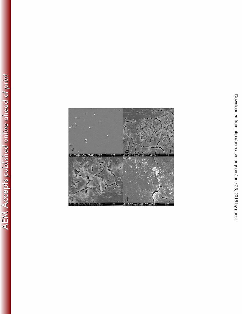
To further examine the effects of burial on the surface of PU coupons, ESEM was 242
conducted on PU coupons after 12 weeks of incubation (Figure 2). Cracking of the PU 243
surface was observed at all temperatures but was more extensive at 45° and 50°C. By 244
contrast, no physical changes were observed in polyether PU coupons after 12 weeks 245
burial at different temperatures (data not shown). 246
Impact of soil and compost burial on tensile strength of PU 247
In order to quantify the extent of biodegradation during burial, tensile strength of buried 248
PU dumb-bells were determined periodically following incubation at 25°, 45° and 50°C 249
over 12 weeks (Figure 3 a-d). In order to distinguish between microbiological effects and 250
potential effects of chemical components in soil and compost, PU dumb-bells were also 251
immersed in sterile soil and compost extracts. Tensile strength of PU dumb-bells buried 252
in compost or soil at all temperatures after 12 weeks showed major loss (p< 0.05) in 253
tensile strength (>75%) compared to dumb-bells immersed in sterile soil or compost 254
extract. There was slight variation (p>0.05) in the tensile strength loss at different 255
temperatures, however, the variation in loss of tensile strength amongst replicate coupons 256
on June 23, 2018 by guesthttp://aem
.asm.org/
Dow
nloaded from
increased with increase in temperature (Figure 3e). No considerable physical degradation 257
was seen in polyether PU dumb-bells after 12 weeks under any condition (data not 258
shown). 259
To further assess biodegradation, dry weight loss was determined after 12 weeks of 260
burial. Despite the major reduction in tensile strength and physical disruption of the 261
surface of PU, weight loss in all cases was <1% and was not significant (p>0.05)(data not 262
shown). 263
Fungal colonization of PU coupons 264
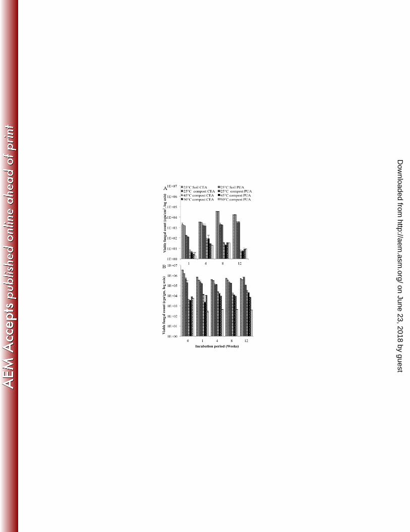
To investigate fungal colonisation of PU coupons following burial, total fungal viable 265
counts and putative fungal PU degrader counts recovered from the surface of coupons 266
were enumerated on CEA and PUA plates respectively (Figure 4a). Total fungal viable 267
and putative fungal degrader counts in the compost and soil environments in which the 268
PU coupons were buried were also enumerated (Figure 4b). All fungal colonies recovered 269
on PUA agar displayed a zone of clearing around the colony margin indicating PU 270
degradation. 271
Total viable counts in soil and in compost in which the coupons were buried remained 272
similar for any temperature over 12 weeks. At 25°C, total viable counts in soil and 273
compost were similar, however counts in compost were significantly (P<0.05) lower at 274
45°C and 50°C, temperatures at which only thermotolerant and thermophilic fungi can 275
grow (34). When total viable counts and putative PU degrader counts were compared, PU 276
degraders composed ca. 40-70% of all colonies recovered (Figure 4b) indicating that a 277
large proportion of the viable fungal population was putative degraders. 278
on June 23, 2018 by guesthttp://aem
.asm.org/
Dow
nloaded from

Total fungal viable counts from the surface of PU coupons buried in soil and compost at 279
25°C increased up to week 8 and then remained similar. Total viable counts from the PU 280
surface also increased up to week 8 when incubated 45°C and 50°C, but decreased at 281
week 12. A comparison of total viable counts and putative degrader counts demonstrated 282
that ca. >70% of fungi colonizing the PU surface were putative degraders (Figure 4A). 283
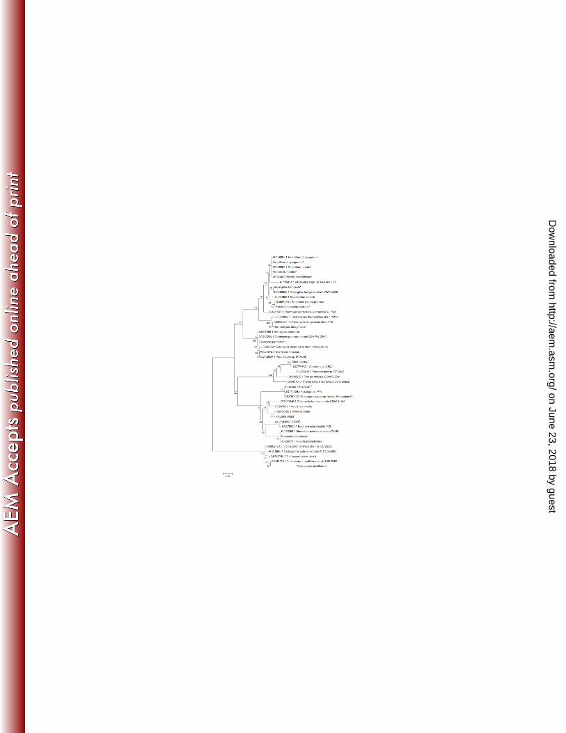
Identification of polyester PU degrading fungal isolates 284
Fungal colonies growing on PUA plates were difficult to distinguish morphologically. 285
Therefore, colonies were transferred onto PDA and incubated up to 5 days. Differences in 286
development of pigments, sporulation and colonial morphology on PDA enabled colonies 287
to be grouped into different morphotypes and quantified (Figure 5). 4-5 isolates were 288
randomly chosen, total genomic DNA was extracted, the ITS1-5.8s-ITS2 region of rDNA 289
amplified by PCR, sequenced and sequences used to interrogate the NCBI database 290
(Table 1). All the isolates were then aligned with the published sequences and a 291
phylogenetic tree constructed (Figure 6). Although the two environments were very 292
different (soil and compost), a number of common species were isolated at 25°C from the 293
surface of PU from both soil and compost with only an Alternaria sp (morphotype 1) and 294
Volutella ciliata (morphotype 5) uniquely recovered from the surface of soil buried PU 295
and Penicillium spp. (morphotype 6) uniquely recovered from the surface of compost 296
buried PU. However, the relative proportions of the species recovered from the surface 297
of PU differed between soil and compost and changed over the twelve week incubation 298
period (Figure 5). From the surface of soil buried PU at 25°C, the proportion of the 299
different species recovered was relatively constant except that Volutella ciliata could not 300
be recovered after week 4, Geomyces pannorum could not be recovered after week 8 and 301
on June 23, 2018 by guesthttp://aem
.asm.org/
Dow
nloaded from

Bionectria ochroleuca was the dominant species by week 12. The proportion of species 302
recovered from the surface of PU buried in compost was also variable over the twelve 303
weeks incubation period, Penicillium spp. were dominant at week 1 but from weeks 4-12 304
Fusarium solani/oxysporum was the dominant phenotype. At 45° and 50°C, Aspergillus 305
fumigatus and Thermomyces lanuginosus respectively were the only species recovered at 306
any time point. For morphotypes 6 and 7, subsequent sequencing of random isolates 307
revealed that they were composed of more than one species (Penicillium mandriti, P. 308
chrysogenum and P. roseopurpureum for morphotype 6 and Fusarium solani and F. 309
oxysporum for morphotype 7) (Table 1). 310
Fungal community diversity on the surface of buried polyester PU 311
TRFLP was used to study the temporal change in fungal communities on the surface of 312
polyester PU during burial in soil at 25°C and in compost at 25°, 45° and 50°C. TRF 313
electropherograms were compared using Principle Component Analysis (PCA) and 314
displayed as a scatter graph for each separate temperature for both the communities on 315
the PU surface and in the surrounding compost/soil (Figure 7). At 25°C, the fungal 316
communities in soil and in compost separated into two distinct groups (soil and compost, 317
group I and III) indicating two distinctive communities (Figure 7a). Within soil and 318
compost at all temperatures, the community changed over the first four weeks and then 319
remained stable. However, the communities colonizing the PU surface were separate 320
from the compost community, indicating that the community on the PU surface was 321
different from the surrounding soil or compost. Though, the community on the surface 322
clustered together but was not stable and changed over the 12 week period. However, at 323
on June 23, 2018 by guesthttp://aem
.asm.org/
Dow
nloaded from
50°C the clustering was much weaker than at 25° & 45°C on the PU surface suggesting 324
greater community change over time at the higher temperature (Figure 7 a, b and c). 325
TRFLP profiles generated from compost, soil and PU coupons were subjected to 326
statistical analysis using MVSP and the total number of TRFs and their relative 327
abundance was used to calculate Shannon-Weaver index and evenness as indicators of 328
diversity and equitability respectively (Table 2). When soil or compost was incubated at 329
25°C, Shannon index increased from week 0 to week 4 and then remained approximately 330
constant indicating an initial increase in species diversity. The Shannon index from the 331
surface of PU remained approximately constant over 12 weeks but was significantly 332
lower (P<0.05) indicating the community on the surface was far less diverse than the 333
surrounding compost or soil. At 45° and 50°C, the Shannon Weaver index decreased at 334
week 4 and then remained approximately constant indicating selection for thermophilic 335
and thermotolerant species and a consequent reduction in species diversity. Again, the 336
Shannon weaver index from the PU surface was significantly (P<0.05) lower than the 337
surrounding compost (Table 2). Evenness remained approximately constant in soil and 338
compost and on the surface of PU at all temperatures indicating no emergence of 339
dominant species in the community over time, however, the evenness value from the PU 340
surface was lower than the surrounding soil or compost indicating a less even distribution 341
in the surface community. 342
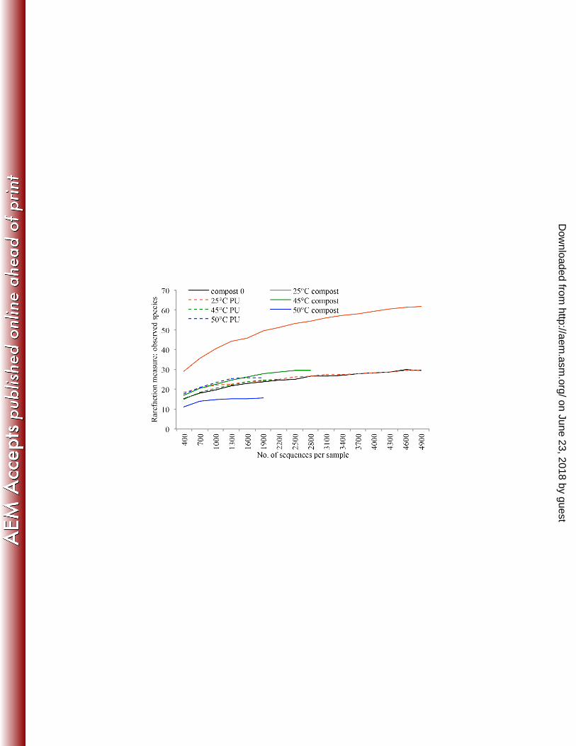
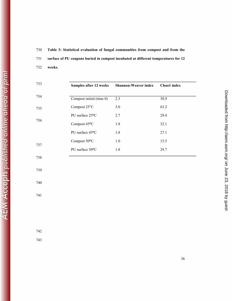
Biodiversity of fungal communities by pyrosequencing 343
A total of 94,502 sequences were obtained after discarding low quality and short reads. 344
Analysis of rarefaction showed that sample curves leveled off at the 97% cut-off level 345
on June 23, 2018 by guesthttp://aem
.asm.org/
Dow
nloaded from

and that the majority of the community had been captured. The number of observed 346
species ranged between 14 and 51, with the highest number detected at 25°C (Figure 8a). 347
Chao1 index, an estimator of OTU richness, was higher at 25°C in the compost and 348
lowest in 50°C compost followed by population of the surface of PU coupons incubated 349
at 45° and 50°C (Table 3). In compost at 25°C, Geomyces pannorum was the dominant 350
species prior to incubation and after 12 weeks (49.6% and 42.2% respectively) while 351
other species declined or were not detected (Thielavia sp., Arthrographis kalrae, 352
Pseudallescheria boydii, Arthrobotrys flagran and Doratomyces nanus). After 12 weeks, 353
other species appeared at levels >1% that were not detected or present at less than 1% 354
prior to incubation (Scytalidium thermophilum, Fusarium solani, Mortierella sp., 355
Pseudallescheria fimeti and Thermomyces lanuginosus). Incubation of compost at 45°C 356
and 50°C for 12 weeks led to the development of a very different community compared 357
to compost prior to incubation due to the selection of thermophilic and thermotolerant 358
species. Three species, Emericella rugulosa, an unidentified fungal clone A and 359
Scytalidium thermophilum were present at both 45°C and 50°C and accounted for 81% 360
and 88.9% of all sequences respectively although Scytalidium thermophilum was 361
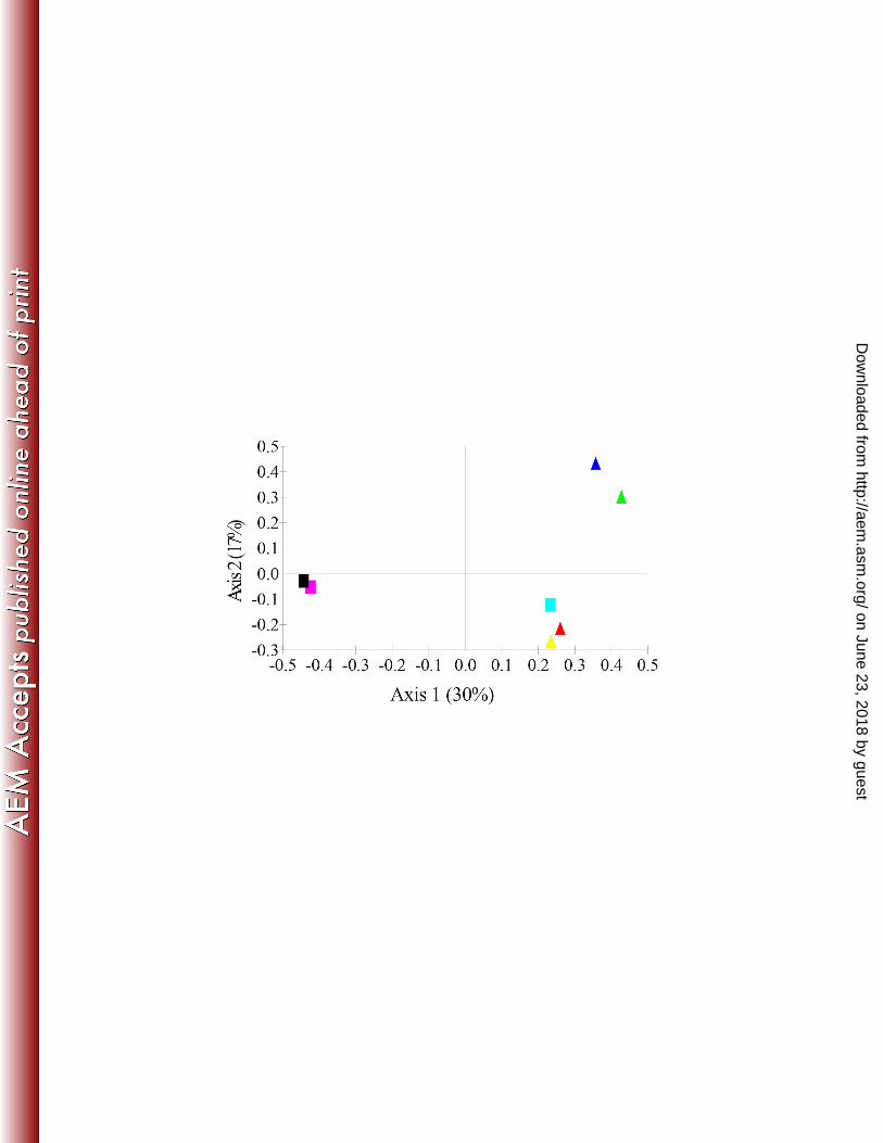
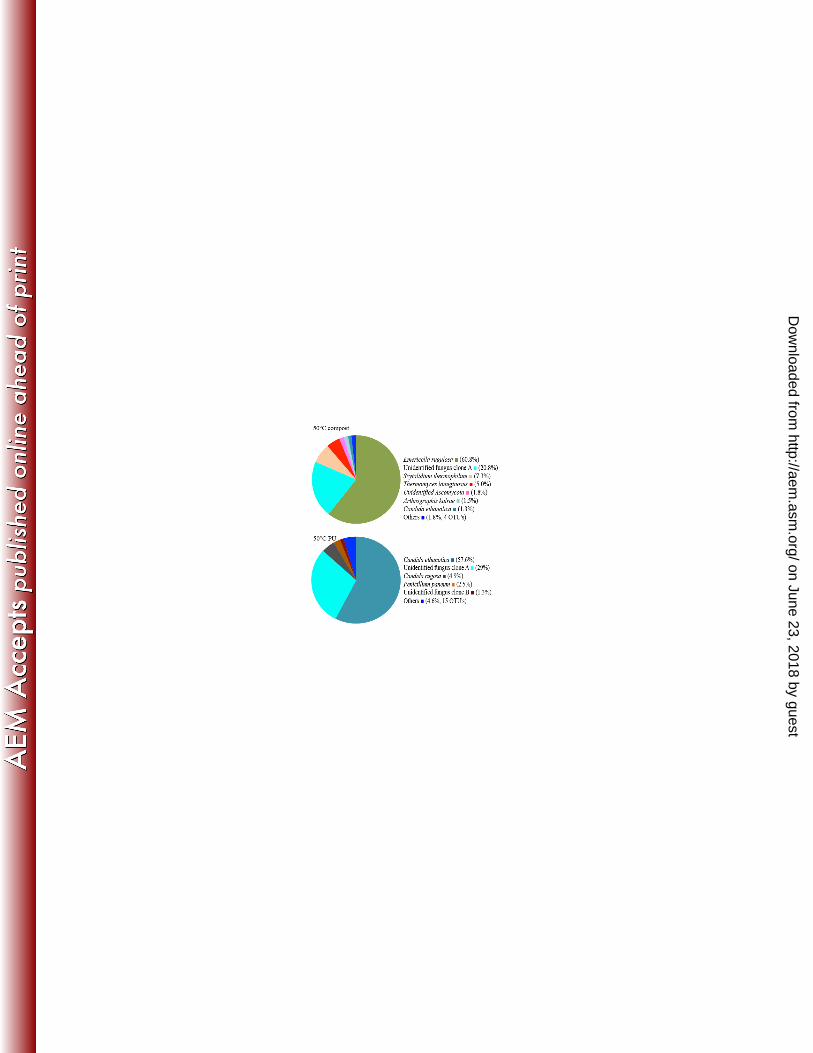
predominant at 45°C and Emericella rugulosa at 50°C (Figure 9). 362
Pyrosequencing revealed clear differences in the fungal communities in the compost 363
compared to the surface of the PU coupons at each temperature (Figure 9). At 25°C, the 364
population of Geomyces pannorum in compost was 42.4% but on the surface of PU 365
coupons was present at <1% with Fusarium solani dominating (53%). At 45°C and 50°C, 366
the fungal community on the surface of PU were similar to each other and dominated by 367
Candida ethanolica (60.3% and 57.6% respectively), an unidentified fungal clone A 368
on June 23, 2018 by guesthttp://aem
.asm.org/
Dow
nloaded from
(32.3% and 29.0% respectively) and Penicillium paneum (2.4% and 2.5% respectively). 369
Candida rugosa (4.9%) was the only species present at >1% that was only found only at 370
50°C. The compost community at 45°C was dominated by Scytalidium thermophilum 371
(68.1%) wheras at 50°C Emericella rugulosa was dominant (60.8%). PCA analysis of the 372
OTU’s also demonstrated that the compost community at 45°C and 50°C clustered 373
together and a second distinct cluster composed communities on the surface of PU at 45° 374
and 50°C (Figure 8B). PCA analysis also showed that incubation of compost at 45° and 375
50°C had a greater effect on the fungal community than incubating at 25°C, but 376
nonetheless, the community on the surface of PU at 25°C was different from the 377
surrounding compost. 378
DISCUSSION 379
Polyurethanes are a diverse group of plastics with a wide range of applications ranging 380
from packaging materials, shoe soles, coatings and paints and are therefore common 381
environmental pollutants (2, 35). Polyester PU’s are known to be susceptible to microbial 382
enzymatic degradation through hydrolysis of the polymer (21) and it has been shown 383
previously, that fungi are the dominant microbes responsible for colonization and 384
biodegradation of PU’s in soil environments, particularly when the moisture content falls 385
between 20 and 70 % (16–18). 386
Composting is a natural process involving the aerobic decomposition of organic wastes 387
by a mixed microbial consortium that also involves thermophilic microbes appearing due 388
to the increase in heat that occurs during microbial respiration and in recent years has 389
been increasingly used commercially as a waste management treatment for green and 390
on June 23, 2018 by guesthttp://aem
.asm.org/
Dow
nloaded from

food wastes (36). In this study, we investigated whether microbes, and in particular fungi 391
present in compost, had the capacity to degrade PU and whether selection of particular 392
communities on the surface of PU were associated with this process. For this purpose, we 393
incubated PU coupons for up to 12 weeks in mature compost at 25˚C (to emulate the 394
maturation phase) and at 45˚C and 50˚C (to emulate the thermophilic stage). Coupons 395
were also buried in soil at 25°C to enable the rate of degradation in soil to be compared to 396
compost. Significant colonization by fungal mycelia was clearly observed in all cases 397
along with significant macroscopic changes to the PU coupons (Figure 1) and severe 398
cracking of the PU surface and fungal hyphae were visible under environmental scanning 399
electron microscopy (Figure 2). Barratt et al. (16) also observed a high coverage of 400
polyurethane film by fungal hyphae and spores and extensive surface damage in soil 401
buried polyester PU. Despite these major changes to the integrity of the coupons, little 402
loss in dry weight was observed. Weight loss over 12 weeks was not therefore a reliable 403
indicator for polyester PU biodegradation. Barratt et al. (16) and Pathirana & Seal (21) 404
also reported little change in weight loss in soil buried PU. 405
Significant changes were observed in the tensile strength of buried polyester PU (Figure 406
3). Tensile strength is defined as the load at break divided by the original cross-sectional 407
area. A number of studies have analyzed the rate of degradation of PU by analyzing loss 408
in tensile strength as it is a sensitive measure of polymer integrity (16–19, 37–40). Umare 409
and Chandre (41) studied the degradation of polyester PU under three different 410
treatments; alkaline hydrolysis, enzymatic hydrolysis and soil burial and reported that soil 411
burial gave the greatest level of degradation. Previously Barratt et al. (16) buried 412
polyurethane in soil microcosms and reported a reduction in tensile strength of 60% after 413
on June 23, 2018 by guesthttp://aem
.asm.org/
Dow
nloaded from
44 days. In another study, Cosgrove et al. (18) investigated the rate of degradation of 414
polyester PU in situ in the environment and found a ca. 95% reduction in tensile strength 415
after 5 months. In our study, percentage loss in tensile strength of buried polyester PU 416
coupons at 25°, 45° and 50°C in compost was ca. 90, 80 and 70%, respectively 417
suggesting that in compost, PU degradation occurs to a similar extent at all phases of the 418
composting process, albeit by different fungal species. Loss in tensile strength has been 419
shown to be due to the secretion of extracellular enzymes that degrade the ester and 420
urethane linkages of polyurethane causing polymer chain scission (21, 42, 43). A fall in 421
tensile strength was also observed when polyester PU coupons were incubated for the 422
same length of time in sterile compost extract, although to a much lower extent (Figure 423
3). Aquino et al (44) studied hydrolysis of polyester PU over a wide temperature range 424
(10°-70°C) and found that PU was stable at 50°C but significant polymer hydrolysis 425
occurred at 70°C after 177 days of incubation. In another study, Zuidema et al. (45) also 426
reported significant hydrolysis of PU at two different temperatures (37°C and 60°C) after 427
3 years. Thus, while polyester PU is vulnerable to chemical abiotic hydrolysis it is a slow 428
process and does not account for the large drop in tensile strength observed when buried 429
in soil or compost. 430
By contrast, no change in tensile strength was observed after 3 months burial under any 431
condition for polyether PU (data not shown). Polyether PU is known to be far more 432
recalcitrant to degradation compared to polyester PU (3, 46). Darby & Kaplan (9) also 433
compared the biodegradation of polyether and polyester based PU and suggested that 434
polyether PU’s are moderately to highly resistant to fungal attack than polyester PU’s 435
because of the presence of high molecular weight branched polyol chains. A similar 436
on June 23, 2018 by guesthttp://aem
.asm.org/
Dow
nloaded from
observation was made by Filip (47), when PU biodegradation was tested in landfills. 437
Jansen et al. (48) reported that polyether PU degradation occurred very slowly when 438
Staphylococcus epidermis was used as a test organism and polyether based PU was also 439
found to be highly resistant to anaerobic bacterial degradation. Krasowska et al. (3) 440
reported that polyether based PU turned yellow/brown when incubated in a compost pile 441
but reported no physical deterioration. However, Matsumiya et al. (49) reported that an 442
Alternaria sp. could degrade polyether polyurethane physically and released metabolites 443
were detected in the media indicating physical degradation. This appeared to be due to 444
the hydrolysis of urea and urethane bonds releasing polyols and polyisocyanates. In our 445
study, polyether PU was incubated for 12 weeks and may require a much longer 446
incubation period before a significant change in tensile strength is observed. 447
TRFLP has previously been successfully used to analyze species diversity in fungal 448
communities from a wide variety of environments (50–52). PCA analysis of TRFLP 449
electropherograms also demonstrated clear differences in the communities colonizing the 450
surface of PU compared to the surrounding environment indicating selection on the PU 451
surface (Figure 7). Previously, Cosgrove et al. (18) also reported the polyester PU buried 452
in two soil types using DGGE showed different fungal communities on the surface of PU 453
compared to the surrounding compost. 454
The fungal species isolated and cultured from the surface of polyester PU at 25˚C in both 455
soil and compost after 12 weeks were the same (Bionectria ochroleuca, a Penicillium sp., 456
Fusarium oxysporum/solani and Trichosporon moniliforme) although recovered to 457
different extents (Figure 5). The only exception was Geomyces pannorum, which was 458
recovered from the PU surface in compost but not in soil although it was recovered at 459
on June 23, 2018 by guesthttp://aem
.asm.org/
Dow
nloaded from

weeks 1, 4 and 8. Many of the fungi recovered have been reported to be associated with 460
PU degradation previously (9, 16, 18, 21, 23, 37). Thus, culturing suggests a limited 461
species diversity on the surface of PU when buried in either soil or compost, but 462
cultivation is known to be unreliable and to under report the true diversity of microbial 463
populations as only a highly limited proportion of species (estimated to be <1-5%) grow 464
readily on synthetic media (53, 54). 465
In order to examine the diversity of the fungal communities on the surface of PU buried 466
in compost in more detail, pyrosequencing was performed with total genomic DNA 467
extracted from mycelium colonising the surface of the PU coupons after 12 weeks burial 468
at 25°, 45° and 50°C and compared to the surrounding compost fungal community 469
(Figures 8 and 9). 454 pyrosequencing is a high throughput sequencing technique 470
particularly useful in the detection of rare community members that may represent <1% 471
of the microbiota (55). Recently 454 pyrosequencing demonstrated that fungal diversity 472
is rich in soil with detection of rare OTUs that were previously under estimated (56, 57). 473
Since the rarefaction curves leveled off suggested that >97% of species present in all 474
samples had been captured (Figure 8a). PCA analysis of the pyrosequencing data was 475
broadly congruent with the PCA analysis of the TRFLP electropherograms in 476
demonstrating that the fungal community on the surface of PU was different from the 477
surrounding compost community at each temperature, and that the PU surface community 478
at 45° and 50°C were broadly similar (Figure 8b). 479
On the surface of PU at 25°C, Fusarium solani, Bionectria ochroleuca and an Alternaria 480
sp. accounted for 53.0%, 17.4% and 16.6% of the sequences recovered. After 12 weeks, 481
both F. solani and B. ochroleuca were also the dominant organisms recovered by 482
on June 23, 2018 by guesthttp://aem
.asm.org/
Dow
nloaded from

cultivation (Figure 5). F. solani has previously been associated with the degradation of 483
polyester PU (23, 37) and B. ochroleuca was also identified as a dominant organism 484
from the surface of polyester PU buried in soil after pre-treatment with impranil DLN (a 485
polyester PU dispersion) following cloning and sequencing of a major band in DGGE 486
(17). Interestingly, it was also isolated from soil as a major degrader of poly (butylene 487
succinate) films, a biodegradable polyester polymer suggesting it possesses the ability to 488
efficiently degrade esters within long chain polymers (58). Alternaria spp. have also been 489
ubiquitously found in soil and compost and several species has been extensively reported 490
as polyurethane degrader (9, 18, 20, 39, 49, 59) 491
While the most dominant species cultivated from the surface of PU at 25°C were amongst 492
the most dominant identified by pyrosequencing, at 45° and 50°C there was little 493
correlation. At 45° and 50°C, A. fumigatus and T. lanuginosus were the only fungi 494
isolated via culture-based technique (Table 1). However, pyrosequencing revealed that in 495
both cases, Candida ethanolica was the dominant organism on the surface of PU. C. 496
ethanolica was first isolated as an ethanol tolerant yeast with a limited ability to ferment 497
sugars and is closely related to the Pichia group (60). It has however, been reported to be 498
amongst a group of yeasts identified from a clone library which dominated the early 499
stages of composting in a commercial compost facility (61). Also dominant was an 500
unidentified fungal clone A, present at both 45°C and 50°C that has been found in soils 501
but has yet to be isolated or taxonomically assigned. The third most dominant species was 502
Candida rugosa comprising 4.9% of the population on the surface of PU coupons at 503
50°C. Gautam, et al. (62) have demonstrated that a lipase enzyme from C. rugosa was 504
able to cause significant PU degradation. Penicillium paneum was also detected on the 505
on June 23, 2018 by guesthttp://aem
.asm.org/
Dow
nloaded from
surface of PU coupons and is closely related to Penicillium roqueforti and has been 506
reported to grow under high acidic environments (63). Together, C. ethanolica and 507
unidentified fungus clone A accounted for >85% of the sequences recovered with the 508
remaining sequences belonging to 14 to 16 other species (Figure 9). While A. fumigatus 509
and T. lanuginosus were detected by pyrosequencing on the surface of PU, they 510
accounted for <0.35% of the sequences, despite appearing to be the only organisms 511
isolated from the surface of PU through conventional cultivation. 512
In summary, this study has demonstrated that polyester PU is susceptible to fungal 513
biodegradation in compost under both thermophilic (thermophilic stage) and mesophilic 514
(maturation phase) conditions and that positive selection for rare taxa from the existing 515
compost community on the PU surface occurs. Thus there may be the potential for 516
optimizing existing commercial composting processes to enable polyester PU waste to be 517
diverted into commercial composting streams thereby reducing the burden on landfill 518
sites. 519
REFERENCES 520
1. Kaplan AM, Darby RT, Greenberger M, R. RM. 1968. Microbial deterioration 521 of polyurethane. Developments in Industrial Microbiology 9:201–217. 522
2. Howard GT. 2012. Polyurethane biodegradation, p. 371–394. In Singh, SN (ed.), 523 Microbial Degradation of Xenobiotics. Springer Berlin Heidelberg, Berlin, 524 Heidelberg. 525
3. Krasowska K, Janik H, Gradys A, Rutkowska M. 2012. Degradation of 526 polyurethanes in compost under natural conditions. Journal of Applied 527 Microbiology 125:4252–4260. 528
4. PlasticEurope. 2012. Plastics – the Facts 2012. An analysis of European plastics 529 production , demand and waste data for 2011. 530
on June 23, 2018 by guesthttp://aem
.asm.org/
Dow
nloaded from
5. Oehlmann J, Schulte-Oehlmann U, Kloas W, Jagnytsch O, Lutz I, Kusk KO, 531 Wollenberger L, Santos EM, Paull GC, Van Look KJW, Tyler CR. 2009. A 532 critical analysis of the biological impacts of plasticizers on wildlife. Philosophical 533 Transactions of the Royal Society of London. Series B, Biological sciences 534 364:2047–2062. 535
6. Jayasekara R, Harding I, Bowater I, Lonergan G. 2005. Biodegradability of a 536 selected range of polymers and polymer blends and standard methods for 537 assessment of biodegradation. Journal of Polymers and the Environment 13:231–538 251. 539
7. Morton LHG, Surman SB. 1994. Biofilms in Biodeterioration-a review. 540 International Biodeterioration & Biodegradation 34:203–221. 541
8. Darby RT, Kaplan AM. 1968. Fungal susceptibility of polyurethanes. Applied 542 Microbiology 16:900–905. 543
9. Pommer EH, Lorenz G. 1985. The behaviour of polyester and polyether 544 polyurethanes towards microorganisms, p. 77–86. In Seal, KJ (ed.), 545 Biodeterioration and Biodegradation of Polymers, 1st ed. biodeterioration society, 546 New york. 547
10. Arvanitoyannis IS, Bosnea LA. 2001. Recycling of polymeric materials used for 548 food packaging: current status and perspectives. Food Reviews International 549 17:291–346. 550
11. Epstein E. 1997. The science of composting. CRC press. USA. 551
12. Ryckeboer J, Mergaert J, Coosemans J, Deprins K, Swings J. 2003. 552 Microbiological aspects of biowaste during composting in a monitored compost 553 bin. Journal of Applied Microbiology 94:127–137. 554
13. Kim YD, Kim SC. 1998. Effect of chemical structure on the biodegradation of 555 polyurethanes under composting conditions. Polymer Degradation and Stability 556 62:343–352. 557
14. Ishii K, Fukui M, Takii S. 2000. Microbial succession during a composting 558 process as evaluated by denaturing gradient gel electrophoresis analysis. Journal of 559 Applied Microbiology 89:768–777. 560
15. Gannes V De, Eudoxie G, Hickey WJ. 2013. Prokaryotic successions and 561 diversity in composts as revealed by 454-pyrosequencing. Bioresource Technology 562 133:573–580. 563
16. Barratt SR, Ennos AR, Greenhalgh M, Robson GD, Handley PS. 2003. Fungi 564 are the predominant micro-organisms responsible for degradation of soil-buried 565
on June 23, 2018 by guesthttp://aem
.asm.org/
Dow
nloaded from

polyester polyurethane over a range of soil water holding capacities. Journal of 566 Applied Microbiology 95:78–85. 567
17. Cosgrove L, McGeechan PL, Handley PS, Robson GD. 2010. Effect of 568 biostimulation and bioaugmentation on degradation of polyurethane buried in soil. 569 Applied and Environmental Microbiology 76:810–819. 570
18. Cosgrove L, McGeechan PL, Robson GD, Handley PS. 2007. Fungal 571 communities associated with degradation of polyester polyurethane in soil. 572 Applied and Environmental Microbiology 73:5817–5824. 573
19. Mathur G, Prasad R. 2012. Degradation of polyurethane by Aspergillus flavus 574 (ITCC 6051) isolated from soil. Applied Biochemistry and Biotechnology 575 167:1595–1602. 576
20. Russell JR, Huang J, Anand P, Kucera K, Sandoval AG, Dantzler KW, 577 Hickman D, Jee J, Kimovec FM, Koppstein D, Marks DH, Mittermiller P a, 578 Núñez SJ, Santiago M, Townes M a, Vishnevetsky M, Williams NE, Vargas 579 MPN, Boulanger L-A, Bascom-Slack C, Strobel S a. 2011. Biodegradation of 580 polyester polyurethane by endophytic fungi. Applied and Environmental 581 Microbiology 77:6076–6084. 582
21. Pathirana RA, Seal KJ. 1984. Studies on polyurethane deteriorating fungi. Part 1. 583 Isolation and characterization of the test fungi employed. Internationali 584 Biodeterioration 20:163–168. 585
22. Forster J. 1995. Soil sampling, handling, storage and analysis, p. 106. In Alef, K, 586 Nannipieri, P (eds.), Methods in applied soil microbiology and biochemistry. 587 Academic Press, London,U.K. 588
23. Crabbe JR, Campbell JR, Thompson L, Walz SL, Schultz WW. 1994. 589 Biodegradation of a colloidal ester-based polyurethane by soil fungi. International 590 Biodeterioration & Biodegradation 33:103–113. 591
24. Lorch HJ, Benckieser G, Ottow JC. 1995. Enrichment, isolation and counting of 592 soil microorganisms, p. 147. In Alef, K, Nannipieri, P (eds.), Methods in applied 593 soil microbiology and biochemistry. Academic Press, London. 594
25. Feng J, Hwang R, Chang KF, Hwang SF, Strelkov SE, Gossen BD, Zhou QA. 595 2010. An inexpensive method for extraction of genomic DNA from fungal 596 mycelia. Canadian Journal of Plant Pathology 32:396–401. 597
26. Webb JS, Nixon M, Eastwood IM, Greenhalgh M, Robson GD, Handley PS. 598 2000. Fungal colonization and biodeterioration of plasticized polyvinyl chloride. 599 Applied and Environmental Microbiology 66:3194–3200. 600
on June 23, 2018 by guesthttp://aem
.asm.org/
Dow
nloaded from

27. White TJ, Bruns TD, Lee S, Taylor J. 1990. Analysis of phylogenetic 601 relationshipd by amplificaion and direct sequencing of ribosomal RNA genes, p. 602 315–322. In Innis, MA, Gelfand, DH, Sninsky, JJ, White, TJ (eds.), PCR Protocol: 603 a Guide to Methods and Applications. Academic Press, New York, US. 604
28. Kawasaki A, Watson ER, Kertesz M a. 2011. Indirect effects of polycyclic 605 aromatic hydrocarbon contamination on microbial communities in legume and 606 grass rhizospheres. Plant and Soil 358:169–182. 607
29. Smith CJ, Danilowicz BS, Clear AK, Costello FJ, Wilson B, Meijer WG. 2005. 608 T-Align, a web-based tool for comparison of multiple terminal restriction fragment 609 length polymorphism profiles. FEMS Microbiology Ecology 54:375–380. 610
30. Tiquia SM. 2005. Microbial community dynamics in manure composts based on 611 16S and 18S rDNA T-RFLP profiles. Environmental Technology 26:1101–1113. 612
31. Roche. 2011. 454 Sequencing system guidelines for amplicon experimental 613 design. 614
32. Caporaso JG, Kuczynski J, Stombaugh J, Bittinger K, Bushman FD, Costello 615 EK, Fierer N, Peña AG, Goodrich JK, Gordon JI, Huttley G a, Kelley ST, 616 Knights D, Koenig JE, Ley RE, Lozupone C a, McDonald D, Muegge BD, 617 Pirrung M, Reeder J, Sevinsky JR, Turnbaugh PJ, Walters W a, Widmann J, 618 Yatsunenko T, Zaneveld J, Knight R. 2010. QIIME allows analysis of high-619 throughput community sequencing data. Nature methods 7:335–336. 620
33. Reeder J, Knight R. 2010. Rapidly denoising pyrosequencing amplicon reads by 621 exploiting rank-abundance distributions. Nature Methods 7:668–669. 622
34. Dix NJ, Webster J. 1995. Fungal ecology. Chapman & Hall, London, 623 London,U.K. 624
35. Howard GT. 2002. Biodegradation of polyurethane: a review. International 625 Biodeterioration & Biodegradation 49:245–252. 626
36. Ryckeboer J, Mergaert J, Vaes K, Klammer S. 2003. A survey of bacteria and 627 fungi occurring during composting and self-heating processes. Annals of 628 Microbiology 53:349–410. 629
37. Bentham RH, Morton LHG, Allen NG. 1987. Rapid assessment of the microbial 630 deterioration of polyurethanes. International Biodeterioration 23:377–386. 631
38. Dale R, Squirrell DJ. 1990. A rapid method for assessing the resistance of 632 polyurethanes to biodeterioration. International Biodeterioration 26:355–367. 633
on June 23, 2018 by guesthttp://aem
.asm.org/
Dow
nloaded from

39. Ibrahim IN, Maraqa A, Hameed KM, Saadoun IM, Maswadeh HM, 634 Nakajima-kambe T. 2009. Polyester-polyurethane biodegradation by Alternaria 635 solani, isolated from northern Jordan. Advances in Environmental Biology 3:162–636 170. 637
40. Kay MJ, Morton LHG, Prince EL. 1991. Bacterial degradation of polyester 638 polyurethane. International Biodeterioration & Biodegradation 27:205–222. 639
41. Umare SS, Chandure AS. 2008. Synthesis, characterization and biodegradation 640 studies of poly(ester urethane)s. Chemical Engineering Journal 142:65–77. 641
42. Akutsu Y, Nakajima-Kambe T, Nomura N, Nakahara T. 1998. Purification and 642 properties of a polyester polyurethane-degrading enzyme from Comamonas 643 acidovorans TB-35. Applied and Environmental Microbiology 64:62–67. 644
43. Nakajima-Kambe T, Shigeno-Akutsu Y, Nomura N, Onuma F, Nakahara T. 645 1999. Microbial degradation of polyurethane, polyester polyurethanes and 646 polyether polyurethanes. Applied Microbiology and Biotechnology 51:134–140. 647
44. Aquino F, Sheldrake T, J. C, Pires F, Coutinho FMB. 2010. Study of aging of 648 polyurethane applied to the offshore industry. Polymers 20:33–38. 649
45. Zuidema J, van Minnen B, Span MM, Hissink CE, van Kooten TG, Bos 650 RRM. 2009. In vitro degradation of a biodegradable polyurethane foam, based on 651 1,4-butanediisocyanate: a three-year study at physiological and elevated 652 temperature. Journal of Biomedical Materials Research. Part A 90:920–930. 653
46. Rutkowska M, Krasowska K, Heimowska A, Steinka I, Janik H. 2002. 654 Degradation of polyurethanes in sea water. Polymer Degradation and Stability 655 76:233–239. 656
47. Filip Z. 1985. Microbial degradation of polyurethanes, p. 51–55. In Seal, KJ (ed.), 657 Biodeterioration and Biodegradation of Polymers. Biodeterioration Society, New 658 York, US. 659
48. Jansen B, Schumacher-Perdreau F, Peters G, Pulverer G. 1991. Evidence for 660 degradation of synthetic polyurethanes by Staphylococcus epidermidis. 661 Zentralblatt für Bakteriologie�: international journal of medical microbiology 662 276:36–45. 663
49. Matsumiya Y, Murata N, Tanabe E, Kubota K, Kubo M. 2010. Isolation and 664 characterization of an ether-type polyurethane-degrading micro-organism and 665 analysis of degradation mechanism by Alternaria sp. Journal of Applied 666 Microbiology 108:1946–1953. 667
on June 23, 2018 by guesthttp://aem
.asm.org/
Dow
nloaded from
50. Wu B, Hogetsu T, Isobe K, Ishii R. 2007. Community structure of arbuscular 668 mycorrhizal fungi in a primary successional volcanic desert on the southeast slope 669 of Mount Fuji. Mycorrhiza 17:495–506. 670
51. Lord NS, Kaplan CW, Shank P, Kitts CL, Elrod SL. 2002. Assessment of 671 fungal diversity using terminal restriction fragment (TRF) pattern analysis: 672 comparison of 18S and ITS ribosomal regions. FEMS Microbiology Ecology 673 42:327–337. 674
52. Dickie IA, FitzJohn RG. 2007. Using terminal restriction fragment length 675 polymorphism (T-RFLP) to identify mycorrhizal fungi: a methods review. 676 Mycorrhiza 17:259–270. 677
53. Magnuson JK, Lasure LL. 2002. Fungal diversity in soil as assessed by direct 678 culture and molecular techniques.Paper presented at the 102nd general meeting of 679 the American society of Microbiology. Salt lake City. 19-23 May 2002. 680
54. Salar RK, Aneja KR. 2006. Thermophilous fungi from temperate soils of 681 northern India. Journal of Agricultural Technology 1:49–58. 682
55. Ronaghi M. 2001. Pyrosequencing sheds light on DNA sequencing. Genome 683 Research 11:3–11. 684
56. Danielsen L, Thürmer A, Meinicke P, Buée M, Morin E, Martin F, Pilate G, 685 Daniel R, Polle A, Reich M. 2012. Fungal soil communities in a young transgenic 686 poplar plantation form a rich reservoir for fungal root communities. Ecology and 687 Evolution 2:1935–1948. 688
57. Buée AM, Reich M, Murat C, Morin E, Nilsson RH, Uroz S, Martin F, Mur 689 C. 2013. 454 Pyrosequencing analyses of forest soils reveal an unexpectedly 690 fungal diversity. New Phytologist 184:449–456. 691
58. Mei X, Tian C, Dong Q, Liang Y. 2012. Influencing factors and process on in 692 situ degradation of poly(Butylene Succinate) film by strain Bionectria ochroleuca 693 BFM-X1 in soil. Journal of Environmental Protection 03:523–532. 694
59. Stranger-Johannessen M. 1985. Microbial degradation of polyurethane products 695 in service, p. 264–267. In Seal, KJ (ed.), Biodeterioration and Biodegradation of 696 Polymers, 1st ed. Biodeterioration Society, New york. 697
60. Rybarova J, Stros F, Kockova-Kratochvilova A. 1980. Candida ethanolica n. 698 sp. Zeitschrift fur Allgemeine Mikrobiologie 20:579–581. 699
61. Hultman J, Vasara T, Partanen P, Kurola J, Kontro MH, Paulin L, Auvinen 700 P, Romantschuk M. 2010. Determination of fungal succession during municipal 701
on June 23, 2018 by guesthttp://aem
.asm.org/
Dow
nloaded from

solid waste composting using a cloning-based analysis. Journal of Applied 702 Microbiology 108:472–487. 703
62. Gautam R, Bassi AS, Yanful EK. 2007. Candida rugosa lipase-catalyzed 704 polyurethane degradation in aqueous medium. Biotechnology Letters 29:1081–705 1086. 706
63. Chitarra GS, Abee T, Rombouts FM, Posthumus MA, Dijksterhuis J. 2004. 707 Germination of Penicillium paneum conidia is regulated by 1-Octen-3-ol , a 708 volatile self-inhibitor. Applied and Environmental Microbiology 70:2823–2829. 709
710
on June 23, 2018 by guesthttp://aem
.asm.org/
Dow
nloaded from
Figure Legends
Figure 1: Visual changes to polyester polyurethane coupons after burial at different
temperatures. Physical deterioration of polyester coupons buried in soil at (a) 25°C and
compost at (b) 25°C (c) 45° and (d) 50°C for 0, 4, 8 and 12 weeks. The white and cream
discoloration visible on the surface is fungal mycelium. The scale bar represents 4 cm.
Figure 2: Effect of compost burial on the surface of PU coupons visualised by
environmental scanning microscopy. PU coupons were recovered after 12 weeks of
burial in compost at unburied (a), 25°C (b), 45°C (c), 50°C (d) and changes in the surface
properties visualised by environmental electron microscopy. Prominent cracks can be
seen on the surface of the PU. Scale bar represents 20 µm.
Figure 3: Effect of compost burial on the loss of tensile strength of PU coupons. PU
coupons were buried in soil or compost and loss of tensile strength measured over a 12
week period. Control coupons were immersed in sterile soil or compost extract () and
buried () in soil at 25°C (a), compost at 25°C (b), compost at 45°C (c), compost at 50°C
(d). The results are the means of 5 replicates ±SEM.
Figure 4: Changes in the total fungal viable counts and total fungal PU degrader
counts over a 12 weeks incubation period. (A) Total viable and putative PU degrading
fungal counts (cfu/cm2) recovered from the surface of polyester PU coupons. (B) Total
viable and putative PU degrading fungal counts in compost/soil (cfu/g compost/soil).
Data represent the mean of three replicates ±SEM.
Figure 5: Relative distribution of the colony morphotypes recovered from the
surface of PU coupons buried over 12 weeks at 25°C. Polyester PU coupons were
on June 23, 2018 by guesthttp://aem
.asm.org/
Dow
nloaded from
buried in soil at 25°C and in compost at 25°, 45° and 50°C for 12 weeks and viable
fungal colonies recovered periodically from the surface of the coupons on PUA.
Following transfer onto PDA, colonies were classified into different morphotypes and the
percentage of fungal colonies recovered for each fungal morphotype calculated. A total of
seven major morphotypes were recovered. Morphotype 1 (■, Alternaria sp.), 2 (■,
Bionectria ochroleuca), 3 (■, Geomyces pannorum), 4 (■,Trichosporon moniliforme), 5
(■,Volutella ciliata), 6 (■, Penicillium spp.), 7 (■, Fusarium spp.). At 45°C and 50 °C
only Aspergillus fumigatus and Thermomyces lanuginosus respectively were recovered.
Figure 6: Neighbour joining phylogenetic analysis of putative isolated PU degrading 711
fungi. Fungi isolated and identified from the surface of PU coupons (Table 1) (*) were 712
subjected to phylogenetic analysis with closely related species using Neighbour Joining 713
Phylogenetic Tree (bootstrap corrected with 500 samples). Sequences were BLAST on 714
July’2013. 715
Figure 7: Principal Component Analysis of microbial community obtained from
surface of PU and compost. Scatter plot for Principal Component Analysis of TRFLP
pattern obtained from the surface of the buried polyester PU coupon and compost/soil at
(A) 25° (B) 45° & (C) 50°C after week 4, 8 and 12. Fungal community in compost/soil
and on the surface of PU showed obvious demarcation. PU surface under compost; week
4 (●), week 8 (●) and week 12(●), compost; week 0 (▲) week 4 (▲), week 8 (▲) and
week 12(▲), PU surface under soil; week 4 (), week 8 () and week 12() and soil;
week 0 (Δ) week 4 (Δ), week 8 (Δ) and week 12(Δ).
on June 23, 2018 by guesthttp://aem
.asm.org/
Dow
nloaded from
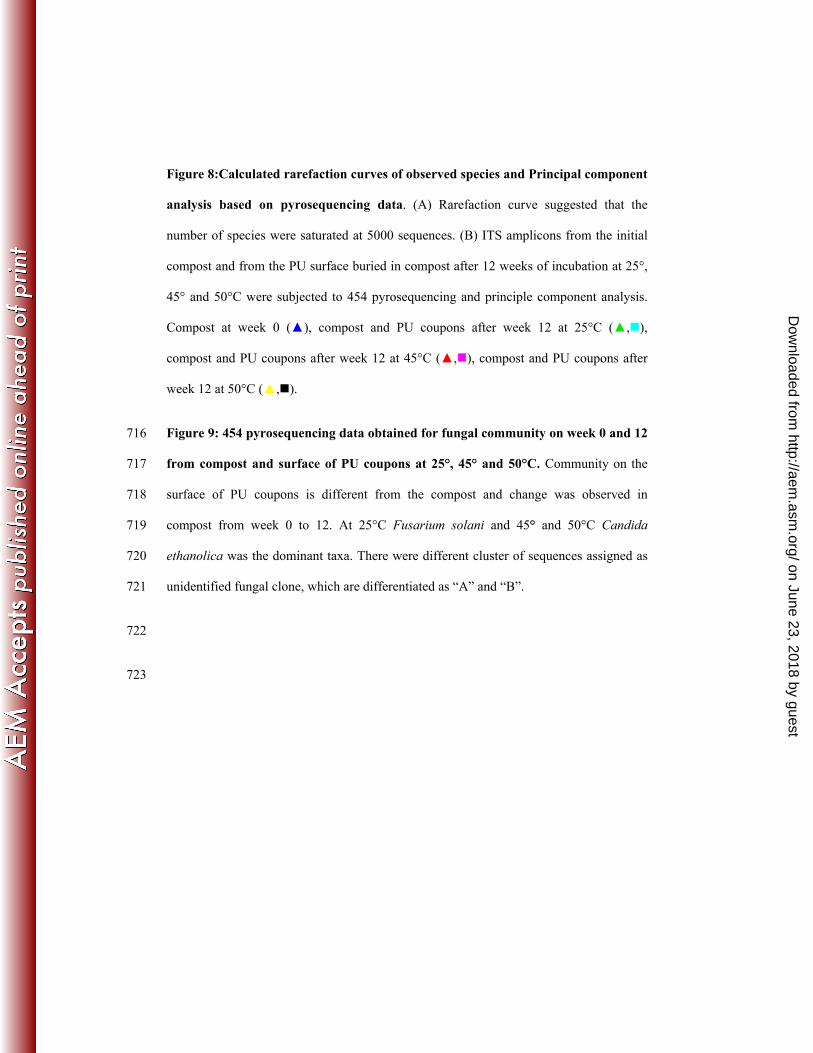
Figure 8:Calculated rarefaction curves of observed species and Principal component
analysis based on pyrosequencing data. (A) Rarefaction curve suggested that the
number of species were saturated at 5000 sequences. (B) ITS amplicons from the initial
compost and from the PU surface buried in compost after 12 weeks of incubation at 25°,
45° and 50°C were subjected to 454 pyrosequencing and principle component analysis.
Compost at week 0 (▲), compost and PU coupons after week 12 at 25°C (▲,),
compost and PU coupons after week 12 at 45°C (▲,), compost and PU coupons after
week 12 at 50°C (▲,).
Figure 9: 454 pyrosequencing data obtained for fungal community on week 0 and 12 716
from compost and surface of PU coupons at 25°, 45° and 50°C. Community on the 717
surface of PU coupons is different from the compost and change was observed in 718
compost from week 0 to 12. At 25°C Fusarium solani and 45° and 50°C Candida 719
ethanolica was the dominant taxa. There were different cluster of sequences assigned as 720
unidentified fungal clone, which are differentiated as “A” and “B”. 721
722
723
on June 23, 2018 by guesthttp://aem
.asm.org/
Dow
nloaded from
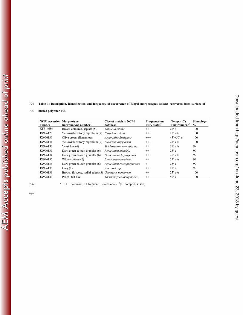
Table 1: Description, identification and frequency of occurrence of fungal morphotypes isolates recovered from surface of 724
buried polyester PU. 725
NCBI accession number
Morphotype (morphotype number)
Closest match in NCBI database
Frequency on PUA plates*
Temp. (˚C) Environment#
Homology %
KF314689 Brown coloured, septate (5) Volutella ciliata ++ 25° s 100
JX996129 Yellowish cottony mycelium (7) Fusarium solani +++ 25° c+s 100
JX996130 Olive green, filamentous Aspergillus fumigatus +++ 45°+50° c 100
JX996131 Yellowish cottony mycelium (7) Fusarium oxysporum +++ 25° c+s 100
JX996132 Yeast like (4) Trichosporon moniliforme +++ 25° c+s 99
JX996133 Dark green colour, granular (6) Penicillium mandriti ++ 25° c 99
JX996134 Dark green colour, granular (6) Penicillium chrysogenum ++ 25° c+s 99
JX996135 White cottony (2) Bionectria ochroleuca ++ 25° c+s 99
JX996136 Dark green colour, granular (6) Penicillium roseopurpureum + 25° c 99
JX996137 Grey (1) Alternaria sp. ++ 25° s 98
JX996139 Brown, floccose, radial edges (3) Geomyces pannorum ++ 25° c+s 100
JX996140 Peach, felt like Thermomyces lanuginosus +++ 50° c 100
* +++ = dominant, ++ frequent, + occasional). #(c =compost, s=soil) 726
727
on June 23, 2018 by guesthttp://aem
.asm.org/
Dow
nloaded from

35
Table 2: Variation in the Shannon-Weaver and Evenness indices derived from
TRFLP electropherograms from fungal communities
# No fungi were detected from the PU surface prior to burial.
728
729
Samples (temperature and weeks)
Shannon-Weaver Index
Evenness
Compost or soil
PU surface
Compost or soil
PU surface
25°C soil 0 2.9 n/a# 0.7 n/a#
25°C soil 4 3.4 2.5 0.7 0.6
25°C soil 8 3.7 2.7 0.8 0.6
25°C soil 12 3.2 2.4 0.8 0.7
25°C compost 0 2.8 n/a# 0.8 n/a#
25°C compost 4 3.5 2.6 0.8 0.7
25°C compost 8 3.3 2.5 0.8 0.7
25°C compost 12 3.4 2.5 0.8 0.8
45°C compost 0 2.8 n/a# 0.7 n/a#
45°C compost 4 2.2 1.4 0.8 0.6
45°C compost 8 1.9 1.1 0.7 0.7
45°C compost 12 2.1 1.5 0.6 0.6
50°C compost 0 2.9 n/a# 0.7 n/a#
50°C compost 4 2.0 1.1 0.6 0.6
50°C compost 8 2.0 1.8 0.7 0.6
50°C compost 12 2.1 1.7 0.7 0.7
on June 23, 2018 by guesthttp://aem
.asm.org/
Dow
nloaded from
36
Table 3: Statistical evaluation of fungal communities from compost and from the 730
surface of PU coupons buried in compost incubated at different temperatures for 12 731
weeks. 732
733
734
735
736
737
738
739
740
741
742
743
Samples after 12 weeks Shannon-Weaver index Chao1 index
Compost initial (time 0) 2.3 30.9
Compost 25°C 3.0 63.2
PU surface 25°C 2.7 29.4
Compost 45°C 1.8 32.1
PU surface 45°C 1.8 27.1
Compost 50°C 1.0 15.5
PU surface 50°C 1.8 29.7
on June 23, 2018 by guesthttp://aem
.asm.org/
Dow
nloaded from


![Appl. Environ. Microbiol. doi:10.1128/AEM.03300-12 ...aem.asm.org/content/early/2012/12/14/AEM.03300-12.full.pdf · 22 KP: kurt.pfister@tropa.vetmed.uni-muenchen.de ... [15] was used](https://img.dokumen.tips/doc/110x75/5b1657b77f8b9a4f6d8b6b96/appl-environ-microbiol-doi101128aem03300-12-aemasmorgcontentearly20121214aem03300-12fullpdf.jpg)