Embed Size (px)
Citation preview
International Journal of Science and Research (IJSR) ISSN: 2319-7064
ResearchGate Impact Factor (2018): 0.28 | SJIF (2018): 7.426
Volume 8 Issue 12, December 2019
www.ijsr.net Licensed Under Creative Commons Attribution CC BY
A Prospective Observational Study of Temporalis
Fascia with Tragal Perichondrium as Graft Material
in Type I Tympanoplasty
Dr Sai Kiran Gova1, Dr. V. Krishna Chaitanya
2
1M.B.B.S, Reg. No: M175413021, Department of ENT Narayana Medical College & Hospital Chintha Reddy Palem, Nellore - 524 002,
Andhra Pradesh, India 2M. S., E.N.T., Professor, Department of ENT Narayana Medical College & Hospital Chintha Reddy Palem, Nellore - 524 002, Andhra
Pradesh, India
Dr N.T.R. University of Health Sciences, Vijayawada, A.P., India
Abstract: A prospective observational study is performed on patients undergoing Type 1 Tympanoplasty after proper evaluation and
obtaining a written informed consent for comparison of Temporalis fascia with Tragal Perichondrium as graft material in Type 1
Tympanoplasty and improvement is assessed with hearing improvement, time taken for graft uptake. A sample size of 100 patients in age
group of 18 to 60 years in CSOM cases were selected for the study. Duration of study 2 years i.e. from October 2017 to September 2019.
Keywords: Temporalis fascia, Tragal perichondrium, Tympanoplasty, Chronic suppurative otitis media
1. Introduction
Tympanoplasty is a surgical procedure to eradicate the
disease in the middle ear and to reconstruct the hearing
mechanism with or without tympanic membrane grafting,
whereas Myringoplasty is a surgery in which the
reconstructive procedure is limited to repair of tympanic
membrane perforation assuming that middle ear ossicles
are functioning normally, eustachian tube is patent, and the
patient has a good cochlear reserve [1, 2].
Type-1 tympanoplasty is surgical reconstruction limited to
repair of tympanic membrane alone. The implicit
definition is that the ossicular chain is intact and mobile
and that there is no middle ear disease. In early centuries,
an ear infection with complication was a life threatening
condition. The introduction of antibiotics and the use of
the operative microscope in the surgical field were
revolutionary advances in the control of the disease.
Chronic suppurative otitis media (CSOM) is a long
standing infection of a part or whole of middle ear cleft.
CSOM is characterized by ear discharge, a permanent
perforation, and impairment of hearing. Tympanic
membrane (TM) perforations lead to recurrent ear
infections and hearing loss [1].
If the perforations are bilateral, hearing handicap becomes
more evident. Persistent perforations occur either due to
improper treatment of recurrent middle ear infections or
infected traumatic perforation. Repair of TM perforation
was attempted for many years. Different techniques and
different graft materials like temporalis fascia, tragal
perichondrium and tragal perichondrium with cartilage
were used [3].
It is the work of two Germans, Wullstein and Zollner,
which started in 1949, lead to a new concept of the
treatment of deafness secondary to chronic infection in the
middle ear and mastoid and the new method was called
―tympanoplasty‖ [4].
Chronic suppurative otitis media is highly prevalent
middle ear disease, particularly in the developing countries
like India [5, 6]. It is defined as a persistent, disease
affecting the mucoperiosteal lining of the middle ear cleft
more than month which is insidious in onset, and capable
of causing the destruction and some irreversible sequela,
and also it clinically manifests with ear discharge and hard
of hearing [7]. It causes numerous pathological changes in
tympanic membrane, and middle ear such as perforation,
ossicular destruction, myringosclerosis,
tymapanosclerosis, granulation tissue polyp,
cholesteatoma, etc. it causes significant conductive hearing
loss. The surgical treatment of chronic suppurative otitis
media primarily aims at eradication of disease process, and
reconstruction of conductive hearing mechanism
Tympanoplasty is surgical procedure to reconstruct sound
conducting apparatus, tympanic membrane, ossicular
system with or without grafting.
The primary goals of tympanoplasty are:
1. Eradication of the disease
2. To improve or maintain hearing mechanism
3. Establish middle ear cleft ventilation
For reconstruction of tympanic membrane, different types
of materials are used. The most commonly used are
temporalis fascia, cartilage, fascia lata etc. Temporalis
fascia the most frequently used graft material, with closure
of the tympanic membrane perforation. Cartilage has
shown to be better graft material, to close perforations in
the tympanic membrane. It gains popularity due to its
resistance to retraction, resorption and reperforation, even
with Eustachian tube dysfunction. The stiffness of
cartilage that prevents reperforations, but it interfere with
the sound conduction properties of the tympanic
membrane, than temporalis fascia [1].
Paper ID: ART20203370 DOI: 10.21275/ART20203370 509
International Journal of Science and Research (IJSR) ISSN: 2319-7064
ResearchGate Impact Factor (2018): 0.28 | SJIF (2018): 7.426
Volume 8 Issue 12, December 2019
www.ijsr.net Licensed Under Creative Commons Attribution CC BY
The present study is a prospective study which describes
the influence of various parameters like type of graft used,
surgical approach, the time taken for graft uptake,
Valsalva maneuver in postoperative period on audiological
outcome after surgery.
2. Objectives
To observe and assess the difference between temporalis
fascia, tragal perichondrium as graft material in Type 1
Tympanoplasty.
Objectives
To evaluate the Improvement in hearing by audiology
with Pure Tone Audiometry
To study the effect of time taken for graft uptake on
the audiological outcome.
3. Review of Literature
Development of the ear
The human ear development begins at the fourth week of
embryonic life.
External ear
The auricle develops from auricular hillocks, derived from
first and second branchial arches. The six hillocks develop
in the 6-week of embryonic life. The external auditory
meatus formed by deepening of the groove between the
two arches. At the dorsal end of the first branchial groove,
the ectodermal cells thicken and form the external meatus.
These ectodermal cells proliferation forms a meatal ―plug‖
that progress medially. Resorption of cells in the center of
this meatal plug forms a tube-like structure which becomes
ear canal. When complete canalization fails to occur that
leads to External canal atresia [8].
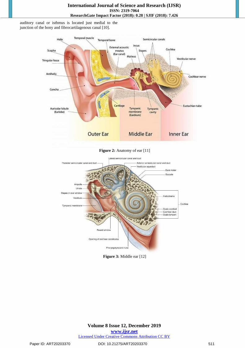
Middle ear
The middle ear cavity forms as a lateral extension of the
first pharyngeal pouch. The proximal end of this extension
becomes the eustachian tube. The lateral extension joins
with the ectoderm of the meatal plug and forms the
tympanic membrane. From Mesoderm of the first
branchial arch, the malleus, incus, anterior malleolar
ligaments and tensor tympani muscle are derived. From
the mesoderm of second arch, Stapes, stapedius muscle
derived [9].
Inner ear
It has two components,
1.Membranous labyrinth. -derived from the ectoderm
2.Bony labyrinth- derived from the mesoderm and neural
crest.
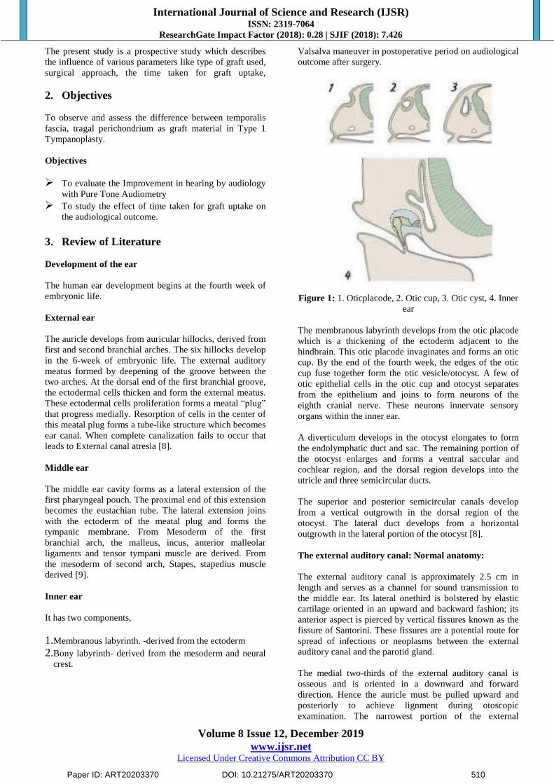
Figure 1: 1. Oticplacode, 2. Otic cup, 3. Otic cyst, 4. Inner
ear
The membranous labyrinth develops from the otic placode
which is a thickening of the ectoderm adjacent to the
hindbrain. This otic placode invaginates and forms an otic
cup. By the end of the fourth week, the edges of the otic
cup fuse together form the otic vesicle/otocyst. A few of
otic epithelial cells in the otic cup and otocyst separates
from the epithelium and joins to form neurons of the
eighth cranial nerve. These neurons innervate sensory
organs within the inner ear.
A diverticulum develops in the otocyst elongates to form
the endolymphatic duct and sac. The remaining portion of
the otocyst enlarges and forms a ventral saccular and
cochlear region, and the dorsal region develops into the
utricle and three semicircular ducts.
The superior and posterior semicircular canals develop
from a vertical outgrowth in the dorsal region of the
otocyst. The lateral duct develops from a horizontal
outgrowth in the lateral portion of the otocyst [8].
The external auditory canal: Normal anatomy:
The external auditory canal is approximately 2.5 cm in
length and serves as a channel for sound transmission to
the middle ear. Its lateral onethird is bolstered by elastic
cartilage oriented in an upward and backward fashion; its
anterior aspect is pierced by vertical fissures known as the
fissure of Santorini. These fissures are a potential route for
spread of infections or neoplasms between the external
auditory canal and the parotid gland.
The medial two-thirds of the external auditory canal is
osseous and is oriented in a downward and forward
direction. Hence the auricle must be pulled upward and
posteriorly to achieve lignment during otoscopic
examination. The narrowest portion of the external
Paper ID: ART20203370 DOI: 10.21275/ART20203370 510
International Journal of Science and Research (IJSR) ISSN: 2319-7064
ResearchGate Impact Factor (2018): 0.28 | SJIF (2018): 7.426
Volume 8 Issue 12, December 2019
www.ijsr.net Licensed Under Creative Commons Attribution CC BY
auditory canal or isthmus is located just medial to the
junction of the bony and fibrocartilagenous canal [10].
Figure 2: Anatomy of ear [11]
Figure 3: Middle ear [12]
Paper ID: ART20203370 DOI: 10.21275/ART20203370 511

International Journal of Science and Research (IJSR) ISSN: 2319-7064
ResearchGate Impact Factor (2018): 0.28 | SJIF (2018): 7.426
Volume 8 Issue 12, December 2019
www.ijsr.net Licensed Under Creative Commons Attribution CC BY
The Tympanic membrane
Figure 4: Tympanic membrane [13]
The tympanic membrane is irregularly round and slightly
conical in shape. The apex of the cone is located at the
umbo, which marks the tip of the manubrium. In the adult,
it is angulated approximately 140° with respect to the
superior wall of the external auditory canal. The vertical
diameter of the tympanic membrane as determined along
the axis of the manubrium ranges from 8.5 to 10 mm,
while the horizontal diameter varies from 8 to 9 mm.
The anterior and posterior tympanic striae extend from the
lateral process of the malleus to the anterior and posterior
tympanic spines, respectively. These striae divide the
tympanic membrane into larger pars tensa below, and
smaller triangular pars flaccida (or Shrapnell‘s membrane)
above.
The thickened periphery of the pars tensa, the tympanic
annulus anchors the tympanic membrane in a groove
known as the tympanic sulcus. The tympanic annulus and
sulcus are absent superiorly in the area of the notch of
Rivinus.
The pars tensa, as its name suggests, is taut and consists of
three layers:
A lateral epidermal layer
A medial mucosal layer
An intermediate fibrous layer.
The pars flaccida, first described by Shrapnell, also
consists of epidermal, fibrous, and mucosal layers. Here,
the fibrous layer is scanty and consists of irregularly
arranged collagen and elastic fibers [13].
The ossicles:
The three ossicles (malleus, incus, stapes) serve to transmit
sound energy from the tympanic membrane to the inner
ear [14].
Figure 5: Ossicles [14]
The malleus:
It has a head, neck, lateral process, anterior process, and
manubrium. It is held to the walls of the petrotympanic
fissure by the anterior malleal ligament which, with the
posterior incudal ligament, serves to establish the axis of
rotation of the ossicles. On its thinner, medial aspect runs
the chorda tympani nerve as it passes anteriorly to enter
the iter chordae anterius at the Glaserian fissure.
The lateral process of the malleus contains a cartilaginous
cap attached to the pars tensa of the tympanic membrane.
The inferior end of the manubrium is firmly attached to the
tympanic membrane as the pars propria splits to envelop it
(the umbo).
The malleus is held in place by five ligaments, one
articulation, the tensor tympani tendon, and the tympanic
membrane.
Three of the five ligaments have a suspensory function.
They are:
The anterior suspensory ligament
The lateral suspensory ligament
Paper ID: ART20203370 DOI: 10.21275/ART20203370 512
International Journal of Science and Research (IJSR) ISSN: 2319-7064
ResearchGate Impact Factor (2018): 0.28 | SJIF (2018): 7.426
Volume 8 Issue 12, December 2019
www.ijsr.net Licensed Under Creative Commons Attribution CC BY
The superior suspensory ligament [14].
The incus:
The incus, the largest of the auditory ossicles, consists of a
body, short process, long process, and lenticular process.
The body of the incus rests in the epitympanum in
association with the head of the malleus. The short process
of the incus extends posteriorly, occupying the posterior
incudal recess (fossa incudis).The long process reaches
inferiorly, to end in the lenticular process; the convex
surface of this process articulates with the concave surface
of the head of the stapes. The long process of the incus is
highly susceptible to osteitic resorption caused by chronic
otitis media.
The stapes:
The stapes is the smallest ossicle. It consists of a head,
footplate (the basis stapedis), and two crura or legs. The
stapedius tendon attached to the superior aspect of the
posterior crus. The footplate, in association with the
annular ligament, seals the oval Window. The head
articulates with the lenticular process of the incus at its
fovea.
The muscles
The Stapedius muscle:
The stapedius muscle, the smallest of the skeletal muscles,
emerges from the pyramidal eminence and attaches to the
head and/or posterior crus of the stapes. It is supplied by
facial nerve.
The tensor tympani muscle:
The tensor tympani muscle, arises from the cartilage of the
eustachian tube, attach to the concave surface of the
cochleariform (spoon-shaped) process, at which point the
main body of the tendon turns laterally to attach to the
medial and anterior surfaces of the neck and the
manubrium of the malleus. Its innervation is from the
trigeminal nerve. The action of the tensor tympani muscle
is to draw the manubrium medially, thus tensing the
tympanic membrane [2, 13, 14].
The middle ear spaces:
The tympanic cavity is a cleft in the sagittal plane
measuring about 15 mm in the vertical and anteroposterior
dimensions. In its transverse dimension, it expands
superiorly to 6 mm and inferiorly to 4 mm from a central
constriction of 2 mm. At the floor of the tympanic cavity
(jugular wall) a small plate of bone separates the jugular
bulb. In the posterior wall, the chordal eminence is lateral
to the pyramidal eminence and medial to the posterior rim
of the tympanic membrane. There is a foramen in this
eminence, known as the iter chordae posterius, through
which the chorda tympani nerve gains access to the middle
ear. The facial recess is interposed between the chordal
eminence laterally and the pyramidal eminence medially
and superiorly bounded by the fossa incudis.
The sinus tympani lies between the ponticulus (which
bridges the gap between the pyramidal eminence and the
promontory superiorly) and the subiculum. The anterior
wall of the middle ear (carotid wall) narrows inferiorly
where it is formed by the thin bony shell of the carotid
canal. Located more superiorly in the anterior wall is the
orifice of the eustachian tube and above it the tensor
tympani muscle lies in its semicanal.
The roof (tegmental wall, tegmen tympani) separates the
tympanic cavity from the middle cranial fossa. The lateral
boundary (membranous wall) is composed of the tympanic
membrane, the bony tympanic ring, and a layer of bone
from the squama - the scutum or shield of Leidy. The
medial wall (labyrinthine wall) of the tympanic cavity is
marked by two main depressions:
The round window niche
The oval window niche
The round window niche is located anteroinferior to the
subiculum and posteroinferior to the promontory.The latter
structure is the bulge of the bone overlying the basal turn
of the cochlea.
The oval window niche is anterosuperior to the ponticulus.
Located posterosuperiorly is the prominence of the facial
canal as it traverses the medial wall and then descends
along the mastoid wall of the tympanic cavity.
The middle ear space is divided into four regions:
1. The mesotympanum (middle ear proper) is that area
located medial to the tympanic membrane and the bony
tympanic annulus.
2. The epitympanum is that area that lies medial to the pars
flaccida and scutum.
3. The protympanum lies anterior to a frontal plane drawn
through to the anterior margin of the tympanic annulus.
It leads to the tympanic orifice of the eustachian tube.
4. The hypotympanum is that part of the middle ear located
inferior to a horizontal plane through the most inferior
part of the tympanic annulus.
The Eustachian tube:
The eustachian tube, a mucosally lined pathway between
the nasopharynx and the middle ear, permits ventilation of
the pneumatized spaces of the temporal bone while
safeguarding against bacterial contamination of these
spaces. The posterolateral one-third is bony while the
anteromedial two-thirds is fibrocartilaginous; these two
sections are joined at the tubal isthmus. The overall length
of the eustachian tube in the adult is 36mm.
The middle ear mucosa:
In electron microscopic observations, Hentzer
distinguished five types of cells in the middle ear mucosa:
1. Nonciliated without secretory granules
2. Nonciliated with secretory granules
Paper ID: ART20203370 DOI: 10.21275/ART20203370 513
International Journal of Science and Research (IJSR) ISSN: 2319-7064
ResearchGate Impact Factor (2018): 0.28 | SJIF (2018): 7.426
Volume 8 Issue 12, December 2019
www.ijsr.net Licensed Under Creative Commons Attribution CC BY
3. Ciliated
4. Intermediate
5. Basal
The mucosa of the middle ear represents a modified
respiratory mucosa.
The Mastoid region:
At birth the mastoid has a single cavity consisting of the
antrum and small adjacent mastoid. It occupies a
superficial position and is surrounded by diploic bone. In
adult life, the normal mastoid may be fully pneumatized,
diploic, or sclerotic. The anterolateral portion of the
mastoid arises from the squamous part of the temporal
bone; the posteromedial portion, including the mastoid tip,
arises from the petrous part. In most mastoids, the plane of
junction of these two parts is marked internally by an
incomplete plate of bone, the petrosquamosal septum, also
known as Koerner‘s septum. The mastoid antrum area is a
large superior central space which communicates with the
epitympanic space of the middle ear via the aditus [15,
16].
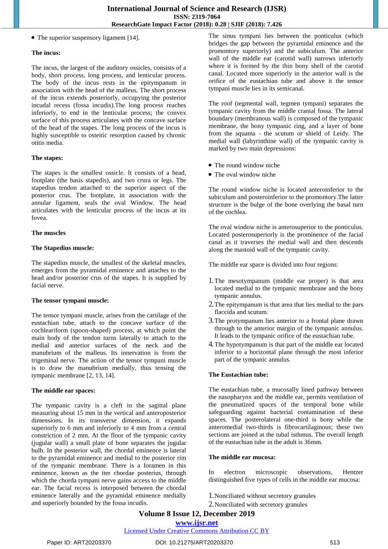
The inner ear
The bony labyrinth:
The long axis of the bony labyrinth, measuring 20 mm in
length, roughly parallels the posterior surface of the
petrous pyramid. Its components are the vestibule, the
semicircular canals, and the cochlea [17].
Figure 6: Inner ear [17]
The vestibule:
The vestibule is the central chamber. At the
posterosuperior aspect of its medial wall is a depression
known as the elliptical recess which accommodates part of
the utricular macula. The spherical recess is a similar
depression for the saccular macula, located
anteroinferiorly. The vestibular crest is an oblique
elevation between these two recesses. The opening for the
cochlea lies anteriorly, while the openings for the
semicircular canals are located posteriorly. The oval
window is an opening on the lateral wall, adjoining the
tympanic cavity. The vestibular aqueduct with its
contained endolymphatic duct opens into the
posteroinferior aspect of the vestibule [16].
The canals:
The osseous semicircular canals are:
1. The lateral
2. Posterior
3. Superior canals
Each canal expands to double its diameter at its osseous
ampulla where it communicates with the vestibule. The
non ampullated ends of the posterior and superior canals
fuse, forming the common crus, while the non ampullated
end of the lateral canal remains independent. Thus, the
vestibule has five apertures for the semicircular canals.
The membranous labyrinth:
The membranous labyrinth is encased within the bony
labyrinth and is surrounded by the perilymphatic space.
The constituents are
1. The cochlear duct,
2. The three semicircular ducts and their ampullae,
3. The otolithic organs (the utricle and saccule), and the
endolymphatic (otic) duct and sac.
This system of epithelially lined channels and spaces is
filled with endolymph (Scarpa‘s fluid); the utricular duct,
the saccular duct, and the ductus reuniens interconnect the
major structures [18].
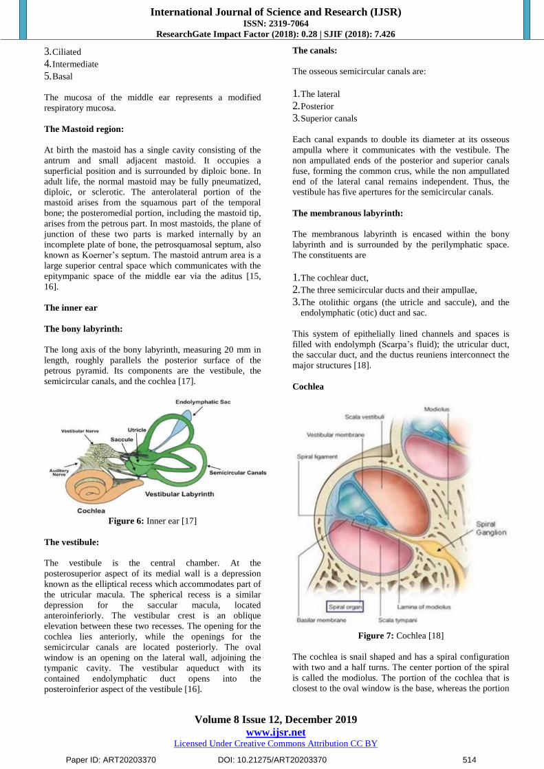
Cochlea
Figure 7: Cochlea [18]
The cochlea is snail shaped and has a spiral configuration
with two and a half turns. The center portion of the spiral
is called the modiolus. The portion of the cochlea that is
closest to the oval window is the base, whereas the portion
Paper ID: ART20203370 DOI: 10.21275/ART20203370 514

International Journal of Science and Research (IJSR) ISSN: 2319-7064
ResearchGate Impact Factor (2018): 0.28 | SJIF (2018): 7.426
Volume 8 Issue 12, December 2019
www.ijsr.net Licensed Under Creative Commons Attribution CC BY
of the cochlea that is farthest away from the oval window
is the apex.
The cochlea is having three compartments: scala tympani,
scala vestibuli and scala media. The basilar membrane
separates the scala tympani and the scala media, Reissner's
membrane separates the scala media and the scala vestibuli
The scala tympani and the scala vestibuli communicates
with each other at helicotrema. In the scala media, the
organ of Corti rests on the basilar membrane. Basilar
membrane and organ of Corti are referred to as the
cochlear partition. The organ of Corti has the inner and the
outer hair cells. The inner hair cells are arranged in a
single row and outer hair cells are arranged in three rows.
These hair cells have hair like projections called
stereocilia, which is responsible for the signal transduction
in hair cells. The scala vestibuli and the scala tympani are
filled with perilymph, which resemble the extracellular
fluid (high in sodium, low in potassium) in composition.
The scala media filled with end lymph, which resemble
intracellular fluid (low in sodium, high in potassium) in
composition. The electrolyte composition of the scala
media causes the endocochlear potential, which is +60 to
+100 mV relative to the perilymph [18].
Physiology
The difference between the impedance of air and the
impedance of fluid is great; thus, in the transmission of
sound energy from Air to Fluid medium, there would be a
99.9% loss which is approximately 30 dB loss. The above
loss can be overcome by impedence matching, which
allows optimum sound energy transmission [19].
Hydraulic lever
This is the ratio of the surface area of the tympanic
membrane to that of the oval window. The tympanic
membrane surface area is 55 mm2 and stapes foot plate
surface area 3.2 mm2. This difference represents a 17-fold
increase in surface area. Sound energy striking the much
larger tympanic membrane is transmitted through to a
much smaller surface area of the stapes footplate.
Lever ratio
The length of the manubrium, when compared the long
process of the incus, is 1.3times longer. Hence the
leverage gain is 1.3. Combined effects of these two
mechanisms, the Hydrualic ratio and the lever ratio, the
approximate gain is 22 dB.
The two factors help in the transmission of sound energy
are
1.Optimal Eustachian tube Function
2.Gas exchange within the middle ear Mucosa
Eustachian tube
The Eustachian tube has cartilaginous and bony portion.
The lateral bony portion of the canal opens in the anterior
wall of Middle Ear. The medial cartilaginous part, opens
into the nasopharynx and is closed at rest. The tensor veli
palatini opens the Eustachian tube orifice during
swallowing, for a transient period that lasts 0.3–0.5s.This
results in pressure equalization with the atmospheric
pressure.
Middle ear mucosal gas exchange
Middle ear mucosa has a well-developed capillary
structure close to its surface. This helps in gas exchange In
normal conditions middle ear pressure is equal to
atmospheric pressure, approximately760 mmHg at sea
level. These gases are bi-directionally exchanged.
Nitrogen gas level in middle ear is higher than that of
venous blood. This gradient results in gas exchange
between the middle ear space and venous blood. The
absorption of nitrogen into the venous blood results in
negative pressure in the middle ear. This is equalized by
opening of the Eustachian tube. Prolonged Eustachian tube
dysfunction hampers this mechanism results in negative
middle ear pressure, transudation of fluid, and the
development of a middle ear effusion and increases the
middle ear acoustic impedence.
Transmission of sound energy in the Cochlea
When sound energy travels through the ear, it causes the
stapes footplate to vibrate. The vibration of the stapes
footplate produces a compressional wave in the perilymph,
which travels to the scala vestibuli, through the
helicotrema, and out across the scala tympani towards the
round window. An inward motion of the stapes results in
outward movement of the round window. When the organ
of corti and basilar membrane are deflected in response to
the compression wave, it produces a shearing force
between the tectorial membrane and the stereocilia of the
hair cells. This shearing force produces a deflection of
stereocilia toward the direction of tallest row results in
opening of stretch- sensitive cationic channels located on
the stereocilia. The opening of these stretch- sensitive
cationic channels causes a influx of cationic current, which
results in hair cell depolarization.
When inner hair cells are depolarized, it opens voltage-
gated calcium channels. The resulting calcium current
triggers neurotransmitter release across the synapse, which
results in activation of the auditory nerve fibers [16, 19].
Tympanoplasty - An overview
Tympanoplasty (TM) is the procedure of removal of
disease from the middle ear and reconstruction of the
hearing mechanism along with TM grafting.
History of Tympanoplasty:
1640-Banzer
First attempt at repair of TM
Used pigs bladder as a lateral graft
Paper ID: ART20203370 DOI: 10.21275/ART20203370 515
International Journal of Science and Research (IJSR) ISSN: 2319-7064
ResearchGate Impact Factor (2018): 0.28 | SJIF (2018): 7.426
Volume 8 Issue 12, December 2019
www.ijsr.net Licensed Under Creative Commons Attribution CC BY
1853-Toynbee
Placed a rubber disc attached to a silver wire over the
TM
Reported significant hearing improvement
1863-Yearsley
Placed a cotton ball over a perforation
1877-Blake
Paper patch
1876-Roosa
Chemical cautery
1878-Berthold
Coined the term Myringoplasty
1950-Wullstein and Zollner
Described 5 types of Tympanoplasty
1960-Heerman
First used temporalis fascia grafting material in
tympanoplasty.
1961-Storrs
Temporalis fascia grafting
1967-House Glasscock and Sheehy
Techniques for lateral grafting
Indications for surgery:
1. Conductive hearing loss due to TM perforation or
ossicular dysfunction
2. Chronic or recurrent otitis media secondary to
contamination
3. Progressive hearing loss due to chronic middle ear
pathology
Contraindications for surgery:
1. Malignant tumours
2. Unusual infections like malignant otitis externa.
3. Intracranial complications
4. Cholesteatoma
Goals of the surgery:
1. Establish an intact TM
2. Eradicate middle ear disease and create an air containing
middle ear space
3. Restore hearing by sound pressure transformation
between the eardrum and the cochlea
Techniques:-
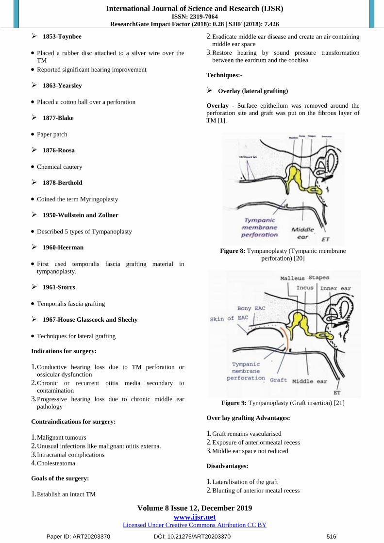
Overlay (lateral grafting)
Overlay - Surface epithelium was removed around the
perforation site and graft was put on the fibrous layer of
TM [1].
Figure 8: Tympanoplasty (Tympanic membrane
perforation) [20]
Figure 9: Tympanoplasty (Graft insertion) [21]
Over lay grafting Advantages:
1. Graft remains vascularised
2. Exposure of anteriormeatal recess
3. Middle ear space not reduced
Disadvantages:
1. Lateralisation of the graft
2. Blunting of anterior meatal recess
Paper ID: ART20203370 DOI: 10.21275/ART20203370 516
International Journal of Science and Research (IJSR) ISSN: 2319-7064
ResearchGate Impact Factor (2018): 0.28 | SJIF (2018): 7.426
Volume 8 Issue 12, December 2019
www.ijsr.net Licensed Under Creative Commons Attribution CC BY
3. Chance of iatrogenic cholesteatoma formation Healing
may take longer (4-8 weeks)
1. Technically more demanding
2. Formation of epithelial pearl
Underlay (Medial grafting)
Underlay technique was introduced by SHEA. The graft
was placed medial to the handle of malleus and TM
remnant.
ADVANTAGES:
1. Less blunting or lateralisation
2. High graft uptake
Disadvantages:
1. Limited visualization of anterior meatal recess
2. Difficult with small EAC.
3. Less suitable in large anterior perforation
4. Reduction in middle ear space TM grafts
Histologically TM grafts become lined by squamous
epithelium on the ear canal side and the middle ear mucosa
on the tympanic cavity side [22].
Grafting materials
1. Temporalis fascia graft
2. Cartilage graft
3. Fat graft
4. Hyaluronic acid fat graft
5. Tragal perichondrium and cartilage
6. Vein graft
7. Conchal cartilage
8. Fascia lata
9. Subcutaneous tissue
10. Periosteum
Approach
1. Transcanal
Posterior moderate sized perforations Favourable EAC
anatomy.
2. Endaural
Visualisation of annulus and anterior sulcus is difficult.
3. Postaural
All perforation sizes
Better angle of visualization
Various surgical techniques
1. Overlay-underlay technique
2. Combined technique
Two grafts-one under the handle of malleus Second on the
fibrous layer of tm
3. Circumferential sub annular graft technique
4. Swing door technique
5. Butterfly and palisade technique cartilage
Tympanoplasty
6. Cartilage shield tympanoplasty
7. The button graft technique
8. Cartilage tympanoplasty with island technique
9. Endoscopic vs microscopic tympanoplaty [23].
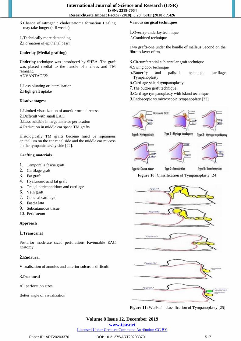
Figure 10: Classification of Tympanoplasty [24]
Figure 11: Wullstein classification of Tympanoplasty [25]
Paper ID: ART20203370 DOI: 10.21275/ART20203370 517
International Journal of Science and Research (IJSR) ISSN: 2319-7064
ResearchGate Impact Factor (2018): 0.28 | SJIF (2018): 7.426
Volume 8 Issue 12, December 2019
www.ijsr.net Licensed Under Creative Commons Attribution CC BY
Wullstein and Zollner (1956): TYPE I
TM is grafted to an intact ossicular chain
TYPE II
Malleus is partiallyeroded
TM is grafted to the long process of incus/ remaining
malleus
TYPE III
Columella effect /Myringostapediopexy Malleus and incus
are eroded
TM is grafted to the stapes suprastructure with cartilage in
between
TYPE –IV
Stapes suprastructure is eroded but foot plate is mobile TM
is grafted to a mobile footplate
Sound protection of the roundwindow and formation of
airspace in the hypotympanum
TYPE V
TM is grafted to a fenestration in the lateral semicircular
canal in cases with no ossicles and a fixed footplate.
In this thesis, study about the comparison of two different
grafting materials were Temporalis fascia and Tragal
perichondrium used in the patients undergoing type 1
tymnoplasty [25].
Temporalis fascia:
Temporalis fascia was first used in myringoplasty by
Ortegtran (1958-59), Heerman (1961) and Storrs (1961). It
is most commonly used autogenous material. It is
preferred for various reasons:
1. It is easy to harvest.
2. It can be used asonlay, intermediate or underlay graft.
3. For primary operation, there are no size limitations.
4. Fascia is quite similar to tympanic membrane with low
basal metabolic rate.
5. For reconstruction of the tympanic cavity and ear canal,
fascia is the only suitable autogenous material, because
of its size.
Tragal perichondrium:
Tragal perichondrium was introduced into myringoplasty
by Victor Goodhill et al (1964), after being used in
stapedectomy as an oval window graft for some years
before that. Like temporalis fascia, tragal perichondrium
has several advantages:
1. It is easily accessible.
2. It is a mesodermal graft.
3. It has a good chance of postoperative survival.
4. It has a conical contour.
5. It is sufficiently large for myringoplasty of a total
perforation [26].
4. Clinical Review
In 2013 Adip K. Shetty et al used tragal perichondrium as
graft material in type 1 tympanoplasty with a success rate
of 96% in the perichondrium group as compared with 92%
in the control group. In perichondrium- cartilage group
shows mean gain in AirBone gap was 16.5 + 7.27 dB as
compared to 15 + 7.07 dB in temporalis fascia group. In
this study there is no statistical significant difference in
graft taken up and hearing gain between both groups [27].
In the study by M. Mohsen Wafaie et al in 2010 shows.
Anatomical closure of TM perforations in 95% of patients
in the cartilage group, and in 90% of patients in the
temporalis fascia group with value p more than 0.05. The
mean acoustic gain in two group with no significant
statistical difference [28].
R. K. Mundra et al (2013) did study. to evaluate the
results of closure of subtotal perforation by tympanoplasty
using underlay technique., with the perichondrium
temporalis fascia graft supported by single sliced cartilage
[29].
Emily Iacovou et al, 2012 to compare the hearing results.,
and graft take up rate in patients undergoing
myringoplasty for the reconstruction of the tympanic
membrane, with the use of cartilage or fascia temporalis.
The mean graft uptake rate was 92.4 % in the cartilage
group and 84.3 % in the fascia temporalis group. The
obtained audiometric results are comparable to temporalis
muscle facia group and the rate of reperforation is lower
[30].
In 2011 Matthew Yung, Senthilnathan, Vivekanandan
and Philip Smith study compares outcomes of
myringoplasty procedures, using fascia and cartilage
grafts. The graft integration of fascia and cartilage grafts at
24 months, were 84.2% and 80%, respectively. The
postoperative AB gaps and audiological gains at 24
months were 16.97 dB and 13.63 dB, respectively, in the
fascia group and 20.63 dB and 12.60 dB respectively in
the cartilage group. There was no statistical significant
difference, in the graft taken up or postoperative
audiological gain between the two groups. In this study,
they pointed out their limitation as very low sample size,
so interpreted with caution [31]. In 2014 Rajeev Reddy
conducted ―study of results. of cartilage-perichondrium vs
temporalis fascia grafting in chronic suppurative otitis
media.‖ temporalis fascia group showed a good
neotympanum about 60 patients (84.5%), 7 patient
(9.85%) had reperforation and 5 (7.04%) had retraction
pockets. About 60 (98.6%) Patients showed a healed
Tympanic membrane and only 1 (1.63%) had
reperforations in tragal cartilage perichondrium group.
Patients with temporalis fascia graft showed an AB gap of
less than 10 dB in 49 (82%) patients and more than 10 dB
in 11 (18%) patients. AB gap closure with tragal cartilage
Paper ID: ART20203370 DOI: 10.21275/ART20203370 518
International Journal of Science and Research (IJSR) ISSN: 2319-7064
ResearchGate Impact Factor (2018): 0.28 | SJIF (2018): 7.426
Volume 8 Issue 12, December 2019
www.ijsr.net Licensed Under Creative Commons Attribution CC BY
perichondrium was less than 10 dB in 45 (78%) patients
and more than 10 dB in 13 patients (12%). ―Tragal
cartilage perichondrium (<0.5 mm) seems to be an ideal
graft material for tympanic membrane, in terms of
postoperative healing and acoustic properties‖ [32].
In the study by Kazikdas et al, 2007 reported graft
acceptance rate about 97.5% with palisade cartilage
tympanoplasty, for the anatomical closure of subtotal
perforations compare with 75% of the fascia group.
Regarding acoustic gain, they found no statistical
difference between cartilage and fascia groups [33].
Zahnert et al. (13) reported that the ideal acoustic
thickness of cartilage should be approximately 0.5 mm
instead.of full thickness of 0.7 to 1 mm to achieve better
audiological outcome [34].
Yakup Yegi̇ n et al between 2009 to 2014 .did
comparative ―study of temporalis muscle fascia and full-
thickness cartilage grafts in type 1 tympanoplasty ―in 247
patients. in the cartilage tympanoplasty patien.t show
higher graft take rate 91.3% than fascia tympanoplasty
patient 68.9% with p<0.001, but audioulogical gain are not
statistically significance, in with full thickness cartilage
tympanoplasty.compare to fascia [35].
M M KHAN, S R PARAB conduct.study on sliced
cartilage. and temporalis fascia myringoplasty. Period of
study 2005-2008. On four year follow up anatomic closure
of perforation 97.5%. in cartilage group, and 82.63% in
fascia group. Hearing gain comparable.on both group
(average AirBone gap 7.10 dB in cartilage vs 8.05db in
other group) [36].
In 2010 M. Mohsen, Wafaie Abdelaziz., M.Elsherif M.
Salama Bakr were did study at ENT Dept. AL-Azhar
university. They got 95% of tympanicmembrane closure in
cartilage as compared to 90% fascia group. Audiological
gain in. cartilage group was 12.4±6.4 dB, and14.8±9.9 dB
in fascia group with no significant statistical difference
between the two groups [37].
In 2016 Rahul K. Jaiswal did study. in Nepal
‗Comparison of outcomes of palisade cartilage with
temporalis fascia.following tympanoplasty‖. 40 His study
shows closure of AB gap within. 30 dB was achieved in
71.87% of temporalis fascia group and 88.89% in palisade
cartilage group and anatomical closure. of perforation was
comparable in both group [38].
In 2015 Sohil Vadiya et al study conducted in Gujarat to
compare the outcome of cartilage shield tympanoplasty
with TM fascia type 1 tympanoplasty in cartilage group.
Graft uptake about 98.46% as compare to 89.61 of TM
fascia group. In this study graft uptake rate. show
statistical significant in both group. And audiological
improvement in both group almost equal except at 8 kHz
frequency where improvement in TM fascia group than
cartilage group [39].
Uzun et al. achieved 100%.graft up, take with type 1
palisade - cartilage grafting, whereas 84.2% only success
rate was observed in temporalis fascia type 1
tympanoplasty [40].
In Korea LEE HY et al conducted. a study of change of
middle ear mechanics after sliced cartilage type 1
tympanoplasty and compare with fascia tympanoplasty.43
Period of study. 2002 to 2005 and followed up after 6
month. They founded there was no statistical. significant
between two group in impedance audiometry [41].
Cabra et al did at randomized. controlled trial between
1997 to 2002 in general hospital. They allocated 64 patient
to cartilage group. and 59 patient into fascia temporalis,
group. 1st they analyzed outcome after 24 month
anatomical. success was 82.26% (51 of 64) in cartilage.
tympanoplasty and 64.41 % (38 of 58) in TM fascia group.
The functional; outcome comparable. between both 2
groups [42].
Juveria Majeed, Naveed Ahamed, 2016 reported the
patients with tubotympanic type of chronic suppurative
otitis media were selected from all patients attending to the
ENT department of Gandhi Hospital, Secunderabad
between July 2014 to March 2016. This study includes 60
patients out of which 30 were subjected to myringoplasty
with temporalis fascia and remaining 30 to myringoplasty
with tragal perichondrium. In all patients, discharging ears
and associated nasal pathologies were excluded. The
comparative study was done on following parameters-
graft uptake, audiological outcome, donor site
complications and any late complications such as
reperforation, retraction, worsening of hearing and
adhesions. Our study included a follow up of post
operative cases for 1 year 8 months. Out of 60 cases
operated, 11 cases didn‘t come for follow up. Hence they
were excluded. The remaining 49 cases were 25 temporalis
fascia group and 24 tragal cartilage group. The youngest
patient in our group was 13 years while the oldest was 56
years old. The overall male: female ratio was 27:22. The
patients who underwent temporalis fascia grafting, 86.73%
had a gain of 15dB while 13.7% had a gain of > 15 dB. Of
the patients underwent tragal perichondrium grafting 50%
had a gain of 15 dB while 10% had a gain of >15dB. The
graft uptake rate was 85.7% for both temporalis fascia as
well as tragal perichondrium.4% of the patients of the
temporalis fascia group had seroma and 4% had
persistantpain..Residual perforation was seen in 3 patients
of temporalis fascia group and 4 patients of tragal
perichondrium group. 1 case of each group showed canal
stenosis [43].
Methodology
Source of data
A sample size of in age group of 18 to 60 years in CSOM
cases were selected for the study. Duration of study 2
years i.e. from a total of 100 patients in age group of 18 to
60 years with CSOM tubo tympanic disease who
underwent Type 1 tympanoplasty by underlay technique in
the Department of ENT, Narayana Medical College &
Paper ID: ART20203370 DOI: 10.21275/ART20203370 519
International Journal of Science and Research (IJSR) ISSN: 2319-7064
ResearchGate Impact Factor (2018): 0.28 | SJIF (2018): 7.426
Volume 8 Issue 12, December 2019
www.ijsr.net Licensed Under Creative Commons Attribution CC BY
Hospital were studied in the period of two years. (Between
October 2017 to September 2019).
Inclusion criteria
1. Patient with CSOM (Tubotympanic) with a hearing loss
upto 45 dB
2. Patient in the age group of 17 — 60 years of either sex
were selected for the study.
Exclusion criteria
1. Patients with active discharge.
2. Patients with attic disease.
3. Patients with systemic diseases like diabetest Hyper
tension, Ischemic Heart disease.
All the patients who presented with symptoms and signs
suggesting tubotympanic type CSOM were submitted to an
assessment protocol, based on a guided history taking,
specific physical exam (otoscopy), and subjected to
audiogram. During history taking, the patients were
questioned about disease onset, and if they had undergone
previous otologic surgeries.
A detailed proforma was filled for each patient with regard
to history, clinical examination, investigations, surgical
procedures, postoperative period &follow up visits.
Audiological evaluation (pure tone audiometry) done
preoperatively, 1 month & 3 months after surgery and the
results were tabulated.
Pure tone threshold audiometry has become the standard
behavioral procedure for describing audiometry
sensitivity. Therefore pure tone audiometry had been used
for assessment of hearing level in this study.
The Audiometer (Elcon 3N3 Multi) used in this study was
manual.
Figure 12: Audiometer (Elcon 3N3 Multi)
The test was performed in acoustically treated room with
no ambience noise the audiometry was done following
standard protocol. Patient was explained about the
procedure before audiometry and adequate time was taken
for testing. The technique followed was Carhart & Jerger‘s
technique which is mostly used (technique of 5 up and 10
down method).
For calculation of average of hearing loss (air conduction
threshold) three frequencies were selected. They were 500
Hz, 1000Hz and 2000Hz. These frequencies were selected
because they represent speech frequency range and
elevation of threshold in these frequencies will be
clinically significant.
All surgeries were performed under local or general
anesthesia using a microscope with a lens of 250mm. In
most of the cases postauricular approach was used. In
cases where the external acoustic meatus was wide and the
perforation borders were visible endaural or transcanal
approach was performed. Temporalis fascias, Tragal
perichondrium, were taken as graft materials for cases and
underlay grafting done in all cases.
All the patients were followed after surgery every week for
1 month but the puretone audiograms were done at 1
month and 3 month postoperativerly
Graft materials used for the procedure were
Autologous temporalis fascia
Autologous tragal perichondrium
Temporalis fascia:
Temporalis fascia was obtained during the surgical
procedure. In cases using post- auricular incision &
endaural incision as shown in the figure 16 & 17
respectively. The same incision was extended to harvest
the temporalis fascia. In transcanal surgeries, a separate
transverse incision was placed above the pinna on the
temporal region to obtain a graft from temporalis fascia or
over tragus (Figure 18) to obtain perichondrium and
cartilage. After obtaining the graft, it was spread on to a
graft spreader and teased to remove excessive muscle
fibres; fat and fibrous tissues so that it appears like
parchment when dry (Figure 19). The temporalis fascia
graft is seen after one month and three months of
tympanoplasty as shown in figure 20 and 21 respectively.
Autologous tragal perichondrium: It was obtained
during the surgical procedure from the tragal cartilage is as
shown in the figure.
Paper ID: ART20203370 DOI: 10.21275/ART20203370 520

International Journal of Science and Research (IJSR) ISSN: 2319-7064
ResearchGate Impact Factor (2018): 0.28 | SJIF (2018): 7.426
Volume 8 Issue 12, December 2019
www.ijsr.net Licensed Under Creative Commons Attribution CC BY
Figure 13: Tympanoplasty instrument set
Figure 14: Post aural incision
Paper ID: ART20203370 DOI: 10.21275/ART20203370 521

International Journal of Science and Research (IJSR) ISSN: 2319-7064
ResearchGate Impact Factor (2018): 0.28 | SJIF (2018): 7.426
Volume 8 Issue 12, December 2019
www.ijsr.net Licensed Under Creative Commons Attribution CC BY
Figure 15: Endaural incision
Figure 16: Transcanal approach with tragal perichondrium as graft
Figure 17: Prepared temporalis fascia
Paper ID: ART20203370 DOI: 10.21275/ART20203370 522

International Journal of Science and Research (IJSR) ISSN: 2319-7064
ResearchGate Impact Factor (2018): 0.28 | SJIF (2018): 7.426
Volume 8 Issue 12, December 2019
www.ijsr.net Licensed Under Creative Commons Attribution CC BY
Figure 18: Tragal perichondrium
Figure 19: Temporalis fascia graft-1 month post tympanoplasty
Paper ID: ART20203370 DOI: 10.21275/ART20203370 523
International Journal of Science and Research (IJSR) ISSN: 2319-7064
ResearchGate Impact Factor (2018): 0.28 | SJIF (2018): 7.426
Volume 8 Issue 12, December 2019
www.ijsr.net Licensed Under Creative Commons Attribution CC BY
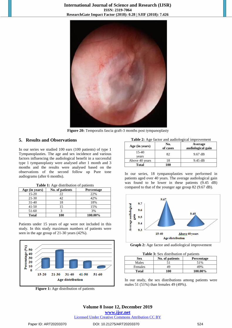
Figure 20: Temporalis fascia graft-3 months post tympanoplasty
5. Results and Observations
In our series we studied 100 ears (100 patients) of type 1
Tympanoplasties. The age and sex incidence and various
factors influencing the audiological benefit in a successful
type 1 tympanoplasty were analysed after 1 month and 3
months and the results were analysed based on the
observations of the second follow up Pure tone
audiograms (after 6 months).
Table 1: Age distribution of patients
Age (in years) No. of patients Percentage
15-20 22 22%
21-30 42 42%
31-40 18 18%
41-50 15 15%
51-60 3 3%
Total 100 100.00%
Patients under 15 years of age were not included in this
study. In this study maximum numbers of patients were
seen in the age group of 21-30 years (42%).
Figure 1: Age distribution of patients
Table 2: Age factor and audiological improvement
Age (in years) No.
of cases
Average
audiological gain
15-40
years 82 9.67 dB
Above 40 years 18 9.45 dB
Total 100
In our series, 18 tympanoplasties were performed in
patients aged over 40 years. The average audiological gain
was found to be lower in these patients (9.45 dB)
compared to that of the younger age group 82 (9.67 dB).
Graph 2: Age factor and audiological improvement
Table 3: Sex distribution of patients
Sex No. of patients Percentage
Males 51 51%
Females 49 49%
Total 100 100.00%
In our study, the sex distributions among patients were
males 51 (51%) than females 49 (49%).
Paper ID: ART20203370 DOI: 10.21275/ART20203370 524

International Journal of Science and Research (IJSR) ISSN: 2319-7064
ResearchGate Impact Factor (2018): 0.28 | SJIF (2018): 7.426
Volume 8 Issue 12, December 2019
www.ijsr.net Licensed Under Creative Commons Attribution CC BY
Figure 3: Pie chart of sex distribution of patients
Table 4: Sex incidence and audiological benefit
Gender No.
of cases
Average
audiological Gain
Males 51 9.48 dB
Females 49 9.83 dB
Total 100
Figure 4: Graph of sex incidence and audiological benefit
In this study, the audiological benefit observed more in
females could be an incidental finding as no reason could
be attributed to this observation.
Table 5: Time taken for graft uptake and audiological
benefit
TIME (in
weeks)
No.
of cases
Average
audiological gain
3 86 9.61 dB
>3 14 9.93 dB
Total 100
Audiological benefit was found to correlate with the time
taken for graft take up, as the time taken for graft uptake
increases, the hearing gain was found to decrease post
operatively.
Table 6: Audiological assessment in Type-I
tympanoplasty
Hearing results Audiological outcome
Improved 97 (97%)
No much change 3 (3%)
Total cases 100
In our study of 100 cases, 97 (97%) cases showed
audiological improvement. There is no big difference of
average audiological gain in rest of the cases.
Figure 5: Pie chart of audiological assessment in Type-I
tympanoplasty
Table 7: Effect of different grafts on audiological
improvement in Type I tympanoplasty
Type of graft No. of cases Average audiological
gain
Temporalis fascia 47 10.07 dB
Tragal perichondrium 53 9.28 dB
Total no. of cases 100
The audiological improvement (average audiological gain)
is more with temporalis fascia (10.07 dB) when compared
to tragal perichondrium (9.28 dB).
Graph 6: Effect of different grafts on auological
improvement in Type I tympanoplasty
Table 8: Statistical analysis of different graft on
audiological improvement in Type I tympanoplasty N Mean SD F Value P Value
Preop ave.AC
threshold
(dB)
TF 47 31.03 3.44271
0.675
0.781
(Not Sig.)
TP 53 30.72 3.75556
Total 100 30.87 3.59723
Postop 3 months
ave.AC
threshold (dB)
TF 47 20.96 2.56191
0.743
0.667
(Not
Sig.)
TP 53 21.42 2.78343
Total 100 21.20 2.67800
Audiological gain (dB)
TF 47 10.07 3.37184
1.553
0.161
(Not Sig.)
TP 53 9.28 2.85766
Total 100 9.65 3.16370
Statistical analysis has been done by Annova test using
IBM SPSS Version 22.0. P values which are less than 0.05
are considered as statistical significant.
Paper ID: ART20203370 DOI: 10.21275/ART20203370 525
International Journal of Science and Research (IJSR) ISSN: 2319-7064
ResearchGate Impact Factor (2018): 0.28 | SJIF (2018): 7.426
Volume 8 Issue 12, December 2019
www.ijsr.net Licensed Under Creative Commons Attribution CC BY
6. Discussion
Type I tympanoplasty is a surgical procedure in which the
reconstruction procedure is limited to the repair of
tympanic membrane perforation alone. Implicit definition
is that the ossicular chain is intact and mobile, and that
there is no middle ear disease such as infected mucosa or
in growth of skin [1]. The present study describes various
parameters in assessing the hearing improvement after
tympanoplasty. Post operative audiological evaluations
were done after 1 month and 3 months following type I
tympanoplasty.
Table 9: Age Incidence
Present study Ortegren [44]
Age in
years
No. of
patients Percentage
Age in
years
No. of
patients Percentage
0-10 0 0 0-10 5 5
11-20
22
22% 11-20 20 22.9
21-30
42
42% 21-30 13 14.9
31-40
18
18% 31-40 12 13.7
>40 18 18% >40 37 42.5
Total 100 100 Total 87 100
Patients under 15 years of age are not included in this
study. In this study maximum numbers of patients are seen
in the age group of 21-30 years (42%). This is in contrast
to the study conducted by Ortergren, where the maximum
number of the patients are in the age group of >40 years
(42.5%) [44].
Table 10: Age distribution in different studies
Sl. No. Author Year of
study
Total no.
of cases
Commonest
age group
1 Saha A K et al
[45] 2006 40 14-34
2 Fukuchi et al [46] 2006 37 15-35
3 Nagle et al [47] 2009 100 21-30
4 Goyal Rashmi [48] 2010 80 11-40
5 Present study 2019 82 11-40
All the studies showed 15 – 40 yrs of age as the most
common group and are correlating with the present study.
The reason behind this may be that this is the socially
active and health conscious age group.
Table 11: Age factor and audiological improvement
Age (in years) No. of cases Audiological benefit
15-40 years 82 9.67 dB
Above 40 years 18 9.45 dB
Total 100
In our series, 18 tympanoplasties were performed in
patients aged over 40 years. The audiological benefit was
found to be lower in these patients (9.45 dB) compared to
that of the younger age group (9.67 dB).
Ortegren has reported that there is a limit at 40 years
beyond which hearing results are markedly worse than in
younger cases. In Ortegren‘s study, the hearing
improvement in the various groups below 40 years was
obviously very much alike. Hearing results were worse in
the above 40 years age group, compared to young age
group. Vartiainen10 et al found that results in elderly
patients were found to be as good as in younger patients
[44].
Table 12: Sex incidence and audiological benefit
Age (in years) No. of cases Audiological benefit
Males 51 9.48 dB
Females 49 9.83 dB
Total 100
In this study, the audiological benefit observed more in
males could be an incidental finding as no reason could be
attributed to this observation.
Table 13: Graft taking time and audiological benefit
Time (in weeks) No. of cases Audiological benefit
3 86 9.61 dB
> 3 14 9.93 dB
Total 100
In our study, the audiological benefit was found to
correlate with graft take up time.
Akayleh R et al has similar outcomes as that of our study
[49].
Table 14: Audiological assessment in tympanoplasty
Hearing results Audiological benefit
Improvement 97 (97%)
No change 3 (3%)
Total 100 (100.00%)
In our study of 100 cases, 97 (97 %) cases showed
audiological improvement. There is no big difference of
average audiological gain in rest of the cases.
Table 15: Comparison of hearing improvement in various
studies
Sl.No Study Hearing improvement
(% of cases)
1 Saleem et al [50] 85.88
2 Karela et al [51] 91.5
3 Fukuchi et al [46] 92
4 Present study 97
The most likely explanation for lack of complete success
from a hearing stand point is that in most cases of CSOM,
even though ossicular chain may appear normal, there is
some factor of scar tissue that prevents total restoration of
hearing (Sheehy et al 1980). Sheehy has reported a loss of
BC of 10dB or more at 2K or 4K in 3% of cases, probably
due to trauma to the ossicular chain [52].
Gibb & Klat (1982) have also found that a persistent
conductive hearing loss can result from underlay
technique. If the handle of malleus is severely retracted
especially if it is touching or adherent to the promontory,
difficulties arise due to possible reduction in the depth of
the tympanic cavity when the graft is placed medial to the
malleus handle. To overcome this problem, they suggested
that one could leave the malleus in its original retracted
position and a split graft be pulled upon each size of
Paper ID: ART20203370 DOI: 10.21275/ART20203370 526
International Journal of Science and Research (IJSR) ISSN: 2319-7064
ResearchGate Impact Factor (2018): 0.28 | SJIF (2018): 7.426
Volume 8 Issue 12, December 2019
www.ijsr.net Licensed Under Creative Commons Attribution CC BY
malleus handle and tucked behind its upper part or
amputate 2-3mm from the tip of handle [53].
Vartiainen and Nauutinene (1993) in their series had 11
audiological failures. The cause of persistent hearing loss
was found to be due to fixation or erosion of ossicles
overlooked by the surgeon [54].
Saeed Ghamdi et al 1994 reported a permanent hearing
loss in 3% of the patients. Ghamdi also reported that one
patient developed a profound SN hearing loss in the
operated ear in his study. The unchanged audiological
status in tympanoplasty can be explained by disorders that
can interfere with the ventilatory or conducting function of
the middle ear viz. tympanosclerosis, stiffness of ossicles
and Eustachian tube dysfunction that have not been dealt
during the surgery (Rance. W. Rancy in 1995) [55].
Alan G Gibb proposed the following reasons for
worsening of hearing loss. They stated that a conductive
loss can result from damage to the ossicular chain.
Sensorineural hearing loss appearing for the first time post
operatively generally iatrogenic and the result of some
technical error at operation. Thus it is essential to exercise
extreme care throughout the operation and avoid undue
commotion of ossicular chain which might cause cochlear
damage [53].
If underlay grafting is employed, special care should be
taken to avoid touching the incus or stapes when scarifying
the postero superior are of the tympanic membrane
remnant. If due care is not taken in this procedure it is
possible to hook up the ossicles or incudostapedial joint
with a scraper. It is also important to exercise extreme care
when taking the graft into position in this area, as at this
stage of operation, the ossicles are often obscured by the
graft itself. In either event the stapes may be shaken and if
the trauma is severe, the foot plate may even be cracked,
sever or even total SN deafness may result.
The introduction of toxic solutions in the middle ear at the
time of operation is another possible cause of SN deafness
as previously reported by Alan G. Gibb et al [53].
Valsalva maneuver may add some audiological benefit
when effectively incorporated which is supported by
Seung Hyo Choi et al. The criteria of successful
myringoplasty and tympanoplasty-1 surgery was a positive
graft take and followed by improvement in hearing [56].
Table 16: Effect of different grafts on audiological
improvement in Type I tympanoplasty
Ear discharge No. of cases Average audiological
gain
Temporalis fascia 47 10.07
Tragal perichondrium 53 9.28
Total 100
The average audiological gain is more with temporalis
fascia (10.07) in the present study. Jain K, Pandey A,
Gupta S, Rahul, 2016, did a clinical Study on hearing
outcome after type1 tympanoplasty using temporalis fascia
as graft in 45 patients. They observed a mean hearing gain
of 11.14 dB in their study [57].
According to study done by Lee et al., 2012, in a
retrospective analysis of 40 patients with CSOM,
compared the anatomical and audiological results of type I
tympanoplasty using fascia temporalis, cartilage tissue,
and cartilage palisade. No statistically significant
differences were observed between the three groups
regarding the closure of tympanic membrane perforations.
Regarding auditory improvement, the cartilage palisade
technique showed slightly poorer results than the others
[58].
Ortegren during the period 1957 to 1961 did a comparison
study of 87 patients of tympanoplasty-1 using temporalis
fascia and canal skin graft. The relation of age on hearing
and hearing results, importance of tubal function and
causes of failures were discussed [44].
Palva et al in 1969 reported that they achieved a
practically useful hearing (0 to 40 dB, ISO standard) in
93% of cases postoperatively out of 160 myringoplasties.
They used an operative method termed as ‗swing door
myringoplasty‘ [59].
In 1973, Glasscock analysed 237 myringoplasty cases
using temporalis fascia. He found that the results were
better using the underlay technique [60].
P. Packer et al, in 1982 compared preoperative and
postoperative hearing evaluation in 604 patients. He also
compared the different techniques and different graft
materials used in his series [61].
G.S. Bawa et al in 1987 studied 50 cases of
myringoplasties and showed a postoperative hearing
improvement in 74% patients [62].
Sato H, Nakamura H, Honjo I, Hayashi M. in 1990
examined about the Prognostic value of preoperative
Eustachian tube function in 77 ears, subjected to type 1
tympanoplasty. Eustachian tube function was evaluated by
positive and negative pressure equalization tests, and
clearance test and found that positive pressure equalization
and clearance tests of the tube were correlated with the
outcome of ear surgery, although the negative pressure
equalization test had no correlation with it and concluded
that the preoperative tubal function test including positive
pressure test and clearance test are useful for predicting the
prognosis of ear surgery [63].
Vartiainen et al in 1993 did a follow up study 404 cases of
myringoplasties. In audiological failures the cause of
persistent conductive hearing loss was found to be due to
fixation or erosion of ossicles overlooked by the surgeon
[64].
Kotecha B et al in 1999 presented a prospective audit
study of myringoplasty.73 surgeons participated in this
study and they got data from 1070 individual patients.
Where hearing loss was the main indication for surgery,
hearing improvement was seen in 67% [65].
Paper ID: ART20203370 DOI: 10.21275/ART20203370 527

International Journal of Science and Research (IJSR) ISSN: 2319-7064
ResearchGate Impact Factor (2018): 0.28 | SJIF (2018): 7.426
Volume 8 Issue 12, December 2019
www.ijsr.net Licensed Under Creative Commons Attribution CC BY
Mak D et al in 2000 January published a paper on a field
assessment of the surgical outcome in middle ear disease
in remote aboriginal Australia during the period 1986 to
1995.Success was defined by an intact tympanic
membrane and air-bone gap of 25 dB at 6 month follow up
after operation. A success rate of 53% was observed.
Successful outcomes were more likely in adults and
children aged more than 10 years [66].
Elluru RG, Dhanda R, Neely JG, Goebel JA. in 2001
studied about the efficacy and safety of anterior
subannular tympanostomy in 38 consecutive patients with
a diagnosis of Eustachian tube dysfunction, adhesive otitis
media, or chronic otitis media with a perforation who
underwent a tympanoplasty and concluded that Anterior
subannular tympanostomy is a safe and effective method
for long-term middle ear ventilation in patients with
chronic eustachian tube dysfunction [67].
Nepal A, Bhandary S, Mishra SC, Singh I, Kumar P. in
2003-04 studied 100 cases with dry, clean central
tympanic membrane perforations due to various causes
like chronic suppurative otitis media-tubotympanic, post
acute suppurative otitis media residual perforations or
simple traumatic perforations with conductive hearing loss
and without pre existing hearing loss were
clinicoaudiologically evaluated and analyzed. The study
concluded that hearing loss was found to be directly
proportional to the size of perforation irrespective of their
cause. Overall, perforations involving posterioinferior
quadrant were found to have maximum hearing loss [68].
Gierek T, Slaska-Kaspera A et al.2004, compared the
audiological results with temporalis fascia and tragal
perichondrium and they concluded no statistically
significant difference between the two [69].
Yetiser S, Hidir Y, Karatas E, Karapinar U. conducted a
study on 30 patients who underwent ossicular chain
reconstruction between 1990 and 2005, concluded that the
success of the surgery was dictated by the location and the
extent of tympanosclerotic involvement [70].
Mehta RP, Rosowski JJ, Voss SE, O'Neil E, Merchant SN.
in 2006 studied patients with tympanic membrane
perforations without other middle-ear disease. They
concluded that the conductive hearing loss resulting from a
tympanic membrane perforation is frequency-dependent,
with the largest losses occurring at the lowest sound
frequencies; increases as size of the perforation increases;
varies inversely with volume of the middle ear and
mastoid air space (losses are larger in ears with small
volumes); and does not vary appreciably with location of
the perforation. Effects of location, if any, are small [71].
Gierek T, Slaska-Kaspera A, Majzel K, Klimczak-Gołab
L. in 2006 published a study that aimed to establish
through a systemic review what is the best technique to
treat tympanic perforations and concluded that there is no
technique considered sure for every perforation neither
technique definitive for type of perforation [69].
Haruo Takahashia, Hiroaki Satob, Hajime Nakamurac,
Yasushi Naitod, Hiroshi Umekia. In 2007 examined the
correlation between the middle-ear pressure regulation
functions including active eustachian tube (ET) functions
and transmucosal gas exchange function, and outcome of
tympanoplasty and concluded that impairment of all the
middle- ear pressure-regulation functions was likely to
cause poor outcome of tympanoplasty, and also allowed us
reconfirm that ears with mechanically obstructed ETs were
contraindicated for tympanoplasty. Therefore, assessment
of mastoid condition is important as well as the ET
function before tympanoplasty [72].
H. Vijayendra, C. J. Ittop and R. Sangeetha in 2008 found
that canaloplasty is an integral part of tympanoplasty and
concluded that Canaloplasty gives 9 dB gain in hearing
compared to without canaloplasty and gives better
visualization, better graft placemen and better post-
operative care [73].
Matsuda Y, Kurita T, Ueda Y, Ito S, Nakashima T. in
2009 found significant correlation between the degree of
sound conduction disturbance and the perforation area; this
correlation was greater at low frequencies following a
traumatic perforation [74].
Ibekwe TS, Nwaorgu OG, Ijaduola TG, in 2009 studied
the relationship between the location of perforation on TM
and hearing loss and concluded that the location of
perforation on the tympanic membrane (TM) has no effect
on the magnitude of hearing loss in acute TM perforations
while it is significant in chronic ones [75].
Warren Y. Adkins, M.D., Benjamin White, M.D.in 2009
studied about Type I tympanoplasties utilizing an underlay
technique with temporalis fascia performed at the Medical
University associated hospitals over a 5-year period were
reviewed. In their study 40 adults were analyzed for
influencing factors. The overall success rate was 89% and
concluded that the age of the patient, the length of time the
ear had been dry, and the presence of infection at the time
of surgery had no influence on the success rate [76].
Kazim Bozdenir et al., 2011, in their comparative study,
Tympanoplasty with island cartilage graft versus
Temporalis fascia concluded that postoperatively, the pure
tone averages and air-bone gap closure were better with
temporalis fascia compared to cartilage grafting [77].
Rasha A, Ahmed SAO, 2015, in their study on outcome of
hearing improvement in myringoplasty of 51 Sudanese
Patients concluded that the temporalis fascia graft had
better hearing threshold improvement [78].
Abhay Kumar, Prabhu Narayan, Prem Narain, Jaypal
Singh, Prateek Kumar Porwal, Sanjay Sharma, Daya
Shankar in 2018 did a comparative study between result of
Temporalis muscle fascia and tragal cartilage
perichondrium as a graft material in Type 1 tympanoplasty
in 60 patients, dividing them into two groups. They
concluded that in comparison to tragal cartilage
perichondrium, tympanoplasty with temporalis fascia had
better mean air conduction values [79].
Paper ID: ART20203370 DOI: 10.21275/ART20203370 528
International Journal of Science and Research (IJSR) ISSN: 2319-7064
ResearchGate Impact Factor (2018): 0.28 | SJIF (2018): 7.426
Volume 8 Issue 12, December 2019
www.ijsr.net Licensed Under Creative Commons Attribution CC BY
Singh SP, Nagi RS, Singh J, 2018, did a comparative study
on evaluation of audiological and graft uptake results of
reinforced sliced cartilage versus temporalis muscle fascia
graft in type I tympanoplasty. They concluded that sliced
cartilage reinforced with temporalis fascia is a reliable
technique for tympanoplasty, especially in large
perforations with better graft uptake rates compared to
temporalis fascia without affecting audiometric results
[80].
7. Conclusion
In our study we concluded both temporalis fascia and
tragal perichondrium are suitable graft materials for
tympanoplasty. Graft uptake was superior with temporalis
fascia, while hearing improvement was better with tragal
perichondrium, although the results were not statistically
significant.
Tragal perichondrial graft is an effective grafting material
used for tympanoplasty due to its good qualities. Graft
success rate was 97% in this study, which signifies yield of
tragal perichondrial graft tympanoplasty.
Thus, to conclude, both temporalis fascia and tragal
perichondrium are acceptable graft materials for successful
closure of tympanic membrane perforations. The overall
graft uptake appeared to be better with temporalis fascia,
while the hearing improvement was better with tragal -
perichondrium. However, none of the results were
statistically significant.
References
[1] Aristides Sismanis, MD. Tympanoplasty. In:
Glasscock- Shambaugh SURGERY of the EAR. Ed;
Micheal E Glasscock III, MD, Aina Julianna Gulya,
MD.5th Ed; Elsevier publishers.2007: (Chapter 24),
463-485.
[2] Palva T. Surgical treatment of chronic middle ear
disease. Myringoplasty and tympanoplasty. Acta
Otolaryngol. 1987 Sep-Oct; 104 (3-4):279-84.
[3] Perkins R. Grafting materials and methods in
reconstructive ear surgery. Ann Sep Otol Rhinol
Laryngol. 1975 Jul-Aug; 84 (4 Pt 1): 518-26.
[4] Michael E Glasscock 111, George E. Shambaugh, jr.,
Surgery of the ear, 4th edition 1990.W.B.Saunder‘s
Company.
[5] World Health Organisation/CIBA Foundation
workshop reports, Prevention of Hearing Impairment
from chronic otitis media, 1996.
[6] Rupa V, Jacob A, Joseph A.Chronic suppurative otitis
media: prevalence and practices among rural South
Indian children. Int J Pediatr Otorhinolaryngol.1999
May 25; 48 (3):21721.
[7] Salah Mansour et al Comprehensive and Clinical
Anatomy of the Middle Ear 152- 165.
[8] Casale J, Giwa AO. Embryology, Branchial Arches.
InStatPearls [Internet] 2019 Feb 27, Stat Pearls
Publishing.
[9] Anthwal N, Thompson H. The development of the
mammalian outer and middle ear. Journal of anatomy.
2016 Feb;228 (2):217-32.
[10] Szymanski A, Geiger Z. Anatomy, Head and Neck,
Ear Tympanic Membrane. InStatPearls [Internet] 2019
Apr 6. StatPearls Publishing.
[11] Daniel E. Noise and hearing loss: a review. Journal of
School Health. 2007 May;77 (5):225-31.
[12] Toyoda S, Shiraki N, Yamada S, Uwabe C, Imai H,
Matsuda T, Yoneyama A, Takeda T, Takakuwa T.
Morphogenesis of the inner ear at different stages of
normal human development. The Anatomical Record.
2015 Dec;298 (12):2081-90.
[13] Ječmenica J, Bajec-Opančina A, Ječmenica D.
Genetic hearing impairment. Child's Nervous System.
2015 Apr 1;31 (4):515-9.
[14] Sánchez‐Villagra MR, Gemballa S, Nummela S,
Smith KK, Maier W. Ontogenetic and phylogenetic
transformations of the ear ossicles in marsupial
mammals. Journal of Morphology. 2002 Mar;251
(3):219-38.
[15] Hindi K, Alazzawi S, Raman R, Prepageran N,
Rahmat K. Pneumatization of mastoid air cells,
temporal bone, ethmoid and sphenoid sinuses. Any
correlation?. Indian Journal of Otolaryngology and
Head & Neck Surgery. 2014 Dec 1;66 (4):429-36.
[16] Siddarthan M. A comparative study of cortical
mastoidectomy with myringoplasty vs myringoplasty
alone in active cases of chronic otitis media (Doctoral
dissertation, Stanley Medical College, Chennai).
[17] Hussain B, Ali M, Qasim M, Masoud MS, Khan L.
Hearing impairments, presbycusis and the possible
therapeutic interventions. Biomedical Research and
Therapy. 2017 Apr 20;4 (4):1228-45.
[18] Morrill S, He DZ. Apoptosis in inner ear sensory hair
cells. Journal of otology. 2017 Dec 1;12 (4):151-64
[19] Branch IV MS, Chennai T. A Comparative Study of
Cortical Mastoidectomy With Myringoplasty vs
Myringoplasty Alone in Active Cases of Chronic
Otitis Media.
[20] Mundra RK, Sinha R, Agrawal R. Tympanoplasty in
subtotal perforation with graft supported by a slice of
cartilage: a study with near 100% results. Indian
Journal of Otolaryngology and Head & Neck Surgery.
2013 Dec 1;65 (3):631-5.
[21] Barake R, El Natout T, Bassim M, El Natout MA.
Loop underlay tympanoplasty for anterior, subtotal
and total tympanic membrane perforations: a
retrospective review. Journal of Otolaryngology-Head
& Neck Surgery. 2019 Dec;48 (1):12.
[22] Sergi B, Galli J, De Corso E, Parrilla C, Paludetti G.
Overlay versus underlay myringoplasty: report of
outcomes considering closure of perforation and
hearing function. Acta Otorhinolaryngologica Italica.
2011 Dec;31 (6):366.
[23] Patil K, Baisakhiya N, Deshmukh PT. Evaluation of
different graft material in type 1 tympanoplasty.
Indian Journal of Otology. 2014 Jul 1;20 (3):106.
[24] Freitas MR, Oliveira TC. The role of different types
of grafts in tympanoplasty. Brazilian journal of
otorhinolaryngology. 2014 Aug;80 (4):275-6.
[25] Merkus P, Kemp P, Ziylan F, Yung M. Classifications
of mastoid and middle ear surgery: A scoping review.
The journal of international advanced otology. 2018
Aug;14 (2):227.
Paper ID: ART20203370 DOI: 10.21275/ART20203370 529

International Journal of Science and Research (IJSR) ISSN: 2319-7064
ResearchGate Impact Factor (2018): 0.28 | SJIF (2018): 7.426
Volume 8 Issue 12, December 2019
www.ijsr.net Licensed Under Creative Commons Attribution CC BY
[26] Sarkar S. A review on the history of tympanoplasty.
Indian Journal of Otolaryngology and Head & Neck
Surgery. 2013 Dec 1;65 (3):455-60.
[27] Adip K. Shetty, Haritosh K. Velankar, Akanksha A.
Saberwal, Yogesh G. Dabholkar, Sharad Bhalekar,
Devayani Shinde.―Temporalis Fascia versus Tragal
Perichondrium -A Comparative study of Outcomes as
Graft Material in Tympanoplasty‖.Journal of
Evolution of Medical and Dental Sciences 2013; Vol.
2, Issue 52, December 30; Page: 10172-10176
[28] M. Mohsen Wafaie comparative study of the clinical
and audiological outcome of myringoplasty using
temporalis fascia graft versus tragal cartilage graft
aamj, vol. 8, n. 2, april, 2010.
[29] R. K. Mundra • Richi Sinha • Richa
AgrawalTympanoplasty in Subtotal Perforation with
Graft Supported by a Slice of Cartilage: A Study with
near 100 % ResultsIndian J Otolaryngol Head Neck
Surg (December 2013) 65 (Suppl 3):S631–S635; DOI
10.1007/s12070-013-0673
[30] Emily IacovouIs et al cartilage better than temporalis
muscle fascia in type I Tympanoplasty? Implications
for current surgical practice Eur Arch
Otorhinolaryngol, DOI 10.1007/s00405-012-2329-4
[31] Matthew Yung et al Randomized Study Comparing
Fascia and Cartilage Grafts in Myringoplasty Annals
of Otology, Rhinology & Laryngology 120 (8):535-
541.
[32] Rajeev Reddy. ―Comparative Study of Results of
Cartilage-Perichondrium vs. Temporalis Fascia
Grafting in Active Tubotympanic Type of Chronic
Suppurative Otitis Media‖. Journal of Evolution of
Medical and Dental Sciences 2014; Vol. 3, Issue 24,
June 16; Page: 6714-6720, DOI:
10.14260/jemds/2014/2809
[33] K. Cagdas Kazikdas et al. Palisade cartilage
tympanoplasty for management of subtotal
perforations: a comparison with the temporalis fascia
technique Eur Arch Otorhinolaryngol (2007)
264:985–989 DOI 10.1007/s00405-007-0291-3
[34] Zahnert T, Huttenbrink KB, Murbe D, Bornitz M
(2000) Experimental investigations of the use of
cartilage in tympanic membrane reconstruction. Am J
Otol 21:322–328?
[35] Yakup Yegi̇n Comparison of temporalis muscle fascia
and full-thickness cartilage grafts in type 1
tympanoplasty Int J Clin Exp Med 2016; 9 (5):8731-
8736
[36] M M KHAN, S R PARABComparative study of
sliced tragal cartilage and Temporalis fascia in type I
tympanoplastyThe Journal of Laryngology & Otology
(2015), 129, 16–22
[37] M. Mohsen Wafaie comparative study of the clinical
and audiological outcome of myringoplasty using
temporalis fascia graft versus tragal cartilage graft
aamj, vol. 8, n. 2, april, 2010
[38] Rahul K. Jaiswal1Comparison of outcomes of
palisade cartilage with temporalis fascia following
tympanoplasty at a tertiary care centre in Nepal. Int J
Otorhinolaryngol Head Neck Surg. 2016 Oct; 2
(4):224- 229doi:10.1017/S0022215114003132
[39] Sohil Vadiya Comparison of Modified Cartilage
Shieldf Tympanoplasty with Tympanoplasty Using
Temporalis Fascia Only: Retrospective Analysis of
142 Cases Scientifica Volume 2016, Article ID
8092328, 4 pages
http://dx.doi.org/10.1155/2016/8092328
[40] Uzun C, Caye-Thomasen P, Andersen J, Tos M. A
tympanometric comparison of tympanoplasty with
cartilage palisades or fasciaafter surgery for tensa
holesteatoma in children. Laryngoscope2003;
113:1751Y7.
[41] Lee CF, Chen JH, Chou YF, Hsu LP, Chen PR, Liu
TC. Optimal graftthickness for different sizes of
tympanic membrane perforation in
cartilagemyringoplasty: a finite element analysis.
Laryngoscope 2007; 117:725–730.
[42] Cabra J, Monoux A. Efficacy of cartilage palisade
tympanoplasty: randomised controlled trial. Otol
Neurotol 2010; 31: 589-95.
[43] Juveria Majeed, Naveed Ahamed. Comparative Study
Between Temporalis Fascia And Tragal
Perichondrium In Myringoplasty. Journal of Dental
and Medical Sciences. 2016; 15 (11): 64-72.
[44] Octegren U. Atca Otolaryngol Suppl.1964, 193:1-43.
[45] Ashok K. Saha, D.M. Munsi, S.N.Ghosh. Evaluation
of improvement of hearing in type I tympoanoplasty
& its influencing factors. Indian J. of otolaryngology
and H & N surgery. 2006; 58 (3): 253-57
[46] Ilana Fukuchi, Dafne Patrícia Cerchiari, Eduardo
Garcia, Carlos Eduardo Borges Rezende, Priscila
Bogar Rapoport. Tympanoplasty: Surgical results and
a comparison of the factors that may interfere in their
success. Rev Bras Otorrinolaringol 2006; 72 (2):267-
71.
[47] S. K. Nagle, M. V. Jagade, S. R. Gandhi, P.V. Pawar.
Comparative study of outcome of type 1
tympanoplasty in dry and wet ear. Indian J
Otolaryngol Head Neck Surg (April- June 2009);
61:138-140.
[48] Goyal Rashmi. Role of cortical mastoidectomy in type
1 tympanoplasty. Indian Journal of Otology
2010;16:8-12.
[49] R Akayleh, Amman-Jordon, M Alroosan. The surgical
outcome of myringoplasty in adults in the Royal
Medical Services. Khartoum Medical Journal (2012);
Vol. 05, No.3, pp. 816 – 818.
[50] Adnan Saleem Umar, Zubair Ahmed. Anatomical and
functional outcome following type-1 tympanoplasty in
chronic tubotympanic suppurative otitis media. Pak
Armed Forces Med J Mar 2008; 58 (1):62-7.
[51] Karela M. Myringoplasty: surgical outcomes and
hearing improvement: is it worth performing to
improve hearing? Eur Arch Otorhinolaryngol
September2008; 265 (9): 1039-42.
[52] Sheehy JL, Anderson RG. Myringoplasty. A review of
472 cases. Ann Otol Rhinol Laryngol. 1980 Jul-Aug;
89 (4 Pt 1):331-4.
[53] Gibb AG, Chang SK. Myringoplasty (A review of 365
operations). J Laryngol Otol. 1982, Oct; 96 (10):915-
30.
[54] Vartiainen E, Nuutinen J. Success and pitfalls in
myringoplasty: follow-up study of 404 cases. Am J
Otol. 1993 May; 14 (3):301-5
Paper ID: ART20203370 DOI: 10.21275/ART20203370 530

International Journal of Science and Research (IJSR) ISSN: 2319-7064
ResearchGate Impact Factor (2018): 0.28 | SJIF (2018): 7.426
Volume 8 Issue 12, December 2019
www.ijsr.net Licensed Under Creative Commons Attribution CC BY
[55] Al-Ghamdi SA. Tympanoplasty: Factors influencing
surgical outcome. Ann Saudi Med. 1994 Nov; 14
(6):483-5.
[56] Seung Hyo Choi, Ju Hee Han, Jong Woo Chung. Pre-
operative evaluation of Eustachian tube function using
a modified pressure equilibration test is predictive of
good postoperative hearing and middle ear aeration in
type 1 tympanoplasty patients. Clinical and
Experimental Otorhinolaryngology. 2009; 2 (2):61-65.
[57] Jain K, Pandey A, Gupta S, Rahul. A Clinical Study
of Hearing Outcome after Type I Tympanoplasty.
IAIM, 2016; 3 (10): 48-54.
[58] Lee JC, Lee SR, Nam JK, Lee TH, Kwon JK.
Comparison of different grafting techniques in type I
tympanoplasty. Otol.Neurotol. 2012; 33:586---90.9
[59] Palva T. Surgical treatment of chronic middle ear
disease. Myringoplasty and tympanoplasty. Acta
Otolaryngol. 1987 Sep-Oct;104 (3-4):279-84.
[60] Michael E Glasscock 111, George E. Shambaugh, jr.,
Surgery of the ear, 4th edition 1990.W.B.Saunder‘s
Company.
[61] Packer P.et al. What‘s best in Myringoplasty:
Underlay or Overlay, dura or fascia? The journal of
Laryngology and otology, January 1982, 96.pp 25-41.
[62] Bawa G.S., A.S. Khurana, and Harvinder Singh.
Comparitive evaluation of Auto graft Temporalis
fascia and Homogta ft duramater in Tympanoplasty.
Indian journal of otolaryngology, 1987; 39.No
3:September, pp.102-103.
[63] Sato H, Nakamura H, Honjo I, Hayashi M. Eustachian
tube function in tympanoplasty. Acta Otolaryngol
Suppl. 1990; 471:9-12.
[64] Vartiainen E, Nuutinen J. Success and pitfalls in
myringoplasty: follow-up study of 404 cases. Am J
Otol. 1993 May; 14 (3):301-5.
[65] Kotecha B, Fowler S, Topham J. Myringoplasty: a
prospective audit study. Clin Otolaryngol Allied Sci.
1999 Apr; 24 (2):126-9
[66] Mak D, MacKendrick A, Weeks S, Plant AJ. Middle-
ear disease in remote Aboriginal Australia: a field
assessment of surgical outcomes. J Laryngol Otol.
2000 Jan; 114 (1):26- 32.
[67] Elluru RG, Dhanda R, Neely JG, Goebel JA. Anterior
subannular T-tube for prolonged middle ear
ventilation during tympanoplasty: evaluation of
efficacy and complications. Otol Neurotol. 2001 Nov;
22 (6):761-5.
[68] Nepal A, Bhandary S, Mishra SC, Singh I, Kumar P.
The morphology of central tympanic membrane
perforations. Nepal Med Coll J. 2007 Dec; 9 (4):239-
44.
[69] Gierek T, Slaska-Kaspera A, Majzel K, Klimczak-
Gołab L. Results of myringoplasty and type I
tympanoplasty with the use of fascia, cartilage and
perichondrium grafts. Otolaryngol Pol. 2004; 58
(3):529-33.
[70] Yetiser S, Hidir Y. Temporalis fascia and cartilage-
perichondrium composite shield grafts for
reconstruction of the tympanic membrane. Ann Otol
Rhinol Laryngol. 2009 Aug; 118 (8):570-4.
[71] Mehta RP, Rosowski JJ, Voss SE, O'Neil E, Merchant
SN. Determinants of hearing loss in perforations of
the tympanic membrane. Otol Neurotol. 2006 Feb; 27
(2):136-43.
[72] Haruo Takahashia, Hiroaki Satob, Hajime
Nakamurac, Yasushi Naitod, Hiroshi Umekia.
Correlation between middle-ear pressure-regulation
functions and outcome of type-I tympanoplasty. (June
2007); Volume 34, Issue 2, Pages 173-176.
[73] H. Vijayendra. C.J.Ittop. R.Sangeetha. Comparitive
study of hearing improvement in type 1 tympaoplasty
with and without canaloplasty. Indian J. Otolaryngol.
Head Neck surg. 2008; 60:341-344.
[74] Matsuda Y, Kurit T, Ueda Y, Ito S, Nakashima T.
Effect of tympanic membrane perforation on middle-
ear sound transmission J Laryngol Otol Suppl. 2009
May; (31):81- 9
[75] Ibekwe TS, Nwaorgu OG, Ijaduola TG. BMC.
Correlating the site of tympanic membrane perforation
with Hearing loss. Ear Nose Throat Disorders 2009
Jan4; 9:1.
[76] Adkins WY, White B. Type I tympanoplasty:
influencing factors. Laryngoscope. 2009 January; 94
(7):916-8.
[77] Kazim Bozdemir et.al., Tympanoplasty with island
cartilage or Tempoalis fascia: A comparative
study.2011 Nov; 10:14:02.
[78] Rasha A, Ahmed SAO. Outcome of Hearing
Improvement in Myringoplasty. A Study of Fifty One
Sudanese Patients. International Journal of
Otorhinolaryngology. 2015; Vol. 1, No. 1, pp. 5-8.
[79] Abhay Kumar, Prabhu Narayan, Prem Narain, Jaypal
Singh, Prateek Kumar Porwal, Sanjay Sharma, Daya
Shankar.Comparative study between result of
Temporalis muscle fascia and tragal cartilage
perichondrium as a graft material in Type 1
tympanoplasty. International Journal of
Otorhinolaryngology and Head and Neck Surgery
2018 Mar; 4 (2):565-568
[80] Singh SP, Nagi RS, Singh J a comparative evaluation
of audiological and graft uptake results of reinforced
sliced conchal cartilage versus temporalis muscle
fascia graft in type I tympanoplasty. Int J Clin Exp
Otolaryngol. 2018; 4 (1):96100
Paper ID: ART20203370 DOI: 10.21275/ART20203370 531