Embed Size (px)
Citation preview

REVIEW
ABSTRACT
A Concise Review on Surgical Fixation of Distal Humerus Fractures
THE ORTHOPAEDIC JOURNAL AT HARVARD MEDICAL SCHOOL68
Grace Xiong, MD1Abhiram R. Bhashyam, MD, PhD1
AUTHOR AFFILIATIONS1Harvard Combined Orthopaedic Residency Program, Harvard Medical School, Boston, MA
CORRESPONDING AUTHOR
Grace Xiong, MDHarvard Combined Orthopaedic Residency Program55 Fruit StreetBoston, MA 02114Phone: (617) 726-2942Fax: (617) [email protected]
©2020 by The Orthopaedic Journal at Harvard Medical School
The authors report no conflict of interest related to this work.
Distal humerus fractures are an increasingly common injury with complex
anatomy which can be challenging to treat. The primary goal of treatment is
to recreate a mobile, painless, stable joint. In this review, we address anatomic
considerations, fracture classification systems, as well as options for exposure,
fixation principles, and available implants.
LEVEL OF EVIDENCE Level V Review
KEYWORDS Distal humerus fractures, trauma, geriatric trauma
Distal humerus fractures are becoming increasingly common, especially among older male patients.1,2 These injuries are challenging to manage, often adversely affecting patient independence and with poor outcomes. As with many fractures, distal humerus fractures have a bimodal distribution-typically affecting younger patients via high energy mecha-nisms and older patients via low energy trauma or falls.3 In the elderly population, distal humerus fractures often portend a decline in independence in activities of daily living from which it can be difficult to recover.4 The primary goal of treatment is to recreate a stable, mobile, painless joint.5-7 Ideally, these aims are achieved in a durable manner that diminishes the risk of post-traumatic arthritis and reoperation.
ANATOMIC CONSIDERATIONS
The anatomy of the elbow joint contributes to challenges in the management of distal humerus fractures. The distal humerus is composed of a medial and lateral column con-nected at the articular surface by a “tie arch,” similar to arched bridges supported by the underlying bridge deck rather than the ground. This keystone, which connects the medial and lateral columns, is what gives the distal humerus its intrinsic stability, even in osteopo-rotic bone. The trochlea and capitellar articular surfaces project approximately 40 degrees anterior to the anatomic axis of the humerus. When viewed in the coronal plane, the “tie arch” also sits in a position approximately 4-8 degrees valgus compared to the mechanical axis which allows the physiologic carrying angle to carry objects away from the body.8,9
FRACTURE PATTERNS AND CLASSIFICATIONS
Due to the complexity of distal humerus anatomy, several classifications have been de-veloped to describe these potentially complex fractures.10 The AO/OTA classification is commonly used and, following standard AO nomenclature for all fractures, and encom-

Volume 21 October 2020 69
A Concise Review on Surgical Fixation of Distal Humerus Fractures
passes several components.⁵ The first component, “1”, refers to the humerus in standard AO referencing with “3” indicates the distal segment, followed by letter designation according to degree of ar-ticular involvement. “A” fractures are entirely extraarticular, type B fractures are partial articular fractures with some portion of the humerus still intact with the articular surface, and type C fractures are intraarticular fractures with no preserved diaphyseal-epiphy-seal connection. Further fracture sub-types are based on specific patterns and degree of comminution.
Other classifications include the Milch11 and Jupiter9 systems. The Milch system is designated based on if the lateral trochlea is still attached to the humeral shaft. In type I fractures, the lateral eminence of the trochlea remains attached and the fracture line passes medial, whereas type II fractures involve the trochlear ridge and articular surface. The Milch system is useful for interpreting distal humerus fractures, however was more important before the advent of locked plating and is primarily used for research purpos-es in the current clinical environment. The Jupiter classification is based off the concept of a mechanical triangle, with the medial column, lateral column, and trochlea comprising the sides of the triangle or “arch”. The classification is divided into intraarticular, extraarticular intracapsular, and extracapsular fractures. Intraar-ticular fractures are further divided into single- and bi-columnar fractures, capitellar fractures, and trochlear fractures.
INDICATIONS FOR OPERATIVE MANAGEMENT
In general, operative management is indicated for most distal humerus fractures to optimize functional outcomes and achieve a stable, mobile, painless joint. The standard operative treatment is open reduction and internal fixation with either unicolumnar or bicolumnar plating. Exceptions to this treatment approach in-clude geriatric, low-demand patients who are unable to tolerate or participate in postoperative rehabilitation protocols,12,13 as well as medically unwell patients for whom the risk of surgery would outweigh benefits. For these patients, some have advocated a con-servative management strategy (“bag of bones”), in highly commi-nuted fractures in low demand patients, while others have recom-mended total elbow arthroplasty for highly comminuted fractures for patients who still retain enough functional demand that the “bag of bones” technique does not afford adequate function.12,14,15 It is imperative that appropriate counseling be undertaken to in-form the patient of risks of non-operative management such as the decrement in range of motion.
PRE-OPERATIVE PLANNING
In addition to standard radiographs of the elbow (anteroposte-rior and lateral views), two additional imaging techniques can sig-nificantly aid in pre-operative planning. A traction radiograph can help to define articular fragments and aid in pre-operative classi-fication of the fracture. In addition, recent studies have demon-strated that computed tomography, especially with three-dimen-sional reconstruction, can help improve identification of single fragments, articular surfaces and fracture edges. This additional
information has been shown to be especially useful for pre-opera-tive planning of fracture reduction and implant selection.7,8,16,17 CT is particularly useful for identifying impacted fracture fragments that make reduction challenging.
TECHNIQUES FOR SURGICAL FIXATION
Strategy for surgical fixation
Operative management of distal humerus fractures should prioritize stable fixation. Given the multi-directional physiologic forces on the elbow joint during functional activity, stable fixation devices are necessary to avoid subsequent implant failure. A typ-ical tactic based on AO principles for reconstruction of the distal humerus is:
1. Anatomic articular reduction with stable internal fixation2. Restoration of articular axial alignment with stable internal fix-
ation of the articular segment to the metaphysis and diaphysis
3. Early range of motion
For fracture patterns with large supracondylar or cartilage defects, additional treatment principles involve:
1. Judicious use of provisional internal fixation to restore the articular block
2. Bone graft (allograft or autograft) to fill any bony meta-di-aphyseal bony defects
In this review, we do not cover total elbow arthroplasty for man-agement of comminuted distal humerus fractures in older-age, low-demand patients or those with severely osteoporotic bone as this has been covered in other recent publications.14,15
Surgical Approaches for Exposure
The three most common posterior approaches used for expo-sure of distal humerus fractures are the triceps split, paratricipital, and the olecranon osteotomy. The primary considerations in se-lection of one of these approaches is the amount of exposed artic-ular surface that can be visualized or preservation of alternate sur-gical strategies (e.g. keeping the option for total elbow arthroplasty if appropriate reduction and stable fixation cannot be obtained in elderly patients). Anatomical studies have demonstrated that the median exposed articular surface for the paratricipital, triceps splitting and olecranon osteotomy approaches are 26%, 35-37%, and 52-57% respectively.18,19 Despite wide exposure, achieving an anatomical reduction of the articular surface in a highly commi-nuted fracture can be challenging.
Another more recent approach that has been described is the triceps-reflecting aconeus pedicle (TRAP) approach.20 In this ap-proach, the triceps is reflected off of its olecranon attachment and the anconeus off its ulnar attachments via a longitudinal posterior approach. The triceps and the anconeus pedicle are then retracted proximally, allowing full visualization of the joint once the elbow is placed in full flexion. This approach provides similar exposure as that of an olecranon osteotomy, save for a portion of the ante-

THE ORTHOPAEDIC JOURNAL AT HARVARD MEDICAL SCHOOL70
Xiong et al.
rior trochlea. Proponents of the technique cite the use of the ex-isting olecranon, coronoid, and radial head as anatomic templates which can aid in the reduction of distal humerus fragments.21
O'Driscoll principles and technique
In the management of intra-articular distal humerus fracture fractures, O’Driscoll proposed two principles to guide operative planning for distal humerus fractures:8
1. Maximize fixation in the distal fragments
2. All fixation in the distal fragments should contribute to stabil-ity between the shaft and the distal fragments
He went on to propose eight technical details, which we condense and summarize here:
1. Every screw in the distal fragment should pass through a plate from the opposite column to maximize distal fixation as well as fixation to the shaft.
2. Every screw in the distal fragment should engage as many articular fragments as possible
3. The screws in distal fragments from the medial and lateral columns should interdigitate, indirectly providing a fixed angle device with as many screws (of maximum possible length) as possible. This reconstitutes what O’Driscoll termed a “keystone” concept, also referred to as the troch-lea in “tie arch” concept.
4. The plates should be applied with compression at the su-pracondylar level and be strong enough and stiff enough to resist failure until union occurs
Parallel versus orthogonal plating
Multiple studies have compared clinical and biomechanical re-sults of parallel plating versus orthogonal plating for distal humer-us fixation (parallel: Figure 1A, orthogonal: Figure 1B). A recent meta-analysis of seventeen biomechanical studies demonstrated greater strength and stiffness in axial (difference 225.0 N/mm) and torsional loads (difference 0.58 Nm/deg) in parallel plating compared with orthogonal plating.22 In clinical practice, a recent systematic review and meta-analysis of 6 RCTs and 2 cohort stud-ies demonstrated no difference between groups in postoperative range of motion, however this analysis did note a decreased time to union in parallel plating versus the orthogonal plating group (standard mean difference 0.36 although absolute times not re-ported, P = 0.018).23 Based on these reports, the decision to use parallel versus orthogonal plating strategies should remain at the discretion of the surgeon based on the fracture type.
Implant options
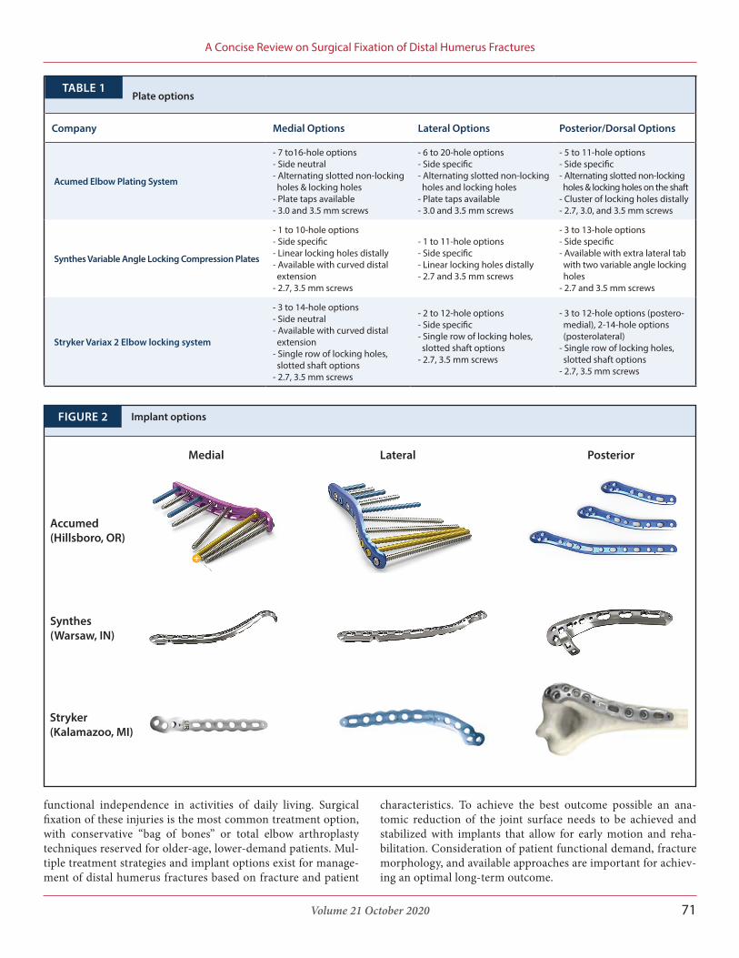
To address the specific needs of distal humerus fractures, sever-al implants are available. Most companies offer some combination of medial, lateral, and dorsal-specific plating with locking options. Locking options may be favored, particularly in osteoporotic bone to aid in resistance to torsional loads.24,25 These options are sum-marized in Table 1 by company with example images in Figure 2.
CONCLUSION
In summary, distal humerus fractures are an increasingly common injury that has significant implications for patient’s
(A)Parallel plating (B) Orthogonal platingFIGURE 1
A. B.
Volume 21 October 2020 71
A Concise Review on Surgical Fixation of Distal Humerus Fractures
functional independence in activities of daily living. Surgical fixation of these injuries is the most common treatment option, with conservative “bag of bones” or total elbow arthroplasty techniques reserved for older-age, lower-demand patients. Mul-tiple treatment strategies and implant options exist for manage-ment of distal humerus fractures based on fracture and patient
characteristics. To achieve the best outcome possible an ana-tomic reduction of the joint surface needs to be achieved and stabilized with implants that allow for early motion and reha-bilitation. Consideration of patient functional demand, fracture morphology, and available approaches are important for achiev-ing an optimal long-term outcome.
Company Medial Options Lateral Options Posterior/Dorsal Options
Acumed Elbow Plating System
- 7 to16-hole options- Side neutral- Alternating slotted non-locking holes & locking holes- Plate taps available- 3.0 and 3.5 mm screws
- 6 to 20-hole options- Side specific- Alternating slotted non-locking holes and locking holes- Plate taps available- 3.0 and 3.5 mm screws
- 5 to 11-hole options- Side specific- Alternating slotted non-locking holes & locking holes on the shaft- Cluster of locking holes distally- 2.7, 3.0, and 3.5 mm screws
Synthes Variable Angle Locking Compression Plates
- 1 to 10-hole options- Side specific- Linear locking holes distally- Available with curved distal extension- 2.7, 3.5 mm screws
- 1 to 11-hole options- Side specific- Linear locking holes distally- 2.7 and 3.5 mm screws
- 3 to 13-hole options- Side specific- Available with extra lateral tab with two variable angle locking holes- 2.7 and 3.5 mm screws
Stryker Variax 2 Elbow locking system
- 3 to 14-hole options- Side neutral- Available with curved distal extension- Single row of locking holes, slotted shaft options- 2.7, 3.5 mm screws
- 2 to 12-hole options- Side specific- Single row of locking holes, slotted shaft options- 2.7, 3.5 mm screws
- 3 to 12-hole options (postero- medial), 2-14-hole options (posterolateral)- Single row of locking holes, slotted shaft options- 2.7, 3.5 mm screws
Plate optionsTABLE 1
Implant optionsFIGURE 2
Medial Lateral Posterior
Accumed(Hillsboro, OR)
Synthes(Warsaw, IN)
Stryker(Kalamazoo, MI)

THE ORTHOPAEDIC JOURNAL AT HARVARD MEDICAL SCHOOL72
Xiong et al.
REFERENCES
1. Mahabier KC, Hartog DD, Van Veldhuizen J, et al. Trends in inci-dence rate, health care consumption, and costs for patients admitted with a humeral fracture in The Netherlands between 1986 and 2012. Injury. 2015 Oct;46(10):1930-7. doi: 10.1016/j.injury.2015.07.025.
2. Court-Brown CM, Duckworth AD, Clement ND, McQueen MM. Fractures in older adults. A view of the future? Injury. 2018 Dec;49(12):2161-2166. doi: 10.1016/j.injury.2018.11.009.
3. Robinson CM, Hill RM, Jacobs N, Dall G, Court-Brown CM. Adult distal humeral metaphyseal fractures: epidemiology and results of treatment. J Orthop Trauma. 2003 Jan;17(1):38-47. doi: 10.1097/00005131-200301000-00006.
4. Charissoux JL, Vergnenegre G, Pelissier M, Fabre T, Mansat P, Sofcot. Epidemiology of distal humerus fractures in the elderly. Or-thop Traumatol Surg Res. 2013 Nov;99(7):765-9. doi: 10.1016/j.otsr.2013.08.002. Epub 2013 Sep 26.
5. Anglen J. Distal humerus fractures. J Am Acad Orthop Surg. 2005 Sep;13(5):291-7. doi: 10.5435/00124635-200509000-00001.
6. Varecka TF, Myeroff C. Distal Humerus Fractures in the Elderly Population.J Am Acad Orthop Surg. 2017 Oct;25(10):673-683. doi: 10.5435/JAAOS-D-15-00683.
7. Bhashyam AR, Jupiter JB. Revision Fixation of Distal Humerus Fracture Nonunions in Older Age Patients with Poor Bone Quality or Bone Loss - Is This Viable as a Long-term Treatment Option? Arch Bone Jt Surg. 2019 May;7(3):251-257.
8. O'Driscoll SW. Optimizing stability in distal humeral fracture fixation. J Shoulder Elbow Surg. Jan-Feb 2005;14(1 Suppl S):186S-194S. doi: 10.1016/j.jse.2004.09.033.
9. Jupiter JB, Mehne DK. Fractures of the distal humerus. Orthope-dics. 1992 Jul;15(7):825-33.
10. Wainwright AM, Williams JR, Carr AJ. Interobserver and in-traobserver variation in classification systems for fractures of the distal humerus. J Bone Joint Surg Br. 2000 Jul;82(5):636-42. doi: 10.1302/0301-620x.82b5.9673.
11. Milch H. Fractures and Fracture Dislocations of the Humeral Condyles. J Trauma. 1964 Sep;4:592-607. doi: 10.1097/00005373-196409000-00004.
12. Aitken SA, Jenkins PJ, Rymaszewski L. Revisiting the 'bag of bones': functional outcome after the conservative manage-ment of a fracture of the distal humerus. Bone Joint J. 2015 Aug;97-B(8):1132-8. doi: 10.1302/0301-620X.97B8.35410.
13. Desloges W, Faber KJ, King GJ, Athwal GS. Functional out-comes of distal humeral fractures managed nonoperatively in med-ically unwell and lower-demand elderly patients. J Shoulder Elbow Surg. 2015 Aug;24(8):1187-96. doi: 10.1016/j.jse.2015.05.032.
14. Choo A, Ramsey ML. Total elbow arthroplasty: current options. J Am Acad Orthop Surg. 2013 Jul;21(7):427-37. doi: 10.5435/JAAOS-21-07-427.
15. Sanchez-Sotelo J, Morrey BF. Total elbow arthroplasty. J Am Acad Orthop Surg. 2011 Feb;19(2):121-5. doi: 10.5435/00124635-201102000-00007.
16. Guitton TG, Ring D. Three-dimensional computed tomo-graphic imaging and modeling in the upper extremity. Hand Clin. 2010 Aug;26(3):447-53, viii. doi: 10.1016/j.hcl.2010.04.007. Epub 2010 Jun 11.
17. O'Driscoll SW, Sanchez-Sotelo J, Torchia ME. Management of the smashed distal humerus. Orthop Clin North Am. 2002 Jan;33(1):19-33, vii. doi: 10.1016/s0030-5898(03)00070-1.
18. Dakoure PW, Ndiaye A, Ndoye JM, et al. Posterior surgical approaches to the elbow: a simple method of comparison of the articular exposure. Surg Radiol Anat. 2007 Dec;29(8):671-4. doi: 10.1007/s00276-007-0263-8. Epub 2007 Oct 19.
19. Wilkinson JM, Stanley D. Posterior surgical approaches to the elbow: a comparative anatomic study. J Shoulder Elbow Surg. Jul-Aug 2001;10(4):380-2. doi: 10.1067/mse.2001.116517.
20. O'Driscoll SW. The triceps-reflecting anconeus pedicle (TRAP) approach for distal humeral fractures and nonunions. Orthop Clin North Am. 2000 Jan;31(1):91-101. doi: 10.1016/s0030-5898(05)70130-9.
21. Chou YC, Hsu YH, Yu YH, Wu CC. Triceps-reflecting ancone-us pedicle approach with double precontoured locking plate fixa-tion is efficient in the treatment of orthopaedic trauma association type C distal humerus fracture. Injury. 2016 Oct;47(10):2240-2246. doi: 10.1016/j.injury.2016.06.036. Epub 2016 Jul 5.
22. Shih CA, Su WR, Lin WC, Tai TW. Parallel versus orthogonal plate osteosynthesis of adult distal humerus fractures: a meta-anal-ysis of biomechanical studies. Int Orthop. 2019 Feb;43(2):449-460. doi: 10.1007/s00264-018-3937-4. Epub 2018 Apr 20.
23. Yu X, Xie L, Wang J, Chen C, Zhang C, Zheng W. Orthogonal plating method versus parallel plating method in the treatment of distal humerus fracture: A systematic review and meta-analy-sis. Int J Surg. 2019 Sep;69:49-60. doi: 10.1016/j.ijsu.2019.07.028. Epub 2019 Jul 27.
24. Gardner MJ, Griffith MH, Demetrakopoulos D, et al. Hybrid locked plating of osteoporotic fractures of the humerus. J Bone Joint Surg Am. 2006 Sep;88(9):1962-7. doi: 10.2106/JBJS.E.00893.
25. Freeman AL, Tornetta P, 3rd, Schmidt A, Bechtold J, Ricci W, Fleming M. How much do locked screws add to the fixation of "hybrid" plate constructs in osteoporotic bone? J Orthop Trauma. 2010 Mar;24(3):163-9. doi: 10.1097/BOT.0b013e3181d35c29.

![DERLEME Distal humerus kaynamamalarıdergi.totbid.org.tr/20176/totbid.dergisi.2017.69.pdf · ilerlemesi ile karmaşık distal humerus kırık-larının stabil tespitine[1] rağmen](https://img.dokumen.tips/doc/110x75/5e42168b70e2a2311d559499/derleme-distal-humerus-kaynamamalardergi-ilerlemesi-ile-karmak-distal-humerus.jpg)