Embed Size (px)
Citation preview
ORIGINAL ARTICLE
Retrospective analysis of extra-articular distal humerus shaftfractures treated with the use of pre-contoured lateral columnmetaphyseal LCP by triceps-sparing posterolateral approach
Yatinder Kharbanda1 • Yashwant Singh Tanwar1 • Vishal Srivastava2 •
Vikas Birla1 • Ashok Rajput2 • Ramsagar Pandit1
Received: 25 August 2015 / Accepted: 21 October 2016 / Published online: 3 November 2016
� The Author(s) 2016. This article is published with open access at Springerlink.com
Abstract Management of extra-articular distal humerus
fractures presents a challenge to the treating surgeon due to
the complex anatomy of the distal part of the humerus and
complicated fracture morphology. Although surgical
treatment has shown to provide a more stable reduction and
alignment and predictable return to function, it has been
associated with complications like iatrogenic radial nerve
palsy, infection, non-union and Implant failure. We in the
present series retrospectively analysed 20 patients with
extra-articular distal humerus shaft fractures surgically
treated using the extra-articular distal humeral locking
plate approached by the triceps-sparing posterolateral
approach. The outcome was assessed using the DASH
score, range of motion at the elbow and the time to union.
The mean time to radiographic fracture union was
12 weeks.
Keywords Distal humerus fracture � Extra-articular distalhumerus LCP � Posterolateral approach humerus
Introduction
Extra-articular fractures of the distal humeral shaft are
relatively rare injuries and have been in the limelight
owing to a higher incidence of radial nerve injuries, as well
as the dilemmas surrounding their management [1, 2]. Both
conservative and surgical treatment options exist for these
fractures, with the ideal treatment still being debatable.
Bracing has been an acceptable option for humeral shaft
fractures; however, in the distal third of the humerus in
adults it can cause problems owing to difficulty in con-
trolling angulation. Sarmiento reported his results of
functional bracing for comminuted extra-articular fractures
of the distal third humerus. There was varus deformity
averaging 9 degrees in 81% of patients, but loss of range of
movement was minimal and functional results were good
[3]. However, O‘Driscoll et al. [4] showed that cubitus
varus deformity secondary to supracondylar malunion or
congenital deformity of the distal part of the humerus may
not always be a benign condition and may have important
long-term clinical implications including tardy posterolat-
eral instability.
Although surgical treatment seems to provide a more
reliable and predictable alignment and potentially quicker
return of function, iatrogenic radial nerve palsy is a
cause of major concern [5]. If the decision to proceed to
surgical intervention has been made, then plate
osteosynthesis is the usual standard option [6]. The
classical teaching for fixation of a humeral shaft fracture
has been with a narrow/broad 4.5 mm low-contact
dynamic compression plate, purchasing a minimum of
eight cortices (i.e. 4 screws) on either side of the fracture
zone or at least six cortices (3 screws) on either side if a
lag screw has been used [6]. This, however, becomes
difficult to achieve in distal humeral shaft fractures
owing to the limited space available distally, as well as
the curved shape of the distal humerus when approaching
anteriorly and the presence of the olecranon fossa pos-
teriorly (Fig. 1). Double-column plating using two 3.5-
mm plates in orthogonal or parallel patterns is another
& Yashwant Singh Tanwar
1 Department of Orthopedics, Apollo Hospital, HNo299,
Pocket B, DDA Flats, Sarita Vihar, New Delhi,
Delhi 110076, India
2 Department of Orthopedics, Dr. RML Hospital and PGIMER,
New Delhi, Delhi 110001, India
123
Strat Traum Limb Recon (2017) 12:1–9
DOI 10.1007/s11751-016-0270-6

option [7], but it requires greater soft tissue stripping and
exposure, leading to a potentially higher non-union and
infection rate and elbow stiffness reported in some series
[5, 8].
In the present retrospective case series, we present our
clinical experience with use of a single column pre-con-
toured extra-articular distal humeral locking compression
plate (J plate Synthes, Solothurn, Switzerland) for treat-
ment of extra-articular distal humeral fractures. It was a
retrospective study aimed to evaluate the clinical and
radiographic results after fixation of fractures of the distal
humerus shaft with this single column system.
Materials and methods
Implant
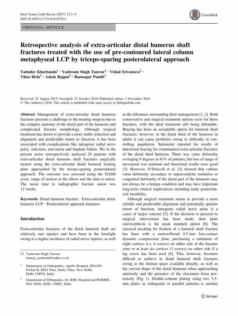
3.5-mm LCP (Locking Compression Plate) extra-articular
distal humerus plate (AO Synthes) is an anatomically
shaped and angular stable fixation system for extra-artic-
ular fractures of the distal humerus. Distally, the plate
accepts five 3.5-mm locking screws and is tapered to
minimize soft tissue irritation and the screw hole density is
greater to allow larger number of screws to be placed in the
distal fragment (Fig. 2). The two most distal screw holes
are angled towards the capitellum and trochlea, which
allows longer locking screws to be placed distally. Proxi-
mally, the thickness of the plate is based on LCP 4.5/5.0,
narrow and has combi-holes. Locking screws create a
fixed-angle construct, providing angular stability, whereas
the combi-holes can be used to provide inter-fragmentary
or dynamic axial compression. As the plates are anatomi-
cally contoured, there are different plates for the right and
left sides and it is available from 4 hole (122 mm) to 14
(302 mm) hole length.
Patients
Between Sept 2010 to Feb 2013, 20 patients with meta-
physeal extra-articular distal humerus fractures—AO Type
12 A/B/C—were treated at our institution using the
EADHP (Table 1). Inclusion criteria for the patients were:
fractures of the distal humeral shaft which could not be
fixed with conventional LCDCP’s with minimum of six/
eight cortices distally, age[18 years, closed fractures of
the distal humeral shaft, with or without radial nerve palsy,
recent fractures and non-unions. Patients who did not sat-
isfy these inclusion criteria were not included in the study.
All the surgeries were performed by the same senior author
(YK) at one institution only.
Clinical outcome was assessed using Disabilities of the
Arm, Shoulder and Hand (DASH) score and the range of
motion of the elbow joint for each patient. The union was
assessed clinically and radiologically; clinically by absence
of pain and tenderness on palpation and range of motion at
elbow joint, ability to perform activities of daily living
without pain. Anteroposterior and lateral radiographs were
done, and the healing progress of the distal humerus frac-
ture was assessed. Union was defined by the absence of
fracture line or bridging of the fracture site on at least 3 of
the 4 cortices and the absence of implant loosening or
failure.
Surgical technique
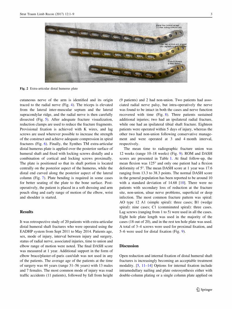
Patient is placed in the lateral position under general
anaesthesia, with the arm hanging by the side. A triceps-
reflecting posterolateral approach of Gerwin et al. [9] is
utilized to expose the fracture site. After performing a
midline skin incision on the posterior aspect of arm, full
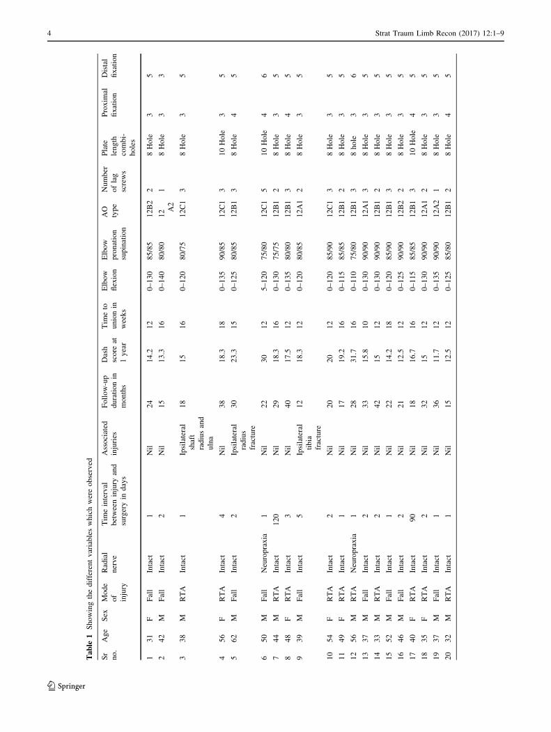
thickness flaps are developed on the lateral side (Fig. 3).
On the lateral side, using blunt dissection, the lower lateral
Fig. 1 Showing AP and lateral views of X-rays with low distal humeral ‘‘extra articular’’ fracture
2 Strat Traum Limb Recon (2017) 12:1–9
123
cutaneous nerve of the arm is identified and its origin
traced to the radial nerve (Fig. 4). The triceps is elevated
from the lateral inter-muscular septum and the lateral
supracondylar ridge, and the radial nerve is then carefully
dissected (Fig. 5). After adequate fracture visualization,
reduction clamps are used to reduce the fracture fragments.
Provisional fixation is achieved with K wires, and lag
screws are used wherever possible to increase the strength
of the construct and achieve adequate compression in spiral
fractures (Fig. 6). Finally, the Synthes TM extra-articular
distal humerus plate is applied over the posterior surface of
humeral shaft and fixed with locking screws distally and a
combination of cortical and locking screws proximally.
The plate is positioned so that its shaft portion is located
centrally on the posterior aspect of the humerus, while the
distal end curved along the posterior aspect of the lateral
column (Fig. 7). Plate bending is required in some cases
for better seating of the plate to the bone surface. Post-
operatively, the patient is placed in a soft dressing and arm
pouch sling and early range of motion of the elbow, wrist
and shoulder is started.
Results
It was retrospective study of 20 patients with extra-articular
distal humeral shaft fractures who were operated using the
EADHP system from Sept 2011 to May 2014. Patients age,
sex, mode of injury, interval between injury and surgery,
status of radial nerve, associated injuries, time to union and
elbow range of motion were noted. The final DASH score
was measured at 1 year. Additional support in the form of
elbow brace/plaster-of-paris cast/slab was not used in any
of the patients. The average age of the patients at the time
of surgery was 44 years (range 31–56 years) with 13 males
and 7 females. The most common mode of injury was road
traffic accidents (11 patients), followed by fall from height
(9 patients) and 2 had non-union. Two patients had asso-
ciated radial nerve palsy, but intra-operatively the nerve
was found to be intact in both the cases and nerve function
recovered with time (Fig. 8). Three patients sustained
additional injuries; two had an ipsilateral radial fracture,
while one had an ipsilateral tibial shaft fracture. Eighteen
patients were operated within 5 days of injury, whereas the
other two had non-union following conservative manage-
ment and were operated at 3 and 4 month interval,
respectively.
The mean time to radiographic fracture union was
12 weeks (range 10–18 weeks) (Fig. 9). ROM and DASH
scores are presented in Table 1. At final follow-up, the
mean flexion was 125� and only one patient had a flexion
deformity of 5�. The mean DASH score at 1 year was 17.6
ranging from 13.3 to 38.3 points. The normal DASH score
in the general population has been reported to be around 10
with a standard deviation of 14.68 [10]. There were no
patients with secondary loss of reduction at the fracture
site, non-union, ulnar nerve problems, superficial or deep
infection. The most common fracture pattern was spiral:
AO type 12 A1 (simple spiral): three cases; B1 (wedge
spiral): nine cases; C1 (comminuted spiral): three cases.
Lag screws (ranging from 1 to 5) were used in all the cases.
Eight hole plate length was used in the majority of the
cases (18 out of 20), and in the rest ten hole plate was used.
A total of 3–4 screws were used for proximal fixation, and
5–6 were used for distal fixation (Fig. 9).
Discussion
Open reduction and internal fixation of distal humeral shaft
fractures is increasingly becoming an acceptable treatment
modality. [5, 11–14] Options for internal fixation include
intramedullary nailing and plate osteosynthesis either with
double-column plating or a single column plate applied on
Fig. 2 Extra-articular distal humerus plate
Strat Traum Limb Recon (2017) 12:1–9 3
123
Table
1Showingthedifferentvariableswhichwereobserved
Sr
no.
Age
Sex
Mode
of
injury
Radial
nerve
Tim
einterval
betweeninjury
and
surgeryin
days
Associated
injuries
Follow-up
durationin
months
Dash
score
at
1year
Tim
eto
unionin
weeks
Elbow
flexion
Elbow
pronation
supination
AO
type
Number
oflag
screws
Plate
length
combi-
holes
Proxim
al
fixation
Distal
fixation
131
FFall
Intact
1Nil
24
14.2
12
0–130
85/85
12B2
28Hole
35
242
MFall
Intact
2Nil
15
13.3
16
0–140
80/80
12 A2
18Hole
33
338
MRTA
Intact
1Ipsilateral
shaft
radiusand
ulna
18
15
16
0–120
80/75
12C1
38Hole
35
456
FRTA
Intact
4Nil
38
18.3
18
0–135
90/85
12C1
310Hole
35
562
MFall
Intact
2Ipsilateral
radius
fracture
30
23.3
15
0–125
80/85
12B1
38Hole
45
650
MFall
Neuropraxia
1Nil
22
30
12
5–120
75/80
12C1
510Hole
46
744
MRTA
Intact
120
Nil
29
18.3
16
0–130
75/75
12B1
28Hole
35
848
FRTA
Intact
3Nil
40
17.5
12
0–135
80/80
12B1
38Hole
45
939
MFall
Intact
5Ipsilateral
tibia
fracture
12
18.3
12
0–120
80/85
12A1
28Hole
35
10
54
FRTA
Intact
2Nil
20
20
12
0–120
85/90
12C1
38Hole
35
11
49
FRTA
Intact
1Nil
17
19.2
16
0–115
85/85
12B1
28Hole
35
12
56
MRTA
Neuropraxia
1Nil
28
31.7
16
0–110
75/80
12B1
38hole
36
13
37
MFall
Intact
2Nil
33
15.8
10
0–130
90/90
12A1
38Hole
35
14
33
MRTA
Intact
2Nil
42
15
12
0–130
90/90
12B1
28Hole
35
15
52
MFall
Intact
1Nil
22
14.2
18
0–120
85/90
12B1
38Hole
35
16
46
MFall
Intact
2Nil
21
12.5
12
0–125
90/90
12B2
28Hole
35
17
40
FRTA
Intact
90
Nil
18
16.7
16
0–115
85/85
12B1
310Hole
45
18
35
FRTA
Intact
2Nil
32
15
12
0–130
90/90
12A1
28Hole
35
19
37
MFall
Intact
1Nil
36
11.7
12
0–135
90/90
12A2
18Hole
35
20
32
MRTA
Intact
1Nil
15
12.5
12
0–125
85/80
12B1
28Hole
45
4 Strat Traum Limb Recon (2017) 12:1–9
123
the posterior or posterolateral side. Biomechanical studies
have shown superior bending properties of humeral frac-
tures fixed with a plate and screw system versus intrame-
dullary devices. Also, the distal fragment is short and the
medullary canal is narrow, rendering it difficult to perform
nail osteosynthesis in distal third fractures [15].
Dual plating although offers a better biomechanical
strength [16] does so at the expense of greater soft tissue
dissection. It requires almost circumferential exposure of
both the medial and lateral column. Such an enormous soft
tissue dissection and exposure although justifiable for intra-
articular fractures seems unreasonable for extra-articular
shaft fractures. Preservation of the soft tissue envelope is
an important aspect in fracture healing, and it has led to the
change in the earlier concept of anatomic reduction and
rigid fixation [17]. This concept is no longer valid for most
of the extra-articular fractures with complex fracture pat-
terns, where minimal soft tissue dissection and stable fixa-
tion has shown to have better results and is now the
standard principle [18]. Although there have been no
comparative studies of dual column vs. single column
fixation for distal humerus fractures, we believe and sug-
gest that the higher infection and non-union rates quoted in
many series of distal humerus fractures may in part be due
to greater soft tissue dissection and a longer operative time
required for dual column plating [5, 8].
Yang et al. [18] also suggested that the excessive soft
tissue dissection required for dual plating may be respon-
sible for the increased incidence of iatrogenic radial nerve
palsy reported in some series. Placement of implant over
the distal medial aspect of humerus which has a scant soft
tissue cover also leads to a high incidence of implant-re-
lated complications such as ulnar neuropathy [19]. To
circumvent these problems, single column plating has been
suggested by many to be the answer. Standard single col-
umn plating techniques fail to achieve adequate stabiliza-
tion owing to many factors; the most important being
inadequate distal purchase. Levy et al. [20] used modified
Synthes Lateral Tibial Head Buttress Plate (Synthes, Paoli,
PA) that allowed for a centrally placed posterior plating of
the humeral shaft that angled anatomically along the lateral
column to treat far distal humeral shaft fractures.
The advent of modern locking plates has allowed
improved fixation of the peri-articular fractures. NumerousFig. 3 Midline skin incision and elevation of full thickness lateral
flap
Fig. 4 Lower lateral cutaneous nerve of the arm which can be traced proximally to the radial nerve
Strat Traum Limb Recon (2017) 12:1–9 5
123

studies have demonstrated and confirmed the increased
stability provided by locking plates at the distal femur,
proximal tibia, calcaneum, distal radius and proximal
humerus [21–25]. This increased strength of fixation has in
some cases obviated the need for dual column fixation.
Several studies have demonstrated that the mechanical
stability and overall stiffness of a laterally placed locked
plate in the proximal tibia is equivalent to the control of
historical dual plating [26–28].
The extra-articular distal humeral locking plate is based
on a similar concept of single column plating. Owing to
greater screw hole density distally, it allows the placement
of adequate number of screws in the distal fragment and the
locking construct increases the stability. Since only the
lateral column is exposed, it decreases both the soft tissue
dissection and the surgical time. As compared to the tro-
chlea, the posterior aspect of the lateral column is non-
articular and allows for posterior placement of implant
without risk of injury to the cartilage or risk of impinge-
ment with flexion and extension. We in the present series
used the posterolateral approach of Gerwin et al. [9] which
has several advantages over the traditional triceps splitting
approach. Sparing the triceps muscle limits the formation
of intramuscular adhesions and scar formation and theo-
retically reduces the chances of elbow contracture and
improves post-operative triceps function. The exposure can
Fig. 5 Elevation of the triceps from the lateral inter-muscular septum and radial nerve dissection
Fig. 6 Lag screw fixation
6 Strat Traum Limb Recon (2017) 12:1–9
123
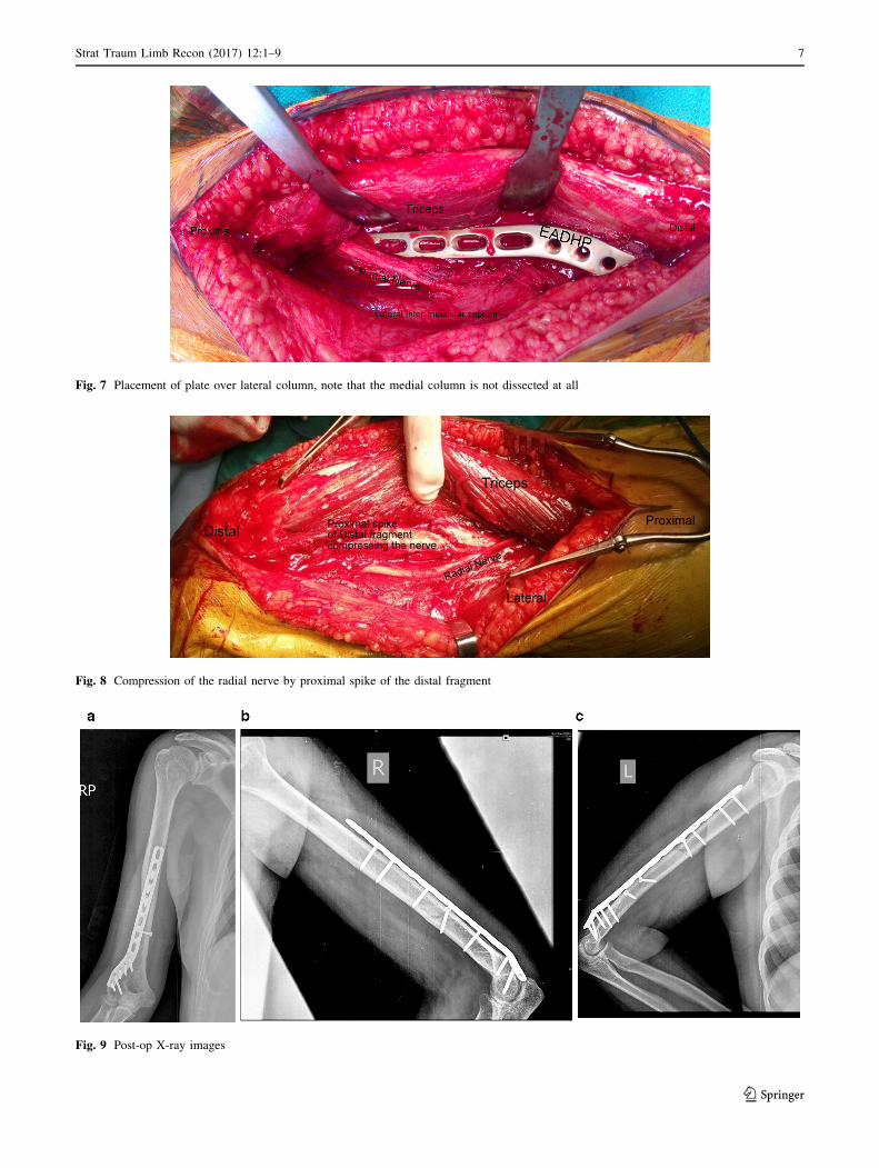
Fig. 7 Placement of plate over lateral column, note that the medial column is not dissected at all
Fig. 8 Compression of the radial nerve by proximal spike of the distal fragment
Fig. 9 Post-op X-ray images
Strat Traum Limb Recon (2017) 12:1–9 7
123
be extended proximally and distally; proximal extension is
by elevating the triceps off the humerus and mobilizing the
radial nerve, and distal extension can be accomplished by
converting the approach into an olecranon osteotomy
approach, TRAP approach [29] or Bryan and Morrey [30]
approach if there is an intra-articular extension of the shaft
fracture. Triceps-reflecting anconeus pedicle (TRAP)
approach involves complete detachment of triceps from
proximal ulna along with anconeus using sharp dissec-
tion. The entire flap is then lifted off the posterior aspect of
distal humerus. Lewisky, Sheppard and Ruth described
how the posterolateral approach can be extended proxi-
mally and distally to expose most of the posterior humeral
shaft and elbow joint for complex fracture treatment. They
described the combined olecranon osteotomy, lateral
paratricipital sparing and deltoid insertion splitting
(COLD) approach [31]. Approximately 94% of the hum-
eral diaphysis can be exposed with the posterolateral
approach (Fig. 6c) as compared to the triceps splitting
approach which provides exposure to only 76% of the shaft
[9]. This enhanced exposure also provides complete visu-
alization of the radial nerve on both sides of the inter-
muscular septum and since it exploits a relatively blood
less plane, this approach can be performed without a
tourniquet.
DASH score was used to assess the functional outcome.
This questionnaire asks the patient about symptoms as well
as their ability to perform certain activities. The questions
are answered based on the condition in the last week. If
patient did not have an opportunity to perform an activity
in the last week, the best estimate is made. It does not
matter which hand or arm is use to perform the activity.
The normal DASH score in the general population has been
reported to be around 10 with a standard deviation of 14.68
[10].
Our study has a few limitations, namely a small sample
size, and the lack of a biomechanical study to test and
compare the strength of a single column vs. double-column
locking plate.
As the plate is pre-contoured, it does not seat equally
well in all patients and bending the plate can potentially
damage the locking hole screw threads and can also change
the screw direction to a certain extent. Improperly locked
screws can compromise the stability of the construct, and
the change in screw direction can pose a problem in the
distal screws which are directed into the capitellum and
trochlea. To circumvent this problem, plate bending should
be done after blocking the screw holes with locking sleeves
and bending the plate only in between the screw holes.
Tejwani et al. [16] in their laboratory study demon-
strated that a double plating construct is stiffer than one
single-locking plate, especially in varus stress when the
medial column is absent. We, however, in our series of 20
patients did not encounter any patient with a comminuted
medial column; those who had so, also had some intra-
articular extension of the fracture and were treated by
conventional dual plating system. The increased stress
placed on a single (lateral) column fixation in the absence
or comminution of the other (medial) column leads to
increased strain over the implant at the fracture site, which
can lead to implant failure in absence of union. This can to
some extent be negated by using a longer plate with widely
spaced screws to increase the working length.
Conclusion
The EADHP system using the modified posterior approach
to the humerus is a useful treatment option for managing
extra-articular distal humerus fractures. The provision of
greater screw hole density of the plate distally and using
3.5-mm screws instead of 4.5 mm allows adequate number
of screws to be placed in the distal fragment. Bi-columnar
fixation of distal humerus provides increased stability, but
requires increased soft tissue dissection. EADHP fixation
of distal humerus fractures using the modified posterior
approach provides stable fracture fixation with adequate
exposure of the radial nerve and[90% of posterior hum-
eral shaft surface.
Compliance with ethical standards
Conflict of interest The authors declare that they have no conflict of
interest.
Ethical standards The study design was approved by the IRB and
conducted in accordance with the Declaration of Helsinki.
Informed consent A proper written and informed consent was taken
from all the patients.
Open Access This article is distributed under the terms of the
Creative Commons Attribution 4.0 International License (http://crea
tivecommons.org/licenses/by/4.0/), which permits unrestricted use,
distribution, and reproduction in any medium, provided you give
appropriate credit to the original author(s) and the source, provide a
link to the Creative Commons license, and indicate if changes were
made.
References
1. Horne G (1980) Supracondylar fractures of the humerus in adults.
J Trauma 20:71–74
2. Aitken GK, Rorabeck CH (1986) Distal humeral fractures in the
adult. Clin Orthop 207:191–197
3. Sarmiento A, Horowitch A, Aboulafia A, Vangsness CT Jr (1990)
Functional bracing for comminuted extra-articular fractures of
the distal third of the humerus. J Bone Joint Surg Br
72(2):283–287
4. O’Driscoll SW, Spinner RJ, McKee MD, Kibler WB, Hastings H
2nd, Morrey BF et al (2001) Tardy posterolateral rotatory
8 Strat Traum Limb Recon (2017) 12:1–9
123
instability of the elbow due to cubitus varus. J Bone Joint Surg
Am 83-A(9):1358–1369
5. Jawa A, McCarty P, Doornberg J, Harris M, Ring D (2006) Extra-
articular distal-third diaphyseal fractures of the humerus. A
comparison of functional bracing and plate fixation. J Bone Joint
Surg Am 88(11):2343–2347
6. McKee MD, Larsson S (2010) Humeral shaft fractures. In:
Bucholz RW, Court-Brown CM, Heckman JD, Tornetta P III
(eds) Rockwood and Green’s fractures in adults, 7th edn. Lip-
pincott William & Wilkins, Philadelphia, p 1015
7. Prasarn ML, Ahn J, Paul O, Morris EM, Kalandiak SP, Helfet DL
et al (2011) Dual plating for fractures of the distal third of the
humeral shaft. J Orthop Trauma 25:57–63
8. Paris H, Tropiano P, ClouetD’orval B, Chaudet H, Poitout DG
(2000) Fractures of the shaft of the humerus: systematic plate
fixation. Anatomic and functional results in 156 cases and a
review of the literature. Rev Chir Orthop Reparatrice Appar Mot
86(4):346–359
9. Gerwin M, Hotchkiss RN, Weiland AJ (1996) Alternative oper-
ative exposures of the posterior aspect of the humeral diaphysis
with reference to the radial nerve. J Bone Joint Surg Am
78(11):1690–1695
10. Hunsaker FG, Cioffi DA, Amadio PC, Wright JG, Caughlin B
(2002) The American academy of orthopaedic surgeon’s out-
comes instruments: normative values from the general popula-
tion. J Bone Joint Surg Am 84-A(2):208–215
11. Scolaro JA, Voleti P, Makani A, Namdari S, Mirza A, Mehta S
(2014) Surgical fixation of extra-articular distal humerus fractures
with a posterolateral plate through a triceps-reflecting technique.
J Shoulder Elbow Surg 23(2):251–257
12. Capo JT, Debkowska MP, Liporace F, Beutel BG, Melamed E
(2014) Outcomes of distal humerus diaphyseal injuries fixed with
a single-column anatomic plate. Int Orthop 38(5):1037–1043
13. Jawa A (2010) Treatment of distal diaphyseal humerus fractures.
J Hand Surg Am. 35(2):301–302
14. Meloy GM, Mormino MA, Siska PA, Tarkin IS (2013) A para-
digm shift in the surgical reconstruction of extra-articular distal
humeral fractures: single-column plating. Injury
44(11):1620–1624
15. Zimmerman MC, Waite AM, Deehan M, Tovey J, Oppenheim W
(1994) A biomechanical analysis of four humeral fracture fixation
systems. J Orthop Trauma 8(3):233–239
16. Tejwani NC, Murthy A, Park J, McLaurin TM, Egol KA, Kum-
mer FJ (2009) Fixation of extra-articular distal humerus fractures
using one locking plate versus two reconstruction plates: a lab-
oratory study. J Trauma 66(3):795–799
17. Thomas PR, Richard EB, Christopher GM (2007) AO philosophy
and evolution. In: Thomas PR, Richard EB, Christopher GM
(eds) AO principles of fracture management, 2nd edn. Thieme,
Stuttgart, pp 1–9
18. Yang Qing, Wang Fang, Wang Qiugen, Gao Wei, Huang Jianhua,
Xiaofeng Wu et al (2012) Surgical treatment of adult extra-
articular distal humeral diaphyseal fractures using an oblique
metaphyseal locking compression plate via a posterior approach.
Med Princ Pract 21:40–45
19. Chen RC, Harris DJ, Leduc S, Borrelli JJ Jr, Tornetta P III, Ricci
WM (2010) Is ulnar nerve transposition beneficial during open
reduction and internal fixation of distal humerus fractures?
J Orthop Trauma 24(7):391–394
20. Levy JC, Kalandiak SP, Hutson JJ, Zych G (2005) An alternative
method of osteosynthesis for distal humeral shaft fractures.
J Orthop Trauma 19(1):43–47
21. Liporace FA, Gupta S, Jeong GK, Stracher M, Kummer F, Egol
KA et al (2005) A biomechanical comparison of a dorsal 3.5-mm
T-plate and a volar fixed-angle plate in a model of dorsally
unstable distal radius fractures. J Orthop Trauma 19(3):187–191
22. Stoffel K, Booth G, Rohrl SM, Kuster M (2007) A comparison of
conventional versus locking plates in intraarticular calcaneus
fractures: a biomechanical study in human cadavers. Clin Bio-
mech (Bristol, Avon) 22:100–105
23. Weinstein DM, Bratton DR, Ciccone WJ 2nd, Elias JJ (2006)
Locking plates improve torsional resistance in the stabilization of
three-part proximal humeral fractures. J Shoulder Elbow Surg
15:239–243
24. Ratcliff JR, Werner FW, Green JK, Harley BJ (2007) Medial
buttress versus lateral locked plating in a cadaver medial tibial
plateau fracture model. J Orthop Trauma 21:444–448
25. Higgins TF, Pittman G, Hines J, Bachus KN (2007) Biome-
chanical analysis of distal femur fracture fixation: fixed-angle
screw-plate construct versus condylar blade plate. J Orthop
Trauma 21:43–46
26. Gosling T, Schandelmaier P, Marti A, Hufner T, Partenheimer A,
Krettek C (2004) Less invasive stabilization of complex tibial
plateau fractures: a biomechanical evaluation of a unilateral
locked screw plate and double plating. J Orthop Trauma
18(8):546–551
27. Mueller KL, Karunakar MA, Frankenburg EP, Scott DS (2003)
Bicondylar tibial plateau fractures: a biomechanical study. Clin
Orthop Relat Res 412:189–195
28. Egol KA, Su E, Tejwani NC, Sims SH, Kummer FJ, Koval KJ
(2004) Treatment of complex tibial plateau fractures using the
less invasive stabilization system plate: clinical experience and a
laboratory comparison with double plating. J Trauma
57(2):340–346
29. Shawn WO (2000) Driscoll: the tricep reflecting anconeus pedicle
(TRAP) approach for distal humerus fractures and nonunions.
Orthop Clin North Am 31(1):91–101
30. Bryan RS, Morrey BF (1982) Extensive posterior exposure of the
elbow: a triceps-sparing approach. Clin Orthop 166:188–192
31. Lewicky YM, Sheppard JE, Ruth JT (2007) The combined ole-
cranon osteotomy, lateral paratricipital sparing, deltoid insertion
splitting approach for concomitant distal intra-articular and
humeral shaft fractures. J Orthop Trauma 21(2):133–139
Strat Traum Limb Recon (2017) 12:1–9 9
123
![DERLEME Distal humerus kaynamamalarıdergi.totbid.org.tr/20176/totbid.dergisi.2017.69.pdf · ilerlemesi ile karmaşık distal humerus kırık-larının stabil tespitine[1] rağmen](https://img.dokumen.tips/doc/110x75/5e42168b70e2a2311d559499/derleme-distal-humerus-kaynamamalardergi-ilerlemesi-ile-karmak-distal-humerus.jpg)