Embed Size (px)
Citation preview

9-1
Chapter 9 DNA-Protein Interactions in BacteriaStudent learning outcomes:• Describe examples of structure /function
relationships in phage repressors• Appreciate that altered specificity repressors and
operator mutants clarify mechanisms of amino acid: base pair recognition
Impt. Figs. 1*, 2, 3, 4, 6, 7, 8,
14, 16, 17
Q: 1, 2, 3, 4, 5, 7, 10, 11
Cro binding DNA

9-2
The Family of Repressors: 434, P22
• Repressors have recognition helices that lie in major groove of appropriate operator
• Helix-turn-helix motif (HTH)• Specificity of bp binding
depends on amino acids in recognition helices
• Phages are not immune to super-infection by each other
Fig. 2

9-3
Binding Specificity of lambda-like Repressor: Operator DNA
Fig. 1
• Recognition helices fit sideways in major groove of operator DNA
• Certain amino acids on DNA side of recognition helix 2 make specific contact with bases in operator
• Contacts determine specificity of protein-DNA binding• ** Changing amino acids can change specificity of
repressor to different DNA sequence
9-4
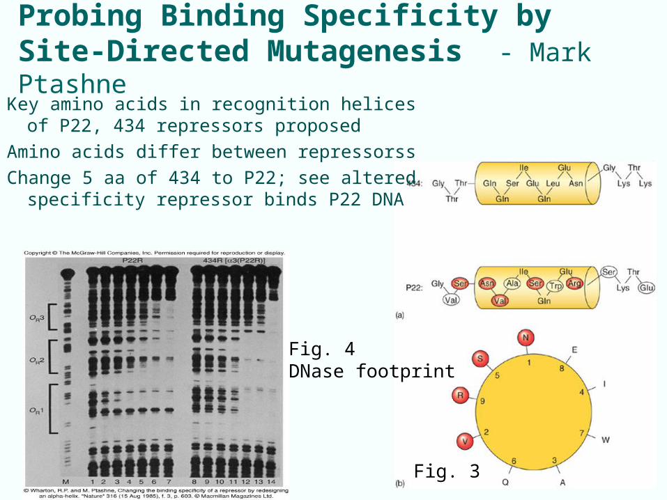
Probing Binding Specificity by Site-Directed Mutagenesis - Mark Ptashne
Fig. 3
Fig. 4DNase footprint
Key amino acids in recognition helices of P22, 434 repressors proposed
Amino acids differ between repressorss
Change 5 aa of 434 to P22; see altered specificity repressor binds P22 DNA

9-5
Repressor
repressor has extra motif, N-terminal arm that aids binding by embracing DNA
• Cro and repressors share affinity for same operators, but micro-specificities for OR1(l) or OR3 (cro)
• Specificities determined by interactions between different amino acids in recognition helices and different base pairs in operators
Fig. 4 repressor dimer on OR2
9-6
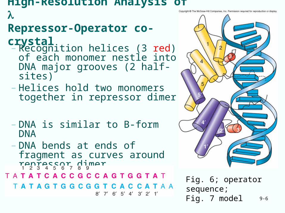
High-Resolution Analysis of Repressor-Operator co-crystal
– Recognition helices (3 red) of each monomer nestle into DNA major grooves (2 half-sites)
– Helices hold two monomers together in repressor dimer
– DNA is similar to B-form DNA– DNA bends at ends of fragment
as curves around repressor dimer
Fig. 6; operator sequence;Fig. 7 model
9-7
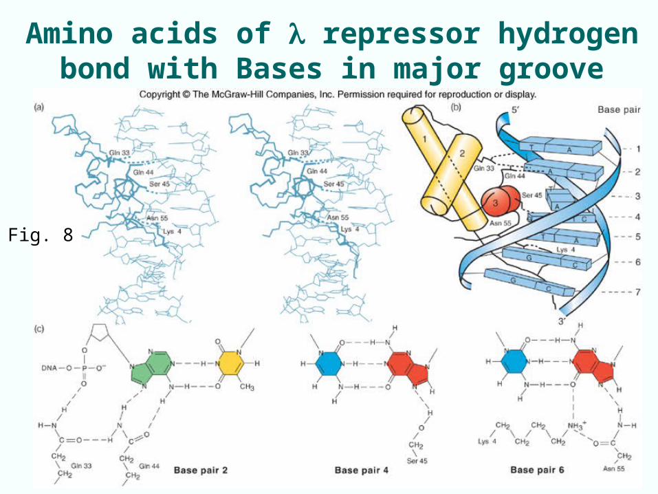
Amino acids of repressor hydrogen bond with Bases in major groove
Fig. 8

9-8
Amino Acid: DNA Backbone Interactions
• Hydrogen bond at Gln33 maximizes electrostatic attraction between positively charged amino end of -helix and negatively charged DNA
• Attraction works to stabilize bond
Fig. 9

9-9
High-Resolution Analysis of 434 Repressor-Operator Interactions
• Genetic and biochemical data predicted R-O contacts
• X-ray crystallography of 434 repressor-fragment/ operator-fragment shows H bonding at Gln residues in recognition helix to 3 bp in DNA
• Potential van der Waals contact between Gln29 and 5Me of T3
Fig. 10

9-10
Phage 434: Effects on DNA Conformation
• R-O complex DNA deviates from normal shape• DNA bends to accommodate base /aa contacts• Central part of helix is wound extra tightly• Outer parts are wound more loosely than normal• DNA sequence of operator facilitates bending
Fig. 11 Normal DNA;DNA bent by 434 repressor binding

9-11
9.2 trp Repressor and role of Tryptophan• trp repressor uses helix-turn-helix (HTH) DNA
binding motif to contact operator• Aporepressor is not active in binding DNA• Tryptophan forces recognition helices of trp repressor
dimer into proper position to bind trp operator
Fig. 12
9-12
9.3 General Considerations on Protein-DNA Interactions; multimeric proteins
• Specificity of binding between protein and specific stretch of DNA relates to:– Specific interactions between bases and amino acids– Ability of DNA to assume shape that directly relates to
DNA’s base sequence
• Target sites for DNA-binding proteins usually symmetric or repeated
• Most DNA-binding proteins are dimers: greatly enhances binding between DNA and protein as protein subunits bind cooperatively

9-13
Hydrogen Bonding Capabilities of Different Base Pairs
• Protein ‘reads the DNA’
• Different base pairs present four different hydrogen-bonding profiles to amino acids approaching either major or minor groove
Fig. 14

9-14
9.4 DNA-Binding Proteins: Action at a Distance
• DNA-binding proteins can influence interactions at remote sites in DNA – often looping intervening DNA
• Common in eukaryotes
• Occurs in several prokaryote systems:
lac operon multiple operators
ara operon looping
gal operon looping
repressor
9-15
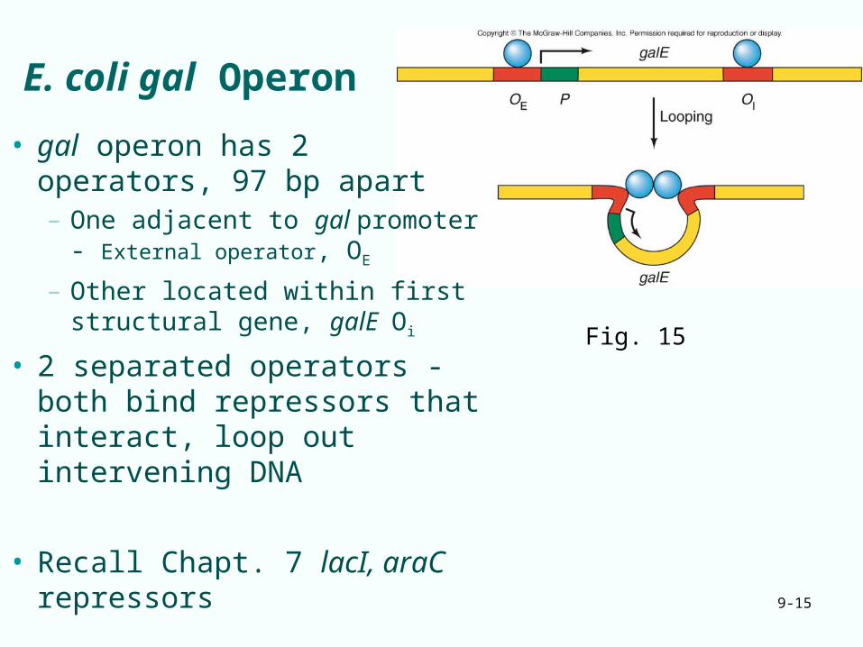
E. coli gal Operon
• gal operon has 2 operators, 97 bp apart– One adjacent to gal promoter -
External operator, OE
– Other located within first structural gene, galE Oi
• 2 separated operators - both bind repressors that interact, loop out intervening DNA
• Recall Chapt. 7 lacI, araC repressors
Fig. 15

9-16
DNA Looping affects DNase Susceptibility
Operators separated by – Integral number of double-
helical turns loop out DNA to allow cooperative binding
– Nonintegral number of turns requires proteins to bind opposite faces of DNA, no cooperative binding
Fig. 16
Fig. 17 repressor binds cooperatively to operators

9-17
Enhancers Enhancers are nonpromoter DNA elements that bind
protein factors and stimulate transcription– Can act at a distance– Originally found in eukaryotes (lots chapt. 12)– Recently found in prokaryotes. E. coli glnA gene:
• NtrC protein binds enhancer, • Binds RNAP 70 bp away• NtrC hydrolyzes ATP,
lets RPo form. • Insert 350 bp, see loop
Fig. 20NtrC: RNAP

Review questions
• 1. Draw rough diagram of helix-turn-helix domain interacting with DNA double helix
• 2. Describe experiment that shows which amino acids bind which base pairs in -like phage repressors.
• 10. Explain fact that protein oligomers (dimers, tetramers) bind better to DNA than monomeric proteins
9-18