Embed Size (px)
Citation preview
doi:10.1016/j.jmb.2007.09.081 J. Mol. Biol. (2007) 374, 1200–1212
Available online at www.sciencedirect.com
PIAS Proteins as Repressors of Oct4 Function
Elena Tolkunova1,3, Anna Malashicheva1,3, Vladimir N. Parfenov3,Claudio Sustmann2, Rudolf Grosschedl2 and Alexey Tomilin1,3⁎
1Department of DevelopmentalBiology, Max-Planck Institutefor Immunobiology, Stübeweg51, 79108 Freiburg, Germany2Department of Cellularand Molecular Immunology,Max-Planck Institute forImmunobiology, Stübeweg 51,79108 Freiburg, Germany3Institute of Cytology,Russian Academy of Science,Tikhoretski Ave. 4, 194064St-Petersburg, Russia
Received 28 May 2007;received in revised form9 August 2007;accepted 27 September 2007Available online3 October 2007
*Corresponding author. E-mail [email protected] used: SUMO, smal
modifier; PIAS, protein inhibitor ofprimordial germ cell; ES, embryonicepitope; tk, thymidine kinase; TE, trinner cell mass.
0022-2836/$ - see front matter © 2007 E
The POU domain transcription factor Oct4 plays essential functions in themaintenance of pluripotent embryonic and germ cells of mammals.Molecular mechanisms of Oct4 action remain poorly understood. To isolatemodulators of Oct4 activity, we performed a yeast two-hybrid screen withthe Oct4 POU domain as a bait and isolated PIASy as an Oct4-interactingprotein. Oct4 and PIASy interact in vivo via their POU domain and SAP-domain-containing N terminus, respectively. PIASy does not enhance Oct4sumoylation but acts as a potent inhibitor of Oct4-mediated transcriptionalactivation, sequestering Oct4 protein from the vicinity of Cajal bodies andsplicing speckles to the nuclear periphery. These modes of PIASy action areuncoupled from its sumoylation activity. Other PIAS family members,PIAS1 and PIAS3, can also interact with Oct4 in vivo and target Oct4 to thenuclear periphery, depending on cellular context. We propose that Oct4inhibition, mediated by this new class of transcriptional partners, might beinstrumental during mammalian development.
© 2007 Elsevier Ltd. All rights reserved.
Edited by M. Yaniv
Keywords: PIAS; POU; Oct4; SUMO; transcriptional repressionIntroduction
Development of multicellular organisms is char-acterized by an intricate series of genetic andepigenetic events that generate the complex adultbody from a single cell, the zygote. A refined andsophisticated regulatory network that is establishedduring embryogenesis relies on a relatively smallnumber of transcriptional regulators. The diversityin transcriptional control is achieved through acomplex network of combinatorial protein–proteinand protein–DNA interactions affecting the stabilityand subcellular and subnuclear localization of thesetranscriptional regulators. The primary structure ofcis-regulatory DNA elements, superimposed by
ess:
l ubiquitin-relatedactivated STAT; PGC,stem; MT, Mycophectoderm; ICM,
lsevier Ltd. All rights reserve
their epigenetic status, defines the composition andarchitecture of the transcriptional activation com-plexes that ultimately control gene expression in theappropriate temporospatial context of the develop-ing organism.The POU domain transcription factor Oct4 (also
called Oct3, Oct3/4, and Pou5f1) cloned 17 yearsago1–3 has a unique place in the array of transcrip-tional regulators because it is an indispensablecomponent of the core regulatory circuitry control-ling the self-renewal and maintenance of theundifferentiated state of early pluripotent cellswithin the epiblast, the primordial germ cells(PGCs), as well as of the cultured counterparts ofthese embryonic cell types, most notably theembryonic stem (ES) cells.4–10 Significant effort wasput into defining the regulatory network operated byOct4, as well as dissecting the molecular mechanismof its action. Increasing evidence suggests that Oct4does not activate transcription of target genes alonebut requires DNA-dependent heterodimerizationwith another DNA-binding transcription factor, theHMG-box protein Sox2. These two transcriptionfactors are found to cooperatively bind regulatory
d.
1201PIAS Proteins Inhibit Oct4
regions of a number of target genes, including theirownpromoters.10–14 The recruitment of another classof interacting partners, either DNA-binding tran-scription regulators or viral coactivators, have alsobeen described forOct4.15–17 In addition, aDNA site-dependent differential recruitment of transcriptionalcofactors by distinct types of dimers has beendescribed for the POU domain factors Pit1, Oct1,and Oct2;18–21 however, a biological role of Oct4dimerization has not been well characterized.Finally, no negative coregulators of Oct4 functionhave been reported to date.The family of PIAS proteins, initially described as
protein inhibitors of activated STATs, comprise atleast five genes and/or splice variants (PIAS1,PIAS3, PIASxα, PIASxβ, and PIASy). These pro-teins share the conserved N-terminal SAP domain,which confers the interaction with both nuclearmatrix-associated DNA sequences and the centralRING-like domain that is required for E3 smallubiquitin-related modifier (SUMO) ligase activity.PIAS proteins indeed have been reported topromote sumoylation of a number of proteintargets, affecting their stability, localization, andactivity, and hence are involved in diverse cellularprocesses. However, the major function of PIASfamily member proteins seems to modulate theactivity of transcription factors, which is frequentlyuncoupled from their sumoylating function.22,23 Itis currently believed that PIAS proteins operatethrough relocalization of target proteins to differentnuclear compartments. For instance, PIASy targetsthe Wnt/β-catenin pathway mediator LEF1 to PMLnuclear bodies, resulting in the repression of LEF1activity. Although PIASy can markedly stimulatesumoylation of LEF1, this modification is not re-quired either for the repression or for the relo-calization.24 Another PIAS family member, PIAS1,has been shown to relocalize the homeobox tran-scription factor Msx1 to the nuclear periphery,allowing Msx1 to engage and repress MyoD andMyf5 gene promoters. Again, this event does notrequire thePIAS1-dependent sumoylationofMsx1.25
In this paper we report a novel class of Oct4partners belonging to the PIAS family of transcrip-tional repressors and SUMO modifying enzymes:
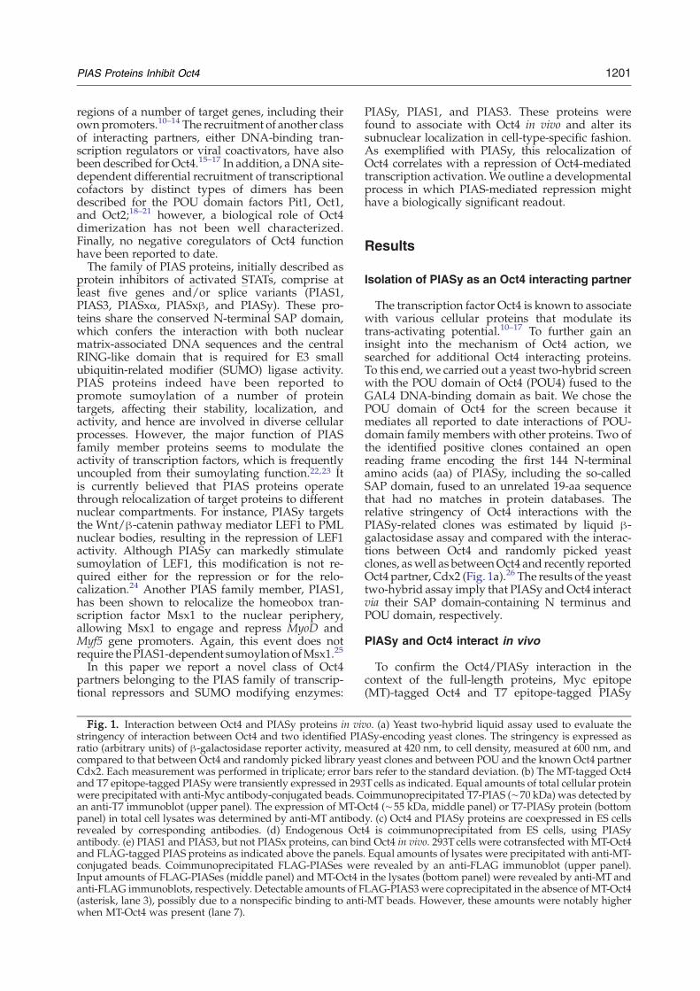
Fig. 1. Interaction between Oct4 and PIASy proteins in vivstringency of interaction between Oct4 and two identified PIAratio (arbitrary units) of β-galactosidase reporter activity, meacompared to that between Oct4 and randomly picked library yCdx2. Each measurement was performed in triplicate; error baand T7 epitope-tagged PIASy were transiently expressed in 293were precipitated with anti-Myc antibody-conjugated beads. Can anti-T7 immunoblot (upper panel). The expression of MT-Opanel) in total cell lysates was determined by anti-MT antibodrevealed by corresponding antibodies. (d) Endogenous Octantibody. (e) PIAS1 and PIAS3, but not PIASx proteins, can binand FLAG-tagged PIAS proteins as indicated above the panelsconjugated beads. Coimmunoprecipitated FLAG-PIASes werInput amounts of FLAG-PIASes (middle panel) and MT-Oct4 ianti-FLAG immunoblots, respectively. Detectable amounts of F(asterisk, lane 3), possibly due to a nonspecific binding to antiwhen MT-Oct4 was present (lane 7).
PIASy, PIAS1, and PIAS3. These proteins werefound to associate with Oct4 in vivo and alter itssubnuclear localization in cell-type-specific fashion.As exemplified with PIASy, this relocalization ofOct4 correlates with a repression of Oct4-mediatedtranscription activation. We outline a developmentalprocess in which PIAS-mediated repression mighthave a biologically significant readout.
Results
Isolation of PIASy as an Oct4 interacting partner
The transcription factor Oct4 is known to associatewith various cellular proteins that modulate itstrans-activating potential.10–17 To further gain aninsight into the mechanism of Oct4 action, wesearched for additional Oct4 interacting proteins.To this end, we carried out a yeast two-hybrid screenwith the POU domain of Oct4 (POU4) fused to theGAL4 DNA-binding domain as bait. We chose thePOU domain of Oct4 for the screen because itmediates all reported to date interactions of POU-domain family members with other proteins. Two ofthe identified positive clones contained an openreading frame encoding the first 144 N-terminalamino acids (aa) of PIASy, including the so-calledSAP domain, fused to an unrelated 19-aa sequencethat had no matches in protein databases. Therelative stringency of Oct4 interactions with thePIASy-related clones was estimated by liquid β-galactosidase assay and compared with the interac-tions between Oct4 and randomly picked yeastclones, aswell as betweenOct4 and recently reportedOct4 partner, Cdx2 (Fig. 1a).26 The results of the yeasttwo-hybrid assay imply that PIASy andOct4 interactvia their SAP domain-containing N terminus andPOU domain, respectively.
PIASy and Oct4 interact in vivo
To confirm the Oct4/PIASy interaction in thecontext of the full-length proteins, Myc epitope(MT)-tagged Oct4 and T7 epitope-tagged PIASy
o. (a) Yeast two-hybrid liquid assay used to evaluate theSy-encoding yeast clones. The stringency is expressed assured at 420 nm, to cell density, measured at 600 nm, andeast clones and between POU and the known Oct4 partnerrs refer to the standard deviation. (b) The MT-tagged Oct4T cells as indicated. Equal amounts of total cellular proteinoimmunoprecipitated T7-PIAS (∼70 kDa) was detected byct4 (∼55 kDa, middle panel) or T7-PIASy protein (bottomy. (c) Oct4 and PIASy proteins are coexpressed in ES cells4 is coimmunoprecipitated from ES cells, using PIASyd Oct4 in vivo. 293T cells were cotransfected with MT-Oct4. Equal amounts of lysates were precipitated with anti-MT-e revealed by an anti-FLAG immunoblot (upper panel).n the lysates (bottom panel) were revealed by anti-MT andLAG-PIAS3 were coprecipitated in the absence of MT-Oct4-MT beads. However, these amounts were notably higher
1202 PIAS Proteins Inhibit Oct4
were analyzed by coimmunoprecipitation assay intransfected 293T cells. Towards this end, we coim-munoprecipitated T7-PIASy with antibodies direc-ted against MT (Fig. 1b). The coimmunoprecipitatedPIASy was detected by immunoblot analysis withanti-T7 antibody. The interaction between full-lengthOct4 and PIASy proteinswas additionally confirmed
Fig. 1 (legend on
by a reciprocal coimmunoprecipitation assay (seebelow).Next we examined the interaction of endogenous
Oct4 and PIASy. First, we found that PIASy isexpressed in ES cells at a relatively high levelcompared to COS7 cells (Fig. 1c). Subsequently, wewere able to coimmunoprecipitate the endogenous
previous page)
1203PIAS Proteins Inhibit Oct4
Oct4 from ES cell lysate, using anti-PIASy antibody(Fig. 1d), suggesting that these two proteins interactin vivo in the context of physiological expressionlevels.Because PIASy is a member of a highly conserved
protein family that additionally comprises PIAS1,PIAS3, and PIASxα/β, we assessed whether Oct4can interact with other family members as well.Plasmids expressing different PIAS proteins taggedwith the FLAG epitope were transfected into 293Tcells, alone or together with MT-Oct4. Coimmuno-precipitation with anti-MT antibody and anti-FLAGimmunoblot analysis resulted in the detection ofPIAS1 and PIAS3, but not of PIASxα and β (Fig. 1e).
PIASy does not act as SUMO ligase for Oct4
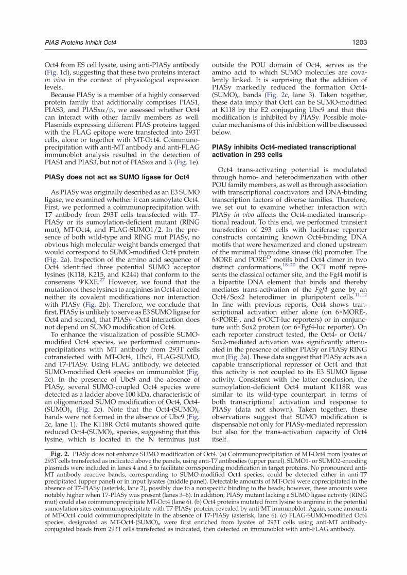
As PIASywas originally described as an E3 SUMOligase, we examined whether it can sumoylate Oct4.First, we performed a coimmunoprecipitation withT7 antibody from 293T cells transfected with T7-PIASy or its sumoylation-deficient mutant (RINGmut), MT-Oct4, and FLAG-SUMO1/2. In the pre-sence of both wild-type and RING mut PIASy, noobvious high molecular weight bands emerged thatwould correspond to SUMO-modified Oct4 protein(Fig. 2a). Inspection of the amino acid sequence ofOct4 identified three potential SUMO acceptorlysines (K118, K215, and K244) that conform to theconsensus ΨKXE.27 However, we found that themutation of these lysines to arginines inOct4 affectedneither its covalent modifications nor interactionwith PIASy (Fig. 2b). Therefore, we conclude thatfirst, PIASy is unlikely to serve as E3 SUMO ligase forOct4 and second, that PIASy–Oct4 interaction doesnot depend on SUMO modification of Oct4.To enhance the visualization of possible SUMO-
modified Oct4 species, we performed coimmuno-precipitations with MT antibody from 293T cellscotransfected with MT-Oct4, Ubc9, FLAG-SUMO,and T7-PIASy. Using FLAG antibody, we detectedSUMO-modified Oct4 species on immunoblot (Fig.2c). In the presence of Ubc9 and the absence ofPIASy, several SUMO-coupled Oct4 species weredetected as a ladder above 100 kDa, characteristic ofan oligomerized SUMO modification of Oct4, Oct4-(SUMO)n (Fig. 2c). Note that the Oct4-(SUMO)nbands were not formed in the absence of Ubc9 (Fig.2c, lane 1). The K118R Oct4 mutants showed quitereduced Oct4-(SUMO)n species, suggesting that thislysine, which is located in the N terminus just
Fig. 2. PIASy does not enhance SUMO modification of Oc293Tcells transfected as indicated above the panels, using anti-plasmids were included in lanes 4 and 5 to facilitate corresponMT antibody reactive bands, corresponding to SUMO-modprecipitated (upper panel) or in input lysates (middle panel). Dabsence of T7-PIASy (asterisk, lane 2), possibly due to a nonspnotably higher when T7-PIASy was present (lanes 3–6). In addmut) could also coimmunoprecipitate MT-Oct4 (lane 6). (b) Ocsumoylation sites coimmunoprecipitate with T7-PIASy proteinof MT-Oct4 could coimmunoprecipitate in the absence of T7species, designated as MT-Oct4-(SUMO)n were first enrichconjugated beads from 293T cells transfected as indicated, the
outside the POU domain of Oct4, serves as theamino acid to which SUMO molecules are cova-lently linked. It is surprising that the addition ofPIASy markedly reduced the formation Oct4-(SUMO)n bands (Fig. 2c, lane 3). Taken together,these data imply that Oct4 can be SUMO-modifiedat K118 by the E2 conjugating Ubc9 and that thismodification is inhibited by PIASy. Possible mole-cular mechanisms of this inhibition will be discussedbelow.
PIASy inhibits Oct4-mediated transcriptionalactivation in 293 cells
Oct4 trans-activating potential is modulatedthrough homo- and heterodimerization with otherPOU family members, as well as through associationwith transcriptional coactivators and DNA-bindingtranscription factors of diverse families. Therefore,we set out to examine whether interaction withPIASy in vivo affects the Oct4-mediated transcrip-tional readout. To this end, we performed transienttransfection of 293 cells with luciferase reporterconstructs containing known Oct4-binding DNAmotifs that were hexamerized and cloned upstreamof the minimal thymidine kinase (tk) promoter. TheMORE and PORED motifs bind Oct4 dimer in twodistinct conformations,18–20 the OCT motif repre-sents the classical octamer site, and the Fgf4 motif isa bipartite DNA element that binds and therebymediates trans-activation of the Fgf4 gene by anOct4/Sox2 heterodimer in pluripotent cells.11,12
In line with previous reports, Oct4 shows tran-scriptional activation either alone (on 6×MORE-,6×PORE-, and 6×OCT-luc reporters) or in conjunc-ture with Sox2 protein (on 6×Fgf4-luc reporter). Oneach reporter construct tested, the Oct4- or Oct4/Sox2-mediated activation was significantly attenu-ated in the presence of either PIASy or PIASy RINGmut (Fig. 3a). These data suggest that PIASy acts as acapable transcriptional repressor of Oct4 and thatthis activity is not coupled to its E3 SUMO ligaseactivity. Consistent with the latter conclusion, thesumoylation-deficient Oct4 mutant K118R wassimilar to its wild-type counterpart in terms ofboth transcriptional activation and response toPIASy (data not shown). Taken together, theseobservations suggest that SUMO modification isdispensable not only for PIASy-mediated repressionbut also for the trans-activation capacity of Oct4itself.
t4. (a) Coimmunoprecipitation of MT-Oct4 from lysates ofT7 antibodies (upper panel). SUMO1- or SUMO2-encodingding modification in target proteins. No pronounced anti-ified Oct4 species, could be detected either in anti-T7etectable amounts of MT-Oct4 were coprecipitated in theecific binding to the beads; however, these amounts wereition, PIASy mutant lacking a SUMO ligase activity (RINGt4 proteins mutated from lysine to arginine in the potential, revealed by anti-MT immunoblot. Again, some amounts-PIASy (asterisk, lane 6). (c) FLAG-SUMO-modified Oct4ed from lysates of 293T cells using anti-MT antibody-n detected on immunoblot with anti-FLAG antibody.

1204 PIAS Proteins Inhibit Oct4
As tested using the 6×MORE-luc reporter, theability to repress Oct4-mediated transcriptionalactivation in 293 cells is restricted to PIASy, as the
Fig. 2 (legend on
other known members of the PIAS family, includingthe established Oct4-interacting PIAS1 and PIAS3(see above), are neutral towards Oct4 (Fig. 3b).
previous page)
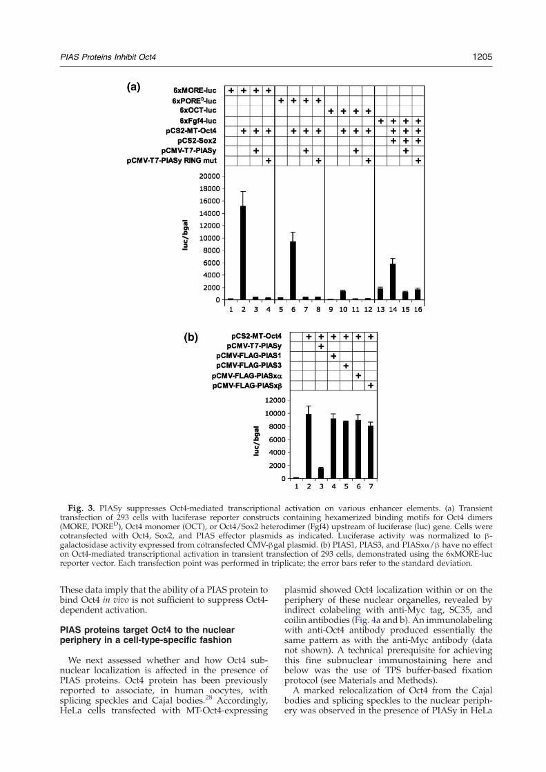
Fig. 3. PIASy suppresses Oct4-mediated transcriptional activation on various enhancer elements. (a) Transienttransfection of 293 cells with luciferase reporter constructs containing hexamerized binding motifs for Oct4 dimers(MORE, PORED), Oct4 monomer (OCT), or Oct4/Sox2 heterodimer (Fgf4) upstream of luciferase (luc) gene. Cells werecotransfected with Oct4, Sox2, and PIAS effector plasmids as indicated. Luciferase activity was normalized to β-galactosidase activity expressed from cotransfected CMV-βgal plasmid. (b) PIAS1, PIAS3, and PIASxα/β have no effecton Oct4-mediated transcriptional activation in transient transfection of 293 cells, demonstrated using the 6xMORE-lucreporter vector. Each transfection point was performed in triplicate; the error bars refer to the standard deviation.
1205PIAS Proteins Inhibit Oct4
These data imply that the ability of a PIAS protein tobind Oct4 in vivo is not sufficient to suppress Oct4-dependent activation.
PIAS proteins target Oct4 to the nuclearperiphery in a cell-type-specific fashion
We next assessed whether and how Oct4 sub-nuclear localization is affected in the presence ofPIAS proteins. Oct4 protein has been previouslyreported to associate, in human oocytes, withsplicing speckles and Cajal bodies.28 Accordingly,HeLa cells transfected with MT-Oct4-expressing
plasmid showed Oct4 localization within or on theperiphery of these nuclear organelles, revealed byindirect colabeling with anti-Myc tag, SC35, andcoilin antibodies (Fig. 4a and b). An immunolabelingwith anti-Oct4 antibody produced essentially thesame pattern as with the anti-Myc antibody (datanot shown). A technical prerequisite for achievingthis fine subnuclear immunostaining here andbelow was the use of TPS buffer-based fixationprotocol (see Materials and Methods).A marked relocalization of Oct4 from the Cajal
bodies and splicing speckles to the nuclear periph-ery was observed in the presence of PIASy in HeLa
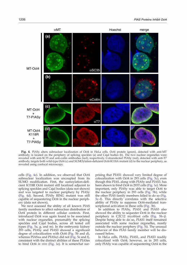
Fig. 4. PIASy alters subnuclear localization of Oct4 in HeLa cells. Oct4 protein (green), detected with anti-MTantibody, is located on the periphery of splicing speckles (a) and Cajal bodies (b). The two nuclear organelles wererevealed with anti-SC35 and anti-coilin antibodies (red), respectively. Cotransfected PIASy (red), detected with anti-T7antibody, targets both wild-type Oct4 (c) and SUMOylation-deficient Oct4-K118A mutant (d) to the nuclear periphery, asrevealed using confocal microscopy.
1206 PIAS Proteins Inhibit Oct4
cells (Fig. 4c). In addition, we observed that Oct4subnuclear localization was uncoupled from itsSUMO modification. First, the sumoylation-defi-cient K118R Oct4 mutant still localized adjacent tosplicing speckles and Cajal bodies (data not shown)and was targeted to nuclear periphery by PIASy(Fig. 4d). Second, PIASy RING mutant was stillcapable of sequestering Oct4 in the nuclear periph-ery (data not shown).We next assessed the ability of all known PIAS
family members to affect subnuclear distribution ofOct4 protein in different cellular contexts. First,introduced Oct4 was again found to be associatedwith nuclear organelles, presumably the splicingspeckles and Cajal bodies, across all tested celltypes (Fig. 5a, g, and m). In the embryonic kidney293 cells, PIASy and PIAS3 showed a significantdegree of colocalization with Oct4 (Fig. 5b and d),whereas PIASxα and PIASxβ did not (Fig. 5e and f),consistent with the distinct abilities of these PIASesto bind Oct4 in vivo (Fig. 1e). It is somewhat sur-
prising that PIAS1 showed very limited degree ofcolocalization with Oct4 in 293 cells (Fig. 5c), eventhough this PIAS, along with PIASy and PIAS3, hasbeen shown to bind Oct4 in 293T cells (Fig. 1e). Moreimportant, only PIASy was able to target Oct4 tothe nuclear periphery in 293 cells (Fig. 5b), whilethe other PIAS family members failed to do so (Fig.5c–f). This directly correlates with the selectiveability of PIASy to suppress Oct4-mediated tran-scriptional activation in these cells (Fig. 3b).In addition to PIASy, PIAS1 and PIAS3 also
showed the ability to sequester Oct4 in the nuclearperiphery in C2C12 myoblast cells (Fig. 5h–j).Despite being able to do so, PIAS1 itself remainedassociated with some nuclear organelles, mostlyoutside the nuclear periphery (Fig. 5i). The unusualbehavior of this PIAS family member will be dis-cussed below.In HeLa cells, PIASy, PIAS1, and PIAS3 readily
colocalized with Oct4; however, as in 293 cells,only PIASy was capable of sequestering Oct4 in the
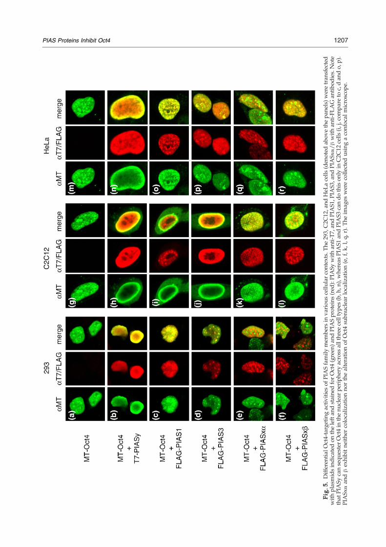
Fig.
5.Differen
tialO
ct4-targetingactiv
ities
ofPIASfamily
mem
bers
inva
riou
scellu
larcontexts.T
he29
3,C2C
12,a
ndHeL
acells
(den
oted
abov
ethepa
nels)w
eretran
sfected
with
plasmidsindicatedon
theleftan
dstaine
dforOct4(green
)and
PIASproteins
(red
):PIASy
with
anti-T7
,and
PIAS1
,PIA
S3,and
PIASx
α/β
with
anti-FL
AG
antib
odies.Note
that
PIASy
cansequ
esterOct4in
thenu
clearpe
riph
eryacross
allthree
celltype
s(b,h
,n),whe
reas
PIAS1
andPIAS3
cando
thison
lyin
C2C
12cells
(i,j,compa
reto
c,dan
do,
p).
PIASx
αan
dβexhibitne
ither
colocaliz
ationno
rthealteratio
nof
Oct4su
bnuc
lear
localiz
ation(e,f,k
,l,q
,r).Th
eim
ages
werecolle
cted
usingaconfocal
microscop
e.
1207PIAS Proteins Inhibit Oct4
1208 PIAS Proteins Inhibit Oct4
nuclear periphery (Fig. 5n–p). As in two other cellstypes, PIASxα and β neither colocalized nor alteredOct4 subnuclear distribution (Fig. 5e and f, k and l,q and r).In sum, these data demonstrate that the members
of the PIAS family exhibit differential activitiestowards Oct4 protein: PIASxα and PIASxβ are
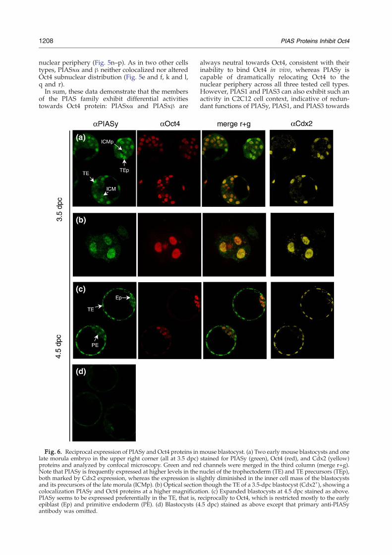
Fig. 6. Reciprocal expression of PIASy and Oct4 proteins inlate morula embryo in the upper right corner (all at 3.5 dpc)proteins and analyzed by confocal microscopy. Green and redNote that PIASy is frequently expressed at higher levels in theboth marked by Cdx2 expression, whereas the expression is sand its precursors of the late morula (ICMp). (b) Optical sectioncolocalization PIASy and Oct4 proteins at a higher magnificatPIASy seems to be expressed preferentially in the TE, that is, repiblast (Ep) and primitive endoderm (PE). (d) Blastocysts (4antibody was omitted.
always neutral towards Oct4, consistent with theirinability to bind Oct4 in vivo, whereas PIASy iscapable of dramatically relocating Oct4 to thenuclear periphery across all three tested cell types.However, PIAS1 and PIAS3 can also exhibit such anactivity in C2C12 cell context, indicative of redun-dant functions of PIASy, PIAS1, and PIAS3 towards
mouse blastocyst. (a) Two early mouse blastocysts and onestained for PIASy (green), Oct4 (red), and Cdx2 (yellow)channels were merged in the third column (merge r+g).
nuclei of the trophectoderm (TE) and TE precursors (TEp),lightly diminished in the inner cell mass of the blastocyststhough the TE of a 3.5-dpc blastocyst (Cdx2+), showing a
ion. (c) Expanded blastocysts at 4.5 dpc stained as above.eciprocally to Oct4, which is restricted mostly to the early.5 dpc) stained as above except that primary anti-PIASy
1209PIAS Proteins Inhibit Oct4
modulating Oct4 activity during mammalian devel-opment in certain cell types.
PIASy and Oct4 in preimplantation mousedevelopment
We next looked at preimplantation development,when PIASy-mediated restriction of Oct4 functioncould be of biological significance. Oct4 expressionduring this developmental period has been wellstudied: zygotic Oct4 expression starts at 4 to 8-cellstage and continues in all cells throughout morula[2.5 days postcoitum (dpc)] and early blastocyststage (3.5 dpc). By expanded blastocyst stage(4.5 dpc), Oct4 expression becomes restricted to theepiblast and primitive endoderm, being excludedfrom the trophectoderm (TE).Contrary to Oct4, PIASy expression during pre-
implantation development has not been reported.Therefore, we performed an immunofluorescentlabeling of preimplantation mouse embryos usinganti-PIASy antibody. PIASy protein was not detect-able at 1- though 4-cell cleavage stage; however, itsexpression was revealed in most cells of 8- and 16-cell embryos at around 2.5 dpc (data not shown). At3.5 dpc, PIASy protein was still present in all cells,although a tendency towards its extinction in theinner cells of late morula and the inner cell mass(ICM) of early blastocysts could be observed (Fig.6a). We further took a closer look at subnucleardistribution of PIASy and Oct4 proteins at 3.5 dpc,that is, during the time window where theirexpression domains overlap in the TE. The twoproteins seemed to colocalize in the nuclei of TE cells.However, in contrast to the above HeLa cell results,no obvious sequestration of Oct4 in the nuclearperiphery was seen in the nuclei of TE cells (Fig. 6b).A reason for that could be different fixation/permeabilization protocols used for cultured cellsand embryos. The TPS buffer-based method (seeMaterials and Methods), which allowed one toresolve fine subnuclear structures in HeLa cells, didnot produce a satisfactory signal in preimplantationembryos, for both PIASy and Oct4 proteins. Thus, itremains to be definedwhether PIASy-mediatedOct4targeting to the nuclear periphery occurs in vivo.By the expanded blastocyst stage (4.5 dpc), PIASy
seems to be expressed predominantly in the TE (Fig.6c). Therefore, during the transition from early to lateblastocyst stage,Oct4 andPIASy expressiondomainsbecome segregated: Oct4 protein is restricted to theepiblast and primitive endoderm and is excludedfrom the TE, whereas PIASy is restricted to the TE.The reciprocal pattern of expression of the twoproteins is consistent with the idea that PIASy mayplay a restrictive function towards Oct4 activityduring preimplantation development.
Discussion
Oct4 is an important transcriptional regulatorrequired for the maintenance of early pluripotent
cell types of mammalian embryo, PGCs, and EScells.4–10 Several transcriptional modulators of Oct4function have been described in the past.10–17
However, no negative transcriptional coregulatorshave been reported to date. Here we show that somemembers of the PIAS family, most notably PIASy,represent a novel class of transcriptional modulatorsof Oct4 function that act to suppress Oct4-mediatedtranscriptional activation through a sequestration ofthe protein in the nuclear periphery. In line withprevious reports,24,25 this repressive PIASy functionis uncoupled from its E3 SUMO ligase activity.Genetic ablation of Oct4 function leads to peri-
implantation lethality due to a differentiation of theICM cells into trophoblast cells.9 According to theproposed model, which suggests that PIASy inhibitsOct4 function in the TE, a loss of PIASy should leadto an increased Oct4 activity in the TE of blastocyst.However, PIASy-deficient embryos and mice do notmanifest any obvious developmental defects,29,30
suggesting that PIASy-mediated inhibition of Oct4is either not detrimental for the TE or is obscured bya redundant function of coexpressed PIAS familymembers. In support of the latter possibility, ourdata suggest that first, PIAS3 is coexpressed withPIASy in the TE and is mostly absent from theepiblast and primitive endoderm (SupplementaryFig. 1) and second, PIASy, PIAS1, and PIAS3 areredundant in targeting Oct4 to the nuclear peripheryin C2C12 cells (Fig. 5).Contrary to PIASy, PIAS1- and PIAS3-mediated
targeting of Oct4 to the nuclear periphery dependson the cellular context. The cell-type-specific en-gagement of Oct4 by these PIAS family membersmight be of importance, considering that Oct4is expressed and, in some cases, was shown toplay essential functions in different cell types, suchas the epiblast, PGCs, and spermatogonial stemcells.7,9,31,32There is a notable difference in the mode of PIAS1
action towards Oct4 in C2C12 cells compared toPIASy and PIAS3. Despite being able to promoteOct4 relocalization to the nuclear periphery, PIAS1itself is not enriched in this nuclear compartment(Fig. 5i). This observation is especially surprisingconsidering that PIAS1 is retained together with itsinteracting partner, Msx1, in the nuclear peripheryof the same type of cells.25 On the other hand, PIAS1and Oct4 show a substantial degree of colocalizationin C2C12 cells examined shortly after transfection(see Supplementary Fig. 2), suggesting a transientassociation between the two proteins. Molecularaspects and functional significance of such anintriguing behavior of this PIAS family memberneed further investigation.Although dispensable for PIASy-mediated Oct4
repression, the sumoylation of Oct4 might have animportant readout in other processes, for example,in the regulation of Oct4 stability. However, our datasuggest that PIASy does not serve as an E3 SUMOligase for Oct4. Moreover, it appears that PIASyinhibits K118 Oct4 sumoylation promoted by the E2SUMO conjugating enzyme Ubc9 (Fig. 2c). To our
1210 PIAS Proteins Inhibit Oct4
knowledge, this is the first example of an inhibitoryfunction of PIASy in sumoylation, although itcannot be excluded that this effect is due to theoverexpression of PIASy. On the other hand, Ubc9has been shown to sumoylate some protein targetswithout the help of E3 SUMO ligases.33–35 Ubc9,likewise PIASy, was also isolated in our two-hybridscreen as a protein interacting with the POU domainof Oct4 (data not shown). Therefore, it is possiblethat the mechanism by which PIASy inhibits Ubc9-dependent sumoylation involves a displacement ofUbc9 from the complex with the POU domain. Theparticipation of two other E3 SUMO ligases, RanBP2and Pc2,36,37 in Oct4 sumoylation cannot be ruledout as well.Dissecting regulatory mechanisms underlying
Oct4 functioning is of a high priority because thisknowledge will eventually help to outline basicprinciples of pluripotency and to manipulate thiscellular state. Members of the PIAS family mightrepresent important molecular components of reg-ulatory network controlling pluripotent cell fate viathe modulation of Oct4 function.
Materials and Methods
Plasmids
To construct the pCS2-MT-Oct4 and pCS2-Ubc9 plas-mid, an NcoI–EcoRI 1.3-kb fragment of Oct4 cDNA andPCR-amplified Ubc9 were cloned in pCS2+MT and pCS2vector, respectively (D. Turner, R. Rupp, and H. Wein-traub). The K118R, K215R, K244R, and K118/K244R Oct4mutant-encoding plasmids were derived from the pCS2-MT-Oct4 by PCR-based site-directed mutagenesis. ThepBD-GAL4-POU4 plasmid was made by inserting PCR-amplified POU domain-encoding part of Oct4 (127–282aa) at EcoRI–XmaI of pBD-GAL4/Cam (Stratagene). The0.95-kb Cdx2 cDNA was excised from pCX-GR*cdx2ire-sHyg plasmid38 with NcoI–XhoI and cloned into pACT2(Clontech), resulting in pACT2-Cdx2 control plasmid. For6×Fgf4-luc, the oligonucleotide 5′-AACTCTTTGTTTG-GATGCTAATGGGA-3′, spanning the octamer site(ATGCTAAT) and Sox2-binding motifs (CTTTGTT) froman Fgf4 enhancer, was hexamerized and cloned upstreamof the minimal tk promoter of the −37tk-luc plasmid (giftof A. Hecht). The 6×OCT-luc, 6×MORE-luc, 6×PORED-luc,pCMV-T7-PIASy, pCMV-T7-PIASy RING mut, andpCMV-FLAG-SUMO1/2 plasmids have been previouslydescribed.18,19,24 The pCS2-Sox2 plasmid was kindlyprovided by C. Wehrle and R. Kemler. The pCMV-FLAG-PIAS1, pCMV-FLAG-PIAS3, and pCMV-FLAG-PIASxα/β were a kind gift of K. Shuai and J. Palvimo.
Yeast two-hybrid screening
Mouse 11-day embryo MATCHMAKER cDNA Library(Clontech) cloned in pACT2 vector and pretransformedinto yeast strain Y187 was screened by mating it to theyeast strain AH109 carrying the pBD-GAL4-POU4 baitvector. In a control assay, the Y187 strain carried thepACT2-Cdx2 plasmid in place of the library. Colony-liftfilter and liquid culture assay was carried out according tothe manufacturer's guidelines (Clontech).
Coimmunoprecipitation
293T cells transfected by calcium phosphate method ornon-transfected R1 ES and COS7 cells were washed inphosphate-buffered saline (PBS), then lysed by sonicationin the CoIP buffer (50 mM Tris–HCl at pH 7.5, 100 mMNaCl, 15 mM EGTA, 0.1% Triton X-100, a proteaseinhibitor mix, 1 mM NaF, and 0.4 mM sodium orthovana-date). The lysates were cleared of cell debris by centrifuga-tion at 16,000g for 15 min at 4 °C. Equivalent amounts oftotal protein were precleared by incubation with a 1:1 mixof protein A- and protein G-conjugated beads (Sigma) andimmunoprecipitated overnight at 4 °C with either 1 μg ofmouse anti-T7 (Novagen) or rat anti-PIASy supernatant24
(1:10), followed by 2-h incubation at 4 °Cwith protein A+Gbeads (25 μl) or with rabbit anti-Myc antibody conjugatedwith agarose (25 μl, Sigma). Beads were washed severaltimes with CoIP buffer, boiled in 1×SDS loading buffer,and the supernatants were loaded onto SDS-PAGE.Western blots were carried out by the ECL detectionmethod (Amersham-Pharmacia), using peroxidase-conju-gated rabbit anti-Myc or mouse anti-FLAG tag antibodies(1:5000, Sigma), or using mouse anti-T7 (1:10,000, Nova-gen), mouse anti-Oct4 (1:500, Santa Cruz), followed byperoxidase-conjugated goat anti-mouse F(ab′)2 fragment(1:10,000, Jackson ImmunoResearch).
Transient transfections
293T cells were transfected in 24-well tissue cultureplates using SuperFect reagent (Qiagen). The total amountof DNA per well was equalized with a carrier plasmid.After 24–36 h, cells were washed in PBS and lysed directlyin wells in Glo-Lysis buffer (Promega). Approximatelyone-tenth of the crude extract was used to measure theluciferase and β-galactosidase activities (Bright-Glo and β-gal assays, Promega).
Immunostaining
HeLa, C2C12, and 293 cells were plated on LabTek IIchambered coverslips (Nunc) and transfected the next daywith the SuperFect (Qiagen) or Lipofectamine 2000(Invitrogen). After 24 h, the cells were fixed with 1%paraformaldehyde in the TPS buffer (10 mM Tris–HCl, pH8.0, 13.5 mM KCl, 20 mM NaCl, 0.5 mM EDTA, 0.1%Triton X-100) at room temperature for 10 min. Preimplan-tation mouse embryos were flushed from superovulatedfemales, fixed in 2% paraformaldehyde in PBS for 10 min,and permeabilized with 0.25% Triton X-100 for 8 min atroom temperature. The cells and embryos were subse-quently blocked in 2% bovine serum albumin, 1% heat-inactivated normal sheep serum in TPS or PBS for 30 min,and incubated for 2–5 h at room temperature in the sameblocking solution plus 0.1% Tween 20 with the followingprimary antibodies: rabbit anti-Myc tag (1:500, Sigma),mouse anti-T7 tag (1:1000, Novagen), mouse anti-FLAGM2 (1:250, Sigma), rat anti-PIASy supernatant (1:2; Ref.24), mouse anti-Oct4 (1:50, Santa Cruz), rabbit anti-coilin(1:400; see Ref. 28), mouse anti-SC35 (1:10; see Ref. 28),rabbit anti-Cdx2 (1:100, gift of Felix Beck), and rabbitanti-PIAS3 (1:25, Santa Cruz). Secondary antibodies usedwere goat anti-rabbit/mouse/rat IgG conjugated withAlexa488, Alexa546, or Alexa633 fluorochromes (1:500,Molecular Probes). Following the immunostaining pro-cedure, the cells were counterstained with Hoechst or4′,6-diamidino-2-phenylindole. Confocal analysis wasperformed with a Leica DMIRE2 microscope.
1211PIAS Proteins Inhibit Oct4
Acknowledgements
We are grateful to D. Turner, R. Rupp, H.Weintraub, A. Hecht, C. Wehrle, R. Kemler, F. Beck,K. Shuai, and J. Palvimo for providing valuablereagents. This work was supported by Alexander-von-Humboldt Stiftung to A.M., the Max-PlanckGesellschaft, and partially by the grant program“Molecular and Cellular Biology” to E.T. and A.T.
Supplementary Data
Supplementary data associated with this articlecan be found, in the online version, at doi:10.1016/j.jmb.2007.09.081
References
1. Scholer, H. R., Dressler, G. R., Balling, R., Rohdewohld,H. & Gruss, P. (1990). Oct-4: a germline-specifictranscription factor mapping to the mouse t-complex.EMBO J. 9, 2185–2195.
2. Okamoto, K., Okazawa, H., Okuda, A., Sakai, M.,Muramatsu, M. & Hamada, H. (1990). A noveloctamer binding transcription factor is differentiallyexpressed in mouse embryonic cells. Cell, 60, 461–472.
3. Rosner, M. H., Vigano, M. A., Ozato, K., Timmons,P. M., Poirier, F., Rigby, P. W. & Staudt, L. M. (1990).A POU-domain transcription factor in early stemcells and germ cells of the mammalian embryo.Nature, 345, 686–692.
4. Boyer, L. A., Mathur, D. & Jaenisch, R. (2006).Molecular control of pluripotency. Curr. Opin. Genet.Dev. 16, 455–462.
5. Loh, Y. H., Wu, Q., Chew, J. L., Vega, V. B., Zhang, W.,Chen, X. et al. (2006). The Oct4 and Nanog transcrip-tion network regulates pluripotency in mouse em-bryonic stem cells. Nat. Genet. 38, 431–440.
6. Boiani, M. & Scholer, H. R. (2005). Regulatorynetworks in embryo-derived pluripotent stem cells.Nat. Rev. Mol. Cell Biol. 6, 872–884.
7. Kehler, J., Tolkunova, E., Koschorz, B., Pesce, M.,Gentile, L., Boiani, M. et al. (2004). Oct4 is requiredfor primordial germ cell survival. EMBO Rep. 5,1078–1083.
8. Niwa, H., Miyazaki, J. & Smith, A. G. (2000).Quantitative expression of Oct-3/4 defines differen-tiation, dedifferentiation or self-renewal of ES cells.Nat. Genet. 24, 372–376.
9. Nichols, J., Zevnik, B., Anastassiadis, K., Niwa, H.,Klewe-Nebenius, D., Chambers, I. et al. (1998).Formation of pluripotent stem cells in the mammalianembryo depends on the POU transcription factorOct4. Cell, 95, 379–391.
10. Boyer, L. A., Lee, T. I., Cole, M. F., Johnstone, S. E.,Levine, S. S., Zucker, J. P. et al. (2005). Coretranscriptional regulatory circuitry in human embryo-nic stem cells. Cell, 122, 947–956.
11. Yuan, H., Corbi, N., Basilico, C. & Dailey, L. (1995).Developmental-specific activity of the FGF-4 enhancerrequires the synergistic action of Sox2 andOct-3.GenesDev. 9, 2635–2645.
12. Remenyi, A., Lins, K., Nissen, L. J., Reinbold, R.,Scholer, H. R. & Wilmanns, M. (2003). Crystal struc-ture of a POU/HMG/DNA ternary complex suggests
differential assembly of Oct4 and Sox2 on two en-hancers. Genes Dev. 17, 2048–2059.
13. Okumura-Nakanishi, S., Saito, M., Niwa, H. &Ishikawa, F. (2005). Oct-3/4 and Sox2 regulate Oct-3/4 gene in embryonic stem cells. J. Biol. Chem. 280,5307–5317.
14. Tomioka,M.,Nishimoto,M.,Miyagi, S., Katayanagi, T.,Fukui, N., Niwa, H. et al. (2002). Identification of Sox-2regulatory region which is under the control of Oct-3/4-Sox-2 complex. Nucleic Acids Res. 30, 3202–3213.
15. Brehm, A., Ohbo, K., Zwerschke, W., Botquin, V.,Jansen-Durr, P. & Scholer, H. R. (1999). Synergismwith germ line transcription factor Oct-4: viral onco-proteins share the ability to mimic a stem cell-specificactivity. Mol. Cell Biol. 19, 2635–2643.
16. Scholer, H. R., Ciesiolka, T. & Gruss, P. (1991). A nexusbetween Oct-4 and E1A: implications for gene regula-tion in embryonic stem cells. Cell, 66, 291–304.
17. Guo, Y., Costa, R., Ramsey, H., Starnes, T., Vance, G.,Robertson, K. et al. (2002). The embryonic stem celltranscription factors Oct-4 and FoxD3 interact toregulate endodermal-specific promoter expression.Proc. Natl Acad. Sci. USA, 99, 3663–3667.
18. Botquin, V., Hess, H., Fuhrmann, G., Anastassiadis,C., Gross, M. K., Vriend, G. & Scholer, H. R. (1998).New POU dimer configuration mediates antagonisticcontrol of an osteopontin preimplantation enhancerby Oct-4 and Sox-2. Genes Dev. 12, 2073–2090.
19. Tomilin, A., Remenyi, A., Lins, K., Bak, H., Leidel, S.,Vriend, G. et al. (2000). Synergism with the coactivatorOBF-1 (OCA-B, BOB-1) is mediated by a specific POUdimer configuration. Cell, 103, 853–864.
20. Remenyi, A., Tomilin, A., Pohl, E., Lins, K., Philippsen,A., Reinbold, R. et al. (2001). Differential dimeractivities of the transcription factor Oct-1 by DNA-induced interface swapping. Mol. Cell, 8, 569–580.
21. Jacobson, E. M., Li, P., Leon-del-Rio, A., Rosenfeld, M.G. & Aggarwal, A. K. (1997). Structure of Pit-1 POUdomain bound to DNA as a dimer: unexpectedarrangement and flexibility. Genes Dev. 11, 198–212.
22. Shuai, K. & Liu, B. (2005). Regulation of gene-activation pathways by PIAS proteins in the immunesystem. Nat. Rev. Immunol. 5, 593–605.
23. Schmidt, D. & Muller, S. (2003). PIAS/SUMO: newpartners in transcriptional regulation. Cell Mol. LifeSci. 60, 2561–2574.
24. Sachdev, S., Bruhn, L., Sieber, H., Pichler, A., Melchior,F. & Grosschedl, R. (2001). PIASy, a nuclear matrix-associated SUMO E3 ligase, represses LEF1 activityby sequestration into nuclear bodies. Genes Dev. 15,3088–3103.
25. Lee, H., Quinn, J. C., Prasanth, K. V., Swiss, V. A.,Economides, K. D., Camacho, M. M. et al. (2006).PIAS1 confers DNA-binding specificity on the Msx1homeoprotein. Genes Dev. 20, 784–794.
26. Niwa, H., Toyooka, Y., Shimosato, D., Strumpf, D.,Takahashi, K., Yagi, R. & Rossant, J. (2005). Interactionbetween Oct3/4 and Cdx2 determines trophectodermdifferentiation. Cell, 123, 917–929.
27. Rodriguez, M. S., Dargemont, C. & Hay, R. T. (2001).SUMO-1 conjugation in vivo requires both a consensusmodification motif and nuclear targeting. J. Biol. Chem.276, 12654–12659.
28. Parfenov, V. N., Pochukalina, G. N., Davis, D. S.,Reinbold, R., Scholer, H. R. & Murti, K. G. (2003).Nuclear distribution of Oct-4 transcription factor intranscriptionally active and inactive mouse oocytesand its relation to RNA polymerase II and splicingfactors. J. Cell Biochem. 89, 720–732.
1212 PIAS Proteins Inhibit Oct4
29. Roth, W., Sustmann, C., Kieslinger, M., Gilmozzi, A.,Irmer, D., Kremmer, E. et al. (2004). PIASy-deficientmice display modest defects in IFN and Wnt signal-ing. J. Immunol. 173, 6189–6199.
30. Wong, K. A., Kim, R., Christofk, H., Gao, J., Lawson,G. & Wu, H. (2004). Protein inhibitor of activatedSTAT Y (PIASy) and a splice variant lacking exon6 enhance sumoylation but are not essential forembryogenesis and adult life. Mol. Cell Biol. 24,5577–5586.
31. Yeom, Y. I., Fuhrmann, G., Ovitt, C. E., Brehm, A.,Ohbo, K., Gross, M. et al. (1996). Germline regulatoryelement of Oct-4 specific for the totipotent cycle ofembryonal cells. Development, 122, 881–894.
32. Pesce, M., Wang, X., Wolgemuth, D. J. & Scholer, H.(1998). Differential expression of the Oct-4 transcrip-tion factor during mouse germ cell differentiation.Mech. Dev. 71, 89–98.
33. Tatham, M. H., Jaffray, E., Vaughan, O. A., Desterro,J. M., Botting, C. H., Naismith, J. H. & Hay, R. T.(2001). Polymeric chains of SUMO-2 and SUMO-3 areconjugated to protein substrates by SAE1/SAE2 andUbc9. J. Biol. Chem. 276, 35368–35374.
34. Bernier-Villamor, V., Sampson, D. A., Matunis, M. J. &Lima, C. D. (2002). Structural basis for E2-mediatedSUMO conjugation revealed by a complex betweenubiquitin-conjugating enzyme Ubc9 and RanGAP1.Cell, 108, 345–356.
35. Lee, G. W., Melchior, F., Matunis, M. J., Mahajan, R.,Tian, Q. & Anderson, P. (1998). Modification of RanGTPase-activating protein by the small ubiquitin-related modifier SUMO-1 requires Ubc9, an E2-typeubiquitin-conjugating enzyme homologue. J. Biol.Chem. 273, 6503–6507.
36. Mahajan, R., Delphin, C., Guan, T., Gerace, L. &Melchior, F. (1997). A small ubiquitin-related poly-peptide involved in targeting RanGAP1 to nuclearpore complex protein RanBP2. Cell, 88, 97–107.
37. Kagey, M. H., Melhuish, T. A. & Wotton, D. (2003).The polycomb protein Pc2 is a SUMO E3. Cell, 113,127–137.
38. Tolkunova, E., Cavaleri, F., Eckardt, S., Reinbold, R.,Christenson, L. K., Scholer, H. R. & Tomilin, A. (2006).The caudal-related protein cdx2 promotes trophoblastdifferentiation of mouse embryonic stem cells. StemCells, 24, 139–144.

![APolycombRepressionSignatureinMetastaticProstateCancer ... · survival. [CancerRes2007;67(22):10657–63] Introduction Polycomb group (PcG) proteins are transcriptional repressors](https://img.dokumen.tips/doc/110x75/5fb3d33c686ac07cd643bad0/apolycombrepressionsignatureinmetastaticprostatecancer-survival-cancerres2007672210657a63.jpg)