Embed Size (px)
Citation preview
JOURNAL OF VIROLOGY, Apr. 2010, p. 3631–3643 Vol. 84, No. 70022-538X/10/$12.00 doi:10.1128/JVI.02380-09Copyright © 2010, American Society for Microbiology. All Rights Reserved.
Biphasic Recruitment of Transcriptional Repressors to the MurineCytomegalovirus Major Immediate-Early Promoter during
the Course of Infection In Vivo�
Xue-feng Liu,1 Shixian Yan,1 Michael Abecassis,1,2§* and Mary Hummel1,2,3§Comprehensive Transplant Center, Department of Surgery,1 Department of Microbiology and Immunology,2 and
Robert H. Lurie Cancer Center,3 Northwestern University Feinberg School of Medicine, Chicago, Illinois
Received 11 November 2009/Accepted 19 January 2010
Our previous studies showed that establishment of murine cytomegalovirus (MCMV) latency in vivo isassociated with repression of immediate-early gene expression, deacetylation of histones bound to the majorimmediate-early promoter (MIEP), changes in patterns of methylation of histones, and recruitment of cellularrepressors of transcription to the MIEP. Here, we have quantitatively analyzed the kinetics of changes in viralRNA expression, DNA copy number, and recruitment of repressors and activators of transcription to viralpromoters during the course of infection. Our results show that changes in viral gene expression correlate withchanges in recruitment of RNA polymerase and acetylated histones to viral promoters. Binding of the tran-scriptional repressors histone deacetylase type 2 (HDAC2), HDAC3, YY1, CBF-1/RBP-Jk, Daxx, and CIR to theMIEP and HDACs to other promoters showed a biphasic pattern: some binding was detectable prior toactivation of viral gene expression, then decreased with the onset of transcription and increased again asrepression of viral gene expression occurred. Potential binding sites for CBF-1/RBP-Jk and YY1 in the MIEPand for YY1 in the M100 promoter (M100P) were identified by in silico analysis. While recruitment of HDACswas not promoter specific, binding of CBF-1/RBP-Jk and YY1 was restricted to promoters with their cognatesites. Our results suggest that sequences within viral promoters may contribute to establishment of latencythrough recruitment of transcriptional repressors to these genes. The observation that repressors are boundto the MIEP and other promoters immediately upon infection suggests that latency may be established in somecells very early in infection.
Cytomegalovirus (CMV), like other herpesviruses, has theability to establish a lifelong latent infection. Reactivation fromlatency, characterized by shedding of virus, occurs frequentlyand asymptomatically in immunocompetent hosts. In immuno-compromised recipients of solid organs and bone marrowtransplants, reactivation of CMV is associated with increasedrisk of acute and chronic allograft rejection, opportunistic in-fection, graft failure, and patient mortality (54). CMV infec-tion of immunologically immature fetuses in utero is associatedwith hearing loss, cognitive impairment, cerebral palsy, andvisual impairment (16). Thus, CMV is an important opportu-nistic human pathogen, and an understanding of the mecha-nisms by which CMV establishes latent infection and reacti-vates from latency is essential in developing new tools tocombat viral infection and its sequelae.
CMV latency has been defined as the absence of infectiousvirus, despite the presence of viral DNA (56). However, themolecular mechanisms by which latency is established andmaintained have not been clear. Immune control of cells ex-pressing viral genes associated with productive infection playsan important role in preventing recurrence of infectious virus.However, transcriptional control of viral gene expression is
also thought to be important in controlling viral latency andreactivation. Studies with HCMV have suggested that latencyis established through repression of ie-1 and ie-2 gene expres-sion (4, 58). The IE-1 and IE-2 proteins, which are among thefirst proteins expressed by the virus during productive infec-tion, are transcriptional regulatory proteins that are requiredfor induction of early and late gene expression, viral DNAsynthesis, and production of infectious virus (46). Transcrip-tion of IE-1 and IE-2 RNAs, which are differentially splicedtranscripts initiated at the major immediate-early (ie) pro-moter (MIEP), is regulated by the MIE enhancer (6, 13).Previous studies have shown that in latently infected cells, theMIEP is bound to deacetylated histones and cellular repressorsof transcription, suggesting that latency is established throughrecruitment of these factors to the MIEP (59). However, stud-ies of HCMV latency in naturally infected cells have been verylimited, due to the low frequency of latently infected cells andthe inability to manipulate the virus during infection in its host.Thus, it has not been possible to determine the mechanisms bywhich these factors are recruited to the MIEP.
To circumvent these difficulties, we and others have usedmurine CMV (MCMV), which is similar to HCMV with re-spect to genome organization, regulation of transcription,pathogenesis, and ability to establish latent infection and toreactivate, as a model system to study CMV latency in vivo.Studies of HCMV latency have shown that it establishes latentinfection in cells of the monocyte lineage and have suggestedthat, within this lineage, there may be cell-type-specific differ-ences in the ability to establish latency (58). A molecular basis
* Corresponding author. Mailing address: Transplant Center,Northwestern Memorial Hospital, 675 N. St. Clair St., Galter Pavilion,Suite 17-200, Chicago, IL 60611. Phone: (312) 695-0359. Fax: (312)695-1817. E-mail: [email protected].
§ These authors contributed equally to the work.� Published ahead of print on 27 January 2010.
3631
Dow
nloa
ded
from
http
s://j
ourn
als.
asm
.org
/jour
nal/j
vi o
n 12
Dec
embe
r 20
21 b
y 92
.52.
207.
83.
for these differences has not been determined. Because of thelack of availability of other tissues and the low copy number,there have been very few studies that have addressed othersites of HCMV latency. However, in the MCMV model, it isclear that the virus establishes latent infection in multiple or-gans (5, 31, 44, 51, 64). We previously showed that ie geneexpression is repressed during latent infection in mouse kid-neys (25, 40). Establishment of latency was also correlated withdeacetylation of histones bound to the major immediate-earlypromoter, with changes in patterns of methylation of histones,and with recruitment of cellular repressors of transcription tothe MIEP (40). These changes were specific to viral chromatin,as viral infection had no effect on modifications of histonesbound to the promoters of the �-actin and Ant4 cellular genes.These studies support the hypothesis that latency is establishedthrough repression of the MIEP but left open the importantquestion of how these repressors are recruited. In order tofurther investigate this question, we have now quantitativelyanalyzed viral RNA expression, DNA copy number, changes inhistone modifications, and recruitment of transcriptional re-pressors to the MIEP and other viral promoters at varioustimes postinfection. Our results show that viral promoters inaddition to the MIEP are bound to repressors at the veryearliest stage of infection. The proportion of genomes boundto these repressors decreased as binding of RNA polymeraseand acetylated histones to promoters and activation of viralgene expression occurred, and it subsequently increased whenlatency was established. Some repressors were recruited in apromoter-specific fashion, suggesting that sequences in thepromoters could be involved in the establishment of latency.
MATERIALS AND METHODS
Mice and virus. Three-week-old, female, specific-pathogen-free BALB/c micewere purchased from Jackson Laboratory, Bar Harbor, ME. Mice were main-tained in isolation cages and fed and watered ad libitum. MCMV (Smith strain,ATCC 194-VR) was purchased from the American Type Culture Collection(Rockville, MD). Virus stocks were generated by propagation in salivary glandsas previously described (31). Mice were infected by intraperitoneal injection with105 PFU of MCMV and sacrificed at various times postinfection. This studyprotocol was reviewed and approved by the Northwestern University Institu-tional Animal Care and Use Committee.
Nucleic acid extraction. Kidney DNA was prepared by using a Puregene tissuekit (Gentra, Minneapolis, MN) according to the manufacturer’s instruction.Total RNA was isolated and purified with a TRIzol Plus RNA purification kit(Invitrogen, Carlsbad, CA). On-column DNase � digestion was performed duringRNA purification. DNA and RNA were quantified by A260 with a DU640Bspectrophotometer (Beckman Coulter, Fullerton, CA).
Reverse transcription (RT). Oligo(dT)-primed cDNA was generated from 5�g of total RNA by using an AffinityScript multiple temperature cDNA synthesiskit (Stratagene, La Jolla, CA) in a 20-�l reaction mixture. Reverse transcriptasewas inactivated by heating at 70°C for 15 min. Samples were cooled to 37°C andtreated for 30 min with 0.15 U/�l RNaseH (Invitrogen) and 1 �l RNase cocktail(Ambion, Austin, TX). Subsequently, the cDNA was purified with a QIAquickPCR purification kit (Qiagen, Valencia, CA) and quantified using a Quant-iTOligreen single-stranded DNA (ssDNA) assay kit (Invitrogen).
Real-time PCR analysis. All real-time PCRs were performed using TaqMangene expression master mix in an ABI 7500 Fast system using the standard curve(absolute quantitation) assay and regular 7500 mode. Primers and TaqManminor groove binder (MGB) probes described in Table 1 were designed by ABIor by using Primer Express 3.0 software. Reaction mixtures contained 900 nMprimers and 250 nM TaqMan MGB probes. Each sample was analyzed in trip-licate. The thermal cycling conditions were 50°C for 2 min and 95°C for 10 minfollowed by 50 cycles of 95°C for 15 s and 60°C for 1 min.
Quantification of MCMV DNA and RNA by real-time PCR. For absolutequantification of MCMV DNA copy number in kidney tissue, a standard curve
was generated by serial dilution of plasmid pIE111 (45), which contains theMIEP and IE genes, in genomic DNA isolated from kidneys of uninfected mice,such that 13.5 �l of the template contained 3 � 106, 3 � 105, 3 � 104, 3 � 103,3 � 102, or 3 � 101 copies of pIE111 in 800 ng cellular DNA. A standard curvefor mouse Eef1� DNA was generated by serial dilution of plasmid pDRIVE-mEF1� (InvivoGene, San Diego, CA), which contains the promoter region of themouse eukaryotic translation elongation factor 1 alpha (Eef1�) gene, such that13.5 �l of the template contained 1 � 106, 1 � 105, 1 � 104, 1 � 103, 1 � 102,or 10 copies of pDRIVE-mEF1� in 800 ng sheared salmon sperm DNA. Samplegenomic DNA for each reaction (800 ng) was used as a template for PCR in a30-�l reaction mixture. Genomic DNA from uninfected mouse kidneys (800 ng)was used as a control for detection of nonspecific amplification. The primers andprobes used to detect MCMV MIEP and Eef1� promoter DNA are listed inTable 1. The target DNA copy number in each sample was calculated from astandard curve generated in each individual assay. The viral DNA copy numberper million cells was determined by dividing the average copy number of MCMVDNA by the Eef1� copy number in the same sample and multiplying by 2 � 106.
Quantification of MCMV IE-1, IE-3, M112, and M100 mRNA was performedusing the relative standard curve method as described in the Applied Biosystemssupport material “Chemistry Guide: Applied Biosystems Real-Time PCR Sys-tems” (http://www.appliedbiosystems.com/support/apptech/, part no. 4348358)with the primers and probes specific to each gene (Table 1). Serially dilutedcDNA derived from RNA isolated from kidneys at 5 days postinfection was usedto generate the standard curve. Sample cDNA (10 ng) was added to a 20-�lreaction mixture for amplification of virus cDNA; the same amount of cDNAfrom uninfected mouse kidneys was used as a control for nonspecific amplifica-tion. The abundance of the target cDNA in each sample was determined on thebasis of a standard curve produced in each run. Mouse eukaryotic Eef2 RNA wasemployed as an internal control. The relative quantity of MCMV RNA in thesample was determined by dividing the average quantity of MCMV mRNA bythat of Eef2, as determined from the standard curve. As a control to detectcontamination of the cDNA by genomic DNA, we analyzed cDNAs for thepresence of the intronic region between exons 1 and 2 in the immediate-earlygene 1/3.
Chromatin immunoprecipitation (ChIP). Chromatin immunoprecipitation as-says were performed essentially as described previously (40). Antibodies againsthistone H4 (pan), acetyl histone H4 (Lys5, Lys8, Lys12, Lys16), and Daxx wereobtained from Millipore Corporation, Billerica, MA. Antibodies for histonedeacetylase type 2 (HDAC2) and HDAC3 were purchased from Sigma-AldrichInc., St. Louis, MO. Antibodies specifc for RNA polymerase �� (N-20) andCBF-1/RBP-Jk(H-50) were obtained from Santa Cruz Biotechnology, SantaCruz, CA. Anti-YY1 and anti-CIR were purchased from Abcam, Cambridge,MA, and Novus Biologicals, Littleton, CO, respectively.
Quantitative analysis of ChIP DNA samples by real-time PCR. The relativeamount of MCMV MIEP DNA in each ChIP sample was determined using therelative standard curve method. Serial dilutions of sheared genomic DNA puri-fied from acutely infected mouse kidneys were used to generate the standardcurve. The same set of standards was used for all analyses. For analysis of totalhistone H4, HDACs, RNA Pol II, YY1, CBF-1/RBP-Jk, Daxx, and CIR, theresults are expressed as the percentage of immunoprecipitated chromatin rela-tive to the total input of chromatin in the immunoprecipitation mixture. Foranalysis of acetylated histone H4, the results are expressed as the ratio of themodified histone to corresponding total histone after normalization to inputDNA. ChIP assays were analyzed in triplicate, and the results presented are theaverages of results of two to four independent assays plus standard error.
Statistical analysis. A two-tailed Student’s t test was used to determine sta-tistical significance. P values of �0.05 were considered significant.
RESULTS
Repression of MCMV ie gene expression in the kidney oc-curs between day 7 and day 10 postinfection. In order tounderstand the mechanism of establishment of latency, it isimportant to know when repression of viral gene expressionoccurs during the course of infection. We therefore examinedthe kinetics of expression of IE-1 and IE-3 RNAs and accu-mulation of viral DNA during acute infection in the kidney.IE-1 and IE-3 RNAs were analyzed by real-time PCR analysisof purified cDNA using the relative standard curve method asdescribed in Materials and Methods. The intron region be-
3632 LIU ET AL. J. VIROL.
Dow
nloa
ded
from
http
s://j
ourn
als.
asm
.org
/jour
nal/j
vi o
n 12
Dec
embe
r 20
21 b
y 92
.52.
207.
83.

tween exons 1 and 2 was analyzed as a control for genomicDNA contamination of the cDNA. DNA and RNA from un-infected mice were used as controls for nonspecific amplifica-tion. These controls were negative (data not shown). Our re-sults show that expression of IE-1 RNA was detectable at day2 postinfection (p.i.), peaked at day 7, and dropped signifi-cantly at day 10 relative to the peak at day 7 (Fig. 1A). Ex-pression of IE-3, which is transcribed from the same promoteras IE-1, followed the same trend as expression of IE-1.
MCMV DNA was detectable in the kidney within 1 day p.i.but did not accumulate significantly above this level until day 4.In contrast to IE RNA expression, which peaked at day 7,MCMV DNA copy number peaked at day 10 and fell graduallyuntil day 28, where it plateaued to levels seen in latently in-fected mice (Fig. 1B). The decrease in DNA copy numberlikely reflects clearance of infected cells by the host immuneresponse. However, our results show that loss of ie gene ex-pression precedes the decrease in DNA copy number and is
TABLE 1. List of primers and probes
Target region Sequence Accession no. (nucleotide no.)
Mouse �-actin promoter 5�CGTTCCGAAAGTTGCCTTTTA 3� (forward) DD173001 (429–489)5�GCCGCCGGGTTTTATAGG 3� (reverse)5�CTCGAGTGGCCGCTG 3� (probe)
Mouse Ant4 promoter 5�CAGGCTAGTGTCTGCACCTG 3� (forward) AC146980 (102991–103117)5�ACCAACCCGGTGATTAACTG 3� (reverse)5�ATTACAGGCGTGCAGCATCT 3� (probe)
Mouse RpL30 gene YY1 5�GGCTGGTGTTGGTGAGTGA 3� (forward) K02928 (489–599)binding region 5�ACACAGAGGACAGAAGAGAGGATT 3� (reverse)
5�CCAGAGCGTCAAACAC 3� (probe)
Mouse HES1 gene RBP-Jk 5�GGCCTGCGGATCACACA 3� (forward) D16464 (188–262)binding region 5�GGACCAAGGAGAGAGGTAGACA 3� (reverse)
5�CACCAGCTCCAGATCC 3� (probe)
Mouse Eef2 mRNA 5�CCACGGGCCTGAAGGT 3� (forward) NM_007907.1(1345–1417)5�GGCTTCAGGTATAGGTCCTCTTTC 3� (reverse)5�ATGGGCCCCAACTAC 3� (probe)
Mouse EeF1� promoter 5�CGGGTTTGCCGTCAGAAC 3� (forward) AC158987 (121163–121239)5�GCTCGGAGCAGGACCTC 3� (reverse)5�CCACACCCGCCCCTC 3� (probe)
MCMV MIEP 5�GGTGGTCAGACCGAAGACT 3� (forward) U68299 (182873–182944)5�GCTGAGCTGCGTTCTACGT 3� (reverse)5�CTGGTCGCGCCTCTTA 3� (probe)
MCMV ie-1 mRNA 5�CAACAGCGGCAGCTTCTTC 3� (forward) U68299 (179823–179884)5�CATGGCGTACTGCCTCTTGA 3� (reverse)5�ACTGCTCCTCGCCC 3� (probe)
MCMV ie-3 mRNA 5�CAGCCGGAGGCTACGT 3� (forward) U68299 (178572–178641)5�GCTGGGTACTTCCTGGACTTATC 3� (reverse)5�TTCCCTGCACTTCTTG 3� (probe)
IE intron 1 5�CCTGTGTTCACACACCAGATATTACA 3� (forward) U68299 (182115–182204)5�GCAGCCTCTGAGTACAATATTCTGT 3� (reverse)5�CCGCCATGATAAGTCC 3� (probe)
MCMV M100 (gM) 5�GGGTCAAATAGTGAGAGCATCCTT 3� (forward) U68299 (145392–145465)promoter 5�GAAGCGGCGACGTTACC 3� (reverse)
5�CTCGGACGGTCTCTCT 3� (probe)
MCMV M100 mRNA 5�TCGCGCGACTCGTAGTG 3� (forward) U68299 (144804–144880)5�GCGTTCAGCAAGTGCATGTAC 3� (reverse)5�CCTGACGGCCTTCGTG 3� (probe)
MCMV M112 (e1) 5�GCAGACCAAATGCTGATAGTTCCT 3� (forward) U68299 (163008–163072)promoter 5�GGTCGCGACGAGAAAAGTG 3� (reverse)
5�TCGCGGTAGATTACG 3� (probe)
MCMV M112 mRNA 5�CGTCTGTAACAAGACCGTCTCTTC 3� (forward) U68299 (163423–163533)5�ACCAGGATGACTTGCAGCAA 3� (reverse)5�CCGCCACCAGAGTCA 3� (probe)
VOL. 84, 2010 BIPHASIC RECRUITMENT OF REPRESSORS TO THE MCMV MIEP 3633
Dow
nloa
ded
from
http
s://j
ourn
als.
asm
.org
/jour
nal/j
vi o
n 12
Dec
embe
r 20
21 b
y 92
.52.
207.
83.
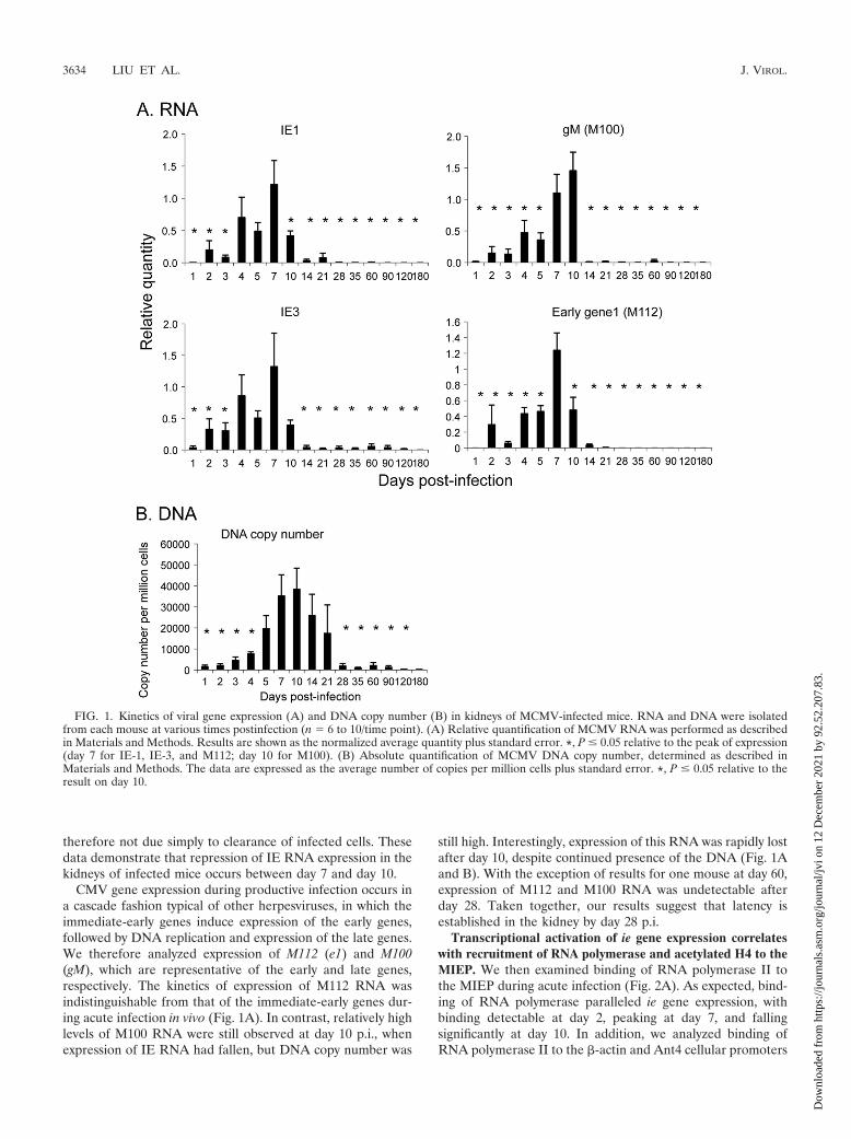
therefore not due simply to clearance of infected cells. Thesedata demonstrate that repression of IE RNA expression in thekidneys of infected mice occurs between day 7 and day 10.
CMV gene expression during productive infection occurs ina cascade fashion typical of other herpesviruses, in which theimmediate-early genes induce expression of the early genes,followed by DNA replication and expression of the late genes.We therefore analyzed expression of M112 (e1) and M100(gM), which are representative of the early and late genes,respectively. The kinetics of expression of M112 RNA wasindistinguishable from that of the immediate-early genes dur-ing acute infection in vivo (Fig. 1A). In contrast, relatively highlevels of M100 RNA were still observed at day 10 p.i., whenexpression of IE RNA had fallen, but DNA copy number was
still high. Interestingly, expression of this RNA was rapidly lostafter day 10, despite continued presence of the DNA (Fig. 1Aand B). With the exception of results for one mouse at day 60,expression of M112 and M100 RNA was undetectable afterday 28. Taken together, our results suggest that latency isestablished in the kidney by day 28 p.i.
Transcriptional activation of ie gene expression correlateswith recruitment of RNA polymerase and acetylated H4 to theMIEP. We then examined binding of RNA polymerase II tothe MIEP during acute infection (Fig. 2A). As expected, bind-ing of RNA polymerase paralleled ie gene expression, withbinding detectable at day 2, peaking at day 7, and fallingsignificantly at day 10. In addition, we analyzed binding ofRNA polymerase II to the �-actin and Ant4 cellular promoters
FIG. 1. Kinetics of viral gene expression (A) and DNA copy number (B) in kidneys of MCMV-infected mice. RNA and DNA were isolatedfrom each mouse at various times postinfection (n � 6 to 10/time point). (A) Relative quantification of MCMV RNA was performed as describedin Materials and Methods. Results are shown as the normalized average quantity plus standard error. *, P � 0.05 relative to the peak of expression(day 7 for IE-1, IE-3, and M112; day 10 for M100). (B) Absolute quantification of MCMV DNA copy number, determined as described inMaterials and Methods. The data are expressed as the average number of copies per million cells plus standard error. *, P � 0.05 relative to theresult on day 10.
3634 LIU ET AL. J. VIROL.
Dow
nloa
ded
from
http
s://j
ourn
als.
asm
.org
/jour
nal/j
vi o
n 12
Dec
embe
r 20
21 b
y 92
.52.
207.
83.
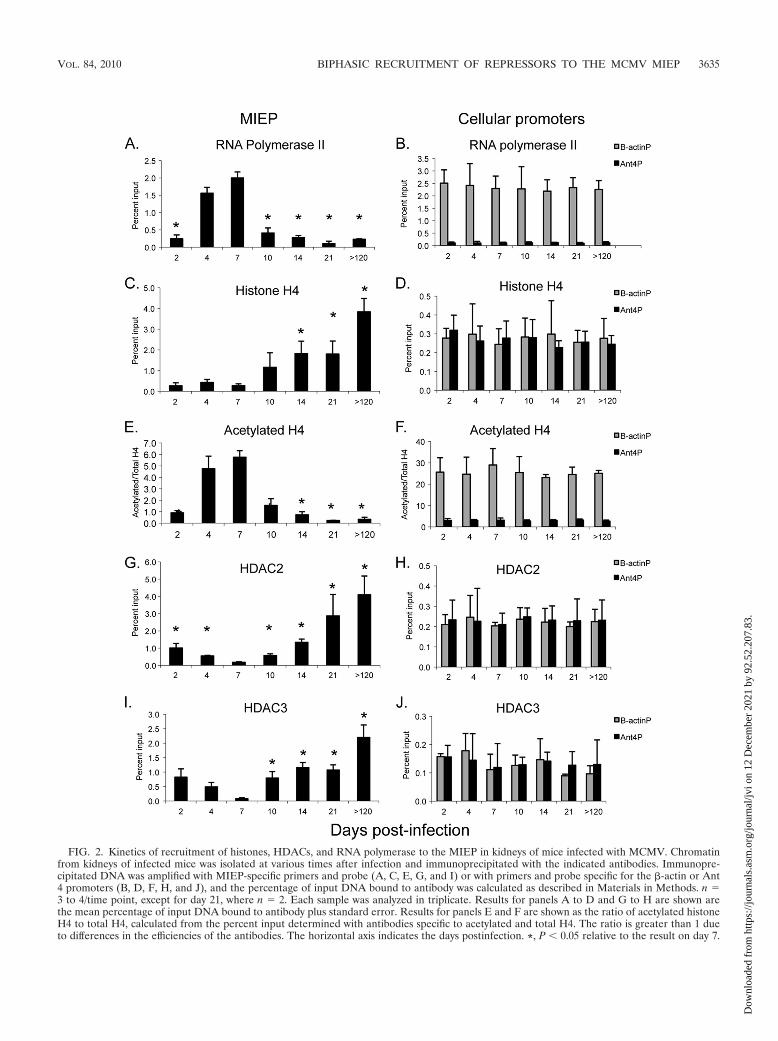
FIG. 2. Kinetics of recruitment of histones, HDACs, and RNA polymerase to the MIEP in kidneys of mice infected with MCMV. Chromatinfrom kidneys of infected mice was isolated at various times after infection and immunoprecipitated with the indicated antibodies. Immunopre-cipitated DNA was amplified with MIEP-specific primers and probe (A, C, E, G, and I) or with primers and probe specific for the �-actin or Ant4 promoters (B, D, F, H, and J), and the percentage of input DNA bound to antibody was calculated as described in Materials in Methods. n �3 to 4/time point, except for day 21, where n � 2. Each sample was analyzed in triplicate. Results for panels A to D and G to H are shown arethe mean percentage of input DNA bound to antibody plus standard error. Results for panels E and F are shown as the ratio of acetylated histoneH4 to total H4, calculated from the percent input determined with antibodies specific to acetylated and total H4. The ratio is greater than 1 dueto differences in the efficiencies of the antibodies. The horizontal axis indicates the days postinfection. *, P � 0.05 relative to the result on day 7.
VOL. 84, 2010 BIPHASIC RECRUITMENT OF REPRESSORS TO THE MCMV MIEP 3635
Dow
nloa
ded
from
http
s://j
ourn
als.
asm
.org
/jour
nal/j
vi o
n 12
Dec
embe
r 20
21 b
y 92
.52.
207.
83.
as controls for transcriptionally active and inactive genes, re-spectively (Fig. 2B). �-Actin is widely expressed in all organs;The Ant4 gene is a developmentally regulated gene which issilenced through DNA methylation in adult mouse tissues (61).Binding of RNA polymerase to the �-actin promoter was ob-served at all time points at comparable levels. Thus, thechanges in binding of RNA polymerase to the MIEP duringthe course of infection are not due to differences in the chro-matin preparations. At the peak of infection, the percentage ofMIEP DNA bound to RNA polymerase was similar to that of�-actin, indicating that the proportion of viral genomes beingactively transcribed was similar to that of a highly active cel-lular gene and, thus, is biologically relevant. In contrast, verylittle binding of RNA polymerase to the transcriptionally in-active Ant4 promoter was observed, indicating the specificityof the binding to the MIEP that we observed.
We previously demonstrated that MCMV DNA is bound tohistones during infection and that the percentage of genomesassociated with histones rises dramatically in latent infection(40). Here, we have examined the kinetics of histone deposi-tion to the MIEP (Fig. 2C). Our previous studies showed thatbinding of histone H4 to the MIEP was detectable during acuteinfection; here, we show that binding is detectable as early asday 2. Statistically significant accumulation of histones abovethis level was observed starting at day 14, when repression of iegene expression was apparent (Fig. 1). At the peak of ie geneexpression at day 7, binding of H4 to the MIEP was compara-ble to that of cellular genes (Fig. 2C and D). In contrast tocellular promoters, whose association with H4 did not changeduring the course of infection, binding of H4 to the MIEPincreased more than 14-fold in latently infected mice com-pared to that in mice at 7 days postinfection.
Transcriptional activity of cellular genes is controlled byposttranslational modifications of histones bound to promoterregions (32). Acetylation of histone H4 is associated with ac-tive transcription, while deacetylation is associated with tran-scriptional repression. Because the percentage of genomes as-sociated with histones changes during the course of infection,we analyzed the ratio of acetylated histones to total histonesbound to the MIEP. Our results show that binding of acety-lated histone H4 to the MIEP paralleled that of RNA poly-merase, with a peak at day 7 and a significant drop at day 10(Fig. 2E). In contrast, binding of acetylated H4 to the �-actinpromoter, which was analyzed as a positive control, remainedhigh throughout the course of infection (Fig. 2F). Very littlebinding of acetylated H4 to the Ant4 promoter, which wasanalyzed as a negative control, was observed at any time point.
Biphasic recruitment of repressors to the MIEP. Deacetyla-tion of histones is catalyzed by histone deacetylases (HDACs).Relative to the situation on day 7, binding of HDAC2 to theMIEP began to increase at day 10 and continued to increase aslatency was established (Fig. 2G). Thus, binding of HDAC2 tothe MIEP correlated inversely with binding of RNA polymer-ase and acetylated histone H4. Interestingly, binding ofHDAC2 to the MIEP was initially observed at day 2 and day 4and then fell at day 7 before increasing again at day 10. Bindingof HDAC3 to the MIEP followed the same trend (Fig. 2I). Ascontrols, we analyzed binding of HDACs to the �-actin andAnt4 promoters (Fig. 2H and J). As expected, little binding ofHDACs to the transcriptionally active �-actin promoter was
observed. We also observed little binding of HDACs to thetranscriptionally silent Ant4 promoter. As we noted previously,this gene is silenced by DNA methylation, and its expression isapparently not regulated by histone deacetylation (40, 61). Nochanges in the levels of HDACs bound to cellular promoterswere observed at different time points, indicating that differ-ences in binding of HDACs to the MIEP were not due todifferences in chromatin preparations.
Repression of ie gene expression is likely to occur throughinteraction of repressive factors in addition to HDACs withthe MIEP. Recruitment of these factors may occur directlythrough recognition of specific DNA sequences in the promoter/enhancer region or indirectly through interaction with otherfactors. We used MatInspector (release 8.01; Genomatix) (9)to identify potential binding sites for repressive transcriptionfactors in the MCMV enhancer. This analysis identifies sitesusing position weight matrices, in which every nucleotide in thesequence is assigned a score based on its conservation withother residues at that position in the database and, thus, ac-counts for the fact that some positions are more highly con-served than others (73). The algorithm defines a short, highlyconserved core sequence common to all sequences in the da-tabase and calculates a conservation score (Ci value) at eachposition of the matrix. This is used to identify sequences in thegene of interest with perfect matches for the core sequence andcalculates the similarity of the sequence to the matrix. A matrixscore of 1 indicates that the test sequence corresponds to themost conserved nucleotide at each position of the matrix.Many of the sites in the MIEP identified by MatInspector aresequences recognized by positively acting transcription factors,such as NF-B, AP-1, Sp1, and CREB, which may have a rolein driving IE gene expression during productive infection orduring reactivation from latency.
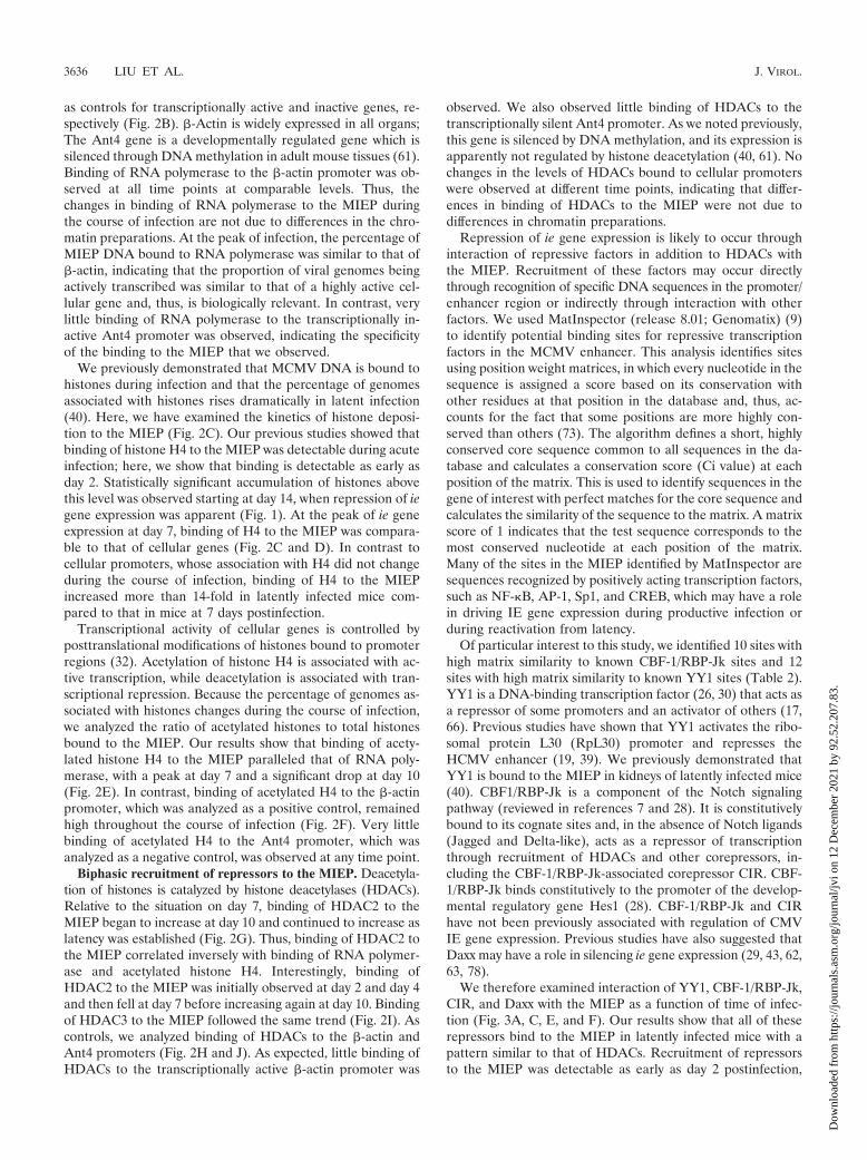
Of particular interest to this study, we identified 10 sites withhigh matrix similarity to known CBF-1/RBP-Jk sites and 12sites with high matrix similarity to known YY1 sites (Table 2).YY1 is a DNA-binding transcription factor (26, 30) that acts asa repressor of some promoters and an activator of others (17,66). Previous studies have shown that YY1 activates the ribo-somal protein L30 (RpL30) promoter and represses theHCMV enhancer (19, 39). We previously demonstrated thatYY1 is bound to the MIEP in kidneys of latently infected mice(40). CBF1/RBP-Jk is a component of the Notch signalingpathway (reviewed in references 7 and 28). It is constitutivelybound to its cognate sites and, in the absence of Notch ligands(Jagged and Delta-like), acts as a repressor of transcriptionthrough recruitment of HDACs and other corepressors, in-cluding the CBF-1/RBP-Jk-associated corepressor CIR. CBF-1/RBP-Jk binds constitutively to the promoter of the develop-mental regulatory gene Hes1 (28). CBF-1/RBP-Jk and CIRhave not been previously associated with regulation of CMVIE gene expression. Previous studies have also suggested thatDaxx may have a role in silencing ie gene expression (29, 43, 62,63, 78).
We therefore examined interaction of YY1, CBF-1/RBP-Jk,CIR, and Daxx with the MIEP as a function of time of infec-tion (Fig. 3A, C, E, and F). Our results show that all of theserepressors bind to the MIEP in latently infected mice with apattern similar to that of HDACs. Recruitment of repressorsto the MIEP was detectable as early as day 2 postinfection,
3636 LIU ET AL. J. VIROL.
Dow
nloa
ded
from
http
s://j
ourn
als.
asm
.org
/jour
nal/j
vi o
n 12
Dec
embe
r 20
21 b
y 92
.52.
207.
83.
prior to the activation of ie gene expression (Fig. 1). This wasfollowed by a loss of binding as activation of ie gene expressionoccurred at days 4 to 7, with a nadir of binding at day 7, whentranscription of the IE genes reached its peak. Relative to thefindings for day 7, there was a statistically significant increasein the percentage of genomes bound to these factors at day 14,which did not increase further at later times. As positive con-trols, we analyzed binding of YY1 and CBF-1/RBP-Jk to thepromoter regions of RpL30 and Hes1, respectively (Fig. 3Band D). In contrast to the MIEP, binding of these factors didnot change during the course of infection, indicating that dif-ferences in binding of YY1 and CBF-1/RBP-Jk to the MIEPwere not due to variability in the chromatin preparations. Fur-thermore, the level of binding to the MIEP in latently infectedmice was similar to or higher than that observed with cellularpromoters, indicating that the binding activity is likely to bebiologically relevant. Very little binding to the negative con-trols (the Ant4 promoter for YY1 and the �-actin promoter forCBF-1/RBP-Jk) was observed, indicating the specificity of theinteraction with the MIEP. Changes in the levels of binding ofthese factors to the MIEP are unlikely to be due to differencesin the levels of expression, since real-time PCR analysis of YY1and CBF-1 RNA levels and Western blot analysis of YY1protein levels showed no change in expression of these genesover the course of infection (data not shown). Binding of CIRand Daxx to the MIEP followed the same biphasic patternobserved for YY1 and CBF-1/RBP-Jk (Fig. 3E and F). Collec-tively, our results demonstrate that (i) cellular repressors arerecruited to the MIEP during infection and (ii) there are dra-matic changes in the proportion of MIEP molecules bound tothese repressors as the infection progresses from the very earlystages of infection prior to activation of IE gene expression toacute infection and then to latency.
YY1 and CBF-1/RBP-Jk are specifically recruited to theMIEP in latent infection. The observations that the MIEP
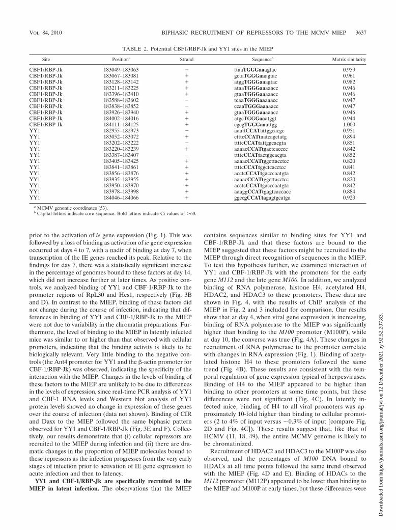
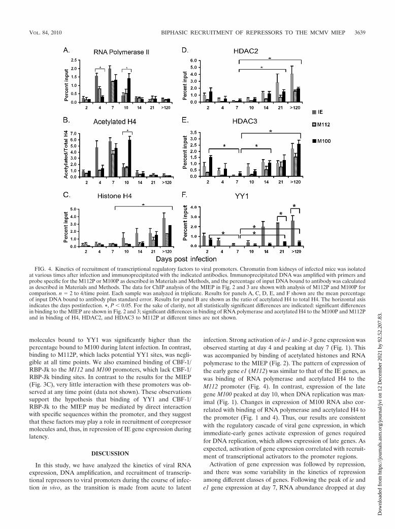
contains sequences similar to binding sites for YY1 andCBF-1/RBP-Jk and that these factors are bound to theMIEP suggested that these factors might be recruited to theMIEP through direct recognition of sequences in the MIEP.To test this hypothesis further, we examined interaction ofYY1 and CBF-1/RBP-Jk with the promoters for the earlygene M112 and the late gene M100. In addition, we analyzedbinding of RNA polymerase, histone H4, acetylated H4,HDAC2, and HDAC3 to these promoters. These data areshown in Fig. 4, with the results of ChIP analysis of theMIEP in Fig. 2 and 3 included for comparison. Our resultsshow that at day 4, when viral gene expression is increasing,binding of RNA polymerase to the MIEP was significantlyhigher than binding to the M100 promoter (M100P), whileat day 10, the converse was true (Fig. 4A). These changes inrecruitment of RNA polymerase to the promoter correlatewith changes in RNA expression (Fig. 1). Binding of acety-lated histone H4 to these promoters followed the sametrend (Fig. 4B). These results are consistent with the tem-poral regulation of gene expression typical of herpesviruses.Binding of H4 to the MIEP appeared to be higher thanbinding to other promoters at some time points, but thesedifferences were not significant (Fig. 4C). In latently in-fected mice, binding of H4 to all viral promoters was ap-proximately 10-fold higher than binding to cellular promot-ers (2 to 4% of input versus 0.3% of input [compare Fig.2D and Fig. 4C]). These results suggest that, like that ofHCMV (11, 18, 49), the entire MCMV genome is likely tobe chromatinized.
Recruitment of HDAC2 and HDAC3 to the M100P was alsoobserved, and the percentages of M100 DNA bound toHDACs at all time points followed the same trend observedwith the MIEP (Fig. 4D and E). Binding of HDACs to theM112 promoter (M112P) appeared to be lower than binding tothe MIEP and M100P at early times, but these differences were
TABLE 2. Potential CBF1/RBP-Jk and YY1 sites in the MIEP
Site Positiona Strand Sequenceb Matrix similarity
CBF1/RBP-Jk 183049–183063 � ttaaTGGGaaagtac 0.959CBF1/RBP-Jk 183067–183081 � gctaTGGGaaagtac 0.961CBF1/RBP-Jk 183128–183142 � atggTGGGaaagtac 0.982CBF1/RBP-Jk 183211–183225 � ataaTGGGaaaaacc 0.946CBF1/RBP-Jk 183396–183410 � gtaaTGGGaaaaacc 0.946CBF1/RBP-Jk 183588–183602 � tcaaTGGGaaaaacc 0.947CBF1/RBP-Jk 183838–183852 � ccaaTGGGaaaaacc 0.947CBF1/RBP-Jk 183926–183940 � gtaaTGGGaaaaacc 0.946CBF1/RBP-Jk 184002–184016 � atgcTGGGaaatggt 0.944CBF1/RBP-Jk 184111–184125 � cgcgTGGGaaattgg 1.000YY1 182955–182973 � aaattCCATattggcacgc 0.951YY1 183052–183072 � ctttcCCATtaatcagctatg 0.894YY1 183202–183222 � ttttcCCATtattggcacgta 0.851YY1 183220–183239 � aaaacCCATtgactcacccc 0.842YY1 183387–183407 � ttttcCCATtactggcacgta 0.852YY1 183405–183425 � aaaacCCATtggcttacctcc 0.820YY1 183841–183861 � ttttcCCATtggctcacctcc 0.841YY1 183856–183876 � acctcCCATtgacccaatgta 0.842YY1 183935–183955 � aaaacCCATtggcttacctcc 0.820YY1 183950–183970 � acctcCCATtgacccaatgta 0.842YY1 183978–183998 � aaaggCCATtgagtcaccacc 0.884YY1 184046–184066 � ggccgCCATtagagtgcatga 0.923
a MCMV genomic coordinates (53).b Capital letters indicate core sequence. Bold letters indicate Ci values of 60.
VOL. 84, 2010 BIPHASIC RECRUITMENT OF REPRESSORS TO THE MCMV MIEP 3637
Dow
nloa
ded
from
http
s://j
ourn
als.
asm
.org
/jour
nal/j
vi o
n 12
Dec
embe
r 20
21 b
y 92
.52.
207.
83.
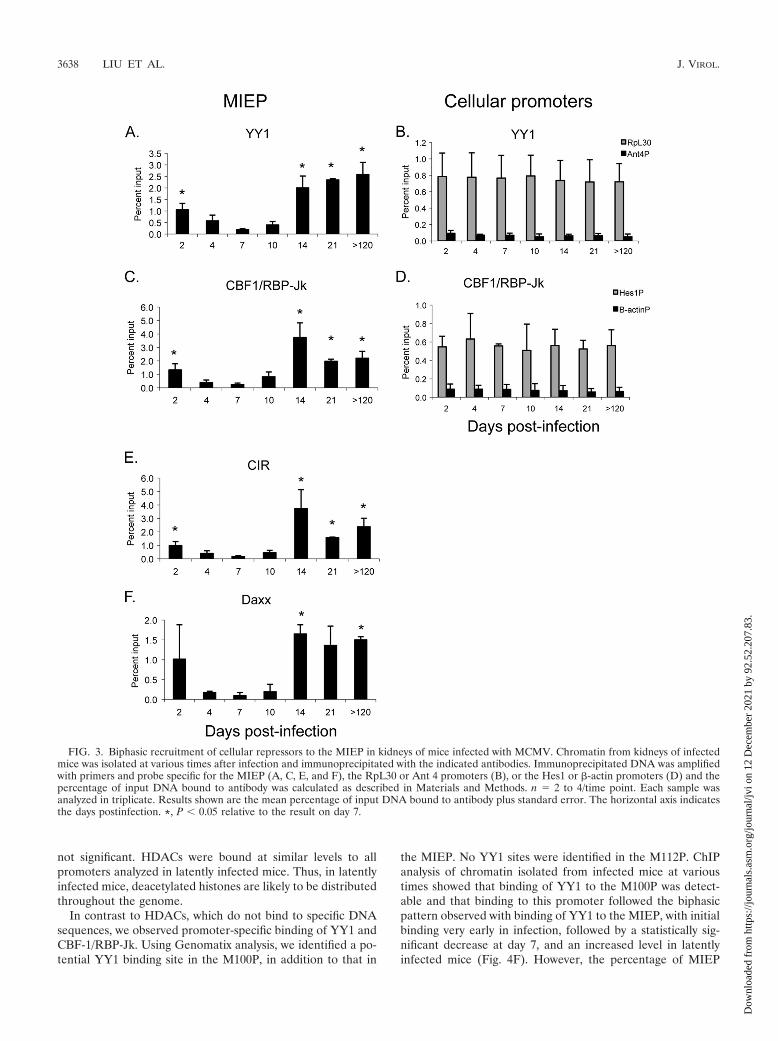
not significant. HDACs were bound at similar levels to allpromoters analyzed in latently infected mice. Thus, in latentlyinfected mice, deacetylated histones are likely to be distributedthroughout the genome.
In contrast to HDACs, which do not bind to specific DNAsequences, we observed promoter-specific binding of YY1 andCBF-1/RBP-Jk. Using Genomatix analysis, we identified a po-tential YY1 binding site in the M100P, in addition to that in
the MIEP. No YY1 sites were identified in the M112P. ChIPanalysis of chromatin isolated from infected mice at varioustimes showed that binding of YY1 to the M100P was detect-able and that binding to this promoter followed the biphasicpattern observed with binding of YY1 to the MIEP, with initialbinding very early in infection, followed by a statistically sig-nificant decrease at day 7, and an increased level in latentlyinfected mice (Fig. 4F). However, the percentage of MIEP
FIG. 3. Biphasic recruitment of cellular repressors to the MIEP in kidneys of mice infected with MCMV. Chromatin from kidneys of infectedmice was isolated at various times after infection and immunoprecipitated with the indicated antibodies. Immunoprecipitated DNA was amplifiedwith primers and probe specific for the MIEP (A, C, E, and F), the RpL30 or Ant 4 promoters (B), or the Hes1 or �-actin promoters (D) and thepercentage of input DNA bound to antibody was calculated as described in Materials and Methods. n � 2 to 4/time point. Each sample wasanalyzed in triplicate. Results shown are the mean percentage of input DNA bound to antibody plus standard error. The horizontal axis indicatesthe days postinfection. *, P � 0.05 relative to the result on day 7.
3638 LIU ET AL. J. VIROL.
Dow
nloa
ded
from
http
s://j
ourn
als.
asm
.org
/jour
nal/j
vi o
n 12
Dec
embe
r 20
21 b
y 92
.52.
207.
83.
molecules bound to YY1 was significantly higher than thepercentage bound to M100 during latent infection. In contrast,binding to M112P, which lacks potential YY1 sites, was negli-gible at all time points. We also examined binding of CBF-1/RBP-Jk to the M112 and M100 promoters, which lack CBF-1/RBP-Jk binding sites. In contrast to the results for the MIEP(Fig. 3C), very little interaction with these promoters was ob-served at any time point (data not shown). These observationssupport the hypothesis that binding of YY1 and CBF-1/RBP-Jk to the MIEP may be mediated by direct interactionwith specific sequences within the promoter, and they suggestthat these factors may play a role in recruitment of corepressormolecules and, thus, in repression of IE gene expression duringlatency.
DISCUSSION
In this study, we have analyzed the kinetics of viral RNAexpression, DNA amplification, and recruitment of transcrip-tional repressors to viral promoters during the course of infec-tion in vivo, as the transition is made from acute to latent
infection. Strong activation of ie-1 and ie-3 gene expression wasobserved starting at day 4 and peaking at day 7 (Fig. 1). Thiswas accompanied by binding of acetylated histones and RNApolymerase to the MIEP (Fig. 2). The pattern of expression ofthe early gene e1 (M112) was similar to that of the IE genes, aswas binding of RNA polymerase and acetylated H4 to theM112 promoter (Fig. 4). In contrast, expression of the lategene M100 peaked at day 10, when DNA replication was max-imal (Fig. 1). Changes in expression of M100 RNA also cor-related with binding of RNA polymerase and acetylated H4 tothe promoter (Fig. 1 and 4). Thus, our results are consistentwith the regulatory cascade of viral gene expression, in whichimmediate-early genes activate expression of genes requiredfor DNA replication, which allows expression of late genes. Asexpected, activation of gene expression correlated with recruit-ment of transcriptional activators to the promoter regions.
Activation of gene expression was followed by repression,and there was some variability in the kinetics of repressionamong different classes of genes. Following the peak of ie ande1 gene expression at day 7, RNA abundance dropped at day
FIG. 4. Kinetics of recruitment of transcriptional regulatory factors to viral promoters. Chromatin from kidneys of infected mice was isolatedat various times after infection and immunoprecipitated with the indicated antibodies. Immunoprecipitated DNA was amplified with primers andprobe specific for the M112P or M100P as described in Materials and Methods, and the percentage of input DNA bound to antibody was calculatedas described in Materials and Methods. The data for ChIP analysis of the MIEP in Fig. 2 and 3 are shown with analysis of M112P and M100P forcomparison. n � 2 to 4/time point. Each sample was analyzed in triplicate. Results for panels A, C, D, E, and F shown are the mean percentageof input DNA bound to antibody plus standard error. Results for panel B are shown as the ratio of acetylated H4 to total H4. The horizontal axisindicates the days postinfection. *, P � 0.05. For the sake of clarity, not all statistically significant differences are indicated: significant differencesin binding to the MIEP are shown in Fig. 2 and 3; significant differences in binding of RNA polymerase and acetylated H4 to the M100P and M112Pand in binding of H4, HDAC2, and HDAC3 to M112P at different times are not shown.
VOL. 84, 2010 BIPHASIC RECRUITMENT OF REPRESSORS TO THE MCMV MIEP 3639
Dow
nloa
ded
from
http
s://j
ourn
als.
asm
.org
/jour
nal/j
vi o
n 12
Dec
embe
r 20
21 b
y 92
.52.
207.
83.
10, while the DNA copy number remained high. Thus, repres-sion of ie and e1 gene expression occurred in the kidney be-tween day 7 and day 10 postinfection, while repression of M100was not observed until day 14. Repression of gene expressioncorrelated with loss of RNA polymerase and acetylated H4 topromoters.
Repression of viral gene expression is likely due to recruit-ment of chromatin remodeling complexes that render the pro-moters inaccessible to the transcription apparatus. As previ-ously noted (40), histone H4 and HDACs were bound to theMIEP at significantly higher levels in latently infected micethan in acutely infected mice. Here, we show that H4 andHDACs are also bound to other viral promoters at higherlevels in latently infected mice, suggesting that the entire ge-nome may be in a highly chromatinized, repressive state inlatency. In addition to HDACs, we have shown that a numberof cellular repressors, including YY1, CBF-1/RBP-Jk, CIR,and Daxx, are bound to the MIEP during latent infection.
Surprisingly, recruitment of repressors to viral promoterswas biphasic: binding was detectable at very early times postin-fection, prior to activation of ie gene expression, then de-creased with the onset of ie gene expression and increasedagain at day 10 to 14, when repression of gene expressionoccurred. Loss of these repressors correlated with increasedbinding of RNA polymerase and acetylated histones and acti-vation of immediate-early and early gene expression betweenday 4 and day 7 p.i., followed by amplification of viral DNAand activation of late gene expression. These results are similarto recent studies of HCMV infection of permissive cells, whichshowed that the genome becomes rapidly associated with his-tones bearing markers of repressed chromatin, which are thenlost as the viral program of transcription becomes activated(18).
Previous studies with HCMV have shown that during infec-tion of fibroblasts in vitro, the viral genome becomes rapidlyassociated with ND10 bodies in the nucleus (2, 43). Thesemultiprotein complexes are dynamic structures comprised oftranscriptional repressors, including PML, Daxx, and ATRX,which are thought to act as a form of host intrinsic defenseagainst infection through repression of ie gene expression (62,63, 75, 78). Two viral proteins that activate ie gene expression,the tegument protein pp71 and the immediate-early proteinIE-1, antagonize Daxx and HDACs and disrupt formation ofND10 bodies (1, 2, 35, 42, 48, 62, 63). Like HCMV IE-1,MCMV IE-1 binds to host cellular repressors and blocksHDAC activity (74). It is tempting to speculate that cellularrepressors are recruited to MCMV DNA when the genomeenters the nucleus and that the decrease in binding of theseproteins to the MIEP observed from day 2 to 7 is due todisruption of these interactions mediated by viral tegumentand IE-1 proteins. However, there may be other explanationsfor the loss of binding of repressors to the enhancer. Activationof cellular signaling pathways triggered by viral entry (27, 82)may help to derepress viral promoters. Alternatively, the ap-parent loss of repressors bound to the MIEP could be due tonew DNA being synthesized that lacks repressors, rather thanloss of repressors from viral DNA that was present at day 2.
Following activation of gene expression and loss of repres-sors, a second phase of binding of repressors to viral promoterswas observed from day 10 to day 21, which correlated with
repression of viral gene expression. During the course of in-fection in vivo, the adaptive immune response eliminates pro-ductively infected cells, leaving behind latently infected cells,which are presumably invisible to the host immune responsedue to transcriptional silencing of gene expression. Thus, theapparent increase in binding of repressive factors to the viralpromoters observed during the second phase of binding couldbe due largely to a change in the ratio of productively versuslatently infected cells as productively infected cells are clearedby the immune response.
While infected cells are abundant and readily detectableduring acute infection, the frequency of latently infected cellsis very low, suggesting that establishment of latency is a rareevent (5, 31, 50, 55, 64, 71). At the peak of IE RNA expression,binding of repressors to the MIEP fell significantly but was stilldetectable. These observations suggest that while most ge-nomes are free of cellular repressors between day 4 and day 7,allowing for expression of the ie genes and productive infec-tion, some viral genomes may remain in a repressive state withthese factors bound to the MIEP. These cells may form areservoir of latently infected cells which never transition toproductive infection in the days immediately following infec-tion. Thus, latency and productive infection may be mutuallyexclusive pathways which are chosen at the outset of infection.The observation that only 10% of infecting HCMV genomesare actively transcribed in permissive cells is consistent withthis model (18). However, our results do not exclude the pos-sibility that latency may be established in some cells as a resultof abortive infection subsequent to activation of viral geneexpression.
Recruitment of some cellular repressors to viral promoters islikely to be mediated by recognition of specific DNA sequencesin the promoter, while others may be recruited indirectly, as aresult of interactions with these sequence-specific DNA-bind-ing factors. The MCMV IE-3 protein, like HCMV IE-2, is arepressor of the MIEP as well as an activator of early geneexpression (45). Studies with HCMV IE-2 have shown that itrepresses the MIEP through binding to a cis repression se-quence immediately upstream of the transcription start siteand recruitment of chromatin remodeling complexes (11, 57,72). Thus, recruitment of repressors to the MIEP and repres-sion of ie gene expression observed between day 7 and day 10may be due in part to autorepression. However, repressorsmust be recruited to viral promoters through alternative meansprior to the activation of ie gene expression during acute in-fection and following the establishment of latency, when the IEgenes are not expressed in most latently infected cells (56).
Our studies of latently infected mice show that whileHDACs bind to all viral promoters tested, YY1 and CBF-1/RBP-Jk bind specifically to promoters containing their cognaterecognition sequences: CBF-1/RBP-Jk bound only to theMIEP, while YY1 bound to both the MIEP and M100P. Theseresults support the hypothesis that these factors are recruitedto these promoters though direct interaction with DNA se-quences in the promoter region. Since these repressors arepart of large corepressor complexes (7, 17), binding of thesefactors to the DNA may recruit additional repressors to viralpromoters. Interestingly, although YY1 bound to both theMIEP and the M100P at comparable levels at day 2, binding ofYY1 to the M100P was significantly lower than binding to the
3640 LIU ET AL. J. VIROL.
Dow
nloa
ded
from
http
s://j
ourn
als.
asm
.org
/jour
nal/j
vi o
n 12
Dec
embe
r 20
21 b
y 92
.52.
207.
83.
MIEP in latent infection. The significance of this observationwill require further analysis.
YY1 is a ubiquitously expressed (3, 14) member of thepolycomb group of proteins, which form chromatin-modifyingcomplexes that mediate transcriptional silencing (70). YY1 hasfundamental roles in embryogenesis, differentiation, replica-tion, cellular proliferation, and inflammation and has beenshown to regulate expression of many genes (17). YY1 was sonamed because it can function as either an activator or repres-sor of transcription, depending on a variety of factors, includ-ing its binding position relative to the transcription start site,intracellular concentration, and posttranslational modifica-tions, such as acetylation (8, 67, 80). These dual functions aremediated by different domains of the protein (66) which inter-act with repressors, including HDACs and SAP30 (23, 79),transcription factors, including Sp1 and c-myc (65, 68), or co-activators such as CBP, pCAF, and p300 (3, 8, 80). YY1 bindsdirectly to DNA through recognition of specific sequences (26,30) and thus can serve as an adaptor to recruit either activatingor repressive complexes (37) to promoter regions. YY1 hasbeen shown to be a repressor of the HCMV enhancer, whichhas multiple YY1 binding sites (39).
Intracellular signaling events can modulate YY1 activity.For example, under resting conditions, YY1 constitutively oc-cupies a single, high-affinity proximal site in the beta-interferonpromoter, leading to recruitment of HDACs and transcrip-tional repression. In response to viral infection, however, YY1occupies a second, lower-affinity distal site in the promoter,leading to recruitment of CBP and histone acetyltransferases(HATs), acetylation of histones bound to the promoter, andactivation of transcription (47, 77). In other cases, YY1 mayact solely as a repressor whose activity can be abrogatedthrough competition with other transcription factors, such asserum response factor, AP-1, or NF-B, that recognize over-lapping sites in promoter regions (36, 41, 81). AP-1 and NF-Bare activated in response to many inflammatory signaling path-ways and control expression of many genes that mediate innateand adaptive immunity. Thus, inflammation can alter the ac-tivity of YY1, either through conversion of YY1 from a re-pressor to an activator or through competition with activatingfactors for DNA binding sites.
Like YY1, CBF-1 is a DNA-binding protein that recruitscorepressors or coactivators and, thus, can act as a repressor oran activator of expression, depending on the cellular signalingenvironment. CBF-1 is the major downstream effector of theNotch signaling pathway. In the absence of Notch signaling,CBF-1 binds to promoters of target genes and recruits core-pressor complexes (7, 28). Interaction of transmembrane formsof Notch with its ligands, Jagged and Delta-like, results incleavage of Notch and translocation of the intracellular do-main into the nucleus, where it binds to CBF-1, displacestranscriptional repressors, and recruits activators that driveexpression of target genes. The Notch signaling system is con-served from Drosophila to vertebrates and regulates the ex-pression of genes involved in embryonic development (7, 28).In the immune system, Notch signaling is involved in the de-velopment and function of T cells, macrophages, NK cells, anddendritic cells (21). Recent studies indicate that CBF1 medi-ates Toll-like receptor-induced expression of some inflamma-tory cytokines, including TNF, IL-6, and IL-12, independent of
Notch signaling (22). Thus, like YY1, CBF-1 is an importantmediator of inflammation as well as embryonic development.
CBF-1 and/or YY1 has been implicated in the control oflatency and reactivation of human immunodeficiency virus(HIV), human papillomavirus (HPV), and Kaposi’s sarcoma-associated herpesvirus (KSHV) (20, 33, 34, 38, 76). Previousstudies have suggested that an inflammatory immune responsecan lead to reactivation of latent CMV through activation ofthe ie enhancer (10, 12, 15, 24, 25, 52, 60, 69). The observationsthat (i) YY1 and CBF-1 are bound to the MIEP in MCMVlatently infected mice and (ii) the activity of these factors canbe modulated by cellular signaling pathways, and particularlyby inflammatory stimuli, suggest that they could also have arole in regulating the switch from CMV latency to reactivation.
In summary, we have demonstrated in this study that severalcellular repressors are bound to the MIEP and other viralpromoters in latently infected mice. While these data are cor-relative, they provide strong circumstantial evidence in supportof the hypothesis that (i) transcriptional silencing of viral geneexpression through recruitment of cellular repressors leads tothe establishment of latency; (ii) some of these repressors maybe recruited directly to promoters through recognition of spe-cific sequences, leading to binding of additional corepressorsthrough protein-protein interactions; and (iii) latency may beestablished in some cells at very early times after infection.Investigation of the requirement for the putative YY1 andCBF-1/RBP-Jk binding sites in the MIEP for establishment oflatency should provide further insight into the mechanisms bywhich MCMV establishes latent infection.
ACKNOWLEDGMENT
This work was supported by Public Health Service grant R21AI76771 from the NIH.
REFERENCES
1. Ahn, J. H., E. J. Brignole III, and G. S. Hayward. 1998. Disruption of PMLsubnuclear domains by the acidic IE1 protein of human cytomegalovirus ismediated through interaction with PML and may modulate a RING finger-dependent cryptic transactivator function of PML. Mol. Cell. Biol. 18:4899–4913.
2. Ahn, J. H., and G. S. Hayward. 1997. The major immediate-early proteinsIE1 and IE2 of human cytomegalovirus colocalize with and disrupt PML-associated nuclear bodies at very early times in infected permissive cells.J. Virol. 71:4599–4613.
3. Austen, M., B. Luscher, and J. M. Luscher-Firzlaff. 1997. Characterizationof the transcriptional regulator YY1. The bipartite transactivation domain isindependent of interaction with the TATA box-binding protein, transcrip-tion factor IIB, TAFII55, or cAMP-responsive element-binding protein(CPB)-binding protein. J. Biol. Chem. 272:1709–1717.
4. Bain, M., M. Reeves, and J. Sinclair. 2006. Regulation of human cytomeg-alovirus gene expression by chromatin remodeling, p. 167–183. In M. Red-dehase (ed.), Cytomegaloviruses: molecular biology and immunology.Caister Academic Press, Norfolk, UK.
5. Balthesen, M., M. Messerle, and M. J. Reddehase. 1993. Lungs are a majororgan site of cytomegalovirus latency and recurrence. J. Virol. 67:5360–5366.
6. Boshart, M., F. Weber, G. Jahn, K. Dorsch-Hasler, B. Fleckenstein, and W.Schaffner. 1985. A very strong enhancer is located upstream of an immediateearly gene of human cytomegalovirus. Cell 41:521–530.
7. Bray, S. J. 2006. Notch signalling: a simple pathway becomes complex. Nat.Rev. Mol. Cell Biol. 7:678–689.
8. Bushmeyer, S., K. Park, and M. L. Atchison. 1995. Characterization offunctional domains within the multifunctional transcription factor, YY1.J. Biol. Chem. 270:30213–30220.
9. Cartharius, K., K. Frech, K. Grote, B. Klocke, M. Haltmeier, A. Klingenhoff,M. Frisch, M. Bayerlein, and T. Werner. 2005. MatInspector and beyond:promoter analysis based on transcription factor binding sites. Bioinformatics21:2933–2942.
10. Cook, C. H., J. Trgovcich, P. D. Zimmerman, Y. Zhang, and D. D. Sedmak.2006. Lipopolysaccharide, tumor necrosis factor alpha, or interleukin-1�
VOL. 84, 2010 BIPHASIC RECRUITMENT OF REPRESSORS TO THE MCMV MIEP 3641
Dow
nloa
ded
from
http
s://j
ourn
als.
asm
.org
/jour
nal/j
vi o
n 12
Dec
embe
r 20
21 b
y 92
.52.
207.
83.
triggers reactivation of latent cytomegalovirus in immunocompetent mice.J. Virol. 80:9151–9158.
11. Cuevas-Bennett, C., and T. Shenk. 2008. Dynamic histone H3 acetylationand methylation at human cytomegalovirus promoters during replication infibroblasts. J. Virol. 82:9525–9536.
12. Docke, W. D., S. Prosch, E. Fietze, V. Kimel, H. Zuckermann, C. Klug, U.Syrbe, D. H. Kruger, R. von Baehr, and H. D. Volk. 1994. Cytomegalovirusreactivation and tumour necrosis factor. Lancet 343:268–269.
13. Dorsch-Hasler, K., G. M. Keil, F. Weber, M. Jasin, W. Schaffner, and U. H.Koszinowski. 1985. A long and complex enhancer activates transcription ofthe gene coding for the highly abundant immediate early mRNA in murinecytomegalovirus. Proc. Natl. Acad. Sci. U. S. A. 82:8325–8329.
14. Drews, D., M. Klar, C. Dame, and A. U. Brauer. 2009. Developmentalexpression profile of the YY2 gene in mice. BMC Dev. Biol. 9:45.
15. Fietze, E., S. Prosch, P. Reinke, J. Stein, W. D. Docke, G. Staffa, S. Loning,S. Devaux, F. Emmrich, and R. von Baehr. 1994. Cytomegalovirus infectionin transplant recipients. The role of tumor necrosis factor. Transplantation58:675–680.
16. Gaytant, M. A., E. A. Steegers, B. A. Semmekrot, H. M. Merkus, and J. M.Galama. 2002. Congenital cytomegalovirus infection: review of the epidemi-ology and outcome. Obstet. Gynecol. Surv. 57:245–256.
17. Gordon, S., G. Akopyan, H. Garban, and B. Bonavida. 2006. Transcriptionfactor YY1: structure, function, and therapeutic implications in cancer biol-ogy. Oncogene 25:1125–1142.
18. Groves, I. J., M. B. Reeves, and J. H. Sinclair. 2009. Lytic infection ofpermissive cells with human cytomegalovirus is regulated by an intrinsic‘pre-immediate-early’ repression of viral gene expression mediated by his-tone post-translational modification. J. Gen. Virol. 90:2364–2374.
19. Hariharan, N., D. E. Kelley, and R. P. Perry. 1989. Equipotent mouseribosomal protein promoters have a similar architecture that includes inter-nal sequence elements. Genes Dev. 3:1789–1800.
20. He, G., and D. M. Margolis. 2002. Counterregulation of chromatin deacety-lation and histone deacetylase occupancy at the integrated promoter ofhuman immunodeficiency virus type 1 (HIV-1) by the HIV-1 repressor YY1and HIV-1 activator Tat. Mol. Cell. Biol. 22:2965–2973.
21. Hertzog, P. 2008. A notch in the toll belt.[Comment]. Immunity 29:663–665.22. Hu, X., A. Y. Chung, I. Wu, J. Foldi, J. Chen, J. D. Ji, T. Tateya, Y. J. Kang,
J. Han, M. Gessler, R. Kageyama, and L. B. Ivashkiv. 2008. Integratedregulation of Toll-like receptor responses by Notch and interferon-gammapathways. Immunity 29:691–703.
23. Huang, N. E., C. H. Lin, Y. S. Lin, and W. C. Yu. 2003. Modulation of YY1activity by SAP30. Biochem. Biophys. Res. Commun. 306:267–275.
24. Hummel, M., and M. I. Abecassis. 2002. A model for reactivation of CMVfrom latency. J. Clin. Virol. 25:S123–S136.
25. Hummel, M., Z. Zhang, S. Yan, I. DePlaen, P. Golia, T. Varghese, G.Thomas, and M. I. Abecassis. 2001. Allogeneic transplantation induces ex-pression of cytomegalovirus immediate-early genes in vivo: a model forreactivation from latency. J. Virol. 75:4814–4822.
26. Hyde-DeRuyscher, R. P., E. Jennings, and T. Shenk. 1995. DNA bindingsites for the transcriptional activator/repressor YY1. Nucleic Acids Res.23:4457–4465.
27. Isaacson, M. K., L. K. Juckem, and T. Compton. 2008. Virus entry andinnate immune activation. Curr. Top. Microbiol. Immunol. 325:85–100.
28. Kageyama, R., T. Ohtsuka, and T. Kobayashi. 2007. The Hes gene family:repressors and oscillators that orchestrate embryogenesis. Development 134:1243–1251.
29. Kalejta, R. F. 2008. Functions of human cytomegalovirus tegument proteinsprior to immediate early gene expression. Curr. Top. Microbiol. Immunol.325:101–115.
30. Kim, J. 2009. YY1’s longer DNA-binding motifs. Genomics 93:152–158.31. Koffron, A. J., M. Hummel, B. K. Patterson, S. Yan, D. B. Kaufman, J. P.
Fryer, F. P. Stuart, and M. I. Abecassis. 1998. Cellular localization of latentmurine cytomegalovirus. J. Virol. 72:95–103.
32. Kouzarides, T. 2007. Chromatin modifications and their function. Cell 128:693–705.
33. Lace, M. J., Y. Yamakawa, M. Ushikai, J. R. Anson, T. H. Haugen, and L. P.Turek. 2009. Cellular factor YY1 downregulates the human papillomavirus16 E6/E7 promoter, P97, in vivo and in vitro from a negative elementoverlapping the transcription-initiation site. J. Gen. Virol. 90:2402–2412.
34. Lan, K., D. A. Kuppers, and E. S. Robertson. 2005. Kaposi’s sarcoma-associated herpesvirus reactivation is regulated by interaction of latency-associated nuclear antigen with recombination signal sequence-binding pro-tein Jkappa, the major downstream effector of the Notch signaling pathway.J. Virol. 79:3468–3478.
35. Lee, H. R., D. J. Kim, J. M. Lee, C. Y. Choi, B. Y. Ahn, G. S. Hayward, andJ. H. Ahn. 2004. Ability of the human cytomegalovirus IE1 protein to mod-ulate sumoylation of PML correlates with its functional activities in tran-scriptional regulation and infectivity in cultured fibroblast cells. J. Virol.78:6527–6542.
36. Lee, T. C., Y. Shi, and R. J. Schwartz. 1992. Displacement of BrdUrd-induced YY1 by serum response factor activates skeletal alpha-actin tran-
scription in embryonic myoblasts. Proc. Natl. Acad. Sci. U. S. A. 89:9814–9818.
37. Le May, N., Z. Mansuroglu, P. Leger, T. Josse, G. Blot, A. Billecocq, R. Flick,Y. Jacob, E. Bonnefoy, and M. Bouloy. 2008. A SAP30 complex inhibitsIFN-beta expression in Rift Valley fever virus infected cells. PLoS Pathog.4:e13.
38. Liang, Y., and D. Ganem. 2003. Lytic but not latent infection by Kaposi’ssarcoma-associated herpesvirus requires host CSL protein, the mediator ofNotch signaling. Proc. Natl. Acad. Sci. U. S. A. 100:8490–8495.
39. Liu, R., J. Baillie, J. G. Sissons, and J. H. Sinclair. 1994. The transcriptionfactor YY1 binds to negative regulatory elements in the human cytomega-lovirus major immediate early enhancer/promoter and mediates repressionin non-permissive cells. Nucleic Acids Res. 22:2453–2459.
40. Liu, X. F., S. Yan, M. Abecassis, and M. Hummel. 2008. Establishment ofmurine cytomegalovirus latency in vivo is associated with changes in histonemodifications and recruitment of transcriptional repressors to the majorimmediate-early promoter. J. Virol. 82:10922–10931.
41. Lu, S. Y., M. Rodriguez, and W. S. Liao. 1994. YY1 represses rat serumamyloid A1 gene transcription and is antagonized by NF-kappa B duringacute-phase response. Mol. Cell. Biol. 14:6253–6263.
42. Lukashchuk, V., S. McFarlane, R. D. Everett, and C. M. Preston. 2008.Human cytomegalovirus protein pp71 displaces the chromatin-associatedfactor ATRX from nuclear domain 10 at early stages of infection. J. Virol.82:12543–12554.
43. Maul, G. G. 2008. Initiation of cytomegalovirus infection at ND10. Curr.Top. Microbiol. Immunol. 325:117–132.
44. Mercer, J. A., C. A. Wiley, and D. H. Spector. 1988. Pathogenesis of murinecytomegalovirus infection: identification of infected cells in the spleen duringacute and latent infections. J. Virol. 62:987–997.
45. Messerle, M., B. Buhler, G. M. Keil, and U. H. Koszinowski. 1992. Structuralorganization, expression, and functional characterization of the murinecytomegalovirus immediate-early gene 3. J. Virol. 66:27–36.
46. Mocarski, E. S., T. Shenk, and R. F. Pass. 2007. Cytomegaloviruses, p. 2702–2772. In D. M. Knipe and P. M. Howley (ed.), Fields virology, 5th ed., vol. 2.Wolters Kluwer Health/Lippincott Williams & Wilkins, Philadelphia, PA.
47. Mokrani, H., O. Sharaf el Dein, Z. Mansuroglu, and E. Bonnefoy. 2006.Binding of YY1 to the proximal region of the murine beta interferon pro-moter is essential to allow CBP recruitment and K8H4/K14H3 acetylation onthe promoter region after virus infection. Mol. Cell. Biol. 26:8551–8561.
48. Nevels, M., C. Paulus, and T. Shenk. 2004. Human cytomegalovirus imme-diate-early 1 protein facilitates viral replication by antagonizing histonedeacetylation. Proc. Natl. Acad. Sci. U. S. A. 101:17234–17239.
49. Nitzsche, A., C. Paulus, and M. Nevels. 2008. Temporal dynamics of cyto-megalovirus chromatin assembly in productively infected human cells. J. Vi-rol. 82:11167–11180.
50. Pollock, J. L., R. M. Presti, S. Paetzold, and H. W. Virgin IV. 1997. Latentmurine cytomegalovirus infection in macrophages. Virology 227:168–179.
51. Pollock, J. L., and H. W. Virgin IV. 1995. Latency, without persistence, ofmurine cytomegalovirus in the spleen and kidney. J. Virol. 69:1762–1768.
52. Prosch, S., K. Staak, J. Stein, C. Liebenthal, T. Stamminger, H.-D. Volk, andD. Kruger. 1995. Stimulation of the human cytomegalovirus IE enhancer/promoter in HL-60 cells by TNF alpha is mediated via induction of NF-kB.Virology 208:197–206.
53. Rawlinson, W. D., H. E. Farrell, and B. G. Barrell. 1996. Analysis of thecomplete DNA sequence of murine cytomegalovirus. J. Virol. 70:8833–8849.
54. Razonable, R. R., and C. V. Paya. 2002. Beta-herpesviruses in transplanta-tion. Rev. Med Microbiol. 13:163–176.
55. Reddehase, M. J., M. Balthesen, M. Rapp, S. Jonjic, I. Pavic, and U. H.Koszinowski. 1994. The conditions of primary infection define the load oflatent viral genome in organs and the risk of recurrent cytomegalovirusdisease. J. Exp. Med. 179:185–193.
56. Reddehase, M. J., C. O. Simon, C. K. Seckert, N. Lemmermann, and N. K.Grzimek. 2008. Murine model of cytomegalovirus latency and reactivation.Curr. Top. Microbiol. Immunol. 325:315–331.
57. Reeves, M., J. Murphy, R. Greaves, J. Fairley, A. Brehm, and J. Sinclair.2006. Autorepression of the human cytomegalovirus major immediate-earlypromoter/enhancer at late times of infection is mediated by the recruitmentof chromatin remodeling enzymes by IE86. J. Virol. 80:9998–10009.
58. Reeves, M., and J. Sinclair. 2008. Aspects of human cytomegalovirus latencyand reactivation. Curr. Top. Microbiol. Immunol. 325:297–313.
59. Reeves, M. B., P. A. MacAry, P. J. Lehner, J. G. Sissons, and J. H. Sinclair.2005. Latency, chromatin remodeling, and reactivation of human cytomeg-alovirus in the dendritic cells of healthy carriers. Proc. Natl. Acad. Sci.U. S. A. 102:4140–4145.
60. Reinke, P., S. Prosch, F. Kern, and H. D. Volk. 1999. Mechanisms of humancytomegalovirus (HCMV) (re)activation and its impact on organ transplantpatients. Transpl. Infect. Dis. 1:157–164.
61. Rodic, N., M. Oka, T. Hamazaki, M. R. Murawski, M. Jorgensen, D. M.Maatouk, J. L. Resnick, E. Li, and N. Terada. 2005. DNA methylation isrequired for silencing of ant4, an adenine nucleotide translocase selectivelyexpressed in mouse embryonic stem cells and germ cells. Stem Cells 23:1314–1323.
3642 LIU ET AL. J. VIROL.
Dow
nloa
ded
from
http
s://j
ourn
als.
asm
.org
/jour
nal/j
vi o
n 12
Dec
embe
r 20
21 b
y 92
.52.
207.
83.
62. Saffert, R. T., and R. F. Kalejta. 2007. Human cytomegalovirus gene expres-sion is silenced by Daxx-mediated intrinsic immune defense in model latentinfections established in vitro. J. Virol. 81:9109–9120.
63. Saffert, R. T., and R. F. Kalejta. 2006. Inactivating a cellular intrinsic im-mune defense mediated by Daxx is the mechanism through which the humancytomegalovirus pp71 protein stimulates viral immediate-early gene expres-sion. J. Virol. 80:3863–3871.
64. Seckert, C. K., A. Renzaho, H. M. Tervo, C. Krause, P. Deegen, B. Kuh-napfel, M. J. Reddehase, and N. K. Grzimek. 2009. Liver sinusoidal endo-thelial cells are a site of murine cytomegalovirus latency and reactivation.J. Virol. 83:8869–8884.
65. Seto, E., B. Lewis, and T. Shenk. 1993. Interaction between transcriptionfactors Sp1 and YY1. Nature 365:462–464.
66. Shi, Y., J.-S. Lee, and K. Galvin. 1997. Everything you ever wanted to knowabout Yin Yang 1. Biochim. Biophys. Acta 1332:F49–F66.
67. Shi, Y., E. Seto, L. S. Chang, and T. Shenk. 1991. Transcriptional repressionby YY1, a human GLI-Kruppel-related protein, and relief of repression byadenovirus E1A protein. Cell 67:377–388.
68. Shrivastava, A., S. Saleque, G. V. Kalpana, S. Artandi, S. P. Goff, and K.Calame. 1993. Inhibition of transcriptional regulator Yin-Yang-1 by associ-ation with c-Myc. Science 262:1889–1892.
69. Simon, C. O., C. K. Seckert, D. Dreis, M. J. Reddehase, and N. K. Grzimek.2005. Role for tumor necrosis factor alpha in murine cytomegalovirus tran-scriptional reactivation in latently infected lungs. J. Virol. 79:326–340.
70. Simon, J. A., and R. E. Kingston. 2009. Mechanisms of polycomb genesilencing: knowns and unknowns. Nat. Rev. Mol. Cell Biol. 10:697–708.
71. Slobedman, B., and E. S. Mocarski. 1999. Quantitative analysis of latenthuman cytomegalovirus. J. Virol. 73:4806–4812.
72. Stinski, M. F., and D. T. Petrik. 2008. Functional roles of the human cyto-megalovirus essential IE86 protein. Curr. Top. Microbiol. Immunol. 325:133–152.
73. Stormo, G. D. 2000. DNA binding sites: representation and discovery. Bioin-formatics 16:16–23.
74. Tang, Q., and G. G. Maul. 2003. Mouse cytomegalovirus immediate-earlyprotein 1 binds with host cell repressors to relieve suppressive effects on viraltranscription and replication during lytic infection. J. Virol. 77:1357–1367.
75. Tavalai, N., P. Papior, S. Rechter, M. Leis, and T. Stamminger. 2006.Evidence for a role of the cellular ND10 protein PML in mediating intrinsicimmunity against human cytomegalovirus infections. J. Virol. 80:8006–8018.
76. Tyagi, M., and J. Karn. 2007. CBF-1 promotes transcriptional silencingduring the establishment of HIV-1 latency. EMBO J. 26:4985–4995.
77. Weill, L., E. Shestakova, and E. Bonnefoy. 2003. Transcription factorYY1 binds to the murine beta interferon promoter and regulates itstranscriptional capacity with a dual activator/repressor role. J. Virol.77:2903–2914.
78. Woodhall, D. L., I. J. Groves, M. B. Reeves, G. Wilkinson, and J. H. Sinclair.2006. Human Daxx-mediated repression of human cytomegalovirus geneexpression correlates with a repressive chromatin structure around the majorimmediate early promoter. J. Biol. Chem. 281:37652–37660.
79. Yang, W. M., C. Inouye, Y. Zeng, D. Bearss, and E. Seto. 1996. Transcrip-tional repression by YY1 is mediated by interaction with a mammalianhomolog of the yeast global regulator RPD3. Proc. Natl. Acad. Sci. U. S. A.93:12845–12850.
80. Yao, Y. L., W. M. Yang, and E. Seto. 2001. Regulation of transcription factorYY1 by acetylation and deacetylation. Mol. Cell. Biol. 21:5979–5991.
81. Ye, J., M. Cippitelli, L. Dorman, J. R. Ortaldo, and H. A. Young. 1996. Thenuclear factor YY1 suppresses the human gamma interferon promoterthrough two mechanisms: inhibition of AP1 binding and activation of asilencer element. Mol. Cell. Biol. 16:4744–4753.
82. Yurochko, A. D. 2008. Human cytomegalovirus modulation of signal trans-duction. Curr. Top. Microbiol. Immunol. 325:205–220.
VOL. 84, 2010 BIPHASIC RECRUITMENT OF REPRESSORS TO THE MCMV MIEP 3643
Dow
nloa
ded
from
http
s://j
ourn
als.
asm
.org
/jour
nal/j
vi o
n 12
Dec
embe
r 20
21 b
y 92
.52.
207.
83.


![Chimeric Activators and Repressors Define HY5 Activity and … · Chimeric Activators and Repressors Define HY5 Activity and Reveal a Light-Regulated Feedback Mechanism[OPEN] Yogev](https://img.dokumen.tips/doc/110x75/5f10fef97cde8b41974dd02a/chimeric-activators-and-repressors-define-hy5-activity-and-chimeric-activators-and.jpg)