Embed Size (px)
Citation preview
GLOBAL STRATEGY FOR THE DIAGNOSIS, MANAGEMENT, AND PREVENTION OF
CHRONIC OBSTRUCTIVE PULMONARY DISEASE2006
Global Initiative for ChronicObstructiveLungDisease
Copyright © 2006 MCR VISION, Inc.All Rights Reserved
GOLD_WR_06 1/22/07 12:50 PM Page A1
i
GLOBAL INITIATIVE FORCHRONIC OBSTRUCTIVE LUNG DISEASE
GLOBAL STRATEGY FOR THE DIAGNOSIS, MANAGEMENT, AND PREVENTION OF CHRONIC OBSTRUCTIVE PULMONARY DISEASE (2006)
© 2006 Global Initative for Chronic Obstructive Lung Disease
GOLD_WR_06 1/22/07 12:50 PM Page i

Global Strategy for the Diagnosis, Management, and Prevention ofChronic Obstructive Pulmonary Disease (2006)
GOLD EXECUTIVE COMMITTEE*
A. Sonia Buist, MD, ChairOregon Health & Science University Portland, Oregon, USA
Antonio Anzueto, MD (Representing the American Thoracic Society)University of Texas Health Science CenterSan Antonio, Texas, USA
Peter Calverley, MDUniversity Hospital AintreeLiverpool, UK
Teresita S. deGuia, MDPhilippine Heart CenterQuezon City, Philippines
Yoshinosuke Fukuchi, MD(Representing the Asian Pacific Society for Respirology)Tokyo, Japan
Christine Jenkins, MDWoolcock Institute of Medical ResearchSydney, NSW, Australia
Nikolai Khaltaev, MD(Representing the World Health Organization)Geneva, Switzerland
James Kiley, PhD(Representing the National Heart, Lung, and Blood Institute,National Institutes of Health, Department of Health and Human Services) Bethesda, Maryland, USA
Ali Kocabas, MD Cukurova University School of MedicineBalcali, Adana, Turkey
Mará Victorina López, MD(Representing the Latin American Thoracic Society)Montevideo, Uruguay
Ewa Nizankowska-Mogilnicka, MDUniversity School of MedicineKrakow, Poland
Klaus F. Rabe, MD, PhD Leiden University Medical Center Leiden, The Netherlands
Roberto Rodriguez Roisin, MDHospital ClinicBarcelona, Spain
Thys van der Molen, MDUniversity of GroningenGroningen, The Netherlands
Chris van Weel, MD(Representing the World Organization of Family Doctors(WONCA))University of NijmegenNijmegen, The Netherlands
GOLD SCIENCE COMMITTEE*
Klaus F. Rabe, MD, PhD, ChairLeiden University Medical Center Leiden, The Netherlands
A. G. Agusti, MD (Effective June 2006)Hospital Universitari Son DuretaPalma de Mallorca, Spain
Antonio Anzueto, MD University of Texas Health Science CenterSan Antonio, Texas, USA
Peter J. Barnes, MDNational Heart and Lung InstituteLondon, UK
A. Sonia Buist, MDOregon Health & Science UniversityPortland, Oregon, USA
Peter Calverley, MDUniversity Hospital AintreeLiverpool, UK
Marc Decramer, MD (Effective June 2006)University HospitalLeuven, Belgium
Yoshinosuke Fukuchi, MDPresident Asian Pacific Society for RespirologyTokyo, Japan
Paul Jones, MD (Effective June 2006)St. George's Hospital Medical SchoolLondon, UK
ii
*Disclosure forms for GOLD Committees are posted on the GOLD Website, www.goldcopd.org
GOLD_WR_06 1/22/07 12:50 PM Page ii

Roberto Rodriguez Roisin, MDHospital ClinicBarcelona, Spain
Jorgen Vestbo, MD (Effective June 2006)Hvidovre University HospitalHvidovre, Denmark
Jan Zielinski, MDInstitute of TB and Lung DiseasesWarsaw, Poland
CHAPTER CONTRIBUTORS
Leonardo Fabbri, MDUniversity of Modena & Reggio EmiliaModena, Italy
James C. Hogg, MDSt. Paul’s HospitalVancouver, British Columbia, Canada
Christine Jenkins, MDWoolcock Institute of Medical ResearchSydney, NSW, Australia
Ewa Nizankowska-Mogilnicka, MDUniversity School of MedicineKrakow, Poland
Sean Sullivan, MDUniversity of WashingtonSeattle, Washington, USA
Thys van der Molen, MDUniversity of GroningenGroningen, The Netherlands
Chris van Weel, MDUniversity of NijmegenNijmegen, The Netherlands
REVIEWERS
Bart Celli, MDCaritas St. Elizabeth's Medical CenterBrighton, Massachusetts, USA
M.W. Elliott, MDSt. James's University HospitalWest Yorkshire, UK
H.A.M. Kerstjens, MD, PhD University Medical Center Groningen Groningen, The Netherlands
Peter Lange, MDHvidovre HospitalHvidovre, Denmark
Carlos M. Luna, MDPresident, ALATBuenos Aires, Argentina
Dennis Niewoehner, MDUniversity of MinnesotaMinneapolis, Minnesota, USA
Jim Reid, MDDunedin School of MedicineUniversity of Otago Dunedin, New Zealand
Sanjay Sethi, MDVA Medical ResearchBuffalo, New York, USA
Peter Sterk, MDLeiden University Medical CenterLeiden, The Netherlands
GOLD NATIONAL LEADERS WHO SUBMITTED COMMENTS
Lorenzo Corbetta, MDUniversità di Firenze Firenze, Italy
Maia Gotua, MD, PhDCenter of Allergy & ImmunologyTbilisi, Georgia
Gérard Huchon, MDUniversity of ParisParis, France
Prof. E.M. Irusen South Africa Thoracic SocietyUniversity of StellenboschCape Town, South Africa
Yousser Mohammad, MDTishreen University School of MedicineLattakia, Syria
Jaromir Musil, PhDStanislav Kos, MD, PhDF. Salajka, PhDVladimir Vondra, MD, PhDCzech Association Against COPDPrague, Czech Republic
Júlio A. Oliveira, MDFernando Lundgren, MDJosé R. Jardim, MDBrazil
Vesna Petrovic, MDJUDAH Association for Asthma and COPDSerbia
iii
GOLD_WR_06 1/22/07 12:50 PM Page iii

Chronic Obstructive Pulmonary Disease (COPD) remainsa major public health problem. It is the fourth leadingcause of chronic morbidity and mortality in the UnitedStates, and is projected to rank fifth in 2020 in burden of disease caused worldwide, according to a study published by the World Bank/World Health Organization.Furthermore, although COPD has received increasingattention from the medical community in recent years, itis still relatively unknown or ignored by the public as wellas public health and government officials.
In 1998, in an effort to bring more attention to COPD, itsmanagement, and its prevention, a committed group ofscientists encouraged the US National Heart, Lung, andBlood Institute and the World Health Organization to formthe Global Initiative for Chronic Obstructive Lung Disease(GOLD). Among the important objectives of GOLD are toincrease awareness of COPD and to help the millions ofpeople who suffer from this disease and die prematurelyfrom it or its complications.
The first step in the GOLD program was to prepare aconsensus report, Global Strategy for the Diagnosis,Management, and Prevention of COPD, which was published in 2001. The report was written by an ExpertPanel, which was chaired by Professor Romain Pauwelsof Belgium and included a distinguished group of healthprofessionals from the fields of respiratory medicine, epidemiology, socioeconomics, public health, and healtheducation. The Expert Panel reviewed existing COPDguidelines and new information on pathogenic mechanismsof COPD, bringing all of this material together in the consensus document. The present, newly revised documentfollows the same format as the original consensus report,but has been updated to reflect the many publications onCOPD that have appeared since 2001.
Since the original consensus report was published in2001, a network of international experts known as GOLDNational Leaders has been formed to implement thereport’s recommendations. Many of these experts haveinitiated investigations of the causes and prevalence ofCOPD in their countries, and developed innovativeapproaches for the dissemination and implementation of COPD management guidelines. We appreciate theenormous amount of work the GOLD National Leadershave done on behalf of their patients with COPD.
In spite of the achievements in the five years since theGOLD report was originally published, considerable additional work is ahead of all of us if we are to controlthis major public health problem. The GOLD initiative willcontinue to bring COPD to the attention of governments,public health officials, health care workers, and the general public, but a concerted effort by all involved inhealth care will be necessary.
I would like to acknowledge the work of the members ofthe GOLD Science Committee who prepared this revisedreport. We look forward to our continued work with interested organizations and the GOLD National Leadersto meet the goals of this initiative.
We are most appreciative of the unrestricted educationalgrants from Altana, AstraZeneca, Boehringer Ingelheim,Chiesi, GlasoSmithKline, Mitsubishi Pharma Corporation,Nikken Chemicals, Co,. Ltd., Novartis, and Pfizer thatenabled development of this report.
A. Sonia Buist, MDPortland, Oregon, USAChair, GOLD Executive Committee
iv
PREFACE
GOLD_WR_06 1/22/07 12:50 PM Page iv
Introduction . . . . . . . . . . . . . . . . . . . . . . . . . . . . . . .viii
1. Definition . . . . . . . . . . . . . . . . . . . . . . . . . . . . . . . .1Key Points . . . . . . . . . . . . . . . . . . . . . . . . . . . . . . . . . .2Definition . . . . . . . . . . . . . . . . . . . . . . . . . . . . . . . . . . .2
Airflow limitation in COPD . . . . . . . . . . . . . . . . . . . .2COPD and Co morbidities . . . . . . . . . . . . . . . . . . . .3
Natural History . . . . . . . . . . . . . . . . . . . . . . . . . . . . . . .3Spirometric Classification of Severity . . . . . . . . . . . .3Stages of COPD . . . . . . . . . . . . . . . . . . . . . . . . . . .4
Scope of the Report . . . . . . . . . . . . . . . . . . . . . . . . . . .5Asthma and COPD . . . . . . . . . . . . . . . . . . . . . . . . . .5Pulmonary Tuberculosis and COPD . . . . . . . . . . . . .5
References . . . . . . . . . . . . . . . . . . . . . . . . . . . . . . . . .5
2. Burden of COPD . . . . . . . . . . . . . . . . . . . . . . . . . . .7Key Points . . . . . . . . . . . . . . . . . . . . . . . . . . . . . . . . . .8Introduction . . . . . . . . . . . . . . . . . . . . . . . . . . . . . . . . .8Epidemiology . . . . . . . . . . . . . . . . . . . . . . . . . . . . . . . .8Prevalence . . . . . . . . . . . . . . . . . . . . . . . . . . . . . . . . . .8
Morbidity . . . . . . . . . . . . . . . . . . . . . . . . . . . . . . . . .9Mortality . . . . . . . . . . . . . . . . . . . . . . . . . . . . . . . . .10
Economic and Social Burden of COPD . . . . . . . . . . . .11Economic Burden . . . . . . . . . . . . . . . . . . . . . . . . . .11Social Burden . . . . . . . . . . . . . . . . . . . . . . . . . . . .12
References . . . . . . . . . . . . . . . . . . . . . . . . . . . . . . . . .12
3. Risk Factors . . . . . . . . . . . . . . . . . . . . . . . . . . . . .15Key Points . . . . . . . . . . . . . . . . . . . . . . . . . . . . . . . . .16Introduction . . . . . . . . . . . . . . . . . . . . . . . . . . . . . . . .16Risk Factors . . . . . . . . . . . . . . . . . . . . . . . . . . . . . . . .16
Genes . . . . . . . . . . . . . . . . . . . . . . . . . . . . . . . . . .16Inhalational Exposures . . . . . . . . . . . . . . . . . . . . . .17
Tobacco smoke . . . . . . . . . . . . . . . . . . . . . . . . . .17Occupational dusts and chemicals . . . . . . . . . . .17Indoor air pollution . . . . . . . . . . . . . . . . . . . . . . .17Outdoor air pollution . . . . . . . . . . . . . . . . . . . . . .18
Lung Growth and Development . . . . . . . . . . . . . . .18Oxidative Stress . . . . . . . . . . . . . . . . . . . . . . . . . . .18Gender . . . . . . . . . . . . . . . . . . . . . . . . . . . . . . . . .18Infections . . . . . . . . . . . . . . . . . . . . . . . . . . . . . . . .18Socioeconomic Status . . . . . . . . . . . . . . . . . . . . . .18Nutrition . . . . . . . . . . . . . . . . . . . . . . . . . . . . . . . . .18Asthma . . . . . . . . . . . . . . . . . . . . . . . . . . . . . . . . .19
References . . . . . . . . . . . . . . . . . . . . . . . . . . . . . . . . .19
4. Pathology, Pathogenesis, and Pathophysiology . .23Key Points . . . . . . . . . . . . . . . . . . . . . . . . . . . . . . . . .24Introduction . . . . . . . . . . . . . . . . . . . . . . . . . . . . . . . .24Pathology . . . . . . . . . . . . . . . . . . . . . . . . . . . . . . . . . .24Pathogenesis . . . . . . . . . . . . . . . . . . . . . . . . . . . . . . .25
Inflammatory Cells . . . . . . . . . . . . . . . . . . . . . . . . .25Inflammatory Mediators . . . . . . . . . . . . . . . . . . . . .25Oxidative Stress . . . . . . . . . . . . . . . . . . . . . . . . . . .25Protease-Antiprotease Imbalance . . . . . . . . . . . . . .26Differences in Inflammation Between COPD
and Asthma . . . . . . . . . . . . . . . . . . . . . . . . . . . .26Pathophysiology . . . . . . . . . . . . . . . . . . . . . . . . . . . . .26
Airflow Limitation and Air Trapping . . . . . . . . . . . . .26Gas Exchange Abnormalities . . . . . . . . . . . . . . . . .26Mucus Hypersecretion . . . . . . . . . . . . . . . . . . . . . .26Pulmonary Hypertension . . . . . . . . . . . . . . . . . . . .28Systemic Features . . . . . . . . . . . . . . . . . . . . . . . . .28
Exacerbations . . . . . . . . . . . . . . . . . . . . . . . . . . . . . .28References . . . . . . . . . . . . . . . . . . . . . . . . . . . . . . . . .28
5. Management of COPD . . . . . . . . . . . . . . . . . . . . .31Introduction . . . . . . . . . . . . . . . . . . . . . . . . . . . . . . . .32
Component 1: Assess and Monitor Disease . . . . . .33Key Points . . . . . . . . . . . . . . . . . . . . . . . . . . . . . . . . .33Initial Diagnosis . . . . . . . . . . . . . . . . . . . . . . . . . . . . .33
Assessment of Symptoms . . . . . . . . . . . . . . . . . . .33Dyspnea . . . . . . . . . . . . . . . . . . . . . . . . . . . . . . .34Cough . . . . . . . . . . . . . . . . . . . . . . . . . . . . . . . . .34Sputum production . . . . . . . . . . . . . . . . . . . . . . .34Wheezing and chest tightness . . . . . . . . . . . . . . .34Additional features in severe disease . . . . . . . . . .35
Medical History . . . . . . . . . . . . . . . . . . . . . . . . . . .35Physical Examination . . . . . . . . . . . . . . . . . . . . . . .35
Inspection . . . . . . . . . . . . . . . . . . . . . . . . . . . . . .35Palpation and percussion . . . . . . . . . . . . . . . . . .35Auscultation . . . . . . . . . . . . . . . . . . . . . . . . . . . .35
Measurement of Airflow Limitation . . . . . . . . . . . . .36Assessment of COPD Severity . . . . . . . . . . . . . . . .37Additional Investigations . . . . . . . . . . . . . . . . . . . . .37
Bronchodilator reversibility testing . . . . . . . . . . . .37Chest X-ray . . . . . . . . . . . . . . . . . . . . . . . . . . . . .38Arterial blood gas measurement . . . . . . . . . . . . .38Alpha-1 antitrypsin deficiency screening . . . . . . .38
Differential Diagnosis . . . . . . . . . . . . . . . . . . . . . . .38Ongoing Monitoring and Assessment . . . . . . . . . . . . .39
v
TABLE OF CONTENTS
GOLD_WR_06 1/22/07 12:50 PM Page v
Monitor Disease Progression andDevelopment of Complications . . . . . . . . . . . . . . .40
Pulmonary function . . . . . . . . . . . . . . . . . . . . .40Arterial blood gas measurement . . . . . . . . . . .40Assessment of pulmonary hemodynamics . . . .40Diagnosis of right heart failure or cor pulmonale . .40CT and ventilation-perfusion scanning . . . . . . .40Hematocrit . . . . . . . . . . . . . . . . . . . . . . . . . . .40Respiratory muscle function . . . . . . . . . . . . . .40Sleep studies . . . . . . . . . . . . . . . . . . . . . . . . .40Exercise testing . . . . . . . . . . . . . . . . . . . . . . .40
Monitor Pharmacotherapy andOther Medical Treatment . . . . . . . . . . . . . . . . . .40
Monitor Exacerbation History . . . . . . . . . . . . . . . . .41Monitor Comorbidities . . . . . . . . . . . . . . . . . . . . . .41
Component 2: Reduce Risk Factors . . . . . . . . . . . .42Key Points . . . . . . . . . . . . . . . . . . . . . . . . . . . . . . . . .42Introduction . . . . . . . . . . . . . . . . . . . . . . . . . . . . . . . .42Tobacco Smoke . . . . . . . . . . . . . . . . . . . . . . . . . . . . .42
Smoking Prevention . . . . . . . . . . . . . . . . . . . . . . . .42Smoking Cessation . . . . . . . . . . . . . . . . . . . . . . . .43
The role of health care providers in smoking cessation . . . . . . . . . . . . . . . . . . . .43
Counseling . . . . . . . . . . . . . . . . . . . . . . . . . . .44Pharmacotherapy . . . . . . . . . . . . . . . . . . . . . .45
Occupational Exposures . . . . . . . . . . . . . . . . . . . . . . .45Indoor/Outdoor Air Pollution . . . . . . . . . . . . . . . . . . . .46
Regulation of Air Quality . . . . . . . . . . . . . . . . . . . . .46Steps for Health Care Providers/Patients . . . . . . . .46
Component 3: Manage Stable COPD . . . . . . . . . . . .47Key Points . . . . . . . . . . . . . . . . . . . . . . . . . . . . . . . . .47Introduction . . . . . . . . . . . . . . . . . . . . . . . . . . . . . . . .47Education . . . . . . . . . . . . . . . . . . . . . . . . . . . . . . . . . .47
Goals and Educational Strategies . . . . . . . . . . . . . .48Components of an Education Program . . . . . . . . . .48Cost Effectiveness of Education
Programs for COPD Patients . . . . . . . . . . . . . .49Pharmacologic Treatment . . . . . . . . . . . . . . . . . . . . . .49
Overview of the Medications . . . . . . . . . . . . . . . . . .49Bronchodilators . . . . . . . . . . . . . . . . . . . . . . . . . . .50
�2-agonists . . . . . . . . . . . . . . . . . . . . . . . . . . .51Anticholinergics . . . . . . . . . . . . . . . . . . . . . . . .52Methylxanthines . . . . . . . . . . . . . . . . . . . . . . .52Combination bronchodilator therapy . . . . . . . .53
Glucocorticosteroids . . . . . . . . . . . . . . . . . . . . . . . .53Oral glucocorticosteroids: short-term . . . . . . . .53Oral glucocorticosteroids: long-term . . . . . . . .53Inhaled glucocorticosteroids . . . . . . . . . . . . . .53
Pharmacologic Therapy by Disease Severity . . . . .54Other Pharmacologic Treatments . . . . . . . . . . . . . .55
Vaccines . . . . . . . . . . . . . . . . . . . . . . . . . . . . .55Alpha-1 antitrypsin augmentation therapy . . . .55Antibiotics . . . . . . . . . . . . . . . . . . . . . . . . . . . .55Mucolytic agents . . . . . . . . . . . . . . . . . . . . . . .55Antioxidant agents . . . . . . . . . . . . . . . . . . . . .55Immunoregulators . . . . . . . . . . . . . . . . . . . . . .55Antitussives . . . . . . . . . . . . . . . . . . . . . . . . . .55Vasodilators . . . . . . . . . . . . . . . . . . . . . . . . . .55Narcotics (morphine) . . . . . . . . . . . . . . . . . . . .55Others . . . . . . . . . . . . . . . . . . . . . . . . . . . . . .56
Non-Pharmacologic Treatment . . . . . . . . . . . . . . . . . .56Rehabilitation . . . . . . . . . . . . . . . . . . . . . . . . . . . . .56
Patient selection and program design . . . . . . .56Components of pulmonary rehabilitation
programs . . . . . . . . . . . . . . . . . . . . . . . . . . .57Assessment and follow-up . . . . . . . . . . . . . . . .58Economic cost of rehabilitation programs . . . .58
Oxygen Therapy . . . . . . . . . . . . . . . . . . . . . . . . . . .58Cost considerations . . . . . . . . . . . . . . . . . . . .59Oxygen use in air travel . . . . . . . . . . . . . . . . .59
Ventilatory Support . . . . . . . . . . . . . . . . . . . . . . . . .60Surgical Treatments . . . . . . . . . . . . . . . . . . . . . . . .60
Bullectomy . . . . . . . . . . . . . . . . . . . . . . . . . . .60Lung volume reduction surgery . . . . . . . . . . . .60Lung transplantation . . . . . . . . . . . . . . . . . . . .60
Special Considerations . . . . . . . . . . . . . . . . . . . . . .61Surgery in COPD . . . . . . . . . . . . . . . . . . . . . .61
Component 4: Manage Exacerbations . . . . . . . . . . .62Key Points . . . . . . . . . . . . . . . . . . . . . . . . . . . . . . . . .62Introduction . . . . . . . . . . . . . . . . . . . . . . . . . . . . . . . .62Diagnosis and Assessment of Severity . . . . . . . . . . . .62
Medical History . . . . . . . . . . . . . . . . . . . . . . . . . . .62Assessment of Severity . . . . . . . . . . . . . . . . . . . . .63
Spirometry and PEF . . . . . . . . . . . . . . . . . . . .63Pulse oximetry/Arterial blood gases . . . . . . . . .63Chest X-ray and ECG . . . . . . . . . . . . . . . . . . .63Other laboratory tests . . . . . . . . . . . . . . . . . . .63
Differential Diagnoses . . . . . . . . . . . . . . . . . . . . . .63Home Management . . . . . . . . . . . . . . . . . . . . . . . . . .64
Bronchodilator Therapy . . . . . . . . . . . . . . . . . . . . .64Glucocorticosteroids . . . . . . . . . . . . . . . . . . . . . . . .64Antibiotics . . . . . . . . . . . . . . . . . . . . . . . . . . . . . . .64
Hospital Management . . . . . . . . . . . . . . . . . . . . . . . . .64Emergency Department or Hospital . . . . . . . . . . . .65
Controlled oxygen therapy . . . . . . . . . . . . . . .65Bronchodilator therapy . . . . . . . . . . . . . . . . . .65Glucocorticosteroids . . . . . . . . . . . . . . . . . . . .66
vi
GOLD_WR_06 1/22/07 12:50 PM Page vi

Antibiotics . . . . . . . . . . . . . . . . . . . . . . . . . . . .66Respiratory Stimulants . . . . . . . . . . . . . . . . . .67Ventilatory support . . . . . . . . . . . . . . . . . . . . .67Other measures . . . . . . . . . . . . . . . . . . . . . . .69
Hospital Discharge and Follow-Up . . . . . . . . . . . . .69References . . . . . . . . . . . . . . . . . . . . . . . . . . . . . . . . .69
6. Translating Guideline Recommendations to theContext of (Primary) Care . . . . . . . . . . . . . . . . . .85
Key Points . . . . . . . . . . . . . . . . . . . . . . . . . . . . . . . . .86Introduction . . . . . . . . . . . . . . . . . . . . . . . . . . . . . . . .86Diagnosis . . . . . . . . . . . . . . . . . . . . . . . . . . . . . . . . . .86
Respiratory Symptoms . . . . . . . . . . . . . . . . . . . . . .86Spirometry . . . . . . . . . . . . . . . . . . . . . . . . . . . . . . .86
Comorbidities . . . . . . . . . . . . . . . . . . . . . . . . . . . . . . .87Reducing Exposure to Risk Factors . . . . . . . . . . . . . .87Implementation of COPD Guidelines . . . . . . . . . . . . . .87References . . . . . . . . . . . . . . . . . . . . . . . . . . . . . . . . .88
vii
GOLD_WR_06 1/22/07 12:50 PM Page vii
INTRODUCTION
Chronic Obstructive Pulmonary Disease (COPD) is amajor cause of chronic morbidity and mortality throughoutthe world. Many people suffer from this disease for yearsand die prematurely from it or its complications. COPD isthe fourth leading cause of death in the world1, and furtherincreases in its prevalence and mortality can be predictedin the coming decades2.
The goals of the Global Initiative for Chronic ObstructiveLung Disease (GOLD) are to increase awareness ofCOPD and decrease morbidity and mortality from the disease. GOLD aims to improve prevention and manage-ment of COPD through a concerted worldwide effort ofpeople involved in all facets of health care and health carepolicy, and to encourage an expanded level of researchinterest in this highly prevalent disease. A nihilistic attitude toward COPD continues among some healthcare providers, due to the relatively limited success of primary and secondary prevention (i.e., avoidance of factors that cause COPD or its progression), the prevailingnotion that COPD is largely a self-inflicted disease, anddisappointment with available treatment options. Anotherimportant goal of the GOLD initiative is to work towardcombating this nihilistic attitude by disseminating informationabout available treatments (both pharmacologic and nonpharmacologic), and by working with a network ofexperts—the GOLD National Leaders—to implementeffective COPD management programs developed inaccordance with local health care practices.
Tobacco smoking continues to be a major cause ofCOPD, as well as of many other diseases. A worldwidedecline in tobacco smoking would result in substantialhealth benefits and a decrease in the prevalence ofCOPD and other smoking-related diseases. There is anurgent need for improved strategies to decrease tobaccoconsumption. However, tobacco smoking is not the onlycause of COPD, and it may not even be the major causein some parts of the world. Furthermore, not all smokersdevelop clinically significant COPD, which suggests that additional factors are involved in determining eachindividual's susceptibility. Thus, investigations of COPDrisk factors, ways to reduce exposure to these factors,and the molecular and cellular mechanisms involved inCOPD pathogenesis continue to be important areas ofresearch to develop more effective treatments that slowor halt the course of the disease.
One strategy to help achieve the objectives of GOLD isto provide health care workers, health care authorities,and the general public with state-of-the-art informationabout COPD and specific recommendations on the most appropriate management and prevention strategies. The GOLD report, Global Strategy for the Diagnosis,Management, and Prevention of COPD, is based on thebest-validated current concepts of COPD pathogenesisand the available evidence on the most appropriate management and prevention strategies. The report,developed by individuals with expertise in COPD researchand patient care and reviewed by many additional experts,provides state-of-the-art information about COPD for pulmonary specialists and other interested physicians.The document serves as a source for the production ofvarious communications for other audiences, including an Executive Summary, a Pocket Guide for Health CareProfessionals, and a Patient Guide2.
The GOLD report is not intended to be a comprehensivetextbook on COPD, but rather to summarize the currentstate of the field. Each chapter starts with Key Pointsthat crystallize current knowledge. The chapters on theBurden of COPD and Risk Factors demonstrate the globalimportance of COPD and the various causal factorsinvolved. The chapter on Pathology, Pathogenesis, andPathophysiology documents the current understanding of, and remaining questions about, the mechanism(s) thatlead to COPD, as well as the structural and functionalabnormalities of the lung that are characteristic of the disease.
A major part of the GOLD report is devoted to the clinicalManagement of COPD and presents a management planwith four components: (1) Assess and Monitor Disease;(2) Reduce Risk Factors; (3) Manage Stable COPD; (4)Manage Exacerbations.
Management recommendations are presented accordingto the severity of the disease, using a simple classificationof severity to facilitate the practical implementation of the available management options. Where appropriate,information about health education for patients is includ-ed. A new chapter at the end of the document will assistreaders in Translating Guideline Recommendations to theContext of (Primary) Care.
viii
GLOBAL STRATEGY FOR THE DIAGNOSIS, MANAGEMENT, AND PREVENTION OF COPD
GOLD_WR_06 1/22/07 12:50 PM Page viii
A large segment of the world’s population lives in areaswith inadequate medical facilities and meager financialresources, and fixed international guidelines and rigid scientific protocols will not work in many locations. Thus,the recommendations found in this report must be adaptedto fit local practices and the availability of health careresources. As the individuals who participate in theGOLD program expand their work, every effort will bemade to interact with patient and physician groups atnational, district, and local levels, and in multiple healthcare settings, to continuously examine new and innovativeapproaches that will ensure the delivery of the best carepossible to COPD patients, and the initiation of programsfor early detection and prevention of this disease. GOLDis a partner organization in a program launched in March2006 by the World Health Organization, the GlobalAlliance Against Chronic Respiratory Diseases (GARD).Through the work of the GOLD committees, and in cooperation with GARD initiatives, progress toward bettercare for all patients with COPD should be substantial inthe next decade.
METHODOLOGY
A. Preparation of yearly updates: Immediately followingthe release of the first GOLD report in 2001, the GOLDExecutive Committee appointed a Science Committee,charged with keeping the GOLD documents up-to-dateby reviewing published research, evaluating the impact of this research on the management recommendations in the GOLD documents, and posting yearly updates ofthese documents on the GOLD Website. The first updateto the GOLD report was posted in July 2003, based onpublications from January 2001 through December 2002.A second update appeared in July 2004, and a third inJuly 2005, each including the impact of publications fromJanuary through December of the previous year.
Producing the yearly updates began with a PubMed(http://www.nlm.nih.gov) search using search fields established by the Science Committee: 1) COPD ORchronic bronchitis OR emphysema, All Fields, All Adult,19+ years, only items with abstracts, Clinical Trial,Human, sorted by Author; and 2) COPD OR chronicbronchitis OR emphysema AND systematic, All Fields, All Adult, 19+ years, only items with abstracts, Human,sorted by Author. In addition, publications in peer-reviewed journals not captured by PubMed could be sub-mitted to individual members of the Science Committee,provided that an abstract and the full paper were submittedin (or translated into) English.
All members of the committee received a summary ofcitations and all abstracts. Each abstract was assignedto two committee members (members were not assignedpapers they had authored), although any member wasoffered the opportunity to provide an opinion on anyabstract. Each member evaluated the assigned abstractsor, where s/he judged necessary, the full publication, byanswering specific written questions from a short questionnaire, and indicating whether the scientific datapresented affected recommendations in the GOLD report.If so, the member was asked to specifically identify modifications that should be made. The GOLD ScienceCommittee met on a regular basis to discuss each individual publication indicated by at least one member ofthe committee to have an impact on COPD management,and to reach a consensus on the changes needed in thereport. Disagreements were decided by vote.
The publications that met the search criteria for eachyearly update (between 100 and 200 articles per year)mainly affected Chapter 5, Management of COPD. Listsof the publications considered by the Science Committeeeach year, along with the yearly updated reports, areposted on the GOLD Website, www.goldcopd.org.
B. Preparation of the New 2006 Report: In January2005, the GOLD Science Committee initiated its work ona comprehensively updated version of the GOLD report.During a two-day meeting, the committee established thatthe report structure should remain the same as in the2001 document, but that each chapter would be carefullyreviewed and modified in accordance with new publishedliterature. The committee met in May and September2005 to evaluate progress and to reach consensus on themessages to be provided in each chapter. Throughout itswork, the committee made a commitment to develop adocument that would reach a global audience, be basedon the most current scientific literature, and be as conciseas possible, while at the same time recognizing that oneof the values of the GOLD report has been to providebackground information on COPD management and thescientific principles on which management recommendationsare based.
In January 2006, the Science Committee met with theExecutive Committee for a two-day session during whichanother in-depth evaluation of each chapter was conducted.At this meeting, members reviewed the literature thatappeared in 2005—using the same criteria developed for the update process. The list of 2005 publications thatwere considered is posted on the GOLD website. At theJanuary meeting, it was clear that work remaining would
ix
GOLD_WR_06 1/22/07 12:50 PM Page ix

permit the report to be finished during the summer of2006, and the Science Committee requested that, aspublications appeared throughout early 2006, they bereviewed carefully for their impact on the recommenda-tions. At the committee’s next meeting, in May 2006,publications meeting the search criteria were consideredand incorporated into the current drafts of the chapterswhere appropriate. A final meeting of the committee washeld in September 2006, at which time publications thatappeared prior to July 31, 2006 were considered for theirimpact on the document.
Periodically throughout the preparation of this report (May and September 2005, May and September 2006),representatives from the GOLD Science Committee metwith the GOLD National Leaders to discuss COPD man-agement and issues specific to each of the chapters.The GOLD National Leaders include representatives fromover 50 countries and many participated in these interimdiscussions. In addition, GOLD National Leaders wereinvited to submit comments on a DRAFT document andtheir comments were considered by the committee.When the committee completed its work, several otherindividuals were invited to submit comments on the document as reviewers. The names of reviewers andGOLD National Leaders who submitted comments are in the front material.
NEW ISSUES PRESENTED IN THIS REPORT
1. Throughout the document, emphasis has been madethat COPD is characterized by chronic airflow limitationand a range of pathological changes in the lung, somesignificant extrapulmonary effects, and important comorbidities that may contribute to the severity of thedisease in individual patients.
2. In the definition of COPD, the phrase “preventable and treatable” has been incorporated following theATS/ERS recommendations to recognize the need to present a positive outlook for patients, to encourage thehealth care community to take a more active role indeveloping programs for COPD prevention, and to stimulate effective management programs to treat thosewith the disease.
3. The spirometric classification of severity of COPD now includes four stages—Stage I: Mild; Stage II:Moderate; Stage III: Severe; Stage IV: Very Severe. Afifth category - “Stage 0: At Risk,” - that appeared in the2001 report is no longer included as a stage of COPD, as there is incomplete evidence that the individuals whomeet the definition of “At Risk” (chronic cough and sputum
production, normal spirometry) necessarily progress on toStage I. Nevertheless, the importance of the publichealth message that chronic cough and sputum are notnormal is unchanged.
4. The spirometric classification of severity continues torecommend use of the fixed ratio, postbronchodilatorFEV1/FVC < 0.7, to define airflow limitation. Using thefixed ratio (FEV1/FVC) is particularly problematic inmilder patients who are elderly as the normal process ofaging affects lung volumes. Postbronchodilator referencevalues in this population are urgently needed to avoidpotential overdiagnosis.
5. Chapter 2, Burden of COPD, provides references topublished data from prevalence surveys carried out in anumber of countries, using standardized methods andincluding spirometry, to estimate that about one-quarterof adults aged 40 years and older may have airflow limitation classified as Stage I: Mild COPD or higher.Evidence is also provided that the prevalence of COPD(Stage I: Mild COPD and higher) is appreciably higher insmokers and ex-smokers than in nonsmokers, in thoseover 40 years than those under 40, and higher in menthan in women. The chapter also provides new data onCOPD morbidity and mortality.
6. Throughout it is emphasized that cigarette smoke isthe most commonly encountered risk factor for COPDand elimination of this risk factor is an important steptoward prevention and control of COPD. However, otherrisk factors for COPD should be taken into account wherepossible. These include occupational dusts andchemicals, and indoor air pollution from biomass cookingand heating in poorly ventilated dwellings—the latterespecially among women in developing countries.
7. Chapter 4, Pathology, Pathogenesis, andPathophysiology, continues with the theme that inhaledcigarette smoke and other noxious particles cause lunginflammation, a normal response which appears to beamplified in patients who develop COPD. The chapterhas been considerably updated and revised.
8. Management of COPD continues to be presented infour components: (1) Assess and Monitor Disease; (2)Reduce Risk Factors; (3) Manage Stable COPD; (4)Manage Exacerbations. All components have beenupdated based on recently published literature. Throughoutthe document, it is emphasized that the overall approachto managing stable COPD should be individualized toaddress symptoms and improve quality of life.
x
GOLD_WR_06 1/22/07 12:50 PM Page x
9. In Component 4, Manage Exacerbations, a COPDexacerbation is defined as: an event in the naturalcourse of the disease characterized by a change in thepatient’s baseline dyspnea, cough, and/or sputum that isbeyond normal day-to-day variations, is acute in onset,and may warrant a change in regular medication in apatient with underlying COPD.
10. It is widely recognized that a wide spectrum of healthcare providers are required to assure that COPD is diagnosed accurately, and that individuals who haveCOPD are treated effectively. The identification of effectivehealth care teams will depend on the local health caresystem, and much work remains to identify how best tobuild these health care teams. A chapter on COPDimplementation programs and issues for clinical practicehas been included but it remains a field that requires considerable attention.
LEVELS OF EVIDENCE
Levels of evidence are assigned to management recommendations where appropriate in Chapter 5,Management of COPD. Evidence levels are indicated inboldface type enclosed in parentheses after the relevantstatement–e.g., (Evidence A). The methodologicalissues concerning the use of evidence from meta-analy-ses were carefully considered3.
This evidence level scheme (Table A) has been used inprevious GOLD reports, and was in use throughout thepreparation of this document. The GOLD ScienceCommittee was recently introduced to a new approach toevidence levels4 and plans to review and consider thepossible introduction of this approach in future reports.
xi
REFERENCES
1. World Health Report. Geneva: World Health Organization. Available from URL: http://www.who.int/whr/2000/en/statistics.htm; 2000.2. Lopez AD, Shibuya K, Rao C, Mathers CD, Hansell AL, Held LS, et al. Chronic obstructive pulmonary disease: current burden and
future projections. Eur Respir J 2006;27(2):397-412.3. Jadad AR, Moher M, Browman GP, Booker L, Sigouin C, Fuentes M, et al. Systematic reviews and meta-analyses on treatment of
asthma: critical evaluation. BMJ 2000;320(7234):537-40.4. Guyatt G, Vist G, Falck-Ytter Y, Kunz R, Magrini N, Schunemann H. An emerging consensus on grading recommendations? ACP J
Club 2006;144(1):A8-9. Available from URL: http://www.evidence-basedmedicine.com.
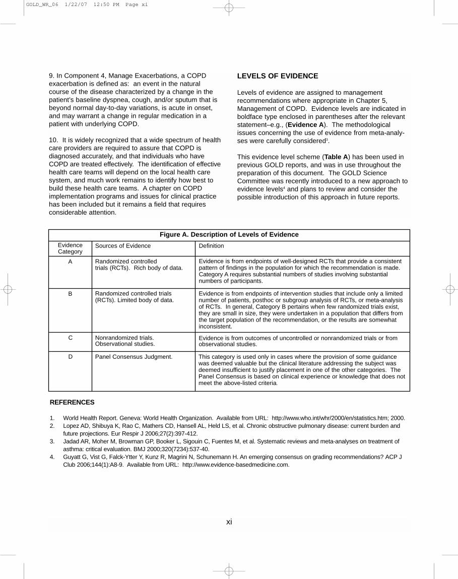
Figure A. Description of Levels of Evidence
Randomized controlled trials (RCTs). Rich body of data.
EvidenceCategory
A
B
C
D
Sources of Evidence Definition
Evidence is from endpoints of well-designed RCTs that provide a consistentpattern of findings in the population for which the recommendation is made.Category A requires substantial numbers of studies involving substantial numbers of participants.
Randomized controlled trials(RCTs). Limited body of data.
Evidence is from endpoints of intervention studies that include only a limitednumber of patients, posthoc or subgroup analysis of RCTs, or meta-analysisof RCTs. In general, Category B pertains when few randomized trials exist,they are small in size, they were undertaken in a population that differs fromthe target population of the recommendation, or the results are somewhatinconsistent.
Nonrandomized trials.Observational studies.
Evidence is from outcomes of uncontrolled or nonrandomized trials or fromobservational studies.
Panel Consensus Judgment. This category is used only in cases where the provision of some guidancewas deemed valuable but the clinical literature addressing the subject wasdeemed insufficient to justify placement in one of the other categories. ThePanel Consensus is based on clinical experience or knowledge that does notmeet the above-listed criteria.
GOLD_WR_06 1/22/07 12:50 PM Page xi

CHAPTER
1
DEFINITION
GOLD_WR_06 1/22/07 12:50 PM Page 1
2 DEFINITION
DEFINITION
Chronic obstructive pulmonary disease (COPD) is characterized by chronic airflow limitation and a range of pathological changes in the lung, some significantextra-pulmonary effects, and important comorbiditieswhich may contribute to the severity of the disease inindividual patients. Thus, COPD should be regarded asa pulmonary disease, but these significant comorbiditiesmust be taken into account in a comprehensive diagnostic assessment of severity and in determiningappropriate treatment.
Based on current knowledge, a working definition is:
Chronic Obstructive Pulmonary Disease (COPD) is apreventable and treatable disease with some significantextrapulmonary effects that may contribute to the severity in individual patients. Its pulmonary componentis characterized by airflow limitation that is not fullyreversible. The airflow limitation is usually progressiveand associated with an abnormal inflammatory responseof the lung to noxious particles or gases.
Worldwide, cigarette smoking is the most commonlyencountered risk factor for COPD, although in manycountries, air pollution resulting from the burning of woodand other biomass fuels has also been identified as aCOPD risk factor.
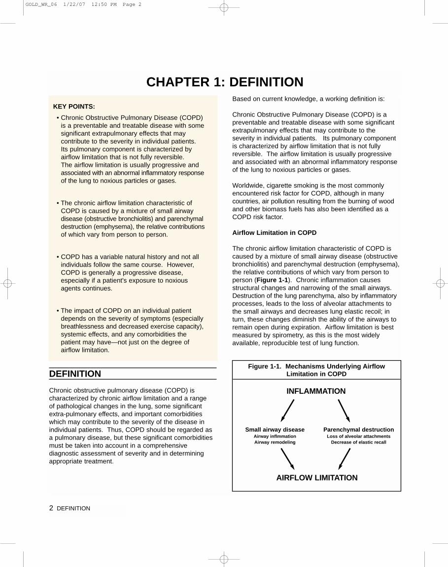
Airflow Limitation in COPD
The chronic airflow limitation characteristic of COPD iscaused by a mixture of small airway disease (obstructivebronchiolitis) and parenchymal destruction (emphysema),the relative contributions of which vary from person toperson (Figure 1-1). Chronic inflammation causes structural changes and narrowing of the small airways.Destruction of the lung parenchyma, also by inflammatoryprocesses, leads to the loss of alveolar attachments tothe small airways and decreases lung elastic recoil; inturn, these changes diminish the ability of the airways toremain open during expiration. Airflow limitation is bestmeasured by spirometry, as this is the most widely available, reproducible test of lung function.
Figure 1-1. Mechanisms Underlying AirflowLimitation in COPD
KEY POINTS:
• Chronic Obstructive Pulmonary Disease (COPD)is a preventable and treatable disease with somesignificant extrapulmonary effects that may contribute to the severity in individual patients.Its pulmonary component is characterized by airflow limitation that is not fully reversible. The airflow limitation is usually progressive and associated with an abnormal inflammatory responseof the lung to noxious particles or gases.
• The chronic airflow limitation characteristic ofCOPD is caused by a mixture of small airwaydisease (obstructive bronchiolitis) and parenchymaldestruction (emphysema), the relative contributionsof which vary from person to person.
• COPD has a variable natural history and not allindividuals follow the same course. However,COPD is generally a progressive disease, especially if a patient's exposure to noxiousagents continues.
• The impact of COPD on an individual patientdepends on the severity of symptoms (especiallybreathlessness and decreased exercise capacity),systemic effects, and any comorbidities thepatient may have—not just on the degree of airflow limitation.
INFLAMMATION
AIRFLOW LIMITATION
Small airway diseaseAirway inflmmationAirway remodeling
Parenchymal destructionLoss of alveolar attachments
Decrease of elastic recall
CHAPTER 1: DEFINITION
GOLD_WR_06 1/22/07 12:50 PM Page 2
DEFINITION 3
Many previous definitions of COPD have emphasized the terms “emphysema” and “chronic bronchitis,” whichare not included in the definition used in this and earlierGOLD reports. Emphysema, or destruction of the gas-exchanging surfaces of the lung (alveoli), is a pathologicalterm that is often (but incorrectly) used clinically anddescribes only one of several structural abnormalitiespresent in patients with COPD. Chronic bronchitis, or thepresence of cough and sputum production for at least 3 months in each of two consecutive years, remains aclinically and epidemiologically useful term. However, it does not reflect the major impact of airflow limitation on morbidity and mortality in COPD patients. It is alsoimportant to recognize that cough and sputum productionmay precede the development of airflow limitation; conversely, some patients develop significant airflow limitation without chronic cough and sputum production.
COPD and Comorbidities
Because COPD often develops in long-time smokers inmiddle age, patients often have a variety of other diseasesrelated to either smoking or aging1. COPD itself also hassignificant extrapulmonary (systemic) effects that lead tocomorbid conditions2. Data from the Netherlands showthat up to 25% of the population 65 years and older sufferfrom two comorbid conditions and up to 17% have three3.Weight loss, nutritional abnormalities and skeletal muscledysfunction are well-recognized extrapulmonary effects ofCOPD and patients are at increased risk for myocardialinfarction, angina, osteoporosis, respiratory infection,bone fractures, depression, diabetes, sleep-disorders,anemia, and glaucoma4. The existence of COPD mayactually increase the risk for other diseases; this is particularly striking for COPD and lung cancer5-8.Whether this association is due to common risk factors(e.g., smoking), involvement of susceptibility genes, orimpaired clearance of carcinogens is not clear.
Thus, COPD should be managed with careful attentionalso paid to comorbidities and their effect on the patient’squality of life. A careful differential diagnosis and comprehensive assessment of severity of comorbid conditions should be performed in every patient withchronic airflow limitation.
NATURAL HISTORY
COPD has a variable natural history and not all individualsfollow the same course. However, COPD is generally aprogressive disease, especially if a patient's exposure tonoxious agents continues. Stopping exposure to theseagents, even when significant airflow limitation is present,may result in some improvement in lung function and
slow or even halt progression of the disease. However,once developed, COPD and its comorbidities cannot becured and thus must be treated continuously. COPDtreatment can reduce symptoms, improve quality of life,reduce exacerbations, and possibly reduce mortality.
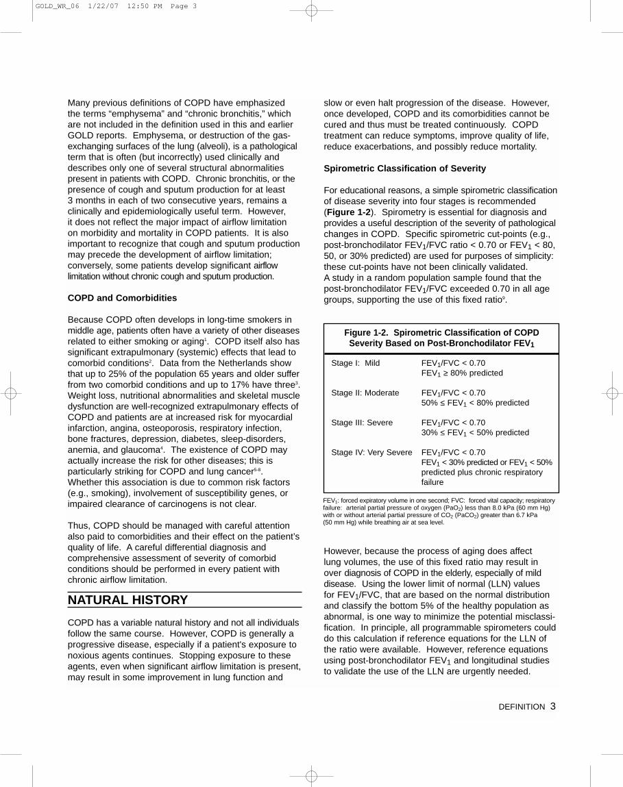
Spirometric Classification of Severity
For educational reasons, a simple spirometric classificationof disease severity into four stages is recommended(Figure 1-2). Spirometry is essential for diagnosis andprovides a useful description of the severity of pathologicalchanges in COPD. Specific spirometric cut-points (e.g.,post-bronchodilator FEV1/FVC ratio < 0.70 or FEV1 < 80,50, or 30% predicted) are used for purposes of simplicity:these cut-points have not been clinically validated. A study in a random population sample found that thepost-bronchodilator FEV1/FVC exceeded 0.70 in all agegroups, supporting the use of this fixed ratio9.
However, because the process of aging does affect lung volumes, the use of this fixed ratio may result inover diagnosis of COPD in the elderly, especially of mild disease. Using the lower limit of normal (LLN) values for FEV1/FVC, that are based on the normal distributionand classify the bottom 5% of the healthy population asabnormal, is one way to minimize the potential misclassi-fication. In principle, all programmable spirometers coulddo this calculation if reference equations for the LLN ofthe ratio were available. However, reference equationsusing post-bronchodilator FEV1 and longitudinal studiesto validate the use of the LLN are urgently needed.
FEV1: forced expiratory volume in one second; FVC: forced vital capacity; respiratoryfailure: arterial partial pressure of oxygen (PaO2) less than 8.0 kPa (60 mm Hg)with or without arterial partial pressure of CO2 (PaCO2) greater than 6.7 kPa (50 mm Hg) while breathing air at sea level.
Figure 1-2. Spirometric Classification of COPDSeverity Based on Post-Bronchodilator FEV1
Stage I: Mild FEV1/FVC < 0.70 FEV1 ≥ 80% predicted
Stage II: Moderate FEV1/FVC < 0.7050% ≤ FEV1 < 80% predicted
Stage III: Severe FEV1/FVC < 0.7030% ≤ FEV1 < 50% predicted
Stage IV: Very Severe FEV1/FVC < 0.70FEV1 < 30% predicted or FEV1 < 50%predicted plus chronic respiratoryfailure
GOLD_WR_06 1/22/07 12:50 PM Page 3
4 DEFINITION
Spirometry should be performed after the administrationof an adequate dose of an inhaled bronchodilator (e.g.,400 �g salbutamol)10 in order to minimize variability. In arandom population study to determine spirometry referencevalues, post-bronchodilator values differed markedly from pre-bronchodilator values9. Furthermore, post-bronchodilator lung function testing in a community settinghas been demonstrated to be an effective method toidentify individuals with COPD11.
While post-bronchodilator FEV1/FVC and FEV1 measure-ments are recommended for the diagnosis and assessmentof severity of COPD, the degree of reversibility of airflowlimitation (e.g., ∆FEV1 after bronchodilator or gluco-corticosteroids) is no longer recommended for diagnosis,differential diagnosis with asthma, or predicting theresponse to long-term treatment with bronchodilators or glucocorticosteroids.
Stages of COPD
The impact of COPD on an individual patient depends not just on the degree of airflow limitation, but also on the severity of symptoms (especially breathlessness anddecreased exercise capacity). There is only an imperfectrelationship between the degree of airflow limitation and the presence of symptoms. Spirometric staging,therefore, is a pragmatic approach aimed at practicalimplementation and should only be regarded as an educational tool and a general indication to the initialapproach to management.
The characteristic symptoms of COPD are chronic andprogressive dyspnea, cough, and sputum production.Chronic cough and sputum production may precede thedevelopment of airflow limitation by many years. This pattern offers a unique opportunity to identify smokers and others at risk for COPD (Figure 1-3), and intervenewhen the disease is not yet a major health problem.
Conversely, significant airflow limitation may develop without chronic cough and sputum production. AlthoughCOPD is defined on the basis of airflow limitation, inpractice the decision to seek medical help (and so permitthe diagnosis to be made) is normally determined by theimpact of a particular symptom on a patient's lifestyle.Thus, COPD may be diagnosed at any stage of the illness.
Stage I: Mild COPD - Characterized by mild airflow limitation (FEV1/FVC < 0.70; FEV1 ≥ 80% predicted).Symptoms of chronic cough and sputum production maybe present, but not always. At this stage, the individual isusually unaware that his or her lung function is abnormal.
Stage II: Moderate COPD - Characterized by worseningairflow limitation (FEV1/FVC < 0.70; 50% ≤ FEV1 < 80%predicted), with shortness of breath typically developingon exertion and cough and sputum production sometimesalso present. This is the stage at which patients typicallyseek medical attention because of chronic respiratorysymptoms or an exacerbation of their disease.
Stage III: Severe COPD - Characterized by further wors-ening of airflow limitation (FEV1/FVC < 0.70; 30% ≤ FEV1< 50% predicted), greater shortness of breath, reducedexercise capacity, fatigue, and repeated exacerbations thatalmost always have an impact on patients’ quality of life.
Stage IV: Very Severe COPD - Characterized by severeairflow limitation (FEV1/FVC < 0.70; FEV1 < 30% predictedor FEV1 < 50% predicted plus the presence of chronicrespiratory failure). Respiratory failure is defined as anarterial partial pressure of O2 (PaO2) less than 8.0 kPa(60 mm Hg), with or without arterial partial pressure ofCO2 (PaCO2) greater than 6.7 kPa (50 mm Hg) whilebreathing air at sea level. Respiratory failure may alsolead to effects on the heart such as cor pulmonale (rightheart failure). Clinical signs of cor pulmonale include elevation of the jugular venous pressure and pitting ankleedema. Patients may have Stage IV: Very Severe COPDeven if the FEV1 is > 30% predicted, whenever thesecomplications are present. At this stage, quality of life is very appreciably impaired and exacerba-tions may belife threatening.
The common statement that only 15-20% of smokersdevelop clinically significant COPD is misleading12. Amuch higher proportion may develop abnormal lung function at some point if they continue to smoke13. Not allindividuals with COPD follow the classical linear courseas outlined in the Fletcher and Peto diagram, which isactually the mean of many individual courses14. Causesof death in patients with COPD are mainly cardiovasculardiseases, lung cancer, and, in those with advancedCOPD, respiratory failure15.
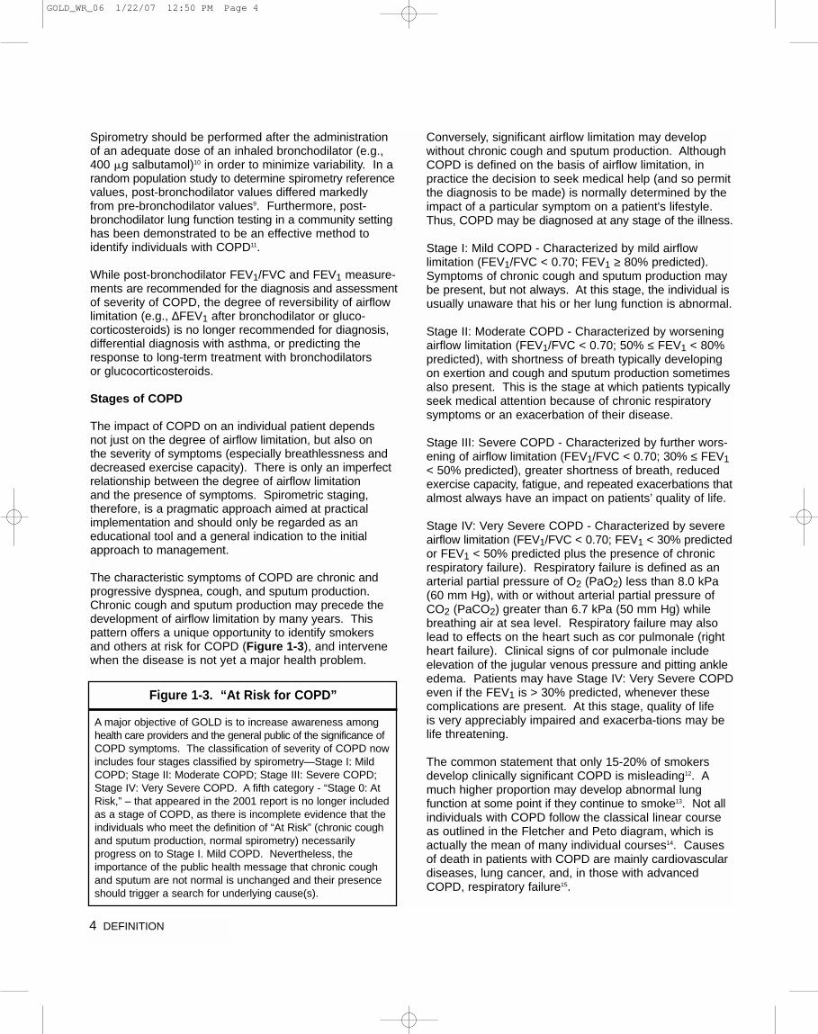
Figure 1-3. “At Risk for COPD”
A major objective of GOLD is to increase awareness amonghealth care providers and the general public of the significance ofCOPD symptoms. The classification of severity of COPD nowincludes four stages classified by spirometry—Stage I: MildCOPD; Stage II: Moderate COPD; Stage III: Severe COPD;Stage IV: Very Severe COPD. A fifth category - “Stage 0: AtRisk,” – that appeared in the 2001 report is no longer includedas a stage of COPD, as there is incomplete evidence that theindividuals who meet the definition of “At Risk” (chronic coughand sputum production, normal spirometry) necessarilyprogress on to Stage I. Mild COPD. Nevertheless, the importance of the public health message that chronic coughand sputum are not normal is unchanged and their presenceshould trigger a search for underlying cause(s).
GOLD_WR_06 1/22/07 12:50 PM Page 4
DEFINITION 5
SCOPE OF THE REPORT
It is not the scope of this report to provide a comprehensivediscussion of the natural history of comorbidities associated with COPD but to focus primarily on chronicairflow limitation caused by inhaled particles and gases,the most common of which worldwide is cigarette smoke.However, chronic airflow limitation may develop also innonsmokers who present with similar symptoms and may be associated with other diseases, e.g., asthma,congestive heart failure, lung carcinoma, bronchiectasis,pulmonary tuberculosis, bronchiolitis obliterans, and interstitial lung diseases. Poorly reversible airflow limitationassociated with these conditions is not addressed exceptinsofar as these conditions overlap with COPD.
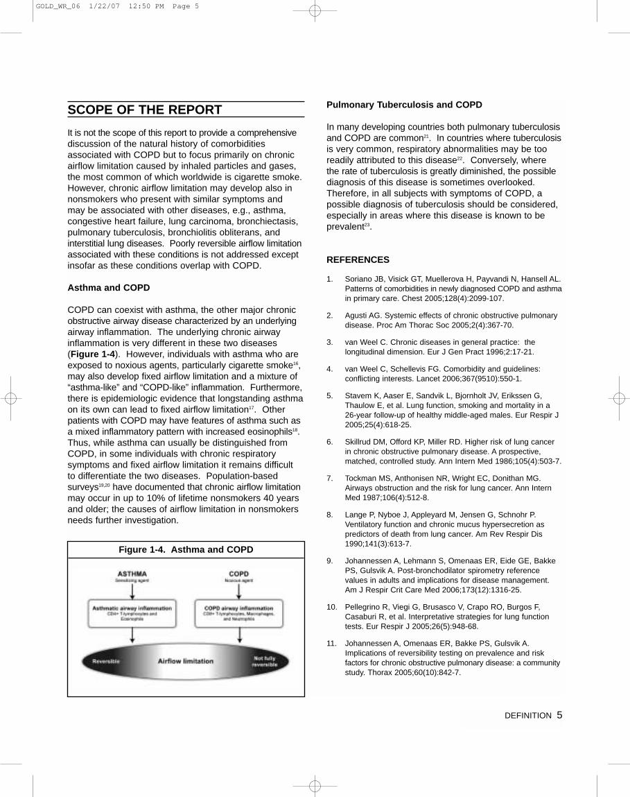
Asthma and COPD
COPD can coexist with asthma, the other major chronicobstructive airway disease characterized by an underlyingairway inflammation. The underlying chronic airwayinflammation is very different in these two diseases(Figure 1-4). However, individuals with asthma who areexposed to noxious agents, particularly cigarette smoke16,may also develop fixed airflow limitation and a mixture of“asthma-like” and “COPD-like” inflammation. Furthermore,there is epidemiologic evidence that longstanding asthmaon its own can lead to fixed airflow limitation17. Otherpatients with COPD may have features of asthma such asa mixed inflammatory pattern with increased eosinophils18.Thus, while asthma can usually be distinguished fromCOPD, in some individuals with chronic respiratory symptoms and fixed airflow limitation it remains difficult to differentiate the two diseases. Population-based surveys19,20 have documented that chronic airflow limitationmay occur in up to 10% of lifetime nonsmokers 40 yearsand older; the causes of airflow limitation in nonsmokersneeds further investigation.
Pulmonary Tuberculosis and COPD
In many developing countries both pulmonary tuberculosisand COPD are common21. In countries where tuberculosisis very common, respiratory abnormalities may be tooreadily attributed to this disease22. Conversely, where the rate of tuberculosis is greatly diminished, the possiblediagnosis of this disease is sometimes overlooked.Therefore, in all subjects with symptoms of COPD, a possible diagnosis of tuberculosis should be considered,especially in areas where this disease is known to beprevalent23.
REFERENCES
1. Soriano JB, Visick GT, Muellerova H, Payvandi N, Hansell AL.Patterns of comorbidities in newly diagnosed COPD and asthmain primary care. Chest 2005;128(4):2099-107.
2. Agusti AG. Systemic effects of chronic obstructive pulmonarydisease. Proc Am Thorac Soc 2005;2(4):367-70.
3. van Weel C. Chronic diseases in general practice: the longitudinal dimension. Eur J Gen Pract 1996;2:17-21.
4. van Weel C, Schellevis FG. Comorbidity and guidelines: conflicting interests. Lancet 2006;367(9510):550-1.
5. Stavem K, Aaser E, Sandvik L, Bjornholt JV, Erikssen G,Thaulow E, et al. Lung function, smoking and mortality in a26-year follow-up of healthy middle-aged males. Eur Respir J2005;25(4):618-25.
6. Skillrud DM, Offord KP, Miller RD. Higher risk of lung cancer in chronic obstructive pulmonary disease. A prospective,matched, controlled study. Ann Intern Med 1986;105(4):503-7.
7. Tockman MS, Anthonisen NR, Wright EC, Donithan MG.Airways obstruction and the risk for lung cancer. Ann InternMed 1987;106(4):512-8.
8. Lange P, Nyboe J, Appleyard M, Jensen G, Schnohr P.Ventilatory function and chronic mucus hypersecretion as predictors of death from lung cancer. Am Rev Respir Dis1990;141(3):613-7.
9. Johannessen A, Lehmann S, Omenaas ER, Eide GE, BakkePS, Gulsvik A. Post-bronchodilator spirometry reference values in adults and implications for disease management. Am J Respir Crit Care Med 2006;173(12):1316-25.
10. Pellegrino R, Viegi G, Brusasco V, Crapo RO, Burgos F,Casaburi R, et al. Interpretative strategies for lung functiontests. Eur Respir J 2005;26(5):948-68.
11. Johannessen A, Omenaas ER, Bakke PS, Gulsvik A.Implications of reversibility testing on prevalence and risk factors for chronic obstructive pulmonary disease: a communitystudy. Thorax 2005;60(10):842-7.
Figure 1-4. Asthma and COPD
GOLD_WR_06 1/22/07 12:50 PM Page 5

6 DEFINITION
12. Rennard S, Vestbo J. COPD: the dangerous underestimate of 15%. Lancet 2006;367:1216-9.
13. Lokke A, Lange P, Scharling H, Fabricius P, Vestbo J.Developing COPD - a 25 years follow-up study of the generalpopulation. Thorax 2006;61:935-9.
14. Fletcher C, Peto R. The natural history of chronic airflowobstruction. BMJ 1977;1(6077):1645-8.
15. Mannino DM, Doherty DE, Sonia Buist A. Global Initiative onObstructive Lung Disease (GOLD) classification of lung disease and mortality: findings from the Atherosclerosis Risk inCommunities (ARIC) study. Respir Med 2006;100(1):115-22.
16. Thomson NC, Chaudhuri R, Livingston E. Asthma and cigarettesmoking. Eur Respir J 2004;24(5):822-33.
17. Lange P, Parner J, Vestbo J, Schnohr P, Jensen G. A 15-yearfollow-up study of ventilatory function in adults with asthma. N Engl J Med 1998;339(17):1194-200.
18. Chanez P, Vignola AM, O'Shaugnessy T, Enander I, Li D,Jeffery PK, et al. Corticosteroid reversibility in COPD is related to features of asthma. Am J Respir Crit Care Med1997;155(5):1529-34.
19. Menezes AM, Perez-Padilla R, Jardim JR, Muino A, Lopez MV,Valdivia G, et al. Chronic obstructive pulmonary disease in fiveLatin American cities (the PLATINO study): a prevalencestudy. Lancet 2005;366(9500):1875-81.
20. Centers for Disease Control and Prevention. SurveillanceSummaries. MMWR 2002:51(No. SS-6).
21. Fairall LR, Zwarenstein M, Bateman ED, Bachmann M,Lombard C, Majara BP, et al. Effect of educational outreach to nurses on tuberculosis case detection and primary care ofrespiratory illness: pragmatic cluster randomised controlledtrial. BMJ 2005;331(7519):750-4.
22. de Valliere S, Barker RD. Residual lung damage after completion of treatment for multidrug-resistant tuberculosis.Int J Tuberc Lung Dis 2004;8(6):767-71.
23. Bateman ED, Feldman C, O'Brien J, Plit M, Joubert JR.Guideline for the management of chronic obstructive pulmonary disease (COPD): 2004 revision. S Afr Med J2004;94(7 Pt 2):559-75.
GOLD_WR_06 1/22/07 12:50 PM Page 6

CHAPTER
2
BURDEN OF COPD
GOLD_WR_06 1/22/07 12:50 PM Page 7
8 BURDEN OF COPD
INTRODUCTION
COPD is a leading cause of morbidity and mortalityworldwide and results in an economic and social burdenthat is both substantial and increasing. COPD prevalence,morbidity, and mortality vary across countries and acrossdifferent groups within countries but, in general, aredirectly related to the prevalence of tobacco smokingalthough in many countries, air pollution resulting fromthe burning of wood and other biomass fuels has alsobeen identified as a COPD risk factor. The prevalenceand burden of COPD are projected to increase in thecoming decades due to continued exposure to COPD risk factors and the changing age structure of the world’spopulation (with more people living longer, and thusreaching the age at which COPD normally develops).
EPIDEMIOLOGY
In the past, imprecise and variable definitions of COPDhave made it difficult to quantify prevalence, morbidityand mortality. Furthermore, the underrecognition and
underdiagnosis of COPD lead to significant underreporting.The extent of the underreporting varies across countriesand depends on the level of awareness and understandingof COPD among health professionals, the organization ofhealth care services to cope with chronic diseases, andthe availability of medications for the treatment of COPD1.
There are several sources of information on the burdenof COPD: publications such as the 2003 European Lung White Book2, international Websites such as theWorld Health Organization (http://www.who.int) and theWorld Bank/WHO Global Burden of Disease Study(http://www.who.int/topics/global_burden_of_disease), andcountry-specific Websites such as the US Centers forDisease Control and Prevention (http://www.cdc.gov) andthe UK Health Survey for England (http://www.doh.gov.uk).
Prevalence
Existing COPD prevalence data show remarkable variationdue to differences in survey methods, diagnostic criteria,and analytic approaches3,4. Survey methods can include:
• Self-report of a doctor diagnosis of COPD or equivalentcondition
• Spirometry with or without a bronchodilator • Questionnaires that ask about the presence of
respiratory symptoms
The lowest estimates of prevalence are usually thosebased on self-reporting of a doctor diagnosis of COPD or equivalent condition. For example, most national datashow that less than 6% of the population has been toldthat they have COPD3. This likely reflects the wide-spread underrecognition and underdiagnosis of COPD5
as well as the fact that those with Stage I: Mild COPDmay have no symptoms, or else symptoms (such aschronic cough and sputum) that are not perceived byindividuals or their health care providers as abnormal and possibly indicative of early COPD5. These estimatesmay have value, however, since they may most accuratelyreflect the burden of clinically significant disease that is ofsufficient severity to require health services, and thereforeis likely to generate significant direct and indirect costs.
By contrast, data from prevalence surveys carried out ina number of countries, using standardized methods andincluding spirometry, estimate that up to about one-quarterof adults aged 40 years and older may have airflow limitation classified as Stage I: Mild COPD or higher6-9.
CHAPTER 2: BURDEN OF COPD
KEY POINTS:
• COPD is a leading cause of morbidity and mortalityworldwide and results in an economic and socialburden that is both substantial and increasing.
• COPD prevalence, morbidity, and mortality varyacross countries and across different groupswithin countries but, in general, are directly relatedto the prevalence of tobacco smoking, althoughin many countries, air pollution resulting from theburning of wood and other biomass fuels hasalso been identified as a COPD risk factor.
• The prevalence and burden of COPD are projectedto increase in the coming decades due to continuedexposure to COPD risk factors and the changingage structure of the world’s population.
• COPD is a costly disease with both direct costs(value of health care resources devoted to diagnosis and medical management) and indirectcosts (monetary consequences of disability,missed work, premature mortality, and caregiveror family costs resulting from the illness).
GOLD_WR_06 1/22/07 12:50 PM Page 8
BURDEN OF COPD 9
Because of the large gap between the prevalence ofCOPD as defined by the presence of airflow limitationand the prevalence of COPD as defined by clinically significant disease, the debate continues as to which ofthese it is better to use in estimating the burden ofCOPD. Early diagnosis and intervention may help toidentify the number of individuals who progress to a clinically significant stage of disease, but there is insufficient evidence at this time to recommend community-based spirometric screening for COPD10.
Different diagnostic criteria also give widely different estimates and there is little consensus regarding themost appropriate criteria for different settings (e.g., epidemiologic surveys, clinical diagnosis), or the strengthsand weaknesses of the different criteria. It is recognizedthat defining irreversible airflow obstruction as a post-bronchodilator FEV1/FVC ratio less than 0.70 leads to the potential for significant misclassification, with underdiagnosis (false negatives) in younger adults andover-diagnosis (false positives) over age 50 years11-13.This has led to the recommendation that the use of thelower limit of normal (LLN) of the post-bronchodilatorFEV1/FVC ratio rather than the fixed ratio be used todefine irreversible airflow obstruction14,15. However, moreinformation is needed from population-based longitudinalstudies to determine the outcome of individuals classifiedusing either definition.
Many additional sources of variation can affect estimatesof COPD prevalence, including sampling methods,response rates, quality control of spirometry, and whetherspirometry is performed pre- or post-bronchodilator.Samples that are not population-based and poor responserates may give biased estimates of prevalence, with thedirection of bias sometimes hard to determine. Inadequateemptying of the lungs during the spirometric maneuver is common and leads to an artificially high ratio ofFEV1/FVC and therefore to an underestimate of theprevalence of COPD. Failure to use post-bronchodilatorvalue instead of pre-bronchodilator values leads to anoverdiagnosis of irreversible airflow limitation In futureprevalence surveys, post-bronchodilator spirometryshould be used to confirm the diagnosis of COPD16.
Despite these complexities, data are emerging thatenable some conclusions to be drawn regarding COPDprevalence. A systematic review and meta-analysis ofstudies carried out in 28 countries between 1990 and20043, and an additional study from Japan17, provide evidence that the prevalence of COPD (Stage I: MildCOPD and higher) is appreciably higher in smokers andex-smokers than in nonsmokers, in those over 40 yearsthan those under 40, and in men than in women.
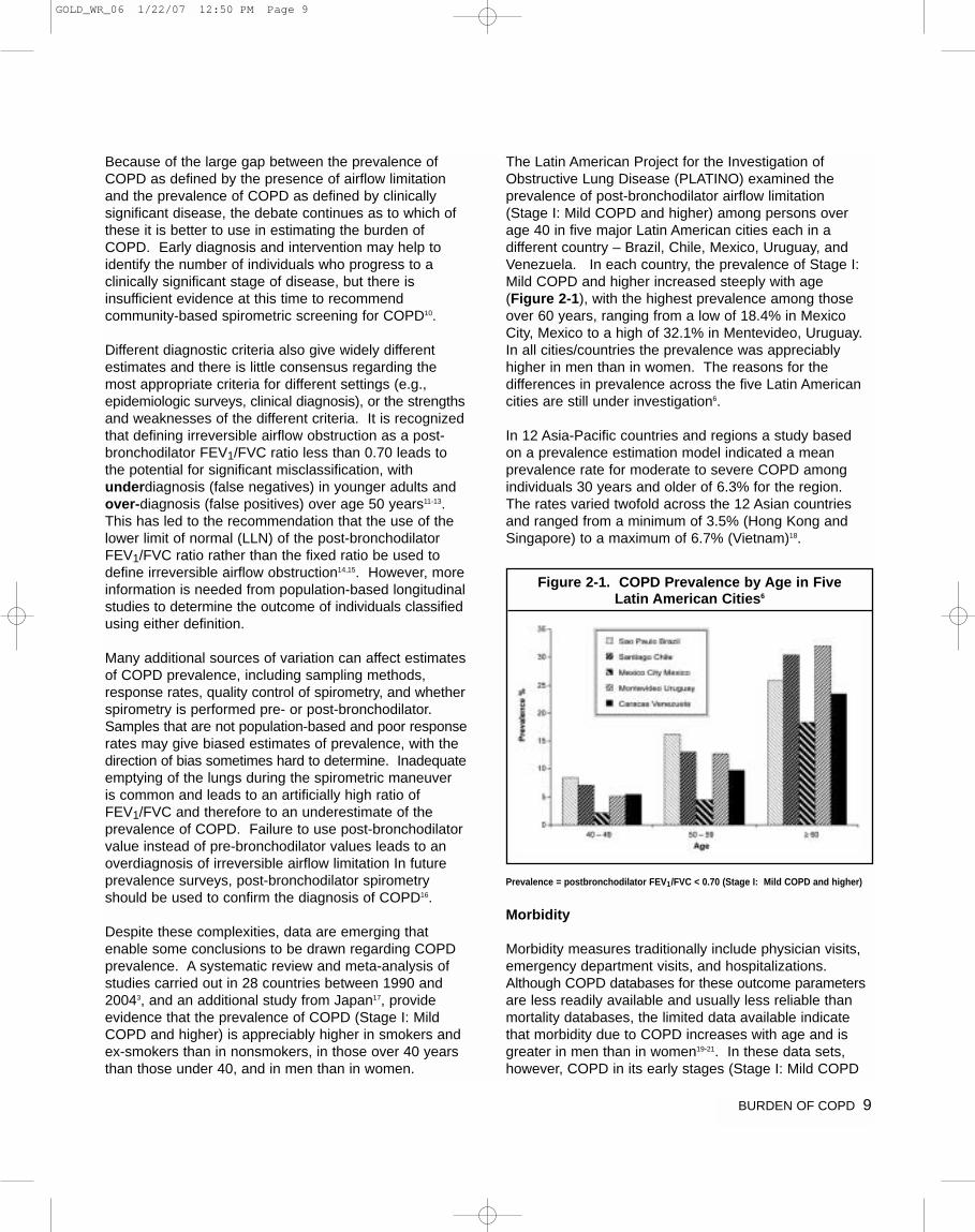
The Latin American Project for the Investigation ofObstructive Lung Disease (PLATINO) examined theprevalence of post-bronchodilator airflow limitation (Stage I: Mild COPD and higher) among persons overage 40 in five major Latin American cities each in a different country – Brazil, Chile, Mexico, Uruguay, andVenezuela. In each country, the prevalence of Stage I:Mild COPD and higher increased steeply with age(Figure 2-1), with the highest prevalence among thoseover 60 years, ranging from a low of 18.4% in MexicoCity, Mexico to a high of 32.1% in Mentevideo, Uruguay.In all cities/countries the prevalence was appreciablyhigher in men than in women. The reasons for the differences in prevalence across the five Latin Americancities are still under investigation6.
In 12 Asia-Pacific countries and regions a study based on a prevalence estimation model indicated a meanprevalence rate for moderate to severe COPD amongindividuals 30 years and older of 6.3% for the region.The rates varied twofold across the 12 Asian countriesand ranged from a minimum of 3.5% (Hong Kong andSingapore) to a maximum of 6.7% (Vietnam)18.
Prevalence = postbronchodilator FEV1/FVC < 0.70 (Stage I: Mild COPD and higher)
Morbidity
Morbidity measures traditionally include physician visits,emergency department visits, and hospitalizations.Although COPD databases for these outcome parametersare less readily available and usually less reliable thanmortality databases, the limited data available indicatethat morbidity due to COPD increases with age and isgreater in men than in women19-21. In these data sets,however, COPD in its early stages (Stage I: Mild COPD
Figure 2-1. COPD Prevalence by Age in Five Latin American Cities6
GOLD_WR_06 1/22/07 12:50 PM Page 9
and Stage 2: Moderate COPD) is usually not recognized,diagnosed, or treated, and therefore may not be includedas a diagnosis in a patient’s medical record.
Morbidity from COPD may be affected by other comorbidchronic conditions22 (e.g., musculoskeletal disease, diabetes mellitus) that are not directly related to COPDbut nevertheless may have an impact on the patient’shealth status, or may negatively interfere with COPDmanagement. In patients with more advanced disease(Stage III: Severe COPD and Stage IV: Very SevereCOPD), morbidity from COPD may be misattributed toanother comorbid condition.
Morbidity data are greatly affected by the availability ofresources (e.g,, hospitalization rates are highly dependenton the availability of hospital beds) and thus have to beinterpreted cautiously and with a clear understanding ofthe possible biases inherent in the dataset. Despite thelimitations in the data for COPD, the European WhiteBook provides good data on the mean number of consultations for major respiratory diseases across 19 countries of the European Economic Community2. In most countries, consultations for COPD greatly out-numbered consultations for asthma, pneumonia, lung and tracheal cancer, and tuberculosis. In the UnitedStates in 2000, there were 8 million physician office/hospital outpatient visits for COPD, 1.5 million emergencydepartment visits, and 673,000 hospitalizations23.
Another way of estimating the morbidity burden of diseaseis to calculate years of living with disability (YLD). TheGlobal Burden of Disease Study estimates that COPDresults in 1.68 YLD per 1,000 population, representing1.8% of all YLDs, with a greater burden in men than inwomen (1.93% vs. 1.42%)8,24,25.
Mortality
The World Health Organization publishes mortality statistics for selected causes of death annually for allWHO regions; additional information is available from the WHO Evidence for Health Policy Department(http://www.who.int/evidence). Data must be interpretedcautiously, however, because of inconsistent use of terminology for COPD. Prior to about 1968 and theEighth Revision of the International Classification ofDiseases (ICD), the terms “chronic bronchitis” and“emphysema” were used extensively. During the 1970s,the term “COPD” increasingly replaced those terms insome but not all countries, making COPD mortality comparisons in different countries very difficult. However,the situation has improved with the Ninth and Tenth
Revisions of the ICD, in which deaths from COPD orchronic airways obstruction are included in the broad category of “COPD and allied conditions” (ICD-9 codes490-496 and ICD-10 codes J42-46).
Thus, the problem of labeling has been partly solved, butunderrecognition and underdiagnosis of COPD still affectthe accuracy of mortality data. Although COPD is often aprimary cause of death, it is more likely to be listed as acontributory cause of death or omitted from the deathcertificate entirely, and the death attributed to anothercondition such as cardiovascular disease.
Despite the problems with the accuracy of the COPDmortality data, it is clear that COPD is one of the mostimportant causes of death in most countries. The GlobalBurden of Disease Study8,24,25 has projected that COPD,which ranked sixth as the cause of death in 1990, willbecome the third leading cause of death worldwide by2020. This increased mortality is driven by the expandingepidemic of smoking and the changing demographics inmost countries, with more of the population living longer.Of these two forces, demographics is the stronger driverof the trend.
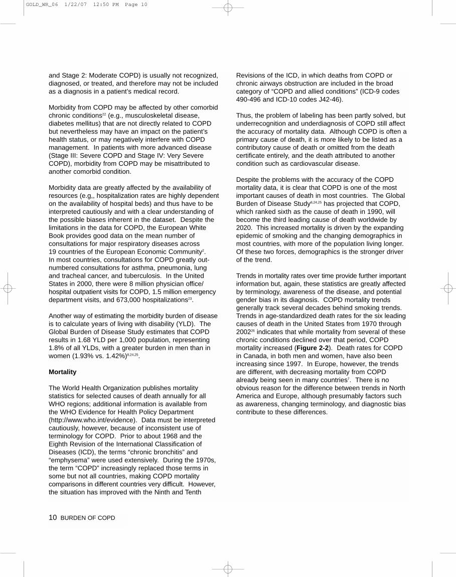
Trends in mortality rates over time provide further importantinformation but, again, these statistics are greatly affectedby terminology, awareness of the disease, and potentialgender bias in its diagnosis. COPD mortality trends generally track several decades behind smoking trends.Trends in age-standardized death rates for the six leadingcauses of death in the United States from 1970 through200226 indicates that while mortality from several of thesechronic conditions declined over that period, COPD mortality increased (Figure 2-2). Death rates for COPDin Canada, in both men and women, have also beenincreasing since 1997. In Europe, however, the trendsare different, with decreasing mortality from COPDalready being seen in many countries7. There is no obvious reason for the difference between trends in NorthAmerica and Europe, although presumably factors suchas awareness, changing terminology, and diagnostic biascontribute to these differences.
10 BURDEN OF COPD
GOLD_WR_06 1/22/07 12:50 PM Page 10
Reprinted from Jemal A, Ward E, Hao Y, Thun M. Trends in the leading causesof death in the United States, 1970-2002. JAMA 2005;294(10):1255-9. withpermission from JAMA
The mortality trends for COPD have been particularlystriking for women. In Canada, the death rate from COPDamong women accelerated in the 1990s and is expectedto soon overtake the rate among men21. In the UnitedStates, COPD deaths among women have been risingsteeply since the 1970s. In 2000, the number of deathsfrom COPD in the United States was greater amongwomen than men (59,936 vs. 59,118), although the mortality rates among women remain somewhat lowerthan among men27.
Worldwide, recent increases in COPD deaths are likely to continue. The Global Burden of Disease Study8,24,25
projected baseline, optimistic, and pessimistic models forCOPD mortality from 1990 to 2020 that take into accountthe expected aging of the world’s population, projectedincreases in smoking rates, and projected declines inother causes of death such as diarrheal and HIV-relateddiseases.
ECONOMIC AND SOCIAL BURDEN OF COPD
Economic Burden
COPD is a costly disease with both direct costs (value of health care resources devoted to diagnosis and medical management) and indirect costs (monetary consequences of disability, missed work, premature mortality, and caregiver or family costs resulting from theillness)2. In developed countries, exacerbations of COPDaccount for the greatest burden on the health care system.In the European Union, the total direct costs of respiratorydisease are estimated to be about 6% of the total healthcare budget, with COPD accounting for 56% (38.6 billionEuros) of this2. In the United States in 2002, the directcosts of COPD were $18 billion and the indirect coststotaled $14.1 billion28. Costs per patient will vary acrosscountries since these costs depend on how health care is provided and paid7.
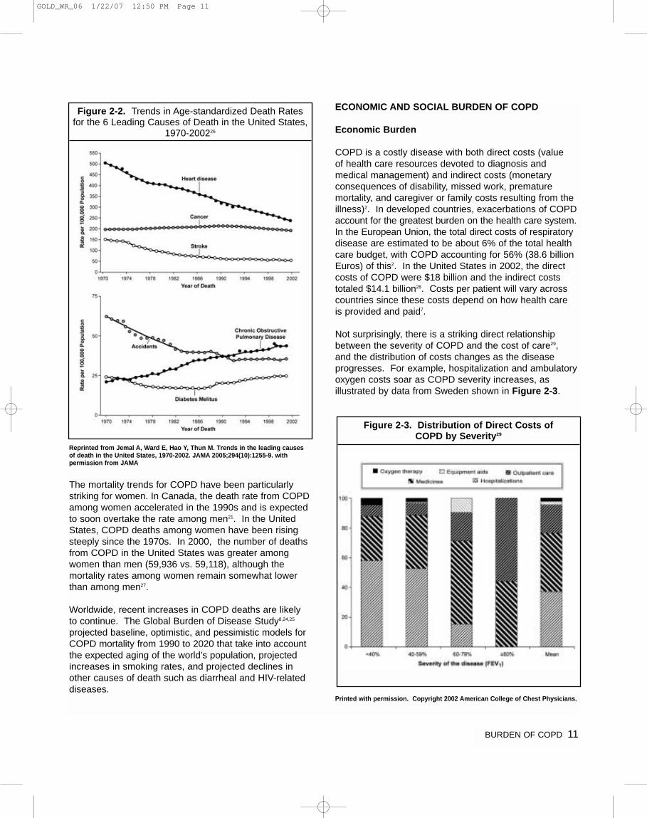
Not surprisingly, there is a striking direct relationshipbetween the severity of COPD and the cost of care29, and the distribution of costs changes as the disease progresses. For example, hospitalization and ambulatoryoxygen costs soar as COPD severity increases, as illustrated by data from Sweden shown in Figure 2-3.
Printed with permission. Copyright 2002 American College of Chest Physicians.
BURDEN OF COPD 11
Figure 2-2. Trends in Age-standardized Death Rates for the 6 Leading Causes of Death in the United States,
1970-200226
Figure 2-3. Distribution of Direct Costs of COPD by Severity29
GOLD_WR_06 1/22/07 12:50 PM Page 11
12 BURDEN OF COPD
The presence of COPD greatly increases the total cost of care for patients, especially when inpatient costs areconsidered. In a study of COPD-related illness costs inthe United States based on the 1987 National MedicalExpenditure Survey, per capita expenditures for hospital-izations of COPD patients were 2.7 times the expendituresfor patients without COPD ($5,409 vs. $2,001)30. In a1992 study of Medicare, the US government health insurance program for individuals over 65, annual percapita expenditures for people with COPD ($8,482) werenearly 2.5 times the expenditures for people withoutCOPD ($3,511)31.
Individuals with COPD frequently receive professionalmedical care in their homes. In some countries, nationalhealth insurance plans provide coverage for oxygen therapy, visiting nursing services, rehabilitation, and evenmechanical ventilation in the home, although coveragefor specific services varies from country to country32. Any estimate of direct medical expenditures for homecare underrepresents the true cost of home care to society, because it ignores the economic value of thecare provided to those with COPD by family members.In developing countries, direct medical costs may be lessimportant than the impact of COPD on workplace andhome productivity. Because the health care sector mightnot provide long-term supportive care services forseverely disabled individuals, COPD may force two individuals to leave the workplace—the affected individualand a family member who must now stay home to carefor the disabled relative. Since human capital is often themost important national asset for developing countries,the indirect costs of COPD may represent a seriousthreat to their economies.
Social Burden
Since mortality offers a limited perspective on the humanburden of a disease, it is desirable to find other measuresof disease burden that are consistent and measurableacross nations. The authors of the Global Burden ofDisease Study designed a method to estimate the fractionof mortality and disability attributable to major diseasesand injuries using a composite measure of the burden ofeach health problem, the Disability-Adjusted Life Year(DALY)8,24,25. The DALYs for a specific condition are thesum of years lost because of premature mortality andyears of life lived with disability, adjusted for the severityof disability. In 1990, COPD was the twelfth leadingcause of DALYs lost in the world, responsible for 2.1% of the total. According to the projections, COPD will bethe fifth leading cause of DALYs lost worldwide in 2020,behind ischemic heart disease, major depression, trafficaccidents, and cerebrovascular disease. This substantial
increase in the global burden of COPD projected over thenext twenty years reflects, in large part, the continuedhigh use of tobacco in many countries and the changingage structure of populations in developing countries.
REFERENCES
1. Tirimanna PR, van Schayck CP, den Otter JJ, van Weel C,van Herwaarden CL, van den Boom G, et al. Prevalence ofasthma and COPD in general practice in 1992: has it changedsince 1977? Br J Gen Pract 1996;46(406):277-81.
2. European Respiratory Society. European Lung White Book:Huddersfield, European Respiratory Society Journals, Ltd; 2003.
3. Halbert RJ, Natoli JL, Gano A, Badamgarav E, Buist AS,Mannino DM. Global burden of COPD: systematic review andmeta-analysis. Eur Respir J 2006.
4. Halbert RJ, Isonaka S, George D, Iqbal A. Interpreting COPDprevalence estimates: what is the true burden of disease?Chest 2003;123(5):1684-92.
5. van den Boom G, van Schayck CP, van Mollen MP, TirimannaPR, den Otter JJ, van Grunsven PM, et al. Active detection ofchronic obstructive pulmonary disease and asthma in the general population. Results and economic consequences ofthe DIMCA program. Am J Respir Crit Care Med1998;158(6):1730-8.
6. Menezes AM, Perez-Padilla R, Jardim JR, Muino A, LopezMV, Valdivia G, et al. Chronic obstructive pulmonary diseasein five Latin American cities (the PLATINO study): a prevalence study. Lancet 2005;366(9500):1875-81.
7. Chapman KR, Mannino DM, Soriano JB, Vermeire PA, BuistAS, Thun MJ, et al. Epidemiology and costs of chronicobstructive pulmonary disease. Eur Respir J 2006;27(1):188-207.
8. Lopez AD, Shibuya K, Rao C, Mathers CD, Hansell AL, Held LS,et al. Chronic obstructive pulmonary disease: current burdenand future projections. Eur Respir J 2006;27(2):397-412.
9. Buist AS, Vollmer WM, Sullivan SD, Weiss KB, Lee TA, MenezesAM, et al. The burden of obstructive lung disease initiative(BOLD): Rationale and Design. J COPD 2005;2:277-83.
10. Wilt TJ, Niewoehner D, Kim C, Kane RL, Linabery A, TacklindJ, et al. Use of spirometry for case finding, diagnosis, andmanagement of chronic obstructive pulmonary disease(COPD). Evid Rep Technol Assess (Summ) 2005(121):1-7.
11. Hnizdo E, Glindmeyer HW, Petsonk EL, Enright P, Buist AS.Case Definitions for Chronic Obstructive Pulmonary Disease.J COPD 2006;3:1-6.
12. Roberts SD, Farber MO, Knox KS, Phillips GS, Bhatt NY,Mastronarde JG, et al. FEV1/FVC ratio of 70% misclasifiespatients with obstructin at the extremes of age. Chest2006;130:200-6.
GOLD_WR_06 1/22/07 12:50 PM Page 12

13. Celli BR, Halbert RJ, Isonaka S, Schau B. Population impactof different definitions of airway obstruction. Eur Respir J2003;22(2):268-73.
14. Pellegrino R, Viegi G, Brusasco V, Crapo RO, Burgos F,Casaburi R, et al. Interpretative strategies for lung functiontests. Eur Respir J 2005;26(5):948-68.
15. Hankinson JL, Odencrantz JR, Fedan KB. Spirometric referencevalues from a sample of the general US population. Am JRespir Crit Care Med 1999;159:179-87.
16. Sterk PJ. Let's not forget: the GOLD criteria for COPD arebased on post-bronchodilator FEV1. Eur Respir J2004;23:497-8.
17. Fukuchi Y, Nishimura M, Ichinose M, Adachi M, Nagai A,Kuriyama T, et al. COPD in Japan: the Nippon COPDEpidemiology study. Respirology 2004;9(4):458-65.
18. COPD Prevalence in 12 Asia-Pacific Countries and regions:Projections based on the COPD prevalence estimation model.Regional COPD Working Group. Respirology 2003;8:192-8.
19. National Heart, Lung, and Blood Institute. Morbidity &Mortality: Chartbook on Cardiovascular, Lung, and BloodDiseases. Bethesda, MD: US Department. of Health andHuman Services, Public Health Service, National Institutes ofHealth; 1998.
20. Soriano JR, Maier WC, Egger P, Visick G, Thakrar B, Sykes J,et al. Recent trends in physician diagnosed COPD in womenand men in the UK. Thorax 2000;55:789-94.
21. Chapman KR. Chronic obstructive pulmonary disease: arewomen more susceptible than men? Clin Chest Med2004;25(2):331-41.
22. Schellevis FG, Van de Lisdonk EH, Van der Velden J,Hoogbergen SH, Van Eijk JT, Van Weel C. Consultation ratesand incidence of intercurrent morbidity among patients withchronic disease in general practice. Br J Gen Pract1994;44(383):259-62.
23. Centers for Disease Control and Prevention. SurveillanceSummaries. MMWR 2002:51(No. SS-6).
24. Murray CJL, Lopez AD, editors. In: The global burden of dis-ease: a comprehensive assessment of mortality and disabilityfrom diseases, injuries and risk factors in 1990 and projectedto 2020. Cambridge, MA: Harvard University Press; 1996.
25. Murray CJ, Lopez AD. Alternative projections of mortality anddisability by cause 1990-2020: Global Burden of DiseaseStudy. Lancet 1997;349(9064):1498-504.
26. Jemal A, Ward E, Hao Y, Thun M. Trends in the leading caus-es of death in the United States, 1970-2002. JAMA2005;294(10):1255-9.
27. Mannino DM, Homa DM, Akinbami LJ, Ford ES, Redd SC.Chronic obstructive pulmonary disease surveillance--UnitedStates, 1971-2000. MMWR Surveill Summ 2002;51(6):1-16.
28. National Heart, Lung, and Blood Institute. Morbidity and mortalitychartbook on cardiovascular, lung and blood diseases. Bethesda,Maryland: US Department of Health and Human Services,Public Health Service, National Institutes of Health. Accessed at:http://www.nhlbi.nih.gov/resources/docs/cht-book.htm; 2004.
29. Jansson SA, Andersson F, Borg S, Ericsson A, Jonsson E,Lundback B. Costs of COPD in Sweden according to diseaseseverity. Chest 2002;122(6):1994-2002.
30. Sullivan SD, Strassels S, Smith DH. Characterization of theincidence and cost of COPD in the US. Eur Respir J1996;9(Supplement 23):S421.
31. Grasso ME, Weller WE, Shaffer TJ, Diette GB, Anderson GF.Capitation, managed care, and chronic obstructive pulmonarydisease. Am J Respir Crit Care Med 1998;158:133-8.
32. Fauroux B, Howard P, Muir JF. Home treatment for chronicrespiratory insufficiency: the situation in Europe in 1992. TheEuropean Working Group on Home Treatment for ChronicRespiratory Insufficiency. Eur Respir J 1994;7:1721-6.
BURDEN OF COPD 13
GOLD_WR_06 1/22/07 12:50 PM Page 13

14 BURDEN OF COPD
GOLD_WR_06 1/22/07 12:50 PM Page 14

CHAPTER
3
RISK FACTORS
GOLD_WR_06 1/22/07 12:50 PM Page 15
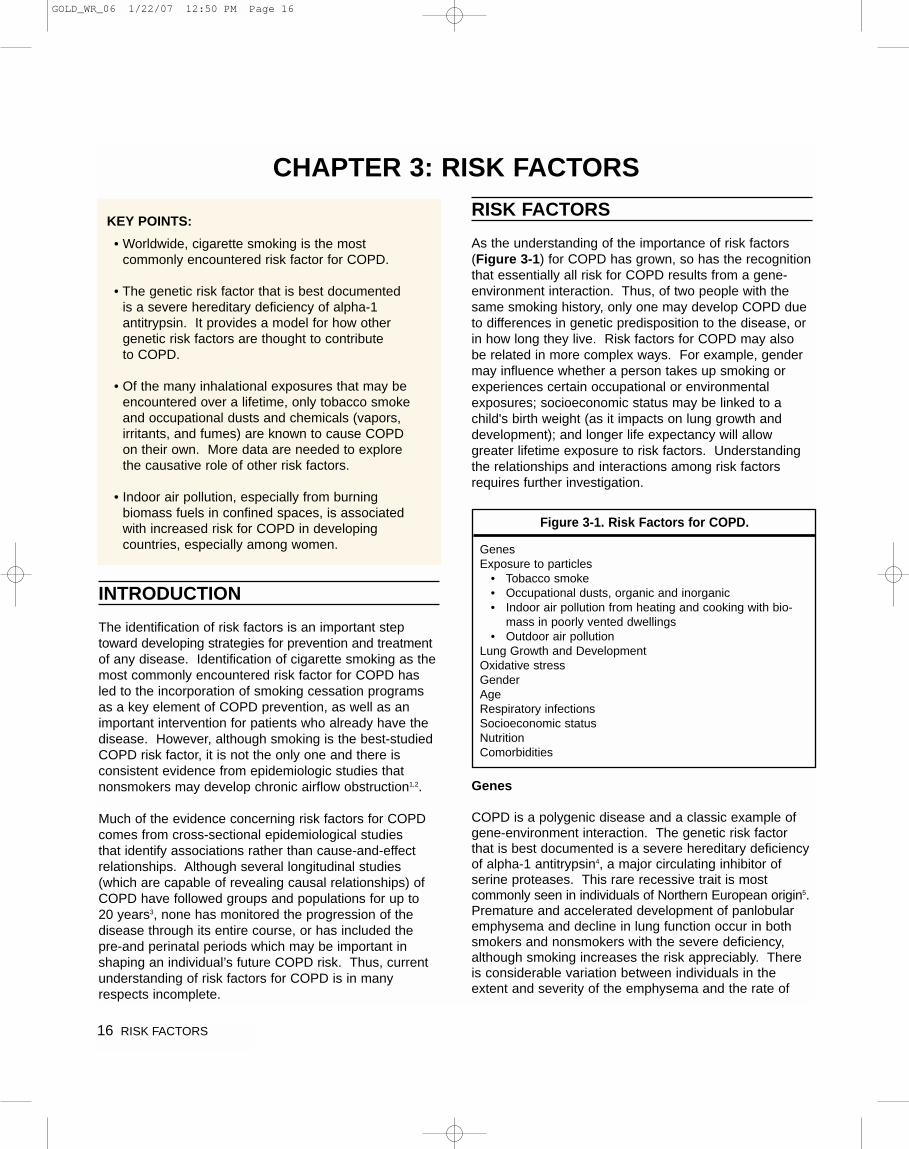
16 RISK FACTORS
INTRODUCTION
The identification of risk factors is an important steptoward developing strategies for prevention and treatmentof any disease. Identification of cigarette smoking as themost commonly encountered risk factor for COPD hasled to the incorporation of smoking cessation programsas a key element of COPD prevention, as well as animportant intervention for patients who already have thedisease. However, although smoking is the best-studiedCOPD risk factor, it is not the only one and there is consistent evidence from epidemiologic studies that nonsmokers may develop chronic airflow obstruction1,2.
Much of the evidence concerning risk factors for COPDcomes from cross-sectional epidemiological studies that identify associations rather than cause-and-effectrelationships. Although several longitudinal studies(which are capable of revealing causal relationships) ofCOPD have followed groups and populations for up to 20 years3, none has monitored the progression of the disease through its entire course, or has included thepre-and perinatal periods which may be important inshaping an individual’s future COPD risk. Thus, currentunderstanding of risk factors for COPD is in manyrespects incomplete.
RISK FACTORS
As the understanding of the importance of risk factors(Figure 3-1) for COPD has grown, so has the recognitionthat essentially all risk for COPD results from a gene-environment interaction. Thus, of two people with thesame smoking history, only one may develop COPD dueto differences in genetic predisposition to the disease, orin how long they live. Risk factors for COPD may alsobe related in more complex ways. For example, gendermay influence whether a person takes up smoking orexperiences certain occupational or environmental exposures; socioeconomic status may be linked to achild's birth weight (as it impacts on lung growth anddevelopment); and longer life expectancy will allowgreater lifetime exposure to risk factors. Understandingthe relationships and interactions among risk factorsrequires further investigation.
Genes
COPD is a polygenic disease and a classic example ofgene-environment interaction. The genetic risk factorthat is best documented is a severe hereditary deficiencyof alpha-1 antitrypsin4, a major circulating inhibitor of serine proteases. This rare recessive trait is most commonly seen in individuals of Northern European origin5.Premature and accelerated development of panlobularemphysema and decline in lung function occur in bothsmokers and nonsmokers with the severe deficiency,although smoking increases the risk appreciably. Thereis considerable variation between individuals in theextent and severity of the emphysema and the rate of
KEY POINTS:
• Worldwide, cigarette smoking is the most commonly encountered risk factor for COPD.
• The genetic risk factor that is best documented is a severe hereditary deficiency of alpha-1 antitrypsin. It provides a model for how othergenetic risk factors are thought to contribute to COPD.
• Of the many inhalational exposures that may beencountered over a lifetime, only tobacco smokeand occupational dusts and chemicals (vapors,irritants, and fumes) are known to cause COPDon their own. More data are needed to explorethe causative role of other risk factors.
• Indoor air pollution, especially from burning biomass fuels in confined spaces, is associatedwith increased risk for COPD in developing countries, especially among women.
CHAPTER 3: RISK FACTORS
Figure 3-1. Risk Factors for COPD.
GenesExposure to particles
• Tobacco smoke• Occupational dusts, organic and inorganic• Indoor air pollution from heating and cooking with bio-
mass in poorly vented dwellings• Outdoor air pollution
Lung Growth and DevelopmentOxidative stressGenderAgeRespiratory infectionsSocioeconomic statusNutritionComorbidities
GOLD_WR_06 1/22/07 12:50 PM Page 16
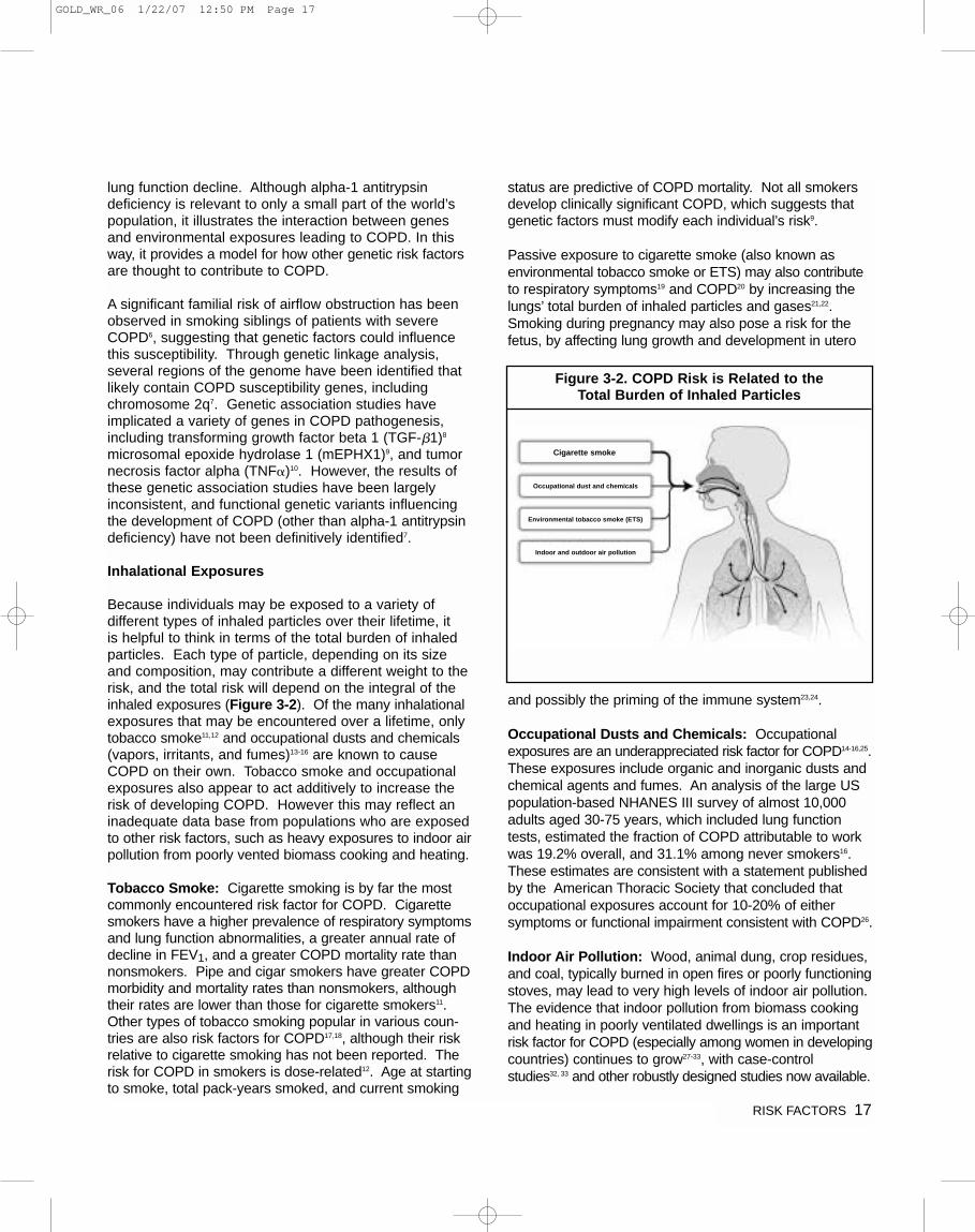
RISK FACTORS 17
lung function decline. Although alpha-1 antitrypsin deficiency is relevant to only a small part of the world’spopulation, it illustrates the interaction between genesand environmental exposures leading to COPD. In thisway, it provides a model for how other genetic risk factorsare thought to contribute to COPD.
A significant familial risk of airflow obstruction has beenobserved in smoking siblings of patients with severeCOPD6, suggesting that genetic factors could influencethis susceptibility. Through genetic linkage analysis, several regions of the genome have been identified thatlikely contain COPD susceptibility genes, including chromosome 2q7. Genetic association studies haveimplicated a variety of genes in COPD pathogenesis,including transforming growth factor beta 1 (TGF-�1)8
microsomal epoxide hydrolase 1 (mEPHX1)9, and tumornecrosis factor alpha (TNF�)10. However, the results ofthese genetic association studies have been largelyinconsistent, and functional genetic variants influencingthe development of COPD (other than alpha-1 antitrypsindeficiency) have not been definitively identified7.
Inhalational Exposures
Because individuals may be exposed to a variety of different types of inhaled particles over their lifetime, it is helpful to think in terms of the total burden of inhaledparticles. Each type of particle, depending on its sizeand composition, may contribute a different weight to therisk, and the total risk will depend on the integral of theinhaled exposures (Figure 3-2). Of the many inhalationalexposures that may be encountered over a lifetime, onlytobacco smoke11,12 and occupational dusts and chemicals(vapors, irritants, and fumes)13-16 are known to causeCOPD on their own. Tobacco smoke and occupationalexposures also appear to act additively to increase therisk of developing COPD. However this may reflect aninadequate data base from populations who are exposedto other risk factors, such as heavy exposures to indoor airpollution from poorly vented biomass cooking and heating.
Tobacco Smoke: Cigarette smoking is by far the mostcommonly encountered risk factor for COPD. Cigarettesmokers have a higher prevalence of respiratory symptomsand lung function abnormalities, a greater annual rate ofdecline in FEV1, and a greater COPD mortality rate thannonsmokers. Pipe and cigar smokers have greater COPDmorbidity and mortality rates than nonsmokers, althoughtheir rates are lower than those for cigarette smokers11.Other types of tobacco smoking popular in various coun-tries are also risk factors for COPD17,18, although their riskrelative to cigarette smoking has not been reported. Therisk for COPD in smokers is dose-related12. Age at startingto smoke, total pack-years smoked, and current smoking
status are predictive of COPD mortality. Not all smokersdevelop clinically significant COPD, which suggests thatgenetic factors must modify each individual’s risk9.
Passive exposure to cigarette smoke (also known as environmental tobacco smoke or ETS) may also contributeto respiratory symptoms19 and COPD20 by increasing thelungs’ total burden of inhaled particles and gases21,22.Smoking during pregnancy may also pose a risk for thefetus, by affecting lung growth and development in utero
and possibly the priming of the immune system23,24.
Occupational Dusts and Chemicals: Occupationalexposures are an underappreciated risk factor for COPD14-16,25.These exposures include organic and inorganic dusts andchemical agents and fumes. An analysis of the large USpopulation-based NHANES III survey of almost 10,000adults aged 30-75 years, which included lung functiontests, estimated the fraction of COPD attributable to workwas 19.2% overall, and 31.1% among never smokers16.These estimates are consistent with a statement publishedby the American Thoracic Society that concluded thatoccupational exposures account for 10-20% of eithersymptoms or functional impairment consistent with COPD26.
Indoor Air Pollution: Wood, animal dung, crop residues,and coal, typically burned in open fires or poorly functioningstoves, may lead to very high levels of indoor air pollution.The evidence that indoor pollution from biomass cookingand heating in poorly ventilated dwellings is an importantrisk factor for COPD (especially among women in developingcountries) continues to grow27-33, with case-control studies32, 33 and other robustly designed studies now available.
Figure 3-2. COPD Risk is Related to the Total Burden of Inhaled Particles
Cigarette smoke
Occupational dust and chemicals
Environmental tobacco smoke (ETS)
Indoor and outdoor air pollution
GOLD_WR_06 1/22/07 12:50 PM Page 17

18 RISK FACTORS
Almost 3 billion people worldwide use biomass and coal as their main source of energy for cooking, heating, andother household needs, so the population at risk worldwideis very large. In these communities, indoor air pollution isresponsible for a greater fraction of COPD risk than SO2or particulates from motor vehicle emissions, even in citiesdensely populated with people and cars. Biomass fuelsused by women for cooking account for the high prevalenceof COPD among nonsmoking women in parts of the MiddleEast, Africa, and Asia34,35. Indoor air pollution resulting fromthe burning of wood and other biomass fuels is estimatedto kill two million women and children each year36.
Outdoor Air Pollution: High levels of urban air pollutionare harmful to individuals with existing heart or lung disease.The role of outdoor air pollution in causing COPD is unclear,but appears to be small when compared with that of cigarettesmoking. It has also been difficult to assess the effects of single pollutants in long-term exposure to atmosphericpollution. However, air pollution from fossil fuel combustion,primarily from motor vehicle emissions in cities, is associatedwith decrements of respiratory function37. The relativeeffects of short-term, high-peak exposures and long-term,low-level exposures is a question yet to be resolved.
Lung Growth and Development
Lung growth is related to processes occurring during gestation, birth, and exposures during childhood38-40.Reduced maximal attained lung function (as measured byspirometry) may identify individuals who are at increasedrisk for the development of COPD41. Any factor that affectslung growth during gestation and childhood has the potentialfor increasing an individual’s risk of developing COPD.For example, a large study and meta-analysis confirmed apositive association between birth weight and FEV1 inadulthood42.
Oxidative Stress
The lungs are continuously exposed to oxidants generatedeither endogenously from phagocytes and other cell typesor exogenously from air pollutants or cigarette smoke. Inaddition, intracellular oxidants, such as those derived frommitochondrial electron transport, are involved in many cellular signaling pathways. Lung cells are protectedagainst this oxidative challenge by well-developed enzymaticand nonenzymatic systems. When the balance betweenoxidants and antioxidants shifts in favor of the former—i.e.,an excess of oxidants and/or a depletion of antioxidants—oxidative stress occurs. Oxidative stress not only producesdirect injurious effects in the lungs but also activates molecular mechanisms that initiate lung inflammation.Thus, an imbalance between oxidants and antioxidants isconsidered to play a role in the pathogenesis of COPD43.
Gender
The role of gender in determining COPD risk remainsunclear44. In the past, most studies showed that COPDprevalence and mortality were greater among men thanwomen. Studies from developed countries45,46 show thatthe prevalence of the disease is now almost equal in menand women, which probably reflects changing patterns oftobacco smoking. Some studies have suggested thatwomen are more susceptible to the effects of tobaccosmoke than men44,47,48. This is an important question giventhe increasing rate of smoking among women in bothdeveloped and developing countries.
Infections
Infections (viral and bacterial) may contribute to the pathogenesis and progression of COPD49, and the bacterialcolonization associated with airway inflammation50, andmay also play a significant role in exacerbations51. A historyof severe childhood respiratory infection has been associated with reduced lung function and increased respiratory symptoms in adulthood38,41,52. There are severalpossible explanations for this association (which are notmutually exclusive). There may be an increased diagnosisof severe infections in children who have underlying airwayhyperresponsiveness, itself considered a risk factor forCOPD. Susceptibility to viral infections may be related toanother factor, such as birth weight, that is related toCOPD. HIV infection has been shown to accelerate theonset of smoking-related emphysema; HIV-induced pulmonary inflammation may play a role in this process53.
Socioeconomic Status
There is evidence that the risk of developing COPD isinversely related to socioeconomic status54. It is not clear,however, whether this pattern reflects exposures to indoorand outdoor air pollutants, crowding, poor nutrition, or otherfactors that are related to low socioeconomic status55,56.
Nutrition
The role of nutrition as an independent risk factor for thedevelopment of COPD is unclear. Malnutrition and weight loss can reduce respiratory muscle strength andendurance, apparently by reducing both respiratory musclemass and the strength of the remaining muscle fibers57.The association of starvation and anabolic/catabolic statuswith the development of emphysema has been shown inexperimental studies in animals58. Lung CT scans ofwomen chronically malnourished because of anorexia nervosa showed emphysema-like changes59.
GOLD_WR_06 1/22/07 12:50 PM Page 18

RISK FACTORS 19
Asthma
Asthma may be risk factor for the development of COPD,although the evidence is not conclusive. In a report from a longitudinal cohort of the Tucson Epidemiological Studyof Airway Obstructive Disease adults with asthma werefound to have a twelvefold higher risk of acquiring COPDover time than those without asthma, after adjusting forsmoking60. Another longitudinal study of people with asthma found that around 20% of subjects developedfunctional signs of COPD, irreversible airflow limitation,and reduced transfer coefficient61.
REFERENCES
1. Celli BR, Halbert RJ, Nordyke RJ, Schan B. Airway obstructionin never smokers: results from the Third National Health andNutrition Examination Survey. Am J Med 2005;118:1364-72.
2. Behrendt CE. Mild and moderate-to-severe COPD in non-smokers. Distinct demographic profiles. Chest2005;128:1239-44.
3. Anthonisen NR, Connett JE, Murray RP. Smoking and lungfunction of Lung Health Study participants after 11 years. AmJ Respir Crit Care Med 2002;166(5):675-9.
4. Stoller JK, Aboussouan LS. Alpha1-antitrypsin deficiency.Lancet 2005;365(9478):2225-36.
5. Blanco I, de Serres FJ, Fernandez-Bustillo E, Lara B,Miravitlles M. Estimated numbers and prevalence of PI*S andPI*Z alleles of alpha1-antitrypsin deficiency in European countries. Eur Respir J 2006;27(1):77-84.
6. McCloskey SC, Patel BD, Hinchliffe SJ, Reid ED, WarehamNJ, Lomas DA. Siblings of patients with severe chronicobstructive pulmonary disease have a significant risk of airflow obstruction. Am J Respir Crit Care Med 2001;164(8 Pt 1):1419-24.
7. Silverman EK, Palmer LJ, Mosley JD, Barth M, Senter JM,Brown A, et al. Genomewide linkage analysis of quantitativespirometric phenotypes in severe early-onset chronic obstructivepulmonary disease. Am J Hum Genet 2002;70(5):1229-39.
8. Wu L, Chau J, Young RP, Pokorny V, Mills GD, Hopkins R, etal. Transforming growth factor-beta1 genotype and suscepti-bility to chronic obstructive pulmonary disease. Thorax2004;59(2):126-9.
9. Smith CA, Harrison DJ. Association between polymorphism ingene for microsomal epoxide hydrolase and susceptibility toemphysema. Lancet 1997;350(9078):630-3.
10. Huang SL, Su CH, Chang SC. Tumor necrosis factor-alphagene polymorphism in chronic bronchitis. Am J Respir CritCare Med 1997;156(5):1436-9.
11. US Surgeon General. The health consequences of smoking:chronic obstructive pulmonary disease. Washington, D.C.: USDepartment of Health and Human Services; 1984.
12. Burrows B, Knudson RJ, Cline MG, Lebowitz MD. Quantitativerelationships between cigarette smoking and ventilatory function.Am Rev Respir Dis 1977;115(2):195-205.
13. Becklake MR. Occupational exposures: evidence for a causalassociation with chronic obstructive pulmonary disease. AmRev Respir Dis 1989;140(3 Pt 2):S85-91.
14. Trupin L, Earnest G, San Pedro M, Balmes JR, Eisner MD,Yelin E, et al. The occupational burden of chronic obstructivepulmonary disease. Eur Respir J 2003;22(3):462-9.
15. Matheson MC, Benke G, Raven J, Sim MR, Kromhout H,Vermeulen R, et al. Biological dust exposure in the workplaceis a risk factor for chronic obstructive pulmonary disease.Thorax 2005;60(8):645-51.
16. Hnizdo E, Sullivan PA, Bang KM, Wagner G. Associationbetween chronic obstructive pulmonary disease and employmentby industry and occupation in the US population: a study ofdata from the Third National Health and Nutrition ExaminationSurvey. Am J Epidemiol 2002;156(8):738-46.
17. Jindal SK, Aggarwal AN, Chaudhry K, Chhabra SK, D'SouzaGA, Gupta D, et al. A multicentric study on epidemiology ofchronic obstructive pulmonary disease and its relationshipwith tobacco smoking and environmental tobacco smokeexposure. Indian J Chest Dis Allied Sci 2006;48(1):23-9.
18. Al-Fayez SF, Salleh M, Ardawi M, AZahran FM. Effects ofsheesha and cigarette smoking on pulmonary function ofSaudi males and females. Trop Geogr Med 1988;40(2):115-23.
19. The Health Consequences of Involuntary Exposure to TobaccoSmoke: A Report of the Surgeon General, Department ofHealth and Human Services. Washington, DC, US; 2006.
20. Eisner MD, Balmes J, Katz BP, Trupin L, Yelin E, Blanc P.Lifetime environmental tobacco smoke exposure and the riskof chronic obstructive pulmonary disease. Environ HealthPerspect 2005;4:7-15.
21. Leuenberger P, Schwartz J, Ackermann-Liebrich U, Blaser K,Bolognini G, Bongard JP, et al. Passive smoking exposure inadults and chronic respiratory symptoms (SAPALDIA Study).Swiss Study on Air Pollution and Lung Diseases in Adults,SAPALDIA Team. Am J Respir Crit Care Med 1994;150(5 Pt 1):1222-8.
22. Dayal HH, Khuder S, Sharrar R, Trieff N. Passive smoking inobstructive respiratory disease in an industrialized urban population. Environ Res 1994;65(2):161-71.
23. Tager IB, Ngo L, Hanrahan JP. Maternal smoking during preg-nancy. Effects on lung function during the first 18 months oflife. Am J Respir Crit Care Med 1995;152:977-83.
24. Holt PG. Immune and inflammatory function in cigarette smokers. Thorax 1987;42(4):241-9.
25. Hnizdo E, Sullivan PA, Bang KM, Wagner G. Airflow obstructionattributable to work in industry and occupation among U.S.race/ethnic groups: a study of NHANES III data. Am J IndMed 2004;46(2):126-35.
GOLD_WR_06 1/22/07 12:50 PM Page 19

20 RISK FACTORS
26. Balmes J, Becklake M, Blanc P, Henneberger P, Kreiss K,Mapp C, et al. American Thoracic Society Statement:Occupational contribution to the burden of airway disease. Am J Respir Crit Care Med 2003;167(5):787-97.
27. Warwick H, Doig A. Smoke the killer in the kitchen: Indoor airpollution in developing countries. ITDG Publishing, 103-105Southampton Row, London WC1B HLD, UK 2004:URL:http://www.itdgpublishing.org.uk.
28. Ezzati M. Indoor air pollution and health in developing countries. Lancet 2005;366(9480):104-6.
29. Smith KR, Mehta S, Maeusezahl-Feuz M. Indoor air-pollutionfrom household solid fuel use. In: Ezzati, M., Lopez, A. D.,Rodgers, M., Murray, C. J., eds. Comparative quantificationof health risks: global and regional burden of disease attributable to selected major risk factors. Geneva: WorldHealth Organization; 2004.
30. Mishra V, Dai X, Smith KR, Mika L. Maternal exposure to biomass smoke and reduced birth weight in Zimbabwe. AnnEpidemiol 2004;14(10):740-7.
31. Boman C, Forsberg B, Sandstrom T. Shedding new light onwood smoke: a risk factor for respiratory health. Eur Respir J2006;27(3):446-7.
32. Oroczo-Levi M, Garcia -Aymerich J, Villar J, Ramirez-SarmientoA, Anto JM, Gea J. Wood smoke exposure and risk of chronicobstructive pulmonary disease. Eur Respir J 2006;27:542-6.
33. Sezer H, Akkurt I, Guler N, Marakoglu K, Berk S. A case-controlstudy on the effect of exposure to different substances on thedevelopment of COPD. Ann Epidemiol 2006;16(1):59-62.
34. Smith KR. Inaugural article: national burden of disease inIndia from indoor air pollution. Proc Natl Acad Sci U S A2000;97(24):13286-93.
35. Chan-Yeung M, Ait-Khaled N, White N, Ip MS, Tan WC. Theburden and impact of COPD in Asia and Africa. Int J TubercLung Dis 2004;8(1):2-14.
36. Smith K. Pollution management in focus. The World Bank,Washington, DC 1999.
37. Abbey DE, Burchette RJ, Knutsen SF, McDonnell WF,Lebowitz MD, Enright PL. Long-term particulate and other airpollutants and lung function in nonsmokers. Am J Respir CritCare Med 1998;158(1):289-98.
38. Barker DJ, Godfrey KM, Fall C, Osmond C, Winter PD,Shaheen SO. Relation of birth weight and childhood respiratoryinfection to adult lung function and death from chronicobstructive airways disease. BMJ 1991;303(6804):671-5.
39. Todisco T, de Benedictis FM, Iannacci L, Baglioni S, Eslami A,Todisco E, et al. Mild prematurity and respiratory functions.Eur J Pediatr 1993;152(1):55-8.
40. Stein CE, Kumaran K, Fall CH, Shaheen SO, Osmond C,Barker DJ. Relation of fetal growth to adult lung function inSouth India. Thorax 1997;52(10):895-9.
41. Tager IB, Segal MR, Speizer FE, Weiss ST. The natural historyof forced expiratory volumes. Effect of cigarette smoking andrespiratory symptoms. Am Rev Respir Dis 1988;138(4):837-49.
42. Lawlor DA, Ebrahim S, Davey Smith G. Association of birthweight with adult lung function: findings from the BritishWomen's Heart and Health Study and a meta-analysis.Thorax 2005;60(10):851-8.
43. MacNee W. Pulmonary and systemic oxidant/antioxidantimbalance in chronic obstructive pulmonary disease. Proc AmThorac Soc 2005;2(1):50-60.
44. Xu X, Weiss ST, Rijcken B, Schouten JP. Smoking, changesin smoking habits, and rate of decline in FEV1: new insightinto gender differences. Eur Respir J 1994;7(6):1056-61.
45. National Heart, Lung, and Blood Institute. Morbidity and mortalitychartbook on cardiovascular, lung and blood diseases. Bethesda,Maryland: US Department of Health and Human Services,Public Health Service, National Institutes of Health, 2004. Accessedat: http://www.nhlbi.nih.gov/resources/docs/cht-book.htm.
46. Mannino DM, Homa DM, Akinbami LJ, Ford ES, Redd SC.Chronic obstructive pulmonary disease surveillance--UnitedStates, 1971-2000. MMWR Surveill Summ 2002;51(6):1-16.
47. Anthonisen NR, Connett JE, Kiley JP, Altose MD, Bailey WC,Buist AS, et al. Effects of smoking intervention and the use ofan inhaled anticholinergic bronchodilator on the rate of declineof FEV1. The Lung Health Study. JAMA 1994;272(19):1497-505.
48. Silverman EK, Weiss ST, Drazen JM, Chapman HA, Carey V,Campbell EJ, et al. Gender-related differences in severe,early-onset chronic obstructive pulmonary disease. Am JRespir Crit Care Med 2000;162(6):2152-8.
49. Retamales I, Elliott WM, Meshi B, Coxson HO, Pare P,Sciurba FC, et al. Amplification of inflammation in emphysemaand its association with latent adenoviral infection. Am JRespir Crit Care Med 2001;164:469-73.
50. Sethi S, Maloney J, Grove L, Wrona C, Berenson CS. Airwayinflammation and bronchial bacterial colonization in chronicobstructive pulmonary disease. Am J Respir Crit Care Med2006;173:991-8.
51. Seemungal T, Harper-Owen R, Bhowmik A, Moric I, SandersonG, Message S, et al. Respiratory viruses, symptoms, andinflammatory markers in acute exacerbations and stablechronic obstructive pulmonary disease. Am J Respir Crit CareMed 2001;164(9):1618-23.
52. Shaheen SO, Barker DJ, Shiell AW, Crocker FJ, Wield GA,Holgate ST. The relationship between pneumonia in earlychildhood and impaired lung function in late adult life. Am JRespir Crit Care Med 1994;149(3 Pt 1):616-9.
53. Diaz PT, King MA, Pacht ER, Wewers MD, Gadek JE,Nagaraja HN, et al. Increased susceptibility to pulmonaryemphysema among HIV-seropositive smokers. Ann InternMed 2000;132(5):369-72.
GOLD_WR_06 1/22/07 12:50 PM Page 20
RISK FACTORS 21
54. Prescott E, Lange P, Vestbo J. Socioeconomic status, lungfunction and admission to hospital for COPD: results from theCopenhagen City Heart Study. Eur Respir J 1999;13(5):1109-14.
55. Tao X, Hong CJ, Yu S, Chen B, Zhu H, Yang M. Priority amongair pollution factors for preventing chronic obstructive pulmonarydisease in Shanghai. Sci Total Environ 1992;127(1-2):57-67.
56. US Centers for Disease Control and Prevention. Criteria for arecommended standard: occupational exposure to respirablecoal mine dust: National Institute of Occupational Safety andHealth; 1995.
57. Wilson DO, Rogers RM, Wright EC, Anthonisen NR. Bodyweight in chronic obstructive pulmonary disease. The NationalInstitutes of Health Intermittent Positive-Pressure BreathingTrial. Am Rev Respir Dis 1989;139(6):1435-8.
58. Sahebjami H, Vassallo CL. Influence of starvation on enzyme-induced emphysema. J Appl Physiol 1980;48(2):284-8.
59. Coxson HO, Chan IH, Mayo JR, Hlynsky J, Nakano Y,Birmingham CL. Early emphysema in patients with anorexianervosa. Am J Respir Crit Care Med 2004;170(7):748-52.
60. Silva GE, Sherrill DL, Guerra S, Barbee RA. Asthma as a riskfactor for COPD in a longitudinal study. Chest 2004;126(1):59-65.
61. Vonk JM, Jongepier H, Panhuysen CI, Schouten JP, BleeckerER, Postma DS. Risk factors associated with the presence ofirreversible airflow limitation and reduced transfer coefficient inpatients with asthma after 26 years of follow up. Thorax2003;58(4):322-7.
GOLD_WR_06 1/22/07 12:50 PM Page 21

22 RISK FACTORS
GOLD_WR_06 1/22/07 12:50 PM Page 22

CHAPTER
4
PATHOLOGY,PATHOGENESIS,
ANDPATHOPHYSIOLOGY
GOLD_WR_06 1/22/07 12:50 PM Page 23
24 PATHOLOGY, PATHOGENESIS, AND PATHOPHYSIOLOGY
INTRODUCTION
Inhaled cigarette smoke and other noxious particlescause lung inflammation, a normal response whichappears to be amplified in patients who develop COPD.This abnormal inflammatory response may induceparenchymal tissue destruction (resulting in emphysema),and disrupt normal repair and defense mechanisms(resulting in small airway fibrosis). These pathologicalchanges lead to air trapping and progressive airflow limitation. A brief overview follows of the pathologicchanges in COPD, their cellular and molecular mechanisms,and how these underlie physiologic abnormalities andsymptoms characteristic of the disease1.
PATHOLOGY
Pathological changes characteristic of COPD are found inthe proximal airways, peripheral airways, lung parenchyma,and pulmonary vasculature2 (Figure 4-1). The pathologicalchanges include chronic inflammation, with increasednumbers of specific inflammatory cell types in differentparts of the lung, and structural changes resulting from
KEY POINTS:
• Pathological changes characteristic of COPD are found in the proximal airways, peripheral airways, lung parenchyma, and pulmonary vasculature. These changes include chronicinflammation, and structural changes resultingfrom repeated injury and repair.
• Inhaled cigarette smoke and other noxious particles cause lung inflammation, a normalresponse which appears to be amplified inpatients who develop COPD.
• There is a characteristic pattern of inflammationin the lungs of COPD patients, with increasednumbers of neutrophils (in the airway lumen),macrophages (airway lumen, airway wall, andparenchyma), and CD8+ lymphocytes (airwaywall and parenchyma). The pattern is differentfrom that seen in asthma.
• Lung inflammation is further amplified by oxidative stress and an excess of proteases in the lung.
• Physiological changes characteristic of the disease include mucus hypersecretion, airflowlimitation and air trapping (leading to hyper-inflation), gas exchange abnormalities, and cor pulmonale.
• Systemic features of COPD, particularly inpatients with severe disease, include cachexia,skeletal muscle wasting, increased risk of cardio-vascular disease, anemia, osteoporosis, anddepression.
• Exacerbations represent a further amplification of the inflammatory response in the airways of patients with COPD, and may be triggered by infection with bacteria or viruses or by environmental pollutants.
CHAPTER 4: PATHOLOGY, PATHOGENESIS, AND PATHOPHYSIOLOGY
Figure 4-1. Pathological Changes in COPD
Proximal airways (trachea, bronchi > 2 mm internal diameter) Inflammatory cells: Macrophages, CD8+ (cytotoxic) T lymphocytes,few neutrophils or eosinophilsStructural changes: Goblet cells, enlarged submucosal glands (bothleading to mucus hypersecretion), squamous metaplasia of epithelium3
Peripheral airways (bronchioles < 2mm i.d.) Inflammatory cells: Macrophages, T lymphocytes (CD8+ > CD4+),
B lymphocytes, lymphoid follicles, fibroblasts, few neutrophils or eosinophilsStructural changes: Airway wall thickening, peribronchial fibrosis, luminalinflammatory exudate, airway narrowing (obstructive bronchiolitis)Increased inflammatory response and exudate correlated with diseaseseverity4
Lung parenchyma (respiratory bronchioles and alveoli) Inflammatory cells: Macrophages, CD8+ T lymphocytesStructural changes: Alveolar wall destruction, apoptosis of epithelial and endothelial cells5
• Centrilobular emphysema: dilatation and destruction of respiratorybronchioles; most commonly seen in smokers
• Panacinar emphysema: destruction of alveolar sacs as well as respiratorybronchioles; most commonly seen in alpha-1 antitrypsin deficiency
Pulmonary vasculature Inflammatory cells: Macrophages, T lymphocytesStructural changes: Thickening of intima, endothelial cell dysfunction,
smooth muscle pulmonary hypertension6.
➝ ➝
➝➝ ➝
➝ ➝
➝➝
➝ ➝
➝➝
+Illustrations of many of the topics covered in this chapter canbe found on the GOLD Website: http://www.goldcopd.org.
GOLD_WR_06 1/22/07 12:50 PM Page 24
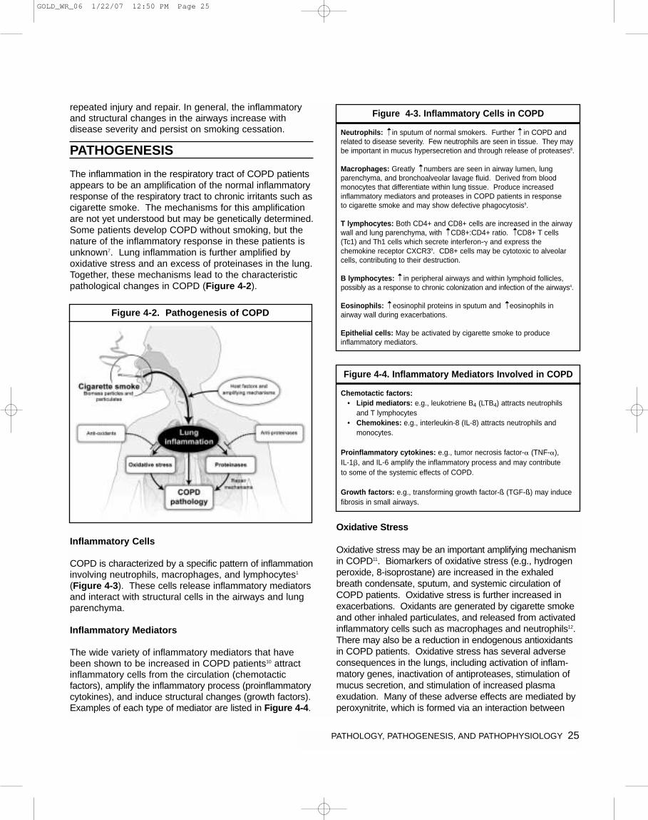
PATHOLOGY, PATHOGENESIS, AND PATHOPHYSIOLOGY 25
repeated injury and repair. In general, the inflammatoryand structural changes in the airways increase with disease severity and persist on smoking cessation.
PATHOGENESIS
The inflammation in the respiratory tract of COPD patientsappears to be an amplification of the normal inflammatoryresponse of the respiratory tract to chronic irritants such ascigarette smoke. The mechanisms for this amplificationare not yet understood but may be genetically determined.Some patients develop COPD without smoking, but thenature of the inflammatory response in these patients isunknown7. Lung inflammation is further amplified byoxidative stress and an excess of proteinases in the lung.Together, these mechanisms lead to the characteristicpathological changes in COPD (Figure 4-2).
Inflammatory Cells
COPD is characterized by a specific pattern of inflammationinvolving neutrophils, macrophages, and lymphocytes1
(Figure 4-3). These cells release inflammatory mediatorsand interact with structural cells in the airways and lungparenchyma.
Inflammatory Mediators
The wide variety of inflammatory mediators that havebeen shown to be increased in COPD patients10 attractinflammatory cells from the circulation (chemotactic factors), amplify the inflammatory process (proinflammatorycytokines), and induce structural changes (growth factors).Examples of each type of mediator are listed in Figure 4-4.
Oxidative Stress
Oxidative stress may be an important amplifying mechanismin COPD11. Biomarkers of oxidative stress (e.g., hydrogenperoxide, 8-isoprostane) are increased in the exhaledbreath condensate, sputum, and systemic circulation ofCOPD patients. Oxidative stress is further increased inexacerbations. Oxidants are generated by cigarette smokeand other inhaled particulates, and released from activatedinflammatory cells such as macrophages and neutrophils12.There may also be a reduction in endogenous antioxidantsin COPD patients. Oxidative stress has several adverseconsequences in the lungs, including activation of inflam-matory genes, inactivation of antiproteases, stimulation ofmucus secretion, and stimulation of increased plasma exudation. Many of these adverse effects are mediated byperoxynitrite, which is formed via an interaction between
Figure 4-2. Pathogenesis of COPD
Figure 4-3. Inflammatory Cells in COPD
Neutrophils: in sputum of normal smokers. Further in COPD andrelated to disease severity. Few neutrophils are seen in tissue. They maybe important in mucus hypersecretion and through release of proteases8.
Macrophages: Greatly numbers are seen in airway lumen, lungparenchyma, and bronchoalveolar lavage fluid. Derived from bloodmonocytes that differentiate within lung tissue. Produce increasedinflammatory mediators and proteases in COPD patients in response to cigarette smoke and may show defective phagocytosis9.
T lymphocytes: Both CD4+ and CD8+ cells are increased in the airwaywall and lung parenchyma, with CD8+:CD4+ ratio. CD8+ T cells(Tc1) and Th1 cells which secrete interferon-� and express thechemokine receptor CXCR39. CD8+ cells may be cytotoxic to alveolarcells, contributing to their destruction.
B lymphocytes: in peripheral airways and within lymphoid follicles,possibly as a response to chronic colonization and infection of the airways4.
Eosinophils: eosinophil proteins in sputum and eosinophils in airway wall during exacerbations.
Epithelial cells: May be activated by cigarette smoke to produce inflammatory mediators.
➝
➝
➝
➝
➝ ➝➝
➝
Figure 4-4. Inflammatory Mediators Involved in COPD
Chemotactic factors:• Lipid mediators: e.g., leukotriene B4 (LTB4) attracts neutrophils
and T lymphocytes• Chemokines: e.g., interleukin-8 (IL-8) attracts neutrophils and
monocytes.
Proinflammatory cytokines: e.g., tumor necrosis factor-� (TNF-�), IL-1�, and IL-6 amplify the inflammatory process and may contribute to some of the systemic effects of COPD.
Growth factors: e.g., transforming growth factor-ß (TGF-ß) may inducefibrosis in small airways.
GOLD_WR_06 1/22/07 12:50 PM Page 25
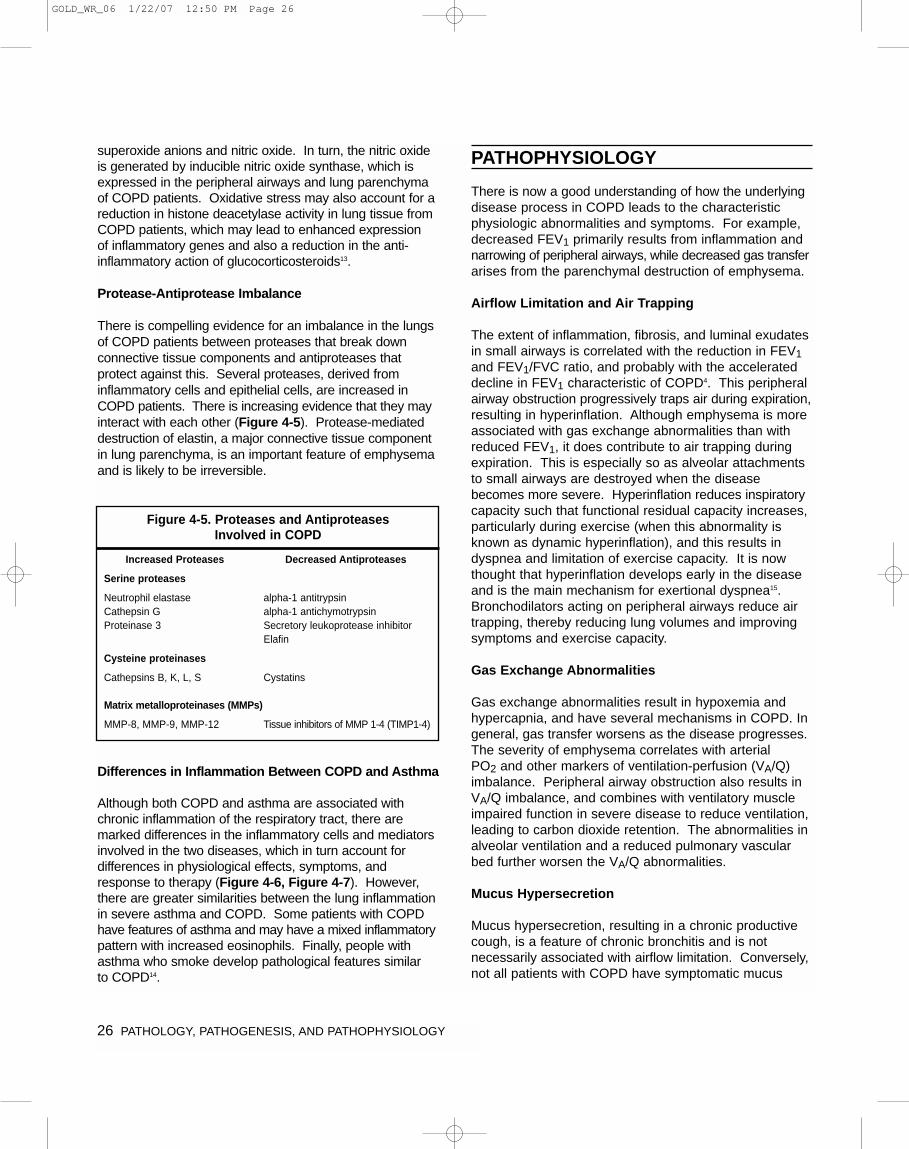
26 PATHOLOGY, PATHOGENESIS, AND PATHOPHYSIOLOGY
superoxide anions and nitric oxide. In turn, the nitric oxideis generated by inducible nitric oxide synthase, which isexpressed in the peripheral airways and lung parenchymaof COPD patients. Oxidative stress may also account for areduction in histone deacetylase activity in lung tissue fromCOPD patients, which may lead to enhanced expression of inflammatory genes and also a reduction in the anti-inflammatory action of glucocorticosteroids13.
Protease-Antiprotease Imbalance
There is compelling evidence for an imbalance in the lungsof COPD patients between proteases that break down connective tissue components and antiproteases that protect against this. Several proteases, derived frominflammatory cells and epithelial cells, are increased inCOPD patients. There is increasing evidence that they mayinteract with each other (Figure 4-5). Protease-mediateddestruction of elastin, a major connective tissue componentin lung parenchyma, is an important feature of emphysemaand is likely to be irreversible.
Differences in Inflammation Between COPD and Asthma
Although both COPD and asthma are associated withchronic inflammation of the respiratory tract, there aremarked differences in the inflammatory cells and mediatorsinvolved in the two diseases, which in turn account for differences in physiological effects, symptoms, andresponse to therapy (Figure 4-6, Figure 4-7). However,there are greater similarities between the lung inflammationin severe asthma and COPD. Some patients with COPDhave features of asthma and may have a mixed inflammatorypattern with increased eosinophils. Finally, people withasthma who smoke develop pathological features similar to COPD14.
PATHOPHYSIOLOGY
There is now a good understanding of how the underlyingdisease process in COPD leads to the characteristicphysiologic abnormalities and symptoms. For example,decreased FEV1 primarily results from inflammation andnarrowing of peripheral airways, while decreased gas transferarises from the parenchymal destruction of emphysema.
Airflow Limitation and Air Trapping
The extent of inflammation, fibrosis, and luminal exudatesin small airways is correlated with the reduction in FEV1and FEV1/FVC ratio, and probably with the accelerateddecline in FEV1 characteristic of COPD4. This peripheralairway obstruction progressively traps air during expiration,resulting in hyperinflation. Although emphysema is moreassociated with gas exchange abnormalities than withreduced FEV1, it does contribute to air trapping duringexpiration. This is especially so as alveolar attachmentsto small airways are destroyed when the diseasebecomes more severe. Hyperinflation reduces inspiratorycapacity such that functional residual capacity increases,particularly during exercise (when this abnormality isknown as dynamic hyperinflation), and this results in dyspnea and limitation of exercise capacity. It is nowthought that hyperinflation develops early in the diseaseand is the main mechanism for exertional dyspnea15.Bronchodilators acting on peripheral airways reduce airtrapping, thereby reducing lung volumes and improvingsymptoms and exercise capacity.
Gas Exchange Abnormalities
Gas exchange abnormalities result in hypoxemia andhypercapnia, and have several mechanisms in COPD. Ingeneral, gas transfer worsens as the disease progresses.The severity of emphysema correlates with arterial PO2 and other markers of ventilation-perfusion (VA/Q)imbalance. Peripheral airway obstruction also results inVA/Q imbalance, and combines with ventilatory muscleimpaired function in severe disease to reduce ventilation,leading to carbon dioxide retention. The abnormalities inalveolar ventilation and a reduced pulmonary vascularbed further worsen the VA/Q abnormalities.
Mucus Hypersecretion
Mucus hypersecretion, resulting in a chronic productivecough, is a feature of chronic bronchitis and is not necessarily associated with airflow limitation. Conversely,not all patients with COPD have symptomatic mucus
Figure 4-5. Proteases and Antiproteases Involved in COPD
Increased Proteases Decreased Antiproteases
Serine proteases
Neutrophil elastase alpha-1 antitrypsinCathepsin G alpha-1 antichymotrypsinProteinase 3 Secretory leukoprotease inhibitor
Elafin
Cysteine proteinases
Cathepsins B, K, L, S Cystatins
Matrix metalloproteinases (MMPs)
MMP-8, MMP-9, MMP-12 Tissue inhibitors of MMP 1-4 (TIMP1-4)
GOLD_WR_06 1/22/07 12:50 PM Page 26
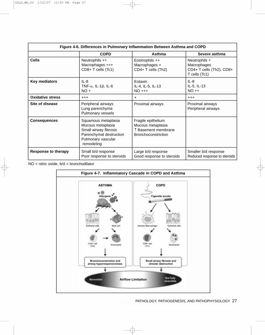
PATHOLOGY, PATHOGENESIS, AND PATHOPHYSIOLOGY 27
Figure 4-6. Differences in Pulmonary Inflammation Between Asthma and COPD
Cells
COPD
Neutrophils ++Macrophages +++CD8+ T cells (Tc1)
Eosinophils ++Macrophages +CD4+ T cells (Th2)
Neutrophils +MacrophagesCD4+ T cells (Th2), CD8+T cells (Tc1)
Key mediators IL-8TNF-�, IL-1�, IL-6NO +
EotaxinIL-4, IL-5, IL-13NO +++
IL-8IL-5, IL-13NO ++
Site of disease Peripheral airwaysLung parenchymaPulmonary vessels
Proximal airways Proximal airwaysPeripheral airways
Consequences Squamous metaplasiaMucous metaplasiaSmall airway fibrosisParenchymal destructionPulmonary vascularremodeling
Fragile epitheliumMucous metaplasia
Basement membrane Bronchoconstriction
Response to therapy Small b/d responsePoor response to steroids
Large b/d responseGood response to steroids
Smaller b/d responseReduced response to steroids
Oxidative stress +++ + +++
➝
Figure 4-7. Inflammatory Cascade in COPD and Asthma
NO = nitric oxide, b/d = bronchodilator
Asthma Severe asthma
GOLD_WR_06 1/22/07 12:50 PM Page 27
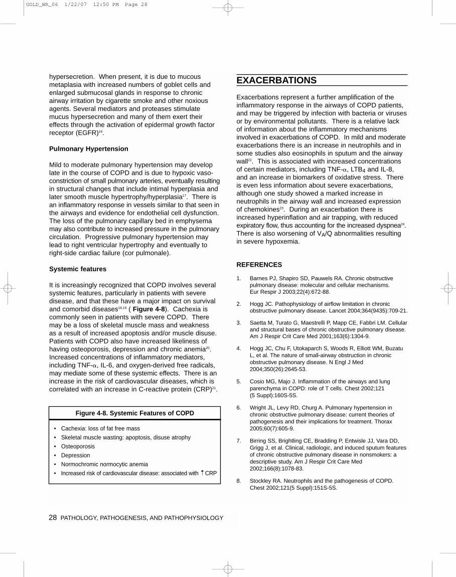
28 PATHOLOGY, PATHOGENESIS, AND PATHOPHYSIOLOGY
hypersecretion. When present, it is due to mucous metaplasia with increased numbers of goblet cells andenlarged submucosal glands in response to chronic airway irritation by cigarette smoke and other noxiousagents. Several mediators and proteases stimulatemucus hypersecretion and many of them exert theireffects through the activation of epidermal growth factorreceptor (EGFR)16.
Pulmonary Hypertension
Mild to moderate pulmonary hypertension may developlate in the course of COPD and is due to hypoxic vaso-constriction of small pulmonary arteries, eventually resultingin structural changes that include intimal hyperplasia andlater smooth muscle hypertrophy/hyperplasia17. There isan inflammatory response in vessels similar to that seen inthe airways and evidence for endothelial cell dysfunction.The loss of the pulmonary capillary bed in emphysemamay also contribute to increased pressure in the pulmonarycirculation. Progressive pulmonary hypertension maylead to right ventricular hypertrophy and eventually toright-side cardiac failure (cor pulmonale).
Systemic features
It is increasingly recognized that COPD involves severalsystemic features, particularly in patients with severe disease, and that these have a major impact on survivaland comorbid diseases18,19 ( Figure 4-8). Cachexia iscommonly seen in patients with severe COPD. Theremay be a loss of skeletal muscle mass and weakness as a result of increased apoptosis and/or muscle disuse.Patients with COPD also have increased likeliness ofhaving osteoporosis, depression and chronic anemia20.Increased concentrations of inflammatory mediators,including TNF-�, IL-6, and oxygen-derived free radicals,may mediate some of these systemic effects. There is anincrease in the risk of cardiovascular diseases, which iscorrelated with an increase in C-reactive protein (CRP)21.
EXACERBATIONS
Exacerbations represent a further amplification of theinflammatory response in the airways of COPD patients,and may be triggered by infection with bacteria or virusesor by environmental pollutants. There is a relative lack of information about the inflammatory mechanismsinvolved in exacerbations of COPD. In mild and moderateexacerbations there is an increase in neutrophils and insome studies also eosinophils in sputum and the airwaywall22. This is associated with increased concentrationsof certain mediators, including TNF-�, LTB4 and IL-8, and an increase in biomarkers of oxidative stress. Thereis even less information about severe exacerbations,although one study showed a marked increase in neutrophils in the airway wall and increased expressionof chemokines23. During an exacerbation there isincreased hyperinflation and air trapping, with reducedexpiratory flow, thus accounting for the increased dyspnea24.There is also worsening of VA/Q abnormalities resultingin severe hypoxemia.
REFERENCES
1. Barnes PJ, Shapiro SD, Pauwels RA. Chronic obstructive pulmonary disease: molecular and cellular mechanisms. Eur Respir J 2003;22(4):672-88.
2. Hogg JC. Pathophysiology of airflow limitation in chronicobstructive pulmonary disease. Lancet 2004;364(9435):709-21.
3. Saetta M, Turato G, Maestrelli P, Mapp CE, Fabbri LM. Cellularand structural bases of chronic obstructive pulmonary disease.Am J Respir Crit Care Med 2001;163(6):1304-9.
4. Hogg JC, Chu F, Utokaparch S, Woods R, Elliott WM, BuzatuL, et al. The nature of small-airway obstruction in chronicobstructive pulmonary disease. N Engl J Med2004;350(26):2645-53.
5. Cosio MG, Majo J. Inflammation of the airways and lungparenchyma in COPD: role of T cells. Chest 2002;121(5 Suppl):160S-5S.
6. Wright JL, Levy RD, Churg A. Pulmonary hypertension inchronic obstructive pulmonary disease: current theories ofpathogenesis and their implications for treatment. Thorax2005;60(7):605-9.
7. Birring SS, Brightling CE, Bradding P, Entwisle JJ, Vara DD,Grigg J, et al. Clinical, radiologic, and induced sputum featuresof chronic obstructive pulmonary disease in nonsmokers: adescriptive study. Am J Respir Crit Care Med2002;166(8):1078-83.
8. Stockley RA. Neutrophils and the pathogenesis of COPD.Chest 2002;121(5 Suppl):151S-5S.
Figure 4-8. Systemic Features of COPD
• Cachexia: loss of fat free mass
• Skeletal muscle wasting: apoptosis, disuse atrophy
• Osteoporosis
• Depression
• Normochromic normocytic anemia
• Increased risk of cardiovascular disease: associated with CRP➝
GOLD_WR_06 1/22/07 12:50 PM Page 28
PATHOLOGY, PATHOGENESIS, AND PATHOPHYSIOLOGY 29
9. Barnes PJ. Macrophages as orchestrators of COPD. J COPD2004;1:59-70.
10. Barnes PJ. Mediators of chronic obstructive pulmonary disease. Pharmacol Rev 2004;56(4):515-48.
11. Rahman I. Oxidative stress in pathogenesis of chronic obstruc-tive pulmonary disease: cellular and molecular mechanisms.Cell Biochem Biophys 2005;43(1):167-88.
12. MacNee W. Oxidative stress and lung inflammation in airwaysdisease. Eur J Pharmacol 2001;429(1-3):195-207.
13. Ito K, Ito M, Elliott WM, Cosio B, Caramori G, Kon OM, et al.Decreased histone deacetylase activity in chronic obstructivepulmonary disease. N Engl J Med 2005;352(19):1967-76.
14. Thomson NC, Chaudhuri R, Livingston E. Asthma and cigarette smoking. Eur Respir J 2004;24(5):822-33.
15. O'Donnell DE, Revill SM, Webb KA. Dynamic hyperinflationand exercise intolerance in chronic obstructive pulmonary disease. Am J Respir Crit Care Med 2001;164(5):770-7.
16. Burgel PR, Nadel JA. Roles of epidermal growth factor receptoractivation in epithelial cell repair and mucin production in air-way epithelium. Thorax 2004;59(11):992-6.
17. Barbera JA, Peinado VI, Santos S. Pulmonary hypertension inchronic obstructive pulmonary disease. Eur Respir J2003;21(5):892-905.
18. Wouters EF, Creutzberg EC, Schols AM. Systemic effects inCOPD. Chest 2002;121(5 Suppl):127S-30S.
19. Agusti AG, Noguera A, Sauleda J, Sala E, Pons J, Busquets X.Systemic effects of chronic obstructive pulmonary disease. EurRespir J 2003;21(2):347-60.
20. Similowski T, Agusti AG, MacNee W, Schonhofer B. The potential impact of anaemia of chronic disease in COPD. Eur Respir J 2006;27(2):390-6.
21. Gan WQ, Man SF, Senthilselvan A, Sin DD. Associationbetween chronic obstructive pulmonary disease and systemicinflammation: a systematic review and a meta-analysis. Thorax2004;59(7):574-80.
22. Wedzicha JA. Exacerbations: etiology and pathophysiologicmechanisms. Chest 2002;121(5 Suppl):136S-41S.
23. Drost EM, Skwarski KM, Sauleda J, Soler N, Roca J, AgustiAG. Oxidative stress and airway inflammation in severe exacerbations of COPD. Thorax 2005;60(4):293-300.
24. Parker CM, Voduc N, Aaron SD, Webb KA, O'Donnell DE.Physiological changes during symptom recovery from moderateexacerbations of COPD. Eur Respir J 2005;26(3):420-8.
GOLD_WR_06 1/22/07 12:50 PM Page 29

30 PATHOLOGY, PATHOGENESIS, AND PATHOPHYSIOLOGY
GOLD_WR_06 1/22/07 12:50 PM Page 30
CHAPTER
5
MANAGEMENTOF COPD
GOLD_WR_06 1/22/07 12:50 PM Page 31
32 MANAGEMENT OF COPD
An effective COPD management plan includes four components: (1) Assess and Monitor Disease; (2) ReduceRisk Factors; (3) Manage Stable COPD; and (4) ManageExacerbations. Management of Mild to Moderate COPD(Stages I and II) involves the avoidance of risk factors toprevent disease progression and pharmacotherapy asneeded to control symptoms. Severe (Stage III) and VerySevere (Stage IV) COPD often require the integration of several different disciplines, a variety of treatmentapproaches, and a commitment of the clinician to thecontinued support of the patient as the illness progresses.In addition to patient education, health advice, and pharmacotherapy, COPD patients may require specificcounseling about smoking cessation, instruction in physicalexercise, nutritional advice, and continued nursing support.Not all approaches are needed for every patient, andassessing the potential benefit of each approach at each stage of the illness is a crucial aspect of effectivedisease management.
While disease prevention is the ultimate goal, once COPDhas been diagnosed, effective management should beaimed at the following goals:
• Relieve symptoms
• Prevent disease progression
• Improve exercise tolerance
• Improve health status
• Prevent and treat complications
• Prevent and treat exacerbations
• Reduce mortality
These goals should be reached with minimal side effectsfrom treatment, a particular challenge in COPD patientsbecause they commonly have comorbidities. The extentto which these goals can be realized varies with eachindividual, and some treatments will produce benefits inmore than one area. In selecting a treatment plan, thebenefits and risks to the individual, and the costs, directand indirect, to the individual, his or her family, and thecommunity must be considered.
Patients should be identified as early in the course of thedisease as possible, and certainly before the end stageof the illness when disability is substantial. Access tospirometry is key to the diagnosis of COPD and shouldbe available to health care workers who care for COPDpatients. However, the benefits of community-basedspirometric screening, of either the general population orsmokers, are still unclear.
Educating patients, physicians, and the public to recognizethat cough, sputum production, and especially breath-lessness are not trivial symptoms is an essential aspectof the public health care of this disease.
Reduction of therapy once symptom control has beenachieved is not normally possible in COPD. Further deterioration of lung function usually requires the progressive introduction of more treatments, both pharmacologic and non-pharmacologic, to attempt to limitthe impact of these changes. Exacerbations of signs andsymptoms, a hallmark of COPD, impair patients' qualityof life and decrease their health status. Appropriate treatment and measures to prevent further exacerbationsshould be implemented as quickly as possible.
Important differences exist between countries in theapproach to chronic illnesses such as COPD and in theacceptability and affordability of particular forms of therapy.Ethnic differences in drug metabolism, especially for oralmedications, may result in different patient preferences in different communities. Little is known about theseimportant issues in relationship to COPD.
CHAPTER 5: MANAGEMENT OF COPDINTRODUCTION
GOLD_WR_06 1/22/07 12:50 PM Page 32

MANAGEMENT OF COPD 33
INITIAL DIAGNOSIS
A clinical diagnosis of COPD should be considered in any patient who has dyspnea, chronic cough or sputumproduction, and/or a history of exposure to risk factors for the disease (Figure 5.1-1). The diagnosis should be confirmed by spirometry. The presence of a postbronchodilator FEV1/FVC < 0.70 and FEV1 < 80%predicted confirms the presence of airflow limitation that is not fully reversible.
Assessment of Symptoms
Although exceptions occur, the general patterns of symptom development in COPD is well established. Themain symptoms of patients in Stage I: Mild COPD arechronic cough and sputum production. These symptomscan be present for many years before the development of airflow limitation and are often ignored or discountedby patients and attributed to aging or lack of conditioning.As airflow limitation worsens in Stage II: Moderate COPD,patients often experience dyspnea, which may interferewith their daily activities1. Typically, this is the stage atwhich they seek medical attention and may be diagnosedwith COPD. However, some patients do not experiencecough, sputum production, or dyspnea in Stage I: MildCOPD or Stage II: Moderate COPD, and do not come to medical attention until their airflow limitation becomesmore severe or their lung function is worsened acutely bya respiratory tract infection. As airflow limitation worsensand the patient enters Stage III: Severe COPD, thesymptoms of cough and sputum production typically continue, dyspnea worsens, and additional symptomsheralding complications (such as respiratory failure, right heart failure, weight loss, and arterial hypoxemia)may develop. It is important to note that, since COPD
KEY POINTS:
• A clinical diagnosis of COPD should be consideredin any patient who has dyspnea, chronic cough orsputum production, and/or a history of exposureto risk factors for the disease. The diagnosisshould be confirmed by spirometry.
• For the diagnosis and assessment of COPD,spirometry is the gold standard as it is the mostreproducible, standardized, and objective way ofmeasuring airflow limitation. The presence of apostbronchodilator FEV1/FVC < 0.70 and FEV1 <80% predicted confirms the presence of airflowlimitation that is not fully reversible.
• Health care workers involved in the diagnosisand management of COPD patients should haveaccess to spirometry.
• Assessment of COPD severity is based on thepatient’s level of symptoms, the severity of thespirometric abnormality, and the presence ofcomplications.
• Measurement of arterial blood gas tensions shouldbe considered in all patients with FEV1 < 50%predicted or clinical signs suggestive of respiratoryfailure or right heart failure.
• COPD is usually a progressive disease and lungfunction can be expected to worsen over time,even with the best available care. Symptoms andobjective measures of airflow limitation should bemonitored to determine when to modify therapyand to identify any complications that may develop.
• Comorbidities are common in COPD and should beactively identified. Comorbidities often complicatethe management of COPD, and vice versa.
COMPONENT 1: ASSESS AND MONITOR DISEASE
Figure 5.1-1. Key Indicators for Considering a Diagnosis of COPD
Consider COPD, and perform spirometry, if any of theseindicators are present in an individual over age 40. Theseindicators are not diagnostic themselves, but the presenceof multiple key indicators increases the probability of adiagnosis of COPD. Spirometry is needed to establish a diagnosis of COPD.
Dyspnea that is: Progressive (worsens over time)Usually worse with exercisePersistent (present every day)Described by the patient as an
“increased effort to breathe,” “heaviness,” “air hunger,” or “gasping.”
Chronic Cough May be intermittent and may beunproductive.
Chronic sputum Any pattern of chronic sputum production: production may indicate COPD.
History of Tobacco smoke.exposure to Occupational dusts and chemicalsrisk factors, Smoke from home cooking andespecially: heating fuels.
GOLD_WR_06 1/22/07 12:50 PM Page 33
34 MANAGEMENT OF COPD
may be diagnosed at any stage, any of the symptomsdescribed below may be present in a patient presentingfor the first time.
Dyspnea. Dyspnea, the hallmark symptom of COPD, isthe reason most patients seek medical attention and is amajor cause of disability and anxiety associated with thedisease. Typical COPD patients describe their dyspneaas a sense of increased effort to breathe, heaviness, air hunger, or gasping2. However, the terms used todescribe dyspnea vary both by individual and by culture3.It is often possible to distinguish the breathlessness ofCOPD from that due to other causes by analysis of theterms used, although there is considerable overlap withdescriptors of bronchial asthma. A simple way to quantifythe impact of breathlessness on a patient’s health status isthe British Medical Research Council (MRC) questionnaire(Figure 5.1-2). This questionnaire relates well to othermeasures of health status4 and predicts future mortality risk5.
Breathlessness in COPD is characteristically persistent andprogressive. Even on “good days” COPD patients experiencedyspnea at lower levels of exercise than unaffected peopleof the same age. Initially, breathlessness is only noted onunusual effort (e.g., walking or running up a flight of stairs)and may be avoided entirely by appropriate behavioralchange (e.g., using an elevator). As lung function deterio-rates, breathlessness becomes more intrusive, and patientsmay notice that they are unable to walk at the same speedas other people of the same age or carry out activities thatrequire use of the accessory respiratory muscles (e.g., car-rying grocery bags)6. Eventually, breathlessness is presentduring everyday activities (e.g., dressing, washing) or atrest, leaving the patient confined to the home.
Cough. Chronic cough, often the first symptom of COPD todevelop7, is often discounted by the patient as an expectedconsequence of smoking and/or environmental exposures.Initially, the cough may be intermittent, but later is presentevery day, often throughout the day. The chronic cough inCOPD may be unproductive8. In some cases, significantairflow limitation may develop without the presence of acough. Figure 5.1-3 lists some of the other causes ofchronic cough in individuals with a normal chest X-ray.
Sputum production. COPD patients commonly raise small quantities of tenacious sputum after coughing bouts.Regular production of sputum for 3 or more months in 2consecutive years (in the absence of any other conditionsthat may explain it) is the epidemiological definition of chronicbronchitis9, but this is a somewhat arbitrary definition thatdoes not reflect the range of sputum production in COPDpatients. Sputum production is often difficult to evaluatebecause patients may swallow sputum rather than expectorateit, a habit subject to significant cultural and gender variation.Patients producing large volumes of sputum may haveunderlying bronchiectasis. The presence of purulent sputumreflects an increase in inflammatory mediators10, and itsdevelopment may identify the onset of an exacerbation11.
Wheezing and chest tightness. Wheezing and chesttightness are nonspecific symptoms that may vary betweendays, and over the course of a single day. These symptomsmay be present in Stage I: Mild COPD, but are more characteristic of asthma or Stage III: Severe COPD andStage IV: Very Severe COPD. Audible wheeze may arise at a laryngeal level and need not be accompanied by auscultatory abnormalities. Alternatively, widespread inspiratory or expiratory wheezes can be present on listeningto the chest. Chest tightness often follows exertion, is poorly
Figure 5.1-2: Modified Medical Research CouncilQuestionnaire for Assessing the
Severity of Breathlessness4
PLEASE TICK IN THE BOX THAT APPLIES TO YOU(ONE BOX ONLY)
I only get breathless with strenuous exercise.
I get short of breath when hurrying on the level or walking up a slight hill.
I walk slower than people of the same age on the level because of breathlessness, or I have to stop for breath when walking on my own pace on the level.
I stop for breath after walking about 100 meters orafter a few minutes on the level.
I am too breathless to leave the house or I ambreathless when dressing or undressing.
Figure 5.1-3. Causes of Chronic Cough with aNormal Chest X-ray
Intrathoracic• Chronic obstructive pulmonary disease• Bronchial asthma• Central bronchial carcinoma• Endobronchial tuberculosis• Bronchiectasis• Left heart failure• Interstitial lung disease• Cystic fibrosis
Extrathoracic• Postnasal drip• Gastroesophageal reflux• Drug therapy (e.g., ACE inhibitors)
GOLD_WR_06 1/22/07 12:50 PM Page 34
MANAGEMENT OF COPD 35
localized, is muscular in character, and may arise from isometric contraction of the intercostal muscles. Anabsence of wheezing or chest tightness does not exclude a diagnosis of COPD, nor does their presence confirm a diagnosis of asthma.
Additional features in severe disease. Weight loss andanorexia are common problems in advanced COPD12. They are prognostically important13 and can also be a signof other diseases (e.g., tuberculosis, bronchial tumors), andtherefore should always be investigated. Cough syncopeoccurs due to rapid increases in intrathoracic pressure during attacks of coughing. Coughing spells may alsocause rib fractures, which are sometimes asymptomatic.Ankle swelling may be the only symptomatic pointer to thedevelopment of cor pulmonale. Finally, psychiatric morbidity,especially symptoms of depression and/or anxiety, is commonin advanced COPD14 and merits specific enquiry in the clinical history.
Medical History
A detailed medical history of a new patient known or thoughtto have COPD should assess:
• Patient’s exposure to risk factors, such as smoking andoccupational or environmental exposures
• Past medical history, including asthma, allergy, sinusitis,or nasal polyps; respiratory infections in childhood; otherrespiratory diseases
• Family history of COPD or other chronic respiratory disease
• Pattern of symptom development: COPD typically develops in adult life and most patients are conscious ofincreased breathlessness, more frequent “winter colds,”and some social restriction for a number of years beforeseeking medical help.
• History of exacerbations or previous hospitalizations forrespiratory disorder: Patients may be aware of periodicworsening of symptoms even if these episodes have notbeen identified as exacerbations of COPD.
• Presence of comorbidities, such as heart disease, malignancies, osteoporosis, and muscloskeletal disorders,which may also contribute to restriction of activity15.
• Appropriateness of current medical treatments: Forexample, beta-blockers commonly prescribed for heartdisease are usually contraindicated in COPD.
• Impact of disease on patient’s life, including limitation ofactivity, missed work and economic impact, effect on family routines, feelings of depression or anxiety
• Social and family support available to the patient
• Possibilities for reducing risk factors, especially smokingcessation
Physical Examination
Though an important part of patient care, a physicalexamination is rarely diagnostic in COPD. Physical signsof airflow limitation are usually not present until significantimpairment of lung function has occurred16,17, and theirdetection has a relatively low sensitivity and specificity. A number of physical signs may be present in COPD, buttheir absence does not exclude the diagnosis.
Inspection.
• Central cyanosis, or bluish discoloration of the mucosalmembranes, may be present but is difficult to detect inartificial light and in many racial groups.
• Common chest wall abnormalities, which reflect thepulmonary hyperinflation seen in COPD, include relatively horizontal ribs, “barrel-shaped” chest, andprotruding abdomen.
• Flattening of the hemi-diaphragms may be associatedwith paradoxical in-drawing of the lower rib cage oninspiration, and widening of the xiphosternal angle.
• Resting respiratory rate is often increased to morethan 20 breaths per minute and breathing can be relatively shallow17.
• Patients commonly show pursed-lip breathing, whichmay serve to slow expiratory flow and permit more efficient lung emptying18.
• COPD patients often have resting muscle activationwhile lying supine. Use of the scalene and sterno-cleidomastoid muscles is a further indicator of respiratory distress.
• Ankle or lower leg edema can be a sign of right heartfailure.
Palpation and percussion.
• These are often unhelpful in COPD.
• Detection of the heart apex beat may be difficult due to pulmonary hyperinflation.
• Hyperinflation also leads to downward displacement ofthe liver and an increase in the ability to palpate thisorgan without it being enlarged.
Auscultation.
• Patients with COPD often have reduced breathsounds, but this finding is not sufficiently characteristicto make the diagnosis19.
GOLD_WR_06 1/22/07 12:50 PM Page 35
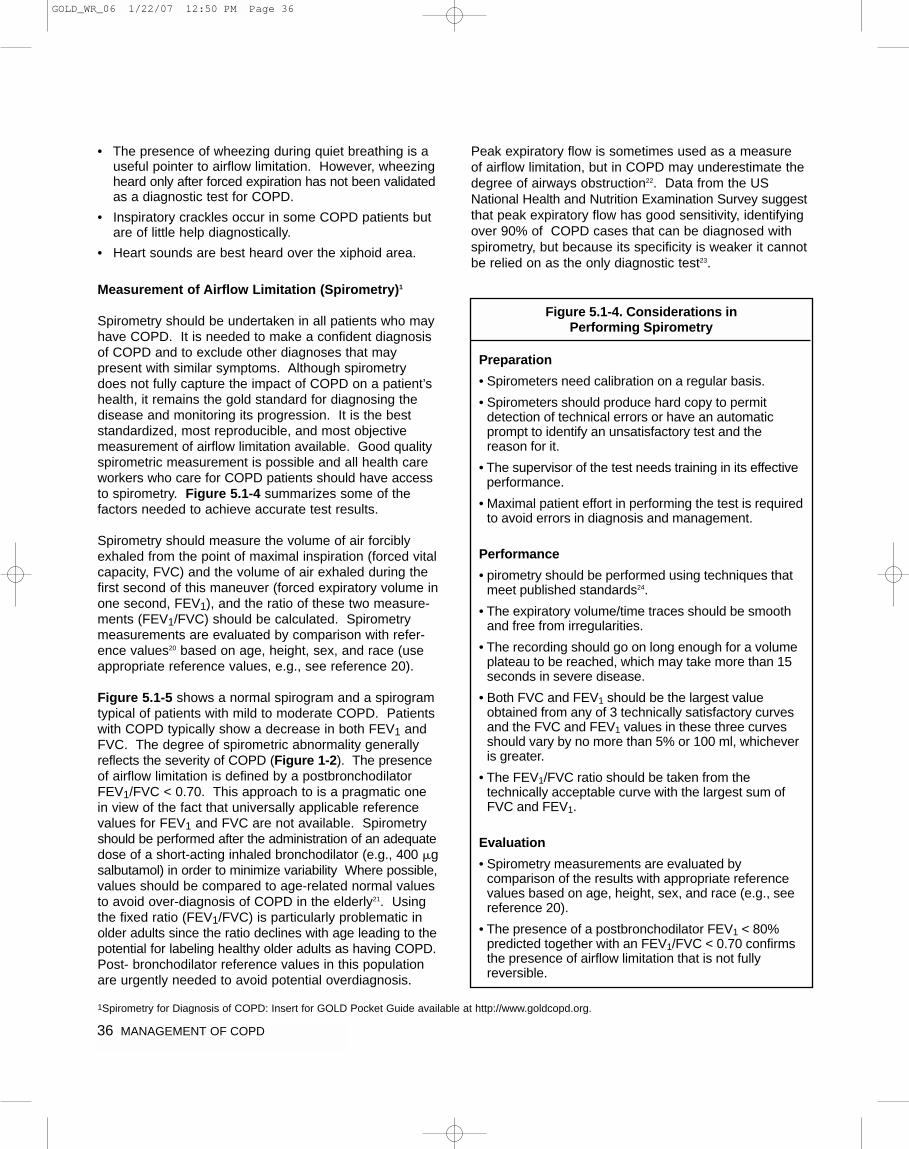
36 MANAGEMENT OF COPD
• The presence of wheezing during quiet breathing is auseful pointer to airflow limitation. However, wheezingheard only after forced expiration has not been validatedas a diagnostic test for COPD.
• Inspiratory crackles occur in some COPD patients butare of little help diagnostically.
• Heart sounds are best heard over the xiphoid area.
Measurement of Airflow Limitation (Spirometry)1
Spirometry should be undertaken in all patients who mayhave COPD. It is needed to make a confident diagnosisof COPD and to exclude other diagnoses that may present with similar symptoms. Although spirometrydoes not fully capture the impact of COPD on a patient’shealth, it remains the gold standard for diagnosing thedisease and monitoring its progression. It is the beststandardized, most reproducible, and most objectivemeasurement of airflow limitation available. Good qualityspirometric measurement is possible and all health careworkers who care for COPD patients should have accessto spirometry. Figure 5.1-4 summarizes some of the factors needed to achieve accurate test results.
Spirometry should measure the volume of air forciblyexhaled from the point of maximal inspiration (forced vitalcapacity, FVC) and the volume of air exhaled during thefirst second of this maneuver (forced expiratory volume inone second, FEV1), and the ratio of these two measure-ments (FEV1/FVC) should be calculated. Spirometrymeasurements are evaluated by comparison with refer-ence values20 based on age, height, sex, and race (useappropriate reference values, e.g., see reference 20).
Figure 5.1-5 shows a normal spirogram and a spirogramtypical of patients with mild to moderate COPD. Patientswith COPD typically show a decrease in both FEV1 andFVC. The degree of spirometric abnormality generallyreflects the severity of COPD (Figure 1-2). The presenceof airflow limitation is defined by a postbronchodilatorFEV1/FVC < 0.70. This approach to is a pragmatic onein view of the fact that universally applicable referencevalues for FEV1 and FVC are not available. Spirometryshould be performed after the administration of an adequatedose of a short-acting inhaled bronchodilator (e.g., 400 �gsalbutamol) in order to minimize variability Where possible,values should be compared to age-related normal valuesto avoid over-diagnosis of COPD in the elderly21. Usingthe fixed ratio (FEV1/FVC) is particularly problematic inolder adults since the ratio declines with age leading to thepotential for labeling healthy older adults as having COPD.Post- bronchodilator reference values in this populationare urgently needed to avoid potential overdiagnosis.
Peak expiratory flow is sometimes used as a measure of airflow limitation, but in COPD may underestimate thedegree of airways obstruction22. Data from the USNational Health and Nutrition Examination Survey suggestthat peak expiratory flow has good sensitivity, identifyingover 90% of COPD cases that can be diagnosed withspirometry, but because its specificity is weaker it cannotbe relied on as the only diagnostic test23.
Figure 5.1-4. Considerations in Performing Spirometry
Preparation
• Spirometers need calibration on a regular basis.
• Spirometers should produce hard copy to permit detection of technical errors or have an automaticprompt to identify an unsatisfactory test and the reason for it.
• The supervisor of the test needs training in its effectiveperformance.
• Maximal patient effort in performing the test is requiredto avoid errors in diagnosis and management.
Performance
• pirometry should be performed using techniques thatmeet published standards24.
• The expiratory volume/time traces should be smoothand free from irregularities.
• The recording should go on long enough for a volumeplateau to be reached, which may take more than 15seconds in severe disease.
• Both FVC and FEV1 should be the largest valueobtained from any of 3 technically satisfactory curvesand the FVC and FEV1 values in these three curvesshould vary by no more than 5% or 100 ml, whicheveris greater.
• The FEV1/FVC ratio should be taken from thetechnically acceptable curve with the largest sum ofFVC and FEV1.
Evaluation
• Spirometry measurements are evaluated bycomparison of the results with appropriate referencevalues based on age, height, sex, and race (e.g., seereference 20).
• The presence of a postbronchodilator FEV1 < 80%predicted together with an FEV1/FVC < 0.70 confirmsthe presence of airflow limitation that is not fullyreversible.
1Spirometry for Diagnosis of COPD: Insert for GOLD Pocket Guide available at http://www.goldcopd.org.
GOLD_WR_06 1/22/07 12:50 PM Page 36

MANAGEMENT OF COPD 37
*Postbronchodilator FEV1 is recommended for the diagnosis and assessmentof severity of COPD.
The role of screening spirometry in the general populationor in a population at risk for COPD is controversial. BothFEV1 and FVC predict all-cause mortality independent oftobacco smoking, and abnormal lung function identifies asubgroup of smokers at increased risk for lung cancer.This has been the basis of an argument that screeningspirometry should be employed as a global healthassessment tool25. However, there are no data to indi-cate that screening spirometry is effective in directingmanagement decisions or in improving COPD outcomesin patients who are identified before the development ofsignificant symptoms26.
Assessment of COPD Severity
Assessment of COPD severity is based on the patient’slevel of symptoms, the severity of the spirometric abnor-mality (Figure 1-2), and the presence of complicationssuch as respiratory failure, right heart failure, weight loss,and arterial hypoxemia.
Although the presence of airflow limitation is key to theassessment of COPD severity, it may be valuable from apublic health perspective to identify individuals at risk forthe disease before significant airflow limitation develops(Figure 1-3). A majority of people with early COPD identified in large studies complained of at least one respiratory symptom, such as cough, sputum production,wheezing, or breathlessness27,28. These symptoms maybe present at a time of relatively minor or even no spirometric abnormality. While not all individuals withsuch symptoms will go on to develop COPD29, the
presence of these symptoms should help define a high-risk population that should be targeted for preventiveintervention. Much depends on the success of convincingsuch people, as well as health care workers, that evenminor respiratory symptoms are not normal and may bemarkers of future ill health.
When evaluating symptomatic patients presenting to aphysician, the severity of the patient’s symptoms and the degree to which they affect his or her daily life, notjust the severity of airflow obstruction, are the majordeterminants of health status30. The severity of a patient’sbreathlessness is important and can be usefully gauged bythe MRC scale (Figure 5.1-2). Other forms of symptomseverity scoring have yet to be validated in different populations and commonly rely on individual clinical judgment, although a clinical COPD questionnaire hasbeen validated in family practice31.
Objectively measured exercise impairment, assessed by a reduction in self-paced walking distance32 or duringincremental exercise testing in a laboratory33, is a powerfulindicator of health status impairment and predictor ofprognosis30. The ratio of inspiratory capacity to total lungcapacity determined plethysmographically has also beenfound to be prognostically useful34. Similarly, weight lossand reduction in the arterial oxygen tension identifypatients at increased risk for mortality35,36.
A relatively simple approach to identifying disease severityusing a combination of most of the above variables hasbeen proposed. The BODE method gives a compositescore (Body mass index, Obstruction, Dyspnea andExercise) that is a better predictor of subsequent survivalthan any component singly37, and its properties as a measurement tool are under investigation.
Additional Investigations
For patients diagnosed with Stage II: Moderate COPDand beyond, the following additional investigations maybe considered:
Bronchodilator reversibility testing. Despite earlierhopes, neither bronchodilator nor oral glucocorticosteroidreversibility testing predicts disease progression, whetherjudged by decline in FEV1, deterioration of health status,or frequency of exacerbations38,39 in patients with a clinicaldiagnosis of COPD and abnormal spirometry39. Smallchanges in FEV1 (e.g., < 400 ml) after administration of a bronchodilator do not reliably predict the patient’sresponse to treatment (e.g., change in exercise capacity40).Minor variations in initial airway caliber can lead to differentclassification of reversibility status depending on the day
Figure 5.1-5. Normal Spirogram and SpirogramTypical of Patients with Mild to Moderate COPD*
GOLD_WR_06 1/22/07 12:50 PM Page 37

38 MANAGEMENT OF COPD
of testing39, and the lower the pre-bronchodilator FEV1, thegreater the chance of a patient being classified as reversibleeven when the 200 ml volume criterion is included.
In some cases (e.g., a patient with an atypical historysuch as asthma in childhood and regular night wakingwith cough or wheeze) a clinician may wish to perform abronchodilator and/or glucocorticosteroid reversibility testand a possible protocol is suggested in Figure 5.1-6.
Chest X-ray. An abnormal chest X-ray is seldom diagnostic in COPD unless obvious bullous disease ispresent, but it is valuable in excluding alternative diagnosesand establishing the presence of significant comorbiditiessuch as cardiac failure. Radiological changes associated
with COPD include signs of hyperinflation (flatteneddiaphragm on the lateral chest film, and an increase inthe volume of the retrosternal air space), hyperlucency of the lungs, and rapid tapering of the vascular markings.Computed tomography (CT) of the chest is not routinelyrecommended. However, when there is doubt about thediagnosis of COPD, high resolution CT (HRCT) scanningmight help in the differential diagnosis. In addition, if asurgical procedure such as lung volume reduction is contemplated, a chest CT scan is necessary since thedistribution of emphysema is one of the most importantdeterminants of surgical suitability41.
Arterial blood gas measurement. In advanced COPD,measurement of arterial blood gases while the patient isbreathing air is important. This test should be performedin stable patients with FEV1 < 50% predicted or with clinical signs suggestive of respiratory failure or right heartfailure. Several considerations are important to ensureaccurate test results. The inspired oxygen concentration(FiO2 – normally 21% at sea level) should be noted, aparticularly important point if patient is using an O2-drivennebulizer. Changes in arterial blood gas tensions taketime to occur, especially in severe disease. Thus, 20-30minutes should pass before rechecking the gas tensionswhen the FiO2 has been changed, e.g., during anassessment for domiciliary oxygen therapy. Adequatepressure must be applied at the arterial puncture site for at least one minute, as failure to do so can lead topainful bruising.
Alpha-1 antitrypsin deficiency screening. In patientsof Caucasian descent who develop COPD at a young age(< 45 years) or who have a strong family history of thedisease, it may be valuable to identify coexisting alpha-1antitrypsin deficiency. This could lead to family screeningor appropriate counseling. A serum concentration ofalpha-1 antitrypsin below 15-20% of the normal value ishighly suggestive of homozygous alpha-1 antitrypsin deficiency.
Differential Diagnosis
In some patients with chronic asthma, a clear distinctionfrom COPD is not possible using current imaging andphysiological testing techniques, and it is assumed thatasthma and COPD coexist in these patients. In thesecases, current management is similar to that of asthma.Other potential diagnoses are usually easier to distinguishfrom COPD (Figure 5.1-7).
Figure 5.1-6. Bronchodilator Reversibility Testing in COPD
Preparation
• Tests should be performed when patients are clinicallystable and free from respiratory infection.
• Patients should not have taken inhaled short-actingbronchodilators in the previous six hours, long-actingbronchodilator n the previous 12 hours, or sustained-release theophylline in the previous 24 hours.
Spirometry
• FEV1 should be measured before a bronchodilator isgiven.
• The bronchodilator should be given by metered doseinhaler through a spacer device or by nebulizer to becertain it has been inhaled.
• The bronchodilator dose should be selected to be highon the dose/response curve.
• Possible dosage protocols are 400 �g �2-agonist, up to 160 �g anticholinergic, or the two combined20.FEV1 should be measured again 10-15 minutes after a short-acting bronchodilator is given; 30-45 minutesafter the combination.
Results
• An increase in FEV1 that is both greater than 200 mland 12% above the pre-bronchodilator FEV1 isconsidered significant20. It is usually helpful to reportthe absolute change as well as the % change frombaseline to set the improvement in a clinical context.
GOLD_WR_06 1/22/07 12:50 PM Page 38
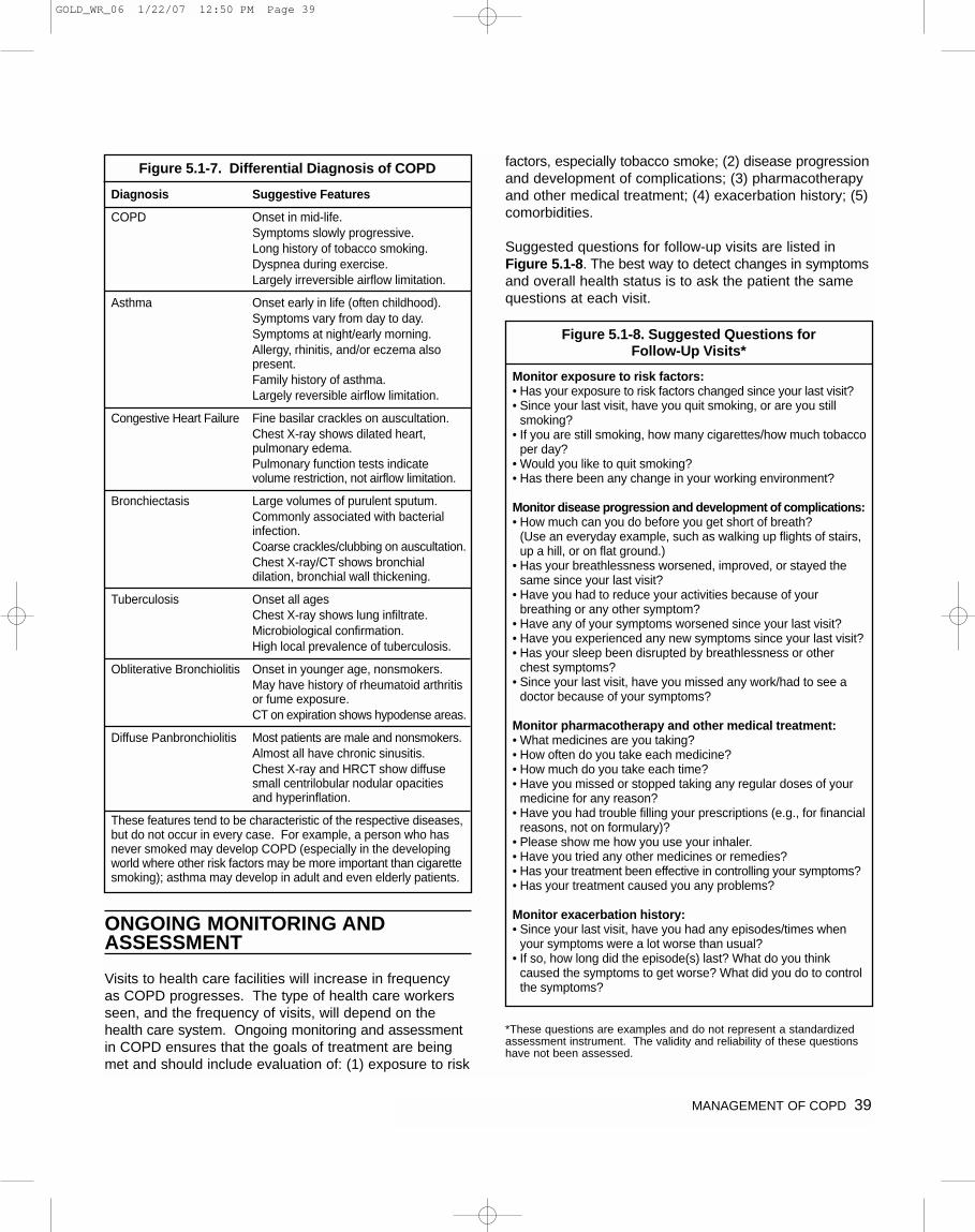
MANAGEMENT OF COPD 39
ONGOING MONITORING ANDASSESSMENT
Visits to health care facilities will increase in frequency as COPD progresses. The type of health care workersseen, and the frequency of visits, will depend on thehealth care system. Ongoing monitoring and assessmentin COPD ensures that the goals of treatment are beingmet and should include evaluation of: (1) exposure to risk
factors, especially tobacco smoke; (2) disease progressionand development of complications; (3) pharmacotherapyand other medical treatment; (4) exacerbation history; (5)comorbidities.
Suggested questions for follow-up visits are listed inFigure 5.1-8. The best way to detect changes in symptomsand overall health status is to ask the patient the samequestions at each visit.
*These questions are examples and do not represent a standardizedassessment instrument. The validity and reliability of these questionshave not been assessed.
Figure 5.1-7. Differential Diagnosis of COPD
Diagnosis Suggestive Features
COPD Onset in mid-life.Symptoms slowly progressive.Long history of tobacco smoking.Dyspnea during exercise.Largely irreversible airflow limitation.
Asthma Onset early in life (often childhood).Symptoms vary from day to day.Symptoms at night/early morning.Allergy, rhinitis, and/or eczema also present.Family history of asthma.Largely reversible airflow limitation.
Congestive Heart Failure Fine basilar crackles on auscultation.Chest X-ray shows dilated heart,pulmonary edema.Pulmonary function tests indicatevolume restriction, not airflow limitation.
Bronchiectasis Large volumes of purulent sputum.Commonly associated with bacterial infection.Coarse crackles/clubbing on auscultation.Chest X-ray/CT shows bronchialdilation, bronchial wall thickening.
Tuberculosis Onset all agesChest X-ray shows lung infiltrate.Microbiological confirmation.High local prevalence of tuberculosis.
Obliterative Bronchiolitis Onset in younger age, nonsmokers.May have history of rheumatoid arthritisor fume exposure.CT on expiration shows hypodense areas.
Diffuse Panbronchiolitis Most patients are male and nonsmokers. Almost all have chronic sinusitis.Chest X-ray and HRCT show diffusesmall centrilobular nodular opacities and hyperinflation.
These features tend to be characteristic of the respective diseases,but do not occur in every case. For example, a person who hasnever smoked may develop COPD (especially in the developingworld where other risk factors may be more important than cigarettesmoking); asthma may develop in adult and even elderly patients.
Figure 5.1-8. Suggested Questions for Follow-Up Visits*
Monitor exposure to risk factors:• Has your exposure to risk factors changed since your last visit?• Since your last visit, have you quit smoking, or are you still
smoking?• If you are still smoking, how many cigarettes/how much tobacco
per day?• Would you like to quit smoking?• Has there been any change in your working environment?
Monitor disease progression and development of complications:• How much can you do before you get short of breath?
(Use an everyday example, such as walking up flights of stairs,up a hill, or on flat ground.)
• Has your breathlessness worsened, improved, or stayed thesame since your last visit?
• Have you had to reduce your activities because of yourbreathing or any other symptom?
• Have any of your symptoms worsened since your last visit?• Have you experienced any new symptoms since your last visit?• Has your sleep been disrupted by breathlessness or other
chest symptoms?• Since your last visit, have you missed any work/had to see a
doctor because of your symptoms?
Monitor pharmacotherapy and other medical treatment:• What medicines are you taking?• How often do you take each medicine?• How much do you take each time?• Have you missed or stopped taking any regular doses of your
medicine for any reason?• Have you had trouble filling your prescriptions (e.g., for financial
reasons, not on formulary)?• Please show me how you use your inhaler.• Have you tried any other medicines or remedies?• Has your treatment been effective in controlling your symptoms?• Has your treatment caused you any problems?
Monitor exacerbation history:• Since your last visit, have you had any episodes/times when
your symptoms were a lot worse than usual?• If so, how long did the episode(s) last? What do you think
caused the symptoms to get worse? What did you do to controlthe symptoms?
GOLD_WR_06 1/22/07 12:50 PM Page 39
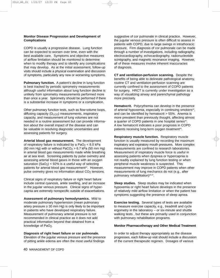
40 MANAGEMENT OF COPD
Monitor Disease Progression and Development ofComplications
COPD is usually a progressive disease. Lung functioncan be expected to worsen over time, even with the best available care. Symptoms and objective measuresof airflow limitation should be monitored to determinewhen to modify therapy and to identify any complicationsthat may develop. As at the initial assessment, follow-upvisits should include a physical examination and discussionof symptoms, particularly any new or worsening symptoms.
Pulmonary function. A patient’s decline in lung functionis best tracked by periodic spirometry measurementsalthough useful information about lung function decline isunlikely from spirometry measurements performed morethan once a year. Spirometry should be performed if thereis a substantial increase in symptoms or a complication.
Other pulmonary function tests, such as flow-volume loops,diffusing capacity (DLCO) measurements, inspiratorycapacity, and measurement of lung volumes are notneeded in a routine assessment but can provide informa-tion about the overall impact of the disease and can be valuable in resolving diagnostic uncertainties andassessing patients for surgery.
Arterial blood gas measurement. The development of respiratory failure is indicated by a PaO2 < 8.0 kPa (60 mm Hg) with or without PaCO2 > 6.7 kPa (50 mm Hg)in arterial blood gas measurements made while breathingair at sea level. Screening patients by pulse oximetry andassessing arterial blood gases in those with an oxygensaturation (SaO2) < 92% is a useful way of selectingpatients for arterial blood gas measurement42. However,pulse oximetry gives no information about CO2 tensions.
Clinical signs of respiratory failure or right heart failureinclude central cyanosis, ankle swelling, and an increasein the jugular venous pressure. Clinical signs of hyper-capnia are extremely nonspecific outside of exacerbations.
Assessment of pulmonary hemodynamics. Mild tomoderate pulmonary hypertension (mean pulmonaryartery pressure ≥ 30 mm Hg) is only likely to be importantin patients who have developed respiratory failure.Measurement of pulmonary arterial pressure is not recommended in clinical practice as it does not add practical information beyond that obtained from a knowledge of PaO2.
Diagnosis of right heart failure or cor pulmonale.Elevation of the jugular venous pressure and the presenceof pitting ankle edema are often the most useful findings
suggestive of cor pulmonale in clinical practice. However,the jugular venous pressure is often difficult to assess inpatients with COPD, due to large swings in intrathoracicpressure. Firm diagnosis of cor pulmonale can be madethrough a number of investigations, including radiography,electrocardiography, echocardiography, radionucleotidescintigraphy, and magnetic resonance imaging. However,all of these measures involve inherent inaccuracies of diagnosis.
CT and ventilation-perfusion scanning. Despite thebenefits of being able to delineate pathological anatomy,routine CT and ventilation-perfusion scanning are currently confined to the assessment of COPD patientsfor surgery. HRCT is currently under investigation as away of visualizing airway and parenchymal pathologymore precisely.
Hematocrit. Polycythemia can develop in the presenceof arterial hypoxemia, especially in continuing smokers43,and can be identified by hematocrit > 55%. Anemia ismore prevalent than previously thought, affecting almosta quarter of COPD patients in one hospital series44. A low hematocrit indicates a poor prognosis in COPDpatients receiving long-term oxygen treatment45.
Respiratory muscle function. Respiratory muscle function is usually measured by recording the maximuminspiratory and expiratory mouth pressures. More complexmeasurements are confined to research laboratories.Measurement of inspiratory muscle force is useful inassessing patients when dyspnea or hypercapnia is not readily explained by lung function testing or whenperipheral muscle weakness is suspected. This measurement may improve in COPD patients when othermeasurements of lung mechanics do not (e.g., after pulmonary rehabilitation)46,47.
Sleep studies. Sleep studies may be indicated whenhypoxemia or right heart failure develops in the presenceof relatively mild airflow limitation or when the patient hassymptoms suggesting the presence of sleep apnea.
Exercise testing. Several types of tests are available to measure exercise capacity, e.g., treadmill and cycleergometry in the laboratory – or six-minute and shuttlewalking tests , but these are primarily used in conjunctionwith pulmonary rehabilitation programs.
Monitor Pharmacotherapy and Other Medical Treatment
In order to adjust therapy appropriately as the diseaseprogresses, each follow-up visit should include a discussionof the current therapeutic regimen. Dosages of various
GOLD_WR_06 1/22/07 12:50 PM Page 40
MANAGEMENT OF COPD 41
medications, adherence to the regimen, inhaler technique,effectiveness of the current regime at controlling symptoms,and side effects of treatment should be monitored.
Monitor Exacerbation History
During periodic assessments, health care workers should question the patient and evaluate any records of exacerbations, both self-treated and those treated byother health care providers. Frequency, severity, andlikely causes of exacerbations should be evaluated.Increased sputum volume, acutely worsening dyspnea,and the presence of purulent sputum should be noted.Specific inquiry into unscheduled visits to providers, telephone calls for assistance, and use of urgent oremergency care facilities may be helpful. Severity canbe estimated by the increased need for bronchodilatormedication or glucocorticosteroids and by the need forantibiotic treatment. Hospitalizations should be documented,including the facility, duration of stay, and any use of critical care or intubation. The clinician then can requestsummaries of all care received to facilitate continuity of care.
Monitor Comorbidities
Comorbidities are common in COPD. Some may be anindirect result of COPD, arising independently but morelikely to occur when COPD is present, e.g., ischemicheart disease, bronchial carcinoma, osteoporosis. Othercomorbid conditions may coexist with COPD becausethey become prevalent as part of the aging process, e.g., arthritis, diabetes, reflux esophagitis. All comorbidconditions become harder to manage when COPD is present, either because COPD adds to the total level ofdisability or because COPD therapy adversely affects thecomorbid disorder. All comorbid conditions amplify thedisability associated with COPD and can potentially complicate its management. Until more integrated guidance about disease management for specific comorbidproblems becomes available, the focus should be onidentification and management of these individual problems in line with local treatment guidance.
GOLD_WR_06 1/22/07 12:50 PM Page 41
42 MANAGEMENT OF COPD
INTRODUCTION
Identification, reduction, and control of risk factors areimportant steps toward prevention and treatment of anydisease. In the case of COPD, these factors includetobacco smoke, occupational exposures, and indoor and outdoor air pollution and irritants. Since cigarettesmoking is the most commonly encountered risk factor forCOPD worldwide, tobacco control (smoking prevention)programs should be implemented and smoking cessationprograms should be readily available and encouraged forall individuals who smoke. Reduction of total personalexposure to occupational dusts, fumes, and gases and to indoor and outdoor air pollutants is also an importantgoal to prevent the onset and progression of COPD.
TOBACCO SMOKE
Smoking Prevention
Comprehensive tobacco control policies and programswith clear, consistent, and repeated nonsmoking messagesshould be delivered through every feasible channel,including health care providers, community activities,schools, and radio, television, and print media. Nationaland local campaigns should be undertaken to reduceexposure to tobacco smoke in public forums. Such bansare proving to be workable and to result in measurablegains in respiratory health48. Legislation to establishsmoke-free schools, public facilities, and work environ-ments should be developed and implemented by govern-ment officials and public health workers, and encouragedby the public. Smoking prevention programs should tar-get all ages, including young children, adolescents, youngadults, and pregnant women. Interventions to preventsmoking uptake and maximize cessation should beimplemented at every level of the health care system.Physicians and public health officials should encouragesmoke-free homes.
An important step toward a collective internationalresponse to tobacco-caused death and disease wastaken in 1996 by the World Health Organization with theimplementation of an International Framework Conventionon Tobacco Control (Figure 5.2-1).
KEY POINTS:
• Reduction of total personal exposure to tobaccosmoke, occupational dusts and chemicals, andindoor and outdoor air pollutants are importantgoals to prevent the onset and progression ofCOPD.
• Smoking cessation is the single most effective—and cost effective—intervention in most people to reduce the risk of developing COPD and stopits progression (Evidence A).
• Comprehensive tobacco control policies and programs with clear, consistent, and repeatednonsmoking messages should be deliveredthrough every feasible channel.
• Efforts to reduce smoking through public healthinitiatives should also focus on passive smokingto minimize risks for nonsmokers.
• Many occupationally induced respiratory disorderscan be reduced or controlled through a variety ofstrategies aimed at reducing the burden ofinhaled particles and gases.
• Reducing the risk from indoor and outdoor airpollution is feasible and requires a combinationof public policy and protective steps taken byindividual patients.
COMPONENT 2: REDUCE RISK FACTORS
Figure 5.2-1. World Health Organization:International Framework Convention on
Tobacco Control
In May, 1996, to address the global tobacco pandemic, the Forty-ninth World Health Assembly requested theDirector-General of the World Health Organization (WHO) to initiate the development of an international frameworkconvention for tobacco control. Included as part of thisframework convention is a strategy to encourage MemberStates to move progressively towards the adoption ofcomprehensive tobacco control policies and to deal withaspects of tobacco control that transcend nationalboundaries.
Information about the work of the WHO tobacco controlprogram can be found athttp://www.who.int/tobacco/resources/publications/fctc/en/index.html
GOLD_WR_06 1/22/07 12:50 PM Page 42
MANAGEMENT OF COPD 43
Environmental tobacco smoke exposure is also an important cause of respiratory symptoms and increasedrisk for COPD, especially in partners and children ofsmokers49. Long-term indoor exposure, combined withcrowded living conditions in poorly ventilated homes,adds to the total burden of particulate exposure andincreases the risk of developing COPD50. Efforts toreduce smoking through public health initiatives shouldalso focus on passive smoking to minimize risks for nonsmokers. Partners and parents should not smoke inthe immediate vicinity of nonsmokers or children, nor inenclosed spaces such as cars and poorly ventilatedrooms that expose others to increased risk.
The first exposure to cigarette smoke may begin in uterowhen the fetus is exposed to blood-borne metabolitesfrom the mother51. Education to reduce in utero risks forunborn children is also of great importance to prevent theeffects of maternal smoking in reducing lung growth andcausing airways disease in early and later life52,53.Neonates and infants may also be exposed passively totobacco smoke in the home if a family member smokes.Children less than 2 years old who are passively exposedto cigarette smoke have an increased prevalence of respiratory infections, and are at a greater risk of developing chronic respiratory symptoms later in life53,54.
Smoking Cessation
Smoking cessation is the single most effective—and costeffective—way to reduce exposure to COPD risk factors.Quitting smoking can prevent or delay the developmentof airflow limitation, or reduce its progression55, and canhave a substantial effect on subsequent mortality56. Allsmokers—including those who may be at risk for COPDas well as those who already have the disease—shouldbe offered the most intensive smoking cessation intervention feasible.
Smoking cessation interventions are effective in bothsexes, in all racial and ethnic groups, and in pregnantwomen. Age influences quit rates, with young peopleless likely to quit, but nevertheless smoking cessationprograms can be effective in all age groups. Internationaldata on the economic impact of smoking cessation arestrikingly consistent: investing resources in smoking cessation programs is cost effective in terms of medicaland societal costs per life-year gained. Effective inter-ventions include nicotine replacement with transdermalpatches, gums, and nasal sprays; counseling from physicians and other health professionals (with or withoutnicotine replacement therapy); self-help and group programs; and community-based stop-smoking challenges.
A review of data from a number of countries estimatedthe median societal cost of various smoking cessationinterventions at $990 to $13,000 (US) per life-year gained57.Smoking cessation programs are a particularly goodvalue for the UK National Health Service, with costs from£212 to £873 (US $320 to $1,400) per life-year gained58.
The role of health care providers in smoking cessation. A successful smoking cessation strategy requires a multi-faceted approach, including public policy, information dissemination programs, and health education through themedia and schools59. However, health care providers,including physicians, nurses, dentists, psychologists,pharmacists, and others, are key to the delivery of smokingcessation messages and interventions. Involving asmany of these individuals as possible will help. Healthcare workers should encourage all patients who smoke to quit, even those patients who come to the health careprovider for unrelated reasons and do not have symptomsof COPD, evidence of airflow limitation, or other smoking-related disease. Guidelines for smoking cessation entitledTreating Tobacco Use and Dependence: A ClinicalPractice Guideline were published by the US PublicHealth Service60. The major conclusions are summarizedin Figure 5.2-2.
Figure 5.2-2. US Public Health Service Report:Treating Tobacco Use and Dependence:
A Clinical Practice Guideline—Major Findings and Recommendations60
1. Tobacco dependence is a chronic condition that warrantsrepeated treatment until long-term or permanent abstinence isachieved.
2. Effective treatments for tobacco dependence exist and alltobacco users should be offered these treatments.
3. Clinicians and health care delivery systems mustinstitutionalize the consistent identification, documentation,and treatment of every tobacco user at every visit.
4. Brief smoking cessation counseling is effective and everytobacco user should be offered such advice at every contactwith health care providers.
5. There is a strong dose-response relation between the intensityof tobacco dependence counseling and its effectiveness.
6. Three types of counseling were found to be especiallyeffective: practical counseling, social support as part oftreatment, and social support arranged outside of treatment.
7. Five first-line pharmacotherapies for tobacco dependence—bupropion SR, nicotine gum, nicotine inhaler, nicotine nasalspray, and nicotine patch—are effective and at least one ofthese medications should be prescribed in the absence ofcontraindications.
8. Tobacco dependence treatments are cost effective relative toother medical and disease prevention interventions.
GOLD_WR_06 1/22/07 12:50 PM Page 43
44 MANAGEMENT OF COPD
The Public Health Service Guidelines recommend a five-step program for intervention (Figure 5.2-3), which providesa strategic framework helpful to health care providersinterested in helping their patients stop smoking60-63. The guidelines emphasize that tobacco dependence is achronic disease (Figure 5.2-4)60 and urge clinicians torecognize that relapse is common and reflects the chronicnature of dependence and addiction, not failure on thepart of the clinician or the patient.
Most individuals go through several stages before theystop smoking (Figure 5.2-5)59. It is often helpful for theclinician to assess a patient's readiness to quit in order to determine the most effective course of action at thattime. The clinician should initiate treatment if the patientis ready to quit. For a patient not ready to make a quitattempt, the clinician should provide a brief interventiondesigned to promote the motivation to quit.
Counseling. Counseling delivered by physicians andother health professionals significantly increases quit ratesover self-initiated strategies64. Even a brief (3-minute)period of counseling to urge a smoker to quit results insmoking cessation rates of 5-10%65. At the very least,this should be done for every smoker at every health careprovider visit65,66. Education in how to offer optimal smokingcessation advice and support should be a mandatory element of curricula for health professionals.
There is a strong dose-response relationship betweencounseling intensity and cessation success18,19. Ways tointensify treatment include increasing the length of thetreatment session, the number of treatment sessions,and the number of weeks over which the treatment isdelivered. Sustained quit rates of 10.9% at 6 monthshave been achieved when clinician tutorials and feed-back are linked to counseling sessions67. With morecomplex interventions (for example, controlled clinical trials that include skills training, problem solving, andpsychosocial support), quit rates can reach 20-30%68. In a multicenter controlled clinical trial, a combination of physician advice, group support, skills training, andnicotine replacement therapy achieved a quit rate of 35%at 1 year and a sustained quit rate of 22% at 5 years55.
Both individual and group counseling are effective formatsfor smoking cessation programs. Several particular itemsof counseling content seem to be especially effective,including problem solving, general skills training, and provision of intra-treatment support59,60. The commonsubjects covered in successful promblem solving/skillstraining programs include:
Figure 5.2-3. Brief Strategies to Help the Patient Willing to Quit60-63
1. ASK: Systematically identify all tobacco users at every visit.Implement an office-wide system that ensures that, for EVERY patientat EVERY clinic visit, tobacco-use status is queried and documented.
2. ADVISE: Strongly urge all tobacco users to quit.In a clear, strong, and personalized manner, urge every tobacco user to quit.
3. ASSESS: Determine willingness to make a quit attempt.Ask every tobacco user if he or she is willing to make a quit attempt atthis time (e.g., within the next 30 days).
4. ASSIST: Aid the patient in quitting.Help the patient with a quit plan; provide practical counseling; provideintra-treatment social support; help the patient obtain extra-treatmentsocial support; recommend use of approved pharmacotherapy exceptin special circumstances; provide supplementary materials.
5. ARRANGE: Schedule follow-up contact.Schedule follow-up contact, either in person or via telephone.
Figure 5.2-4. Tobacco Dependence as a Chronic Disease10
• For most people, tobacco dependence results in true drugdependence comparable to dependence caused by opiates,amphetamines, and cocaine.
• Tobacco dependence is almost always a chronic disorderthat warrants long-term clinical intervention as do otheraddictive disorders. Failure to appreciate the chronic natureof tobacco dependence may impair the clinician's motivationto treat tobacco use consistently in a long-term fashion.
• Clinicians must understand that tobacco dependence is achronic condition requiring sustained effort focused on simplecounseling advice, support, and appropriate pharmacotherapy,and ongoing support for recent quitters to prevent relapse.
• Relapse is common, which is the nature of dependence andnot the failure of the clinician or the patient.
Figure 5.2-5. Stages of Change Model
GOLD_WR_06 1/22/07 12:50 PM Page 44
MANAGEMENT OF COPD 45
• Recognition of danger signals likely to be associatedwith the risk of relapse, such as being around othersmokers, psychosocial stress, being under time pressure, getting into an argument, drinking alcohol,and negative moods
• Enhancement of skills needed to handle these situations, such as learning to anticipate and manageor avoid a particular stress
• Basic information about smoking and successful quitting, such as the nature and time course of withdrawal, the addictive nature of smoking, and thefact that any return to smoking, including even a singlepuff, increases the likelihood of a relapse
Systematic programs to sustain smoking cessationshould be implemented in health care settings17.
Pharmacotherapy. Numerous effective pharma-cotherapies for smoking cessation now exist59-61, andpharmacotherapy is recommended when counseling isnot sufficient to help patients quit smoking. Special consideration should be given before using pharma-cotherapy in selected populations: people with medicalcontraindications, light smokers (fewer than 10 cigarettes/day), and pregnant and adolescent smokers.
Nicotine replacement products. Numerous studies indicate that nicotine replacement therapy in any form(nicotine gum, inhaler, nasal spray, transdermal patch,sublingual tablet, or lozenge) reliably increases long-termsmoking abstinence rates60,69. Nicotine replacement therapy is more effective when combined with counselingand behavior therapy70, although nicotine patch or nicotine gum consistently increases smoking cessationrates regardless of the level of additional behavioral orpsychosocial interventions. Medical contraindications tonicotine replacement therapy include unstable coronaryartery disease, untreated peptic ulcer disease, and recentmyocardial infarction or stroke59. Specific studies do notsupport the use of nicotine replacement therapy forlonger than 8 weeks, although some patients may requireextended use to prevent relapse and, in some studies,use of multiple nicotine replacement therapy modalitieshas been shown to be more effective than only one60,71.
All forms of nicotine replacement therapy are significantlymore effective than placebo. Every effort should bemade to tailor the choice of replacement therapy to theindividual’s culture and lifestyle to improve adherence.The patch is generally favored over the gum because itrequires less training for effective use and is associatedwith fewer compliance problems. No data are availableto help clinicians tailor nicotine patch regimens to theintensity of cigarette smoking. In all cases it seems generally appropriate to start with the higher dose patch.
For most patches, which come in three different doses,patients should use the highest dose for the first fourweeks and drop to progressively lower doses over aneight-week period. Where only two doses are available,the higher dose should be used for the first four weeksand the lower dose for the second four weeks.
When using nicotine gum, the patient needs to beadvised that absorption occurs through the buccalmucosa. For this reason, the patient should be advisedto chew the gum for a while and then put the gumagainst the inside of the cheek to allow absorption tooccur and prolong the release of nicotine. Continuouschewing produces secretions that are swallowed ratherthan absorbed through the buccal mucosa, results in littleabsorption, and can cause nausea. Acidic beverages,particularly coffee, juices, and soft drinks, interfere withthe absorption of nicotine. Thus, the patient needs to beadvised that eating or drinking anything except watershould be avoided for 15 minutes before and duringchewing. Although nicotine gum is an effective smokingcessation treatment, problems with compliance, ease ofuse, social acceptability, risk of developing temporo-mandibular joint symptoms, and unpleasant taste havebeen noted. In highly dependent smokers, the 4 mg gumis more effective than the 2 mg gum72.
Other pharmacotherapy. The antidepressants bupropion73
and nortriptyline have also been shown to increase long-term quit rates59,69,74, but should always be used as oneelement in a supportive intervention program rather than ontheir own. Although more studies need to be conductedwith these medications, a randomized controlled trial withcounseling and support showed quit rates at one year of30% with sustained-release bupropion alone and 35%with sustained-release bupropion plus nicotine patch73.The effectiveness of the antihypertensive drug clonidineis limited by side effects69.
Varenicline, a nicotinic acetylcholine receptor partial agonist that aids smoking cessation by relieving nicotinewithdrawal symptoms and reducing the rewarding properties of nicotine has been demonstrated to be safe and efficacious75-77.
OCCUPATIONAL EXPOSURES
In the United States, it has been estimated that up to19% of COPD in smokers and up to 31% of COPD innonsmokers may be attributable to occupational dust and fume exposure78-81, and the burden may be higher incountries where there is higher exposure to inhaled particles, fumes and gases. Many occupations havebeen shown to be associated with increased risk ofdeveloping COPD, particularly those that involve exposure
GOLD_WR_06 1/22/07 12:50 PM Page 45
46 MANAGEMENT OF COPD
to fumes and mineral and biological dusts. Although it isnot known how many individuals are at risk of developingrespiratory disease from occupational exposures in eitherdeveloping or developed countries, many occupationallyinduced respiratory disorders can be reduced or con-trolled through a variety of strategies aimed at reducingthe burden of inhaled particles and gases82-84.
• Implement, monitor and enforce strict, legally mandatedcontrol of airborne exposure in the workplace.
• Initiate intensive and continuing education of exposedworkers, industrial managers, health care workers, primary care physicians, and legislators.
• Educate employers, workers, and policymakers onhow cigarette smoking aggravates occupational lungdiseases and why efforts to reduce smoking where ahazard exists are important.
The main emphasis should be on primary prevention,which is best achieved by the elimination or reduction of exposures to various substances in the workplace.Secondary prevention, achieved through surveillance andearly case detection, is also of great importance. Bothapproaches are necessary to improve the present situationand to reduce the burden of lung disease. Although studies as yet have not been done to demonstratereduced burden of disease, it is the logical consequenceof effective strategies to reduce workplace exposure torespiratory irritants and toxic inhalants.
INDOOR AND OUTDOOR AIR POLLUTION
Individuals experience diverse indoor and outdoor environments throughout the day, each of which has itsown unique set of air contaminants and particulates thatcause adverse effects on lung function80.
Although outdoor and indoor air pollution are generallyconsidered separately, the concept of total personalexposure may be more relevant for COPD. Reducing the risk from indoor and outdoor air pollution is feasibleand requires a combination of public policy and protectivesteps taken by individual patients. Reduction of exposureto smoke from biomass fuel, particularly among womenand children, is a crucial goal to reduce the prevalence of COPD worldwide. Although efficient non-pollutingcooking stoves have been developed, their adoption hasbeen slow due to social customs and cost.
Regulation of Air Quality
At the national level, achieving a set level of air qualitystandards should be a high priority; this goal will normally require legislative action. Details on setting and maintaining air quality goals are beyond the scope
of this document, but public policy to reduce vehicle andindustrial emissions to safe levels is an urgent priority toreduce the development of COPD as well as symptoms,exacerbations, and hospital admissions in those with disease. Understanding health risks posed by local airpollution sources may be difficult and requires skills incommunity health, toxicology, and epidemiology. Localphysicians may become involved through concerns aboutthe health of their patients or as advocates for the community’s environment.
Steps for Health Care Providers/Patients
The health care provider should consider COPD risk factors including smoking history, family history, exposureto indoor/outdoor pollution) and socioeconomic status foreach individual patient. Some steps to consider:
Individuals at risk for COPD:
• Patients should be counseled concerning the natureand degree of their risk for COPD.
• If various solid fuels are used for cooking and heating,adequate ventilation should be encouraged.
• Respiratory protective equipment has been developedfor use in the workplace in order to minimize exposureto toxic gases and particles. Under most circumstances,vigorous attempts should be made to reduce exposurethrough reducing workplace emissions and improvingventilation measures, rather than simply by using respiratory protection to reduce the risks of ambient air pollution.
• Ventilation and interventions to meet safe air qualitystandards in the workplace offer the greatest opportunityto reduce worker exposure to known atmospheric pollutants and reduce the risk of developing COPD,although to date there are no studies to quantify these benefits.
Patients who have been diagnosed with COPD:
• Persons with advanced COPD should monitor publicannouncements of air quality and be aware that staying indoors when air quality is poor may helpreduce their symptoms.
• The use of medication should follow the usual clinicalindications; therapeutic regimens should not be adjustedbecause of the occurrence of a pollution episode without evidence of worsening of symptoms or lungfunction.
• Those who are at high risk should avoid vigorousexercise outdoors during pollution episodes.
• Air cleaners have not been shown to have health benefits, whether directed at pollutants generated byindoor sources or at those brought in with outdoor air.
GOLD_WR_06 1/22/07 12:50 PM Page 46
MANAGEMENT OF COPD 47
INTRODUCTION
The overall approach to managing stable COPD shouldbe characterized by an increase in treatment, dependingon the severity of the disease and the clinical status ofthe patient. The step-down approach used in the chronictreatment of asthma is not applicable to COPD sinceCOPD is usually stable and very often progressive.Management of COPD involves several objectives (seeChapter 5, Introduction) that should be met with minimalside effects from treatment. It is based on an individualizedassessment of disease severity (Figure 5.3-1) andresponse to various therapies.
The classification of severity of stable COPD incorporatesan individualized assessment of disease severity andtherapeutic response into the management strategy. The severity of airflow limitation (Figure 1-2) provides a general guide to the use of some treatments, but theselection of therapy is predominantly determined by thepatient’s symptoms and clinical presentation. Treatmentalso depends on the patient’s educational level and willingness to apply the recommended management, on cultural and local conditions, and on the availability of medications.
EDUCATION
Although patient education is generally regarded as anessential component of care for any chronic disease, the role of education in COPD has been poorly studied.Assessment of the value of education in COPD may bedifficult because of the relatively long time required toachieve improvements in objective measurements of lung function.
KEY POINTS:
• The overall approach to managing stable COPDshould be individualized to address symptoms andimprove quality of life.
• For patients with COPD, health education plays animportant role in smoking cessation (Evidence A)and can also play a role in improving skills, abilityto cope with illness and health status.
• None of the existing medications for COPD havebeen shown to modify the long-term decline inlung function that is the hallmark of this disease(Evidence A). Therefore, pharmacotherapy forCOPD is used to decrease symptoms and/or complications.
• Bronchodilator medications are central to thesymptomatic management of COPD (Evidence A).They are given on an as-needed basis or on aregular basis to prevent or reduce symptoms and exacerbations.
• The principal bronchodilator treatments are �2-agonists, anticholinergics, and methylxanthinesused singly or in combination (Evidence A).
• Regular treatment with long-acting bronchodilatorsis more effective and convenient than treatmentwith short-acting bronchodilators (Evidence A).
• The addition of regular treatment with inhaled glucocorticosteroids to bronchodilator treatment isappropriate for symptomatic COPD patients withan FEV1 < 50% predicted (Stage III: SevereCOPD and Stage IV: Very Severe COPD) andrepeated exacerbations (Evidence A).
• Chronic treatment with systemic glucocortico-steroids should be avoided because of an unfavorable benefit-to-risk ratio (Evidence A).
• In COPD patients influenza vaccines can reduceserious illness (Evidence A). Pneumococcal polysaccharide vaccine is recommended forCOPD patients 65 years and older and for COPDpatients younger than age 65 with an FEV1 < 40%predicted (Evidence B).
• All COPD patients benefit from exercise trainingprograms, improving with respect to both exercisetolerance and symptoms of dyspnea and fatigue(Evidence A).
• The long-term administration of oxygen (> 15 hoursper day) to patients with chronic respiratory failurehas been shown to increase survival (Evidence A).
COMPONENT 3: MANAGE STABLE COPD
Figure 5.3-1. Factors Affecting the Severity of COPD
• Severity of symptoms
• Severity of airflow limitation
• Frequency and severity of exacerbations
• Presence of one or more complications
• Presence of respiratory failure
• Presence of comorbid conditions
• General health status
• Number of medications needed to manage the disease
GOLD_WR_06 1/22/07 12:50 PM Page 47
48 MANAGEMENT OF COPD
Studies that have been done indicate that patient edu-cation alone does not improve exercise performance orlung function85-88 (Evidence B), but it can play a role inimproving skills, ability to cope with illness, and healthstatus89. These outcomes are not traditionally measuredin clinical trials, but they may be most important in COPDwhere even pharmacologic interventions generally conferonly a small benefit in terms of lung function.
Patient education regarding smoking cessation has thegreatest capacity to influence the natural history of COPD.Evaluation of the smoking cessation component in a long-term, multicenter study indicates that if effective resourcesand time are dedicated to smoking cessation, 25% long-term quit rates can be maintained55 (Evidence A). Educationalso improves patient response to exacerbations90,91
(Evidence B). Prospective end-of-life discussions canlead to understanding of advance directives and effectivetherapeutic decisions at the end of life92 (Evidence B).
Ideally, educational messages should be incorporatedinto all aspects of care for COPD and may take place inmany settings: consultations with physicians or otherhealth care workers, home-care or outreach programs,and comprehensive pulmonary rehabilitation programs.
Goals and Educational Strategies
It is vital for patients with COPD to understand the natureof their disease, risk factors for progression, and theirrole and the role of health care workers in achieving optimal management and health outcomes. Educationshould be tailored to the needs and environment of theindividual patient, interactive, directed at improving qualityof life, simple to follow, practical, and appropriate to theintellectual and social skills of the patient and the caregivers.
In managing COPD, open communication between patientand physician is essential. In addition to being empathic,attentive and communicative, health professionals shouldpay attention to patients’ fears and apprehensions, focuson educational goals, tailor treatment regimens to eachindividual patient, anticipate the effect of functionaldecline, and optimize patients’ practical skills.
Several specific education strategies have been shown toimprove patient adherence to medication and managementregimens. In COPD, adherence does not simply refer towhether patients take their medication appropriately. Italso covers a range of nonpharmacologic treatments,e.g., maintaining an exercise program after pulmonaryrehabilitation, undertaking and sustaining smoking cessation, and using devices such as nebulizers, spacers,and oxygen concentrators properly.
Components of an Education Program
The topics that seem most appropriate for an educationprogram include: smoking cessation; basic informationabout COPD and pathophysiology of the disease; generalapproach to therapy and specific aspects of medicaltreatment; self-management skills; strategies to help minimize dyspnea; advice about when to seek help; self-management and decision-making during exacer-bations; and advance directives and end-of-life issues(Figure 5.3-2). Education should be part of consultationswith health care workers beginning at the time of firstassessment for COPD and continuing with each follow-up visit. The intensity and content of these educationalmessages should vary depending on the severity of thepatient’s disease. In practice, a patient often poses aseries of questions to the physician (Figure 5.3-3). It isimportant to answer these questions fully and clearly, asthis may help make treatment more effective.
Figure 5.3-2. Topics for Patient Education
For all patients:• Information and advice about reducing risk factors
Stage I: Mild COPD through Stage III: Severe COPDAbove topic, plus:• Information about the nature of COPD• Instruction on how to use inhalers and other treatments• Recognition and treatment of exacerbations• Strategies for minimizing dyspnea
Stage IV: Very Severe COPDAbove topics, plus:• Information about complications• Information about oxygen treatment• Advance directives and end-of-life decisions
Figure 5.3-3. Examples of Patient Questions
• What is COPD?• What causes COPD?• How will it affect me?• Can it be treated?• What will happen if my disease gets worse?• What will happen if I need to be admitted to the hospital?• How will I know when I need oxygen at home?• What if I do not wish to be admitted to intensive care for
ventilation?
Answers to these questions can be developed from this document andwill depend on local circumstances. In all cases, it is important thatanswers are clear and use terminology that the patient understands.
GOLD_WR_06 1/22/07 12:50 PM Page 48
MANAGEMENT OF COPD 49
There are several different types of educational programs,ranging from simple distribution of printed materials, toteaching sessions designed to convey information aboutCOPD, to workshops designed to train patients in specific skills (e.g., self-management). In general, case-management approaches to medical problems have been somewhat disappointing93. However, COPDpatients recruited to a comprehensive COPD educationprogram in Canada had significantly fewer exacer-bations and hospitalizations. and used fewer health careresources94. These encouraging results require replicationin other health care systems and patient groups.
Although printed materials may be a useful adjunct toother educational messages, passive dissemination ofprinted materials alone does not improve skills or healthoutcomes. Education is most effective when it is interactiveand conducted in small workshops88 (Evidence B)designed to improve both knowledge and skills. Behavioralapproaches such as cognitive therapy and behavior modification lead to more effective self-managementskills and maintenance of exercise programs.
Cost Effectiveness of Education Programs for COPDPatients
The cost effectiveness of education programs for COPDpatients is highly dependent on local factors that influencethe cost of access to medical services and that will varysubstantially between countries. In one cost-benefitanalysis of education provided to hospital inpatients withCOPD95, an information package resulted in increasedknowledge of COPD and reduced use of health services,including reductions of hospital readmissions and generalpractice consultations. The education package involvedtraining patients to increase knowledge of COPD, medication usage, precautions for exacerbations, andpeak flow monitoring technique. However, this study wasundertaken in a heterogeneous group of patients—65%were smokers and 88% were judged to have an asthmaticcomponent to their disease—and these findings may nothold true for a “pure” COPD population. In a study ofmild to moderate COPD patients at an outpatient clinic,patient education involving one 4-hour group session followed by one to two individual sessions with a nurseand physiotherapist improved patient outcomes andreduced costs in a 12-month follow-up96.
Although a healthy lifestyle is important, and should beencouraged, additional studies are needed to identifyspecific components of self-management programs thatare effective97.
PHARMACOLOGIC TREATMENT
Overview of the Medications
Pharmacologic therapy is used to prevent and controlsymptoms, reduce the frequency and severity of exacer-bations, improve health status, and improve exercise tolerance. None of the existing medications for COPDhave been shown to modify the long-term decline in lung function that is the hallmark of this disease55,98-100
(Evidence A). However, this should not preclude effortsto use medications to control symptoms. Since COPD isusually progressive, recommendations for the pharma-cological treatment of COPD reflect the following generalprinciples:
• Treatment tends to be cumulative with more medications being required as the disease state worsens.
• Regular treatment needs to be maintained at thesame level for long periods of time unless significantside effects occur or the disease worsens.
• Individuals differ in their response to treatment and in the side effects they report during therapy. Carefulmonitoring is needed over an appropriate period toensure that the specific aim of introducing a therapyhas been met without an unacceptable cost to thepatient. The effect of therapy in COPD may occursooner after treatment with bronchodilators andinhaled glucocorticosteroids than previouslythought101, although at present, there is no effectiveway to predict whether or not treatment will reduceexacerbations.
The medications are presented in the order in which theywould normally be introduced in patient care, based on thelevel of disease severity and clinical symptoms. However,each treatment regimen needs to be patient-specific asthe relationship between the severity of symptoms andthe severity of airflow limitation is influenced by other factors, such as the frequency and severity of exacerbations,the presence of one or more complications, the presenceof respiratory failure, comorbidities (cardiovascular disease,sleep-related disorders, etc.), and general health status.
The classes of medications commonly used in treatingCOPD are shown in Figure 5.3-4. The choice within eachclass depends on the availability of medication and thepatient’s response.
GOLD_WR_06 1/22/07 12:50 PM Page 49
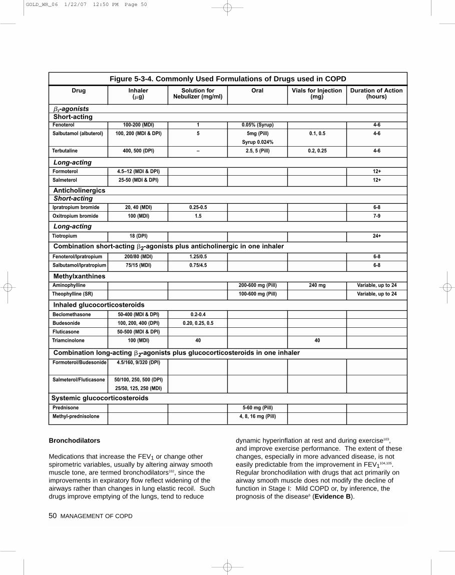
50 MANAGEMENT OF COPD
Bronchodilators
Medications that increase the FEV1 or change otherspirometric variables, usually by altering airway smoothmuscle tone, are termed bronchodilators102, since theimprovements in expiratory flow reflect widening of theairways rather than changes in lung elastic recoil. Suchdrugs improve emptying of the lungs, tend to reduce
dynamic hyperinflation at rest and during exercise103, and improve exercise performance. The extent of thesechanges, especially in more advanced disease, is noteasily predictable from the improvement in FEV1
104,105.Regular bronchodilation with drugs that act primarily onairway smooth muscle does not modify the decline offunction in Stage I: Mild COPD or, by inference, theprognosis of the disease6 (Evidence B).
Figure 5-3-4. Commonly Used Formulations of Drugs used in COPD
Drug Inhaler(�g)
Solution forNebulizer (mg/ml)
Oral Vials for Injection(mg)
Duration of Action(hours)
Fenoterol
Salbutamol (albuterol)
Terbutaline
100-200 (MDI)
100, 200 (MDI & DPI)
400, 500 (DPI)
1
5
–
0.05% (Syrup)
5mg (Pill)
Syrup 0.024%
2.5, 5 (Pill)
0.1, 0.5
0.2, 0.25
4-6
4-6
4-6
Formoterol
Salmeterol
4.5–12 (MDI & DPI)
25-50 (MDI & DPI)
12+
12+
Ipratropium bromide
Oxitropium bromide
20, 40 (MDI)
100 (MDI)
0.25-0.5
1.5
6-8
7-9
Fenoterol/Ipratropium
Salbutamol/Ipratropium
200/80 (MDI)
75/15 (MDI)
1.25/0.5
0.75/4.5
6-8
6-8
Aminophylline
Theophylline (SR)
200-600 mg (Pill)
100-600 mg (Pill)
240 mg Variable, up to 24
Variable, up to 24
Prednisone
Methyl-prednisolone
5-60 mg (Pill)
4, 8, 16 mg (Pill)
Formoterol/Budesonide
Salmeterol/Fluticasone
4.5/160, 9/320 (DPI)
50/100, 250, 500 (DPI)
25/50, 125, 250 (MDI)
Beclomethasone
Budesonide
Fluticasone
Triamcinolone
50-400 (MDI & DPI)
100, 200, 400 (DPI)
50-500 (MDI & DPI)
100 (MDI)
0.2-0.4
0.20, 0.25, 0.5
40 40
Tiotropium 18 (DPI) 24+
�2-agonistsShort-acting
AnticholinergicsShort-acting
Long-acting
Long-acting
Combination short-acting �2-agonists plus anticholinergic in one inhaler
Methylxanthines
Inhaled glucocorticosteroids
Combination long-acting �2-agonists plus glucocorticosteroids in one inhaler
Systemic glucocorticosteroids
GOLD_WR_06 1/22/07 12:50 PM Page 50
MANAGEMENT OF COPD 51
Bronchodilator medications are central to the symptomaticmanagement of COPD106-109 (Evidence A) (Figure 5.3-5).They are given either on an as-needed basis for relief of persistent or worsening symptoms, or on a regularbasis to prevent or reduce symptoms. The side effects ofbronchodilator therapy are pharmacologically predictableand dose dependent. Adverse effects are less likely, andresolve more rapidly after treatment withdrawal, withinhaled than with oral treatment. However, COPD patientstend to be older than asthma patients and more likely tohave comorbidities, so their risk of developing sideeffects is greater.
When treatment is given by the inhaled route, attention toeffective drug delivery and training in inhaler technique isessential. The choice of inhaler device will depend onavailability, cost, the prescribing physician, and the skillsand ability of the patient. COPD patients may have moreproblems in effective coordination and find it harder touse a simple metered-dose inhaler (MDI) than do healthyvolunteers or younger asthmatics. It is essential toensure that inhaler technique is correct and to re-checkthis at each visit.
Alternative breath-activated or spacer devices are availablefor most formulations. Dry powder inhalers (DPIs) maybe more convenient and possibly provide improved drugdeposition, although this has not been established inCOPD. In general, particle deposition will tend to bemore central with the fixed airflow limitation and lowerinspiratory flow rates in COPD110,111. Wet nebulizers arenot recommended for regular treatment because they aremore expensive and require appropriate maintenance112.
Dose-response relationships using the FEV1 as the outcome are relatively flat with all classes of broncho-dilators106-109. Toxicity is also dose related. Increasing thedose of either a �2-agonist or an anticholinergic by anorder of magnitude, especially when given by a wet nebulizer, appears to provide subjective benefit in acuteepisodes113 (Evidence B) but is not necessarily helpful instable disease114 (Evidence C).
All categories of bronchodilators have been shown toincrease exercise capacity in COPD, without necessarilyproducing significant changes in FEV1
115-118 (Evidence A).Regular treatment with long-acting bronchodilators ismore effective and convenient than treatment with short-acting bronchodilators119-122 (Evidence A).
Regular use of a long-acting �2-agonist120 or a short- or long-acting anticholinergic improves health status119-121.Treatment with a long-acting inhaled anti-cholinergic drugreduces the rate of COPD exacerbations123 and improvesthe effectiveness of pulmonary rehabilitation124. Theophyllineis effective in COPD, but due to its potential toxicity inhaledbronchodilators are preferred when available. All studiesthat have shown efficacy of theophylline in COPD weredone with slow-release preparations.
�2-agonists. The principal action of �2-agonists is torelax airway smooth muscle by stimulating �2-adrenergicreceptors, which increases cyclic AMP and producesfunctional antagonism to bronchoconstriction. Oral therapyis slower in onset and has more side effects than inhaledtreatment125 (Evidence A).
Inhaled �2-agonists have a relatively rapid onset of bronchodilator effect although this is probably slower inCOPD than in asthma. The bronchodilator effects ofshort-acting �2-agonists usually wear off within 4 to 6hours126,127 (Evidence A). For single-dose, as-neededuse in COPD, there appears to be no advantage in usinglevalbuterol over conventional nebulized bronchodilators128.Long-acting inhaled �2-agonists, such as salmeterol andformoterol, show a duration of effect of 12 hours or morewith no loss of effectiveness overnight or with regular usein COPD patients129-132 (Evidence A).
Adverse effects. Stimulation of �2-adrenergic receptorscan produce resting sinus tachycardia and has the potential to precipitate cardiac rhythm disturbances invery susceptible patients, although this appears to be aremarkably rare event with inhaled therapy. Exaggeratedsomatic tremor is troublesome in some older patientstreated with higher doses of �2-agonists, whatever theroute of administration, and this limits the dose that canbe tolerated. Although hypokalemia can occur, especially
Figure 5.3-5. Bronchodilators in Stable COPD
• Bronchodilator medications are central to symptommanagement in COPD.
• Inhaled therapy is preferred.
• The choice between �2-agonist, anticholinergic,theophylline, or combination therapy depends onavailability and individual response in terms of symptomrelief and side effects.
• Bronchodilators are prescribed on an as-needed or on a regular basis to prevent or reduce symptoms.
• Long-acting inhaled bronchodilators are more effectiveand convenient.
• Combining bronchodilators may improve efficacy anddecrease the risk of side effects compared to increasingthe dose of a single bronchodilator.
GOLD_WR_06 1/22/07 12:50 PM Page 51

52 MANAGEMENT OF COPD
when treatment is combined with thiazide diuretics133, and oxygen consumption can be increased under restingconditions134, these metabolic effects show tachyphylaxisunlike the bronchodilator actions. Mild falls in PaO2occur after administration of both short-and long-acting�2-agonists135, but the clinical significance of thesechanges is doubtful. Despite the concerns raised someyears ago, further detailed study has found no associa-tion between �2-agonist use and an accelerated loss oflung function or increased mortality in COPD.
Anticholinergics. The most important effect of anti-cholinergic medications, such as ipratropium, oxitropiumand tiotropium bromide, in COPD patients appears to be blockage of acetylcholine’s effect on M3 receptors.Current short-acting drugs also block M2 receptors andmodify transmission at the pre-ganglionic junction, althoughthese effects appear less important in COPD136. The long-acting anticholinergic tiotropium has a pharmacokineticselectivity for the M3 and M1 receptors137. The broncho-dilating effect of short-acting inhaled anticholinergics lastslonger than that of short-acting �2-agonists, with somebronchodilator effect generally apparent up to 8 hoursafter administration126 (Evidence A). Tiotropium has aduration of action of more than 24 hours119,138,139
(Evidence A).
Adverse effects. Anticholinergic drugs are poorly absorbedwhich limits the troublesome systemic effects seen withatropine. Extensive use of this class of inhaled agents in a wide range of doses and clinical settings has shownthem to be very safe. The main side effect is dryness of the mouth. Twenty-one days of inhaled tiotropium, 18 �g/day as a dry powder, does not retard mucus clearance from the lungs140. Although occasional prostaticsymptoms have been reported, there are no data toprove a true causal relationship. A bitter, metallic taste is reported by some patients using ipratropium. An unexpected small increase in cardiovascular events inCOPD patients regularly treated with ipratropium bromidehas been reported and requires further investigation141.
Use of wet nebulizer solutions with a face mask has been reported to precipitate acute glaucoma, probably by a direct effect of the solution on the eye. Mucociliary clearance is unaffected by these drugs, and respiratoryinfection rates are not increased.
Methylxanthines. Controversy remains about the exacteffects of xanthine derivatives. They may act as non-selective phosphodiesterase inhibitors, but have alsobeen reported to have a range of non-bronchodilatoractions, the significance of which is disputed142-146.
Data on duration of action for conventional, or even slow-release, xanthine preparations are lacking in COPD.Changes in inspiratory muscle function have been reportedin patients treated with theophylline142, but whether thisreflects changes in dynamic lung volumes or a primaryeffect on the muscle is not clear (Evidence B). All studiesthat have shown efficacy of theophylline in COPD weredone with slow-release preparations. Theophylline iseffective in COPD but, due to its potential toxicity, inhaledbronchodilators are preferred when available.
Adverse effects. Toxicity is dose related, a particularproblem with the xanthine derivatives because their therapeutic ratio is small and most of the benefit occursonly when near-toxic doses are given144,145 (Evidence A).Methylxanthines are nonspecific inhibitors of all phospho-diesterase enzyme subsets, which explains their widerange of toxic effects. Problems include the developmentof atrial and ventricular arrhythmias (which can provefatal) and grand mal convulsions (which can occur irrespective of prior epileptic history). More common andless dramatic side effects include headaches, insomnia,nausea, and heartburn, and these may occur within thetherapeutic range of serum theophylline. Unlike the otherbronchodilator classes, xanthine derivatives may involvea risk of overdose (either intentional or accidental).
Theophylline, the most commonly used methylxanthine,is metabolized by cytochrome P450 mixed function oxidases. Clearance of the drug declines with age.Many other physiological variables and drugs modifytheophylline metabolism; some of the potentially importantinteractions are listed in Figure 5.3-6.
Figure 5.3-6. Drugs and Physiological Variables thatAffect Theophylline Metabolism in COPD
Increased• Tobacco smoking• Anticonvulsant drugs• Rifampicin• Alcohol
Decreased• Old age• Arterial hypoxemia (PaO2 < 6.0 kPa, 45 mm Hg)• Respiratory acidosis• Congestive cardiac failure• Liver cirrhosis• Erythromycin• Quinolone antibiotics• Cimetidine (not ranitidine)• Viral infections• Herbal remedies (St. John’s Wort)
GOLD_WR_06 1/22/07 12:50 PM Page 52
MANAGEMENT OF COPD 53
Combination bronchodilator therapy. Combining bronchodilators with different mechanisms and durationsof action may increase the degree of bronchodilation forequivalent or lesser side effects. For example, a combi-nation of a short-acting �2-agonist and an anticholinergicproduces greater and more sustained improvements inFEV1 than either drug alone and does not produce evidence of tachyphylaxis over 90 days of treatment126,147,148
(Evidence A).
The combination of a �2-agonist, an anticholinergic, and/or theophylline may produce additional improvements inlung function126,146-151 and health status126,152. Increasing the number of drugs usually increases costs, and anequivalent benefit may occur by increasing the dose ofone bronchodilator when side effects are not a limitingfactor. Detailed assessments of this approach have notbeen carried out.
Glucocorticosteroids
The effects of oral and inhaled glucocorticosteroids inCOPD are much less dramatic than in asthma, and theirrole in the management of stable COPD is limited to specific indications. The use of glucocorticosteroids forthe treatment of acute exacerbations is described inComponent 4: Manage Exacerbations.
Oral glucocorticosteroids: short-term. Many existingCOPD guidelines recommend the use of a short course(two weeks) of oral glucocorticosteroids to identify COPDpatients who might benefit from long-term treatment withoral or inhaled glucocorticosteroids. This recommendationis based on evidence153 that short-term effects predictlong-term effects of oral glucocorticosteroids on FEV1,and evidence that asthma patients with airflow limitationmight not respond acutely to an inhaled bronchodilatorbut do show significant bronchodilation after a shortcourse of oral glucocorticosteroids.
There is mounting evidence, however, that a short course of oral glucocorticosteroids is a poor predictor of the long-term response to inhaled glucocorticosteroidsin COPD38,100. For this reason, there appears to be insufficient evidence to recommend a therapeutic trialwith oral glucocorticosteroids in patients with Stage II:Moderate COPD, Stage III: Severe COPD, or Stage IV:Very Severe COPD and poor response to an inhaledbronchodilator.
Oral glucocorticosteroids: long-term. Two retrospectivestudies154,155 analyzed the effects of treatment with oralglucocorticosteroids on long-term FEV1 changes in clinicpopulations of patients with moderate to very severeCOPD. The retrospective nature of these studies, their
lack of true control groups, and their imprecise definitionof COPD are reasons for a cautious interpretation of thedata and conclusions.
A side effect of long-term treatment with systemic gluco-corticosteroids is steroid myopathy156-158, which con-tributes to muscle weakness, decreased functionality,and respiratory failure in subjects with advanced COPD.In view of the well-known toxicity of long-term treatmentwith oral glucocorticosteroids, prospective studies on thelong-term effects of these drugs in COPD are limited159,160.
Therefore, based on the lack of evidence of benefit, andthe large body of evidence on side effects, long-termtreatment with oral glucocorticosteroids is not recommendedin COPD (Evidence A).
Inhaled glucocorticosteroids. Regular treatment withinhaled glucocorticosteroids does not modify the long-term decline of FEV1 in patients with COPD98-100,161.However, regular treatment with inhaled glucocorticos-teroids is appropriate for symptomatic COPD patientswith an FEV1 < 50% predicted (Stage III: Severe COPDand Stage IV: Very Severe COPD) and repeated exacerbations (for example, 3 in the last 3 years)162-165
(Evidence A). This treatment has been shown to reducethe frequency of exacerbations and thus improve healthstatus140 (Evidence A), and withdrawal from treatmentwith inhaled glucocorticosteroids can lead to exacerbationsin some patients166. Re-analysis of pooled data from several longer studies of inhaled glucocorticosteroids inCOPD suggests that this treatment reduces all-causemortality167, but this conclusion requires confirmation inprospective studies before leading to a change in currenttreatment recommendations. An inhaled glucocortico-steroid combined with a long-acting �2-agonist is moreeffective than the individual components162,164,165,168,169
(Evidence A).
The dose-response relationships and long-term safety of inhaled glucocorticosteroids in COPD are not known.Only moderate to high doses have been used in long-term clinical trials. Two studies showed an increased incidence of skin bruising in a small percentage of theCOPD patients98,100. One long-term study showed noeffect of budesonide on bone density and fracturerate98,170, while another study showed that treatment with triamcinolone acetonide was associated with a decreasein bone density161. The efficacy and side effects ofinhaled glucocorticosteroids in asthma are dependent onthe dose and type of glucocorticosteroid171. This patterncan also be expected in COPD and needs documentationin this patient population. Treatment with inhaled gluco-corticosteroids can be recommended for patients withmore advanced COPD and repeated exacerbations.
GOLD_WR_06 1/22/07 12:50 PM Page 53

54 MANAGEMENT OF COPD
Pharmacologic Therapy by Disease Severity
Figure 5.3-7 provides a summary of recommended treatment at each stage of COPD. For patients with fewor intermittent symptoms (Stage I: Mild COPD), use of ashort-acting inhaled bronchodilator as needed to controldyspnea is sufficient. If inhaled bronchodilators are notavailable, regular treatment with slow-release theo-phylline should be considered.
In patients with Stage II: Moderate COPD to Stage IV:Very Severe COPD) whose dyspnea during daily activitiesis not relieved despite treatment with as-needed short-acting bronchodilators, adding regular treatment with
a long-acting inhaled bronchodilator is recommended(Evidence A). Regular treatment with long-acting bronchodilators is more effective and convenient thantreatment with short-acting bronchodilators (Evidence A).There is insufficient evidence to favor one long-actingbronchodilator over others. For patients on regular long-acting bronchodilator therapy who need additionalsymptom control, adding theophylline may produce additional benefits (Evidence B).
Patients with Stage II: Moderate COPD to Stage IV: VerySevere COPD who are on regular short- or long-actingbronchodilator therapy may also use a short-acting bronchodilator as needed.
Figure 5.3-7. Therapy at Each Stage of COPD*
*Postbronchodilator FEV1 is recommended for the diagnosis and assessment of severity of COPD.
FEV1/FVC < 0.70
FEV1 ≥ 80% predicted
••
FEV1/FVC < 0.70
50% ≤ FEV1 < 80%predicted
Active reduction of risk factor(s); influenza vaccination
Add short-acting bronchodilator (when needed)
Add regular treatment with one or more long-acting bronchodilators(when needed); Add rehabilitation
Add inhaled glucocorticosteroids ifrepeated exacerbations
Add long term oxygen ifchronic respiratory failure.Consider surgical treatments
••
FEV1/FVC < 0.70
30% ≤ FEV1 < 50%predicted
••
FEV1/FVC < 0.70
FEV1 < 30% predictedor FEV1 < 50%predicted plus chronicrespiratory failure
••
I: Mild II: Moderate III: Severe IV: Very Severe
GOLD_WR_06 1/22/07 12:50 PM Page 54
MANAGEMENT OF COPD 55
Some patients may request regular treatment with high-dose nebulized bronchodilators, especially if theyhave experienced subjective benefit from this treatmentduring an acute exacerbation. Clear scientific evidencefor this approach is lacking, but one suggested option is toexamine the improvement in mean daily peak expiratoryflow recording during two weeks of treatment in the homeand continue with nebulizer therapy if a significant changeoccurs112. In general, nebulized therapy for a stablepatient is not appropriate unless it has been shown to bebetter than conventional dose therapy.
In patients with a postbronchodilator FEV1 < 50% predicted (Stage III: Severe COPD to Stage IV: VerySevere COPD) and a history of repeated exacerbations(for example, 3 in the last 3 years), regular treatment with inhaled glucocorticosteroids reduces the frequencyof exacerbations and improves health status. In thesepatients, regular treatment with an inhaled glucocortico-steroid should be added to long-acting inhaled broncho-dilators. Chronic treatment with oral glucocorticosteroidsshould be avoided.
Other Pharmacologic Treatments
Vaccines. Influenza vaccines can reduce serious illness172 and death in COPD patients by about 50%173,174
(Evidence A). Vaccines containing killed or live, inactivatedviruses are recommended175 as they are more effective inelderly patients with COPD176. The strains are adjustedeach year for appropriate effectiveness and should begiven once each year177. Pneumococcal polysaccharidevaccine is recommended for COPD patients 65 yearsand older178,179. In addition, this vaccine has been shownto reduce the incidence of community-acquired pneumonia in COPD patients younger than age 65 with an FEV1 < 40% predicted180 (Evidence B).
Alpha-1 antitrypsin augmentation therapy. Youngpatients with severe hereditary alpha-1 antitrypsin deficiency and established emphysema may be candidatesfor alpha-1 antitrypsin augmentation therapy. However,this therapy is very expensive, is not available in mostcountries, and is not recommended for patients withCOPD that is unrelated to alpha-1 antitrypsin deficiency(Evidence C).
Antibiotics. Prophylactic, continuous use of antibioticshas been shown to have no effect on the frequency ofexacerbations in COPD181-183 and a study that examinedthe efficacy of winter chemoprophylaxis over a period of5 years, concluded that there was no benefit184. There isno current evidence that the use of antibiotics, other thanfor treating infectious exacerbations of COPD and otherbacterial infections, is helpful185,186 (Evidence A).
Mucolytic (mucokinetic, mucoregulator) agents(ambroxol, erdosteine, carbocysteine, iodinated glycerol).The regular use of mucolytics in COPD has been evalu-ated in a number of long-term studies with controversialresults187-189. Although a few patients with viscous sputummay benefit from mucolytics190,191, the overall benefitsseem to be very small, and the widespread use of theseagents cannot be recommended at present (Evidence D).
Antioxidant agents. Antioxidants, in particular N-acetyl-cysteine, have been reported in small studies to reducethe frequency of exacerbations, leading to speculationthat these medications could have a role in the treatmentof patients with recurrent exacerbations192-195 (Evidence B).However, a large randomized controlled trial found no effectof N-acetylcysteine on the frequency of exacerbations,except in patients not treated with inhaled glucocortico-steroids196.
Immunoregulators (immunostimulators, immunomodulators). Studies using an immunoregulatorin COPD show a decrease in the severity and frequencyof exacerbations197,198. However, additional studies toexamine the long-term effects of this therapy are requiredbefore its regular use can be recommended199.
Antitussives. Cough, although sometimes a trouble-some symptom in COPD, has a significant protectiverole200. Thus the regular use of antitussives is not recommended in stable COPD (Evidence D).
Vasodilators. The belief that pulmonary hypertension in COPD is associated with a poorer prognosis has provoked many attempts to reduce right ventricular afterload, increase cardiac output, and improve oxygendelivery and tissue oxygenation. Many agents have beenevaluated, including inhaled nitric oxide, but the resultshave been uniformly disappointing. In patients withCOPD, in whom hypoxemia is caused primarily by ventilation-perfusion mismatching rather than byincreased intrapulmonary shunt (as in noncardiogenicpulmonary edema), inhaled nitric oxide can worsen gasexchange because of altered hypoxic regulation of ventilation-perfusion balance201,202. Therefore, based onthe available evidence, nitric oxide is contraindicated instable COPD.
Narcotics (morphine). Oral and parenteral opioids areeffective for treating dyspnea in COPD patients withadvanced disease. There are insufficient data to concludewhether nebulized opioids are effective203. However, someclinical studies suggest that morphine used to controldyspnea may have serious adverse effects and its benefits may be limited to a few sensitive subjects204-208.
GOLD_WR_06 1/22/07 12:50 PM Page 55
Others. Nedocromil, leukotriene modifiers, and alternativehealing methods (e.g., herbal medicine, acupunture,homeopathy) have not been adequately tested in COPDpatients and thus cannot be recommended at this time.
NON-PHARMACOLOGIC TREATMENT
Rehabilitation
The principal goals of pulmonary rehabilitation are toreduce symptoms, improve quality of life, and increasephysical and emotional participation in everyday activities.To accomplish these goals, pulmonary rehabilitation covers a range of non-pulmonary problems that may notbe adequately addressed by medical therapy for COPD.Such problems, which especially affect patients withStage II: Moderate COPD, Stage III: Severe COPD,and Stage IV: Very Severe COPD, include exercise de-conditioning, relative social isolation, altered moodstates (especially depression), muscle wasting, andweight loss. These problems have complex interrelation-ships and improvement in any one of these interlinkedprocesses can interrupt the “vicious circle” in COPD sothat positive gains occur in all aspects of the illness(Figure 5.3-9). A comprehensive statement on pulmonaryrehabilitation has been prepared by the ATS/ERS209.
Pulmonary rehabilitation has been carefully evaluated ina large number of clinical trials; the various benefits aresummarized in Figure 5.3-1089,210-220. On average, rehabilitation increases peak workload by 18%, peak oxygen consumption by 11%, and endurance time time by87% of baseline. This translates into a 49 m improvementin 6-minute walking distance221. Rehabilitation has beenshown to be at least additive to other forms of therapysuch as bronchodilator treatment124.
Patient selection and program design. Although moreinformation is needed on criteria for patient selection forpulmonary rehabilitation programs, COPD patients at allstages of disease appear to benefit from exercise trainingprograms, improving with respect to both exercise toleranceand symptoms of dyspnea and fatigue222 (Evidence A).Data suggest that these benefits can be sustained evenafter a single pulmonary rehabilitation program223-225.
Benefit does wane after a rehabilitation program ends,but if exercise training is maintained at home the patient’shealth status remains above pre-rehabilitation levels(Evidence B). To date there is no consensus on whetherrepeated rehabilitation courses enable patients to sustainthe benefits gained through the initial course.
Ideally, pulmonary rehabilitation should involve severaltypes of health professionals. Significant benefits canalso occur with more limited personnel, as long as dedicated professionals are aware of the needs of eachpatient. Benefits have been reported from rehabilitationprograms conducted in inpatient, outpatient, and homesettings214,215,226. Considerations of cost and availability mostoften determine the choice of setting. The educationaland exercise training components of rehabilitation areusually conducted in groups, normally with 6 to 8 individuals per class (Evidence D).
The following points summarize current knowledge ofconsiderations important in choosing patients:
Functional status: Benefits have been seen in patientswith a wide range of disability, although those who arechair-bound appear unlikely to respond even to home vis-iting programs227 (Evidence A).
Figure 5.3-9. The Cycle of Physical, Social, andPsychosocial Consequences of COPD
Lack of Fitness
COPD Dyspnea
Depression Social Isolation
Immobility
Figure 5.3-10. Benefits of Pulmonary Rehabilitation in COPD
• Improves exercise capacity (Evidence A).• Reduces the perceived intensity of breathlessness(Evidence A).
• Improves health-related quality of life (Evidence A).• Reduces the number of hospitalizations and days in the
hospital (Evidence A).• Reduces anxiety and depression associated with COPD
(Evidence A).• Strength and endurance training of the upper limbs
improves arm function (Evidence B).• Benefits extend well beyond the immediate period of
training (Evidence B).• Improves survival (Evidence B).• Respiratory muscle training is beneficial, especially when
combined with general exercise training (Evidence C).• Psychosocial intervention is helpful (Evidence C).
56 MANAGEMENT OF COPD
GOLD_WR_06 1/22/07 12:50 PM Page 56
Severity of dyspnea: Stratification by breathlessnessintensity using the MRC questionnaire (Figure 5.1-3)may be helpful in selecting patients most likely to benefitfrom rehabilitation. Those with MRC grade 5 dyspneamay not benefit227 (Evidence B).
Motivation: Selecting highly motivated participants isespecially important in the case of outpatient programs224.
Smoking status: There is no evidence that smokers willbenefit less than nonsmokers, but many clinicians believethat inclusion of a smoker in a rehabilitation programshould be conditional on their participation in a smokingcessation program. Some data indicate that continuingsmokers are less likely to complete pulmonary rehabilitationprograms than nonsmokers224 (Evidence B).
Components of pulmonary rehabilitation programs.The components of pulmonary rehabilitation vary widelyfrom program to program but a comprehensive pulmonaryrehabilitation program includes exercise training, nutritioncounseling, and education.
Exercise training. Exercise tolerance can be assessedby either bicycle ergometry or treadmill exercise with themeasurement of a number of physiological variables,including maximum oxygen consumption, maximum heartrate, and maximum work performed. A less complexapproach is to use a self-paced, timed walking test (e.g.,6-minute walking distance). These tests require at leastone practice session before data can be interpreted.Shuttle walking tests offer a compromise: they providemore complete information than an entirely self-pacedtest, but are simpler to perform than a treadmill test228.
Exercise training ranges in frequency from daily to weekly,in duration from 10 minutes to 45 minutes per session,and in intensity from 50% peak oxygen consumption(VO2 max) to maximum tolerated229. The optimum lengthfor an exercise program has not been investigated in randomized controlled trials but most studies involvingfewer than 28 exercise sessions show inferior resultscompared to those with longer treatment periods221. In practice, the length depends on the resources available and usually ranges from 4 to 10 weeks, withlonger programs resulting in larger effects than shorterprograms213.
Participants are often encouraged to achieve a pre-determined target heart rate230, but this goal may havelimitations in COPD. In many programs, especially thoseusing simple corridor exercise training, the patient isencouraged to walk to a symptom-limited maximum, rest,and then continue walking until 20 minutes of exercise
have been completed. Where possible, endurance exercise training to 60-80% of the symptom-limited maximum is preferred. Endurance training can beaccomplished through continuous or interval exerciseprograms. The latter involve the patient doing the sametotal work but divided into briefer periods of high-intensityexercise, which is useful when performance is limited byother comorbidities231,232. Use of a simple wheeled walkingaid seems to improve walking distance and reducesbreathlessness in severely disabled COPD patients233-235
(Evidence C). Other approaches to improving outcomessuch as use of oxygen during exercise236, exercising whilebreathing heliox gas mixtures237, unloading the ventilatorymuscles while exercising, or use of pursed lip breathingremain experimental at present. Specific strength trainingis possible but its benefits remain uncertain, as do theeffects of supplementation with anabolic steroids and theuse of neuromuscular electrical stimulation.
The minimum length of an effective rehabilitation programis 6 weeks; the longer the program continues, the moreeffective the results238-240 (Evidence B). However, as yet,no effective program has been developed to maintain theeffects over time241. Many physicians advise patientsunable to participate in a structured program to exercise ontheir own (e.g., walking 20 minutes daily). The benefitsof this general advice have not been tested, but it is reasonable to offer such advice to patients if a formalprogram is not available.
Some programs also include upper limb exercises, usuallyinvolving an upper limb ergometer or resistive trainingwith weights. There are no randomized clinical trial datato support the routine inclusion of these exercises, butthey may be helpful in patients with comorbidities thatrestrict other forms of exercise and those with evidenceof respiratory muscle weakness242,243. The addition ofupper limb exercises or other strength training to aerobictraining is effective in improving strength, but does notimprove quality of life or exercise tolerance244.
Nutrition counseling. Nutritional state is an importantdeterminant of symptoms, disability, and prognosis inCOPD; both overweight and underweight can be a problem. Specific nutritional recommendations forpatients with COPD are based on expert opinion andsome small randomized clinical trials209. Approximately25% of patients with Stage II: Moderate COPD to StageIV: Very Severe COPD show a reduction in both theirbody mass index and fat free mass12,245,246. A reduction in body mass index is an independent risk factor for mortality in COPD patients13,247,248 (Evidence A).
MANAGEMENT OF COPD 57
GOLD_WR_06 1/22/07 12:50 PM Page 57

Health care workers should identify and correct the reasons for reduced calorie intake in COPD patients.Patients who become breathless while eating should beadvised to take small, frequent meals. Poor dentitionshould be corrected and comorbidities (pulmonary sepsis,lung tumors, etc.) should be managed appropriately.Improving the nutritional state of COPD patients who arelosing weight can lead to improved respiratory musclestrength249-251. However, controversy remains as towhether this additional effort is cost effective249,250.
Present evidence suggests that nutritional supplementationalone may not be a sufficient strategy. Increased calorieintake is best accompanied by exercise regimes that havea nonspecific anabolic action, and there is some evidencethis also helps even in those patients without severenutritional depletion252. Specific nutritional supplements(e.g., creatine) may improve body composition, but further studies in large numbers of subjects are requiredbefore the routine use of these supplements can be recommended253. Anabolic steroids in COPD patientswith weight loss increase body weight and lean bodymass but have little or no effect on exercise capacity254,255.
Education. Most pulmonary rehabilitation programsinclude an educational component, but the specific contributions of education to the improvements seen after pulmonary rehabilitation remain unclear.
Assessment and follow-up. Baseline and outcomeassessments of each participant in a pulmonary rehabili-tation program should be made to quantify individualgains and target areas for improvement. Assessmentsshould include:
• Detailed history and physical examination• Measurement of spirometry before and after a
bronchodilator drug• Assessment of exercise capacity• Measurement of health status and impact of
breathlessness• Assessment of inspiratory and expiratory muscle
strength and lower limb strength (e.g., quadriceps) in patients who suffer from muscle wasting
The first two assessments are important for establishingentry suitability and baseline status but are not used inoutcome assessment. The last three assessments arebaseline and outcome measures. Several detailed questionnaires for assessing health status are available,including some that are specifically designed for patientswith respiratory disease (e.g., Chronic Respiratory DiseaseQuestionnaire152, St. George Respiratory Questionnaire256),and there is increasing evidence that these questionnaires
may be useful in a clinical setting. Health status can alsobe assessed by generic questionnaires, such as theMedical Outcomes Study Short Form (SF36)257, to enablecomparison of quality of life in different diseases. TheHospital Anxiety and Depression Scale (HADS) has beenused to improve identification and treatment of anxiousand depressed patients258.
Economic cost of rehabilitation programs. A Canadianstudy showing statistically significant improvements indyspnea, fatigue, emotional health, and mastery foundthat the incremental cost of pulmonary rehabilitation was$11,597 (CDN) per person259. A study from the UnitedKingdom provided evidence that an intensive (6-week,18-visit) multidisciplinary rehabilitation program was effective in decreasing use of health services225 (Evidence B).Although there was no difference in the number of hospitaladmissions between patients with disabling COPD in a control group and those who participated in the rehabili-tation program, the number of days the rehabilitationgroup spent in the hospital was significantly lower. Therehabilitation group had more primary-care consultationsat the general practitioner’s premises than did the controlgroup, but fewer primary-care home visits. Comparedwith the control group, the rehabilitation group alsoshowed greater improvements in walking ability and ingeneral and disease-specific health status.
Oxygen Therapy
Oxygen therapy, one of the principal nonpharmacologictreatments for patients with Stage IV: Very SevereCOPD190,260, can be administered in three ways: long-term continuous therapy, during exercise, and to relieveacute dyspnea. The primary goal of oxygen therapy is toincrease the baseline PaO2 to at least 8.0 kPa (60 mmHg) at sea level and rest, and/or produce an SaO2 atleast 90%, which will preserve vital organ function byensuring adequate delivery of oxygen.
The long-term administration of oxygen (> 15 hours perday) to patients with chronic respiratory failure has beenshown to increase survival261,262. It can also have a beneficial impact on hemodynamics, hematologic characteristics, exercise capacity, lung mechanics, andmental state263. Continuous oxygen therapy decreasedresting pulmonary artery pressure in one study261 but notin another study262. Prospective studies have shown thatthe primary hemodynamic effect of oxygen therapy is preventing the progression of pulmonary hypertension264,265.Long-term oxygen therapy improves general alertness,motor speed, and hand grip, although the data are lessclear about changes in quality of life and emotional state.The possibility of walking while using some oxygen
58 MANAGEMENT OF COPD
GOLD_WR_06 1/22/07 12:50 PM Page 58
devices may help to improve physical conditioning andhave a beneficial influence on the psychological state ofpatients266.
Long-term oxygen therapy is generally introduced inStage IV: Very Severe COPD for patients who have:
• PaO2 at or below 7.3 kPa (55 mm Hg) or SaO2 at or below 88%, with or without hypercapnia (Evidence B); or
• PaO2 between 7.3 kPa (55 mm Hg) and 8.0 kPa (60 mm Hg), or SaO2 of 88%, if there is evidence of pulmonary hypertension, peripheral edema suggesting congestive cardiac failure, or polycythemia(hematocrit > 55%) (Evidence D).
A decision about the use of long-term oxygen should bebased on the waking PaO2 values. The prescriptionshould always include the source of supplemental oxygen(gas or liquid), method of delivery, duration of use, andflow rate at rest, during exercise, and during sleep. Adetailed review of the uses of oxygen in COPD, togetherwith possible assessment algorithms and informationabout methods of delivery, is available fromhttp://www.thoracic.org/.
Oxygen is usually delivered by a facemask, with appropriateinspiratory flow rates varying between 24% and 35%.The facemask permits accurate titration of oxygen, whichis particularly valuable in patients who are prone to CO2retention. However, facemasks are easily dislodged andrestrict eating and conversation, so many patients preferoxygen delivered by nasal cannulae. Oxygen delivery by this route requires additional blood gas monitoring toensure that it is satisfactory, and may require individualtitration. Other, more specialized methods of oxygendelivery (e.g., transtracheally) are available but shouldonly be used in specialized centers familiar with the indications and complications of these delivery methods.
Long-term oxygen is usually provided from a fixed oxygenconcentrator with plastic piping allowing the patient touse oxygen in their living area and bedroom. Treatmentshould be for at least 15 hours per day and preferablylonger. In addition, a supply of oxygen should be providedthat will allow the patient to leave the house for an appropriate period of time and to exercise without theiroxygen saturation falling below 90%.
A number of physiological studies have shown that delivering oxygen during exercise can increase the duration of endurance exercise and/or reduce the intensityof end-exercise breathlessness267,268 (Evidence A). This
reflects a reduction in the rate at which dynamic hyper-inflation occurs, which may be secondary to the documented reduction in ventilatory demand andchemoreceptor activation while breathing oxygen duringexercise269,270. These changes occur whether or notpatients are hypoxemic at rest and can translate intoimproved health status if the treatment is used as an outpatient271. However, good data about the use ofambulatory oxygen in representative patient populationsare presently lacking, although a small randomized trialhas suggested that compliance is not high272. Patientsneed encouragement to understand how and when touse ambulatory oxygen and overcome any anxieties orconcerns about using this more conspicuous treatment.
Oxygen therapy reduces the oxygen cost of breathingand minute ventilation, a mechanism that although stilldisputed helps to minimize the sensation of dyspnea.This has led to the use of short burst therapy to controlsevere dyspnea such as occurs after climbing stairs.However, there is no benefit from using short burst oxygen for symptomatic relief before or after exercise273,274
(Evidence B).
Cost considerations. Supplemental home oxygen isusually the most costly component of outpatient therapyfor adults with COPD who require this therapy275. Studiesof the cost effectiveness of alternative outpatient oxygendelivery methods in the US and Europe suggest that oxygen concentrator devices may be more cost effectivethan cylinder delivery systems276,277.
Oxygen use in air travel. Although air travel is safe formost patients with chronic respiratory failure who are onlong-term oxygen therapy, patients should be instructedto increase the flow by 1-2 L/min during the flight278.Ideally, patients who fly should be able to maintain an in-flight PaO2 of at least 6.7 kPa (50 mm Hg). Studiesindicate that this can be achieved in those with moderateto severe hypoxemia at sea level by supplementary oxygen at 3 L/min by nasal cannulae or 31% by Venturifacemask279. Those with a resting PaO2 at sea level of >9.3 kPa (70 mm Hg) are likely to be safe to fly withoutsupplementary oxygen278,280, although it is important toemphasize that a resting PaO2 > 9.3 kPa (70 mm Hg) atsea level does not exclude the development of severehypoxemia when travelling by air (Evidence C). Carefulconsideration should be given to any comorbidity thatmay impair oxygen delivery to tissues (e.g., cardiacimpairment, anemia). Also, walking along the aisle mayprofoundly aggravate hypoxemia281.
MANAGEMENT OF COPD 59
GOLD_WR_06 1/22/07 12:50 PM Page 59
Ventilatory Support
Noninvasive ventilation (using either negative or positivepressure devices) is now widely used to treat acute exacerbations of COPD (see Component 4). Negativepressure ventilation is not indicated for the chronic management of Stage IV: Very Severe COPD patients,with or without CO2 retention. It has been demonstratedto have no effect on shortness of breath, exercise tolerance, arterial blood gases, respiratory musclestrength, or quality of life in a large randomized trial inCOPD patients with chronic respiratory failure282.
Although preliminary studies suggested that combiningnoninvasive intermittent positive pressure ventilation(NIPPV) with long-term oxygen therapy could improvesome outcome variables, current data do not support theroutine use of this combination283. However, comparedwith long-term oxygen therapy alone, the addition ofNIPPV can lessen carbon dioxide retention and improveshortness of breath in some patients284. Thus, althoughat present long-term NIPPV cannot be recommended forthe routine treatment of patients with chronic respiratoryfailure due to COPD, the combination of NIPPV withlong-term oxygen therapy may be of some use in aselected subset of patients, particularly in those with pronounced daytime hypercapnia285.
Surgical Treatments
Bullectomy. Bullectomy is an older surgical procedurefor bullous emphysema. Removal of a large bulla thatdoes not contribute to gas exchange decompresses theadjacent lung parenchyma. Bullectomy can be performedthoracoscopically. In carefully selected patients, this procedure is effective in reducing dyspnea and improvinglung function286 (Evidence C).
Bullae may be removed to alleviate local symptoms suchas hemoptysis, infection, or chest pain, and to allow re-expansion of a compressed lung region. This is theusual indication in patients with COPD. In consideringthe possible benefit of surgery it is crucial to estimate theeffect of the bulla on the lung and the function of the nonbullous lung. A thoracic CT scan, arterial blood gasmeasurement, and comprehensive respiratory functiontests are essential before making a decision regardingsuitability for resection of a bulla. Normal or minimallyreduced diffusing capacity, absence of significant hypoxemia, and evidence of regional reduction in perfusionwith good perfusion in the remaining lung are indicationsa patient will likely benefit from surgery287. However, pulmonary hypertension, hypercapnia, and severeemphysema are not absolute contraindications for
bullectomy. Some investigators have recommended thatthe bulla must occupy 50% or more of the hemithoraxand produce definite displacement of the adjacent lungbefore surgery is performed288.
Lung volume reduction surgery (LVRS). LVRS is asurgical procedure in which parts of the lung are resectedto reduce hyperinflation289, making respiratory musclesmore effective pressure generators by improving theirmechanical efficiency (as measured by length/tensionrelationship, curvature of the diaphragm, and area ofapposition)290,291. In addition, LVRS increases the elasticrecoil pressure of the lung and thus improves expiratoryflow rates292.
A large multicenter study of 1,200 patients comparingLVRS with medical treatment has shown that after 4.3years, patients with upper-lobe emphysema and lowexercise capacity who received the surgery had a greatersurvival rate than similar patients who received medicaltherapy (54% vs. 39.7%)293. In addition, the surgerypatients experienced greater improvements in their maximal work capacity and their health-related quality of life. The advantage of surgery over medical therapywas less significant among patients who had otheremphysema distribution or high exercise capacity prior to treatment.
Hospital costs associated with LVRS in 52 consecutivepatients294 ranged from US$11,712 to $121,829. Hospitalcharges in 23 consecutive patients admitted for LVRS at a single institution295 ranged from US$20,032 to $75,561with a median charge of $26,669. A small number ofindividuals incurred extraordinary costs because of complications. Advanced age was a significant factorleading to higher expected total hospital costs.
Although the results of the large multicenter studyshowed some very positive results of surgery in a selectgroup of patients41,293, LVRS is an expensive palliative surgical procedure and can be recommended only incarefully selected patients.
Lung transplantation. In appropriately selected patientswith very advanced COPD, lung transplantation has beenshown to improve quality of life and functional capacity296-299
(Evidence C), although the Joint United Network forOrgan Sharing in 1998 found that lung transplantationdoes not confer a survival benefit in patients with end-stage emphysema after two years298. Criteria for referralfor lung transplantation include FEV1 < 35% predicted,PaO2 < 7.3-8.0 kPa (55-60 mm Hg), PaCO2 > 6.7 kPa(50 mm Hg), and secondary pulmonary hypertension300,301.
60 MANAGEMENT OF COPD
GOLD_WR_06 1/22/07 12:50 PM Page 60
Lung transplantation is limited by the shortage of donororgans, which has led some centers to adopt the single-lung technique. The common complications seen inCOPD patients after lung transplantation, apart fromoperative mortality, are acute rejection and bronchiolitisobliterans, CMV, other opportunistic fungal (Candida,Aspergillus, Cryptococcus, Carinii) or bacterial(Pseudomonas, Staphylococcus species) infections, lymphoproliferative disease, and lymphomas297.
Another limitation of lung transplantation is its cost. Inthe United States, hospitalization costs associated withlung transplantation have ranged from US$110,000 towell over $200,000. Costs remain elevated for months toyears after surgery due to the high cost of complicationsand the immunosuppressive regimens302-305 that must beinitiated during or immediately after surgery.
Special Considerations
Surgery in COPD. Postoperative pulmonary complicationsare as important and common as postoperative cardiaccomplications and, consequently, are a key component of the increased risk posed by surgery in COPD patients.The principal potential factors contributing to the riskinclude smoking, poor general health status, age, obesity,and COPD severity. A comprehensive definition of post-operative pulmonary complications should include onlymajor pulmonary respiratory complications, namely lunginfections, atelectasis and/or increased airflow obstruction,all potentially resulting in acute respiratory failure andaggravation of underlying COPD306-311.
The incidence of increased risk of postoperative pulmonarycomplications in COPD patients may vary according tothe definition of postoperative pulmonary complicationsand the severity of COPD, with relative ranges of the orderof 2.7 to 4.7306. The surgical site is the most importantpredictor, and risk increases as the incision approachesthe diaphragm. Upper abdominal and thoracic surgeryrepresents the greatest risk, the latter being uncommonafter interventions outside the thorax or abdomen. Mostreports conclude that epidural or spinal anesthesia havea lower risk than general anesthesia, although the resultsare not totally uniform.
Individual patient risk factors are identified by careful history, physical examination, chest radiography, and pulmonary function tests. Although the value of pulmonaryfunction tests remains contentious, there is consensusthat all COPD candidates for lung resection shouldundergo a complete battery, including forced spirometrywith bronchodilator response, static lung volumes, diffusing capacity, and arterial blood gases at rest. One
theoretical rationale behind the assessment of pulmonaryfunction measurement is the identification of COPDpatients in whom the risk is so elevated that surgeryshould be contraindicated.
Several studies in high-risk COPD patients suggest thatthere is a threshold beyond which the risk of surgery isprohibitive. The risk of postoperative respiratory failureappears to be in patients undergoing pneumonectomywith a preoperative FEV1 < 2 L or 50% predicted and/or a DLCO < 50% predicted310. COPD patients at high riskdue to poor lung function should undergo further lungfunction assessment, for example, tests of regional distribution of perfusion and exercise capacity311. To prevent postoperative pulmonary complications, stableCOPD patients clinically symptomatic and/or with limitedexercise capacity should be treated, before surgery,intensely with all the measures already well establishedfor stable COPD patients who are not about to havesurgery. Surgery should be postponed if an exacerbationis present.
Surgery in patients with COPD needs to be differentiatedfrom that aimed to improve lung function and symptomsof COPD. This includes bullectomy, lung volume reduction surgery, and lung transplantation311.
MANAGEMENT OF COPD 61
GOLD_WR_06 1/22/07 12:50 PM Page 61
INTRODUCTION
COPD is often associated with exacerbations of symptoms312-316. An exacerbation of COPD is defined asan event in the natural course of the disease characterizedby a change in the patient’s baseline dyspnea, cough,and/or sputum that is beyond normal day-to-day variations,is acute in onset, and may warrant a change in regularmedication in a patient with underlying COPD317,318.Exacerbations are categorized in terms of either clinicalpresentation (number of symptoms314) and/or health-care
resources utilization317. The impact of exacerbations issignificant and a patient’s symptoms and lung functionmay both take several weeks to recover to the baselinevalues319.
Exacerbations affect the quality of life and prognosis ofpatients with COPD. Hospital mortality of patients admittedfor a hypercarbic COPD exacerbation is approximately10%, and the long-term outcome is poor320. Mortalityreaches 40% at 1 year in those needing mechanical support, and all-cause mortality is even higher (up to49%) 3 years after hospitalization for a COPD exacer-bation316,320-325. In addition, exacerbations of COPD haveserious negative impacts on patients’ quality of life140, lungfunction326,327, and socioeconomic costs325. Thus, prevention,early detection, and prompt treatment of exacerbationsmay impact their clinical progression by ameliorating the effects on quality of life and minimizing the risk ofhospitalization328.
The most common causes of an exacerbation are infectionof the tracheobronchial tree and air pollution329, but thecause of about one-third of severe exacerbations cannotbe identified. The role of bacterial infections is contro-versial, but recent investigations with newer researchtechniques have begun to provide important information.Bronchoscopic studies have shown that at least 50% of patients have bacteria in high concentrations in theirlower airways during exacerbations330-332. However, a significant proportion of these patients also have bacteriacolonizing their lower airways in the stable phase of thedisease.
There is some indication that the bacterial burdenincreases during exacerbations330, and that acquisition of strains of the bacteria that are new to the patient isassociated with exacerbations332. Development of specificimmune responses to the infecting bacterial strains, andthe association of neutrophilic inflammation with bacterialexacerbations, also support the bacterial causation of aproportion of exacerbations333-336.
DIAGNOSIS AND ASSESSMENT OFSEVERITY
Medical History
Increased breathlessness, the main symptom of an exacerbation, is often accompanied by wheezing andchest tightness, increased cough and sputum, change of the color and/or tenacity of sputum, and fever.
62 MANAGEMENT OF COPD
COMPONENT 4: MANAGE EXACERBATIONS
KEY POINTS:
• An exacerbation of COPD is defined as an eventin the natural course of the disease characterizedby a change in the patient’s baseline dyspnea,cough, and/or sputum that is beyond normal day-to-day variations, is acute in onset, and maywarrant a change in regular medication in apatient with underlying COPD.
• The most common causes of an exacerbationare infection of the tracheobronchial tree and airpollution, but the cause of about one-third ofsevere exacerbations cannot be identified(Evidence B).
• Inhaled bronchodilators (particularly inhaled �2-agonists with or without anticholinergics) andoral glucocorticosteroids are effective treatmentsfor exacerbations of COPD (Evidence A).
• Patients experiencing COPD exacerbations withclinical signs of airway infection (e.g., increasedsputum purulence) may benefit from antibiotictreatment (Evidence B).
• Noninvasive mechanical ventilation in exacerbationsimproves respiratory acidosis, increases pH,decreases the need for endotracheal intubation,and reduces PaCO2, respiratory rate, severity ofbreathlessness, the length of hospital stay, andmortality (Evidence A).
• Medications and education to help prevent futureexacerbations should be considered as part offollow-up, as exacerbations affect the quality oflife and prognosis of patients with COPD.
GOLD_WR_06 1/22/07 12:50 PM Page 62

Exacerbations may also be accompanied by a number ofnonspecific complaints, such as tachycardia and tachypnea,malaise, insomnia, sleepiness, fatigue, depression, andconfusion. A decrease in exercise tolerance, fever, and/or new radiological anomalies suggestive of pulmonarydisease may herald a COPD exacerbation. An increasein sputum volume and purulence points to a bacterialcause, as does prior history of chronic sputumproduction314,336.
Assessment of Severity
Assessment of the severity of an exacerbation is basedon the patient’s medical history before the exacerbation,preexisting comorbidities, symptoms, physical examination,arterial blood gas measurements, and other laboratorytests (Figure 5.4-1). Specific information is required onthe frequency and severity of attacks of breathlessnessand cough, sputum volume and color, and limitation ofdaily activities. When available, prior arterial blood gasmeasurements are extremely useful for comparison withthose made during the acute episode, as an acute changein these tests is more important than their absolute values.Thus, where possible, physicians should instruct theirpatients to bring the summary of their last evaluationwhen they come to the hospital with an exacerbation. In patients with Stage IV: Very Severe COPD, the mostimportant sign of a severe exacerbation is a change inthe mental status of the patient and this signals a needfor immediate evaluation in the hospital.
Spirometry and PEF. Even simple spirometric tests can be difficult for a sick patient to perform properly.These measurements are not accurate during an acuteexacerbation; therefore their routine use is not recommended.
Pulse oximetry and arterial blood gas measurement.Pulse oximetry can be used to evaluate a patient’s oxygensaturation and need for supplemental oxygen therapy.For patients that require hospitalization, measurement ofarterial blood gases is important to assess the severity ofan exacerbation. A PaO2 < 8.0 kPa (60 mm Hg) and/orSaO2 < 90% with or without PaCO2 > 6.7 kPa (50 mmHg)when breathing room air indicate respiratory failure. Inaddition, moderate-to-severe acidosis (pH < 7.36) plushypercapnia (PaCO2 > 6-8 kPa, 45-60 mm Hg) in apatient with respiratory failure is an indication formechanical ventilation311,337.
Chest X-ray and ECG. Chest radiographs (posterior/anterior plus lateral) are useful in identifying alternativediagnoses that can mimic the symptoms of an exacerbation.Although the history and physical signs can be confusing,especially when pulmonary hyperinflation masks coexistingcardiac signs, most problems are resolved by the chestX-ray and ECG. An ECG aids in the diagnosis of rightheart hypertrophy, arrhythmias, and ischemic episodes.Pulmonary embolism can be very difficult to distinguishfrom an exacerbation, especially in advanced COPD,because right ventricular hypertrophy and large pul-monary arteries lead to confusing ECG and radiographicresults. A low systolic blood pressure and an inability toincrease the PaO2 above 8.0 kPa (60 mm Hg) despitehigh-flow oxygen also suggest pulmonary embolism. Ifthere are strong indications that pulmonary embolism hasoccurred, it is best to treat for this along with the exacerbation.
Other laboratory tests. The whole blood count mayidentify polycythemia (hematocrit > 55%) or bleeding.White blood cell counts are usually not very informative.The presence of purulent sputum during an exacerbationof symptoms is sufficient indication for starting empiricalantibiotic treatment33. Streptococcus pneumoniae,Hemophilus influenzae, and Moraxella catarrhalis are the most common bacterial pathogens involved in COPDexacerbations. If an infectious exacerbation does notrespond to the initial antibiotic treatment, a sputum cultureand an antibiogram should be performed. Bio- chemicaltest abnormalities can be associated with an exacerbationand include electrolyte disturbance(s) (e.g., hyponatremia,hypokalemia), poor glucose control, metabolic acid-basedisorder. These abnormalities can also be due to associatedco-morbid conditions (see below “Differential Diagnoses”).
Differential Diagnoses
Ten to 30% of patients with apparent exacerbations ofCOPD that do not respond to treatment319,338. In suchcases the patient should be re-evaluated for other medical
MANAGEMENT OF COPD 63
Figure 5.4-1. Assessment of COPD Exacerbations:Medical History and Signs of Severity
Medical History
• Severity of FEV1
• Duration of worsening ornew symptoms
• Number of previousepisodes (exacerbations/hospitalizations)
• Comordibities
• Present treatment regimen
Signs of Severity
• Use of accessory respiratorymuscles
• Paradoxical chest wallmovements
• Worsening or new onsetcentral cyanosis
• Development of peripheraledema
• Hemodynamic instability
• Signs of right heart failure
• Reduced alertness
GOLD_WR_06 1/22/07 12:50 PM Page 63
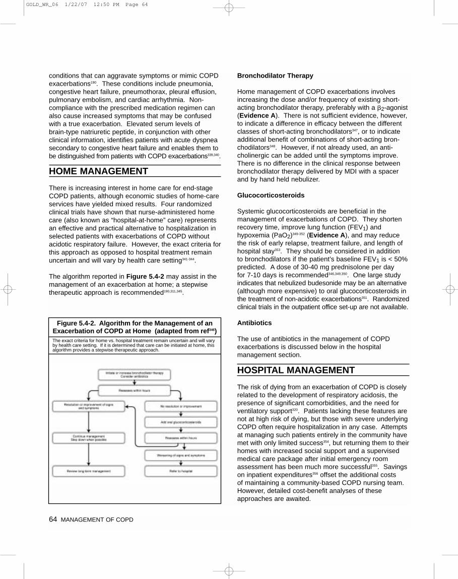
conditions that can aggravate symptoms or mimic COPDexacerbations190. These conditions include pneumonia,congestive heart failure, pneumothorax, pleural effusion,pulmonary embolism, and cardiac arrhythmia. Non-compliance with the prescribed medication regimen canalso cause increased symptoms that may be confusedwith a true exacerbation. Elevated serum levels of brain-type natriuretic peptide, in conjunction with otherclinical information, identifies patients with acute dyspneasecondary to congestive heart failure and enables them tobe distinguished from patients with COPD exacerbations339,340.
HOME MANAGEMENT
There is increasing interest in home care for end-stageCOPD patients, although economic studies of home-careservices have yielded mixed results. Four randomizedclinical trials have shown that nurse-administered homecare (also known as “hospital-at-home” care) representsan effective and practical alternative to hospitalization inselected patients with exacerbations of COPD withoutacidotic respiratory failure. However, the exact criteria forthis approach as opposed to hospital treatment remainuncertain and will vary by health care setting341-344.
The algorithm reported in Figure 5.4-2 may assist in themanagement of an exacerbation at home; a stepwisetherapeutic approach is recommended190,311,345.
Bronchodilator Therapy
Home management of COPD exacerbations involvesincreasing the dose and/or frequency of existing short-acting bronchodilator therapy, preferably with a �2-agonist(Evidence A). There is not sufficient evidence, however,to indicate a difference in efficacy between the differentclasses of short-acting bronchodilators347, or to indicateadditional benefit of combinations of short-acting bron-chodilators348. However, if not already used, an anti-cholinergic can be added until the symptoms improve.There is no difference in the clinical response betweenbronchodilator therapy delivered by MDI with a spacerand by hand held nebulizer.
Glucocorticosteroids
Systemic glucocorticosteroids are beneficial in the management of exacerbations of COPD. They shortenrecovery time, improve lung function (FEV1) and hypoxemia (PaO2)349-352 (Evidence A), and may reducethe risk of early relapse, treatment failure, and length of hospital stay353. They should be considered in addition to bronchodilators if the patient’s baseline FEV1 is < 50%predicted. A dose of 30-40 mg prednisolone per day for 7-10 days is recommended346,349,350. One large studyindicates that nebulized budesonide may be an alternative(although more expensive) to oral glucocorticosteroids inthe treatment of non-acidotic exacerbations351. Randomizedclinical trials in the outpatient office set-up are not available.
Antibiotics
The use of antibiotics in the management of COPD exacerbations is discussed below in the hospital management section.
HOSPITAL MANAGEMENT
The risk of dying from an exacerbation of COPD is closelyrelated to the development of respiratory acidosis, thepresence of significant comorbidities, and the need forventilatory support320. Patients lacking these features arenot at high risk of dying, but those with severe underlyingCOPD often require hospitalization in any case. Attemptsat managing such patients entirely in the community havemet with only limited success354, but returning them to theirhomes with increased social support and a supervisedmedical care package after initial emergency roomassessment has been much more successful355. Savingson inpatient expenditures356 offset the additional costs of maintaining a community-based COPD nursing team.However, detailed cost-benefit analyses of theseapproaches are awaited.
64 MANAGEMENT OF COPD
Figure 5.4-2. Algorithm for the Management of anExacerbation of COPD at Home (adapted from ref346)The exact criteria for home vs. hospital treatment remain uncertain and will varyby health care setting. If it is determined that care can be initiated at home, thisalgorithm provides a stepwise therapeutic approach.
GOLD_WR_06 1/22/07 12:50 PM Page 64
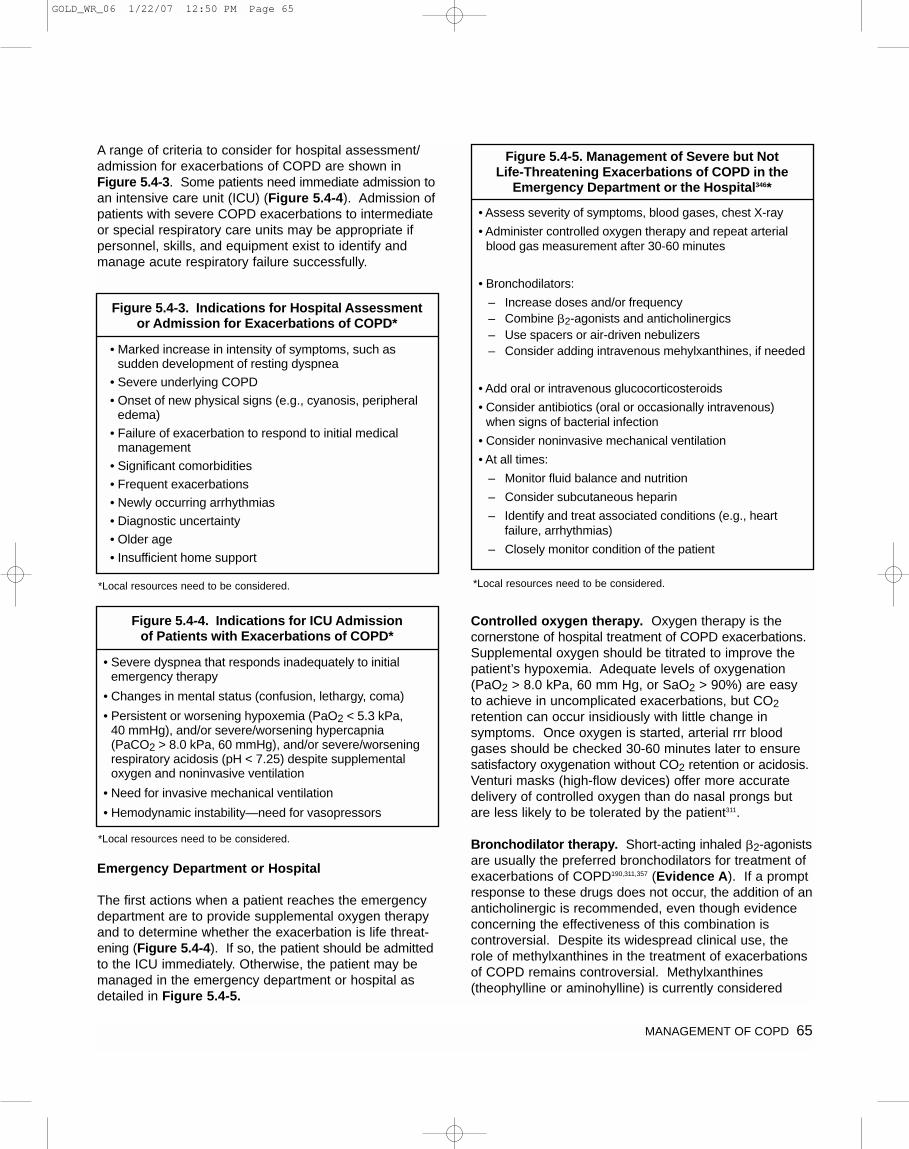
A range of criteria to consider for hospital assessment/admission for exacerbations of COPD are shown inFigure 5.4-3. Some patients need immediate admission toan intensive care unit (ICU) (Figure 5.4-4). Admission ofpatients with severe COPD exacerbations to intermediateor special respiratory care units may be appropriate ifpersonnel, skills, and equipment exist to identify andmanage acute respiratory failure successfully.
Emergency Department or Hospital
The first actions when a patient reaches the emergencydepartment are to provide supplemental oxygen therapyand to determine whether the exacerbation is life threat-ening (Figure 5.4-4). If so, the patient should be admittedto the ICU immediately. Otherwise, the patient may bemanaged in the emergency department or hospital asdetailed in Figure 5.4-5.
Controlled oxygen therapy. Oxygen therapy is the cornerstone of hospital treatment of COPD exacerbations.Supplemental oxygen should be titrated to improve thepatient’s hypoxemia. Adequate levels of oxygenation(PaO2 > 8.0 kPa, 60 mm Hg, or SaO2 > 90%) are easy to achieve in uncomplicated exacerbations, but CO2retention can occur insidiously with little change in symptoms. Once oxygen is started, arterial rrr bloodgases should be checked 30-60 minutes later to ensuresatisfactory oxygenation without CO2 retention or acidosis.Venturi masks (high-flow devices) offer more accuratedelivery of controlled oxygen than do nasal prongs butare less likely to be tolerated by the patient311.
Bronchodilator therapy. Short-acting inhaled �2-agonistsare usually the preferred bronchodilators for treatment ofexacerbations of COPD190,311,357 (Evidence A). If a promptresponse to these drugs does not occur, the addition of ananticholinergic is recommended, even though evidenceconcerning the effectiveness of this combination is controversial. Despite its widespread clinical use, therole of methylxanthines in the treatment of exacerbationsof COPD remains controversial. Methylxanthines (theophylline or aminohylline) is currently considered
MANAGEMENT OF COPD 65
Figure 5.4-3. Indications for Hospital Assessment or Admission for Exacerbations of COPD*
• Marked increase in intensity of symptoms, such assudden development of resting dyspnea
• Severe underlying COPD
• Onset of new physical signs (e.g., cyanosis, peripheraledema)
• Failure of exacerbation to respond to initial medicalmanagement
• Significant comorbidities
• Frequent exacerbations
• Newly occurring arrhythmias
• Diagnostic uncertainty
• Older age
• Insufficient home support
Figure 5.4-5. Management of Severe but Not Life-Threatening Exacerbations of COPD in the
Emergency Department or the Hospital346*
• Assess severity of symptoms, blood gases, chest X-ray
• Administer controlled oxygen therapy and repeat arterialblood gas measurement after 30-60 minutes
• Bronchodilators:
– Increase doses and/or frequency – Combine �2-agonists and anticholinergics– Use spacers or air-driven nebulizers– Consider adding intravenous mehylxanthines, if needed
• Add oral or intravenous glucocorticosteroids
• Consider antibiotics (oral or occasionally intravenous) when signs of bacterial infection
• Consider noninvasive mechanical ventilation
• At all times:
– Monitor fluid balance and nutrition
– Consider subcutaneous heparin
– Identify and treat associated conditions (e.g., heartfailure, arrhythmias)
– Closely monitor condition of the patient
Figure 5.4-4. Indications for ICU Admission of Patients with Exacerbations of COPD*
• Severe dyspnea that responds inadequately to initialemergency therapy
• Changes in mental status (confusion, lethargy, coma)
• Persistent or worsening hypoxemia (PaO2 < 5.3 kPa, 40 mmHg), and/or severe/worsening hypercapnia (PaCO2 > 8.0 kPa, 60 mmHg), and/or severe/worseningrespiratory acidosis (pH < 7.25) despite supplementaloxygen and noninvasive ventilation
• Need for invasive mechanical ventilation
• Hemodynamic instability—need for vasopressors
*Local resources need to be considered. *Local resources need to be considered.
*Local resources need to be considered.
GOLD_WR_06 1/22/07 12:50 PM Page 65

second-line intravenous therapy, used when there isinadequate or insufficient response to short-acting bronchodilators358-362 (Evidence B). Possible beneficialeffects in terms of lung function and clinical endpoints aremodest and inconsistent, whereas adverse effects aresignificantly increased363,364. There are no clinical studiesthat have evaluated the use of inhaled long-acting bronchodilators (either �2-agonists or anticholinergics)with or without inhaled glucocorticosteroids during anacute exacerbation.
Glucocorticosteroids. Oral or intravenous glucocortico-steroids are recommended as an addition to other therapies in the hospital management of exacerbations of COPD350,351 (Evidence A). The exact dose that shouldbe recommended is not known, but high doses are associated with a significant risk of side effects. Thirty to40 mg of oral prednisolone daily for 7-10 days is effectiveand safe (Evidence C). Prolonged treatment does notresult in greater efficacy and increases the risk of sideeffects (e.g., hyperglycemia, muscle atrophy).
Antibiotics. Randomized placebo-controlled studies of antibiotic treatment in exacerbations of COPD havedemonstrated a small beneficial effect of antibiotics onlung function365, and a randomized controlled trial hasprovided evidence for a significant beneficial effect ofantibiotics in COPD patients who presented with anincrease in all three of the following cardinal symptoms:dyspnea, sputum volume, and sputum purulence314.There was also some benefit in those patients with anincrease in only two of these cardinal symptoms.
A study on non-hospitalized patients with exacerbationsof COPD showed a relationship between the purulence of the sputum and the presence of bacteria11, suggestingthat these patients should be treated with antibiotics ifthey also have at least one of the other two cardinalsymptoms (dyspnea or sputum volume). However, thesecriteria for antibiotic treatment of exacerbations of COPDhave not been validated in other studies. A study inCOPD patients with exacerbations requiring mechanicalventilation (invasive or noninvasive) indicated that notgiving antibiotics was associated with increased mortalityand a greater incidence of secondary nosocomial pneumonia366. Based on the current available evidence311,62,antibiotics should be given to:
• Patients with exacerbations of COPD with the followingthree cardinal symptoms: increased dyspnea, increasedsputum volume, and increased sputum purulence(Evidence B).
• Patients with exacerbations of COPD with two of thecardinal symptoms, if increased purulence of sputumis one of the two symptoms (Evidence C).
• Patients with a severe exacerbation of COPD thatrequires mechanical ventilation (invasive or noninvasive)(Evidence B).
The infectious agents in COPD exacerbations can beviral or bacterial177,367. The predominant bacteria recov-ered from the lower airways of patients with COPD exacerbations are H. influenzae, S. pneumoniae, and M. catarrhalis177,330,331,368. So-called atypical pathogens,such as Mycoplasma pneumoniae and Chlamydia pneumoniae368,369, have been identified in patients withCOPD exacerbations, but because of diagnostic limitationsthe true prevalence of these organisms is not known.
Studies in patients with severe underlying COPD whorequire mechanical ventilation370,371 have shown that othermicroorganisms, such as enteric gram-negative bacilliand P. aeruginosa, may be more frequent. Other studieshave shown that the severity of the COPD is an importantdeterminant of the type of microorganism372,373. In patientswith mild COPD exacerbations , S. pneumoniae is pre-dominant. As FEV1 declines and patients have more frequent exacerbations and/or comorbid diseases , H. influenzae and M. catarrhalis become more frequent,and P. aeruginosa may appear in patients with severe airway limitation (Figure 5-4-6)177,311. The risk factors forP. aeruginosa infection are recent hospitalization, frequentadministration of antibiotics (4 courses in the last year),severe COPD exacerbations, and isolation of P. aeruginosaduring a previous exacerbation or colonization during astable period372,373.
Figure 5.4-7177,311,332 provides recommended antibiotictreatment for exacerbations of COPD, although it must be emphasized that most of the published studies relatedto the use of antibiotics were done in chronic bronchitispatients. The route of administration (oral or intravenous)depends on the ability of the patient to eat and the pharmacokinetics of the antibiotic. The oral route is preferred; if the IV route must be used, switching to theoral route is recommended when clinical stabilization permits. Based on studies of the length of use of anti-biotics for chronic bronchitis374-376, antibiotic treatment inpatients with COPD exacerbations could be given for 3 to 7 days (Evidence D).
66 MANAGEMENT OF COPD
GOLD_WR_06 1/22/07 12:50 PM Page 66

Respiratory Stimulants. Respiratory stimulants are notrecommended for acute respiratory failure357. Doxapram,a nonspecific but relatively safe respiratory stimulantavailable in some countries as an intravenous formula-tion, should be used only when noninvasive intermittentventilation is not available or not recommended377.
Ventilatory support. The primary objectives of mechanicalventilatory support in patients with COPD exacerbationsare to decrease mortality and morbidity and to relievesymptoms. Ventilatory support includes both noninvasiveintermittent ventilation using either negative or positivepressure devices, and invasive (conventional) mechanicalventilation by oro-tracheal tube or tracheostomy.
Noninvasive mechanical ventilation. Noninvasive intermittent ventilation (NIV) has been studied in severalrandomized controlled trials in acute respiratory failure,consistently providing positive results with success ratesof 80-85%285,378-380. These studies provide evidence thatNIV improves respiratory acidosis (increases pH, anddecreases PaCO2) , decreases respiratory rate, severity ofbreathlessness, and length of hospital stay (Evidence A).More importantly, mortality—or its surrogate, intubationrate—is reduced by this intervention380-383. However, NIVis not appropriate for all patients, as summarized inFigure 5.4-8285.
MANAGEMENT OF COPD 67
Figure 5.4-6: Stratification of patients with COPDexacerbated for antibiotic treatment and potential
microorganisms involved in each group177,311
Group
Group A Mild exacerbation:No risk factors forpoor outcome
H. influenzaeS. pneumoniaeM. catarrhalisChlamydiapneumoniae
Viruses
Group B Moderateexacerbation withrisk factor(s) forpoor outcome
Group A plus, presence ofresistant organisms(�-lactamaseproducing,penicillin-resistantS. pneumoniae),Enterobacteriaceae(K.pneumoniae,E. coli, Proteus,Enterobacter, etc)
Group C Severeexacerbation withrisk factors for P. aeruginosainfection
Group B plus:P. aeruginosa
Definitiona Microorganisms
a. Risk factors for poor outcome in patients with COPD exacerbation:presence of comorbid diseases, severe COPD, frequent exacerbations(>3 /yr), and antimicrobial use within last 3 months)177,311,372.
5.4-7: Antibiotic treatment in exacerbations of COPDa,b (ref. 177,311,332)
Group A Patients withonly onecardinalsymptomc
should notreceiveantibiotics
If indication then:• �-lactam
(Penicillin,Ampicillin/Amoxicillind)
• Tetracycline
• Trimethoprim/Sulfameth-oxazole
• �-lactam/�-lactamaseinhibitor(Co-amoxiclav)
• Macrolides(Azithromycin, Clarithromycin,Roxithromycine)
• Cephalosporins- 2nd or 3rdgeneration
• Ketolides(Telithromycin)
Group B • �-lactam/�-lactamaseinhibitor(Co-amoxiclav)
• Fluoroquinol-onese
(Gemifloxacin,Levofloxacin,Moxifloxacin)
• �-lactam/�-lactamase inhibitor(Co-amoxiclav,ampicillin/sulbactam)
• Cephalosporins- 2nd or 3rdgeneration
• Fluoroquinol-onese
(Levofloxacin,Moxifloxacin)
Group C In patients at riskfor pseudomonasinfections:• Fluoroquinol-
onese
(Ciprofloxacin,Levofloxacin -high dosef)
• Fluoroquinol-onese
(Ciprofloxacin,Levofloxacin -high dosef) or
• �-lactam withP.aeruginosaactivity
Oral Treatment(No particular order)
Alternative OralTreatment(No particular order)
ParenteralTreatment(No particular order)
a. All patients with symptoms of a COPD exacerbation should be treated with additional bronchodilators ± glucocorticosteroids.
b. Classes of antibiotics are provided (with specific agents in parentheses).In countries with high incidence of S. pneumoniae resistant to penicillin,high dosages of Amoxicillin or Co-amoxiclav are recommended.(See Figure 5-4-6 for definition of Groups A, B, and C.)
c. Cardinal symptoms are increased dyspnea, sputum volume, andsputum purulence.
d. This antibiotic is not appropriate in areas where there is increasedprevalence of �-lactamase producing H. influenzae and M. catarrhalis and/or of S. pneumoniae resistant to penicillin.
e. Not available in all areas of the world.f. Dose 750 mg effective against P. aeruginosa
GOLD_WR_06 1/22/07 12:50 PM Page 67
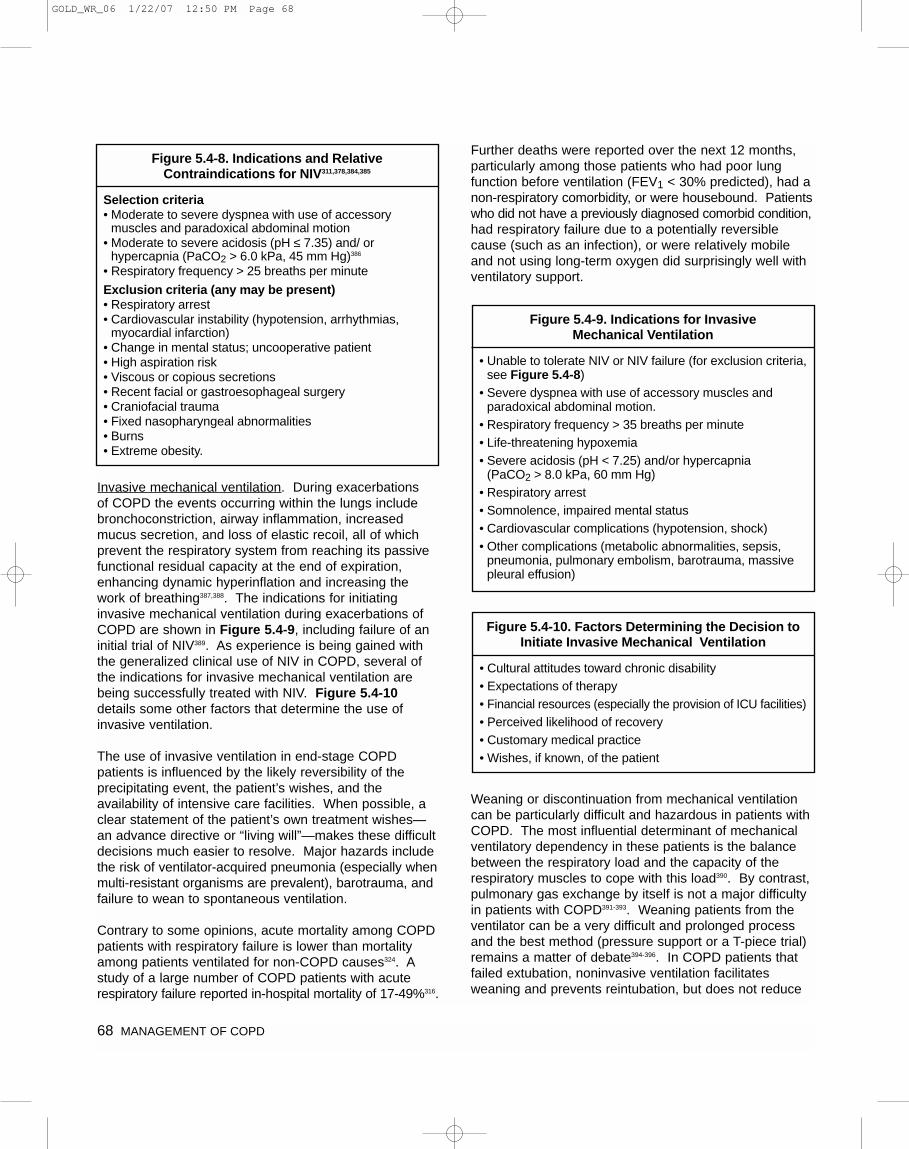
Invasive mechanical ventilation. During exacerbations of COPD the events occurring within the lungs includebronchoconstriction, airway inflammation, increasedmucus secretion, and loss of elastic recoil, all of whichprevent the respiratory system from reaching its passivefunctional residual capacity at the end of expiration,enhancing dynamic hyperinflation and increasing thework of breathing387,388. The indications for initiating invasive mechanical ventilation during exacerbations ofCOPD are shown in Figure 5.4-9, including failure of aninitial trial of NIV389. As experience is being gained withthe generalized clinical use of NIV in COPD, several ofthe indications for invasive mechanical ventilation arebeing successfully treated with NIV. Figure 5.4-10details some other factors that determine the use of invasive ventilation.
The use of invasive ventilation in end-stage COPDpatients is influenced by the likely reversibility of the precipitating event, the patient’s wishes, and the availability of intensive care facilities. When possible, aclear statement of the patient’s own treatment wishes—an advance directive or “living will”—makes these difficultdecisions much easier to resolve. Major hazards includethe risk of ventilator-acquired pneumonia (especially whenmulti-resistant organisms are prevalent), barotrauma, andfailure to wean to spontaneous ventilation.
Contrary to some opinions, acute mortality among COPDpatients with respiratory failure is lower than mortalityamong patients ventilated for non-COPD causes324. Astudy of a large number of COPD patients with acute respiratory failure reported in-hospital mortality of 17-49%316.
Further deaths were reported over the next 12 months,particularly among those patients who had poor lungfunction before ventilation (FEV1 < 30% predicted), had anon-respiratory comorbidity, or were housebound. Patientswho did not have a previously diagnosed comorbid condition,had respiratory failure due to a potentially reversiblecause (such as an infection), or were relatively mobileand not using long-term oxygen did surprisingly well withventilatory support.
Weaning or discontinuation from mechanical ventilationcan be particularly difficult and hazardous in patients withCOPD. The most influential determinant of mechanicalventilatory dependency in these patients is the balancebetween the respiratory load and the capacity of the respiratory muscles to cope with this load390. By contrast,pulmonary gas exchange by itself is not a major difficultyin patients with COPD391-393. Weaning patients from theventilator can be a very difficult and prolonged processand the best method (pressure support or a T-piece trial)remains a matter of debate394-396. In COPD patients thatfailed extubation, noninvasive ventilation facilitates weaning and prevents reintubation, but does not reduce
68 MANAGEMENT OF COPD
Figure 5.4-8. Indications and RelativeContraindications for NIV311,378,384,385
Selection criteria• Moderate to severe dyspnea with use of accessory
muscles and paradoxical abdominal motion• Moderate to severe acidosis (pH ≤ 7.35) and/ or
hypercapnia (PaCO2 > 6.0 kPa, 45 mm Hg)386
• Respiratory frequency > 25 breaths per minute
Exclusion criteria (any may be present)• Respiratory arrest• Cardiovascular instability (hypotension, arrhythmias,
myocardial infarction)• Change in mental status; uncooperative patient• High aspiration risk• Viscous or copious secretions• Recent facial or gastroesophageal surgery• Craniofacial trauma• Fixed nasopharyngeal abnormalities• Burns• Extreme obesity.
Figure 5.4-9. Indications for Invasive Mechanical Ventilation
• Unable to tolerate NIV or NIV failure (for exclusion criteria,see Figure 5.4-8)
• Severe dyspnea with use of accessory muscles andparadoxical abdominal motion.
• Respiratory frequency > 35 breaths per minute• Life-threatening hypoxemia• Severe acidosis (pH < 7.25) and/or hypercapnia
(PaCO2 > 8.0 kPa, 60 mm Hg)• Respiratory arrest• Somnolence, impaired mental status• Cardiovascular complications (hypotension, shock)• Other complications (metabolic abnormalities, sepsis,
pneumonia, pulmonary embolism, barotrauma, massivepleural effusion)
Figure 5.4-10. Factors Determining the Decision toInitiate Invasive Mechanical Ventilation
• Cultural attitudes toward chronic disability• Expectations of therapy• Financial resources (especially the provision of ICU facilities)• Perceived likelihood of recovery• Customary medical practice• Wishes, if known, of the patient
GOLD_WR_06 1/22/07 12:50 PM Page 68
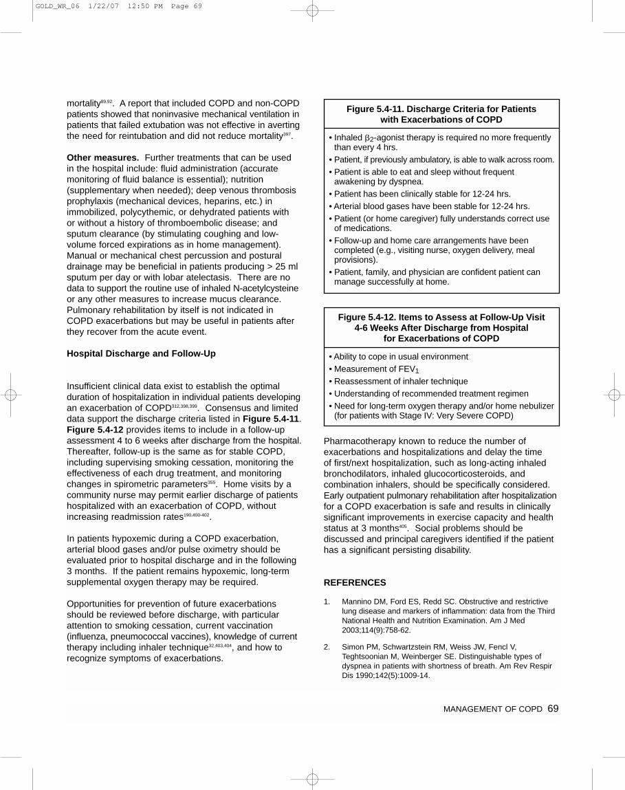
mortality89,92. A report that included COPD and non-COPDpatients showed that noninvasive mechanical ventilation inpatients that failed extubation was not effective in avertingthe need for reintubation and did not reduce mortality397.
Other measures. Further treatments that can be used in the hospital include: fluid administration (accurate monitoring of fluid balance is essential); nutrition (supplementary when needed); deep venous thrombosisprophylaxis (mechanical devices, heparins, etc.) in immobilized, polycythemic, or dehydrated patients with or without a history of thromboembolic disease; and sputum clearance (by stimulating coughing and low-volume forced expirations as in home management).Manual or mechanical chest percussion and posturaldrainage may be beneficial in patients producing > 25 mlsputum per day or with lobar atelectasis. There are nodata to support the routine use of inhaled N-acetylcysteineor any other measures to increase mucus clearance.Pulmonary rehabilitation by itself is not indicated inCOPD exacerbations but may be useful in patients afterthey recover from the acute event.
Hospital Discharge and Follow-Up
Insufficient clinical data exist to establish the optimalduration of hospitalization in individual patients developingan exacerbation of COPD312,398,399. Consensus and limiteddata support the discharge criteria listed in Figure 5.4-11.Figure 5.4-12 provides items to include in a follow-upassessment 4 to 6 weeks after discharge from the hospital.Thereafter, follow-up is the same as for stable COPD,including supervising smoking cessation, monitoring theeffectiveness of each drug treatment, and monitoringchanges in spirometric parameters355. Home visits by acommunity nurse may permit earlier discharge of patientshospitalized with an exacerbation of COPD, withoutincreasing readmission rates190,400-402.
In patients hypoxemic during a COPD exacerbation, arterial blood gases and/or pulse oximetry should beevaluated prior to hospital discharge and in the following3 months. If the patient remains hypoxemic, long-termsupplemental oxygen therapy may be required.
Opportunities for prevention of future exacerbationsshould be reviewed before discharge, with particularattention to smoking cessation, current vaccination(influenza, pneumococcal vaccines), knowledge of currenttherapy including inhaler technique32,403,404, and how to recognize symptoms of exacerbations.
Pharmacotherapy known to reduce the number of exacerbations and hospitalizations and delay the time of first/next hospitalization, such as long-acting inhaledbronchodilators, inhaled glucocorticosteroids, and combination inhalers, should be specifically considered.Early outpatient pulmonary rehabilitation after hospitalizationfor a COPD exacerbation is safe and results in clinicallysignificant improvements in exercise capacity and healthstatus at 3 months405. Social problems should be discussed and principal caregivers identified if the patienthas a significant persisting disability.
REFERENCES
1. Mannino DM, Ford ES, Redd SC. Obstructive and restrictivelung disease and markers of inflammation: data from the ThirdNational Health and Nutrition Examination. Am J Med2003;114(9):758-62.
2. Simon PM, Schwartzstein RM, Weiss JW, Fencl V,Teghtsoonian M, Weinberger SE. Distinguishable types of dyspnea in patients with shortness of breath. Am Rev RespirDis 1990;142(5):1009-14.
MANAGEMENT OF COPD 69
Figure 5.4-11. Discharge Criteria for Patients with Exacerbations of COPD
• Inhaled �2-agonist therapy is required no more frequentlythan every 4 hrs.
• Patient, if previously ambulatory, is able to walk across room.• Patient is able to eat and sleep without frequent
awakening by dyspnea.• Patient has been clinically stable for 12-24 hrs.• Arterial blood gases have been stable for 12-24 hrs.• Patient (or home caregiver) fully understands correct use
of medications.• Follow-up and home care arrangements have been
completed (e.g., visiting nurse, oxygen delivery, mealprovisions).
• Patient, family, and physician are confident patient canmanage successfully at home.
Figure 5.4-12. Items to Assess at Follow-Up Visit 4-6 Weeks After Discharge from Hospital
for Exacerbations of COPD
• Ability to cope in usual environment• Measurement of FEV1
• Reassessment of inhaler technique• Understanding of recommended treatment regimen• Need for long-term oxygen therapy and/or home nebulizer
(for patients with Stage IV: Very Severe COPD)
GOLD_WR_06 1/22/07 12:50 PM Page 69

3. Elliott MW, Adams L, Cockcroft A, MacRae KD, Murphy K, GuzA. The language of breathlessness. Use of verbal descriptorsby patients with cardiopulmonary disease. Am Rev Respir Dis1991;144(4):826-32.
4. Bestall JC, Paul EA, Garrod R, Garnham R, Jones PW,Wedzicha JA. Usefulness of the Medical Research Council(MRC) dyspnoea scale as a measure of disability in patientswith chronic obstructive pulmonary disease. Thorax1999;54(7):581-6.
5. Nishimura K, Izumi T, Tsukino M, Oga T. Dyspnea is a betterpredictor of 5-year survival than airway obstruction in patientswith COPD. Chest 2002;121(5):1434-40.
6. Celli BR, Rassulo J, Make BJ. Dyssynchronous breathing during arm but not leg exercise in patients with chronic airflowobstruction. N Engl J Med 1986;314(23):1485-90.
7. Georgopoulas D, Anthonisen NR. Symptoms and signs ofCOPD. In: Cherniack NS, ed. Chronic obstructive pulmonarydisease. Toronto: WB Saunders Co; 1991:357-63.
8. Burrows B, Niden AH, Barclay WR, Kasik JE. Chronic obstruc-tive lung disease II. Relationships of clinical and physiologicalfindings to the severity of aiways obstruction. Am Rev RespirDis 1965;91:665-78.
9. Definition and classification of chronic bronchitis for clinical andepidemiological purposes. A report to the Medical ResearchCouncil by their Committee on the Aetiology of ChronicBronchitis. Lancet 1965;1(7389):775-9.
10. Hill AT, Bayley D, Stockley RA. The interrelationship of sputuminflammatory markers in patients with chronic bronchitis. Am JRespir Crit Care Med 1999;160(3):893-8.
11. Stockley RA, O'Brien C, Pye A, Hill SL. Relationship of sputumcolor to nature and outpatient management of acute exacerba-tions of COPD. Chest 2000;117(6):1638-45.
12. Schols AM, Soeters PB, Dingemans AM, Mostert R, FrantzenPJ, Wouters EF. Prevalence and characteristics of nutritionaldepletion in patients with stable COPD eligible for pulmonaryrehabilitation. Am Rev Respir Dis 1993;147(5):1151-6.
13. Schols AM, Slangen J, Volovics L, Wouters EF. Weight loss isa reversible factor in the prognosis of chronic obstructive pulmonary disease. Am J Respir Crit Care Med 1998;157(6 Pt 1):1791-7.
14. Calverley PMA. Neuropsychological deficits in chronic obstructivepulmonary disease. [editorial]. Monaldi Arch Chest Dis1996;51(1):5-6.
15. Holguin F, Folch E, Redd SC, Mannino DM. Comorbidity andmortality in COPD-related hospitalizations in the United States,1979 to 2001. Chest 2005;128(4):2005-11.
16. Kesten S, Chapman KR. Physician perceptions and managementof COPD. Chest 1993;104(1):254-8.
17. Loveridge B, West P, Kryger MH, Anthonisen NR. Alteration inbreathing pattern with progression of chronic obstructive pulmonary disease. Am Rev Respir Dis 1986;134(5):930-4.
18. Bianchi R, Gigliotti F, Romagnoli I, Lanini B, Castellani C,Grazzini M, et al. Chest wall kinematics and breathlessnessduring pursed-lip breathing in patients with COPD. Chest2004;125(2):459-65.
19. Badgett RG, Tanaka DJ, Hunt DK, Jelley MJ, Feinberg LE,Steiner JF, et al. Can moderate chronic obstructive pulmonarydisease be diagnosed by historical and physical findingsalone? Am J Med 1993;94(2):188-96.
20. Pellegrino R, Viegi G, Brusasco V, Crapo RO, Burgos F,Casaburi R, et al. Interpretative strategies for lung functiontests. Eur Respir J 2005;26(5):948-68.
21. Hardie JA, Buist AS, Vollmer WM, Ellingsen I, Bakke PS,Morkve O. Risk of over-diagnosis of COPD in asymptomaticelderly never-smokers. Eur Respir J 2002;20(5):1117-22.
22. Kelly CA, Gibson GJ. Relation between FEV1 and peak expiratory flow in patients with chronic airflow obstruction.Thorax 1988;43(4):335-6.
23. Jackson H, Hubbard R. Detecting chronic obstructive pulmonarydisease using peak flow rate: cross sectional survey. BMJ2003;327(7416):653-4.
24. Miller MR, Hankinson J, Brusasco V, Burgos F, Casaburi R,Coates A, et al. Standardisation of spirometry. Eur Respir J2005;26(2):319-38.
25. Ferguson GT, Enright PL, Buist AS, Higgins MW. Officespirometry for lung health assessment in adults: A consensusstatement from the National Lung Health Education Program.Chest 2000;117(4):1146-61.
26. Wilt TJ, Niewoehner D, Kim C, Kane RL, Linabery A, TacklindJ, et al. Use of spirometry for case finding, diagnosis, andmanagement of chronic obstructive pulmonary disease(COPD). Evid Rep Technol Assess (Summ) 2005(121):1-7.
27. Kanner RE, Connett JE, Williams DE, Buist AS. Effects of randomized assignment to a smoking cessation interventionand changes in smoking habits on respiratory symptoms insmokers with early chronic obstructive pulmonary disease: the Lung Health Study. Am J Med 1999;106(4):410-6.
28. Lofdahl CG, Postma DS, Laitinen LA, Ohlsson SV, Pauwels RA,Pride NB. The European Respiratory Society study on chronicobstructive pulmonary disease (EUROSCOP): recruitmentmethods and strategies. Respir Med 1998;92(3):467-72.
29. Peto R, Speizer FE, Cochrane AL, Moore F, Fletcher CM,Tinker CM, et al. The relevance in adults of air-flow obstruction,but not of mucus hypersecretion, to mortality from chronic lungdisease. Results from 20 years of prospective observation. Am Rev Respir Dis 1983;128(3):491-500.
30. Jones PW. Health status measurement in chronic obstructivepulmonary disease. Thorax 2001;56(11):880-7.
31. Van Der Molen T, Willemse BW, Schokker S, Ten Hacken NH,Postma DS, Juniper EF. Development, validity and responsive-ness of the Clinical COPD Questionnaire. Health Qual LifeOutcomes 2003;1(1):13.
70 MANAGEMENT OF COPD
GOLD_WR_06 1/22/07 12:50 PM Page 70
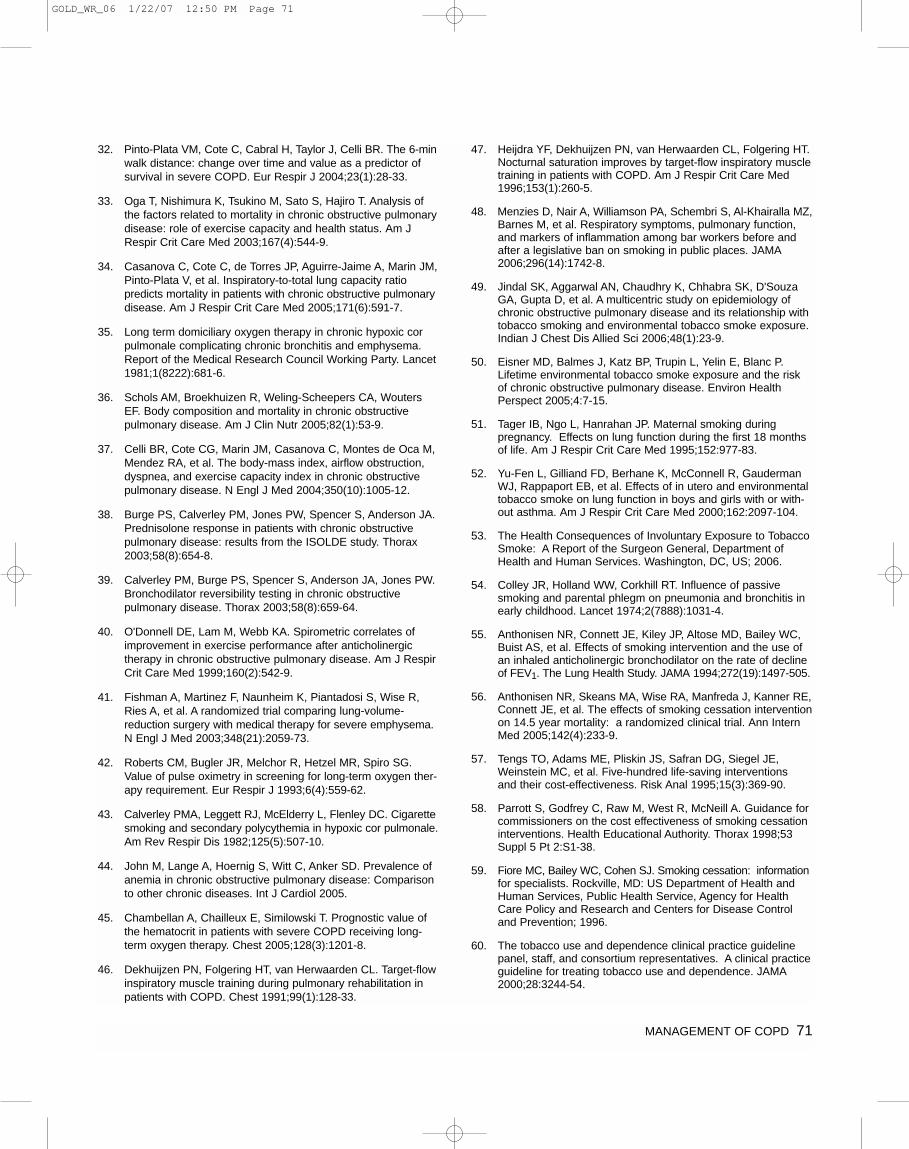
32. Pinto-Plata VM, Cote C, Cabral H, Taylor J, Celli BR. The 6-minwalk distance: change over time and value as a predictor ofsurvival in severe COPD. Eur Respir J 2004;23(1):28-33.
33. Oga T, Nishimura K, Tsukino M, Sato S, Hajiro T. Analysis ofthe factors related to mortality in chronic obstructive pulmonarydisease: role of exercise capacity and health status. Am JRespir Crit Care Med 2003;167(4):544-9.
34. Casanova C, Cote C, de Torres JP, Aguirre-Jaime A, Marin JM,Pinto-Plata V, et al. Inspiratory-to-total lung capacity ratio predicts mortality in patients with chronic obstructive pulmonarydisease. Am J Respir Crit Care Med 2005;171(6):591-7.
35. Long term domiciliary oxygen therapy in chronic hypoxic corpulmonale complicating chronic bronchitis and emphysema.Report of the Medical Research Council Working Party. Lancet1981;1(8222):681-6.
36. Schols AM, Broekhuizen R, Weling-Scheepers CA, WoutersEF. Body composition and mortality in chronic obstructive pulmonary disease. Am J Clin Nutr 2005;82(1):53-9.
37. Celli BR, Cote CG, Marin JM, Casanova C, Montes de Oca M,Mendez RA, et al. The body-mass index, airflow obstruction,dyspnea, and exercise capacity index in chronic obstructivepulmonary disease. N Engl J Med 2004;350(10):1005-12.
38. Burge PS, Calverley PM, Jones PW, Spencer S, Anderson JA.Prednisolone response in patients with chronic obstructive pulmonary disease: results from the ISOLDE study. Thorax2003;58(8):654-8.
39. Calverley PM, Burge PS, Spencer S, Anderson JA, Jones PW.Bronchodilator reversibility testing in chronic obstructive pulmonary disease. Thorax 2003;58(8):659-64.
40. O'Donnell DE, Lam M, Webb KA. Spirometric correlates ofimprovement in exercise performance after anticholinergic therapy in chronic obstructive pulmonary disease. Am J RespirCrit Care Med 1999;160(2):542-9.
41. Fishman A, Martinez F, Naunheim K, Piantadosi S, Wise R,Ries A, et al. A randomized trial comparing lung-volume-reduction surgery with medical therapy for severe emphysema.N Engl J Med 2003;348(21):2059-73.
42. Roberts CM, Bugler JR, Melchor R, Hetzel MR, Spiro SG.Value of pulse oximetry in screening for long-term oxygen ther-apy requirement. Eur Respir J 1993;6(4):559-62.
43. Calverley PMA, Leggett RJ, McElderry L, Flenley DC. Cigarettesmoking and secondary polycythemia in hypoxic cor pulmonale.Am Rev Respir Dis 1982;125(5):507-10.
44. John M, Lange A, Hoernig S, Witt C, Anker SD. Prevalence ofanemia in chronic obstructive pulmonary disease: Comparisonto other chronic diseases. Int J Cardiol 2005.
45. Chambellan A, Chailleux E, Similowski T. Prognostic value ofthe hematocrit in patients with severe COPD receiving long-term oxygen therapy. Chest 2005;128(3):1201-8.
46. Dekhuijzen PN, Folgering HT, van Herwaarden CL. Target-flowinspiratory muscle training during pulmonary rehabilitation inpatients with COPD. Chest 1991;99(1):128-33.
47. Heijdra YF, Dekhuijzen PN, van Herwaarden CL, Folgering HT.Nocturnal saturation improves by target-flow inspiratory muscletraining in patients with COPD. Am J Respir Crit Care Med1996;153(1):260-5.
48. Menzies D, Nair A, Williamson PA, Schembri S, Al-Khairalla MZ,Barnes M, et al. Respiratory symptoms, pulmonary function,and markers of inflammation among bar workers before andafter a legislative ban on smoking in public places. JAMA2006;296(14):1742-8.
49. Jindal SK, Aggarwal AN, Chaudhry K, Chhabra SK, D'SouzaGA, Gupta D, et al. A multicentric study on epidemiology ofchronic obstructive pulmonary disease and its relationship withtobacco smoking and environmental tobacco smoke exposure.Indian J Chest Dis Allied Sci 2006;48(1):23-9.
50. Eisner MD, Balmes J, Katz BP, Trupin L, Yelin E, Blanc P.Lifetime environmental tobacco smoke exposure and the riskof chronic obstructive pulmonary disease. Environ HealthPerspect 2005;4:7-15.
51. Tager IB, Ngo L, Hanrahan JP. Maternal smoking during pregnancy. Effects on lung function during the first 18 monthsof life. Am J Respir Crit Care Med 1995;152:977-83.
52. Yu-Fen L, Gilliand FD, Berhane K, McConnell R, GaudermanWJ, Rappaport EB, et al. Effects of in utero and environmentaltobacco smoke on lung function in boys and girls with or with-out asthma. Am J Respir Crit Care Med 2000;162:2097-104.
53. The Health Consequences of Involuntary Exposure to TobaccoSmoke: A Report of the Surgeon General, Department ofHealth and Human Services. Washington, DC, US; 2006.
54. Colley JR, Holland WW, Corkhill RT. Influence of passivesmoking and parental phlegm on pneumonia and bronchitis inearly childhood. Lancet 1974;2(7888):1031-4.
55. Anthonisen NR, Connett JE, Kiley JP, Altose MD, Bailey WC,Buist AS, et al. Effects of smoking intervention and the use ofan inhaled anticholinergic bronchodilator on the rate of declineof FEV1. The Lung Health Study. JAMA 1994;272(19):1497-505.
56. Anthonisen NR, Skeans MA, Wise RA, Manfreda J, Kanner RE,Connett JE, et al. The effects of smoking cessation interventionon 14.5 year mortality: a randomized clinical trial. Ann InternMed 2005;142(4):233-9.
57. Tengs TO, Adams ME, Pliskin JS, Safran DG, Siegel JE,Weinstein MC, et al. Five-hundred life-saving interventions and their cost-effectiveness. Risk Anal 1995;15(3):369-90.
58. Parrott S, Godfrey C, Raw M, West R, McNeill A. Guidance forcommissioners on the cost effectiveness of smoking cessationinterventions. Health Educational Authority. Thorax 1998;53Suppl 5 Pt 2:S1-38.
59. Fiore MC, Bailey WC, Cohen SJ. Smoking cessation: informationfor specialists. Rockville, MD: US Department of Health andHuman Services, Public Health Service, Agency for HealthCare Policy and Research and Centers for Disease Controland Prevention; 1996.
60. The tobacco use and dependence clinical practice guidelinepanel, staff, and consortium representatives. A clinical practiceguideline for treating tobacco use and dependence. JAMA2000;28:3244-54.
MANAGEMENT OF COPD 71
GOLD_WR_06 1/22/07 12:50 PM Page 71

61. American Medical Association. Guidelines for the diagnosisand treatment of nicotine dependence: how to help patientsstop smoking. Washington DC: American Medical Association;1994.
62. Glynn TJ, Manley MW. How to help your patients stop smoking. A Nattional Cancer Institute manual for physicians.Bethesda, MD: US Department of Health and Human Services,Public Health Service, National Institutes of Health, NationalCancer Institute; 1990.
63. Glynn TJ, Manley MW, Pechacek TF. Physician-initiated smoking cessation program: the National Cancer Institute trials. Prog Clin Biol Res 1990;339:11-25.
64. Baillie AJ, Mattick RP, Hall W, Webster P. Meta-analytic reviewof the efficacy of smoking cessation interventions. Drug andAlcohol Review 1994;13:157-70.
65. Wilson DH, Wakefield MA, Steven ID, Rohrsheim RA, EstermanAJ, Graham NM. “Sick of Smoking”: evaluation of a targetedminimal smoking cessation intervention in general practice.Med J Aust 1990;152(10):518-21.
66. Britton J, Knox A. Helping people to stop smoking: the newsmoking cessation guidelines. Thorax 1999;54(1):1-2.
67. Katz DA, Muehlenbruch DR, Brown RL, Fiore MC, Baker TB.Effectiveness of implementing the agency for healthcareresearch and quality smoking cessation clinical practice guideline: a randomized, controlled trial. J Natl Cancer Inst2004;96(8):594-603.
68. Kottke TE, Battista RN, DeFriese GH, Brekke ML. Attributes ofsuccessful smoking cessation interventions in medical practice.A meta-analysis of 39 controlled trials. JAMA1988;259(19):2883-9.
69. Lancaster T, Stead L, Silagy C, Sowden A. Effectiveness ofinterventions to help people stop smoking: findings from theCochrane Library. BMJ 2000;321(7257):355-8.
70. Schwartz JL. Review and evaluation of smoking cessationmethods: United States and Canada, 1978-1985. Bethesda,MD: National Institutes of Health; 1987.
71. Fiore MC, Smith SS, Jorenby DE, Baker TB. The effectivenessof the nicotine patch for smoking cessation. A meta- analysis.JAMA 1994;271(24):1940-7.
72. Sachs DP, Benowitz NL. Individualizing medical treatment fortobacco dependence. Eur Respir J 1996;9(4):629-31.
73. Tashkin D, Kanner R, Bailey W, Buist S, Anderson P, Nides M,et al. Smoking cessation in patients with chronic obstructivepulmonary disease: a double-blind, placebo-controlled, randomised trial. Lancet 2001;357(9268):1571-5.
74. Jorenby DE, Leischow SJ, Nides MA, Rennard SI, JohnstonJA, Hughes AR, et al. A controlled trial of sustained-releasebupropion, a nicotine patch, or both for smoking cessation. N Engl J Med 1999;340(9):685-91.
75. Jorenby DE, Hays JT, Rigotti NA, Axoulay S, Watsky EJ,Williams KE, et al. Efficacy of varenicline, an alpha4beta2nicotinic acetylcholine receptor partial agonist, vs placebo or sustained-release bupropion for smoking cessation: a randomized controlled trial. JAMA 2006;296(1):56-63.
76. Nides M, Oncken C, Gonzales D, Rennard S, Watsky EJ,Anziano R, et al. Smoking cessation with varenicline, a selectivealpha4beta2 nicotinic receptor partial agonist: results from a 7-week, randomized, placebo- and bupropion-controlled trialwith 1-year follow-up. Arch Intern Med 2006;166(15):1561-8.
77. Tonstad S, Tonnesen P, Hajek P, Williams KE, Billing CB,Reeves KR, et al. Effect of maintenance therapy with vareniclineon smoking cessation: a randomized controlled trial. JAMA2006;296(1):64-71.
78. Celli BR, Halbert RJ, Nordyke RJ, Schan B. Airway obstructionin never smokers: results from the Third National Health andNutrition Examination Survey. Am J Med 2005;118:1364-72.
79. Kunzli N, Kaiser R, Medina M, Studnicka M, Chanel O, Filliger P,et al. Public-health impact of outdoor and traffic-related air pol-lution: a European assessment. Lancet 2000;356:795-801.
80. Ackermann-Liebrich U, Leuenberger P, Schwartz J, SchindlerC, Monn C, Bolognini G, et al. Lung function and long termexposure to air pollutants in Switzerland. Study on Air Pollutionand Lung Diseases in Adults (SAPALDIA) Team. Am J RespirCrit Care Med 1997;155(1):122-9.
81. Oroczo-Levi M, Garcia -Aymerich J, Villar J, Ramirez-SarmientoA, Anto JM, Gea J. Wood smoke exposure and risk of chronicobstructive pulmonary disease. Eur Respir J 2006;27:542-6.
82. Chapman RS, Xingzhou H, Blair AE, Lan Q. Improvement inhousehold stoves and risk of chronic obstructive pulmonarydisease in Xuanwei, China: retrospective cohort study. Br Med J2005;331:1050.
83. Ghambarian MH, Feenstra TL, Zwanikken P, Kalinina AM. Can prevention be improved? Proposal for an integrated intervention strategy. Preventive Medicine 2004;39:337-43.
84. Nichter M. Introducing tobacco cessation in developing countries:an overview of Quit Tobacco International. Tobacco Control2006;15(Supplement 1):12-7.
85. Reis AL. Response to bronchodilators. In: Clausen J, ed.Pulmonary function testing: guidelines and controversies. New York: Academic Press; 1982.
86. Janelli LM, Scherer YK, Schmieder LE. Can a pulmonaryhealth teaching program alter patients' ability to cope withCOPD? Rehabil Nurs 1991;16(4):199-202.
87. Ashikaga T, Vacek PM, Lewis SO. Evaluation of a community-based education program for individuals with chronic obstructivepulmonary disease. J Rehabil 1980;46(2):23-7.
88. Toshima MT, Kaplan RM, Ries AL. Experimental evaluation of rehabilitation in chronic obstructive pulmonary disease:short-term effects on exercise endurance and health status.Health Psychol 1990;9(3):237-52.
72 MANAGEMENT OF COPD
GOLD_WR_06 1/22/07 12:50 PM Page 72
89. Celli BR. Pulmonary rehabilitation in patients with COPD. Am JRespir Crit Care Med 1995;152(3):861-4.
90. Stewart MA. Effective physician-patient communication andhealth outcomes: a review. CMAJ 1995;152(9):1423-33.
91. Clark NM, Nothwehr F, Gong M, Evans D, Maiman LA, HurwitzME, et al. Physician-patient partnership in managing chronic illness. Acad Med 1995;70(11):957-9.
92. Heffner JE, Fahy B, Hilling L, Barbieri C. Outcomes of advancedirective education of pulmonary rehabilitation patients. Am JRespir Crit Care Med 1997;155(3):1055-9.
93. Taylor SJ, Candy B, Bryar RM, Ramsay J, Vrijhoef HJ, EsmondG, et al. Effectiveness of innovations in nurse led chronic disease management for patients with chronic obstructive pulmonary disease: systematic review of evidence. BMJ2005;331(7515):485.
94. Bourbeau J, Julien M, Maltais F, Rouleau M, Beaupre A, BeginR, et al. Reduction of hospital utilization in patients with chronicobstructive pulmonary disease: a disease-specific self-management intervention. Arch Intern Med 2003;163(5):585-91.
95. Tougaard L, Krone T, Sorknaes A, Ellegaard H. Economic benefits of teaching patients with chronic obstructive pulmonarydisease about their illness. The PASTMA Group. Lancet1992;339(8808):1517-20.
96. Gallefoss F. The effects of patient education in COPD in a 1-year follow-up randomised, controlled trial. Patient EducCouns 2004;52(3):259-66.
97. Monninkhof E, van der Valk P, van der Palen J, van HerwaardenC, Zielhuis G. Effects of a comprehensive self-managementprogramme in patients with chronic obstructive pulmonary disease. Eur Respir J 2003;22(5):815-20.
98. Pauwels RA, Lofdahl CG, Laitinen LA, Schouten JP, PostmaDS, Pride NB, et al. Long-term treatment with inhaled bude-sonide in persons with mild chronic obstructive pulmonary disease who continue smoking. European Respiratory SocietyStudy on Chronic Obstructive Pulmonary Disease. N Engl JMed 1999;340(25):1948-53.
99. Vestbo J, Sorensen T, Lange P, Brix A, Torre P, Viskum K.Long-term effect of inhaled budesonide in mild and moderatechronic obstructive pulmonary disease: a randomised controlled trial. Lancet 1999;353(9167):1819-23.
100. Burge PS, Calverley PM, Jones PW, Spencer S, Anderson JA,Maslen TK. Randomised, double blind, placebo controlledstudy of fluticasone propionate in patients with moderate tosevere chronic obstructive pulmonary disease: the ISOLDEtrial. BMJ 2000;320(7245):1297-303.
101. Vestbo J, Pauwels R, Anderson JA, Jones P, Calverley P. Early onset of effect of salmeterol and fluticasone propionate inchronic obstructive pulmonary disease. Thorax 2005;60(4):301-4.
102. Calverley PMA. Symptomatic bronchodilator treatment. In:Calverley PMA, Pride NB, eds. Chronic obstructive pulmonarydisease. London: Chapman and Hall; 1995:419-45.
103. Belman MJ, Botnick WC, Shin JW. Inhaled bronchodilatorsreduce dynamic hyperinflation during exercise in patients withchronic obstructive pulmonary disease. Am J Respir Crit CareMed 1996;153(3):967-75.
104. Berger R, Smith D. Effect of inhaled metaproterenol on exerciseperformance in patients with stable “fixed” airway obstruction.Am Rev Respir Dis 1988;138(3):624-9.
105. Hay JG, Stone P, Carter J, Church S, Eyre-Brook A, PearsonMG, et al. Bronchodilator reversibility, exercise performanceand breathlessness in stable chronic obstructive pulmonarydisease. Eur Respir J 1992;5(6):659-64.
106. Vathenen AS, Britton JR, Ebden P, Cookson JB, Wharrad HJ,Tattersfield AE. High-dose inhaled albuterol in severe chronicairflow limitation. Am Rev Respir Dis 1988;138(4):850-5.
107. Gross NJ, Petty TL, Friedman M, Skorodin MS, Silvers GW,Donohue JF. Dose response to ipratropium as a nebulizedsolution in patients with chronic obstructive pulmonary disease.A three-center study. Am Rev Respir Dis 1989;139(5):1188-91.
108. Chrystyn H, Mulley BA, Peake MD. Dose response relation tooral theophylline in severe chronic obstructive airways disease.BMJ 1988;297(6662):1506-10.
109. Higgins BG, Powell RM, Cooper S, Tattersfield AE. Effect ofsalbutamol and ipratropium bromide on airway calibre andbronchial reactivity in asthma and chronic bronchitis. EurRespir J 1991;4(4):415-20.
110. Ericsson CH, Svartengren K, Svartengren M, Mossberg B,Philipson K, Blomquist M, et al. Repeatability of airway depositionand tracheobronchial clearance rate over three days in chronicbronchitis. Eur Respir J 1995;8(11):1886-93.
111. Kim CS, Kang TC. Comparative measurement of lung depositionof inhaled fine particles in normal subjects and patients withobstructive airway disease. Am J Respir Crit Care Med1997;155(3):899-905.
112. Boe J, Dennis JH, O'Driscoll BR, Bauer TT, Carone M,Dautzenberg B, et al. European Respiratory Society Guidelineson the use of nebulizers. Eur Respir J 2001;18(1):228-42.
113. O'Driscoll BR, Kay EA, Taylor RJ, Weatherby H, Chetty MC,Bernstein A. A long-term prospective assessment of home nebulizer treatment. Respir Med 1992;86(4):317-25.
114. Jenkins SC, Heaton RW, Fulton TJ, Moxham J. Comparison of domiciliary nebulized salbutamol and salbutamol from ametered-dose inhaler in stable chronic airflow limitation. Chest1987;91(6):804-7.
115. Ikeda A, Nishimura K, Koyama H, Izumi T. Bronchodilatingeffects of combined therapy with clinical dosages of ipratropiumbromide and salbutamol for stable COPD: comparison withipratropium bromide alone. Chest 1995;107(2):401-5.
116. Guyatt GH, Townsend M, Pugsley SO, Keller JL, Short HD,Taylor DW, et al. Bronchodilators in chronic air-flow limitation.Effects on airway function, exercise capacity, and quality of life.Am Rev Respir Dis 1987;135(5):1069-74.
MANAGEMENT OF COPD 73
GOLD_WR_06 1/22/07 12:50 PM Page 73
117. Man WD, Mustfa N, Nikoletou D, Kaul S, Hart N, Rafferty GF,et al. Effect of salmeterol on respiratory muscle activity duringexercise in poorly reversible COPD. Thorax 2004;59(6):471-6.
118. O'Donnell DE, Fluge T, Gerken F, Hamilton A, Webb K,Aguilaniu B, et al. Effects of tiotropium on lung hyperinflation,dyspnoea and exercise tolerance in COPD. Eur Respir J2004;23(6):832-40.
119. Vincken W, van Noord JA, Greefhorst AP, Bantje TA, Kesten S,Korducki L, et al. Improved health outcomes in patients withCOPD during 1 yr's treatment with tiotropium. Eur Respir J2002;19(2):209-16.
120. Mahler DA, Donohue JF, Barbee RA, Goldman MD, Gross NJ,Wisniewski ME, et al. Efficacy of salmeterol xinafoate in thetreatment of COPD. Chest 1999;115(4):957-65.
121. Dahl R, Greefhorst LA, Nowak D, Nonikov V, Byrne AM,Thomson MH, et al. Inhaled formoterol dry powder versus ipratropium bromide in chronic obstructive pulmonary disease.Am J Respir Crit Care Med 2001;164(5):778-84.
122. Oostenbrink JB, Rutten-van Molken MP, Al MJ, Van Noord JA,Vincken W. One-year cost-effectiveness of tiotropium versusipratropium to treat chronic obstructive pulmonary disease. Eur Respir J 2004;23(2):241-9.
123. Niewoehner DE, Rice K, Cote C, Paulson D, Cooper JA, Jr.,Korducki L, et al. Prevention of exacerbations of chronicobstructive pulmonary disease with tiotropium, a once-dailyinhaled anticholinergic bronchodilator: a randomized trial. Ann Intern Med 2005;143(5):317-26.
124. Casaburi R, Kukafka D, Cooper CB, Witek TJ, Jr., Kesten S.Improvement in exercise tolerance with the combination oftiotropium and pulmonary rehabilitation in patients with COPD.Chest 2005;127(3):809-17.
125. Shim CS, Williams MH, Jr. Bronchodilator response to oralaminophylline and terbutaline versus aerosol albuterol inpatients with chronic obstructive pulmonary disease. Am J Med1983;75(4):697-701.
126. COMBIVENT Inhalation Aerosol Study Group. In chronicobstructive pulmonary disease, a combination of ipratropiumand albuterol is more effective than either agent alone. An 85-day multicenter trial. Chest 1994;105(5):1411-9.
127. van Schayck CP, Folgering H, Harbers H, Maas KL, van Weel C.Effects of allergy and age on responses to salbutamol andipratropium bromide in moderate asthma and chronic bronchitis.Thorax 1991;46(5):355-9.
128. Datta D, Vitale A, Lahiri B, ZuWallack R. An evaluation of nebulized levalbuterol in stable COPD. Chest 2003;124(3):844-9.
129. Ulrik CS. Efficacy of inhaled salmeterol in the management ofsmokers with chronic obstructive pulmonary disease: a singlecentre randomised, double blind, placebo controlled, crossoverstudy. Thorax 1995;50(7):750-4.
130. Boyd G, Morice AH, Pounsford JC, Siebert M, Peslis N,Crawford C. An evaluation of salmeterol in the treatment ofchronic obstructive pulmonary disease (COPD) [publishederratum appears in Eur Respir J 1997 Jul;10(7):1696]. Eur Respir J 1997;10(4):815-21.
131. Cazzola M, Matera MG, Santangelo G, Vinciguerra A, Rossi F,D'Amato G. Salmeterol and formoterol in partially reversiblesevere chronic obstructive pulmonary disease: a dose-response study. Respir Med 1995;89(5):357-62.
132. Rossi A, Kristufek P, Levine BE, Thomson MH, Till D, KottakisJ, et al. Comparison of the efficacy, tolerability, and safety offormoterol dry powder and oral, slow-release theophylline inthe treatment of COPD. Chest 2002;121(4):1058-69.
133. Lipworth BJ, McDevitt DG, Struthers AD. Hypokalemic andECG sequelae of combined beta-agonist/diuretic therapy.Protection by conventional doses of spironolactone but not triamterene. Chest 1990;98(4):811-5.
134. Uren NG, Davies SW, Jordan SL, Lipkin DP. Inhaled broncho-dilators increase maximum oxygen consumption in chronic leftventricular failure. Eur Heart J 1993;14(6):744-50.
135. Khoukaz G, Gross NJ. Effects of salmeterol on arterial bloodgases in patients with stable chronic obstructive pulmonary disease. Comparison with albuterol and ipratropium. Am JRespir Crit Care Med 1999;160(3):1028-30.
136. Barnes PJ. Bronchodilators: basic pharmacology. In: CalverleyPMA, Pride NB, eds. Chronic obstructive pulmonary disease.London: Chapman and Hall; 1995:391-417.
137. Disse B, Speck GA, Rominger KL, Witek TJ, Jr., Hammer R.Tiotropium (Spiriva): mechanistical considerations and clinicalprofile in obstructive lung disease. Life Sci 1999;64(6-7):457-64.
138. van Noord JA, Bantje TA, Eland ME, Korducki L, CornelissenPJ. A randomised controlled comparison of tiotropium nd ipratropium in the treatment of chronic obstructive pulmonarydisease. The Dutch Tiotropium Study Group. Thorax2000;55(4):289-94.
139. Casaburi R, Mahler DA, Jones PW, Wanner A, San PG,ZuWallack RL, et al. A long-term evaluation of once-dailyinhaled tiotropium in chronic obstructive pulmonary disease.Eur Respir J 2002;19(2):217-24.
140. Spencer S, Calverley PM, Burge PS, Jones PW. Impact of preventing exacerbations on deterioration of health status inCOPD. Eur Respir J 2004;23(5):698-702.
141. Anthonisen NR, Connett JE, Enright PL, Manfreda J.Hospitalizations and mortality in the Lung Health Study. Am JRespir Crit Care Med 2002;166(3):333-9.
142. Aubier M. Pharmacotherapy of respiratory muscles. Clin ChestMed 1988;9(2):311-24.
143. Moxham J. Aminophylline and the respiratory muscles: analternative view. Clin Chest Med 1988;9(2):325-36.
144. Murciano D, Auclair MH, Pariente R, Aubier M. A randomized,controlled trial of theophylline in patients with severe chronicobstructive pulmonary disease. N Engl J Med1989;320(23):1521-5.
145. McKay SE, Howie CA, Thomson AH, Whiting B, Addis GJ.Value of theophylline treatment in patients handicapped bychronic obstructive lung disease. Thorax 1993;48(3):227-32.
74 MANAGEMENT OF COPD
GOLD_WR_06 1/22/07 12:50 PM Page 74
146. Taylor DR, Buick B, Kinney C, Lowry RC, McDevitt DG. Theefficacy of orally administered theophylline, inhaled salbutamol,and a combination of the two as chronic therapy in the management of chronic bronchitis with reversible air-flowobstruction. Am Rev Respir Dis 1985;131(5):747-51.
147. The COMBIVENT Inhalation Solution Study Group. Routinenebulized ipratropium and albuterol together are better thaneither alone in COPD. Chest 1997;112(6):1514-21.
148. Gross N, Tashkin D, Miller R, Oren J, Coleman W, Linberg S.Inhalation by nebulization of albuterol-ipratropium combination(Dey combination) is superior to either agent alone in the treatment of chronic obstructive pulmonary disease. DeyCombination Solution Study Group. Respiration1998;65(5):354-62.
149. van Noord JA, de Munck DR, Bantje TA, Hop WC, Akveld ML,Bommer AM. Long-term treatment of chronic obstructive pulmonary disease with salmeterol and the additive effect ofipratropium. Eur Respir J 2000;15(5):878-85.
150. ZuWallack RL, Mahler DA, Reilly D, Church N, Emmett A,Rickard K, et al. Salmeterol plus theophylline combination therapy in the treatment of COPD. Chest 2001;119(6):1661-70.
151. Bellia V, Foresi A, Bianco S, Grassi V, Olivieri D, Bensi G, et al.Efficacy and safety of oxitropium bromide, theophylline andtheir combination in COPD patients: a double-blind, randomized,multicentre study (BREATH Trial). Respir Med 2002;96(11):881-9.
152. Guyatt GH, Berman LB, Townsend M, Pugsley SO, ChambersLW. A measure of quality of life for clinical trials in chronic lungdisease. Thorax 1987;42(10):773-8.
153. Callahan CM, Dittus RS, Katz BP. Oral corticosteroid therapyfor patients with stable chronic obstructive pulmonary disease.A meta-analysis. Ann Intern Med 1991;114(3):216-23.
154. Postma DS, Peters I, Steenhuis EJ, Sluiter HJ. Moderatelysevere chronic airflow obstruction. Can corticosteroids slowdown obstruction? Eur Respir J 1988;1(1):22-6.
155. Postma DS, Steenhuis EJ, van der Weele LT, Sluiter HJ.Severe chronic airflow obstruction: can corticosteroids slowdown progression? Eur J Respir Dis 1985;67(1):56-64.
156. Decramer M, de Bock V, Dom R. Functional and histologic picture of steroid-induced myopathy in chronic obstructive pulmonary disease. Am J Respir Crit Care Med 1996;153(6 Pt 1):1958-64.
157. Decramer M, Lacquet LM, Fagard R, Rogiers P. Corticosteroidscontribute to muscle weakness in chronic airflow obstruction.Am J Respir Crit Care Med 1994;150(1):11-6.
158. Decramer M, Stas KJ. Corticosteroid-induced myopathyinvolving respiratory muscles in patients with chronic obstructivepulmonary disease or asthma. Am Rev Respir Dis1992;146(3):800-2.
159. Renkema TE, Schouten JP, Koeter GH, Postma DS. Effects oflong-term treatment with corticosteroids in COPD. Chest1996;109(5):1156-62.
160. Rice KL, Rubins JB, Lebahn F, Parenti CM, Duane PG,Kuskowski M, et al. Withdrawal of chronic systemic cortico-steroids in patients with COPD: a randomized trial. Am JRespir Crit Care Med 2000;162(1):174-8.
161. The Lung Health Study Research Group. Effect of inhaled triamcinolone on the decline in pulmonary function in chronicobstructive pulmonary disease: Lung Health Study II. N Engl JMed 2000;343:1902-9.
162. Mahler DA, Wire P, Horstman D, Chang CN, Yates J, FischerT, et al. Effectiveness of fluticasone propionate and salmeterolcombination delivered via the Diskus device in the treatment ofchronic obstructive pulmonary disease. Am J Respir Crit CareMed 2002;166(8):1084-91.
163. Jones PW, Willits LR, Burge PS, Calverley PM. Disease severityand the effect of fluticasone propionate on chronic obstructivepulmonary disease exacerbations. Eur Respir J 2003;21(1):68-73.
164. Calverley P, Pauwels R, Vestbo J, Jones P, Pride N, Gulsvik A,et al. Combined salmeterol and fluticasone in the treatment ofchronic obstructive pulmonary disease: a randomised controlled trial. Lancet 2003;361(9356):449-56.
165. Szafranski W, Cukier A, Ramirez A, Menga G, Sansores R,Nahabedian S, et al. Efficacy and safety of budesonide/formoterol in the management of chronic obstructive pulmonarydisease. Eur Respir J 2003;21(1):74-81.
166. van der Valk P, Monninkhof E, van der Palen J, Zielhuis G, van Herwaarden C. Effect of discontinuation of inhaled cortico-steroids in patients with chronic obstructive pulmonary disease:the COPE study. Am J Respir Crit Care Med2002;166(10):1358-63.
167. Sin DD, Wu L, Anderson JA, Anthonisen NR, Buist AS, BurgePS, et al. Inhaled corticosteroids and mortality in chronicobstructive pulmonary disease. Thorax 2005;60(12):992-7.
168. Hanania NA, Darken P, Horstman D, Reisner C, Lee B, DavisS, et al. The efficacy and safety of fluticasone propionate (250microg)/salmeterol (50 microg) combined in the Diskus inhalerfor the treatment of COPD. Chest 2003;124(3):834-43.
169. Calverley PM, Boonsawat W, Cseke Z, Zhong N, Peterson S,Olsson H. Maintenance therapy with budesonide and formoterolin chronic obstructive pulmonary disease. Eur Respir J2003;22(6):912-9.
170. Johnell O, Pauwels R, Lofdahl CG, Laitinen LA, Postma DS,Pride NB, et al. Bone mineral density in patients with chronicobstructive pulmonary disease treated with budesonideTurbuhaler. Eur Respir J 2002;19(6):1058-63.
171. Workshop Report: Global Strategy for Diagnosis, Managementand Prevention of COPD - Updated 2005. Available fromhttp://wwwgoldcopdorg 2005.
172. Wongsurakiat P, Maranetra KN, Wasi C, Kositanont U,Dejsomritrutai W, Charoenratanakul S. Acute respiratory illnessin patients with COPD and the effectiveness of influenza vaccination: a randomized controlled study. Chest2004;125(6):2011-20.
MANAGEMENT OF COPD 75
GOLD_WR_06 1/22/07 12:50 PM Page 75
173. Nichol KL, Margolis KL, Wuorenma J, Von Sternberg T. Theefficacy and cost effectiveness of vaccination against influenzaamong elderly persons living in the community. N Engl J Med1994;331(12):778-84.
174. Wongsurakiat P, Lertakyamanee J, Maranetra KN, JongriratanakulS, Sangkaew S. Economic evaluation of influenza vaccinationin Thai chronic obstructive pulmonary disease patients. J MedAssoc Thai 2003;86(6):497-508.
175. Edwards KM, Dupont WD, Westrich MK, Plummer WD, Jr.,Palmer PS, Wright PF. A randomized controlled trial of cold-adapted and inactivated vaccines for the prevention of influenzaA disease. J Infect Dis 1994;169(1):68-76.
176. Hak E, van Essen GA, Buskens E, Stalman W, de Melker RA.Is immunising all patients with chronic lung disease in the community against influenza cost effective? Evidence from ageneral practice based clinical prospective cohort study inUtrecht, The Netherlands. J Epidemiol Community Health1998;52(2):120-5.
177. Woodhead M, Blasi F, Ewig S, Huchon G, Ieven M, Ortqvist A,et al. Guidelines for the management of adult lower respiratorytract infections. Eur Respir J 2005;26(6):1138-80.
178. Jackson LA, Neuzil KM, Yu O, Benson P, Barlow WE, AdamsAL, et al. Effectiveness of pneumococcal polysaccharide vaccine in older adults. N Engl J Med 2003;348(18):1747-55.
179. Prevention of Pneumococcal Disease: Recommendations ofthe Advisory Committee on Immunization Practices (ACIP).MMWR 1997;46 (RR-08):1-24 http://www.cdc.gov/mmwr/preview/mmwrhtml/00047135.htm.
180. Alfageme I, Vazquez R, Reyes N, Munoz J, Fernandez A,Hernandez M, et al. Clinical efficiacy of anti-pneumococcalvaccination in patients with COPD. Thorax 2006;61:189-95.
181. Francis RS, May JR, Spicer CC. Chemotherapy of bronchitis:influence of penicillin and tetracylcline administered daily, orintermittently for exacerbations. BMJ 1961;2:979-85.
182. Francis RS, Spicer CC. Chemotherapy in chronic bronchitis:influence of daily penicillin and teracycline on exacerbationsand their cost. A report to the research committee of theBritish Tuberculosis Assoication by their Chronic Bronchitissubcommittee. BMJ 1960;1:297-303.
183. Fletcher CM, Ball JD, Carstairs LW, Couch AHC, Crofton JM,Edge JR, et al. Value of chemoprophylaxis and chemotherapyin early chronic bronchitis. A report to the Medical ResearchCouncil by their Working Party on trials of chemotherpay inearly chronic bronchitis. BMJ 1966;1(5499)(5499):1317-22.
184. Johnston RN, McNeill RS, Smith DH, Dempster MB, Nairn JR,Purvis MS, et al. Five-year winter chemoprophylaxis for chronicbronchitis. Br Med J 1969;4(678):265-9.
185. Isada CM, Stoller JK. Chronic bronchitis: the role of antibiotics.In: Niederman MS, Sarosi GA, Glassroth J, eds. Respiratoryinfections: a scientific basis for management. London: WBSaunders; 1994:621-33.
186. Siafakas NM, Bouros D. Management of acute exacerbation of chronic obstructive pulmonary disease. In: Postma DS,Siafakas NM, eds. Management of chronic obstructive pulmonary disease. Sheffield: ERS Monograph; 1998:264-77.
187. Allegra L, Cordaro CI, Grassi C. Prevention of acute exacer-bations of chronic obstructive bronchitis with carbocysteinelysine salt monohydrate: a multicenter, double- blind, placebo-controlled trial. Respiration 1996;63(3):174-80.
188. Guyatt GH, Townsend M, Kazim F, Newhouse MT. A controlledtrial of ambroxol in chronic bronchitis. Chest 1987;92(4):618-20.
189. Petty TL. The National Mucolytic Study. Results of a randomized,double-blind, placebo-controlled study of iodinated glycerol inchronic obstructive bronchitis. Chest 1990;97(1):75-83.
190. Siafakas NM, Vermeire P, Pride NB, Paoletti P, Gibson J,Howard P, et al. Optimal assessment and management ofchronic obstructive pulmonary disease (COPD). The EuropeanRespiratory Society Task Force. Eur Respir J 1995;8(8):1398-420.
191. American Thoracic Society. Standards for the diagnosis andcare of patients with chronic obstructive pulmonary disease(COPD) and asthma. Adopted by the ATS Board of Directors,November 1986. Am Rev Respir Dis 1987;136(1):225-44.
192. Hansen NC, Skriver A, Brorsen-Riis L, Balslov S, Evald T,Maltbaek N, et al. Orally administered N-acetylcysteine mayimprove general well-being in patients with mild chronic bronchitis. Respir Med 1994;88(7):531-5.
193. British Thoracic Society Research Committee. Oral N-acetyl-cysteine and exacerbation rates in patients with chronic bronchitisand severe airways obstruction. Thorax 1985;40(11):832-5.
194. Boman G, Backer U, Larsson S, Melander B, Wahlander L.Oral acetylcysteine reduces exacerbation rate in chronic bronchitis: report of a trial organized by the Swedish Societyfor Pulmonary Diseases. Eur J Respir Dis 1983;64(6):405-15.
195. Rasmussen JB, Glennow C. Reduction in days of illness afterlong-term treatment with N- acetylcysteine controlled-releasetablets in patients with chronic bronchitis. Eur Respir J1988;1(4):351-5.
196. Decramer M, Rutten-van Molken M, Dekhuijzen PN, TroostersT, van Herwaarden C, Pellegrino R, et al. Effects of N-acetyl-cysteine on outcomes in chronic obstructive pulmonary disease (Bronchitis Randomized on NAC Cost-Utility Study,BRONCUS): a randomised placebo-controlled trial. Lancet2005;365(9470):1552-60.
197. Collet JP, Shapiro P, Ernst P, Renzi T, Ducruet T, Robinson A.Effects of an immunostimulating agent on acute exacerbationsand hospitalizations in patients with chronic obstructive pulmonary disease. The PARI-IS Study Steering Committeeand Research Group. Prevention of Acute Respiratory Infectionby an Immunostimulant. Am J Respir Crit Care Med1997;156(6):1719-24.
198. Li J, Zheng JP, Yuan JP, Zeng GQ, Zhong NS, Lin CY. Protectiveeffect of a bacterial extract against acute exacerbation inpatients with chronic bronchitis accompanied by chronicobstructive pulmonary disease. Chin Med J (Engl)2004;117(6):828-34.
76 MANAGEMENT OF COPD
GOLD_WR_06 1/22/07 12:50 PM Page 76
199. Anthonisen NR. OM-8BV for COPD. Am J Respir Crit CareMed 1997;156(6):1713-4.
200. Irwin RS, Boulet LP, Cloutier MM, Fuller R, Gold PM, Hoffstein V,et al. Managing cough as a defense mechanism and as asymptom. A consensus panel report of the American College ofChest Physicians. Chest 1998;114(2 Suppl Managing):133S-81S.
201. Barbera JA, Roger N, Roca J, Rovira I, Higenbottam TW,Rodriguez-Roisin R. Worsening of pulmonary gas exchangewith nitric oxide inhalation in chronic obstructive pulmonary disease. Lancet 1996;347(8999):436-40.
202. Jones AT, Evans TW. NO: COPD and beyond. Thorax 1997;52Suppl 3:S16-21.
203. Jennings AL, Davies AN, Higgins JP, Gibbs JS, Broadley KE. Asystematic review of the use of opioids in the management ofdyspnoea. Thorax 2002;57(11):939-44.
204. Eiser N, Denman WT, West C, Luce P. Oral diamorphine: lackof effect on dyspnoea and exercise tolerance in the “pinkpuffer” syndrome. Eur Respir J 1991;4(8):926-31.
205. Young IH, Daviskas E, Keena VA. Effect of low dose nebulisedmorphine on exercise endurance in patients with chronic lungdisease. Thorax 1989;44(5):387-90.
206. Woodcock AA, Gross ER, Gellert A, Shah S, Johnson M,Geddes DM. Effects of dihydrocodeine, alcohol, and caffeineon breathlessness and exercise tolerance in patients withchronic obstructive lung disease and normal blood gases. N Engl J Med 1981;305(27):1611-6.
207. Rice KL, Kronenberg RS, Hedemark LL, Niewoehner DE.Effects of chronic administration of codeine and promethazineon breathlessness and exercise tolerance in patients withchronic airflow obstruction. Br J Dis Chest 1987;81(3):287-92.
208. Poole PJ, Veale AG, Black PN. The effect of sustained-releasemorphine on breathlessness and quality of life in severe chronicobstructive pulmonary disease. Am J Respir Crit Care Med1998;157(6 Pt 1):1877-80.
209. Nici L, Donner C, Wouters E, ZuWallack R, Ambrosino N,Bourbeau J, et al. American Thoracic Society/EuropeanRespiratory Society statement on pulmonary rehabilitation. Am J Respir Crit Care Med 2006;173(12):1390-413.
210. American Thoracic Society. Pulmonary rehabilitation-1999. Am J Respir Crit Care Med 1999;159(5 Pt 1):1666-82.
211. Fishman AP. Pulmonary rehabilitation research. Am J RespirCrit Care Med 1994;149(3 Pt 1):825-33.
212. Pulmonary rehabilitation: joint ACCP/AACVPR evidence-basedguidelines. ACCP/AACVPR Pulmonary RehabilitationGuidelines Panel. American College of Chest Physicians.American Association of Cardiovascular and PulmonaryRehabilitation. Chest 1997;112(5):1363-96.
213. Lacasse Y, Wong E, Guyatt GH, King D, Cook DJ, GoldsteinRS. Meta-analysis of respiratory rehabilitation in chronicobstructive pulmonary disease. Lancet 1996;348(9035):1115-9.
214. Goldstein RS, Gort EH, Stubbing D, Avendano MA, Guyatt GH.Randomised controlled trial of respiratory rehabilitation. Lancet1994;344(8934):1394-7.
215. Wijkstra PJ, Van Altena R, Kraan J, Otten V, Postma DS,Koeter GH. Quality of life in patients with chronic obstructivepulmonary disease improves after rehabilitation at home. Eur Respir J 1994;7(2):269-73.
216. Wijkstra PJ, Ten Vergert EM, van Altena R, Otten V, Kraan J,Postma DS, et al. Long term benefits of rehabilitation at homeon quality of life and exercise tolerance in patients with chronicobstructive pulmonary disease. Thorax 1995;50(8):824-8.
217. O'Donnell DE, McGuire M, Samis L, Webb KA. The impact ofexercise reconditioning on breathlessness in severe chronicairflow limitation. Am J Respir Crit Care Med 1995;152(6 Pt 1):2005-13.
218. Lake FR, Henderson K, Briffa T, Openshaw J, Musk AW.Upper-limb and lower-limb exercise training in patients withchronic airflow obstruction. Chest 1990;97(5):1077-82.
219. Ries AL, Ellis B, Hawkins RW. Upper extremity exercise training in chronic obstructive pulmonary disease. Chest1988;93(4):688-92.
220. Martinez FJ, Vogel PD, Dupont DN, Stanopoulos I, Gray A,Beamis JF. Supported arm exercise vs unsupported arm exercise in the rehabilitation of patients with severe chronic airflow obstruction. Chest 1993;103(5):1397-402.
221. Troosters T, Casaburi R, Gosselink R, Decramer M. Pulmonaryrehabilitation in chronic obstructive pulmonary disease. Am JRespir Crit Care Med 2005;172(1):19-38.
222. Berry MJ, Rejeski WJ, Adair NE, Zaccaro D. Exercise rehabili-tation and chronic obstructive pulmonary disease stage. Am JRespir Crit Care Med 1999;160(4):1248-53.
223. Foglio K, Bianchi L, Bruletti G, Battista L, Pagani M, AmbrosinoN. Long-term effectiveness of pulmonary rehabilitation in patientswith chronic airway obstruction. Eur Respir J 1999;13(1):125-32.
224. Young P, Dewse M, Fergusson W, Kolbe J. Improvements inoutcomes for chronic obstructive pulmonary disease (COPD)attributable to a hospital-based respiratory rehabilitation programme. Aust N Z J Med 1999;29(1):59-65.
225. Griffiths TL, Burr ML, Campbell IA, Lewis-Jenkins V, Mullins J,Shiels K, et al. Results at 1 year of outpatient multidisciplinarypulmonary rehabilitation: a randomised controlled trial [published erratum appears in Lancet 2000;355:1280]. Lancet2000;355(9201):362-8.
226. McGavin CR, Gupta SP, Lloyd EL, McHardy GJ. Physical reha-bilitation for the chronic bronchitic: results of a controlled trial ofexercises in the home. Thorax 1977;32(3):307-11.
227. Wedzicha JA, Bestall JC, Garrod R, Garnham R, Paul EA, JonesPW. Randomized controlled trial of pulmonary rehabilitation in severe chronic obstructive pulmonary disease patients, stratified with the MRC dyspnoea scale. Eur Respir J1998;12(2):363-9.
MANAGEMENT OF COPD 77
GOLD_WR_06 1/22/07 12:50 PM Page 77
228. Singh SJ, Morgan MD, Scott S, Walters D, Hardman AE.Development of a shuttle walking test of disability in patientswith chronic airways obstruction. Thorax 1992;47(12):1019-24.
229. Mahler DA. Pulmonary rehabilitation. Chest 1998;113(4 Suppl):263S-8S.
230. American College of Sports Medicine position stand. The recommended quantity and quality of exercise for developingand maintaining cardiorespiratory and muscular fitness inhealthy adults. Med Sci Sports Exerc 1990;22(2):265-74.
231. Vogiatzis I, Nanas S, Roussos C. Interval training as an alternative modality to continuous exercise in patients withCOPD. Eur Respir J 2002;20(1):12-9.
232. Vogiatzis I, Nanas S, Kastanakis E, Georgiadou O, PapazahouO, Roussos C. Dynamic hyperinflation and tolerance to intervalexercise in patients with advanced COPD. Eur Respir J2004;24(3):385-90.
233. Yohannes AM, Connolly MJ. Early mobilization with walkingaids following hospital admission with acute exacerbation ofchronic obstructive pulmonary disease. Clin Rehabil2003;17(5):465-71.
234. Roomi J, Yohannes AM, Connolly MJ. The effect of walkingaids on exercise capacity and oxygenation in elderly patientswith chronic obstructive pulmonary disease. Age Ageing1998;27(6):703-6.
235. Honeyman P, Barr P, Stubbing DG. Effect of a walking aid ondisability, oxygenation, and breathlessness in patients withchronic airflow limitation. J Cardiopulm Rehabil 1996;16(1):63-7.
236. Emtner M, Porszasz J, Burns M, Somfay A, Casaburi R. Benefitsof supplemental oxygen in exercise training in nonhypoxemicchronic obstructive pulmonary disease patients. Am J RespirCrit Care Med 2003;168(9):1034-42.
237. Palange P, Valli G, Onorati P, Antonucci R, Paoletti P, Rosato A,et al. Effect of heliox on lung dynamic hyperinflation, dyspnea,and exercise endurance capacity in COPD patients. J ApplPhysiol 2004;97(5):1637-42.
238. Behnke M, Taube C, Kirsten D, Lehnigk B, Jorres RA,Magnussen H. Home-based exercise is capable of preservinghospital-based improvements in severe chronic obstructive pulmonary disease. Respir Med 2000;94(12):1184-91.
239. Finnerty JP, Keeping I, Bullough I, Jones J. The effectivenessof outpatient pulmonary rehabilitation in chronic lung disease: a randomized controlled trial. Chest 2001;119(6):1705-10.
240. Green RH, Singh SJ, Williams J, Morgan MD. A randomisedcontrolled trial of four weeks versus seven weeks of pulmonaryrehabilitation in chronic obstructive pulmonary disease. Thorax2001;56(2):143-5.
241. Ries AL, Kaplan RM, Myers R, Prewitt LM. Maintenance afterpulmonary rehabilitation in chronic lung disease: a randomizedtrial. Am J Respir Crit Care Med 2003;167(6):880-8.
242. Belman MJ, Botnick WC, Nathan SD, Chon KH. Ventilatoryload characteristics during ventilatory muscle training. Am JRespir Crit Care Med 1994;149(4 Pt 1):925-9.
243. Lotters F, van Tol B, Kwakkel G, Gosselink R. Effects of controlled inspiratory muscle training in patients with COPD: a meta-analysis. Eur Respir J 2002;20(3):570-6.
244. Bernard S, Whittom F, Leblanc P, Jobin J, Belleau R, BerubeC, et al. Aerobic and strength training in patients with chronicobstructive pulmonary disease. Am J Respir Crit Care Med1999;159(3):896-901.
245. Engelen MP, Schols AM, Baken WC, Wesseling GJ, WoutersEF. Nutritional depletion in relation to respiratory and peripheralskeletal muscle function in out-patients with COPD. Eur Respir J1994;7(10):1793-7.
246. Wilson DO, Rogers RM, Wright EC, Anthonisen NR. Bodyweight in chronic obstructive pulmonary disease. The NationalInstitutes of Health Intermittent Positive-Pressure BreathingTrial. Am Rev Respir Dis 1989;139(6):1435-8.
247. Gray-Donald K, Gibbons L, Shapiro SH, Macklem PT, MartinJG. Nutritional status and mortality in chronic obstructive pulmonary disease. Am J Respir Crit Care Med1996;153(3):961-6.
248. Gorecka D, Gorzelak K, Sliwinski P, Tobiasz M, Zielinski J.Effect of long-term oxygen therapy on survival in patients with chronic obstructive pulmonary disease with moderate hypoxaemia. Thorax 1997;52(8):674-9.
249. Efthimiou J, Fleming J, Gomes C, Spiro SG. The effect of supplementary oral nutrition in poorly nourished patients withchronic obstructive pulmonary disease. Am Rev Respir Dis1988;137(5):1075-82.
250. Rogers RM, Donahoe M, Costantino J. Physiologic effects oforal supplemental feeding in malnourished patients with chronicobstructive pulmonary disease. A randomized control study. Am Rev Respir Dis 1992;146(6):1511-7.
251. Whittaker JS, Ryan CF, Buckley PA, Road JD. The effects ofrefeeding on peripheral and respiratory muscle function in malnourished chronic obstructive pulmonary disease patients.Am Rev Respir Dis 1990;142(2):283-8.
252. Steiner MC, Barton RL, Singh SJ, Morgan MD. Nutritionalenhancement of exercise performance in chronic obstructivepulmonary disease: a randomised controlled trial. Thorax2003;58(9):745-51.
253. Fuld JP, Kilduff LP, Neder JA, Pitsiladis Y, Lean ME, Ward SA,et al. Creatine supplementation during pulmonary rehabilitationin chronic obstructive pulmonary disease. Thorax 2005;60:531-7.
254. Yeh SS, DeGuzman B, Kramer T. Reversal of COPD-associatedweight loss using the anabolic agent oxandrolone. Chest2002;122(2):421-8.
255. Weisberg J, Wanger J, Olson J, Streit B, Fogarty C, Martin T,et al. Megestrol acetate stimulates weight gain and ventilationin underweight COPD patients. Chest 2002;121(4):1070-8.
256. Jones PW, Quirk FH, Baveystock CM. The St George'sRespiratory Questionnaire. Respir Med 1991;85 Suppl B:25-31;discussion 3-7.
78 MANAGEMENT OF COPD
GOLD_WR_06 1/22/07 12:50 PM Page 78

257. Ware JE, Jr., Sherbourne CD. The MOS 36-item short-formhealth survey (SF-36). I. Conceptual framework and itemselection. Med Care 1992;30(6):473-83.
258. Dowson C, Laing R, Barraclough R, Town I, Mulder R, NorrisK, et al. The use of the Hospital Anxiety and Depression Scale(HADS) in patients with chronic obstructive pulmonary disease:a pilot study. N Z Med J 2001;114(1141):447-9.
259. Goldstein RS, Gort EH, Guyatt GH, Feeny D. Economic analysisof respiratory rehabilitation. Chest 1997;112(2):370-9.
260. American Thoracic Society. Standards for the diagnosis andcare of patients with chronic obstructive pulmonary disease.Am J Respir Crit Care Med 1995;152:S77-121.
261. Nocturnal Oxygen Therapy Trial Group. Continuous or nocturnaloxygen therapy in hypoxemic chronic obstructive lung disease:a clinical trial. Ann Intern Med 1980;93(3):391-8.
262. Report of the Medical Research Council Working Party. Long term domiciliary oxygen therapy in chronic hypoxic corpulmonale complicating chronic bronchitis and emphysema.Lancet 1981;1(8222):681-6.
263. Tarpy SP, Celli BR. Long-term oxygen therapy. N Engl J Med1995;333(11):710-4.
264. Weitzenblum E, Sautegeau A, Ehrhart M, Mammosser M,Pelletier A. Long-term oxygen therapy can reverse the progression of pulmonary hypertension in patients with chronicobstructive pulmonary disease. Am Rev Respir Dis1985;131(4):493-8.
265. Zielinski J, Tobiasz M, Hawrylkiewicz I, Sliwinski P, PalasiewiczG. Effects of long-term oxygen therapy on pulmonary hemo-dynamics in COPD patients: a 6-year prospective study. Chest1998;113(1):65-70.
266. Petty TL. Supportive therapy in COPD. Chest 1998;113(4 Suppl):256S-62S.
267. O'Donnell DE, Bain DJ, Webb KA. Factors contributing to reliefof exertional breathlessness during hyperoxia in chronic airflowlimitation. Am J Respir Crit Care Med 1997;155(2):530-5.
268. Somfay A, Porszasz J, Lee SM, Casaburi R. Dose-responseeffect of oxygen on hyperinflation and exercise endurance innonhypoxaemic COPD patients. Eur Respir J 2001;18(1):77-84.
269. Somfay A, Porszasz J, Lee SM, Casaburi R. Effect of hyperoxiaon gas exchange and lactate kinetics following exercise onsetin nonhypoxemic COPD patients. Chest 2002;121(2):393-400.
270. O'Donnell DE, D'Arsigny C, Webb KA. Effects of hyperoxia on ventilatory limitation during exercise in advanced chronicobstructive pulmonary disease. Am J Respir Crit Care Med2001;163(4):892-8.
271. Eaton T, Garrett JE, Young P, Fergusson W, Kolbe J, Rudkin S,et al. Ambulatory oxygen improves quality of life of COPDpatients: a randomised controlled study. Eur Respir J2002;20(2):306-12.
272. Lacasse Y, Lecours R, Pelletier C, Begin R, Maltais F.Randomised trial of ambulatory oxygen in oxygen-dependentCOPD. Eur Respir J 2005;25(6):1032-8.
273. Stevenson NJ, Calverley PM. Effect of oxygen on recoveryfrom maximal exercise in patients with chronic obstructive pulmonary disease. Thorax 2004;59(8):668-72.
274. Lewis CA, Eaton TE, Young P, Kolbe J. Short-burst oxygenimmediately before and after exercise is ineffective in nonhypoxic COPD patients. Eur Respir J 2003;22(4):584-8.
275. Petty TL, O'Donohue WJ, Jr. Further recommendations for prescribing, reimbursement, technology development, andresearch in long-term oxygen therapy. Summary of the FourthOxygen Consensus Conference, Washington, D.C., October15-16, 1993. Am J Respir Crit Care Med 1994;150(3):875-7.
276. Pelletier-Fleury N, Lanoe JL, Fleury B, Fardeau M. The cost of treating COPD patients with long-term oxygen therapy in aFrench population. Chest 1996;110(2):411-6.
277. Heaney LG, McAllister D, MacMahon J. Cost minimisationanalysis of provision of oxygen at home: are the drug tariffguidelines cost effective? BMJ 1999;319(7201):19-23.
278. Gong H, Jr. Air travel and oxygen therapy in cardiopulmonarypatients. Chest 1992;101(4):1104-13.
279. Berg BW, Dillard TA, Rajagopal KR, Mehm WJ. Oxygen supplementation during air travel in patients with chronicobstructive lung disease. Chest 1992;101(3):638-41.
280. Gong H, Jr., Tashkin DP, Lee EY, Simmons MS. Hypoxia-altitude simulation test. Evaluation of patients with chronic airway obstruction. Am Rev Respir Dis 1984;130(6):980-6.
281. Christensen CC, Ryg M, Refvem OK, Skjonsberg OH.Development of severe hypoxaemia in chronic obstructive pulmonary disease patients at 2,438 m (8,000 ft) altitude. Eur Respir J 2000;15(4):635-9.
282. Shapiro SH, Ernst P, Gray-Donald K, Martin JG, Wood-Dauphinee S, Beaupre A, et al. Effect of negative pressureventilation in severe chronic obstructive pulmonary disease.Lancet 1992;340(8833):1425-9.
283. Elliott MW. Noninvasive ventilation in chronic ventilatory failuredue to chronic obstructive pulmonary disease. Eur Respir J2002;20(3):511-4.
284. Clini E, Sturani C, Rossi A, Viaggi S, Corrado A, Donner CF, et al. The Italian multicentre study on noninvasive ventilation inchronic obstructive pulmonary disease patients. Eur Respir J2002;20(3):529-38.
285. Consensus conference report. Clinical indications for noninvasivepositive pressure ventilation in chronic respiratory failure due torestrictive lung disease, COPD, and nocturnal hypoventilation.Chest 1999;116(2):521-34.
286. Mehran RJ, Deslauriers J. Indications for surgery and patient work-up for bullectomy. Chest Surg Clin N Am1995;5(4):717-34.
MANAGEMENT OF COPD 79
GOLD_WR_06 1/22/07 12:50 PM Page 79
287. Hughes JA, MacArthur AM, Hutchison DC, Hugh-Jones P.Long term changes in lung function after surgical treatment of bullous emphysema in smokers and ex-smokers. Thorax1984;39(2):140-2.
288. Laros CD, Gelissen HJ, Bergstein PG, Van den Bosch JM,Vanderschueren RG, Westermann CJ, et al. Bullectomy forgiant bullae in emphysema. J Thorac Cardiovasc Surg1986;91(1):63-70.
289. Cooper JD, Trulock EP, Triantafillou AN, Patterson GA, PohlMS, Deloney PA, et al. Bilateral pneumectomy (volume reduction) for chronic obstructive pulmonary disease. J ThoracCardiovasc Surg 1995;109(1):106-16.
290. Criner G, Cordova FC, Leyenson V, Roy B, Travaline J, SudarshanS, et al. Effect of lung volume reduction surgery on diaphragmstrength. Am J Respir Crit Care Med 1998;157(5 Pt 1):1578-85.
291. Martinez FJ, de Oca MM, Whyte RI, Stetz J, Gay SE, Celli BR.Lung-volume reduction improves dyspnea, dynamic hyper-inflation, and respiratory muscle function. Am J Respir CritCare Med 1997;155(6):1984-90.
292. Fessler HE, Permutt S. Lung volume reduction surgery and airflow limitation. Am J Respir Crit Care Med 1998;157(3 Pt 1):715-22.
293. Naunheim KS, Wood DE, Mohsenifar Z, Sternberg AL, CrinerGJ, DeCamp MM, et al. Long-term follow-up of patients receivinglung-volume-reduction surgery versus medical therapy forsevere emphysema by the National Emphysema TreatmentTrial Research Group. Ann Thorac Surg 2006;82(2):431-43.
294. Elpern EH, Behner KG, Klontz B, Warren WH, Szidon JP,Kesten S. Lung volume reduction surgery: an analysis of hospital costs. Chest 1998;113(4):896-9.
295. Albert RK, Lewis S, Wood D, Benditt JO. Economic aspects oflung volume reduction surgery. Chest 1996;110(4):1068-71.
296. Trulock EP. Lung transplantation. Am J Respir Crit Care Med1997;155(3):789-818.
297. Theodore J, Lewiston N. Lung transplantation comes of age. N Engl J Med 1990;322(11):772-4.
298. Hosenpud JD, Bennett LE, Keck BM, Fiol B, Boucek MM,Novick RJ. The Registry of the International Society for Heartand Lung Transplantation: fifteenth official report--1998. J Heart Lung Transplant 1998;17(7):656-68.
299. Annual report of the US scientific registry for transplant recipientsand the Organ Procurement and Transplantation Network.Transplant data: 1988-1994. Washington, D.C.: Division ofTransplantation, Health Resources and Services Administraion,US Department of Health and Human Services; 1995.
300. Hosenpud JD, Bennett LE, Keck BM, Edwards EB, Novick RJ.Effect of diagnosis on survival benefit of lung transplantationfor end- stage lung disease. Lancet 1998;351(9095):24-7.
301. Maurer JR, Frost AE, Estenne M, Higenbottam T, Glanville AR.International guidelines for the selection of lung transplant candidates. The International Society for Heart and LungTransplantation, the American Thoracic Society, the AmericanSociety of Transplant Physicians, the European RespiratorySociety. Transplantation 1998;66(7):951-6.
302. Ramsey SD, Patrick DL, Albert RK, Larson EB, Wood DE,Raghu G. The cost-effectiveness of lung transplantation. A pilotstudy. University of Washington Medical Center Lung TransplantStudy Group. Chest 1995;108(6):1594-601.
303. Al MJ, Koopmanschap MA, van Enckevort PJ, Geertsma A,van der Bij W, de Boer WJ, et al. Cost-effectiveness of lungtransplantation in The Netherlands: a scenario analysis. Chest1998;113(1):124-30.
304. van Enckevort PJ, Koopmanschap MA, Tenvergert EM,Geertsma A, van der Bij W, de Boer WJ, et al. Lifetime costs of lung transplantation: estimation of incremental costs. HealthEcon 1997;6(5):479-89.
305. van Enckevort PJ, TenVergert EM, Bonsel GJ, Geertsma A,van der Bij W, de Boer WJ, et al. Technology assessment ofthe Dutch Lung Transplantation Program. Int J Technol AssessHealth Care 1998;14(2):344-56.
306. Smetana GW. Preoperative pulmonary evaluation. N Engl JMed 1999;340(12):937-44.
307. Trayner E, Jr., Celli BR. Postoperative pulmonary complications.Med Clin North Am 2001;85(5):1129-39.
308. Weisman IM. Cardiopulmonary exercise testing in the preoperative assessment for lung resection surgery. SeminThorac Cardiovasc Surg 2001;13(2):116-25.
309. Bolliger CT, Perruchoud AP. Functional evaluation of the lungresection candidate. Eur Respir J 1998;11(1):198-212.
310. Schuurmans MM, Diacon AH, Bolliger CT. Functional evaluationbefore lung resection. Clin Chest Med 2002;23(1):159-72.
311. Celli BR, MacNee W. Standards for the diagnosis and treatmentof patients with COPD: a summary of the ATS/ERS positionpaper. Eur Respir J 2004;23(6):932-46.
312. Regueiro CR, Hamel MB, Davis RB, Desbiens N, Connors AF,Jr., Phillips RS. A comparison of generalist and pulmonologistcare for patients hospitalized with severe chronic obstructivepulmonary disease: resource intensity, hospital costs, and survival. SUPPORT Investigators. Study to UnderstandPrognoses and Preferences for Outcomes and Risks ofTreatment. Am J Med 1998;105(5):366-72.
313. Gibson PG, Wlodarczyk JH, Wilson AJ, Sprogis A. Severeexacerbation of chronic obstructive airways disease: healthresource use in general practice and hospital. J Qual Clin Pract1998;18(2):125-33.
314. Anthonisen NR, Manfreda J, Warren CP, Hershfield ES,Harding GK, Nelson NA. Antibiotic therapy in exacerbations of chronic obstructive pulmonary disease. Ann Intern Med1987;106(2):196-204.
315. Warren PM, Flenley DC, Millar JS, Avery A. Respiratory failurerevisited: acute exacerbations of chronic bronchitis between1961-68 and 1970-76. Lancet 1980;1(8166):467-70.
316. Gunen H, Hacievliyagil SS, Kosar F, Mutlu LC, Gulbas G,Pehlivan E, et al. Factors affecting survival of hospitalisedpatients with COPD. Eur Respir J 2005;26(2):234-41.
80 MANAGEMENT OF COPD
GOLD_WR_06 1/22/07 12:50 PM Page 80
317. Rodriguez-Roisin R. Toward a consensus definition for COPDexacerbations. Chest 2000;117(5 Suppl 2):398S-401S.
318. Burge S, Wedzicha JA. COPD exacerbations: definitions andclassifications. Eur Respir J Suppl 2003;41:46s-53s.
319. Seemungal TA, Donaldson GC, Bhowmik A, Jeffries DJ,Wedzicha JA. Time course and recovery of exacerbations inpatients with chronic obstructive pulmonary disease. Am JRespir Crit Care Med 2000;161(5):1608-13.
320. Connors AF, Jr., Dawson NV, Thomas C, Harrell FE, Jr.,Desbiens N, Fulkerson WJ, et al. Outcomes following acuteexacerbation of severe chronic obstructive lung disease. TheSUPPORT investigators (Study to Understand Prognoses andPreferences for Outcomes and Risks of Treatments). Am JRespir Crit Care Med 1996;154(4 Pt 1):959-67.
321. Kong GK, Belman MJ, Weingarten S. Reducing length of stayfor patients hospitalized with exacerbation of COPD by using apractice guideline. Chest 1997;111(1):89-94.
322. Fuso L, Incalzi RA, Pistelli R, Muzzolon R, Valente S, PagliariG, et al. Predicting mortality of patients hospitalized for acutelyexacerbated chronic obstructive pulmonary disease. Am J Med1995;98(3):272-7.
323. Seneff MG, Wagner DP, Wagner RP, Zimmerman JE, Knaus WA.Hospital and 1-year survival of patients admitted to intensivecare units with acute exacerbation of chronic obstructive pulmonary disease. JAMA 1995;274(23):1852-7.
324. Esteban A, Anzueto A, Frutos F, Alia I, Brochard L, Stewart TE,et al. Characteristics and outcomes in adult patients receivingmechanical ventilation: a 28-day international study. JAMA2002;287(3):345-55.
325. Wouters EF. The burden of COPD in The Netherlands: resultsfrom the Confronting COPD survey. Respir Med 2003;97 Suppl C:S51-9.
326. Donaldson GC, Seemungal TA, Bhowmik A, Wedzicha JA.Relationship between exacerbation frequency and lung functiondecline in chronic obstructive pulmonary disease. Thorax2002;57(10):847-52.
327. Kanner RE, Anthonisen NR, Connett JE. Lower respiratory illnesses promote FEV(1) decline in current smokers but notex-smokers with mild chronic obstructive pulmonary disease:results from the lung health study. Am J Respir Crit Care Med2001;164(3):358-64.
328. Wilkinson TM, Donaldson GC, Hurst JR, Seemungal TA,Wedzicha JA. Early therapy improves outcomes of exacerbationsof chronic obstructive pulmonary disease. Am J Respir CritCare Med 2004;169(12):1298-303.
329. White AJ, Gompertz S, Stockley RA. Chronic obstructive pulmonary disease . 6: The aetiology of exacerbations ofchronic obstructive pulmonary disease. Thorax 2003;58(1):73-80.
330. Monso E, Ruiz J, Rosell A, Manterola J, Fiz J, Morera J, et al.Bacterial infection in chronic obstructive pulmonary disease. A study of stable and exacerbated outpatients using the protected specimen brush. Am J Respir Crit Care Med1995;152(4 Pt 1):1316-20.
331. Pela R, Marchesani F, Agostinelli C, Staccioli D, Cecarini L,Bassotti C, et al. Airways microbial flora in COPD patients instable clinical conditions and during exacerbations: a broncho-scopic investigation. Monaldi Arch Chest Dis 1998;53(3):262-7.
332. Sethi S, Evans N, Grant BJ, Murphy TF. New strains of bacteriaand exacerbations of chronic obstructive pulmonary disease. N Engl J Med 2002;347(7):465-71.
333. Sethi S, Wrona C, Grant BJ, Murphy TF. Strain-specificimmune response to Haemophilus influenzae in chronicobstructive pulmonary disease. Am J Respir Crit Care Med2004;169(4):448-53.
334. Sethi S, Muscarella K, Evans N, Klingman KL, Grant BJ,Murphy TF. Airway inflammation and etiology of acute exacerbations of chronic bronchitis. Chest 2000;118(6):1557-65.
335. White AJ, Gompertz S, Bayley DL, Hill SL, O'Brien C, Unsal I,et al. Resolution of bronchial inflammation is related to bacterialeradication following treatment of exacerbations of chronicbronchitis. Thorax 2003;58(8):680-5.
336. Murphy TF, Brauer AL, Grant BJ, Sethi S. Moraxella catarrhalisin Chronic Obstructive Pulmonary Disease: Burden of Diseaseand Immune Response. Am J Respir Crit Care Med2005;172(2):195-9.
337. Emerman CL, Connors AF, Lukens TW, Effron D, May ME.Relationship between arterial blood gases and spirometry inacute exacerbations of chronic obstructive pulmonary disease.Ann Emerg Med 1989;18(5):523-7.
338. Adams S, J. M, Luther M. Antibiotics are associated with lowerrelapse rates in outpatients with acute4 exacerbations of chronic obstructive pulmonary disease. Chest 2000;117:1345-52.
339. Mueller C, Laule-Kiliam K, Frana B, Rodriguez D, Rudez J,Swcholer A, et al. The use of B-natriuretic peptide in the managment of elderly patients with acute dyspenae. J InternMed 2005;258:77-85.
340. Richards AM, Nicholls MG, Epiner EA, Lainchbury JD,Troughton RW, Elliott J, et al. B-type natriuretic peptide andejectrion fraction for prognosis after myocardial infarction.Circulation 2003;107:2786.
341. Davies L, Wilkinson M, Bonner S, Calverley PM, Angus RM.“Hospital at home” versus hospital care in patients with exacer-bations of chronic obstructive pulmonary disease: prospectiverandomised controlled trial. BMJ 2000;321(7271):1265-8.
342. Ojoo JC, Moon T, McGlone S, Martin K, Gardiner ED,Greenstone MA, et al. Patients' and carers’ preferences in twomodels of care for acute exacerbations of COPD: results of arandomised controlled trial. Thorax 2002;57(2):167-9.
343. Skwarska E, Cohen G, Skwarski KM, Lamb C, Bushell D,Parker S, et al. Randomized controlled trial of supported discharge in patients with exacerbations of chronic obstructivepulmonary disease. Thorax 2000;55(11):907-12.
344. Hernandez C, Casas A, Escarrabill J, Alonso J, Puig-Junoy J,Farrero E, et al. Home hospitalisation of exacerbated chronicobstructive pulmonary disease patients. Eur Respir J2003;21(1):58-67.
MANAGEMENT OF COPD 81
GOLD_WR_06 1/22/07 12:50 PM Page 81
345. Celli BR. Current thoughts regarding treatment of chronicobstructive pulmonary disease. Med Clin North Am1996;80(3):589-609.
346. Rodriguez-Roisin R. COPD exacerbations.5: management.Thorax 2006;61(6):535-44.
347. Rebuck AS, Chapman KR, Abboud R, Pare PD, Kreisman H,Wolkove N, et al. Nebulized anticholinergic and sympathomimetictreatment of asthma and chronic obstructive airways disease inthe emergency room. Am J Med 1987;82(1):59-64.
348. Moayyedi P, Congleton J, Page RL, Pearson SB, Muers MF.Comparison of nebulised salbutamol and ipratropium bromidewith salbutamol alone in the treatment of chronic obstructivepulmonary disease. Thorax 1995;50(8):834-7.
349. Thompson WH, Nielson CP, Carvalho P, Charan NB, CrowleyJJ. Controlled trial of oral prednisone in outpatients with acuteCOPD exacerbation. Am J Respir Crit Care Med 1996;154(2 Pt 1):407-12.
350. Davies L, Angus RM, Calverley PM. Oral corticosteroids inpatients admitted to hospital with exacerbations of chronicobstructive pulmonary disease: a prospective randomised controlled trial. Lancet 1999;354(9177):456-60.
351. Niewoehner DE, Erbland ML, Deupree RH, Collins D, GrossNJ, Light RW, et al. Effect of systemic glucocorticoids on exacerbations of chronic obstructive pulmonary disease.Department of Veterans Affairs Cooperative Study Group. N Engl J Med 1999;340(25):1941-7.
352. Maltais F, Ostinelli J, Bourbeau J, Tonnel AB, Jacquemet N,Haddon J, et al. Comparison of nebulized budesonide and oral prednisolone with placebo in the treatment of acute exacerbations of chronic obstructive pulmonary disease: a randomized controlled trial. Am J Respir Crit Care Med2002;165(5):698-703.
353. Aaron SD, Vandemheen KL, Hebert P, Dales R, Stiell IG, Ahuja J,et al. Outpatient oral prednisone after emergency treatment ofchronic obstructive pulmonary disease. N Engl J Med2003;348(26):2618-25.
354. Shepperd S, Harwood D, Gray A, Vessey M, Morgan P.Randomised controlled trial comparing hospital at home carewith inpatient hospital care. II: cost minimisation analysis. BMJ1998;316(7147):1791-6.
355. Gravil JH, Al-Rawas OA, Cotton MM, Flanigan U, Irwin A,Stevenson RD. Home treatment of exacerbations of chronicobstructive pulmonary disease by an acute respiratory assessment service. Lancet 1998;351(9119):1853-5.
356. Soderstrom L, Tousignant P, Kaufman T. The health and costeffects of substituting home care for inpatient acute care: a review of the evidence. CMAJ 1999;160(8):1151-5.
357. National Institute for Clinical Excellence (NICE). Chronicobstructive pulmonary disease. National clinical guideline onmanagement of chronic obstructive pulmonary disease inadults in primary and secondary care. Thorax 2004;59 Suppl 1:1-232.
358. Barbera JA, Reyes A, Roca J, Montserrat JM, Wagner PD,Rodriguez-Roisin R. Effect of intravenously administeredaminophylline on ventilation/perfusion inequality during recoveryfrom exacerbations of chronic obstructive pulmonary disease.Am Rev Respir Dis 1992;145(6):1328-33.
359. Mahon JL, Laupacis A, Hodder RV, McKim DA, Paterson NA,Wood TE, et al. Theophylline for irreversible chronic airflow limitation: a randomized study comparing n of 1 trials to standard practice. Chest 1999;115(1):38-48.
360. Lloberes P, Ramis L, Montserrat JM, Serra J, Campistol J,Picado C, et al. Effect of three different bronchodilators duringan exacerbation of chronic obstructive pulmonary disease. Eur Respir J 1988;1(6):536-9.
361. Murciano D, Aubier M, Lecocguic Y, Pariente R. Effects oftheophylline on diaphragmatic strength and fatigue in patientswith chronic obstructive pulmonary disease. N Engl J Med1984;311(6):349-53.
362. Emerman CL, Connors AF, Lukens TW, May ME, Effron D.Theophylline concentrations in patients with acute exacerbationof COPD. Am J Emerg Med 1990;8(4):289-92.
363. Barr RG, Rowe BH, Camargo CA, Jr. Methylxanthines forexacerbations of chronic obstructive pulmonary disease: meta-analysis of randomised trials. BMJ 2003;327(7416):643.
364. Duffy N, Walker P, Diamantea F, Calverley PM, Davies L.Intravenous aminophylline in patients admitted to hospital withnon-acidotic exacerbations of chronic obstructive pulmonarydisease: a prospective randomised controlled trial. Thorax2005;60(9):713-7.
365. Saint S, Bent S, Vittinghoff E, Grady D. Antibiotics in chronicobstructive pulmonary disease exacerbations. A meta-analysis.JAMA 1995;273(12):957-60.
366. Nouira S, Marghli S, Belghith M, Besbes L, Elatrous S, AbrougF. Once daily oral ofloxacin in chronic obstructive pulmonarydisease exacerbation requiring mechanical ventilation: a randomised placebo- controlled trial. Lancet2001;358(9298):2020-5.
367. Seemungal T, Harper-Owen R, Bhowmik A, Moric I, SandersonG, Message S, et al. Respiratory viruses, symptoms, andinflammatory markers in acute exacerbations and stable chronic obstructive pulmonary disease. Am J Respir Crit CareMed 2001;164(9):1618-23.
368. Blasi F, Damato S, Cosentini R, Tarsia P, Raccanelli R,Centanni S, et al. Chlamydia pneumoniae and chronic bronchitis:association with severity and bacterial clearance followingtreatment. Thorax 2002;57(8):672-6.
369. Seemungal TA, Wedzicha JA, MacCallum PK, Johnston SL,Lambert PA. Chlamydia pneumoniae and COPD exacerbation.Thorax 2002;57(12):1087-8; author reply 8-9.
370. Soler N, Torres A, Ewig S, Gonzalez J, Celis R, El-Ebiary M, et al. Bronchial microbial patterns in severe exacerbations of chronic obstructive pulmonary disease (COPD) requiringmechanical ventilation. Am J Respir Crit Care Med 1998;157(5 Pt 1):1498-505.
82 MANAGEMENT OF COPD
GOLD_WR_06 1/22/07 12:50 PM Page 82
371. Fagon JY, Chastre J, Trouillet JL, Domart Y, Dombret MC,Bornet M, et al. Characterization of distal bronchial microfloraduring acute exacerbation of chronic bronchitis. Use of the protected specimen brush technique in 54 mechanically ventilated patients. Am Rev Respir Dis 1990;142(5):1004-8.
372. Miravitlles M, Espinosa C, Fernandez-Laso E, Martos JA,Maldonado JA, Gallego M. Relationship between bacterial florain sputum and functional impairment in patients with acuteexacerbations of COPD. Study Group of Bacterial Infection inCOPD. Chest 1999;116(1):40-6.
373. Eller J, Ede A, Schaberg T, Niederman MS, Mauch H, Lode H.Infective exacerbations of chronic bronchitis: relation betweenbacteriologic etiology and lung function. Chest1998;113(6):1542-8.
374. Fogarty C, de Wet R, Mandel L, Chang J, Rangaraju M, NusratR. Five day Telitromycin once daily is as effective as 10 dayClarithromycin twice daily for the tretment of Acute Exacer-bations of Chronic Bronchitis and is associated with reducedhealth-care recources utilization. Chest 2005;128:1980-8.
375. Wilson R, Allegra L, Huchon G, Izquierdo JL, Jones P,Schaberg T, et al. Short-term and long-term outcomes of moxifloxacin compared to standard antibiotic treatment in acuteexacerbations of chronic bronchitis. Chest 2004;125(3):953-64.
376. Wilson R, Schentag JJ, Ball P, Mandell L. for the 068 StudyGroup. A comparison of Gemifloxacin and Clarithromycin inAcute Exacerbations of Chronic Bronchitis and Long-term clinical outcomes. Clin Ther 2002;4:639-52.
377. Greenstone M, Lasserson TJ. Doxapram for ventilatory failuredue to exacerbations of chronic obstructive pulmonary disease.Cochrane Database Syst Rev 2003(1):CD000223.
378. Lightowler JV, Wedzicha JA, Elliott MW, Ram FS. Non-invasivepositive pressure ventilation to treat respiratory failure resultingfrom exacerbations of chronic obstructive pulmonary disease:Cochrane systematic review and meta-analysis. BMJ2003;326(7382):185.
379. Meyer TJ, Hill NS. Noninvasive positive pressure ventilation totreat respiratory failure. Ann Intern Med 1994;120(9):760-70.
380. Brochard L, Mancebo J, Wysocki M, Lofaso F, Conti G, Rauss A,et al. Noninvasive ventilation for acute exacerbations of chronicobstructive pulmonary disease. N Engl J Med1995;333(13):817-22.
381. Kramer N, Meyer TJ, Meharg J, Cece RD, Hill NS. Randomized,prospective trial of noninvasive positive pressure ventilation inacute respiratory failure. Am J Respir Crit Care Med1995;151(6):1799-806.
382. Bott J, Carroll MP, Conway JH, Keilty SE, Ward EM, BrownAM, et al. Randomised controlled trial of nasal ventilation inacute ventilatory failure due to chronic obstructive airways disease. Lancet 1993;341(8860):1555-7.
383. Plant PK, Owen JL, Elliott MW. Early use of non-invasive ventilation for acute exacerbations of chronic obstructive pulmonary disease on general respiratory wards: a multicentrerandomised controlled trial. Lancet 2000;355(9219):1931-5.
384. Esteban A, Anzueto A, Alia I, Gordo F, Apezteguia C, Palizas F,et al. How is mechanical ventilation employed in the intensivecare unit? An international utilization review. Am J Respir CritCare Med 2000;161(5):1450-8.
385. International Consensus Conferences in Intensive CareMedicine: noninvasive positive pressure ventilation in acuterespiratory failure. Am J Respir Crit Care Med 2001;163(1):283-91.
386. Plant PK, Owen JL, Elliott MW. Non-invasive ventilation inacute exacerbations of chronic obstructive pulmonary disease:long term survival and predictors of in-hospital outcome.Thorax 2001;56(9):708-12.
387. Rossi A, Gottfried SB, Zocchi L, Higgs BD, Lennox S,Calverley PM, et al. Measurement of static compliance of thetotal respiratory system in patients with acute respiratory failureduring mechanical ventilation. The effect of intrinsic positiveend-expiratory pressure. Am Rev Respir Dis 1985;131(5):672-7.
388. Parker CM, Voduc N, Aaron SD, Webb KA, O'Donnell DE.Physiological changes during symptom recovery from moderateexacerbations of COPD. Eur Respir J 2005;26(3):420-8.
389. Conti G, Antonelli M, Navalesi P, Rocco M, Bufi M, Spadetta G,et al. Noninvasive vs. conventional mechanical ventilation inpatients with chronic obstructive pulmonary disease after failureof medical treatment in the ward: a randomized trial. IntensiveCare Med 2002;28(12):1701-7.
390. Purro A, Appendini L, De Gaetano A, Gudjonsdottir M, DonnerCF, Rossi A. Physiologic determinants of ventilator dependencein long-term mechanically ventilated patients. Am J Respir CritCare Med 2000;161(4 Pt 1):1115-23.
391. Torres A, Reyes A, Roca J, Wagner PD, Rodriguez-Roisin R.Ventilation-perfusion mismatching in chronic obstructive pulmonary disease during ventilator weaning. Am Rev RespirDis 1989;140(5):1246-50.
392. Beydon L, Cinotti L, Rekik N, Radermacher P, Adnot S, MeignanM, et al. Changes in the distribution of ventilation and perfusionassociated with separation from mechanical ventilation inpatients with obstructive pulmonary disease. Anesthesiology1991;75(5):730-8.
393. Nava S, Ambrosino N, Clini E, Prato M, Orlando G, Vitacca M,et al. Noninvasive mechanical ventilation in the weaning ofpatients with respiratory failure due to chronic obstructive pulmonary disease. A randomized, controlled trial. Ann InternMed 1998;128(9):721-8.
394. Esteban A, Frutos F, Tobin MJ, Alia I, Solsona JF, Valverdu I, et al. A comparison of four methods of weaning patients frommechanical ventilation. Spanish Lung Failure CollaborativeGroup. N Engl J Med 1995;332(6):345-50.
395. Brochard L, Rauss A, Benito S, Conti G, Mancebo J, Rekik N,et al. Comparison of three methods of gradual withdrawal fromventilatory support during weaning from mechanical ventilation.Am J Respir Crit Care Med 1994;150(4):896-903.
396. Hilbert G, Gruson D, Portel L, Gbikpi-Benissan G, CardinaudJP. Noninvasive pressure support ventilation in COPD patientswith postextubation hypercapnic respiratory insufficiency. Eur Respir J 1998;11(6):1349-53.
MANAGEMENT OF COPD 83
GOLD_WR_06 1/22/07 12:50 PM Page 83

397. Esteban A, Frutos-Vivar F, Ferguson ND, Arabi Y, ApezteguiaC, Gonzalez M, et al. Noninvasive positive-pressure ventilationfor respiratory failure after extubation. N Engl J Med2004;350(24):2452-60.
398. Kessler R, Faller M, Fourgaut G, Mennecier B, Weitzenblum E.Predictive factors of hospitalization for acute exacerbation in aseries of 64 patients with chronic obstructive pulmonary disease. Am J Respir Crit Care Med 1999;159(1):158-64.
399. Mushlin AI, Black ER, Connolly CA, Buonaccorso KM, EberlySW. The necessary length of hospital stay for chronic pulmonary disease. JAMA 1991;266(1):80-3.
400. Cotton MM, Bucknall CE, Dagg KD, Johnson MK, MacGregor G,Stewart C, et al. Early discharge for patients with exacerbationsof chronic obstructive pulmonary disease: a randomized controlled trial. Thorax 2000;55(11):902-6.
401. Hughes SL, Weaver FM, Giobbie-Hurder A, Manheim L,Henderson W, Kubal JD, et al. Effectiveness of team-managedhome-based primary care: a randomized multicenter trial.Jama 2000;284(22):2877-85.
402. Hermiz O, Comino E, Marks G, Daffurn K, Wilson S, Harris M.Randomised controlled trial of home based care of patients withchronic obstructive pulmonary disease. BMJ 2002;325(7370):938.
403. Stoller JK, Lange PA. Inpatient management of chronicobstructive pulmonary disease. Respir Care Clin N Am1998;4(3):425-38.
404. Peach H, Pathy MS. Follow-up study of disability among elderlypatients discharged from hospital with exacerbations of chronicbronchitis. Thorax 1981;36(8):585-9.
405. Man WD, Polkey MI, Donaldson N, Gray BJ, Moxham J.Community pulmonary rehabilitation after hospitalisation foracute exacerbations of chronic obstructive pulmonary disease:randomised controlled study. BMJ 2004;329(7476):1209.
84 MANAGEMENT OF COPD
GOLD_WR_06 1/22/07 12:50 PM Page 84

CHAPTER
6
TRANSLATINGGUIDELINE
RECOMMENDATIONSTO THE CONTEXT
OF (PRIMARY) CARE
GOLD_WR_06 1/22/07 12:50 PM Page 85
INTRODUCTION
The recommendations provided in Chapters 1 through 5define—from a disease perspective—best practices in thediagnosis, monitoring, and treatment of COPD. However,(primary) medical care is based on an engagement withpatients, and this engagement determines the success or failure of pursuing best practice. For this reason, medical practice requires a translation of disease-specificrecommendations to the circumstances of individualpatients – the local communities in which they live, andthe health systems from which they receive medical care.This chapter summarizes a number of key factors in theapplication of the recommendations in clinical practice,particularly primary care. These factors will determine to alarge extent the success with which the GOLD-proposedbest practices will be implemented.
It is recognized that the scope of this chapter is limited.It does not cover the wide range of health care workersthat provide care for COPD patients, nor the ever increasingneed to develop educational curricula that will lead to betterskills for COPD diagnosis and management, nor does itexplore the essential role of national/regional Medical
Societies from many disciplines working together, and incollaboration with public health officials to coordinate keymessages to increase COPD awareness and reduce theburden of this disease. These topics are very importantand will receive increasing attention in the years to come.
DIAGNOSIS
Early diagnosis and implementation of treatment—especially smoking cessation—have been demonstratedto prevent or delay the onset of airflow limitation or reduceits progression. In pursuing early diagnosis, a policy ofidentifying patients at high risk of COPD, followed bywatchful surveillance of these patients, is advised.
Respiratory Symptoms
Of the chronic symptoms characteristic of COPD (dyspnea,cough, sputum production), dyspnea is the symptom thatinterferes most with a patient’s daily life and health status.When taking the medical history of the patient, it is there-fore important to explore the impact of dyspnea and othersymptoms on daily activities, work, and social activities,and provide treatment accordingly. History taking is asmuch listening to the patient as asking questions, andactive listening will often reveal the impact of signs/symptoms on the patient’s health status. If this processyields insufficient clarity, it can be helpful to use a shortquestionnaire such as the British Medical ResearchCouncil (MRC) questionnaire1, which measures theimpact of dyspnea on daily activities, the Clinical COPDQuestionnaire (CCQ)2, which measures COPD-relatedsymptoms, functional status, and mental health, or theInternational Primary Care Airways Group (IPAG)Questionnaire which measures COPD-related symptomsand risk factors (http://www.ipag.org).
Spirometry
COPD is both under-diagnosed and over-diagnosed inmost countries. To avoid this, the use and availability ofhigh-quality spirometry should be encouraged. High-quality spirometry in primary care is possible3,4, providedthat good skills training and an ongoing quality assuranceprogram are provided. An alternative is to ensure thathigh quality spirometry is available in the community, forexample, within the primary care practice itself, in a primarycare laboratory, or in a hospital setting, depending on the structure of the local health care system5. Ongoing
86 TRANSLATING GUIDELINE RECOMMENDATIONS TO THE CONTEXT OF (PRIMARY) CARE
KEY POINTS:
• There is considerable evidence that managementof COPD is generally not in accordance with current guidelines. Better dissemination of guide-lines and their effective implementation in a varietyof health care settings is urgently required.
• In many countries, primary care practitioners treatthe vast majority of patients with COPD and maybe actively involved in public health campaigns andin bringing messages about reducing exposure torisk factors to both patients and the public.
• Spirometric confirmation is a key component of thediagnosis of COPD and primary care practitionersshould have access to high quality spirometry.
• Older patients frequently have multiple chronichealth conditions. Comorbidities can magnify theimpact of COPD on a patient’s health status, andcan complicate the management of COPD.
CHAPTER 6: TRANSLATING GUIDELINE RECOMMENDATIONSTO THE CONTEXT OF (PRIMARY) CARE
GOLD_WR_06 1/22/07 12:50 PM Page 86

collaboration between primary care and respiratory carealso helps assure quality control.
Although confirmation of the diagnosis of COPD andassessment of disease severity are established byspirometry, in many countries primary care practitionersdiagnose COPD on clinical grounds alone6. Several factors are responsible for this situation, including poorrecognition of the essential role of spirometry in the diagnosis of COPD, and lack of adequate training in itsuse and interpretation6-8. There is a clear necessity forfurther education initiatives targeted to all primary carepractitioners in order to address these factors.
However, in many areas practitioners lack access tospirometry, especially state-of-the-art spirometry. Undersuch conditions it is not possible to fully apply the recommendations in this report, and diagnosis of COPDhas to be made with the tools available. Use of peakflow meters may be considered, provided that the limited(positive and negative) predictive value of peak flowmeters for the diagnosis of COPD is clearly understood.Low peak flow is consistent with COPD but has poorspecificity, since it can be caused by other lung diseasesor by poor performance. The use peak flow should notimpede the implementation of spirometry.
COMORBIDITIES
Older patients frequently have multiple chronic healthconditions. It has been estimated that worldwide, 25% of people over age 65 suffer from two of the five mostcommon chronic diseases (which include COPD), and10% suffer from three or more. These figures rise to40% and 25%, respectively, among those 75 and older9.
The severity of comorbid conditions and their impact on apatient’s health status will vary between patients and inthe same patient over time. Comorbidities can be categorized in various ways to aid in the better under-standing of their impact on the patient, and their impacton disease management10.
• Common pathway comorbidities: diseases with acommon pathophysiology—for instance, in the case of COPD, other smoking-related diseases such asischemic heart disease and lung cancer
• Complicating comorbidities: conditions that arise as acomplication of a specific preexisting disease—in thecase of COPD, pulmonary hypertension and consequentheart failure. Early intervention is directed at preventingcomplications and the effectiveness of these earlyinterventions should be monitored.
• Co-incidental comorbidities: Coexisting chronic conditions with unrelated pathogenesis. Particularly in diseases like COPD that are related to aging, thereis a high chance of co-incidental comorbidity such asbowel or prostate cancer, depression, diabetes mellitus,Parkinson’s disease, dementia, and arthritis. Suchconditions may make COPD management more difficult.
• Inter-current comorbidities: Acute illnesses that mayhave a more severe impact in patients with a givenchronic disease. For example, upper respiratory tractinfections are the most frequent health problem in allage groups, but they may have a more severe impactor require different treatment in patients with COPD.
REDUCING EXPOSURE TO RISK FACTORS
Reduction of total personal exposure to tobacco smoke,occupational dusts and chemicals, and indoor and out-door air pollutants, including smoke from cooking overbiomass fueled fires, are important goals to prevent theonset and progression of COPD. In many health caresystems, primary care practitioners may be activelyinvolved in public health campaigns and can play animportant part in bringing messages about reducingexposure to risk factors to patients and the public.Primary care practitioners can also play a very importantrole in reinforcing the dangers of passive smoking and theimportance of implementing smoke-free work environments.
Smoking cessation: Smoking cessation is the mosteffective intervention to reduce the risk of developingCOPD, and simple smoking cessation advice from healthcare professionals has been shown to make patientsmore likely to stop smoking. Primary care practitionersoften have many contacts with a patient over time, whichprovides the opportunity to discuss smoking cessation,enhance motivation for quitting, and identify the need forsupportive pharmacological treatment. It is very importantto align the advice given by individual practitioners withpublic health campaigns in order to send a coherent message to the public.
IMPLEMENTATION OF COPD GUIDELINES
GOLD has developed a network of individuals, the GOLD National Leaders, who are playing an essentialrole in the dissemination of information about prevention,early diagnosis, and management of COPD in health systems around the world. A major GOLD program activity that has helped to bring together health careteams at the local level is World COPD Day, held
TRANSLATING GUIDELINE RECOMMENDATIONS TO THE CONTEXT OF (PRIMARY) CARE 87
GOLD_WR_06 1/22/07 12:50 PM Page 87
annually on the third Wednesday in November(http://www.goldcopd.org/WCDIndex.asp). GOLDNational Leaders, often in concert with local physicians,nurses, and health care planners, have hosted manytypes of activities to raise awareness of COPD. WONCA(the World Organization of Family Doctors) is also anactive collaborator in organizing World COPD Day activities.Increased participation of a wide variety of health careprofessionals in World COPD Day activities in manycountries would help to increase awareness of COPD.
GOLD is a partner organization in a program launched inMarch 2006 by the World Health Organization, the GlobalAlliance Against Chronic Respiratory Diseases (GARD).The goal is to raise awareness of the burden of chronicrespiratory diseases in all countries of the world, and todisseminate and implement recommendations from international guidelines. Information about the GARDprogram can be found athttp://www.who.int/respiratory/gard/en/.
Although awareness and dissemination of guidelines are important goals, the actual implementation of a comprehensive care system in which to coordinate themanagement of COPD will be important to pursue.Evidence is increasing that a chronic disease managementprogram for COPD patients that incorporates a variety ofinterventions, includes pulmonary rehabilitation, and isimplemented by primary care reduce hospital admissionsand bed days. Key elements are patient participationand information sharing among health care providers11.
REFERENCES
1. Bestall JC, Paul EA, Garrod R, Garnham R, Jones PW,Wedzicha JA. Usefulness of the Medical Research Council(MRC) dyspnoea scale as a measure of disability in patientswith chronic obstructive pulmonary disease. Thorax1999;54(7):581-6.
2. Van Der Molen T, Willemse BW, Schokker S, Ten Hacken NH,Postma DS, Juniper EF. Development, validity and responsive-ness of the Clinical COPD Questionnaire. Health Qual LifeOutcomes 2003;1(1):13.
3. Eaton T, Withy S, Garrett JE, Mercer J, Whitlock RM, Rea HH.Spirometry in primary care practice: the importance of qualityassurance and the impact of spirometry workshops. Chest1999;116(2):416-23.
4. Schermer TR, Jacobs JE, Chavannes NH, Hartman J,Folgering HT, Bottema BJ, et al. Validity of spirometric testingin a general practice population of patients with chronicobstructive pulmonary disease (COPD). Thorax2003;58(10):861-6.
5. Schermer T, Eaton T, Pauwels R, van Weel C. Spirometry inprimary care: is it good enough to face demands like WorldCOPD Day? Eur Respir J 2003;22(5):725-7.
6. Bolton CE, Ionescu AA, Edwards PH, Faulkner TA, EdwardsSM, Shale DJ. Attaining a correct diagnosis of COPD in generalpractice. Respir Med 2005;99(4):493-500.
7. Caramori G, Bettoncelli G, Tosatto R, Arpinelli F, Visona G,Invernizzi G, et al. Underuse of spirometry by general practitioners for the diagnosis of COPD in Italy. Monaldi ArchChest Dis 2005;63(1):6-12.
8. Walters JA, Hansen E, Mudge P, Johns DP, Walters EH,Wood-Baker R. Barriers to the use of spirometry in generalpractice. Aust Fam Physician 2005;34(3):201-3.
9. van Weel C. Chronic diseases in general practice: the longitudinaldimension. Eur J Gen Pract 1996;2:17-21.
10. Schellevis FG, Van de Lisdonk EH, Van der Velden J,Hoogbergen SH, Van Eijk JT, Van Weel C. Consultation ratesand incidence of intercurrent morbidity among patients withchronic disease in general practice. Br J Gen Pract1994;44(383):259-62.
11. Rea H, McAuley S, Stewart A, Lamont C, Roseman P,Didsbury P. A chronic disease management programme canreduce days in hospital for patients with chronic obstructivepulmonary disease. Intern Med J 2004;34(11):608-14.
88 TRANSLATING GUIDELINE RECOMMENDATIONS TO THE CONTEXT OF (PRIMARY) CARE
GOLD_WR_06 1/22/07 12:50 PM Page 88