Embed Size (px)
Citation preview

The Diagnostic Value of HIF-2 alpha to Determine The Development and Efficacy 1
of Treatment for Contrast Induced Nephropathy: An Experimental Study 2
Ismail Altintop 1,*, Mehmet Tatli1 , Cigdem Karakukcu 2, Zeynep Soyer Sarıca 3 , Arzu Hanım YAY 4, 3
Esra Balcioglu4 and Ahmet Ozturk 5 4
1 Department of Emergency Medicine, Kayseri Training and Research Hospital, Kayseri, Turkey 5 2 Department of Biochemistry, Kayseri Training and Research Hospital, Kayseri, Turkey 6
3 Erciyes University, Hakan Çetinsaya Experimantal Animal Center, Kayseri, Turkey 7
4 Department of Histology and Embryology, University of Erciyes, Turkey 8
5 Department of Biostatistics, Erciyes University Faculty of Medicine, Kayseri, Turkey 9
* Correspondence: [email protected]; Tel.: +90-536-973-4670 10
11
Background and objectives: 12
Contrast-induced nephropathy (CIN), is an acute renal damage due to contrast agents. 13
This study is conducted to determine the potential diagnostic value of hypoxia-14
inducible factor 2-alpha (HIF2-α) and to evaluate the renal protective effects of N-15
acetyl cysteine (NAC) and sildenafil in a rat CIN model. 16
Material/Methods: 17
This randomized, controlled, interventional animal study was conducted on Wistar 18
rats. Totally, rats (n=36) were randomly assigned to four groups: control (n=9), CIN 19
group (n=9), CIN+NAC group (n=9), and sildenafil (n=9). The rat model was used to 20
form iohexol-originated CIN. During the modelling, prophylactic treatment was 21
performed at 24th and 48th hours. After 48 hours of the modelling; blood, urine, tissue 22
samples were obtained for biochemical analyses. HIF-2-α levels were measured in 23
renal tissue, serum and urine samples. Renal sections were performed in order for 24
histopathologic and immunohistochemical evaluations. 25
26
Preprints (www.preprints.org) | NOT PEER-REVIEWED | Posted: 7 May 2018 doi:10.20944/preprints201805.0106.v1
© 2018 by the author(s). Distributed under a Creative Commons CC BY license.

2 / 23
Results: 27
In the CIN model, HIF-2α levels and other biochemical parameters were significantly 28
increased (p<0.01). Both sildenafil and NAC, efficiently decreased the renal damage 29
due to contrast agents (p<0.05). Similarly, after treatment with sildenafil and NAC, 30
HIF-2α levels were significantly decreased (p<0.05). 31
Conclusions: 32
The current study constructs an experimental base for the use of HIF-2α for clinical 33
prevention and treatment of CIN. Several mechanisms may be postulated for the 34
changes in HIF-2α levels. Besides, the increased HIF-2α levels with CIN and decreased 35
HIF-2α levels after treatment may be used for the treatment and follow-up of patients 36
with CIN. 37
Keywords : Contrast-induced nephropathy, Hypoxia-inducible factor 2α, N-acetyl-38
cysteine, Sildenafil 39
INTRODUCTION 40
Contrast-induced nephropathy (CIN) is an acute renal damage due to the use of 41
contrast agents. The diagnosis is made with the 25% increment or 5 mg/dL increase of 42
serum creatinine (Scr) versus basal levels within the 48 hours of contrast agent use[1]. 43
In recent years, along with the fast development in medical imaging techniques, the 44
examinations and treatments with intravenous contrast agents in emergency services 45
or other services may induce CIN. CIN is one the most important causes of acute renal 46
damage/injury in patients followed at emergency services and inpatients[2]–[4]. In the 47
current literature, the rates for CIN was reported between 0.2-2.0%, after tomography 48
undertaken with contrast agents [1]. 49
Pathophysiologically, CIN is closely related with renal hemodynamic changes, 50
medullar ischemic injury, oxidative stress injury formed with reactive oxygen species 51
(ROS), secondary damage to tubules and tubular obstruction[5]. In many experimental 52
Preprints (www.preprints.org) | NOT PEER-REVIEWED | Posted: 7 May 2018 doi:10.20944/preprints201805.0106.v1
3 / 23
studies, chronic hypoxic damage is pointed out as an eventual common way to cause 53
the progression of chronic renal disease (CRD) to end-stage renal failure [2]. Thus, the 54
therapeutic intervention to hypoxia may be a valid tool to cease the CRD. 55
Heterodimeric nuclear transcription factor, HIF, is a crucial intermediate form for the 56
protection mechanisms against hypoxia. HIF forms reactions to preserve the renal 57
hypoxic tissues and to decrease the damage after the decrease in hypoxia [6]. In chronic 58
and acute renal failure, HIF is being activated [7], [8]. There are studies emphasizing 59
HIF activation in chronic renal fibrosis in CRD [6]. In situations like CIN, acute hypoxic 60
renal damage occurs[2], [5]. The decrement in intramedullar blood flow secondary to 61
hypoxia and direct tubular damage induce CIN[2], [9]. 62
There are two well-known forms of HIFα: HIF1α and HIF2α [6]. In studies, HIF2α is 63
detected higher in renal cells and as responsible for eritropoetin production [10], [11]. 64
In the literature, HIF2α levels were demonstrated to be specific to renal cells [7]. The 65
increment in HIF2α levels under hypoxic conditions is a key mediator for cellular 66
oxygen homeostasis [12]. In a number of experimental studies conducted with 67
unstable metals like cobalt and nickel, hypoxia induced an increment in HIFs and had 68
a renal protective effect [13]. Accordingly, HIF plays and important role in acute renal 69
injury and is the most important factor for the development of hypoxia, inflammation 70
and angiogenesis [7]. Pinelopi et al. [14], detected HIF2α levels to prevent the ischemic 71
renal injury. 72
In a vast of studies, risk factors and prophylaxis strategies for CIN is determined. 73
Except volume therapies, there is no consensus or an exact protocol for the use in 74
emergency services. N-acetyl cysteine (NAC) is commonly used for the treatment of 75
CIN [1]. Besides, in recent years sildenafil is determined to be effective in experimental 76
CIN models [15]. 77
Nephropathy scoring is used in order to test the efficacy of contrast nephropathy 78
treatments [1]. There is no current biomarker to be used in diagnosis and monitoring 79
Preprints (www.preprints.org) | NOT PEER-REVIEWED | Posted: 7 May 2018 doi:10.20944/preprints201805.0106.v1

4 / 23
for CIN. Normal blood urea nitrogen (BUN) and creatinine levels do not point out the 80
absence of CIN. In the current literature, there is no biomarker to demonstrate CIN 81
injury. Therefore, in the current study, we aimed to evaluate the potential diagnostic 82
value of HIF2α and renal protective effects of NAC and sidenofil in rat CIN model. 83
MATERIAL AND METHODS 84
Experimental materials 85
All the procedures with animals in this study were approved by the Ethical Committee 86
of Erciyes University Experimental Research and Application Center (Approval date 87
and number:14.06.2017 17/063). Forty, sixteen weeks-old Wistar albino, female rats 88
weighing 200-250 g in the same condition were selected (Erciyes University 89
Experimental Research Center). They were provided with adequate commercial feed 90
(Produced by Purina, Düzce,Turkey) and tap water. The rats were arranged into four 91
groups and each group were arranged in four cages (25x40x20). Each cage contained 92
two or three rats and provided coarse sawdust bedding (Kayseri, Turkey). Rats were 93
accommodated under conventional experimental animal housing conditions with 94
controlled temperature (23±2°C), humidity (50±5%), air change (12 air change per 95
hour), 12 h of light and darkness and ad libitum feed. General health status of the rats 96
was monitored prior, during and at the end of the study. 97
NAC was purchased from Basel Pharmaceutical Co. Ltd. (Turkey); Sildenafil was 98
purchased from Actavis Pharmaceutical Co. Ltd. (Turkey), The low-osmolar, non-ionic 99
contrast media agent (Iohexol) was obtained from Opakim Pharmaceutical Co. Ltd 100
(Turkey). 101
Model and grouping 102
The rats were randomly assigned to four groups: control group, CIN group, CIN+NAC 103
group, and sildenafil group, with nine rats in each group (Figure 1). CIN rats were 104
subjected to CIN protocol as follows: [16], [17]. Rats in the CIN model, NAC, and 105
Preprints (www.preprints.org) | NOT PEER-REVIEWED | Posted: 7 May 2018 doi:10.20944/preprints201805.0106.v1

5 / 23
sildenafil group were anesthetized with 60 mg/kg pentobarbital. Pentobarbital sodium 106
anesthesia was followed by CIN induction, which was performed with drug 107
administration into a tail vein. Drugs administered were consisted of low-osmolar, 108
non-ionic contrast medium agent (Iohexol) at a dose of 1600 mg iodine/kg. This is the 109
standard contrast medium dose for clinical purposes and other related experiments in 110
rat studies [5], [17], [18]. For each time, control group rats were provided an equivalent 111
amounts of saline, in terms of volume. Rats in the NAC group received intragastric 112
administration of NAC (150 mg/kg) 48 h prior to the CIN-inducing injections. Rats in 113
the sildenafil group also received intragastric administration of sildenafil (50mg/kg) 114
48 h prior to the CIN-inducing injections. The control group and the CIN group were 115
given an equal volume of saline by intragastric administration. 116
After the protocol, rats in all groups were put into their routine nutritional 117
environment. According to the KM providing hours, earliest at 48th hours under 118
anesthetic conditions, blood and tissue samples were obtained from rats and blood 119
and serum markers were measured. 5ml intracardiac blood samples were taken from 120
rats under ketamin/xylasine anesthetics. Control groups and other groups were 121
sacrified concurrently. After taken into dry tubes, blood samples were centrifuged at 122
3000 rpm for 10 minutes. The obtained serum samples were stored at -80◦C until 123
analyses time. 124
Preprints (www.preprints.org) | NOT PEER-REVIEWED | Posted: 7 May 2018 doi:10.20944/preprints201805.0106.v1

6 / 23
125
Figure 1. Flow chart of the study 126
Biochemical analyses 127
Serum, urine and tissue HIF-2a levels were detected by a commercial kit relied on the 128
quantitative sandwich enzyme immunoassay technique (Human [HIF2a] ELISA kit; 129
SunRed Biotechnology Company, Shangai, PRC). Serum creatinine was measured 130
with modified Jaffe’s reaction and urea was measured by coupled 131
enzymatic method by an Autoanalyzer (Beckman Coulter AU 5800, USA) 132
Biochemical evaluation of tissue samples 133
Renal tissue samples of rats were cut on middle and weights are adjusted to 0.25 g. 134
Then, frozen tissues and 1 ml of phosphate buffered saline (pH 7.4) was put on a screw 135
cap 2.0 ml tube with 0.4 g of sterile zirconium beads (0.3 g of 0.1 mm and 0.1 g of 0.5 136
mm). Tubes were placed in the BeadBug™ (D2400 BeadBlaster 24 Microtube 137
Homogenizer, USA) and processed for 1 minute and 6 cycles with 30 seconds intervals, 138
at speed of 6.5 m/s. Tubes were incubated in cold nitrogen tank for 3 minutes and the 139
Preprints (www.preprints.org) | NOT PEER-REVIEWED | Posted: 7 May 2018 doi:10.20944/preprints201805.0106.v1

7 / 23
same process was repeated on homogenizer. Tubes were centrifuged at 4ºC, 16000xg 140
for 10 minutes. Supernatants were transferred to a fresh 2.0 ml tube for further 141
analysis. 142
Histopathological evaluation 143
For histological examination, routine paraffin wax embedding procedures were used. 144
The kidneys were taken out, divided into sections, fixed in 10% formalin and processed 145
by routine histological methods. After embedding in paraffin, 5μm thick paraffin 146
sections were removed from each sample and placed on poly-L-lysine slides. In order 147
to evaluate the morphological characteristics of the tissue and structure before 148
assesment by light microscopy, all sections were coloured with hematoxylin-eosin 149
(H&E) (Olympus BX51, Tokyo, Japan). Renal injury was graded as follows: At least 10 150
random, non-overlapping fields (200×magnification) were observed for each slice and 151
afterwards, the mean percentage of the injured renal tubules was calculated. The 152
following grading system was implemented for the histopathological evaluation of 153
tissues under light microscopy; no damage was marked as 0; <25% damage was 154
marked as 1; 25–50% damage was marked as 2; 50–75% damage was marked as 3 155
and>75% damage was marked as 4 [19]. 156
Immunohistochemistry 157
The renal tissues were fixed in 10% buffered formalin solution, and, after routine 158
laboratory methods, embedded in paraffin. 5μm paraffin tissue sections were placed 159
on poly-L-lysine slides. The slides were air-dried and the tissue was deparaffinized. 5-160
μm tissue sections were rinsed in de-ionized water and antigen retrieval was 161
performed by incubation in 10% citrate buffer (pH 6.0) at 300 W for ten minutes, 162
afterwards cooled to room temperature for 20 minutes. The sections were incubated in 163
3% H2O2 for ten minutes, then rinsed in phosphate-buffered saline (PBS). Anti-164
Polyvalent HRP kit (Thermo Scientific, USA) was used for the following steps. To 165
reduce non-specific staining, sections were pretreated with normal block serum for 20 166
Preprints (www.preprints.org) | NOT PEER-REVIEWED | Posted: 7 May 2018 doi:10.20944/preprints201805.0106.v1

8 / 23
minutes. Primary antibodies used were raised against HIF2α (HIF-2 alpha Polyclonal 167
Antibody, cat no PA1-16510). The slides were incubated overnight at 4°C in a 168
humidified chamber. After washing three times for five minutes in PBS, sections were 169
incubated with the biotinylated secondary antibodies was applied for 15 min. After 170
washing in PBS was applied 3,3 P-diaminobenzidine tetrahydrochloride (DAB) as a 171
chromogen, and the sections were counterstained with hematoxylin. The stained 172
sections were examined for HIF2α immunoreactivity under an Olympus BX-51 light 173
microscope (Olympus BX-51, Tokyo, Japan). Two histologists continuously observed 174
at least 10 high-power fields (×200) for each slice, and calculated the immunoreactivity 175
intensity to reflect the intensity by using Image J software. 176
Quantitative immunohistochemistry 177
Quantitative immunohistochemistry and histomorphometry were performed using 178
Image J software. The TUNEL-positive cells were counted in the kidney tissue sections 179
without distinguishing cortex and medulla. Immunoreactivity intensity values for 180
HIF2α were calculated for sections in which HIF2α staining was applied. 181
Statistical analyses 182
Statistical analyses were performed by SPSS 22.0 (Chicago, USA). One way Anova test 183
(Post hoc test was used to compare the BUN, SCr, Urine BUN , Urine Cre , HIF-2α-184
tissue, HIF-2α-plasma, HIF-2α-urine and QIRIAR results. Kruskal –Wallis (posthoc 185
Dunn’s and Benforini) test was used to compare tubular damage score in groups. 186
Statistical significancy was set at p<0.05 level. 187
3. RESULTS 188
There was no death among the rats in this study. There were no significant anomalies 189
in nutrition or activity of rats in groups. CIN model was formed and the parameters 190
were measured in CIN model. Among groups (Control, CIN, CIN+NAC, CIN+HIF) 191
were compared renal functions, HIF-2α levels and QIRIAR and demonstrated in Table 192
1. 193
Preprints (www.preprints.org) | NOT PEER-REVIEWED | Posted: 7 May 2018 doi:10.20944/preprints201805.0106.v1

9 / 23
Comparison of renal function among four groups 194
When renal function variables were compared between groups, BUN and SCr were 195
detected as significant (p<0.001), while urine BUN and urine Cre variables were non-196
significant (p=0.678 and p=0.788, respectively). According to multiple comparison test 197
(post-hoc test: Tukey), BUN was significantly different in CIN+SIL and CIN groups 198
(p<0.05) versus the control group. According to the same test, SCre was not significant 199
between the second and third groups (p>0.05). Other possible pairwise comparisons 200
were statistically significant (p<0.05) (Table 1). 201
Comparison of HIF-2α levels among four groups 202
HIF-2α-plasma, HIF-2α-tissue, HIF-2α-urine values were measured in the control 203
group and effects of SIL and NAC on CIN rats were shown in Table 1. When SIL and 204
NAC were given to rats in the CIN group compared versus CIN group, plasma HIF-205
2α levels and kidney tissue HIF-2α levels were both decreased. As the HIF-2α levels 206
were compared according to groups, kidney tissue (ng/gr) and plasma levels were 207
significant (p<0.001), however urine levels were non-significant (p=0.382). 208
According to multiple comparison tests(post-hoc test: Tukey); tissue HIF-2α levels 209
were significant for CIN group and control group, CIN+SIL and CIN+NAC groups. 210
Additionally, the difference between control and CIN+NAC groups were also 211
significant (p<0.05) (Table 1 ). 212
The difference between groups in terms of QIRIAR numbers were significant 213
(p<0.001). According to multiple comparison test(post-hoc test: Tukey); all possible 214
dual comparisons were significant (p<0.05) (Table 1 ). 215
216
217
218
219
Preprints (www.preprints.org) | NOT PEER-REVIEWED | Posted: 7 May 2018 doi:10.20944/preprints201805.0106.v1

10 / 23
Table 1. Comparison of four groups according to laboratory variables 220
Variable
Groups
p Control CIN CIN+SIL CIN+NAC
BUN
(mg/dL)
21.440±2.45 17.610±1.61 22.889±1.965 20.111±1.90 <0.001
SCr
(mg/dL)
0.380±0.34 0.344±0.03 0.294±0.022 0.283±0.025 <0.001
Urine
BUN
(mg/dL)
63.250±122.94 170.000±245.85 164.111±202.093 135.333±224.997 0.678
Urine Cr
(mg/dL)
1.220±2.43 2.000±4.09 2.333±3.000 1.000±3.000 0.788
HIF-2α-
tissue
(ng/gr)
49.110±15.74 71.082±13.086 44.811±9.735 31.638±6.448 <0.001
HIF-2α-
plasma
(ng/ml)
5.770±2.01 8.430±1.330 5.252±1.206 3.627±0.839 <0.001
HIF-2α-
urine
(ng/ml)
0.024±0.006 0.044±0.453 0.025±0.007 0.0441±0.453 0.382
QIRIAR 82.159±0.437 91.864±0.634 76.076±0.378 79.423±0.366 <0.001
Results are expressed as mean ± SEM. SIL: Sildenafil; CIN: contrast-induced 221
nephropathy, NAC: N-acetyl cysteine, SCr: serum creatinine, mg: miligram. 222
According to multiple comparison test (post-hoc test: Tukey), BUN was significantly 223
different in CIN+SIL and CIN groups (p<0.05) versus the control group. According to 224
the same test, SCre was not significant between the second and third groups (p>0.05). 225
Other possible pairwise comparisons were statistically significant (p<0.05). 226
227
In Figure 2; plasma and tissue HIF-2α levels were given for all groups in comparison 228
and with box-plot graphics. 229
Preprints (www.preprints.org) | NOT PEER-REVIEWED | Posted: 7 May 2018 doi:10.20944/preprints201805.0106.v1

11 / 23
230
(a) (b)
231
Figure 2 (a,b). Multiple comparison test of HIF-2α-tissue (Figure 2a) and HIF-2α-232
plasma (figure 2b) for all groups. According to multiple comparison tests(post-hoc test: 233
Tukey); tissue HIF-2α levels were significant for CIN group and control group, 234
CIN+SIL and CIN+NAC groups. Additionally, the difference between control and 235
CIN+NAC groups were also significant (p<0.05). 236
237
Effects of Sildenafil on Kidney Histopathological Alterations, Histopathologic 238
findings in CIN Rats and treatment groups. 239
H&E staining of kidney tissues showed that the renal tubular epithelial cells of the 240
control group presented a normal morphology and structure as shown in Figure 3. 241
However, CIN markedly increased hemorrhage, shedding of the brush border, tubular 242
vacuolization and degeneration, infiltration of mononuclear cells and intratubular 243
obstruction by granular casts were detected in rat kidney compared versus the control. 244
Specifically, the most severe alterations were observed in the renal cortico-medullary 245
boundary zone. Moreover, renal injury in Sil-treated CIN group had fewer histological 246
changes than NAC-treated CIN group. 247
20
30
40
50
60
70
80
90
HIF-2α-tissue
CIN CIN+NAC CIN+SIL Control
0
2
4
6
8
10
HIF-2α-plasma
CIN CIN+NAC CIN+SIL Control
Preprints (www.preprints.org) | NOT PEER-REVIEWED | Posted: 7 May 2018 doi:10.20944/preprints201805.0106.v1

12 / 23
248
Figure 3. Pathological observations of kidney tissue in rats after modelling for 24 h 249
(H&E staining, ×200). (A) Control group; (B) Model group; (C) NAC group; (D) 250
Sildenafil group (arrow; hemorrhage,*; mononuclear cell infiltration, thick arrow; 251
tubular damage, for B and C thick arrow; tubular damage) 252
253
In Figure 4; tubular damage scores were given for all groups in comparison and with 254
box-plot graphics. 255
256
Preprints (www.preprints.org) | NOT PEER-REVIEWED | Posted: 7 May 2018 doi:10.20944/preprints201805.0106.v1

13 / 23
257
258
Figure 4. Tubular damage scores of rats after modelling for 48 h. According to multiple 259
comparison tests(post-hoc test: Tukey); the difference between control group and other 260
groups and CIN and CIN+NAC groups were significant (p<0.05) (Table 2). 261
262
263
Table 2: Multiple comparison of tubular damage scores of rats after modelling for 48 264
h. 265
Variable
Groups
p Control CIN CIN+SIL CIN+NAC
Tubuler
damage
score
0 (0-0) 4 (2-4) 2 (1-3) 2 (1-3) <0.001
Results are expressed as median (min-max). 266
According to groups, tubular damage scores were significant (p<0.001). According to 267
multiple comparison test; all possible dual comparisons were significant (p<0.05) 268
0
0,5
1,0
1,5
2,0
2,5
3,0
3,5
4,0Tu
bule
r dam
age
scor
e
CIN CIN+NAC CIN+SIL Control
Preprints (www.preprints.org) | NOT PEER-REVIEWED | Posted: 7 May 2018 doi:10.20944/preprints201805.0106.v1

14 / 23
(Table 1). According to the same test, the difference between control group and other 269
groups and CIN and CIN+NAC groups were significant (p<0.05) (Table 2). 270
271
Observation of renal immunohistochemistry for four groups 272
The conventional immunohistochemistry method was used to perform HIF-2α 273
immunohistochemical staining on paraffin sections. As observed under a light 274
microscope, the tubules in the control group presented a very low immunoreactivity 275
intensity of HIF-2α positive tubules, and the staining was light than CIN group as 276
shown in Figure 5. 277
278
Figure 5. Immunohistochemistry of HIF2α in rat kidney section after modelling 24 h 279
(×200). (A) Control group; (B) Model group; (C) NAC group; (D) Sildenafil group. 280
281
Preprints (www.preprints.org) | NOT PEER-REVIEWED | Posted: 7 May 2018 doi:10.20944/preprints201805.0106.v1
15 / 23
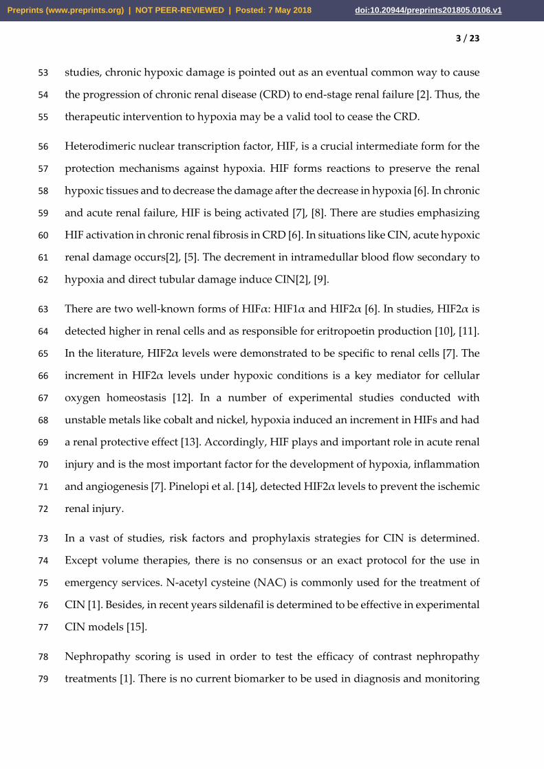
In Figure 6; QIRIAR counts were given for all groups in comparison and with box-plot 282
graphics. 283
284
285
286
287
288
Figure 6. HIF-2α immunostaining in CIN model kidney of control, CIN and treated 289
groups. Ratio of HIF-2α positive tubular with immunolocalization staining in rats 290
after modelling. According to multiple comparison test(post-hoc test: Tukey); all 291
possible dual comparisons were significant (p<0.05). 292
293
DISCUSSION 294
Owing to the increased use of iodine contrast agents worldwide, CIN is increasing 295
day-by-day. Prevalent in all age and patient groups, CIN risk is increased among 296
patients with diabetes and hypertension [1], [20]. In the current literature, the rates of 297
CIN was reported between 0.2-2.0% after contrast tomography taken in emergency 298
services [1]. Generally, along with increased creatinine levels for 3-5 days, it may be 299
65
70
75
80
85
90
95
100
QIR
IAR
CIN CIN+NAC CIN+SIL Control
Preprints (www.preprints.org) | NOT PEER-REVIEWED | Posted: 7 May 2018 doi:10.20944/preprints201805.0106.v1

16 / 23
deleterious in long-term and dialysis requirement may occur. In the current study, in 300
order to form the CIN model, the non-ionic low-osmolar contrast medium agent was 301
used as in the study of Sun et al. [17]. In all rats in the experiment model, CIN was 302
developed significantly versus the control group. The effects of HIF-2α in CIN and 303
treatment groups were demonstrated by using the modified CIN protocol of Sun et al. 304
[17]; with biochemical parameters, histopathological analyses, immunohistochemical 305
tests. 306
Of the biochemical parameters; serum BUN and Cre, urine BUN and Cre levels were 307
significantly increased in CIN group, however, solely serum Cre and urine BUN levels 308
were significantly decreased in treatment groups. Similarly with our results, Wang et 309
al. [16], detected a decrement in increased serum Cre levels in CIN group after 310
treatment with statins. 311
In the entire world, the main aim of the studies conducted to reveal the pathogenesis 312
of CIN is related to diagnosis and treatment. In recent years, all clinicians, especially 313
the ones working at emergency services, conduct studies in order to increase the 314
awareness on CIN. Along with a number of difficulties in CIN diagnosis and 315
treatment, BUN and Cre are the mostly preferred biochemical markers for diagnosis. 316
These markers are necessary for the diagnosis, however they are not sufficient to 317
demonstrate the efficacy of the treatment and the ischemic injury. 318
In the current study, both as a diagnostic agent and also to evaluate the treatment 319
efficacy, HIF-2α is studied on rats modelled with CIN. Our study is among the 320
preliminary studies to determine HIF-2α levels in rats modelled with CIN. A number 321
of studies are performed on a number of biomarkers related with CIN [1]. 322
As the underlying mechanism of contrast agents to induce CIN is complex, diagnosis 323
and treatment are also complex situations. In the clinical treatment, mainly two 324
different mechanisms are used. The first is the sufficient hydration of the patient, and 325
the second is the antioxidant treatment [1], [21]. The mostly known antioxidant 326
Preprints (www.preprints.org) | NOT PEER-REVIEWED | Posted: 7 May 2018 doi:10.20944/preprints201805.0106.v1

17 / 23
treatment is NAC, which scavenges the ROS and increases the vasodilatative effect of 327
nitric oxide [2]. Despite the exact mechanism against renal damage induced with 328
contrast agents is unknown, NAC is the widely used agent in the world in CIN due to 329
its renal protective effect and antioxidant property [2]. Especially, NAC may decrease 330
the oxidative stress formed with contrast agents efficiently [2]. In the current study, in 331
CIN formed rats HIF-2α is significantly increased in both tissue and plasma. After 332
treatment with NAC for 48 hours, a significant decrease was detected in HIF-2α levels. 333
Besides, NAC may protect kidneys with more than one mechanism; as deletion of ROS, 334
inducing glutathion (GSH) synthesis and stabilizing nitric oxide [1]. Apart from NAC; 335
many other antioxidants like sildenafil and vitamin C are used for the treatment of 336
CIN [20], [22]. Thus, we used NAC to compare with sildenafil as a positive control 337
drug. As performing comparisons, we measured both the treatment efficacy of NAC 338
and also the treatment efficacy of sildenafil. 339
Sildenafil is a vasoactive agent used for erectile dysfuncion, pulmonery artery 340
hypertension in humans, besides being used in pig model during cardiac by-pass and 341
in rat model for gentamisin-induced nephrotoxicity [22], [23]. de Almeida et al. [15], 342
regarded sildefanil as successful to prevent nephropathy in CIN-formed rats. In the 343
current study on CIN-modelled rats, sildenafil treatment efficiently protected renal 344
functions, decreased both plasma and tissue HIF2 α levels, and also SCr levels; 345
however not effected the serum BUN, urine BUN and urine Cre levels. In the 346
histopathological evaluation, we detected significant differences in rats modelled with 347
CIN. 348
Important evidences obtained from experimental studies point out to the 349
chronic hypoxic damage of tubulointerstitium as a common eventual pathway to 350
induce the progression of chronic kidney disease to end-stage renal disease [2]. Thus, 351
therapeutic intervention to prevent hypoxia may be a valid way to terminate the 352
progression of CIN. HIF, heterodimeric nuclear transcription factor, is an essential 353
intermediate for defense mechanisms against hypoxia [24]. HIF-α cumulates in the 354
Preprints (www.preprints.org) | NOT PEER-REVIEWED | Posted: 7 May 2018 doi:10.20944/preprints201805.0106.v1

18 / 23
cell, moves to the nucleus and by binding to β-subunit, undertakes functions in 355
erythropoesis, angiogenesis, cell metabolism, cell growth and apoptosis [6]. In chronic 356
and acute renal failure, HIF is activated. There are studies referring to HIF activation 357
to be responsible for renal fibrosis in chronic renal failure [6]. HIF is efficient in 358
preserving the hypoxic tissues, decreasing the hypoxia, decrementing the injury [9]. In 359
hypoxic states, there is no oxygen available for molecular hydroxylation. In states like 360
CIN; there is an acute renal damage secondary to hypoxia, a decrease in intramedullar 361
blood flow and a direct tubular damage [9], [17]. 362
In general, oxidative stress was revealed as an important factor [2]. Thus, 363
several antioxidant agents were used as being important factors for the mechanism of 364
oxidative stress [6]. Although an exact consensus does not exist, in practice, NAC 365
becomes the widely preferred agent [1]. After their injection into the body, the contrast 366
agents produce oxygen radicals through pathophysiologic effects. Contrast agent 367
primarily cause vasoconstriction that plays a directly role on production of oxygen 368
radicals, adenosil residues and calcium ions. Afterwards, glomerular basal membrane 369
and mesengial cells are damaged and oxygen radicals are formed by the increment in 370
leukocyte chemotaxis and xantine oxidase activity. Oxygen radicals are claimed as the 371
causative factor for CIN due to contrast agents and these molecules may lead to toxic 372
ischemic reaction or tissue damage related to immune system [1,22,23]. In the current 373
study, for the model group, we detected significantly increased HIF-2α levels after 374
modelling and the levels were significant in renal tissue. 375
Our study revealed the HIF activation in CIN model and CIN treatment had 376
histopathological and immunohistochemical effects. Several studies demonstrated 377
HIF-2α activation in renal ischemic models[7], [11], [25]. In the current study, in rat 378
models with CIN and in rats treated with NAC and sildenafil, HIF-2α activation is 379
measured and differences were detected. In CIN model, HIF-2α activation is 380
determined and in CIN+NAC and CIN+SIL models, this activation was decreased 381
versus CIN model. Kong et al. [26], demonstrated late phase renal tubular HIF-2α 382
Preprints (www.preprints.org) | NOT PEER-REVIEWED | Posted: 7 May 2018 doi:10.20944/preprints201805.0106.v1

19 / 23
activation to be protective on renal fibrosis and renal dysfunction, and also its use as a 383
therapeutic agent in the late phase of chronic kidney disease. 384
In the current study, HIF-2α levels measured in tissues were as follows: 49.11±15.74 385
ng/mL in control group, 71.082±13.086 ng/mL in CIN group, 44.881±9.735 ng/mL in 386
CIN+SIL group and 31.638±6.448 ng/mL in CIN+NAC group, respectively. 387
Accordingly, the increase for HIF-2α levels in CIN group versus control group was 388
significant (p<0.001). Zheng et al [27], in ischemia/reperfusion injury mice model, 389
detected increased HIF-2α levels in kidney. Again in the same study, treatment with 390
sevoflurance induced a significant decrease in HIF-2α levels. 391
In our study, the decrease in HIF-2α levels in treatment (CIN+SIL, CIN+NAC) groups 392
versus CIN group was significant (p<0.01). HIF-2α levels measured in plasma and also 393
in tissue were significant between groups. Urine HIF-α levels were non-significant in 394
treatment groups versus control group. All these measured values may be used to 395
evaluate the efficacy of treatment with HIF-2α levels in CIN treatment. Similarly, BUN 396
levels were non-significant for CIN and treatment groups. Oppositely, SCr levels were 397
significant in treatment groups versus CIN group. Urine BUN levels were significant 398
for CIN+NAC and CIN groups (p<0.05), however, non-significant for CIN+SIL group. 399
There were no significant differences for urine Cr in treatment groups versus CIN 400
group. Our current results related to BUN and SCr were in consistent with the current 401
literature[1], [18], [22]. 402
CONCLUSION 403
In the current study, HIF-2α levels were significantly increased in CIN model. 404
After CIN treatment with NAC and sildenafil, HIF-2α levels were significantly 405
decreased. NAC and sildenafil efficiently reduced the renal injury due to contrast 406
agent implementation. Increased HIF-2α levels in CIN formation and decreased HIF-407
2α levels after treatment may be beneficial in monitoring and treatment of patients 408
with CIN. The underlying mechanism for the change in HIF-2α levels states, where 409
CIN or acute renal damage is presumed, may be associated with a decrement in 410
Preprints (www.preprints.org) | NOT PEER-REVIEWED | Posted: 7 May 2018 doi:10.20944/preprints201805.0106.v1

20 / 23
regional reactive oxidative stress and renal pathological changes. Thus, these 411
conclusions may construct an experimental base for the use of HIF-2α levels in clinical 412
prevention and treatment of CIN. Despite the use of NAC and sildenafil in CIN 413
treatment, we determined NAC treatment as more significant. 414
415
References 416
[1] R. Xu, A. Tao, Y. Bai, Y. Deng, and G. Chen, “Effectiveness of N-Acetylcysteine 417
for the Prevention of Contrast-Induced Nephropathy: A Systematic Review and 418
Meta-Analysis of Randomized Controlled Trials.,” J. Am. Heart Assoc., vol. 5, no. 419
9, 2016. 420
[2] N. Wang et al., “Renal Protective Effect of Probucol in Rats with Contrast-421
Induced Nephropathy and its Underlying Mechanism.,” Med. Sci. Monit., vol. 422
21, pp. 2886–92, 2015. 423
[3] S. Turedi et al., “The High Risk of Contrast-induced Nephropathy in Patients 424
with Suspected Pulmonary Embolism Despite Three Different Prophylaxis: A 425
Randomized Controlled Trial.,” Acad. Emerg. Med., vol. 23, no. 10, pp. 1136–1145, 426
Oct. 2016. 427
[4] S. J. Traub, J. A. Kellum, A. Tang, L. Cataldo, A. Kancharla, and N. I. Shapiro, 428
“Risk Factors for Radiocontrast Nephropathy After Emergency Department 429
Contrast-enhanced Computerized Tomography,” Acad. Emerg. Med., vol. 20, no. 430
1, pp. 40–45, 2013. 431
[5] S. A. Khaleel, A. A. Alzokaky, N. A. Raslan, A. I. Alwakeel, H. G. Abd El-Aziz, 432
and A. R. Abd-Allah, Lansoprazole halts contrast induced nephropathy through 433
activation of Nrf2 pathway in rats, vol. 270. Elsevier Ireland Ltd, 2017. 434
[6] X. Gong, G. Celsi, K. Carlsson, S. Norgren, and M. Chen, “N-acetylcysteine 435
amide protects renal proximal tubular epithelial cells against iohexol-induced 436
Preprints (www.preprints.org) | NOT PEER-REVIEWED | Posted: 7 May 2018 doi:10.20944/preprints201805.0106.v1

21 / 23
apoptosis by blocking p38 MAPK and iNOS signaling,” Am. J. Nephrol., vol. 31, 437
no. 2, pp. 178–188, 2010. 438
[7] X. Yu et al., “The balance of beneficial and deleterious effects of hypoxia-439
inducible factor activation by prolyl hydroxylase inhibitor in rat remnant kidney 440
depends on the timing of administration,” Nephrol. Dial. Transplant., vol. 27, no. 441
8, pp. 3110–3119, 2012. 442
[8] E. P. Thelin et al., “Lesion size is exacerbated in hypoxic rats whereas hypoxia-443
inducible factor-1 alpha and vascular endothelial growth factor increase in 444
injured normoxic rats: A prospective cohort study of secondary hypoxia in focal 445
traumatic brain injury,” Front. Neurol., vol. 7, no. MAR, pp. 1–17, 2016. 446
[9] M. Cordaro et al., “A novel protective formulation of Palmitoylethanolamide in 447
experimental model of contrast agent induced nephropathy,” Toxicol. Lett., vol. 448
240, no. 1, pp. 10–21, 2016. 449
[10] C. Rosenberger et al., “Up-regulation of HIF in experimental acute renal failure: 450
Evidence for a protective transcriptional response to hypoxia,” Kidney Int., vol. 451
67, no. 2, pp. 531–542, 2005. 452
[11] M. S. Wiesener et al., “Widespread hypoxia-inducible expression of HIF-2α in 453
distinct cell populations of different organs,” FASEB J., vol. 17, no. 2, pp. 271–454
273, 2003. 455
[12] A. Szade, A. Grochot-Przeczek, U. Florczyk, A. Jozkowicz, and J. Dulak, 456
“Cellular and molecular mechanisms of inflammation-induced angiogenesis,” 457
IUBMB Life, vol. 67, no. 3, pp. 145–159, 2015. 458
[13] V. H. Haase, “A breath of fresh air for Diabetic Nephropathy,” Jasn, vol. 26, pp. 459
239–241, 2015. 460
[14] P. P. Kapitsinou et al., “Endothelial HIF-2 mediates protection and recovery from 461
ischemic kidney injury,” J. Clin. Invest., vol. 124, no. 6, pp. 2396–2409, 2014. 462
Preprints (www.preprints.org) | NOT PEER-REVIEWED | Posted: 7 May 2018 doi:10.20944/preprints201805.0106.v1

22 / 23
[15] L. S. de Almeida et al., “Sildenafil prevents renal dysfunction in contrast media-463
induced nephropathy in Wistar rats,” Hum. Exp. Toxicol., pp. 1–9, 2016. 464
[16] Y. Agmon, H. Peleg, Z. Greenfeld, S. Rosen, and M. Brezis, “Nitric oxide and 465
prostanoids protect the renal outer medulla from radiocontrast toxicity in the 466
rat,” J. Clin. Invest., vol. 94, no. 3, pp. 1069–1075, 1994. 467
[17] S. Sun et al., “A novel rat model of contrast-induced acute kidney injury,” Int. J. 468
Cardiol., vol. 172, no. 1, pp. 2013–2015, 2014. 469
[18] K. Özbek et al., “The protective effect of single dose tadalafil in contrast-induced 470
nephropathy: An experimental study.,” Anatol. J. Cardiol., vol. 15, no. 4, pp. 306–471
10, 2015. 472
[19] F. Aksu et al., “Antioxidant and renoprotective effects of 473
sphingosylphosphorylcholine on contrast-induced nephropathy in rats,” Ren. 474
Fail., vol. 38, no. 7, pp. 1089–1098, 2016. 475
[20] S.-I. Hong et al., “Contrast-induced nephropathy in patients with active cancer 476
undergoing contrast-enhanced computed tomography.,” Support. care cancer 477
Off. J. Multinatl. Assoc. Support. Care Cancer, vol. 24, no. 3, pp. 1011–1017, Mar. 478
2016. 479
[21] R. J. Dym, “Solitary Kidney Remains a Risk Factor for Renal Insufficiency, 480
Though Not for Contrast-induced Nephropathy Independently.,” Radiology, vol. 481
280, no. 2, pp. 650–651, Aug. 2016. 482
[22] M. A. Morsy, S. A. Ibrahim, E. F. Amin, M. Y. Kamel, R. A. Rifaai, and M. K. 483
Hassan, “Sildenafil ameliorates gentamicin-induced nephrotoxicity in rats: role 484
of iNOS and eNOS,” J. Toxicol., vol. 2014, 2014. 485
[23] N. N. Patel et al., “Phosphodiesterase-5 inhibition prevents 486
postcardiopulmonary bypass acute kidney injury in swine,” Ann. Thorac. Surg., 487
vol. 92, no. 6, pp. 2168–2176, 2011. 488
Preprints (www.preprints.org) | NOT PEER-REVIEWED | Posted: 7 May 2018 doi:10.20944/preprints201805.0106.v1

23 / 23
[24] C. Xie et al., “Activation of intestinal hypoxia-inducible factor 2α during obesity 489
contributes to hepatic steatosis,” Nat. Med., vol. 23, no. 11, pp. 1298–1308, 2017. 490
[25] U. Florczyk et al., “Opposite effects of HIF-1α and HIF-2α on the regulation of 491
IL-8 expression in endothelial cells,” Free Radic. Biol. Med., vol. 51, no. 10, pp. 492
1882–1892, 2011. 493
[26] K. H. Kong et al., “Selective tubular activation of hypoxia-inducible factor-2α has 494
dual effects on renal fibrosis,” Sci. Rep., vol. 7, no. 1, pp. 1–12, 2017. 495
[27] B. Zheng, Q. Zhan, J. Chen, H. Xu, and Z. He, “Sevoflurane pretreatment 496
enhance HIF-2alpha expression in mice after renal ischemia/reperfusion 497
injury.,” Int. J. Clin. Exp. Pathol., vol. 8, no. 10, pp. 13114–13119, 2015. 498
Preprints (www.preprints.org) | NOT PEER-REVIEWED | Posted: 7 May 2018 doi:10.20944/preprints201805.0106.v1