Embed Size (px)
DESCRIPTION
ASPMH
Citation preview
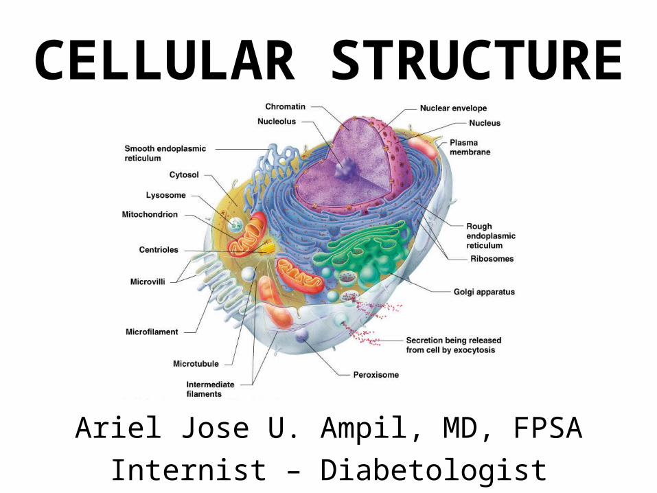
CELLULAR STRUCTURE
Ariel Jose U. Ampil, MD, FPSAInternist – Diabetologist

• Cell- smallest functional and structural unit of the human body
• Cytology- study of the individual cell and the structure it contains
• Tissue- group of cells similar in structure, function and intercellular substances
• Histology- science that deals with the study of tissues
• Microscopic anatomy- study of the human body with the aid of a magnifying object
Microscopy
• Microcopy is the aid in the study of the human body structure by the use of a magnifying instrument called the microscope.
• The microscope renders the details of the cells, tissues and organs.
Microscopy
• The useful magnification of an ordinary light microscope is about 1200X.
• The magnification of lens is the number of times the image of an object is enlarged compared with the actual size of the object.
• The total magnification of the object is solving the product of the separate magnification powers of the objective lens and ocular lens.
Microscopy
• The usefulness of any microscope is not only dependent on its ability to magnify but also on its ability to resolve details.
• The resolving power (resolution) is its capacity to give images of objects close together. It is measured as the smallest distance between two particles that can be distinguished from each other.
Compound Microscope
• A compound microscope is also called a “light microscope”.
• The principle involved is the refraction of light.• The resolving power of the best light
microscope is approximately 0.2 um.• The magnifying image is about 1,500 times or
0.25 um.
Electron Microscope
• This also known as the transmission electron microscope.
• The electron microscope functions on the principle that a beam of electrons can be deflected by electromagnetic fields in a manner similar to light deflection in glass lenses.
Electron Microscope
• Electrons are produced by high temperature heating of a metallic filament (cathode) in a vacuum.
• Its resolving power is very high and objects can be magnified over a hundred thousand times.
• It uses extremely thin preparations of tissues because of the low penetration of electrons.
Advantages of Electron Microscopes
• Very high resolution• Very high magnification• Has greatly extended the understanding of the
ultrastructure or molecular structure of cells
Disadvantages of Electron Microscopes
• Requires a vacuum enclosed system• Requires ultra thin sections of specimens• Requires a high voltage• Requires the service of highly-trained
personnel• Very complex system • Very expensive
Differences between light microscopes and electron microscopes
• Light microscopes use light while electron microscopes use electron beams emitted by tungsten filaments or electron particles
• Light microscopes use glass lens while electron microscopes use electron magnetic fields or electric fields
• Light microscopes have a low resolving power while electron microscopes have a higher resolution
• Light microscopes are open systems while electron microscopes are vacuum enclosed systems
• Light microscopes offer direct viewing of objects with the naked eye, while electron microscopes use a fluorescent screen for viewing
• Light microscopes less expensive• Light microscopes do not require high voltage while
electron microscopes do
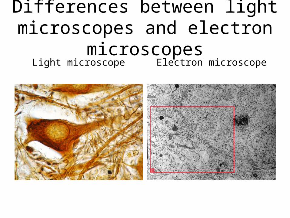
Differences between light microscopes and electron microscopes
Light microscope Electron microscope
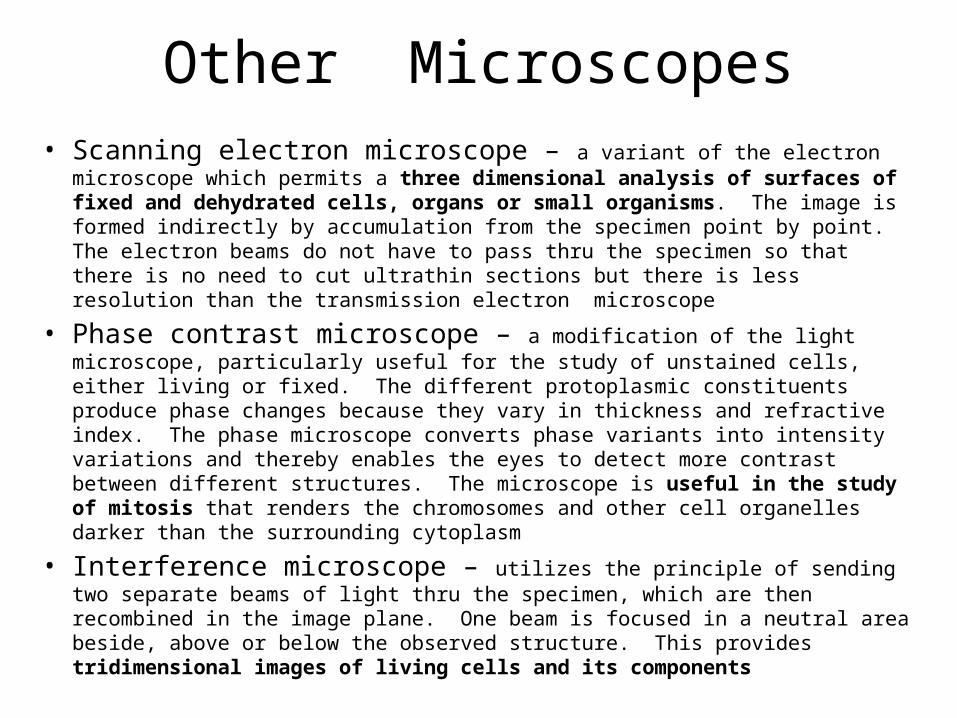
Other Microscopes• Scanning electron microscope – a variant of the electron microscope which
permits a three dimensional analysis of surfaces of fixed and dehydrated cells, organs or small organisms. The image is formed indirectly by accumulation from the specimen point by point. The electron beams do not have to pass thru the specimen so that there is no need to cut ultrathin sections but there is less resolution than the transmission electron microscope
• Phase contrast microscope – a modification of the light microscope, particularly useful for the study of unstained cells, either living or fixed. The different protoplasmic constituents produce phase changes because they vary in thickness and refractive index. The phase microscope converts phase variants into intensity variations and thereby enables the eyes to detect more contrast between different structures. The microscope is useful in the study of mitosis that renders the chromosomes and other cell organelles darker than the surrounding cytoplasm
• Interference microscope – utilizes the principle of sending two separate beams of light thru the specimen, which are then recombined in the image plane. One beam is focused in a neutral area beside, above or below the observed structure. This provides tridimensional images of living cells and its components
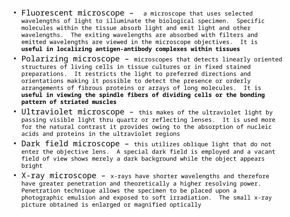
• Fluorescent microscope – a microscope that uses selected wavelengths of light to illuminate the biological specimen. Specific molecules within the tissue absorb light and emit light and other wavelengths. The exiting wavelengths are absorbed with filters and emitted wavelengths are viewed in the microscope objectives. It is useful in localizing antigen-antibody complexes within tissues
• Polarizing microscope – microscopes that detects linearly oriented structures of living cells in tissue cultures or in fixed stained preparations. It restricts the light to preferred directions and orientations making it possible to detect the presence or orderly arrangements of fibrous proteins or arrays of long molecules. It is useful in viewing the spindle fibers of dividing cells or the bonding pattern of striated muscles
• Ultraviolet microscope – this makes of the ultraviolet light by passing visible light thru quartz or reflecting lenses. It is used more for the natural contrast it provides owing to the absorption of nucleic acids and proteins in the ultraviolet regions
• Dark field microscope – this utilizes oblique light that do not enter the objective lens. A special dark field is employed and a vacant field of view shows merely a dark background while the object appears bright
• X-ray microscope – x-rays have shorter wavelengths and therefore have greater penetration and theoretically a higher resolving power. Penetration technique allows the specimen to be placed upon a photographic emulsion and exposed to soft irradiation. The small x-ray picture obtained is enlarged or magnified optically
Microtechnique
• Microtechnique is also known as Histotechnique.
• Microtechnique is a method of preparation of tissue for microscopic examination.
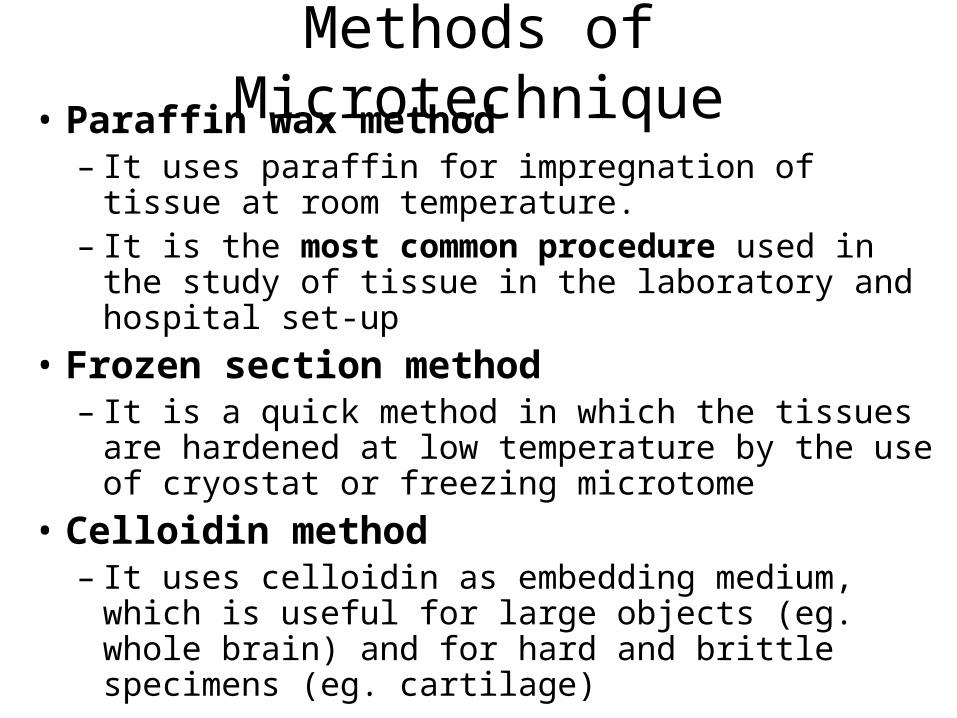
Methods of Microtechnique• Paraffin wax method
– It uses paraffin for impregnation of tissue at room temperature.
– It is the most common procedure used in the study of tissue in the laboratory and hospital set-up
• Frozen section method– It is a quick method in which the tissues are hardened
at low temperature by the use of cryostat or freezing microtome
• Celloidin method– It uses celloidin as embedding medium, which is useful
for large objects (eg. whole brain) and for hard and brittle specimens (eg. cartilage)
Steps in Paraffin Method
• Procurement of tissue specimen• Fixation – submerging the specimen in a
chemical substance in order to preserve the tissue– Purpose:
• To preserve tissue morphology• To act as disinfectant• To kill microorganisms• To harden the tissue• To permit better staining reaction
Steps in Paraffin Method
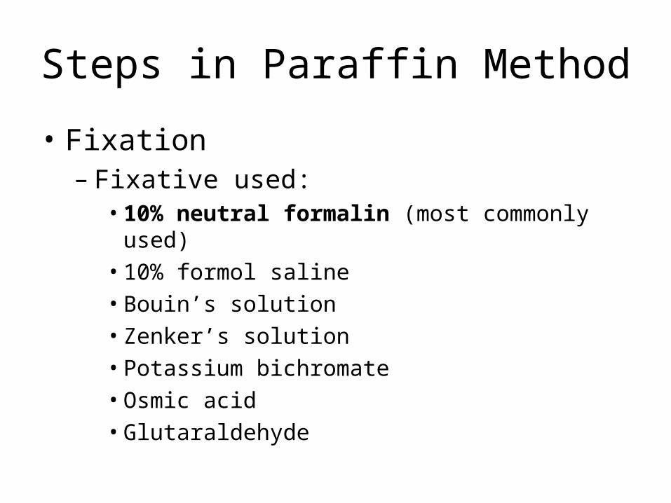
• Fixation– Fixative used:
• 10% neutral formalin (most commonly used)• 10% formol saline• Bouin’s solution• Zenker’s solution• Potassium bichromate• Osmic acid• Glutaraldehyde

Steps in Paraffin Method
• Dehydration – immersing the tissue in increasing concentration of alcohol– Purpose: to remove water from the tissue– Dehydrating agent: graded concentration of ethyl
alcohol (65 to 100% alcohol) • Clearing (dealcoholization) – replacing the
alcohol with clearing agents– Purpose: to impregnate tissue with paraffin
solvent, since alcohol is insoluble with paraffin; it must be replaced by a clearing agent so that it can be impregnated with paraffin wax
Steps in Paraffin Method
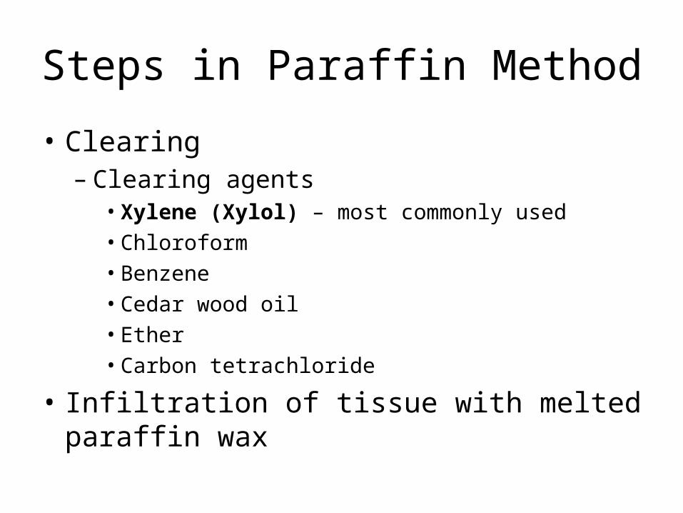
• Clearing– Clearing agents
• Xylene (Xylol) – most commonly used• Chloroform• Benzene• Cedar wood oil• Ether• Carbon tetrachloride
• Infiltration of tissue with melted paraffin wax
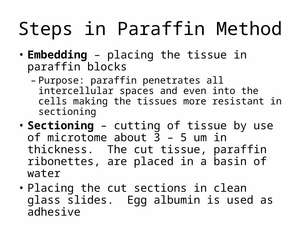
Steps in Paraffin Method• Embedding – placing the tissue in paraffin
blocks– Purpose: paraffin penetrates all intercellular
spaces and even into the cells making the tissues more resistant in sectioning
• Sectioning – cutting of tissue by use of microtome about 3 – 5 um in thickness. The cut tissue, paraffin ribonettes, are placed in a basin of water
• Placing the cut sections in clean glass slides. Egg albumin is used as adhesive
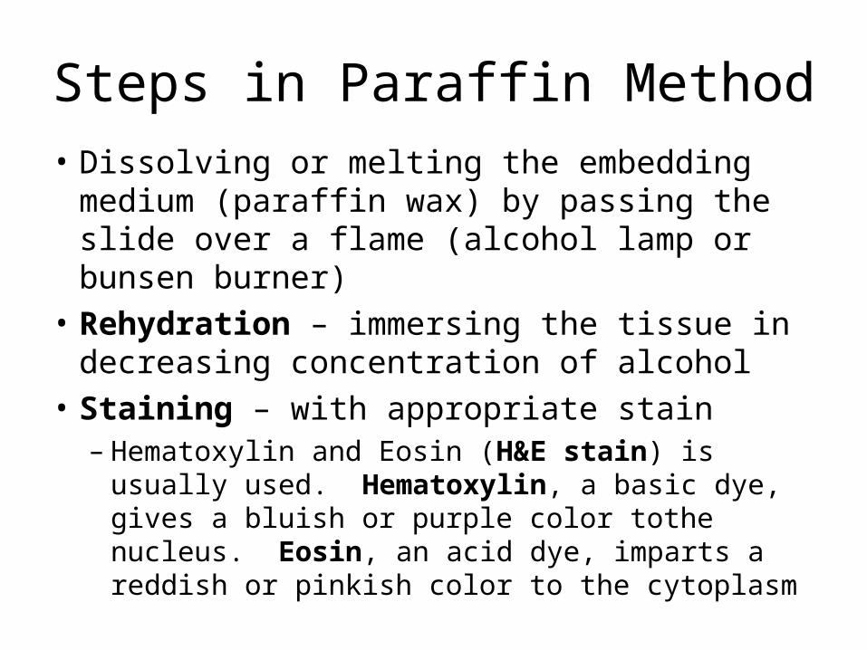
Steps in Paraffin Method• Dissolving or melting the embedding medium
(paraffin wax) by passing the slide over a flame (alcohol lamp or bunsen burner)
• Rehydration – immersing the tissue in decreasing concentration of alcohol
• Staining – with appropriate stain– Hematoxylin and Eosin (H&E stain) is usually used.
Hematoxylin, a basic dye, gives a bluish or purple color tothe nucleus. Eosin, an acid dye, imparts a reddish or pinkish color to the cytoplasm
Steps in Paraffin Method
• Dehydration – immersing again the tissue in increasing concentration of alcohol
• Clearing – with clearing agents• Mounting – placing cover slip with a few
drops of Canada balsam as a mounting medium on processed tissue
• Labelling – label the organ on the prepared slides
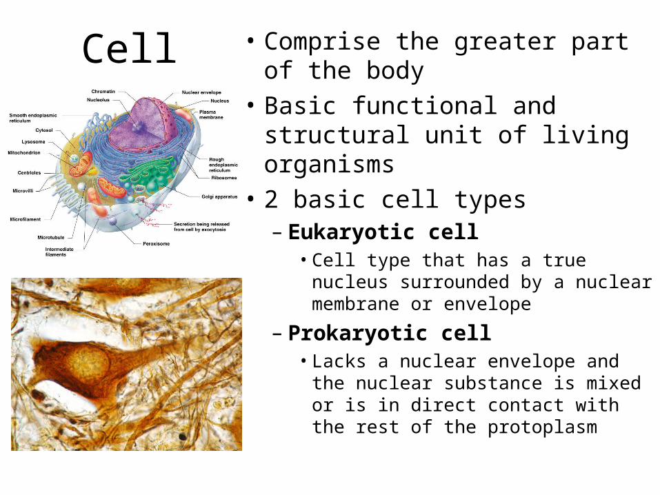
Cell • Comprise the greater part of the body
• Basic functional and structural unit of living organisms
• 2 basic cell types– Eukaryotic cell
• Cell type that has a true nucleus surrounded by a nuclear membrane or envelope
– Prokaryotic cell• Lacks a nuclear envelope and the
nuclear substance is mixed or is in direct contact with the rest of the protoplasm
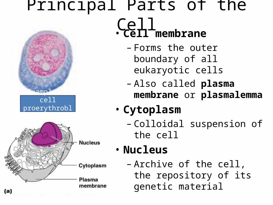
Principal Parts of the Cell• Cell membrane
– Forms the outer boundary of all eukaryotic cells
– Also called plasma membrane or plasmalemma
• Cytoplasm– Colloidal suspension of the
cell
• Nucleus– Archive of the cell, the
repository of its genetic material
Example of cell proerythroblast
Cell Membrane
• Under light microscope, the plasmalemma is invisible.
• Under electron microscope, the cell membrane appears as a trilaminar membrane made up of lipoproteins.
• The membrane is 7-11 mm in thickness and is semi-permeable.
Essential Functions of Plasmalemma• Gives shape to the cell• Regulates the passage of ions and macromolecules in and out
of the cell• It contains devices for cell attachment• Involved in cell to cell communication• Have antigenic molecules that are the basis for recognition
and tissue specificity• Involved in ion pumps for regulating the internal environment• Contains receptors for hormones• Mechanism for generating messenger molecules that activate
the cell’s physiological responses to stimuli
Cell Membrane• Within the membrane, the lipids are presumably
most stable when organized into a double layer with their hydrophobic (non-polar) chains directed toward the center of the membrane and their hydrophilic heads directed outward that lie next to the enveloping protein layer.
• The presence of a continuous hydrophobic region could explain the low permeability of many membranes to water-soluble compounds and their high permeability to lipid-soluble materials.
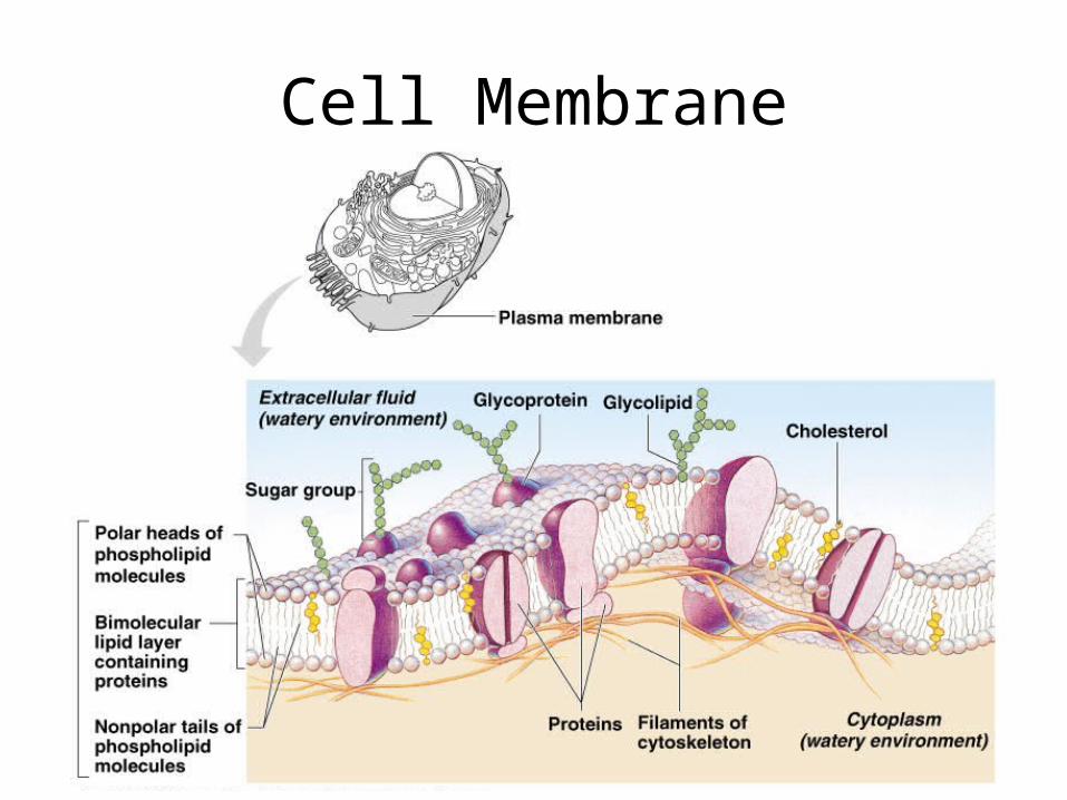
Cell Membrane
Cell Membrane
• The proteins, which constitute a major molecular component of the membrane, can be divided into 2 groups.
• Integral proteins represent a class of proteins that are directly incorporated within the lipid bilayer.
• Peripheral proteins exhibit a lesser association with membrane surfaces.
Cell Membrane
• The cell coat is a surface coat made up of carbohydrate rich or polysaccharide components of the integral glycolipids and oligosaccharides.
• This is found covering the cell membrane specialized for secretions like in the gastro-intestinal tract.
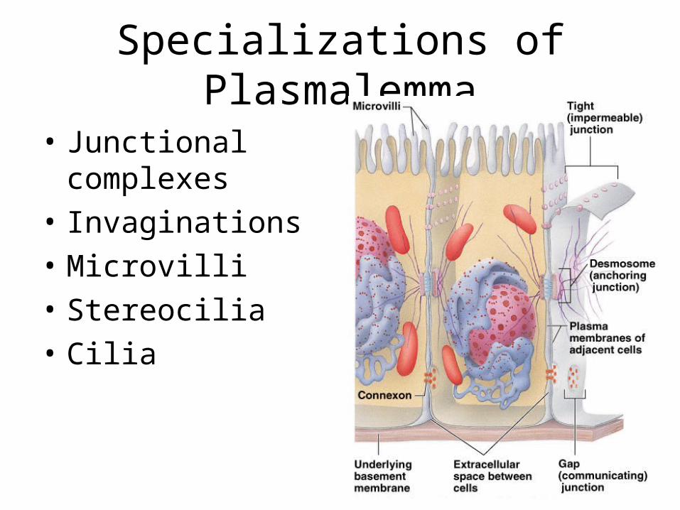
Specializations of Plasmalemma
• Junctional complexes
• Invaginations• Microvilli• Stereocilia• Cilia
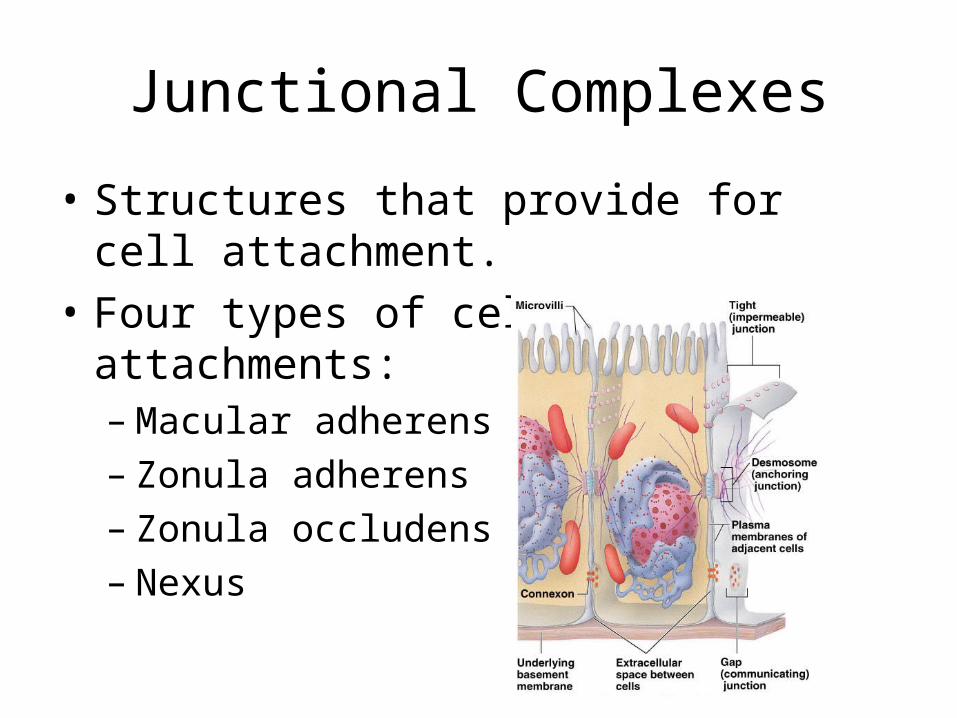
Junctional Complexes
• Structures that provide for cell attachment.• Four types of cellular attachments:
– Macular adherens– Zonula adherens– Zonula occludens– Nexus
Macula Adherens• Also known as desmosome.• Complex disk-shaped structure or cytoplasmic
face of cell membrane of 2 neighboring cells with a spot weld configuration.
• Points of firm intercellular adhesions• Found in stratified epithelia of mouth, esophagus,
vagina and skin• Serve as sites of attachments of the cytoskeleton
to the cell surface and sites of cell to cell adhesions
Zonula Adherens
• Intermediate junctions of fascia adherens• Cellular ring of attachments which increases
the surface contact of cells• The intercellular space is larger than normal
and is filled with electron dense amorphous materials
• Found in intercalated discs of cardiac muscles and is referred to as fascia adherens
Zonula Occludens
• Also known as tight junctions• Has a distinctive reticular pattern found in
epithelium of urinary bladder, GIT and inter-endothelial contacts of brain-tissue capillaries
• In this type, the cell coat is not present and the outer leaflets of the opposing cell membranes have fused and become a single leaflet
• Importance is in the formation of a barrier that prevents the free passage across the epithelium
Nexus
• Also known as gap junction• Communicating junction found in epithelial,
muscular and nervous tissues• These are regions of low electrical resistance
for cell to cell propagation or excitation-contraction, impulse formation and also are preferred crossing over points for molecules being transported from one cell to the next
• Significance is for cell to cell communication
Invaginations
• Modification of cell membranes in the form of infoldings or vesicular pits
• Infolding – This greatly increases the cell surface
• Vesicular pit– This type occurs as pinocytic or phagocytic
vesicles
Vesicular Pit• Pinocytic vesicles are drop-like invaginations of the cell
membrane. They participate in the uptake of extracellular materials called endocytosis, as well as in discharge of aggregations of intercellular materials called exocytosis. Pinocytosis literally means “cell drinking”
• Phagocytic vesicles are larger vesicles which are involved in phagocytosis which literally means “cell engulfing”
• Both pinocytic vesicles and phagocytic vesicles that break off from the cell membrane are referred to as phagosomes
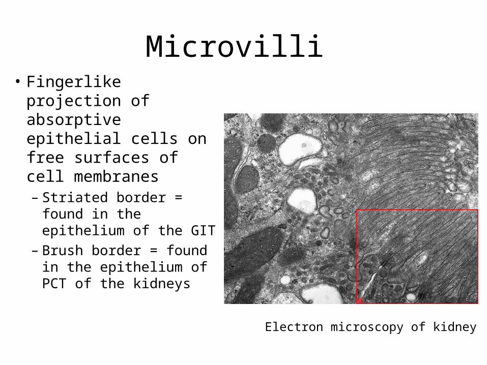
Microvilli • Fingerlike projection of
absorptive epithelial cells on free surfaces of cell membranes– Striated border = found
in the epithelium of the GIT
– Brush border = found in the epithelium of PCT of the kidneys
Electron microscopy of kidney
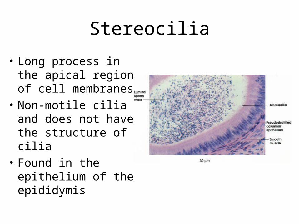
Stereocilia
• Long process in the apical region of cell membranes
• Non-motile cilia and does not have the structure of cilia
• Found in the epithelium of the epididymis
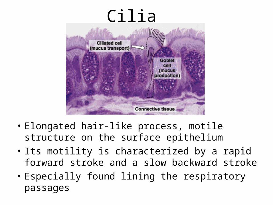
Cilia
• Elongated hair-like process, motile structure on the surface epithelium
• Its motility is characterized by a rapid forward stroke and a slow backward stroke
• Especially found lining the respiratory passages
Cytoplasm
• Colloidal suspension of the cell• Composed of a matrix, termed the “cytosol”,
in which several structures are embedded classified into three groups namely:– Organelles– Inclusions– Cytockeleton
Organelles of the Cytoplasm
• The structures known as organelles present in all eukaryotic cells have a limiting membrane and contain enzymes that participate in cellular metabolic activity
• These are permanent components of the cytoplasm
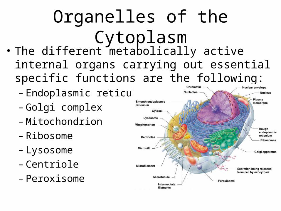
Organelles of the Cytoplasm• The different metabolically active internal organs
carrying out essential specific functions are the following:– Endoplasmic reticulum– Golgi complex– Mitochondrion– Ribosome– Lysosome– Centriole– Peroxisome
Endoplasmic Reticulum
• Extensive system of membrane bounded canaliculi
• Consists of loose network of branching and anastomosing tubules throughout the cytoplasm, the tubules may be expanded locally into broad flat saccules called cisternae
2 forms of Endoplasmic Reticulum
• Rough endoplasmic reticulum (granular endoplasmic reticulum)
• Smooth endoplasmic reticulum (agranular endoplasmic reticulum)
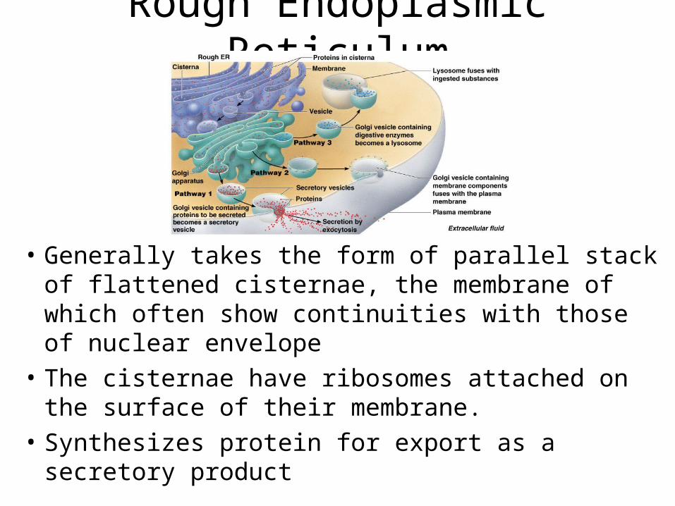
Rough Endoplasmic Reticulum
• Generally takes the form of parallel stack of flattened cisternae, the membrane of which often show continuities with those of nuclear envelope
• The cisternae have ribosomes attached on the surface of their membrane.
• Synthesizes protein for export as a secretory product
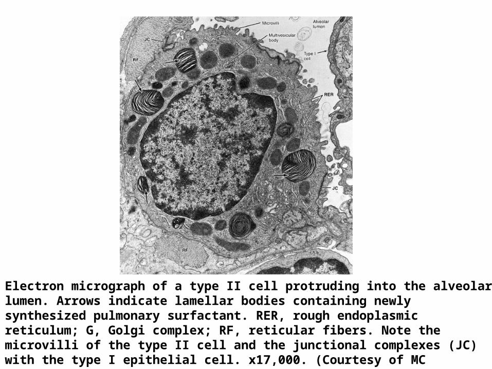
Electron micrograph of a type II cell protruding into the alveolar lumen. Arrows indicate lamellar bodies containing newly synthesized pulmonary surfactant. RER, rough endoplasmic reticulum; G, Golgi complex; RF, reticular fibers. Note the microvilli of the type II cell and the junctional complexes (JC) with the type I epithelial cell. x17,000. (Courtesy of MC Williams.)
Smooth Endoplasmic Reticulum
• Also takes the form of a membranous network within the cell.
• Its ultrastructure differs from the granular endoplasmic reticulum in 2 important ways.– Lacks the associated ribosomes– Its cisternae are more likely to appear as a
profussion of interconnected channels of variable shapes and sizes than as a stack of flattened cisternae
Smooth Endoplasmic Reticulum
• Associated with specialized functions in certain cell types.– Involved in the synthesis of triglycerides, glycogen,
cholesterol and steroid hormones– In the liver, involved in detoxification of exogenous
lipid soluble drugs– In muscle cells, involved in contraction processes,
where it is known as the sarcoplasmic reticulum, which participates in sequestration and release of calcium ions that regulate muscular contraction
Golgi Complex
• Also known as Golgi apparatus or dictysome• Involved in secretory activity of the cell either
distributed throughout the cytoplasm or confined to a zone near the nucleus depending upon the cell type
• Under electron microscope, the Golgi apparatus appears as several membrane limitted flattened saccules or cisternae, with associated vacuoles and vesicles, stacked in parallel arrays
Golgi Complex
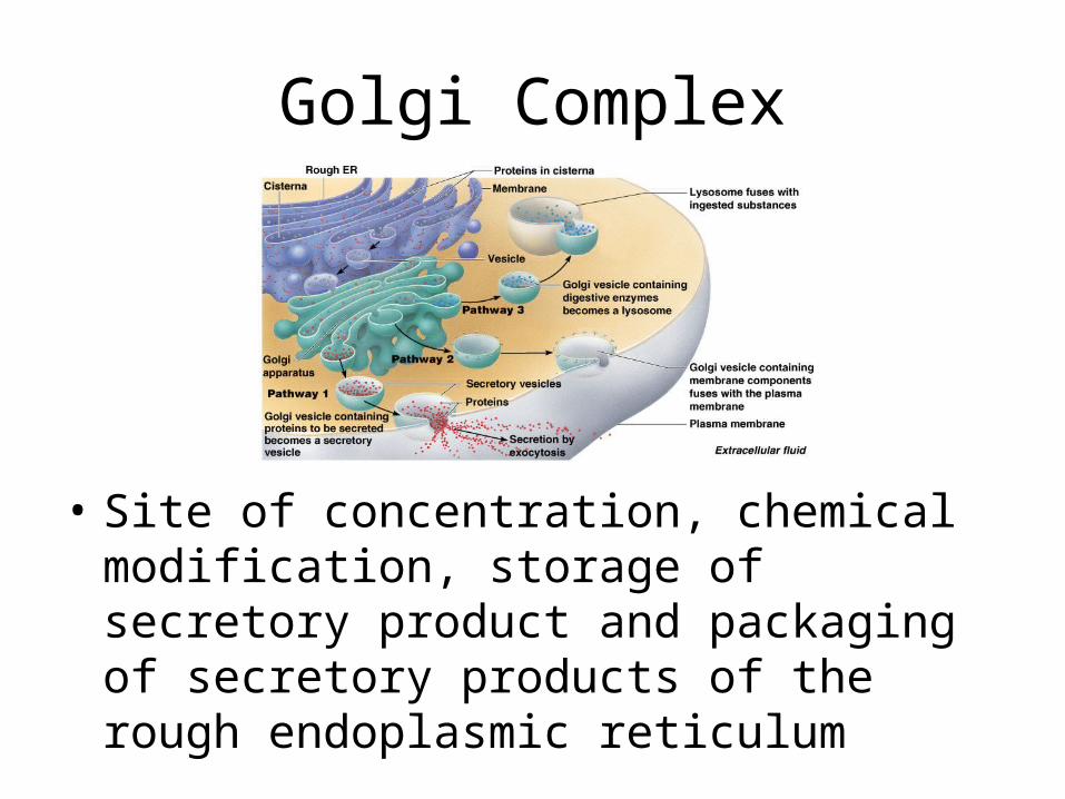
• Site of concentration, chemical modification, storage of secretory product and packaging of secretory products of the rough endoplasmic reticulum
Mitochondrion • Under light microscope, appears as rods or
filaments in both living and fixed cells. It measures about 0.5 to 1 um wide and 2 to 10 um in length.
• Under electron microscope, it has a double membrane with the inner membrane exhibiting folds called cristae. The cristae amplify the surface area of the enzyme rich membrane to increase efficiency of organelles in generating energy
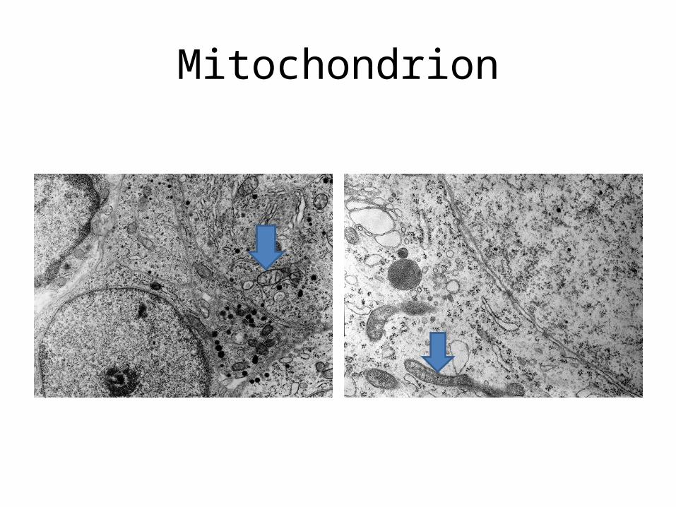
Mitochondrion
Mitochondrion
• Cytoplasmic structure that provides for biosynthesis and motor activity of cells.
• They transform with high efficiency the chemical energy of metabolites present in the cytoplasm into available energy that is easily accessible to the cell. These substances is typified by adenosine triphosphate (ATP)
• Referred to as the “powerhouse of the cell”
Ribosome
• Composed of a smaller and a larger subunit– The small subunit contains a single large molecule
of RNA and some 30 associated small proteins– The larger subunit which is adjacent to the
membrane consists of 2 molecules of RNA and about 40 associated proteins
• Under electron microscope, ribosomes appears as dense granules
Ribosome• The ribonucleoprotein of the ribosome is largely
responsible for the affinity of the cytoplasm for basic dye. Hence, by light microscope, areas rich in ribosomes are intensely basophilic.
• They may take the form of:– Free ribosomes occur singly free in the cytoplasmic matrix.
In synthetically active cells, the great majority of ribosomes occur in clusters of 10 to 20, called polyribosomes or polysomes. These ribosomes are involved in protein synthesis for intracellular use.
– Attached ribosomes involved in the synthesis of proteins destined for export from the cell as a secretory product
Lysosome • Membrane bounded vesicles that contain large variety
of hydrolytic enzymes active in acidic pH, whose main function is related to intracytoplasmic digestions
• Highly heterogenous group of bodies so diverse in size, shape and internal organization that no single description encompasses all of their variation. In general, they are dense bodies, 0.25 to 0.5 um in diameter limited by a membrane. They are ovoid or irregular in outline. Enzymes contained in lysosomes are acid hydrolases (eg. acid phosphatase)
Lysosome• Two types of lysosomes
– Primary lysosomes small vesicles containing the inactive enzyme
– Secondary lysosomes have been involved in enzymatic activities
• Another function of lysosomes concerns with turnover of the cell’s organelles. – Sequestration and digestion of the cell’s organelles called
autophagy. The primary lysosomes with ingested organelles are called autophagosmes
• In pathologic conditions, the lysosomes may rupture, release their enzymes, and ultimately destroy the cell from within, called autolysis
Centrioles
• Under light microscope, a pair of short rods that determine the polarity of the cell
• Under electron microscope, they appear as short, cylindrical structures, embedded in the walls are nine evenly spaced triplets of microtubules
• Considered as the center of activities associated with cell division
• Self-duplicating organelles and are prominent in mitosis
Peroxisomes
• Also known as microbodies• Spherical in shape and has a single limiting
membrane• Contain several enzymes, such as catalase, urase,
oxidase and d-amino acids produced by bacteria in the digestive tract and absorbed by the body, and to the beta-oxidation of fatty acids
• Abundant in the liver, kidneys, bronchioles and odontoblasts

Inclusions
• Generally, temporary components of certain cells. They usually appear as vacuoles, granules, globules and a diversity of sizes and shapes.
• They are inert accumulations of metabolites or cell products such as:– Stored food– Crystals– Pigments– Secretory granules
Cytoskeleton
• Structural framework of the cell composed of several filamentous components
• Important functions include:– Maintenance of cell shape– Stabilization of cell attachments– Plays a role in endocytosis– Movements of local specializations of the cell– Cell motility

Examples of cytoskeletons:
• Microtubules• Microfilaments• Intermediate
filaments• Microtubular lattices
Examples of cytoskeletons:
• Microtubules– Rod-like structures with variable lengths, composed of
proteinaceous heterodimeric subunits known as tubulin dimers (alpha and beta tubulins)
– Play a significant role in the development and maintenance of cell form
– Have a major role in intercellular transport of other organelles
– Provide basis for several complex cytoplasmic organelles, including centrioles, cilia and flagella

• Microfilaments– Contractile filaments of actin-myosin filaments– Responsible for visco-elastic properties and contractility of the
cytoplasm– Necessary for cell motility
• Intermediate filaments– Type of filament whose cross-section falls between
microtubules and microfilaments, hence intermediate filaments– Identified by immuno-chemical analysis as keratins, desmin,
vimentin, neurofilaments and glial filaments, found respectively in epithelium, muscles, mesenchymal cells, neurons and glial cells
Examples of cytoskeletons:
Examples of cytoskeletons:
• Microtubular lattices– 3 dimensional lattices of slender strands in cytoplasmic
matrix forming a gel, solid phase linking together the outer filamentous components and organelles into a single structural and functional unit

Nucleus
• Archive of the cell, the repository of its genetic material
• Found in all cells, except in mature erythrocytes and blood platelets
• Usually basophilic in stain because of the presence of nucleic acids
• Appears as a rounded or elongated structure, usually in the center of the nucleus, but in some cells are peripherally located
Nucleus

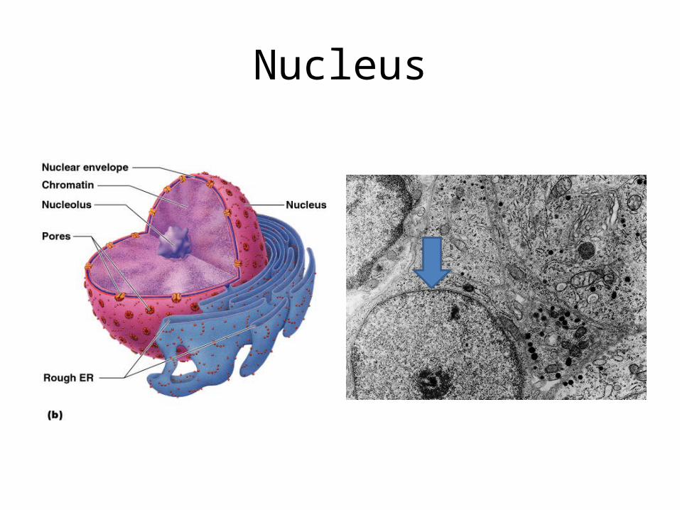
Components of the Nucleus
• Nuclear envelope– This is a bilayer, membrane of lipoprotein
separated by a narrow space called the perinuclear space or cisternae
• Nuclear pores– Openings in the nuclear envelope which are
covered with a thin diaphragm.– These provide ionic transport from the cytoplasm
to the nucleoplasm
Components of the Nucleus• Chromatin
– Composed of colloid strands of DNA bounded to basic proteins – histones
– Carries most of the genetic information– 2 types of chromatin
• Euchromatin – made up of loose network of chromatin fibrils. These are metabolically active
• Heterochromatin – made up of condensed network of chromatin, which appears as coarse granules or patches. These are metabolically inert.
– Rod-shaped or thread-like structures made up of masses of heterochromatin.
– In man, chromosomes are 46 in number (or 23 pairs)
Components of the Nucleus• Nucleolus
– Rounded, refractile body, eccentrically located in the nucleus
– Synthesizes RNA and basic proteins– Under electron microscope, it consists of 3 parts:
• Nucleolemma pars granulosa with dense filaments• Pars fibrosa amorphous part of nucleolus• Nucleolus associated chromatin dispersed filaments
of DNA, which permeates the other 2 regions
• Nucleoplasm– Amorphous matrix that fills the space between
the chromatin and the nucleoli in the nucleus
Nucleus
• During cell death, nuclear changes that may take place include:– Pyknosis
• Shrinkage of the nucleus
– Karyorhexis• Fragmentation of the nucleus
– Karyolysis• Dissolution of the nucleus

Thank you very muchfor your kind attention!


![1st Meeting Cell and Its Function [Autosaved]](https://img.dokumen.tips/doc/110x75/577c7d181a28abe0549d5b7e/1st-meeting-cell-and-its-function-autosaved.jpg)