Embed Size (px)
Citation preview

Red: important.
Black: in male|female slides.
Gray: notes|extra.
Cell structure
Editing File

Histology team 437 | Foundation block | Lecture one
➢ OBJECTIVES
• What is histology and how it is studied?
• Composition of the cell: Light microscopic (L/ M) and
electron microscopic (E/M) .
• Function of each component: Nucleus, Cytoplasm,
Organelles: membranous and non membranous & Inclusions.

Histology team 437 | Foundation block | Lecture one
➢ INTRODUCTION
• Histology is the microscopic study of normal tissues.
• Types of microscopes: LM & EM.
• Organs are made of tissues and tissues are made of cells.
• Thin sections are cut and mounted on glass slides.
Sections are stained with Haematoxylin (H) and Eosin (E).
- Nucleus is always blue (basophilic)
- Cytoplasm may be red (acidophilic)
or blue (basophilic).

Histology team 437 | Foundation block | Lecture one
• it is the structural & functional unit of all
living tissues.
• cells have different shapes & sizes.
• the cell is made of: 1-nucleus 2- cytoplasm.
• Shapes of nuclei.
➢ THE CELL ➢ NUCLEUS (L\M)

Histology team 437 | Foundation block | Lecture one
➢ THE CELL
Appearance of nuclei :
VESICULAR (OPEN FACE) NUCLEUS. DARK NUCLEUS (DEEPLY-STAINED
NUCLEUS) DEEPLY BASOPHILIC NUCLEUS

Histology team 437 | Foundation block | Lecture one
➢ THE CELL (NUCLEUS)
Formed of:
1) Nuclear Envelope:
A double membrane with many pores.- Outer membrane.
- Inner membrane.
- Nuclear pores: (function) provide communication
between nucleus and cytoplasm.
2) Chromatin
• Formed of DNA.
• 2 Forms:
– Heterochromatin: condensed inactive chromatin
(dark = electron dense areas)
– Euchromatin: extended active chromatin
(pale= electron-lucent areas)
• Functions:
– Carries genetic information.
– Directs protein synthesis.

Histology team 437 | Foundation block | Lecture one
➢ THE CELL (NUCLEUS)
3) Nucleolus :
• E/M: It is mostly dark mass (electron-dense) not surrounded by a membrane.
• Usually one.
• L/M: It is a spherical dark basophilic mass.
• Function: formation of ribosomal RNA (rRNA),which is responsible for protein synthesis
in the cytoplasm.
4) Nucleoplasm
• It is a clear fluid medium in which all the contents of
the nucleus are embedded.
• Function: Provides a medium for movement of 3 types
of RNA (ribosomal, messenger and transfer RNA) from the
nucleus to the cytoplasm.

Histology team 437 | Foundation block | Lecture one
➢ THE CELL (NUCLEUS)
*Sex chromatin (Barr Body)
• A dark stained of chromatin , usually adherent to the inner aspect of the nuclear
envelope of female somatic cells. e.g buccal epithelial cells.
• A drumstick mass protruding from the nucleus of neutrophils.
• Represents one of two X chromosomes witch is inactive (condensed) in the normal
female.
• Seen in the normal female cells.
• Absent in the females with turner’s syndrome XO.
• Seen in males with Klinefelter's syndrome XXY.
➢ THE CELL (NUCLEUS)
Function of nucleus:
• It is essential for the vitality and division of the cell.
• It is the site of storage of genetic information.
• It is the site of formation of the three types of RNA.

Histology team 437 | Foundation block | Lecture one
➢ CYTOPLASMIC ORGANELLES
B) NON-MEMBRANOUSA) MEMBRANOUS
1. Ribosomes.
2. Centrioles.
3. Cilia & Flagella.
4. Filaments:
(Actin, Intermediate filaments & Myosin).
5. Cytoskeleton:
(actin, intermediate filaments &
microtubules).
1. Cell membrane.
2. Mitochondria.
3. Golgi apparatus.
4. Endoplasmic reticulum:
(rough & smooth).
5. Lysosomes.
6. Secretory vesicles.
➢ CYTOPLASM
• is formed of:
1-ORGANELLES: They are specialized structures, ESSENTIAL for vital processes of the cell.
2-INCLUSIONS: They are not essential for vitality of cells, may be present or absent.
• Examples are lipids, glycogen and pigments like melanin & lipofuscin.

Histology team 437 | Foundation block | Lecture one
➢ CELL MEMBRANE
• A very thin membrane that surrounds the cell.
• LM: Not visible.
• EM: appears as 2 dark lines (electron dense), separated by a light one
(electron-lucent) (trilaminar appearance).
• Function: selective barrier.
• Chemical Structure:
1- Phospholipid molecules: arranged in 2 layers.
2- Protein molecules: a) Peripheral protein | b) Integral protein
3- Carbohydrate molecules: attached to either proteins or lipids (glycoproteins and
glycolipids), forming the surface or cell coat (Glycocalyx).
• Function of (Glycocalyx):
- Protection of the cell.
- Cell recognition and adhesion.

Histology team 437 | Foundation block | Lecture one
• CILIA
– Long motile hair-like structures surrounded by cell membrane.
– Their core is formed of microtubules.
• MICROVILLI (BRUSH BORDER)
– Cylindrical cytoplasmic projections of apical surface to increase surface area.
– Their core contains actin filaments.
• INTRACELLULAR JUNCTIONS
Occluding (Tight) Junction: seals the intercellular space.
Adherening Junction: fixes adjacent cells together:
Zonula Adhering Junction.
Desmosome (Macula Adherening Junction).
Gap junction: Allow free communication between the cells.
*When a combination of 1 , 2a and 2b is present, this is called a junctional complex.
➢ CELL MEMBRANE (SPECIALIZATIONS)
2a
2b
3
1
2b
3
2a
1
2

Histology team 437 | Foundation block | Lecture one
➢ MITOCHONDRIA
• Each mitochondrion is rod-shaped .
• The wall is composed of 2 membranes.
• The outer is smooth, the inner is folded to form cristae.
• The cavity is filled with mitochondrial matrix, which
contains enzymes. Also contains its own DNA.
• Functions:
1- Generation of ATP which is the source of energy for the cell ,
They are called the power-house of the cell.
2- They can form their own proteins and undergo self
replication.
➢ GOLGI APPARATUS
• The secretory apparatus of the cell.
• Consists of stacked saucer-shaped flattened vesicles.
• Each vesicle has two faces:
1- Convex (forming) face: receives transfer vesicles.
2- Concave (mature) face: forms secretory vesicles.
• Functions:
1- Sorting, modification & packaging of proteins.
2- Secretory vesicles formation.
3- Formation of lysosomes.
Histology team 437 | Foundation block | Lecture one
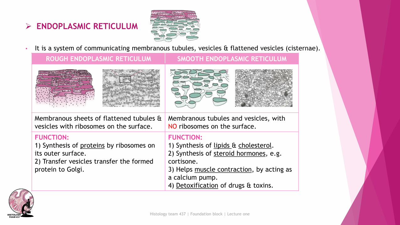
➢ ENDOPLASMIC RETICULUM
• It is a system of communicating membranous tubules, vesicles & flattened vesicles (cisternae).
SMOOTH ENDOPLASMIC RETICULUMROUGH ENDOPLASMIC RETICULUM
Membranous tubules and vesicles, with
NO ribosomes on the surface.
Membranous sheets of flattened tubules &
vesicles with ribosomes on the surface.
FUNCTION:
1) Synthesis of lipids & cholesterol.
2) Synthesis of steroid hormones, e.g.
cortisone.
3) Helps muscle contraction, by acting as
a calcium pump.
4) Detoxification of drugs & toxins.
FUNCTION:
1) Synthesis of proteins by ribosomes on
its outer surface.
2) Transfer vesicles transfer the formed
protein to Golgi.

Histology team 437 | Foundation block | Lecture one
➢ LYSOSOMES
• The digestive apparatus of the cell.
• E/M: Spherical membranous vesicles.
• Contain hydrolytic enzymes.
• Originate from mature surface of the Golgi apparatus, while their
hydrolytic enzymes are formed in the rough endoplasmic reticulum.
• Functions:
intracellular digestion of ingested material or old organelles.
➢ RIBOSOMES
• Formed in the nucleolus.
• LM: Basophilic cytoplasm is due to numerous ribosomes.• Consist of ribosomal RNA (rRNA), combined with proteins.
• EM: Formed of 2 subunits.
• Free in the cytoplasm (may form polyribosomes) or attached to rER.
• Function:
Protein synthesis.

Histology team 437 | Foundation block | Lecture one
➢ MICROTUBULES-CONTAINING ORGANELLES
FLAGELLACILIA CENTRIOLES
- Longer and larger than
cilia.
- Form the tails of sperms.
- Hair-like striations on the
free surface of some cells.
- Basal body is similar to
centriole.
- Shaft is formed of 9
doublets and 2 central
singlets of microtubules,
i.e. 20 microtubules.
- 2 cylinders, perpendicular
to each other.
- Wall is made of 9 triplets
of microtubules, i.e. 27
microtubules.
Function:
Important for movement of
the sperms.
Function:
Movement of particles or
fluids on the free surface of
the cell in one direction.
Functions:
1- Essential for cell
division.
2- Formation of cilia
and flagella.
Histology team 437 | Foundation block | Lecture one
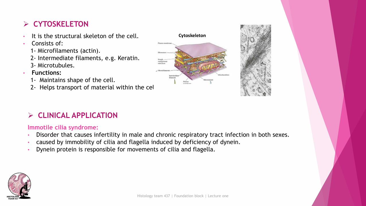
➢ CYTOSKELETON
• It is the structural skeleton of the cell.
• Consists of:
1- Microfilaments (actin).
2- Intermediate filaments, e.g. Keratin.
3- Microtubules.
• Functions:
1- Maintains shape of the cell.
2- Helps transport of material within the cell.
➢ CLINICAL APPLICATION
Immotile cilia syndrome:
• Disorder that causes infertility in male and chronic respiratory tract infection in both sexes.
• caused by immobility of cilia and flagella induced by deficiency of dynein.
• Dynein protein is responsible for movements of cilia and flagella.
Histology team 437 | Foundation block | Lecture one
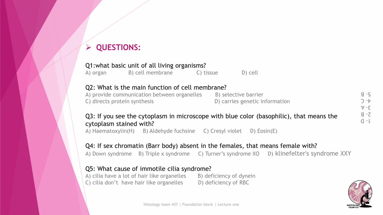
➢ QUESTIONS:
Q1:what basic unit of all living organisms?A) organ B) cell membrane C) tissue D) cell
Q2: What is the main function of cell membrane?A) provide communication between organelles B) selective barrier
C) directs protein synthesis D) carries genetic information
Q3: If you see the cytoplasm in microscope with blue color (basophilic), that means the
cytoplasm stained with?A) Haematoxylin(H) B) Aldehyde fuchsine C) Cresyl violet D) Eosin(E)
Q4: If sex chromatin (Barr body) absent in the females, that means female with?
A) Down syndrome B) Triple x syndrome C) Turner’s syndrome XO D) klinefelter's syndrome XXY
Q5: What cause of immotile cilia syndrome?A) cilia have a lot of hair like organelles B) deficiency of dynein
C) cilia don’t have hair like organelles D) deficiency of RBC
1-D
2-B
3-A
4-C
5-B

Histology team 437 | Foundation block | Lecture one
Q6: Which one of these cytoplasmic organelles is non-membranous?A) ribosome B) lysosome C) mitochondria D) Golgi apparatus
Q7: Which one of these organelles form their own proteins?A) ribosome B) lysosome C) mitochondria D) Golgi apparatus
Q8: Detoxification of drugs & toxins is function of ?A) smooth endoplasmic reticulum B) rough endoplasmic reticulum C) ribosome D) lysosome
Q9: What special enzyme the lysosomes contain?A) Amylase enzyme B) Hyperlytic enzyme C) Pepsin enzyme D) Hydrolytic enzyme
Q10: Wall is made centrioles is made of ?A) 3 central singlets of microtubules B) 9 doublets and 2 central singlets of microtubules
C) 9 triplets of microtubules D) 3 doublets microtubules
Q11: Which organelle is responsible for protein synthesis?A) lysosome B) ribosome C) mitochondria D) Golgi apparatus
6-A
7-C
8-A
9-D
10-C
11-B

Tareq Allhaidan
Marwah Alkhalil
Rinad Alghoraiby
Hussain Alkharboush
Ebtesam Almutairi
Shahad Alzahrani
Team members :
Team leaders : Khalid Fayez Alshehri
Rawan Mohammad Alharbi
Twitter.com/Histology437
Histology team 437 | Foundation block | Lecture one
جنازاتنا" لنكن يداً بيد لريى العامل ا
"وحتّملوا شقاء اليوم لأجل حمل الغد