Embed Size (px)
Citation preview

Supporting Information
Protein Stapling via Azide-Alkyne Ligation
Diya M. Abdeljabbara, Frank J. Piscottaa, Siyan Zhanga, and A. James Linka,b*
Departments of aChemical and Biological Engineering, and bMolecular Biology, Princeton University, Princeton, NJ 08544
*to whom correspondence should be addressed: [email protected]
Materials & Methods
Chemicals and Reagents. The Met analog, azidohomoalanine (AHA), was synthesized as described previously.1, 2 The Phe analog, para-ethynyl-L-phenylalanine hydrochloride (PEP) was synthesized using (S)-4-iodophenylalanine as a starting material (Chem-Impex International Inc.). The procedure has been described previously.3 The triazole ligand tris(3-hydroxypropyltriazolylmethyl)amine (THPTA) was a gift from the Finn Lab (Georgia Tech) and its synthesis has been described previously.4 All natural amino acids were purchased from the USB Corporation. Picomaxx DNA polymerase was purchased from Agilent. The 18 aa M9 media was prepared on a 500 ml scale by mixing sterile water, 10X M9 salt solution, 1M MgSO4, 0.5% thiamine, 5% glucose, and 25x 18 aa solution. Anti-His6-HRP (Sigma) was diluted 4000-fold prior to use in Western blots.
Strains and Plasmids. The E. coli AMF-IQ strain (Met and Phe double auxotroph) was constructed by knocking out the metE gene of the E. coli AF-IQ strain (Phe auxotroph) via P1 transduction, described in detail below. The genes encoding A15 and protein G6 were cloned into pQE80 with N-terminal hexahistidine tags. The gene encoding the mutant phenylalanyl-tRNA synthetase gene7 (pheS*, required for the incorporation of PEP) was cloned into each of the plasmids’ NheI restriction site, including its natural promoter. Met and Phe codons were cloned into the pQE80-his6-A1-pheS* (pDA67 for central staple, pDA77 for C-term staple) and pQE80-his6-protein G-pheS* (pDA75) plasmids. All cloning steps were carried out in XL-1 Blue.
Constructing the AMF-IQ E. coli strain. The E. coli K-12 BW25113 ΔmetE strain was obtained from the Keio Collection,8 streaked on kanamycin LB plates, and grown overnight at 37 °C until large enough colonies formed. A single colony of the donor strain (BW25113 ΔmetE) was used to inoculate 5 ml of LB medium for overnight growth at 37 °C with shaking. The next day, 50 µl of the overnight culture was used to inoculate 5 ml of LB medium containing 0.2% glucose and 5 mM CaCl2. The culture was incubated for 30 min at 37 °C with aeration. 50 µl of a P1vir lysate (~5×10⁸ phage/ml) was added to the 5 ml culture before shaking at 37 °C for 2-3 hrs until the cells lyse.
Electronic Supplementary Material (ESI) for ChemComm.This journal is © The Royal Society of Chemistry 2014

After cell lysis, 100 µl of chloroform was added and agitated by vortexing the sample. The sample was then centrifuged at 4500 g for 10 min to pellet the debris. The supernatant was carefully transferred to another sterile tube and 100 µl of chloroform was added with additional vortexing. The new P1vir lysate was stored at 4 °C until needed. A single colony of the recipient strain (AF-IQ) was used to inoculate 5 ml of LB medium to be grown overnight at 37 °C. The next day, the overnight culture was centrifuged at 1500 g for 10 min and resuspended in 2 ml of 10 mM MgSO4 containing 5 mM CaCl2. Three separate test samples were set up: (1) 100 µl of the cell suspension without P1 lysate (control), (2) 100 µl of the cell suspension plus 50 µl of P1 lysate, and (3) 100 µl of P1 lysate without cells (control). The three test samples were incubated for 30 min at 30 °C without shaking. After 30 min, 1 ml of LB (containing 10 mM sodium citrate) was added to each sample for 30 min at 30 °C without shaking. After 30 min, the samples were centrifuged and decanted. Finally, 100 µl of each test sample was spread and grown on kanamycin plates to select for AMF-IQ colonies containing the kanamycin resistance (kanR) gene. The kanR gene in the individual transductants was eliminated using the FLP recombinase expression plasmid, pCP-209 to yield the final AMF-IQ Met and Phe auxotrophic strain.
Metabolic incorporation of AHA and PEP into E. coli AMF-IQ. Individual colonies of AMF-IQ[pDA67], AMF-IQ[pDA77] or AMF-IQ[pDA75] were grown overnight in M9 minimal medium supplemented with 20 aa and ampicillin. The overnight culture was diluted 100-fold in 50 mL of M9+20 aa and ampicillin and incubated at 37 °C. Once the cultures reached OD600 values of ~ 1.0, the cells were centrifuged at 5000 g for 7 min at 4 °C and the pellet was resuspended in M9+18 aa (excluding Met and Phe). After another centrifugation and resuspension step the cells were allowed to shake at 37 °C for 20 min. The cells were once again pelleted and resuspended in M9+18 aa, ampicillin, AHA, PEP, and IPTG to induce expression of the A1 or protein G. Cells were grown for 3 hrs at 37 °C. After 3 hrs of induction, the cells were pelleted.
Cell lysis, protein purification, and desalting. Following protein expression, 50 mL of culture was pelleted and resuspended in 8 M urea. His-tagged protein was purified under denaturing conditions using Ni-NTA resin in a gravity column (Qiagen) as directed by the manufacturer. Flow through, washes, and purified elutions were analyzed by SDS-PAGE analysis. Purified protein elutions in 8 M urea were diluted 10-fold in water and the final protein yields were determined via the bicinchoninic acid (BCA) assay (Pierce) according to the manufacturer’s protocol. Purified A1 elutions were collected, placed in 6000 Da cut-off dialysis tubing (Spectrum Labs, Inc.), and dialyzed against ultrapure water for 24 hrs (water was replenished 5-6 times). After dialysis, the purified protein was placed in a 15 ml conical tube and frozen on dry ice for 20-30 min. The frozen A1 samples were freeze-dried and resuspended in 10 mM Na2HPO4 buffer (pH 7.0). Purified protein G elutions were pooled and buffer exchanged into 10 mM Na2HPO4 buffer (pH 7.0) via an Econo-Pac 10DG desalting column (Bio-Rad).
Cu(I)-catalyzed azide-alkyne cycloaddition (CuAAC). A1 and protein G were clicked using tris-(hydroxypropyltriazolyl) methylamine (THPTA).4 Briefly, 2 µl of CuSO4 (25 mM) was added to 5 µl of THPTA in water (50 mM) and the mixture was added to 1 ml

of the dissolved recombinant protein. Subsequently, 4 µl of sodium ascorbate (500 mM) was also added to the protein solution. The mixture was incubated at 4 °C for 6-8 h in order to click the azide and alkyne UAA side chains. Purified proteins were passed through a Vivaspin 6 protein concentrator (3000 Da cut-off, GE Healthcare) at 8000 g (20 min, 4 °C) to eliminate the click chemistry reagents prior to circular dichroism (CD) analysis or biolayer interferometry.
Mass spectrometry. In preparation for MALDI, purified A1 protein was diluted 10-fold in ammonium bicarbonate (100 µl total volume) before being incubated overnight at 37 °C with 2 µl of trypsin (1 mg/ml, Promega). Purified protein G in phosphate buffer was denatured by adding 8 M guanidine hydrochloride (GuHCl) to achieve a final GuHCl concentration of 6 M. The solution was heated for 10 min at 95 °C before being diluted to a final GuHCl concentration of 2 M by addition of 1 x chymotrypsin reaction buffer (100 mM Tris-HCl, 10mM CaCl2, pH 7.8). Chymotrypsin (2 µL, 1 mg/mL, Sigma-Aldrich) was added, and the solution was incubated overnight at 30 °C. The next day the protease-digested peptide fragments were purified using C-18 spin columns (Pierce). Briefly, the C-18 spin columns were washed by adding 200 µl of activation buffer (50% acetonitrile in water) and centrifuging for 1 min at 1500 g. Then the columns were washed with equilibration buffer (5% acetonitrile, 0.5% trifluoroacetic acid) in a similar fashion. The protease-digested peptides were then added to the columns and centrifuged as described above. The peptides were recovered and passed through the column a second time to ensure proper binding to the C-18 resin. The columns were subsequently washed three times with 5% acetonitrile solution to remove any impurities. Finally, the samples were eluted in 20 µl of 70% acetonitrile solution. One microliter of alpha-cyano-4-hydroxycinnamic acid matrix was mixed with an equal volume of purified peptide fragments before spotting the sample on the MALDI plate. Spotted samples were analyzed for UAA incorporation and proper azide-alkyne cycloaddition products using a MALDI TOF/TOF Analyzer (Applied Biosystems). MALDI analysis was carried out at the CABM (Rutgers).
Circular Dichroism (CD) Analysis. After UAA incorporation, purification, and CuAAC, the A1 protein secondary structure was analyzed using a CD instrument (Applied Photophysics). Clicked and unclicked samples were scanned from 195 – 260 nm while ramping the temperature from 10 to 90 °C, at 5 °C increments.
Biolayer Interferometry Measurement. After UAA incorporation, purification, and CuAAC, rate and affinity constants (ka, kd, Kd) for binding interactions were measured between protein G and IgG using a BLItz instrument (ForteBio). Protein A biosensors were coated with IgG (400 nM) and used for binding studies of both stapled and unstapled protein G at different concentrations (600 nM and 800 nM). Data were fit and analyzed by the instrument software.

References
1. A. J. Link, M. K. S. Vink and D. A. Tirrell, J. Am. Chem. Soc., 2004, 126, 10598-10602.
2. A. J. Link, M. K. S. Vink and D. A. Tirrell, Nature Protocols, 2007, 2, 1879-1883.3. B. Kayser, J. Altman and W. Beck, Tetrahedron, 1997, 53, 2475-2484.4. V. Hong, S. I. Presolski, C. Ma and M. G. Finn, Angew. Chem.-Int. Edit., 2009,
48, 9879-9883.5. W. A. Petka, J. L. Harden, K. P. McGrath, D. Wirtz and D. A. Tirrell, Science,
1998, 281, 389-392.6. L. Y. Lian, J. P. Derrick, M. J. Sutcliffe, J. C. Yang and G. C. K. Roberts, J. Mol.
Biol., 1992, 228, 1219-1234.7. K. Kirshenbaum, I. S. Carrico and D. A. Tirrell, Chembiochem, 2002, 3, 235-237.8. T. Baba, T. Ara, M. Hasegawa, Y. Takai, Y. Okumura, M. Baba, K. A. Datsenko,
M. Tomita, B. L. Wanner and H. Mori, Molecular Systems Biology, 2006, 2.9. K. A. Datsenko and B. L. Wanner, Proc. Natl. Acad. Sci. U. S. A., 2000, 97,
6640-6645.

Figure S1. Sequences of wild-type, center staple, and C-terminal staple A1 constructs. The helical region is indicated and Met and Phe residues for AHA and PEP incorporation are underlined.

Figure S2. Comparison of A1 expression in M9 minimal media supplemented with 18 aa (lacking Met and Phe), 18 aa with Met and Phe, and 18 aa with AHA and PEP. Protein production was confirmed via anti-6xHis Western blot.

Figure S3. Mass spectrometric analysis of tryptic digest of leucine zipper A1. Top: possible fragments generated by the digest based on AHA and PEP incorporation. Bottom: mass spectrum indicates a high incorporation level of both AHA and PEP into A1.

200 210 220 230 240 250
-30
-25
-20
-15
-10
-5
0
5
1010 °C
20 °C
30 °C
40 °C
50 °C
60 °C
70 °C
80 °C
90 °C
A1, center staple, not clicked
wavelength [nm]
CD
[mde
g]
200 210 220 230 240 250
-30
-25
-20
-15
-10
-5
0
5
1010 °C
20 °C
30 °C
40 °C
50 °C
60 °C
70 °C
80 °C
90 °C
A1, center staple, clicked
wavelength [nm]
CD
[mde
g]
Figure S4: Continued on next page

200 210 220 230 240 250
-140
-105
-70
-35
0
35
70 10 °C
20 °C
30 °C
40 °C
50 °C
60 °C
70 °C
80 °C
90 °C
A1, C-terminal staple, not clicked
wavelength [nm]
CD
[mde
g]
200 210 220 230 240 250
-140
-105
-70
-35
0
35
70 10 °C
20 °C
30 °C
40 °C
50 °C
60 °C
70 °C
80 °C
90 °C
A1, C-terminal staple, clicked
wavelength [nm]
CD
[mde
g]
Figure S4: CD spectra of center staple and C-terminal staple A1 protein, clicked and unclicked, at a range of temperatures.
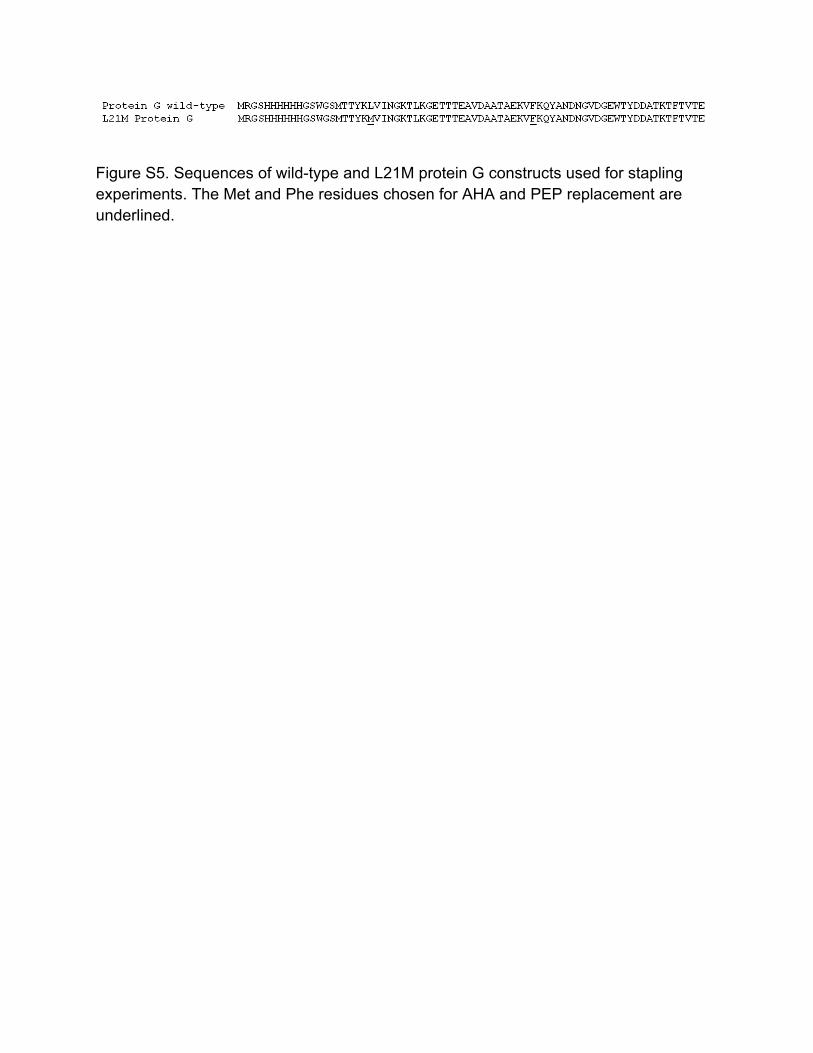
Figure S5. Sequences of wild-type and L21M protein G constructs used for stapling experiments. The Met and Phe residues chosen for AHA and PEP replacement are underlined.

Sensorgrams (raw data)
270
300
330
360
390
420
450
480
510
540
570
600
0
0.1
0.2
0.3
0.4
0.5
0.6
600 nM - click800 nM - click1000 nM - click600 nM - no click800 nM - no click1000 nM - no click
Time (s)
Bin
ding
(nm
)
Computed fits to sensorgrams
270
300
330
360
390
420
450
480
510
540
570
600
00.05
0.10.15
0.20.25
0.30.35
0.40.45
0.50.55
600 nM - click800 nM - click1000 nM - click600 nM - no click800 nM - no click1000 nM - no click
Time (s)
Bin
ding
(nm
)
Figure S6: Sensorgrams from biolayer interferometry measurments of protein G binding to IgG. Top: raw data, Bottom: fits to the raw data by BLItz software.