Embed Size (px)
Citation preview

Yalla, Krishna Chaitanya (2014) Characterisation of DISC1 ubiquitination and its potential as a therapeutic intervention for psychiatric disorders. PhD thesis.
http://theses.gla.ac.uk/5408/
Copyright and moral rights for this work are retained by the author
A copy can be downloaded for personal non-commercial research or study, without prior
permission or charge
This work cannot be reproduced or quoted extensively from without first obtaining
permission in writing from the author
The content must not be changed in any way or sold commercially in any format or
medium without the formal permission of the author
When referring to this work, full bibliographic details including the author, title,
awarding institution and date of the thesis must be given
Enlighten:Theses
http://theses.gla.ac.uk/
Characterisation of DISC1 Ubiquitination and its potential as a therapeutic
intervention for Psychiatric disorders
by
Krishna Chaitanya Yalla B.Sc., M.Sc., M.Res.
A thesis submitted in fulfilment of the requirements for the degree of Doctor of Philosophy
Institute of Cardiovascular and Medical Sciences College of Medical, Veterinary and Life Sciences
University of Glasgow February 2014

This thesis is dedicated to my loving parents
iii
Abstract
Since its discovery over a decade ago, DISC1 has become one of the most promising
candidate genes for Schizophrenia and associated chronic mental disorders. This notion
has been supported by a wealth of evidence from genetic and biochemical studies. With
multiple interacting partners, DISC1 acts as a scaffold protein, orchestrating vital
signalling pathways that underpin neurodevelopment and signalling. While the aetiology
of Schizophrenia is poorly understood, loss of DISC1 protein function remains one of the
proposed disease mechanisms. Furthermore, its tendency to form aggregates is
reminiscent of neurodegenerative illnesses such as Alzheimer’s and Parkinson’s disease.
C-terminal truncation of DISC1 (TrDISC1) is known to decrease neurite outgrowth and
number in the PC12 cell line, abolish protein interaction with proteins such as Ndel1 and
also disrupt vital physiological process such as mitochondrial transport. However, very
little is known about the underlying disease mechanism at the molecular level.
In order to gain insight in to the role of DISC1 pathway in Schizophrenia and associated
mental illnesses, I studied novel post translational modifications of DISC1. The main
conclusion of my thesis is that these modifications affect DISC1 turnover and its scaffold
function.
In the first part of my thesis, I provided the first direct evidence that DISC1 can be
SUMOylated. Peptide array technology was used to SUMOylate and map potential SUMO
acceptor lysines on DISC1, in vitro. As detecting SUMO conjugates in cells is challenging, I
utilised the “Ubc9 fusion directed SUMOylation” method (UFDS) to discover that K643 as
the one of the SUMO target sites. Mass spectrometric analysis was employed to
corroborate this novel post translation modification on DISC1. Preliminary evidence was
also provided that SUMO conjugation of DISC1 obliterates its interaction with DIXIN,
while having no effect on Ndel1 binding. This is an interesting finding since DISC1-DIXIN-
Nudel form a protein complex to regulate neuronal migration during cortical
development. While SUMOylation has been implicated in pathological protein
aggregation, a hallmark of many psychiatric illnesses, this work provides an experimental
framework to understand DISC1 aggregation which disrupts intracellular mitochondrial
transport.
iv
In the second part of my thesis, using biochemical and mass spectroscopy analysis, I
demonstrated that DISC1 protein levels are regulated by the ubiquitin proteasome
system (UPS). Using an siRNA library screen, Fbxw7 was identified as a key Fbox protein
which functions as a key component of an SCF E3 ligase complex that catalyses ubiquitin
transfer to the DISC1 protein. Peptide array studies identified a phosphodegron motif on
DISC1 which interacts with SCFFbxw7 following a dual phosphorylation event by a yet
unidentified kinase. Based on this experimental evidence, a hypothesis was developed
that this novel protein – protein interaction may have a great potential for therapeutic
intervention to address the loss DISC1 of function, a proposed disease mechanism for SCZ
and associated chronic mental disorders. According to my hypothesis, inhibiting SCFFbxw7
mediated ubiquitination of DISC1 stabilises DISC1 which may compensate for the low
cellular levels or dysfunctional DISC1 protein.
The proposed hypothesis was addressed in the third section. Using structure activity
relation analysis and peptide array studies, a disruptor peptide was developed which
interferes with SCFFbxw7 – DISC1 phosphodegron/CPD peptide (Cdc4 phosphodegron).
Two lead peptides were identified which successfully stabilised DISC1 protein levels in
HEK293 cells. These peptides had no effect on the protein levels of other vital Fbxw7
substrates suggesting their specificity towards DISC1- SCFFbxw7 interaction. I also explored
iPS cell technology, which has the potential to provide innumerable patient specific
neurons for disease modelling, to test the effect of the lead peptides. My work provides
preliminary evidence of DISC1 protein stabilisation following treatment with the lead
peptides in the neuronal progenitors differentiated from Schizophrenia patient specific
iPS cells.
In the final section, High throughput screens (HTS) were performed to identify non-
peptide, small molecule inhibitors of this novel PPI. A quantitative and reliable
fluorescence polarization (FP) binding assay was developed and optimized based on
SCFFbxw7 – DISC1 phosphodegron/CPD peptide interaction to perform the HTS. The hits
identified in these screens require more intense characterisation in secondary screens to
validate their effect as modulators of this interaction.

v
The work described in this thesis has uncovered 2 novel post translational modification
and identified the E3 ligase involved in regulating DISC1 turn over. My work has also laid
the foundation for the design and discovery of both peptide and non-peptide, small
molecule inhibitors of the DISC1-Fbxw7 interaction. These inhibitors can serve as both
pharmacological tools and for further investigation of the role of this novel interaction in
DISC1 pathway and the vital physiological functions it is involved in. Furthermore, this
work also indicates the feasibility of controlled and directed differentiation of patient
specific iPS cells in to neurons, which act as a useful tool for disease modelling.

vi
Declaration of Authorship
I declare that, except for where noted, all work contained in this thesis was performed
and composed by myself. Where others have contributed to elements of the work, this is
stated clearly in the text. No element of this work has been submitted for any other
degree of professional qualification.
Krishna Chaitanya Yalla

vii
Acknowledgements
During my long journey as a PhD student I have met some wonderful people
who had made this thesis possible. First and foremost I would like to thank Prof. George
Baillie for his guidance and support. It would have been impossible for the project to turn
out so well without your support and I am really very grateful to you for cheering me up
with your jokes and Scottish proverbs!! A very big thank you to Prof. Miles D Houslay for
securing funding for my project and being my mentor during my pre PhD years in
Gardiner laboratory.
I feel privileged to be a member of Gardiner Laboratory with such lovely people and
would like to thank its past and present members whom have contributed to my work in
every possible way. A very big thank you to Dr. Elaine Huston for your immense support
and I am indebted to your excellent teaching skills. Thank you to Allan Dunlop, Diana,
Helen, Dave, Angie, Hannah, Amy, Allan Paterson, Ashleigh, Aislynn, Nicola, Ryan, Ruth
and Laura who have all been wonderful colleagues and provided their valuable support in
every way possible. Thanks to Christina for continuing my project and making a smooth
transfer. Thank you to Tamara and Jane Findlay for the screening work and I am glad you
guys got to go to Barcelona!! Thank you to Dr. Joe Mountford, Dr. Angela Mccahill and
the stem laboratory members for your kind support during my induction time with stem
cell work in your laboratory.
Thanks to Pfizer Neuroscience, USA for funding my project and it was a real pleasure
being a member of Pfizer Grand Challenge team. Special thanks to Prof. Manuela Zaccolo
and Anna Terrin. Thanks to the Pfizer team Dr.Zoe Hughes and Dr. Michael Popiolek for
your constant feedback and valuable suggestions during the entire course of the project.
Thank you to Jui-Lee and Nancy Drepaul for helping me with all the paper work during the
project. Thanks to Prof.David Porteous , Prof. Kirsty Millar and team for their kind gifts of
DISC1 plasmids and Professor Tetsu Akiyama for α-DISC1 antibody. Thank you to all other
collaborators who have immensely contributed to this project – Dr Jeffrey Walton,
University of Edinburgh; Prof. Bing Hao and Yunfeng Li (University of Connecticut, USA)
for your kind gift of purified Skp1-Fbxw7 protein and crucial X-Ray Crystallography work;

viii
Dr.Eric Kalkman and Susan Baillie (Scottish Bioscreening facility, University of Glasgow)
for help with the compound library screening , Dr. Renza Roncarati and Raul Gomez Riera
(CRG - Centre for Genomic Regulation, Barcelona) for your support with the chemical
library screen at your unit.
Special mention to Pfizer Neuroscience Research Unit members who made my stay in
Boston, an unforgettable experience. Thank you to Dr. Stephen Amato, for sharing your
experiences, Dr.Jan-Philip Schülke, for your valuable discussions on mass spectroscopy,
Veronica Reinhart, for your help with real time PCR; you made it so easy!! Julie and David,
thank you for the drive along the countryside of Boston and wonderful farm ice cream!!!
A very, very big thank you to Dr. Lindsay Wilson and Bryan Sippel for hosting me during
my stay in Boston. Lindsay, you’ve been very patient with me in the lab and had been a
great experience working with you! Your support in every way possible had made my
stay more memorable !
Special thanks to Jon Day for putting up with my constant questioning (well, I call it
scientific discussions!!), providing constant entertainment with your singing and jokes.
Thank you Frank Christian, for your useful discussions and putting up with my endless
questions. Louisa and Faisa, you both have been great colleagues to work with. A huge
thank you to my best buddies Dr. Miranda Jane Willis and Craig Livie (soon to be doctor!!)
for being there for me and providing constant emotional support. Couldn’t have
imagined my PhD journey without you guys. I am gonna miss all those discussions and
debates on every unimaginable topic possible I wish all of you the very best in future!
Hope you guys would stay in touch.
One person needs a special mention here, who has been unimaginably influential,
supportive and been understanding in spite of being deserted during my PhD saga., and
putting up with my constant complaints. Thank you Vishnu for your unconditional love
and support, I am truly a very lucky person to have met you!!!
Last but most importantly, I’d like to thank my wonderful parents Yalla Venkata
Narasimham and Yalla Vasundhara Vani for allowing me to come all the way chasing my
dream of pursuing a career as a scientist and having faith in me all along. This wouldn’t
have been possible without your constant support and encouragement. I hope I made
you feel proud!! And I dedicate this thesis to you
ix
Table of Contents
Characterisation of DISC1 ubiquitination and its potential as a therapeutic intervention for psychiatric disorders
Abstract iii
Declaration of authorship vi
Acknowledgements vii
Table of Contents ix
List of Figures xiii
List of Tables xvi
Abbreviations xvii
CHAPTER 1: INTRODUCTION ............................................................................................... 1
1.1 SCHIZOPHRENIA ..................................................................................................................... 1 1.2. DISC1 (DISRUPTED IN SCHIZOPHRENIA)................................................................................ 3
1.2.1. DISC1 structure ............................................................................................................ 4 1.2.2. DISC1 tissue Expression and sub cellular localisation ................................................. 6 1.2.3. DISC1 in Neuronal signalling and development ................................................................. 7
1.3. POST TRANSLATIONAL MODIFICATION (PTM) ...................................................................... 10 1.4. SUMOYLATION ............................................................................................................. 12
1.4.1. Molecular consequences of protein SUMOylation .................................................... 15 1.4.2. Protein SUMOylation in CNS ..................................................................................... 17
1.5. UBIQUITINATION/UBIQUITYLATION .................................................................................... 19 1.5.1. Introduction............................................................................................................... 19 1.5.2. UPS (Ubiquitin Proteasome System): Components and pathway ............................. 20 1.5.3. Ubiquitin E3 ligases ................................................................................................... 26
1.5.3.1 HECT.............................................................................................................. 26 1.5.3.2 RING (Really Interesting Gene) E3 ligases .................................................... 27 1.5.3.3 SCF (Skp1 Cullin Fbox) ................................................................................... 29 1.5.3.4 FBP: Fbox protein .......................................................................................... 31 1.5.3.5 Fbxw7 ........................................................................................................... 33
1.5.4. UPS impairment in neurological diseases ................................................................. 37 1.5.5. UPS as drug target .................................................................................................... 39
1.6. DISC1 AS A POSSIBLE THERAPEUTIC TARGET FOR NEUROLOGICAL DISORDERS ............................... 41 1.7. AIMS OF MY PH.D. ............................................................................................................... 43
CHAPTER 2 : MATERIALS AND METHODS ........................................................................... 46
2.1 MATERIALS ....................................................................................................................... 46 2.2 MOLECULAR BIOLOGY METHODS ........................................................................................... 46
2.2.1 Transformation of competent cells ............................................................................... 46 2.2.2 Isolation of plasmid DNA from E coli ............................................................................. 47
x
2.2.3 Storage of plasmid DNA ................................................................................................ 47 2.2.4 Quantification of DNA concentration ............................................................................ 47 2.2.5 Total RNA extraction ..................................................................................................... 47 2.2.6 cDNA synthesis .............................................................................................................. 49 2.2.7 TaqMan real-time PCR .................................................................................................. 49 2.2.8 Transient transfection of plasmid DNA ......................................................................... 50 2.2.9 si RNA transfection – Fbox siRNA library screen ........................................................... 51
2.3 CELL CULTURE .................................................................................................................... 51 2.3.1 HEK293 cells .................................................................................................................. 51 2.3.2 iPS cell culture and neuronal progenitor differentiation ............................................... 52
2.3.2.1 iPSc generation and culture .......................................................................... 52 2.3.2.2 Neuronal Progenitor differentiation ............................................................. 52
2.3.3 Preparation of Cell Lysates ............................................................................................ 54 2.4 PROTEIN TECHNIQUES .......................................................................................................... 54
2.4.1 Co-Immunoprecipitation ............................................................................................... 54 2.4.2 Denaturing Co-Immunoprecipitation ............................................................................ 55 2.4.3 SDS-PAGE....................................................................................................................... 55 2.4.4 Western blotting ........................................................................................................... 55 2.4.5 SPOT synthesis of peptides – Mapping DISC1-FBXW7 binding sites ............................. 58 2.4.6 In vitro SUMOylation of hDISC1 peptide array .............................................................. 59 2.4.7 Immunocytochemistry ................................................................................................... 60 2.4.8 Mass Spectroscopy sample preparation ....................................................................... 61 2.4.9 Fluorescence polarization (FP) assay ............................................................................ 62
CHAPTER 3 : DISC1 IS A POTENTIAL SUMOYLATION TARGET .............................................. 64
3.1 INTRODUCTION .................................................................................................................. 64 3.1.1 Evidence of DISC1 aggregation contributing to impaired neuronal function ............... 65 3.1.2 SUMOylation dependent regulation of protein aggregation in chronic mental disease 67
3.2 EXPERIMENTAL AIMS ........................................................................................................... 68 3.3 RESULTS ........................................................................................................................... 70
3.3.1 DISC1 is a putative SUMOylation target ....................................................................... 70 3.3.2 DISC1 peptide array is selectively SUMOylated in vitro ................................................ 72 3.3.3 DISC1 is SUMOylated in HEK293 cells ........................................................................... 75
3.3.3.1 Ubc9 and PIAS E3 ligases enhance DISC1 SUMOylation............................... 75 3.3.3.2 UFDS method based detection of DISC1-SUMO conjugates ......................... 76
3.3.4 Mass Spectroscopy studies to validate SUMO modification of DISC1 protein in vitro.. 82 3.3.5 Consequences of DISC1 SUMOylation – effect on its protein interactions.................... 85
3.4 DISCUSSION ....................................................................................................................... 88 3.4.1 DISC1 is modified by SUMO1 ......................................................................................... 88 3.4.2 Significance of DISC1 as a SUMO substrate .................................................................. 90
3.5 CHAPTER SUMMARY ............................................................................................................ 93
CHAPTER 4 : PROTEASOMAL REGULATION OF HUMAN DISC1 PROTEIN .............................. 94
4.1 INTRODUCTION .................................................................................................................. 94 4.1.1 MG132- Structure, Mechanism of action ...................................................................... 97
4.2 EXPERIMENTAL AIMS ........................................................................................................... 98 4.3 RESULTS ........................................................................................................................... 99
4.3.1 Proteasomal regulation of DISC1 protein in HEK293 cell line ....................................... 99
xi
4.3.2 Ubiquitin chain formation on DISC1 protein – K48 and K63 linked ubiquitin chains detected ................................................................................................................................... 102 4.3.3 DISC1 ubiquitination - Mass spectroscopy evidence ................................................... 105 4.3.4 Analysis of ubiquitination kinetics of the DISC1 K372R mutant (site identified in the Mass spectroscopy studies) ..................................................................................................... 108 4.3.5 Identification of E3 ligase regulating DISC1 ubiquitination – High Throughput Screen for Fbox proteins ...................................................................................................................... 110 4.3.6 Fbxw7/Cdc4 – A Novel interacting partner of DISC1 .................................................. 114 4.3.7 Fbxw7 - DISC1 interaction is mediated by Cdc4 phosphodegron (CPD) ...................... 118
4.4 DISCUSSION ..................................................................................................................... 121 4.4.1 Proteasomal regulation of DISC1 protein ................................................................... 121 4.4.2 Fbxw7 dependent degradation of DISC1 ..................................................................... 123
4.5 CHAPTER SUMMARY .......................................................................................................... 127
CHAPTER 5 : DEVELOPMENT OF DISRUPTOR PEPTIDE TO MODULATE DISC1-SCFFBXW7 COMPLEX INTERACTION ................................................................................................. 128
5.1 INTRODUCTION ................................................................................................................ 128 5.1.1 Protein – protein interactions (PPI) as drug targets ................................................... 129 5.1.2 DISC1 – Fbxw7 interaction as a drug target ................................................................ 130
5.2 EXPERIMENTAL AIMS ......................................................................................................... 131 5.3 RESULTS ......................................................................................................................... 132
5.3.1 Peptide array based screening and optimisation of the putative disruptor peptide to increase its activity, specificity and stability ............................................................................ 132 5.3.2 Stabilisation of endogenous DISC1 protein by the lead peptides in HEK293 cells ...... 137 5.3.3 Application of peptides to cell lines resulted in stabilised endogenous DISC1 protein levels in a dose dependent manner ......................................................................................... 139 5.3.4 The lead peptides specifically stabilised DISC1 while the other known Fbxw7 substrates protein levels remained unaltered ........................................................................................... 141 5.3.5 Induced Pluripotent Stem cells as a cell line model .................................................... 144
5.3.5.1 DISC1 animal models – Limitations ............................................................ 144 5.3.5.2 Induced Pluripotent Stem cells (iPSCs)........................................................ 147 5.3.5.3 iPS cell technology in brain research .......................................................... 148 5.3.5.4 Experimental aims ...................................................................................... 150 5.3.5.5 Results ........................................................................................................ 151 5.3.5.6 Characterisation of iPS cells and differentiated Neuronal progenitors ...... 152 5.3.5.7 DISC1 and Fbxw7 protein and transcript expression profile in the Neuronal progenitors .................................................................................................................. 160 5.3.5.8 Proteasomal inhibition causes DISC1 stabilization in NPs .......................... 163 5.3.5.9 Screening of DISC1-Fbxw7 disruptor peptides on NPs generated from the iPS cells 165
5.4 DISCUSSION ..................................................................................................................... 167 5.4.1 Use of iPS cells and differentiated NPs and neurons as models to facilitate drug discovery .................................................................................................................................. 170
5.5 CHAPTER SUMMARY .......................................................................................................... 172
CHAPTER 6 : HTS FOR SMALL MOLECULES THAT MODULATE DISC1-FBXW7 INTERACTION 173
6.1 INTRODUCTION ................................................................................................................ 173 6.2 HIGH THROUGHPUT SCREENING (HTS) ................................................................................. 176
6.2.1 Fluorescence Polarisation Assay (FP) .......................................................................... 177 6.2.1.1 Principle of FP assay ................................................................................... 177
xii
6.2.2 Ligand-Receptor kinetics - Basics ................................................................................ 179 6.2 EXPERIMENTAL AIMS ......................................................................................................... 181 6.3 RESULTS ......................................................................................................................... 184
6.3.1 Development and optimisation of a FP assay based on the DISC1 peptide -Skp1-Fbxw7 complex .................................................................................................................................... 184 6.3.2 Competitive inhibition experiments ............................................................................ 186 6.3.3 Determination of Z’ factor ........................................................................................... 189 6.3.4 Adapted FP assay protocol for HTS ............................................................................. 190 6.3.5 High Throughput Assay 1 ............................................................................................ 191
6.3.5.1 Secondary screens ...................................................................................... 193 6.3.6 High Throughput Screen 2 ........................................................................................... 196
6.4 DISCUSSION ..................................................................................................................... 198 6.5 CHAPTER SUMMARY .......................................................................................................... 202
CHAPTER 7: FINAL DISCUSSION ....................................................................................... 203
7.1 BACKGROUND .................................................................................................................. 203 7.2 CONSEQUENCES OF DISC1 SUMOYLATION ON ITS NEUROLOGICAL FUNCTIONS ............................. 204 7.3 DISC1 TURN OVER REGULATION VIA UBIQUITIN MEDIATED PROTEASOMAL DEGRADATION ................ 207
7.3.1. Significance of Fbxw7 dependent DISC1 protein turn over ..................................... 209 7.4 TARGETING NOVEL PPI, DISC1-FBXW7 INTERACTION: SCOPE FOR THERAPEUTIC INTERVENTION ........ 210
7.4.1. Small molecule modulators of DISC1-Fbxw7 interaction ........................................ 212 7.5 FINAL CONCLUSION ........................................................................................................... 213
APPENDICES ................................................................................................................... 214
BIBLIOGRAPHY ............................................................................................................... 227
xiii
List of Figures
CHAPTER 1
Figure 1.1 Schematic representation of proposed DSIC1 tertiary structure. .............................. 5 Figure 1.2 Schematic diagram representation of SUMO conjugation cascade. ....................... 14 Figure 1.3 Schematic representation of possible molecular consequences of protein SUMOylation .............................................................................................................................. 16 Figure 1.4 Pictorial representation of the ubiquitin cascade .................................................... 22 Figure 1.5 Various effects of different types of ubiquitin chain conjugation of substrates ...... 24 Figure 1.6 Schematic representation of mechanism of ubiquitin conjugation by HECT and RING class of E3 ligses ............................................................................................................... 27 Figure 1.7 Schematic of the SCF complex ................................................................................. 31 Figure 1.8 Schematic of Fbxw7 isoforms .................................................................................. 34 Figure 1.9 The Skp1-Cdc4 CPD complex .................................................................................... 36
CHAPTER 2 Figure 2.1 Schematic of peptide array workflow ...................................................................... 59 Figure 2.2 Schematic of Mass Spectrometry (MS) technique. .................................................. 61
CHAPTER 3
Figure 3.1 Laser scanning confocal microscopy of DISC1 aggregates in neuroblastoma cells. 66 Figure 3.2 SUMOPlot analysis of DISC1 protein sequence. ....................................................... 71 Figure 3.3 In vitro SUMOylation of DISC1 peptide array .......................................................... 73 Figure 3.4 Alanine substitution peptide arrays of the positive hits from in vitro peptide array analysis ...................................................................................................................................... 74 Figure 3.5 Enhancement of DISC1 SUMOylation by SUMO modification enzymes ................. 76 Figure 3.6 Schematic representation of UFDS (Ubc9 Fusion Directed SUMOylation) method . 78 Figure 3.7 Ubc9 Fusion Directed SUMOylation (UFDS) of human DISC1 in Hek293 cell line .... 79 Figure 3.8 Validation of DISC1-SUMO species using two antibody approach .......................... 80 Figure 3.9 Preparation of mass spectroscopy samples for the detection of DISC1 SUMOylation. ................................................................................................................................................... 83 Figure 3.10a DIXDC1 may interact only with unSUMOylated DISC1 ........................................ 86 Figure 3.10b Ndel1 interacts with both SUMOylated and unSUMOylated DISC1 .................... 87
CHAPTER 4
Figure 4.1 Chemical structure of Proteasome inhibitor MG132 ............................................... 97 Figure 4.2 Proteasome inhibition transiently stabilises endogenous DISC1 levels in HEK293 cell line. .......................................................................................................................................... 100 Figure 4.3 Proteasome inhibition transiently stabilises over expressed DISC1 levels in He293 cell line. .................................................................................................................................... 101 Figure 4.4 Immunoblots showing DISC1-ubiquitin conjugates from HEK293 cell lysate. ....... 102 Figure 4.5 Detection of endogenous DISC1 – Ubiquitin conjugates in HEK293 cells under denaturing conditions. ............................................................................................................. 103 Figure 4.6 Immunoblot analysis of overexpressed human DISC1-Ubiqutin conjugates in HEK293 cell lysate under denaturing conditions: .................................................................... 104
xiv
Figure 4.7 Preparation of Samples for mass spectroscopy studies to detect human DISC1 Ubiquitination in HEK293 cells. ................................................................................................ 106 Figure 4.8 Comparative analysis of Ubiquitination of human DISC1 Ubiquitination mutant K372R to that of wild type DISC1. ........................................................................................... 109 Figure 4.9 Fbox siRNA screen to identify putative Fbox protein that regulates DISC1 Ubiquitination – Primary screen .............................................................................................. 112 Figure 4.10 Validation of Fbox siRNA screen hit - Secondary Screen ..................................... 113 Figure 4.11 siRNA knockdown of Fbxw7 stabilises DISC1 and known substrate, c-Myc – Tertiary screen ......................................................................................................................... 114 Figure 4.12 Immunoblots showing in vivo association of Fbxw7 and DISC1 ......................... 116 Figure 4.13 Effect of Fbxw7 over-expression on the stability of Flag-DISC1........................... 117 Figure 4.14 Schematic of proposed Fbxw7 (SCFFbxw7) mediated ubiquitin conjugation of DISC1. ................................................................................................................................................. 118 Figure 4.15 SCFFbxw7 substrates with Cdc4 conserved CPD motif, consensus sequence and putative DISC1 CPD. ................................................................................................................. 119 Figure 4.16 Peptide array analysis of DISC1 – SCFFbxw7 interaction through the CPD motif on DISC1. ....................................................................................................................................... 120
CHAPTER 5
Figure 5.1 Schematic of the proposed hypothesis addressed in the present chapter ............. 131 Figure 5.2 Designated amino acid positions on the 7-mer peptide identified and shortlisted for developing a disruptor peptide. ............................................................................................... 132 Figure 5.3 (a) Peptide array studies to optimise and increase peptide stability and efficacy . 135 Figure 5.3 (b), (c) Optimisation to increase the efficacy of the disruptor peptide to modulate DISC1-SCFFbxw7 interaction ....................................................................................................... 136 Figure 5.4 Optimised putative disruptor peptides selected for testing in cell lines. ................ 137 Figure 5.5 Preliminary screen of the lead peptides in HEK293 cells for DISC1 protein stabilisation. ............................................................................................................................ 138 Figure 5.6 Lead peptides stabilised DISC1 protein levels in HEK293 cells in a dose dependent manner. .................................................................................................................................... 140 Figure 5.7 Expression of alternative Fbxw7 substrates remained unaltered in response to the lead peptide treatment. ........................................................................................................... 142 Figure 5.8 Amino acid homology showing DISC1 CPD alignment within various species. ..... 146 Figure 5.9 Application of iPS cell technology in research and drug development ................... 150 Figure 5.10 Immunoblot analysis of expression of pluripotent and neuronal markers. .......... 154 Figure 5.11 Schematic of various stages of iPSc differentiation into NPs .............................. 155 ................................................................................................................................................. 156 Figure 5.12a Immunocytochemistry on iPS cell colonies showing the expression of pluripotency marker ..................................................................................................................................... 156 Figure 5.12b Immunocytochemistry on iPS cell colonies showing the expression of pluripotency markers ............................................................................................................... 157 Figure 5.13a Immunocytochemistry on Neuronal progenitors (day 3) showing the expression of neural stem cell markers in cell lines (Detroit, C1-1, D2-1) ................................................. 158 Figure 5.13b Immunocytochemistry on Neuronal progenitors (day 3) showing the expression of neural stem cell markers in cell lines (D2-2, D3-1,D3-2) ..................................................... 159 Figure 5.14 Representative immunoblot showing DISC1 and Fbxw7 protein expression profile in the neuronal progenitors differentiated from iPS cells. ....................................................... 160 Figure 5.15 Relative expression of DISC1 transcripts in NPs analysed at day 15 .................... 161 Figure 5.16 Relative expression of Fbxw7 transcripts in NPs analysed at day 15. .................. 162 Figure 5.17 Effect of proteasome inhibition (MG132 treatment) on DISC1 protein expression in NPs at day 18. .......................................................................................................................... 164
xv
Figure 5.18 Disruptor peptides developed were screened in Neuronal progenitor cells for DISC1 stabilisation using immuno blot analysis................................................................................. 165 Figure 5.19 Quantitative analysis of immuno blots (from Figure 5.18) showing fold ratio of DISC1 normalized GAPDH. ....................................................................................................... 166
CHAPTER 6
Figure 6.1 Schematic representation of FP detection system.................................................. 178 Figure 6.2 Simulated hyperbolic curve representing the binding kinetics of receptor and ligand. ...................................................................................................................................... 180 Figure 6.3 Schematic of targeting DISC1-FBXW7 with small molecule inhibitors. .................. 182 Figure 6.4 Purified recombinant Skp1-Fbxw7 ......................................................................... 183 Figure 6.5 Saturation experiments and Kd determination ....................................................... 185 Figure 6.6 Competitive binding experiments ........................................................................... 188 Figure 6.7 Calculated Z′-factor for competitive FP-based assays ............................................ 190 Figure 6.8 Summary of the HTS screen 1 (Prestwick chemical library and Prestwick phytochemical library). ............................................................................................................ 192 Figure 6.9 Chemical structures of the hits identified in HTS screen 1 with their generic and systemic names ........................................................................................................................ 194 Figure 6.10 Dose response FP assay curves for Cefsulodin and Fusaric acid .......................... 195 Figure 6.11 Dose response curve for Merbromin ................................................................... 195 Figure 6.12 Representative graph of the HTS data from one 384 well plate (plate 20). ......... 197
APPPENDIX
Figure 8.1 Fbox siRNA knock screen in HEK293 cells – Effect on DISC1 protein levels. ........... 215 Figure 8.2a Crystal structure of the Skp1-Fbw7-DISC1 CPD peptide complex ......................... 216 Figure 8.2b Crystal structure of the Skp1-Fbw7-DISC1 CPD peptide complex ......................... 217 ................................................................................................................................................. 218 Figure 8.2c Molecular surface representation of WD40 domain bound DISC1 and CyclinE CPD peptides individualy. ................................................................................................................ 218 Figure 8.2d Summary of key contacts between FBXW7 and DISC1 CPD ................................. 219 Figure 8.3a Peptide array studies to optimise and develop disruptor peptide to inhibit DISC1- [Skp1-Fbxw7] interaction. Optimisation part 1. ...................................................................... 220 Figure 8.3b Peptide array studies to optimise and develop disruptor peptide to inhibit DISC1- [Skp1-Fbxw7] interaction. Optimsiation part 2. ...................................................................... 221 Figure 8.5a Amino acids structures, used in the peptide array studies .................................. 226 Figure 8.5b Schematic of L and D isomers of an amino acid with R side chain ...................... 226
xvi
List of Tables
Table 1.1 Examples of Fbox proteins, their substrates and physiological role………………………33 Table 2.1 Standard PCR reaction conditions…………………………………………………………………………49 Table 2.2 Inventoried primer probe sets purchased from Invitrogen for RT- PCR………………….50 Table 2.3 Standard TaqMan Real Time PCR reaction conditions………………………………………….50 Table 2.4 List of primary and secondary antibodies, their source and working dilutions……...57 Table 2.5 List of secondary antibodies, their source and working dilutions…………………...…….58 Table 2.6 List of Stem cell marker antibodies used to characterise the iPS cell line………..…….60 Table 2.7 List of Neuronal Progenitor cell marker antibodies used to characterise NPs generated from the iPS cell lines………………………………………………………………...………………….…….61 Table 3.1 Summary of DISC1- SUMOylation Mass Spectrocopy studies………………………………..84 Table 4.1 Summary of Mass Spectrocopy studies to detect DISC1-Ubiquitin conjugates…….107 Table 5.1 Recommended amino acid substitutions for disruptor peptide optimisation………134 Table 5.2 Description of the iPS cells used in the study………………………………………………………152
Table 5.3 Table representing stability values of five reference genes obtained using Normfinder software…………………………………………………………………………………………………….161 Table 6.1 Labelled peptides used in the present study……………………………………………………….184 Table 6.2 Putative activators identified in the HTS screen 2 based on fluorescence polarisation…………………………………………………………………………………………………………………………197
xvii
Abbreviations
Ab Antibody
AD Alzheimer’s disorder
AKAP A-Kinase Anchoring Protein
ATF Activating Transcription Factor
ATP Adenosine triphosphate
Bcl-2 B-cell lymphoma-2
bp Base pairs
BP Bipolar disorder
BSA Bovine serum albumin
C Carboxy terminal
cAMP Cyclic 3’5’-adenosine monophosphate
CBP CREB Binding Protein
CNS Central Nervous System
Co-IP Co-Immunoprecipitation
CPD Cdc4 phosphodegron
CREB cAMP Response Element Binding
DAPI 4',6-diamidino-2-phenylindole
DISC1 Disrupted in schizophrenia 1
DIXDC1 DIX domain containing 1
DMEM Dulbecco’s modified Eagle’s medium
DMSO Dimethylsulfoxide
DTT Dithiothreitol
E.coli Escherichia coli
E18 Embryonic day 18
ECL Enhanced Chemiluminescence
EDTA Ethylenediaminetetraacetic acid
EGF Epidermal Growth Factor
eGFP Enhanced green fluorescent protein
EGTA ethylene glycol tetra acetic acid
xviii
ENU N-nitroso-N-ethylurea
Es Extremely Short Isoform (of DISC1)
Esc Embryonic stem cells
Esv Extremely Short Variant Isoform (of DISC1)
FEZ1 Fasciculation of Elongation Factor Zeta 1
FGF2 Fibroblast growth factor 2
FITC Fluorescein isothiocyanate
FRET Förster/fluorescence resonance energy transfer
GABA γ-Amino butyric acid
GAPDH Glyceraldehyde 3-phosphate Dehydrogenase
GFP Green fluorescent protein
GSKβ Glycogen Synthase Kinase 3 Beta
GWAS Genome Wide Association Study
HD Huntington’s Chorea
HDAC Histone deacetylase
HECT Homologous to E6-AP carboxyl terminus
HEK Human embryonic kidney
HPLC High Performance Liquid Chromatography
HRP Horseradish peroxidase
Hsp Heat shock protein
IC50 Half maximal inhibitor concentration
iPSc Induced Pluripotent Stem cells
Jnk C-Jun N-terminal kinase
Kd Dissociation constant for ligand binding
Ki Dissociation constant for inhibitor binding
KM Michaelis constant (Concentration of substrate leading to half maximal
enzyme velocity)
KO Knock Out
L Long isoform (of DISC1)
LB Lysogeny broth
LIS1 Lissencephaly 1
Lv Long Variant isoform (of DISC1)
MAP2 Microtubule Associated Protein 2
MEF Mouse Embryonic Fibroblasts
xix
MOPS 3-(N-morpholino) propanesulfonic acid
mRNA Messenger RNA
MS Mass Spectroscopy
mTOR Mammalian Target of Rapamycin
N Amino terminal
NDE1 Nuclear Distribution Factor Element 1
Ndel1 Nuclear distribution protein nudE-like 1
NGF Nerve Growth Factor
NLS Nuclear Localisation Signals
NMDA N-Methyl-D-Aspartic Acid
NPs Neuronal Progenitors
NRG1 Neuregulin 1
PBS Phosphate buffered saline
PCR Polymerase Chain Reaction
PD Parkinson’s disorder
PDE Phosphodiesterase
PDE4 Phosphodiesterase-4
PKA Protein Kinase A
PML Promyelocytic Leukemia
PPI Protein-Protein Interaction subunit
PSD95 Post Synaptic Density
PSD95 Post Synaptic Density 95
PTM Post Translational Modifications
RING Really Interesting Gene
SAE SUMO activating Enzyme
SCF Skp-Cullin-Fbox E3 ligase
Shh Sonic hedgehog
shRNA Short hairpin RNA
siRNA Small interfering RNA
SNP Single Nucleotide Polymorphism
STAT1 Signal Transducers and Activators of Transcription
SUMO Small Ubiquitin like Modifier
TG Transgenic
TNIK TRAF2- and NCK interacting Kinase
xx
Tris Tris (hydroxymethyl) aminomethane
Ubiquitin Ubiquitin
UFDS Ubc9 Fusion Directed SUMOylation
UPS Ubiquitin Proteasome System
Units Da Dalton
gm Gram
l Litre
m Metre
M Molar
nm Nano Meters
oC Degrees Centigrade
V Volt
Amino Acids
A Ala Alanine
C Cys Cysteine
D Asp Aspartic acid
E Glu Glutamic acid
F Phe Phenylalanine
G Gly Glycine
H His Histidine
I Ile Isoleucine
K Lys Lysine
L Leu Leucine
M Met Methionine
mD N methylated Aspartic acid
mE N methylated Glutamic acid
N Asn Asparagine
P Pro Proline
Pip (S)-N-Fmoc-piperidine-2-carboxylic acid
xxi
pS Phospho Serine
pT Phospho Threonine
Q Gln Glutamine
R Arg Arginine
S Ser Serine
Sar Sarcosine, L-β-homoalanine
T Tyr Tyrosine
Tic N-FMOC-L-1,2,3,4-Tetrahydroisoquinoline-3-carboxylic acid
W Trp Tryptophan
βD β Aspartic acid
βE β Glutamic acid
1
1.
Introduction
1.1 Schizophrenia
Schizophrenia (SCZ) is a chronic and debilitating mental illness, affecting nearly 1% of the
worldwide population (Perälä et al., 2007). It is mainly characterised by a condition
known as psychosis, which is a hallmark of the onset of SCZ. Symptoms are primarily
classified as positive symptoms, such as altered behaviour and thoughts, hallucinations
and/or delusions and negative symptoms such as lack of motivation and interest, poor
speech. On the other hand, it is also believed that other factors such as behavioural and
environmental influences may also contribute collectively to the development of this
disease.
Behavioural and genetic studies over a wide range of populations have implied that the
incidence of SCZ is often prevalent in families, which have a high incidence of other
mental illnesses. It has been established the risk of developing SCZ is directly related to
the biological association to an affected individual (Middleton et al., 2002). Several
studies show that males have a higher incidence of SCZ than females although this
difference was not observed at ages younger than the age of 17 (Jablensky et al., 1992,
Jablensky, 2000, McGrath et al., 2004, Kleinhaus et al., 2011). This difference may be
attributed to the differential neurodevelopmental processes in the sexes, a notion
supported by evidence provided by imaging studies (Lenroot et al., 2007). However,
nearly 60% of patients with SCZ have neither first nor second degree relative(s) with the
2
disorder which highlights the complexity of the disease (Gottesman and Erlenmeyer-
Kimling, 2001). Linkage studies have established an association between SCZ
susceptibility and loci associated with brain development such as 22q11–13, 6p, 13q, and
1q21–22 (Pulver, 2000). This also supports the belief that SCZ is a developmental disease.
Although it is inheriTable, so far, no particular pattern of inheritance has been reported ,
possibly due to the complex interaction between genetic and environment factors.
Stress and smoking were also named as possible risk factors for SCZ (Hays, 2000).
However, these factors don’t explain all the symptoms of the disease. In the absence of a
specific diagnostic marker, SCZ is mainly diagnosed by a comprehensive mental health
evaluation by health care professionals.
Anatomical abnormalities such as reduced relative head circumference was reported at
birth in individuals who later developed this disorder (Lewis and Levitt, 2002).
Researchers reported significant cortical thinning, reduction in grey matter density and an
increase in lateral and third ventricle volumes. However, MRI (magnetic Resonance
Imaging) studies failed to reach a statistical significance in the reduction of total brain
volume In affected individuals (Shenton et al., 2001). Other structural abnormalities in
cerebral volume, glial processes, neurite lengths and synapse number have also been
reported in SCZ patient brains (Bertolino et al., 1996). Despite much research, much
remains to be discovered about the underlying molecular events that lead to the
pathophysiology of this illness.
Several research groups identified many candidate susceptibility genes that confer
considerable risk for SCZ and associated psychiatric illnesses. Various hypothesis have
been proposed linking SCZ to dopamine receptor signalling (Harrison, 2000, Bertolino,
1999, Grima et al., 2003) and serotonin (Slowik, 1967, Robledo, 1961). To date, nearly
100 genes have been proposed to be possible susceptibility factors, though only very few
have been verified in follow-up studies. The first publication identifying ‘DISC1’ as a
possible genetic factor for SCZ triggered much interest in gaining a better understanding
of the molecular mechanisms behind its signalling role in neurons (Millar et al., 2000c).
3
1.2. DISC1 (Disrupted In Schizophrenia)
DISC1 was first discovered as a potential candidate susceptibility gene for SCZ, when a
balanced translocation (1; 11) (q42:q14) was identified in a large Scottish family with a
number of psychological disorders (Millar et al., 2000a). Chromosomal translocation can
be defined as an event in which a segment of a chromosome is detached and
interchanged with a different chromosome segment. This can be detected by
karyotyping. Of all the family members karyotyped, nearly a third of the members
carrying the truncation have been diagnosed with various psychological disorders,
including SCZ, bipolar disorder and unipolar depression. LOD (linkage of the odds ratio) of
3.6 and 4.5 has been achieved for SCZ and schizoaffective disorders respectively
(Blackwood et al., 2001). However, the disease penetrance is insignificant as certain
family members diagnosed with the psychiatric disorder carried a normal karyotype
(Blackwood et al., 2001). Another subgroup within the family also included translocation
carriers, which showed no abnormality in mental state with no known history of
psychiatric illness. This suggests that the translocation may not lead to major
disturbances in brain function in all the members (Blackwood et al., 2001).
Published in 2000, Miller and colleagues cloned the gene directly disrupted by the
translocation and named it as DISC1 (Disrupted in SCZ). A non-coding antisense RNA to
the DISC1 locus was identified and named as DISC2. It was hypothesised that this
antisense RNA could be involved in regulating the DISC1 gene expression (Millar et al.,
2000c). Numerous studies have confirmed these original observations that link
disruption in the DISC1 gene with SCZ (Millar et al., 2001, Chubb et al., 2008, Devon et al.,
2001, Millar et al., 2000c, Brandon et al., 2004, Clapcote et al., 2007). The DISC1 locus has
been found to be associated with SCZ, bipolar disorder, autism, depression and other
associated psychiatric illnesses (Song et al., 2010, Hennah and Porteous, 2009).
Population based case studies identified ultra-rare mutations, which were found to be
associated with a 2% risk for SCZ (Song et al., 2008). Three common amino acid variants
R264Q, L607F and S704C were identified to be associated with the risk of SCZ and
4
associated disorders (Chubb et al., 2008, Bradshaw and Porteous, 2012). In particular, the
DISC1 SNP S704C has been shown to be associated with SCZ and major depression in
several populations (Callicott et al., 2005, Hashimoto et al., 2006). Additionally, studies
reported that F607 allele carriers had significant morphological abnormalities, such as
reduced grey matter in superior frontal gyrus and anterior cingulate cortex in F607
homozygotes, while S704C variants exhibited severe positive SCZ symptoms. In light of
this data, it is surprising that none of the genome wide association studies identified a
common DISC1 variants that may independently increase the risk of developing SCZ or its
associated illness. However, the precise underlying biology still needs to be elucidated
(Nakata et al., 2009).
1.2.1. DISC1 structure
Although DISC1 has been shown to be associated with number of mental disorders, not
much information is available about its tertiary structure. Therefore, it becomes
increasingly difficult to understand the physiological effects of several reported
mutations, truncations and SNPs in DISC1. However, genomic DISC1 structure is well
conserved in low organisms including macaque (Macaca mulatta), mouse (Mus
musculus), rat (Rattus norvegicus) zebrafish (Danio rerio) and pufferfish (Fugu rubripes)
(Chubb et al., 2008).
Located on chromosome 1, the human DISC1 gene spans over 415 kbp with 13 exons.
Exon 9 is the largest and covers about a third of the entire gene in humans. The size of
this exon is relatively conserved in other species and this may be the evidence that this
exon is functionally significant (Chubb et al., 2008). The Scottish translocation occurs in
exon 8, causing exons 9 to 13 to move to chromosome 11. As depicted in Figure 1.1, the
full length human DISC1 protein has been hypothesised to have a globular N terminus
(encoded by exons 1 and 2), a α helical and coiled-coiled region in the C terminus which
carries many protein interaction sites (Millar et al., 2000c, Chubb et al., 2008, Ma et al.,
2002b). C terminus is more conserved within species compared to the N terminus. C
terminus contains self-association domains which lead to formation of stable dimers
(Leliveld et al., 2009). Sequence analysis studies revealed the presence of conserved
nuclear localisation signal (NLS) in the N terminus.
5
Nearly 40 splice variant transcripts of DISC1 have been reported so far. However, no clear
information is available about their existence at the protein level. Characterised DISC1
protein isoforms in humans include Long (L) 854 amino acids (100kDa); Long Variant (Lv)
832 amino acids (~98 kDa); Short (S) 678 amino acids (~75kDa). EST (Extension Sequence
Tags) studies show that L and Lv forms are conserved across various species (Taylor et al.,
2003). RT-PCR analysis of mRNA levels in post mortem brains of adult humans show high
levels of full length DISC1 and low levels of other variant transcripts (Lipska et al., 2006).
Antibodies raised against different regions of DISC1 detect number of bands at various
molecular weights using Western blotting techniques. The bands observed at higher
molecular weights have been speculated to be multimers of various isoforms or post
transnationally modified isoforms (James et al., 2004b). Oligomerisation of DISC1 was
initially proposed by Brandon et al (Brandon et al., 2004). Narayanan and colleagues
reported biochemical characterisation of full length DISC1 oligomerisation using size
exclusion chromatography and analytical ultra-centrifugation studies (Narayanan et al.,
2011). Their studies show that although the common variant S704C, tends to form higher
molecular weight oligomers, this SNP has no effect on its interaction with Ndel1
(Narayanan et al., 2011). Residues 403-504 on DISC1 were identified by biochemical
studies on region specific DISC1 mutants and were shown to constitute a self-association
domain (Kamiya et al., 2005a). Immunoreactive, detergent insoluble DISC1 aggregates
were identified in nearly 20% of post mortem brains with chronic psychiatric illnesses
(Leliveld et al., 2009).
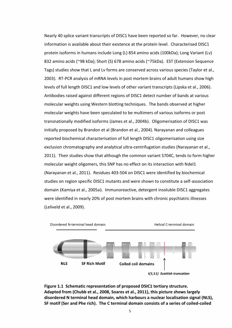
Figure 1.1 Schematic representation of proposed DSIC1 tertiary structure. Adapted from (Chubb et al., 2008, Soares et al., 2011), this picture shows largely disordered N terminal head domain, which harbours a nuclear localisation signal (NLS), SF motif (Ser and Phe rich). The C terminal domain consists of a series of coiled-coiled
6
domains which were implicated in protein interactions and self-association. Translocation break point t(1;11) is indicated with an arrow (at amino acid position 597).
1.2.2. DISC1 tissue Expression and sub cellular localisation
Though DISC1 is mostly discussed in the context of neuronal signalling, its expression is
not just confined to brain but ubiquitously expressed in various other tissues. Placenta,
kidneys and heart have the highest expression of DISC1 transcripts. Abundant DISC1 has
been detected in the dentate gyrus of adult human brain by James et al (2004), although
relatively low expression has been detected in hippocampal pyramidal cells (James et al.,
2004a, Millar et al., 2000b). Northern blotting studies revealed the presence of various
DISC1 transcripts in various parts of the brain (Millar et al., 2000b). Similar DISC1
transcript expression patterns have been reported in mouse brain (Ma et al., 2002c). The
consistent observation of prominent expression of DISC1 transcripts in hippocampus
across species is interesting as the hippocampus is associated with a number of
neurocognitive and other behavioural disorders (Chubb et al., 2008). DISC1 expression
has also been observed in cerebral cortex, sub ventricular zone and olfactory bulb (Ma et
al., 2002a, Schurov et al., 2004b, Miyoshi et al., 2004). DISC1 co localises with Nestin and
SOX2 in these regions, which are sites for neurogenesis. This suggests a role of adult
neurogenesis in the pathophysiology of SCZ (Mao et al., 2009a), (Schurov et al., 2004a,
Kim et al., 2012).
DISC1 protein localisation in mammalian cell lines and cultured neurons has also been
investigated by several groups. DISC1 was shown to be localised to multiple subcellular
compartments such as the centrosome (Miyoshi et al., 2004), microtubules (Morris et al.,
2003), mitochondria (James et al., 2004a) nucleus (Sawamura et al., 2005a) and synapse
(Camargo et al., 2007). It was observed that the localisation varied with the cell type and
the antibodies employed for probing. Co-localisation of overexpressed DISC1 with PSD95
in primary cultures of hippocampal neurons (Bradshaw et al., 2008), presence of 200kDa
DISC1 protein in post synaptic density cell fractions and the presence of other major
DISC1 interacting proteins in the synaptic fractions strongly supports DISC1 localisation as
an important factor for synaptic signalling. Notably, synaptic dysfunction has also been
7
implicated in SCZ (Harrison and Weinberger, 2005). In spite of the data described above,
the two major sites of DISC1 verified by many groups was mitochondria (Atkin et al.,
2011, James et al., 2004a, Millar et al., 2005a) and nucleus (Sawamura et al., 2008,
Brandon et al., 2005, Ma et al., 2002a). Punctate distribution of endogenous DISC1 has
also been reported in mouse cortical neurons, a distribution, which has been replicated
by overexpressed C terminally GFP tagged DSIC1 in HeLa cells and mouse cortical neurons
(Brandon et al., 2005). Immunocytochemistry studies confirmed the localisation of DISC1
with cytoskeleton proteins α-tubulin and MAP2 (Microtubule Associated Protein 2) in
cultured mouse neurons (Brandon et al., 2005). Multi subcellular localisation of DISC1
implicates its involvement in diverse cellular processes mediated by multiple interacting
partners.
1.2.3. DISC1 in Neuronal signalling and development
DISC1 possess multiple interacting partners that demonstrate its pathological relevance
and represent potential for therapeutic intervention. Initial studies that identified a
number of putative DISC1 interacting partners utilised yeast -2-hybrid screens (Ozeki et
al., 2003a) (Camargo et al., 2007). Many of these interacting partners have been
validated by several groups using a range of biochemical techniques.
Most of the interacting partners can be now be categorised in groups of proteins involved
in: cytoskeleton regulation, cell cycle control, signal transduction and central nervous
system signalling. DISC1 forms immune complexes with F actin (Miyoshi et al., 2003b),
alpha tubulin (Brandon et al., 2004), MAP2 (Brandon et al., 2005) suggesting its
association with cytoskeleton. Upon analysis of the DISC1 sequence, it was found to be
similar to those involved in cytoskeleton and transport. Many cytoskeletal proteins are
involved in signalling pathways at the synapse, thereby increasing the chances of DISC1
playing a role in neuronal signalling pathways and suggesting a contribution of DISC1 to
neurological disorders. DISC1 and mitofilin association has been noted in human cell line
and culture mouse neurons (Park et al., 2010). DISC1 knockdown directly effects mitofilin
stability and activity of mitochondrial enzymes like NADH dehydrogenase (Park et al.,
2010).
8
Several groups independently characterised interacting partners of DISC1 such as LIS1,
Ndel1 and Nde1. These are crucial centrosomal proteins, which are involved in neuronal
migration, neurogenesis and neurite outgrowth (Bradshaw et al., 2009, Kamiya et al.,
2005a). Dishevelled axin (DIX) domain containing 1 (DIXDC1) is another DISC1 interacting
partner and DISC1-DIXIN complex is known to modulate neuronal proliferation and
migration via Wnt/GSK3β/ β-catenin signalling (Mao et al., 2009b, Singh et al., 2010).
While knockdown of DISC1 decreases hippocampal progenitor proliferation, DISC1 over
expression has the opposite effect. This effect was also known to be mediated by Wnt
signalling pathway of which GSK3β is one of the major component. The kinase activity of
GSK3β is inhibited upon its interaction with the C terminus region of DISC1, leading to
stabilisation of β catenin protein which translocates in to the nucleus to modulate
translocation of various neurodevelopmental genes (Bradshaw and Porteous, 2010, Ming
and Song, 2009). This has pharmacological relevance as lithium chloride, a GSK3β
inhibitor has been widely employed in management of bipolar disorder (Ming and Song,
2009).
The DISC1 interactome contains a number of proteins specifically localised at the synapse.
TNIK (TRAF2- and NCK interacting Kinase) is one of the PSD (Post Synaptic Density)
localised DISC1 interacting partners whose activity is inhibited when in complex with
DISC1 (Bradshaw and Porteous, 2012). Kal7 which encodes multifunctional Rho GDP/GTP
exchange factor, is one of the candidate genes whose transcripts were found at reduced
levels in SCZ patients (Hill et al., 2006) and an independent study identified KALRN locus
(includes all KAL isoforms) to be associated with SCZ in a GWAS in Japanese population
(Mandela et al., 2012),(Ikeda et al., 2011).
DISC1 role in cAMP signalling is emphasised by its interaction with PDE4enzymes. PDEs, a
large family of phosphodiesterases are the only means of degrading cAMP, a crucial
secondary messenger in many physiological pathways (Houslay et al., 2005). Mammalian
PDE4 enzyme family consists of four gene subtypes with multiple isoforms constitute 4
sub family (PDE4A, PDE4B, PDE4C, and PDE4D)(Houslay, 2001). PDE4 inhibitor, rolipram
9
was elucidated to exhibit anti-psychotic effects in mice. In the absence of PDE4 subtype
specific inhibitors, knockout mice were generated to understand the contribution of each
subtype to the rolipram mediated effects (Zhang et al., 2002). Deficiency of PDE4D gene
produced antidepressant-like effects suggesting that this subtype mediates the effects of
PDE4 inhibitor (Zhang et al., 2002). Biochemical studies also established a direct
interaction between PDE4 isoforms and DISC1. PDE4 isoforms were co-
immunoprecipitated with 100kDa isoform of DISC1 and using peptide array studies, PDE4
binding sites on DISC1 have been mapped (Murdoch et al., 2007). A more recent study
elucidated that ATF4-DISC1 act as repressor complex and regulates PDE4D9 transcription
(Soda et al., 2013). Using ChIP-PCR, it was determined that DISC1 binds to PDE4D gene
locus via ATF4, as it lacks DNA binding domain. Increased levels of PDE4D9 transcripts
and protein levels were reported in embryonic mice brains in which ATF4 and DISC1 were
knocked down using shRNA technology (Soda et al., 2013). PDE4D9 was also shown to
co-localise with DISC1 in the cytosol in cultured hippocampal neurons, emphasising the
involvement of DISC1 in cAMP regulated pathways (Soda et al., 2013). DISC1 interacting
partners in the context of neural signalling and development are discussed in depth in this
review article (reviewed in Bradshaw and Porteous (2012)). As described by Costas et al,
DISC1 can be best termed as a ‘hub’ protein forming a centre point for the orchestration
and regulation of several cellular pathways and exerting its function by protein-protein
interactions (Costas et al., 2013). Interactors of DISC1 and epistatic gene regulation
between them have also been shown to be associated with SCZ risk (Millar et al., 2005b,
Burdick et al., 2008).
The role of DISC1 in psychiatric illness has also been highlighted by a number of genetic
studies in mice. Although it is impossible to model positive, negative and cognitive
symptoms entirely in a mouse, models based on single endophenotype can be developed.
To date, seven different mouse models have been generated to investigate the outcome
of altered DISC1 function. A study by Clapcote et al, reported depression-like behaviour
in mice with ENU induced Q31L mutation, while mice with the L100P mutation exhibited
SCZ-like behaviour. Notably, both had reduced brain volumes, even though they all
expressed normal levels of all DISC1 isoforms (Clapcote et al., 2007). It is interesting to
note that both the above mutations fall in the region on DISC1 known to be involved in
10
PDE4B interaction (Murdoch et al., 2007). Both the mutants have shown reduced binding
to PDE4B while lower PDE4B enzyme activity was reported only in Q31L mutants
(Clapcote et al., 2007). PPI (prepulse inhibition) deficit was observed in both these
mutants, as seen in patients with certain psychiatric illness.
A mutant mouse strain carrying two termination codons in exon 7 and 8 and a premature
polyadenylation site in intron 8 were developed (BAC-DC Tg mice). This led to complete
elimination of the 100kDa and 70kDa DISC1 isoforms, while only a low level of the
truncated DISC1 protein product was detected using Western blot analysis (Kvajo et al.,
2008). Histological studies confirmed reduced cerebral cortex volume and enlarged
lateral ventricles, while reduced neuronal proliferation and neurogenesis was also
reported in this model (Jaaro-Peled, 2009). Three other models have been generated in
which the N terminal DISC1 peptide (1-597 residues, TrDISC1) was overexpressed,
potentially replicating t(1;11) translocation (Pletnikov et al., 2008). A mouse model which
expresses the C terminal DISC1 peptide (671-852 residues) carrying crucial binding sites
for Ndel1 and LIS1 has also been generated (DISC1-cc transgenic mice) (Li et al., 2007). All
these mouse models were intensely characterised for behavioural, histological and
biochemical abnormalities, as summarised in following review papers - (Jaaro-Peled,
2009, Kellendonk et al., 2009). However, the simplest approach, a knockout DISC1
mouse, has proven difficult to produce due to the complexity involved in DISC1 isoforms
which are not fully characterised yet.
1.3. Post Translational Modification (PTM)
Post-translational modification (PTM) is the covalent attachment of additional chemical
groups, lipids, sugars or polypeptides to a protein mediated by various enzyme cascades
(Walsh et al., 2005). Translated proteins are folded and subjected to certain enzyme
catalysed modifications on their amino acid side chains or peptide backbone. Nearly 5%
of the genome encodes for enzymes that catalyse PTMs such as ligases, kinases,
phosphatases, and hydrolases (Walsh et al., 2005). PTM may involve covalent additions
of a co-factor or a co substrate. Examples include ATP-dependent phosphorylation and
11
acetyl CoA dependent acetylation (Walsh et al., 2005). The other type of PTM involve
covalent conjugation of 8 – 10 kDa such as ubiquitin or ubl (ubiquitin like) to the side
chain of amino acids on the target substrates, examples include SUMOylation and
Ubiquitination which are reviewed under sections 1.4 and 1.5 respectively.
These modifications individually or collectively increase the functional diversity within the
proteome. They most often involve a cascade of enzymes, which in turn are tightly
regulated by either another post translational modification, spatial or temporal
separation. They are critical in determining the protein fate – conformational changes,
stability, activity (for enzymes) and sub cellular localisation. However, it should be noted
that PTMs can occur at any stage during or at the end of the life time of a protein. In
most of the cases, they also regulate the cellular signalling cascades responding to
extracellular stimulus, as in the case of the CNS where synaptic communication between
neurons is tightly regulated (Wilkinson et al., 2010). PTMs have been implicated in
cardiovascular disease, psychiatric disorders and cancer (Anderson et al., 2009,
Ciechanover and Iwai, 2004).
To date, phosphorylation was the only PTM, reported on DISC1 using mass spectroscopy
studies. Thr50, Ser58 and Ser713 are three phosphorylation sites on DISC1 identified, in
which the latter two sites are conserved in mouse (Ser54 and Ser710 respectively)
(Ishizuka et al., 2011c). Site directed mutagenesis and in vitro phosphorylation assays
found that PKA and Cdk5 phosphorylated hDISC1 fragment at Ser713 (Ser710 in mouse).
This phosphorylation has also been reported to influence the progression of progenitor
proliferation to post-mitotic neuronal migration. Unphosphorylated form of DISC1
preferentially bind to GSK3β and modulates Wnt signalling. In converse, phosphorylated
DISC1 forms specifically co-localise with BBS proteins (Bardet-Biedl syndrome) at the
centrosome (Ishizuka et al., 2011a). A previously identified PKA mediated
phosphorylation site on hDISC1, Ser58 (Ishizuka et al., 2011c) was known to influence
nuclear localization of DISC1 in HeLa cells (Soda et al., 2013, Ishizuka et al., 2011c).
Phosphorylated WT DISC1 Ser58 was predominantly cytosolic, while the mutant DISC1
12
S58A was nuclear in the presence of okadaic acid, a serine/threonine phosphatase. This
phosphorylation event also reduced DISC1 interaction with ATF4 (Soda et al., 2013).
1.4. SUMOylation
SUMOylation is a post translational modification which involves a covalent attachment of
SUMO proteins (Small Ubiquitin like Modifier) on to substrate. SUMO (also known as
GMP1, UBL1, PIC1) proteins are present in all eukaryotes with and their homologues in
yeast are called Smt3 and Pmt3 (Hay, 2001). Human genome encodes 4 SUMO proteins
(SUMO 1 – SUMO 4). So far, only three of the SUMO proteins have been characterized.
SUMO1 is the best-studied isoform of this family. SUMO 2 and 3 isoforms differ only by
three residues at their C terminus and so collectively they are referred as SUMO 2/3. The
functional role of SUMO4 has not yet been elucidated. SUMO proteins are about 10 kDa
in size and resemble ubiquitin in their three dimensional structures, however, sequence
alignment shows less than 20% similarity with the ubiquitin. A flexible 20 amino acids tail
is absent in ubiquitin and over all surface charge distribution is also significantly different
(Hay, 2005). Like ubiquitination, SUMOylation involves an active enzymatic cascade
where the SUMO moiety (single or as chains) is/are covalently added on to ε-amino group
of lysine residues that present on the surface of substrate proteins.
Figure 1.2, represents a schematic of SUMO conjugation cycle. SUMO proteins are
expressed in immature forms, which are processed by the protease activity of SUMO
specific isopeptidases (SNEPs). In this processing step, variable stretch of the C terminus
(4, 11 and 2 amino acids from SUMO 1, 2, 3 respectively) is cleaved off, to expose the
glycine –glycine domain. Following this action, the protein is considered as the matured
SUMO moiety. This SUMO maturation step is followed by its activation by a
heterodimeric E1 enzyme (SAE1–SAE2 in humans/Uba2–Aos1 in S. cerevisiae) that uses
ATP to adenylate the C-terminal Gly residue of SUMO. This results in formation of a
thioester bond between the C-terminus of SUMO and a Cys residue in active site of SAE2,
releasing AMP (Hay, 2001, Meulmeester and Melchior, 2008). Next step involves the
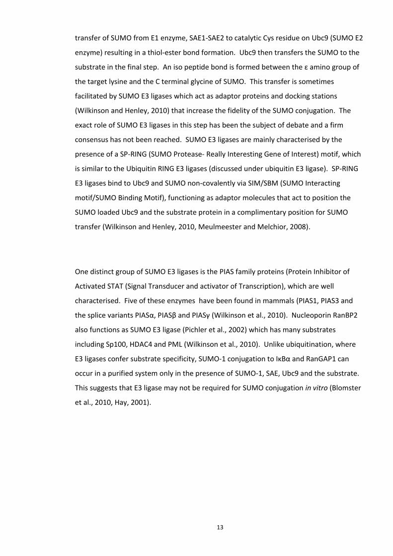
13
transfer of SUMO from E1 enzyme, SAE1-SAE2 to catalytic Cys residue on Ubc9 (SUMO E2
enzyme) resulting in a thiol-ester bond formation. Ubc9 then transfers the SUMO to the
substrate in the final step. An iso peptide bond is formed between the ε amino group of
the target lysine and the C terminal glycine of SUMO. This transfer is sometimes
facilitated by SUMO E3 ligases which act as adaptor proteins and docking stations
(Wilkinson and Henley, 2010) that increase the fidelity of the SUMO conjugation. The
exact role of SUMO E3 ligases in this step has been the subject of debate and a firm
consensus has not been reached. SUMO E3 ligases are mainly characterised by the
presence of a SP-RING (SUMO Protease- Really Interesting Gene of Interest) motif, which
is similar to the Ubiquitin RING E3 ligases (discussed under ubiquitin E3 ligase). SP-RING
E3 ligases bind to Ubc9 and SUMO non-covalently via SIM/SBM (SUMO Interacting
motif/SUMO Binding Motif), functioning as adaptor molecules that act to position the
SUMO loaded Ubc9 and the substrate protein in a complimentary position for SUMO
transfer (Wilkinson and Henley, 2010, Meulmeester and Melchior, 2008).
One distinct group of SUMO E3 ligases is the PIAS family proteins (Protein Inhibitor of
Activated STAT (Signal Transducer and activator of Transcription), which are well
characterised. Five of these enzymes have been found in mammals (PIAS1, PIAS3 and
the splice variants PIASα, PIASβ and PIASγ (Wilkinson et al., 2010). Nucleoporin RanBP2
also functions as SUMO E3 ligase (Pichler et al., 2002) which has many substrates
including Sp100, HDAC4 and PML (Wilkinson et al., 2010). Unlike ubiquitination, where
E3 ligases confer substrate specificity, SUMO-1 conjugation to IκBα and RanGAP1 can
occur in a purified system only in the presence of SUMO-1, SAE, Ubc9 and the substrate.
This suggests that E3 ligase may not be required for SUMO conjugation in vitro (Blomster
et al., 2010, Hay, 2001).
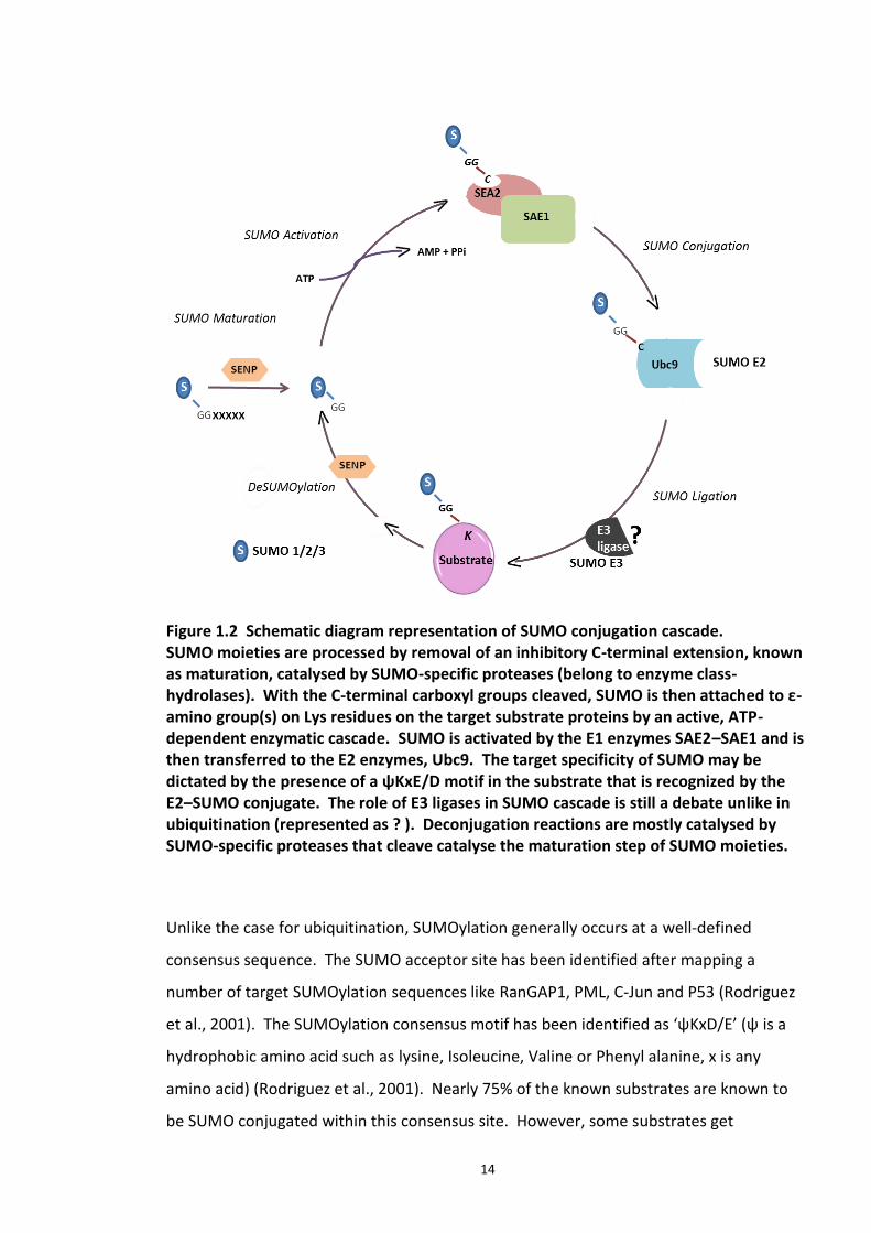
14
Figure 1.2 Schematic diagram representation of SUMO conjugation cascade. SUMO moieties are processed by removal of an inhibitory C-terminal extension, known as maturation, catalysed by SUMO-specific proteases (belong to enzyme class-hydrolases). With the C-terminal carboxyl groups cleaved, SUMO is then attached to ε-amino group(s) on Lys residues on the target substrate proteins by an active, ATP-dependent enzymatic cascade. SUMO is activated by the E1 enzymes SAE2–SAE1 and is then transferred to the E2 enzymes, Ubc9. The target specificity of SUMO may be dictated by the presence of a ψKxE/D motif in the substrate that is recognized by the E2–SUMO conjugate. The role of E3 ligases in SUMO cascade is still a debate unlike in ubiquitination (represented as ? ). Deconjugation reactions are mostly catalysed by SUMO-specific proteases that cleave catalyse the maturation step of SUMO moieties.
Unlike the case for ubiquitination, SUMOylation generally occurs at a well-defined
consensus sequence. The SUMO acceptor site has been identified after mapping a
number of target SUMOylation sequences like RanGAP1, PML, C-Jun and P53 (Rodriguez
et al., 2001). The SUMOylation consensus motif has been identified as ‘ψKxD/E’ (ψ is a
hydrophobic amino acid such as lysine, Isoleucine, Valine or Phenyl alanine, x is any
amino acid) (Rodriguez et al., 2001). Nearly 75% of the known substrates are known to
be SUMO conjugated within this consensus site. However, some substrates get
15
SUMOylated at lysine residues outside this consensus motif by “atypical” SUMO acceptor
lysines (Hoege et al., 2002). Hence, the presence of ‘ψKxD/E’ can be used as a predictor
but is not a definitive indicator of SUMOylation (Wilkinson et al., 2010). In addition to the
covalent SUMO conjugation to an acceptor lysine, recent studies have identified motifs
on substrates that mediate non covalent interactions with SUMO proteins (Minty et al.,
2000, Hecker et al., 2006). These motifs are known as SIMs ( SUMO Interacting Motifs)
which are found to contain a hydrophobic core flanked by acidic residues within the
general consensus sequence V/I-x-V/I-V/I or V/I-V/I-x-V/I/L, where position two or three
can be any amino acid (V,I and L are single letter representation of Valine, Isoleucine and
Leucine respectively) (Song et al., 2004). Since the present study focuses on the covalent
SUMO interactions, it has not been possible to detail SIMs in this review. SIMs have been
reviewed in Song et al. (2005).
Initially on the basis of immunocytochemical studies, it was suggested that SUMOylation
occurs primarily in the nucleus or perinuclear space (Scheschonka et al., 2007).
Subsequently, most of the SUMOylated proteins were found localised in the nucleus
while the SUMO machinery was found distributed both in the nucleus and cytoplasm.
Nuclear SUMO conjugates include transcription factors, nuclear pore proteins and
proteins involved in maintaining genome integrity. Recent evidence of reported
SUMOylation of plasma membrane proteins such as ion channels (K+ channel) and GPCRs
(G-protein-coupled receptors) also suggests the presence of SUMO components at the
plasma membrane (Scheschonka et al., 2007).
1.4.1. Molecular consequences of protein SUMOylation
Covalent linkage of a 10kDa SUMO protein moiety to the protein substrate may have one
or more consequences (schematic in Figure 1.3) - (a) SUMOylation may block interaction
sites on the target protein either by directly competing with the lysine residue which is
also targeted by acetylation, methylation or acetylation cascade or by stearic hindrance
caused by the SUMO moiety (Pichler et al., 2002), (b) SUMO conjugation may add
additional binding surface (sometimes including its own residues) to substrates therefore
16
attracting new interacting partners in a SUMOylation dependent manner (Matunis et al.,
1996). (c) SUMOylation may cause conformational changes to the target protein
uncovering previously masked binding sites (Hay, 2001). The downstream effects of
protein SUMOylation (in neurons), mediated by the above mechanisms are detailed in the
next section.
Figure 1.3 Schematic representation of possible molecular consequences of protein SUMOylation (a) Inhibit interactions: Stearic hindrance caused as a result of covalent attachment of nearly 8kDa protein on to the target will result in abolishing protein interaction either covalent or ionic. (b) New interaction surfaces involving SUMO protein: SUMO moiety on the protein may create new surface(s) which facilitate new interactions. (c) Induce a conformational change: A possible conformational change as a result of SUMOylation may aid result in new interactions. Protein SUMOylation is a dynamic, complex and reversible process. SUMO moieties
covalently attached to the substrates, are recycled by the action of deSUMOylation
enzymes. A single gene family encoding six SUMO specific Cysteine proteases has been
identified in humans known as Sentrin specific proteases (SENP) (Hay, 2013) . The
isoforms SENP1-3 and SENP5-7 are the human homologues. Along with the isopeptidase
function, they are characterised to have the C terminal hydrolase activity which is
required for the maturation of pro SUMO proteins. There are reports of SNEPs
themselves undergoing post translational modification. For example, SENP3 is rapidly
17
turned over under normal cellular conditions by ubiquitination. Extracellular stress
stimulus causes stabilisation of SNEP3 relocating it from cytoplasm to nucleus. SNEP3
rapidly deSUMOylates p300 which regulates transcription of Hif1a, a key regulator of
stress response (Girdwood et al., 2003).
1.4.2. Protein SUMOylation in CNS
Protein SUMOylation has proven to be an essential part in the regulation of many
neuronal cellular pathways. Although, the SUMOylation pathway was linked to diverse
neurological functions and disorders, underlying mechanistic details of how SUMO
conjugation of substrates contributes to health and disease remain largely unknown. This
section is a short review to evaluate the role protein SUMOylation in neurobiological
processes.
The highest levels of Ubc9 expression (SUMO E2 enzyme) and SUMO1 conjugation has
been detected during early brain development in rodents. High expression of Ubc9
reported in dentate gyrus of hippocampus and pyramidal neurons in cerebral cortex
indicates a possible role of Ubc9 and protein SUMOylation in synaptic signalling and
plasticity. Spatio-temporal regulation of SUMOylated substrates and components of
SUMO cascade machinery during rat brain development implies its crucial role during the
neurogenesis (Watanabe et al., 2008). Localisation of SUMO-1 and Ubc9 in Cajal bodies
(CBs) is another example of SUMO mediated regulation of cellular processes in neurons.
CBs are small nuclear organelles, which regulate ribonucleo proteins involved in mRNA
processing (spliceosome machinery). Neuronal proteins coilin and survival of motor
neuron protein (SMN), marker proteins of CBs, were described as putative substrates of
SUMO conjugation (Carvalho et al., 1999). NR2E3 is a key transcription factor, which
activates rod specific genes or represses cone specific genes. This is a key step in
controlling rod photoreceptor differentiation in the retina. NR2E3 is SUMOylated in a
PIAS3 dependent manner. High expression of PIAS3 is observed in mouse retina during
retinal development (Blackshaw et al., 2004, Onishi et al., 2009). MEF2A (Myocyte
Enhancer Factor 2A) is another example of a transcription factor implicated in synaptic
18
activity whose activity is strictly regulated by acetylation and phosphorylation dependent
SUMOylation (Shalizi et al., 2006). It was speculated that SUMOylation may act as switch
between transcriptional activator and repressor roles of MEF2A (Beg and Scheiffele,
2006). Leak K+ channels (example: leak K2P1 channel), crucial for neuronal excitability are
also SUMO modified. It has been proposed that SUMOylation regulates K2P1 channel
activity by directly obstructing the channel pore or altering its charge distribution (Plant
et al., 2005). GSK3β, a key enzyme in Wnt signalling pathway and a DISC1 binding
partner, is also a substrate for SUMOylation (Wilkinson et al., 2010). It is also one of the
few substrates to get SUMOylated on a lysine residue with in a non-consensus region.
GSK3β SUMO mutants (non SUMOylable) are unstable and excluded from the nucleus,
which implies that this modification may be crucial in regulating the enzyme activity (Eun
Jeoung et al., 2008). SUMOylation has also been implicated in the pathogenesis of
Alzheimer’s characterised by progressive dementia. Neurological hallmarks of
Alzheimer’s include neuronal plaques of β amyloid and fibrillar plaques composed of
hyper phosphorylated tau protein, both of which are SUMO substrates (Wilkinson et al.,
2010). A direct genetic link between the SUMO system and AD has also been
hypothesised as an SNP (intron 7) on Ubc9 gene has been shown to be associated with AD
in certain ethnic populations (Ahn et al., 2009). Likewise, α-synuclein, implicated in
Parkinson’s disease interacts with SUMO 1 (Pountney et al., 2005). SUMO protein levels
in cells also alter under conditions of cellular stress. Although, SUMO2/3 levels are low
compared to that of SUMO1, under conditions of oxidative and osmotic stress, SUMO2/3
conjugation of proteins increases, suggesting that they may act as reserve pools (Martin
et al., 2007). SUMO2/3 conjugation is also known to increase in cases of transient global
cerebral ischemia in mice. Hence, it was hypothesised that SUMO conjugation may be a
protective mechanism employed by the cell to down regulate cell surface markers and
receptors and thereby prevent cell death (Martin et al., 2007). In addition, SUMO
proteins are known to be actively transported across the synapse upon neuronal
stimulation (Paul, 2008). SUMO E1 and E3 enzymes were found to be in low levels in post
mortem brain tissue of SCZ patients (Paul, 2008).
Protein aggregation is the hallmark of several psychiatric illnesses such as Alzheimer’s,
Parkinson’s, amyotrophic lateral sclerosis and Huntington’s disease. The proteins
contributing to these diseases Tau, α-synuclein, amyloid precursor protein, SOD1,
19
Huntington and atrophin-1 are SUMO substrates (Krumova and Weishaupt, 2012).
Although, its yet unknown how protein aggregation contributes to the disease
mechanism, it is speculated that it is possibly mediated by SUMOylation as it regulates
protein solubility and stability (Krumova and Weishaupt, 2012). To summarize,
elucidation of SUMOylation mediated regulation of synaptic transmission and neuronal
differentiation has just started to emerge. Possible suggested roles include regulation of
resting membrane potential and synaptic protein-protein interactions. SUMOylation adds
complexity to these processes, as they are already known to be modulated by other PTMs
such as phosphorylation, ubiquitination, and acetylation.
1.5. Ubiquitination/Ubiquitylation
1.5.1. Introduction
A majority of cellular proteins are tagged by ubiquitin (ubiquitin) via an enzyme cascade
known as Ubiquitination. This process is famously known as a prerequisite for targeted
protein degradation by the proteasome. Hershko and collegues, using various
biochemical fractionation and enzymology techniques, discovered that certain proteins
when added to reticulocyte extract, became covalently tagged to a protein known as
ubiquitin, and that these Ubiquitinated proteins were gradually destroyed by an ATP-
dependent protease present in the extract (Hershko et al., 1980, Hershko et al., 1979,
Ciechanover et al., 2012). This team further identified and characterised the enzymes in
the ubiquitin cascade and named them as E1 (ubiquitin-activating enzyme), E2 (ubiquitin-
conjugating enzyme) and E3 (ligase). The other major component of this pathway is the
ATP dependent protease, which gradually degrades the ubiquitin conjugated protein and
is now named as the 26S proteasome (Adams, 2003, Baumeister et al., 1998, Braun et al.,
1999, De Mot et al., 1998, Tanaka, 1998). The ubiquitin moiety, E1, E2, E3 ligases, DUB
(deubiquitinating enzymes) enzymes and the 26S proteasome collectively constitute the
UPS pathway.
20
The UPS (Ubiquitin Proteasome System) is involved in controlling many cellular processes
including protein localisation, transport across membranes, antigen processing and
maintenance of the stoichiometry in protein-protein complexes. The UPS pathway
provides one of the many means through which the cell discards defective, mis-folded,
non-functional and aggregated protein (Hershko and Ciechanover, 1998). Ubiquitin,
which is a major component of this tightly regulated cascade, acts as signalling messenger
to regulating the most complex cellular functions. There has been emerging evidence of
its involvement in other cellular pathways like gene expression regulation, subcellular
targeting of proteins, regulation of protein confirmation, and DNA repair (Di Fiore et al.,
2003, Schwartz et al., 1988). Proteins tagged with a single ubiquitin or ubiquitin chain(s)
are processed by a multi subunit protease complex, the 26S proteasome. However,
ubiquitin itself is spared from the proteasomal action by it release from the substrate by
the action of specific peptidases known as deubiquitinating enzymes (DUBs), making this
post translational modification reversible. Discovery of deubiquitinating enzymes
supports the theory that free ubiquitin moieties can be recycled by the cell (Matsui et al.,
1982). They belong to an enzyme class of hydrolases that hydrolyze the isopeptide bond
between the C-terminal glycine and either the substrate lysine or ubiquitin lysine (if in a
chain). Ubiquitin can bind to proteins either covalently or via ionic interactions. Ubiquitin
may also be conjugated to target substrate proteins and modulate their function without
affecting their stability and other non-destructive roles can also be accomplished by non-
covalent interaction with substrate proteins (Finley et al., 1989).
1.5.2. UPS (Ubiquitin Proteasome System): Components and pathway
Components of the UPS system are well conserved from lower to higher eukaryotes and a
homologous system is absent in prokaryotes. Ubiquitin is a 76 amino acid protein, 8.3kDa
in size. Upon conjugation to the target substrate, ubiquitin may affect their stability,
localisation, conformation and activity. The complex surface design of ubiquitin enables it
to form straight or branched chains of different lengths, which confer great versatility to
this protein modification. The name ‘ubiquitin’ can be justified by its omnipresence and
diverse cellular functions. Ubiquitin was discovered as a thymus hormone by Goldstein et
al in 1975 (Goldstein et al., 1975). However, the exact identity and its extended role in
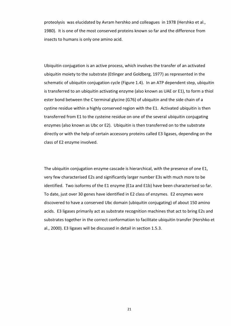
21
proteolysis was elucidated by Avram hershko and colleagues in 1978 (Hershko et al.,
1980). It is one of the most conserved proteins known so far and the difference from
insects to humans is only one amino acid.
Ubiquitin conjugation is an active process, which involves the transfer of an activated
ubiquitin moiety to the substrate (Etlinger and Goldberg, 1977) as represented in the
schematic of ubiquitin conjugation cycle (Figure 1.4). In an ATP dependent step, ubiquitin
is transferred to an ubiquitin activating enzyme (also known as UAE or E1), to form a thiol
ester bond between the C terminal glycine (G76) of ubiquitin and the side chain of a
cystine residue within a highly conserved region with the E1. Activated ubiquitin is then
transferred from E1 to the cysteine residue on one of the several ubiquitin conjugating
enzymes (also known as Ubc or E2). Ubiquitin is then transferred on to the substrate
directly or with the help of certain accessory proteins called E3 ligases, depending on the
class of E2 enzyme involved.
The ubiquitin conjugation enzyme cascade is hierarchical, with the presence of one E1,
very few characterised E2s and significantly larger number E3s with much more to be
identified. Two isoforms of the E1 enzyme (E1a and E1b) have been characterised so far.
To date, just over 30 genes have identified in E2 class of enzymes. E2 enzymes were
discovered to have a conserved Ubc domain (ubiquitin conjugating) of about 150 amino
acids. E3 ligases primarily act as substrate recognition machines that act to bring E2s and
substrates together in the correct conformation to facilitate ubiquitin transfer (Hershko et
al., 2000). E3 ligases will be discussed in detail in section 1.5.3.
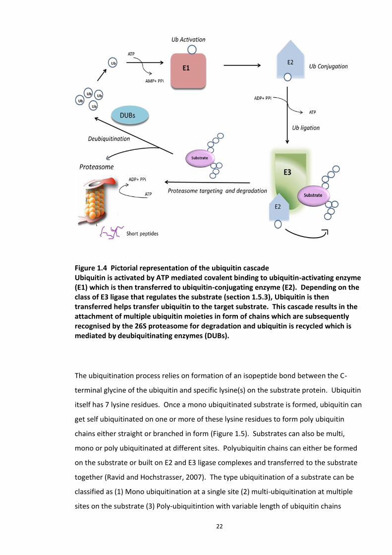
22
Figure 1.4 Pictorial representation of the ubiquitin cascade Ubiquitin is activated by ATP mediated covalent binding to ubiquitin-activating enzyme (E1) which is then transferred to ubiquitin-conjugating enzyme (E2). Depending on the class of E3 ligase that regulates the substrate (section 1.5.3), Ubiquitin is then transferred helps transfer ubiquitin to the target substrate. This cascade results in the attachment of multiple ubiquitin moieties in form of chains which are subsequently recognised by the 26S proteasome for degradation and ubiquitin is recycled which is mediated by deubiquitinating enzymes (DUBs).
The ubiquitination process relies on formation of an isopeptide bond between the C-
terminal glycine of the ubiquitin and specific lysine(s) on the substrate protein. Ubiquitin
itself has 7 lysine residues. Once a mono ubiquitinated substrate is formed, ubiquitin can
get self ubiquitinated on one or more of these lysine residues to form poly ubiquitin
chains either straight or branched in form (Figure 1.5). Substrates can also be multi,
mono or poly ubiquitinated at different sites. Polyubiquitin chains can either be formed
on the substrate or built on E2 and E3 ligase complexes and transferred to the substrate
together (Ravid and Hochstrasser, 2007). The type ubiquitination of a substrate can be
classified as (1) Mono ubiquitination at a single site (2) multi-ubiquitination at multiple
sites on the substrate (3) Poly-ubiquitintion with variable length of ubiquitin chains
23
attached at one or more sites on the substrates. There have been reports of substrates
carrying nearly over a dozen ubiquitin molecules (Hochstrasser, 2006). Using peptide
mapping and site directed mutagenesis studies, K48 and K63 have been identified as the
sites on Ubiquitin involved in chain formation (Chau et al., 1989). However, formation of
chains having both types of the above linkages concomitantly has been reported. Various
models have been hypothesised for poly-ubiquitin chain assembly, reviewed in Mark
Hochstrasser (2006). (Hochstrasser, 2006)). The fate of the ubiquitinated protein
depends on the type of the ubiquitin chains conjugated to it (Figure 1.5). In general,
proteins tagged with K48 chains are targeted to proteasome and K63 linkage chain are
degraded by lysosome (Kirkin et al., 2009). More recent investigations reveal formation
of other type of linkages, like K3 whose functional outcome still needs to be studied.
Multi ubiquitin chain assembly, in some cases also requires an E4 class of enzymes, also
known as E4 chain elongation factors (Hoppe, 2005). There has also been increasing
evidence of ubiquitin tagging to the N terminus of proteins (Ciechanover and Ben-
Saadon, 2004), and the side chains of amino acids like threonine, serine and cysteine
(Cadwell and Coscoy, 2005, Ishikura et al., 2010).
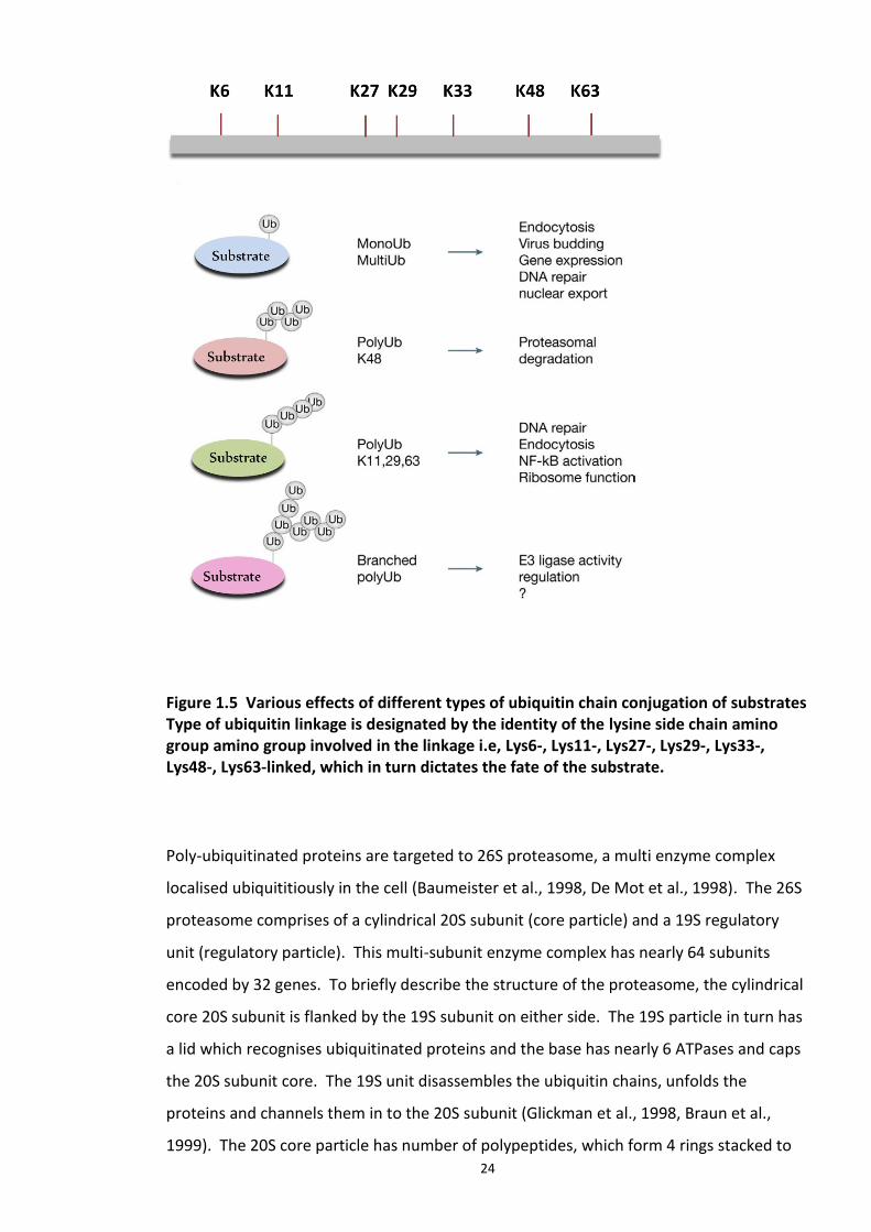
24
Figure 1.5 Various effects of different types of ubiquitin chain conjugation of substrates Type of ubiquitin linkage is designated by the identity of the lysine side chain amino group amino group involved in the linkage i.e, Lys6-, Lys11-, Lys27-, Lys29-, Lys33-, Lys48-, Lys63-linked, which in turn dictates the fate of the substrate.
Poly-ubiquitinated proteins are targeted to 26S proteasome, a multi enzyme complex
localised ubiquititiously in the cell (Baumeister et al., 1998, De Mot et al., 1998). The 26S
proteasome comprises of a cylindrical 20S subunit (core particle) and a 19S regulatory
unit (regulatory particle). This multi-subunit enzyme complex has nearly 64 subunits
encoded by 32 genes. To briefly describe the structure of the proteasome, the cylindrical
core 20S subunit is flanked by the 19S subunit on either side. The 19S particle in turn has
a lid which recognises ubiquitinated proteins and the base has nearly 6 ATPases and caps
the 20S subunit core. The 19S unit disassembles the ubiquitin chains, unfolds the
proteins and channels them in to the 20S subunit (Glickman et al., 1998, Braun et al.,
1999). The 20S core particle has number of polypeptides, which form 4 rings stacked to
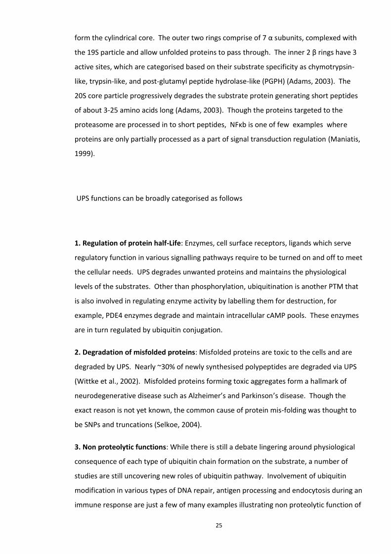
25
form the cylindrical core. The outer two rings comprise of 7 α subunits, complexed with
the 19S particle and allow unfolded proteins to pass through. The inner 2 β rings have 3
active sites, which are categorised based on their substrate specificity as chymotrypsin-
like, trypsin-like, and post-glutamyl peptide hydrolase-like (PGPH) (Adams, 2003). The
20S core particle progressively degrades the substrate protein generating short peptides
of about 3-25 amino acids long (Adams, 2003). Though the proteins targeted to the
proteasome are processed in to short peptides, NFκb is one of few examples where
proteins are only partially processed as a part of signal transduction regulation (Maniatis,
1999).
UPS functions can be broadly categorised as follows
1. Regulation of protein half-Life: Enzymes, cell surface receptors, ligands which serve
regulatory function in various signalling pathways require to be turned on and off to meet
the cellular needs. UPS degrades unwanted proteins and maintains the physiological
levels of the substrates. Other than phosphorylation, ubiquitination is another PTM that
is also involved in regulating enzyme activity by labelling them for destruction, for
example, PDE4 enzymes degrade and maintain intracellular cAMP pools. These enzymes
are in turn regulated by ubiquitin conjugation.
2. Degradation of misfolded proteins: Misfolded proteins are toxic to the cells and are
degraded by UPS. Nearly ~30% of newly synthesised polypeptides are degraded via UPS
(Wittke et al., 2002). Misfolded proteins forming toxic aggregates form a hallmark of
neurodegenerative disease such as Alzheimer’s and Parkinson’s disease. Though the
exact reason is not yet known, the common cause of protein mis-folding was thought to
be SNPs and truncations (Selkoe, 2004).
3. Non proteolytic functions: While there is still a debate lingering around physiological
consequence of each type of ubiquitin chain formation on the substrate, a number of
studies are still uncovering new roles of ubiquitin pathway. Involvement of ubiquitin
modification in various types of DNA repair, antigen processing and endocytosis during an
immune response are just a few of many examples illustrating non proteolytic function of
26
ubiquitin in cell signalling (Chen and Sun, 2009). Ubiquitination and subsequent
proteasomal degradation of IκB leading to activation of NF-κB signalling pathway
implicated in DNA repair.
The following section is a short review on ubiquitin E3 ligases, a crucial component of UPS
which confers specificity towards its substrates.
1.5.3. Ubiquitin E3 ligases
Ubiquitination is temporally and spatially regulated and the complexity is increased by
the presence of diverse number of E3 ligases. To date, 600 E3s have been estimated to
be encoded by the human genome (Bernassola et al., 2008). The two major classes of E3
ligases are the RING finger proteins and HECT domain proteins. The HECT domain forms a
thioester with ubiquitin before the ubiquitin is transferred to the substrate, while the
RING domain present in the RING class proteins, provides a docking surface for E2
enzymes and the target protein facilitating the active transfer of ubiquitin from the
former to later, as represented in the schematic Figure 1.4 (Pickart, 2001). Accumulating
evidence suggests the association of E3 genes with neurogenetic disorders (including
UBR1 (Johanson–Blizzard syndrome), NHLRC1 (Lafora’s disease)) (Tai and Schuman,
2008).
1.5.3.1 HECT
Homologous to E6-AP carboxyl terminus (HECT) class of E3 ligases role has been
emphasised by the study of tumours caused by HPV virus. With the help of the viral
proteins, E6-AP induces the ubiquitination of the tumour suppressor gene p53 (Scheffner
et al., 1995). The important characteristic feature of this family of E3’s is a conserved
stretch of 350 amino acids in their C- terminus called HECT domain, with a cysteine
residue at the catalytic site. The HECT class of E3 ligases differ from the RING E3s by
possessing an intrinsic catalytic site. HECT E 3s form complexes with E2 enzymes and
transfer the active ubiquitin to themselves via the cysteine residue within their active site
forming a ubiquitin-thiolester intermediate. This class of E3 ligases actively catalyse the
transfer and ligation of ubiquitin to the target substrate (Bernassola et al., 2008).
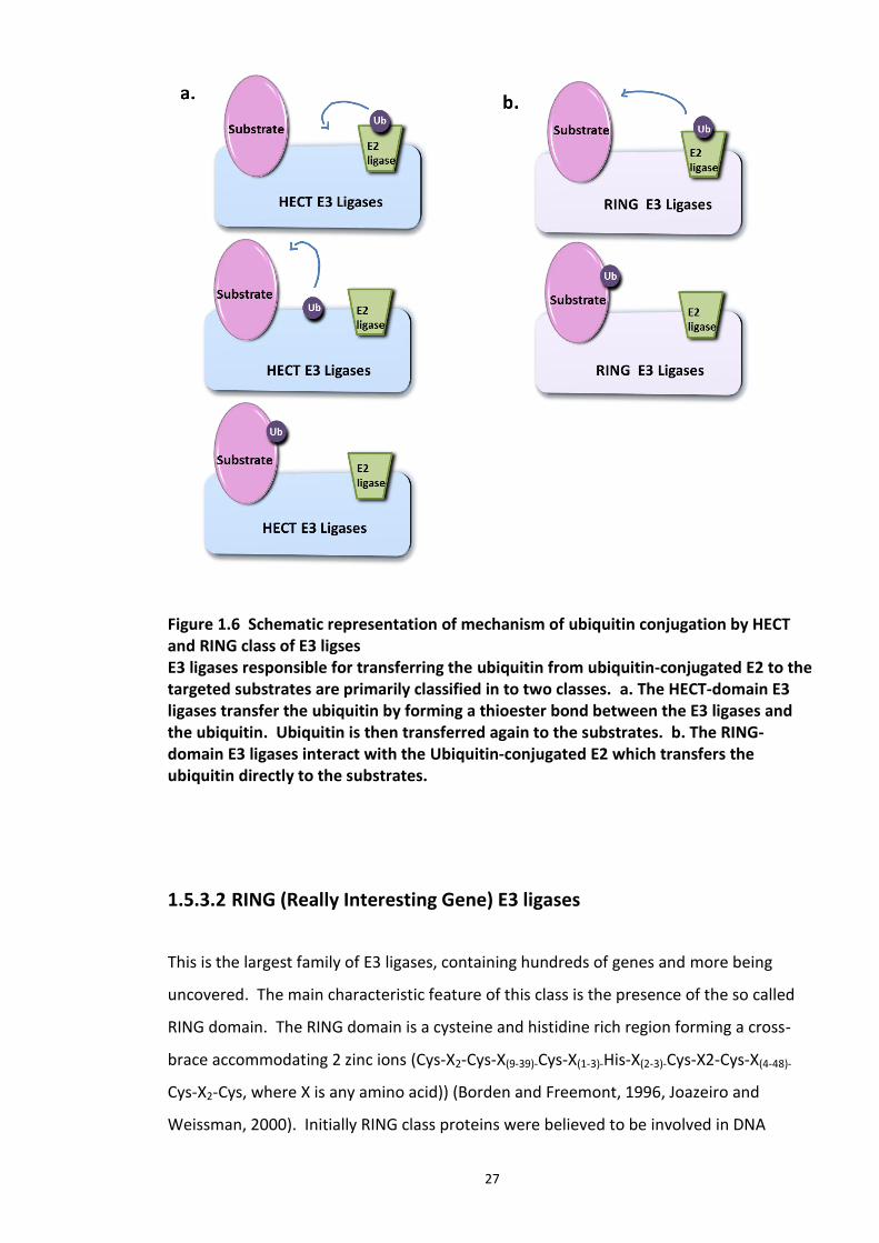
27
Figure 1.6 Schematic representation of mechanism of ubiquitin conjugation by HECT and RING class of E3 ligses E3 ligases responsible for transferring the ubiquitin from ubiquitin-conjugated E2 to the targeted substrates are primarily classified in to two classes. a. The HECT-domain E3 ligases transfer the ubiquitin by forming a thioester bond between the E3 ligases and the ubiquitin. Ubiquitin is then transferred again to the substrates. b. The RING-domain E3 ligases interact with the Ubiquitin-conjugated E2 which transfers the ubiquitin directly to the substrates.
1.5.3.2 RING (Really Interesting Gene) E3 ligases
This is the largest family of E3 ligases, containing hundreds of genes and more being
uncovered. The main characteristic feature of this class is the presence of the so called
RING domain. The RING domain is a cysteine and histidine rich region forming a cross-
brace accommodating 2 zinc ions (Cys-X2-Cys-X(9-39)-Cys-X(1-3)-His-X(2-3)-Cys-X2-Cys-X(4-48)-
Cys-X2-Cys, where X is any amino acid)) (Borden and Freemont, 1996, Joazeiro and
Weissman, 2000). Initially RING class proteins were believed to be involved in DNA
28
repair, however, the breakthrough in the delineation of their real function came via the
discovery of a subunit of the SCF E3 ligase Rbx11 (Kamura et al., 1999, Seol et al., 1999).
These studies revealed the catalytic role of members of a RING family. It has been
suggested that most of the RING class E3 enzymes form a docking platform for the E2
ligase and the substrate protein and catalyse the transfer of the active Ubiquitin from the
former to the later without forming an E3 Ubiquitin intermediate with a covalent bond.
Zheng et al solved the structure of the Cbl-Ubch7 complex, which uncovered a number of
features of this domain (Zheng et al., 2000).
RING E3 ligases are also regulated by ubiquitination, often by autocatalytic function,
consequently regulating its enzymatic activity. For example, Mono ubiquitination of
Mdm2 (E3 ligase which regulates the activity of p53) by autocatalysis is speculated to be
critical in inhibiting its activity. An example, where auto ubiquitination switches on the
ligase activity of an E3 enzyme is that of Bard1-Brca1 heterodimer (Simons et al., 2006).
Diverse mutations in Parkin gene, a RING E3 ligase, were reported in a familial form of
Parkinson's disease known as autosomal recessive juvenile Parkinson's disease (AR-JP) (Yi
and Ehlers, 2007). Brca1, a tumour suppressor gene mutated in nearly 90% breast and
ovarian cancers is an E3 ligase for substrates primarily involved in cell cycle and DNA
repair. It forms a heterodimer with another E3 ligase, Bard1 and it has been established
that this complex formation enhances auto ubiquitination of Brca1, potentially increasing
the E3 ligase activity of Brca1 (Simons et al., 2006).
RING E3 ligases are known to function as oligomers frequently. As mentioned above,
while Brca1-Bard1 is one example another popular example in which these E3 ligases
function as dimer is that of the formation of heterodimer complex of Mdm2 and MDMX.
They play a crucial role in constantly regulating the turnover of p53, a tumour suppressor,
by Ubiquitination. Wang et al, reported that though Mdm2 alone is able to regulate p53
levels, it functions more effectively in complex with MDMX forming a heterodimer (Wang
et al., 2011). As observed in Traf, c-Cbl and Siah RING E3 ligases may also form homo
dimers (Deshaies and Joazeiro, 2009). However, the functional consequence of RING E3
ligase oligomerisation is not well understood yet.
29
Members of the RING family functioning as multi subunit complexes are discussed in
detail in the following section.
1.5.3.3 SCF (Skp1 Cullin Fbox)
These are the well characterised, largest family of RING finger type E3 ligases that form a
protein complex to bridge the E2 and the substrate. SCF complexes and their
components were first discovered during cell cycle studies in yeast. Sic1 is an inhibitor of
Cdk, which is required for the G1 to S phase transition. At the cell cycle checkpoint, Sic1
degradation is initiated by its phosphorylation by G1 cyclin (Cln)-Cdk kinases and targeted
by Ubiquitination via the action of a SCF complex (Schwob et al., 1994). One well-
characterised mammalian E3 ligase belonging to multi subunit RING E3 ligase family is
CDL (Cullin dependent ligase).
A member of CDL family, SCFSkp2 quaternary structure has been elucidated which suggests
that Cullin1 functions as a scaffold protein interacting with all the components of the
protein complex. Its N-terminal helical region that adopts a long stalk-like structure and
interacts with Skp1 and FboxSkp2 while its C terminal domain assumes a globular α/β
domain which interacts with Rbx1 (Zheng et al., 2002). Cullin proteins are modified by
NeDDylation, on their C terminus which induces a conformational change in Cullin
proteins, which inturn leads to activation of the E3 ligase enzyme (NeDDylation is
covalent attachment of ubiquitin like NEDD8 protein(s) catalysed by an enzyme cascade
analogous to ubiquitination) (Duncan et al., 2012). To date, 7 members of Cullin family
have been identified and characterised (Marín, 2009). These are Cullin 1, 2, 3, 4A, 4B, 5,
and 7. Anomalies in their proteins levels, structure and SNPs have been reported in
several tumours. In a comprehensively characterised example of E3 ligase complex Von
Hippel-Lindau (VHL), Cul2 serves as a scaffold. Hif1a, a transcription factor regulated by
cellular oxygen levels, serves as a substrate for the above mentioned complex (Maxwell
et al., 1999). Several mutations in the sequence of VHL have been reported in Von
Hippel-Lindau disease, primarily characterized by tumours of the eye, brain, spinal cord,
30
kidney, pancreas, and adrenal glands (Lee and Zhou, 2010). The role of Cul4B in neural
development has also been reported recently (Liu et al., 2012, Chen et al., 2012). A study
by Liu et al in NT2 cells, revealed the regulatory role of cul4B in neural progenitor growth
and differentiation.
The other component of SCF complex is Rbx protein. Two homologues identified in
humans are Rbx1/ROC1/Hrt1 and Rbx2/SAG/ROC2/Hrt2. In SCFSkp2 , Rbx1 interacts with
Cullin protein via its N terminus (of Rbx1) and E2 enzyme via the RING domain on its C
terminus (Zheng et al., 2002). Rbx1 interacts with all the known Cullin family proteins
(Ohta et al., 1999). The function of Rbx1 is to bind and bring the E2 enzyme into close
proximity with the E3 specific substrate. Apart from recruiting E2 enzyme, other precise
physiological functions of Rbx1 in ubiquitination remain largely unknown. However, it is
known to function as an E3 ligase for Cullin NEDDylation (a PTM essential for the E3
activity of SCF) (Megumi et al., 2005).
The other component of SCF complex is Skp1. Although, there are a number of
homologues reported in lower eukaryotes, humans have only one functional Skp1 gene.
Immunoprecipitation studies and yeast 2 hybrid analysis suggest that Skp1 functions as an
anchor for Cullin and Fbox proteins (Patton et al., 1998). The human Skp1 was initially
characterised as a component of a protein complex containing Cyclin A –Cdk2 and Fbox
Skp2. This complex Ubiquitinates p27Kip, an inhibitor of CDK, which is a regulatory kinase
of the cell cycle check point. Cell cycle is arrested in either G1 or G2 phase in yeast Skp1
mutants suggesting its role in cell cycle (Piva et al., 2002).
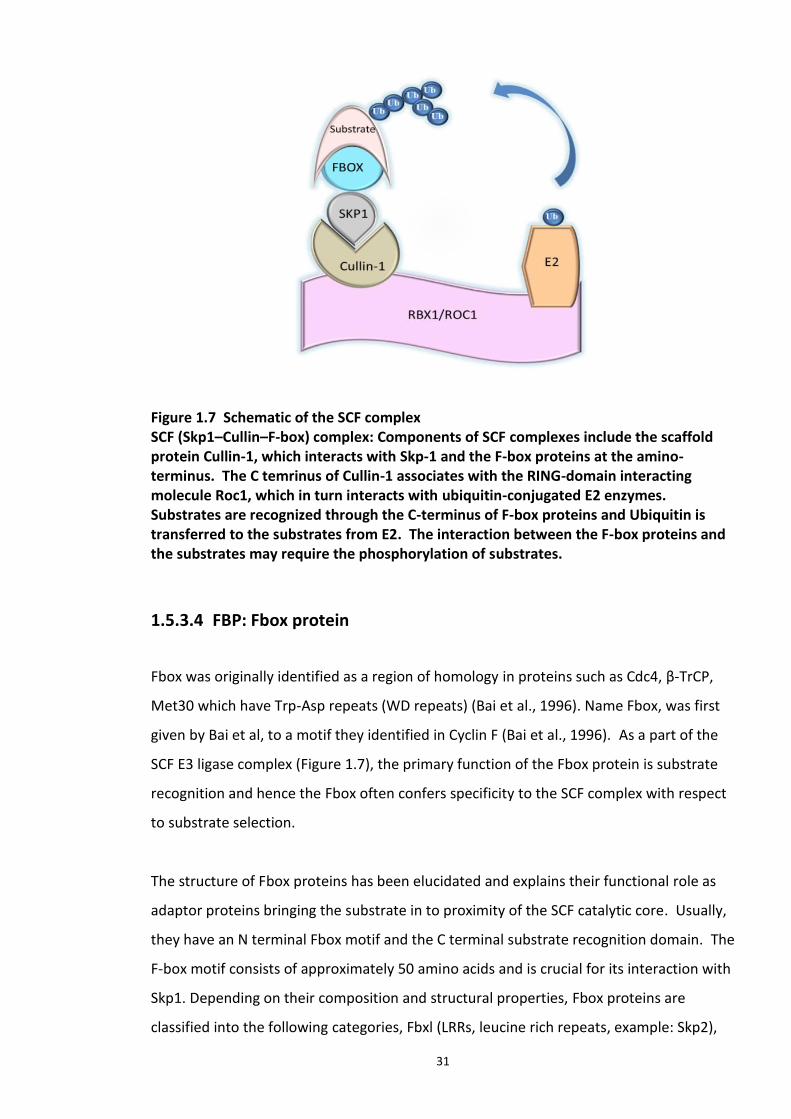
31
Figure 1.7 Schematic of the SCF complex SCF (Skp1–Cullin–F-box) complex: Components of SCF complexes include the scaffold protein Cullin-1, which interacts with Skp-1 and the F-box proteins at the amino-terminus. The C temrinus of Cullin-1 associates with the RING-domain interacting molecule Roc1, which in turn interacts with ubiquitin-conjugated E2 enzymes. Substrates are recognized through the C-terminus of F-box proteins and Ubiquitin is transferred to the substrates from E2. The interaction between the F-box proteins and the substrates may require the phosphorylation of substrates.
1.5.3.4 FBP: Fbox protein
Fbox was originally identified as a region of homology in proteins such as Cdc4, β-TrCP,
Met30 which have Trp-Asp repeats (WD repeats) (Bai et al., 1996). Name Fbox, was first
given by Bai et al, to a motif they identified in Cyclin F (Bai et al., 1996). As a part of the
SCF E3 ligase complex (Figure 1.7), the primary function of the Fbox protein is substrate
recognition and hence the Fbox often confers specificity to the SCF complex with respect
to substrate selection.
The structure of Fbox proteins has been elucidated and explains their functional role as
adaptor proteins bringing the substrate in to proximity of the SCF catalytic core. Usually,
they have an N terminal Fbox motif and the C terminal substrate recognition domain. The
F-box motif consists of approximately 50 amino acids and is crucial for its interaction with
Skp1. Depending on their composition and structural properties, Fbox proteins are
classified into the following categories, Fbxl (LRRs, leucine rich repeats, example: Skp2),
32
Fbxw (WD 40 rich, example: Fbxw7 and β-Trcp) and Fbxo (a different or no Fbox,
example: Cyclin F, Fbxo7) (Cenciarelli et al., 1999). However, mere presence of Fbox
motif does not assure of its E3 ligase activity. For example, Ctf13 forms a part of the
kinetochore complex along with Skp1 as a component of CBF3 (Centromere DNA-binding
protein complex) while other components of the SCF complex are absent and there has
been no reported ubiquitin ligase activity in this complex (Stemmann and Lechner, 1996).
Certain Fbox proteins are also known to possess other intrinsic enzyme activity such as
DNA helicase activity of hFbh1 which regulates homologous recombination (Nelson et al.,
2013).
As this family constitutes a structurally diversified group of proteins, their role in wide
range of cellular processes such as cell division, differentiation, cell survival and apoptosis
has been proposed. Dysregulation of Fbox protein-mediated ubiquitination has been
implicated in many disease pathologies including mood disorders, sleep disorders and
Parkinson’s disease. Table 1.1, presents few examples of Fbox proteins, their specific
substrates and the pathways they regulate.
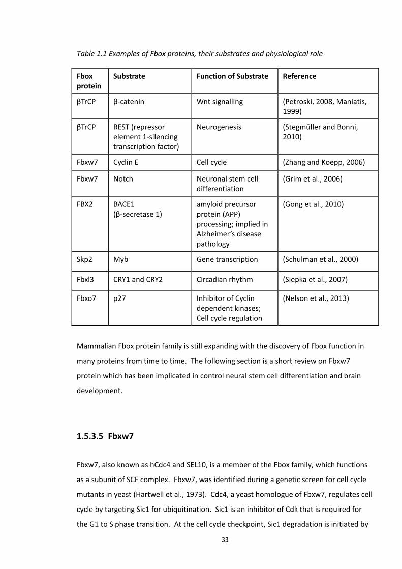
33
Table 1.1 Examples of Fbox proteins, their substrates and physiological role
Fbox protein
Substrate Function of Substrate Reference
βTrCP β-catenin Wnt signalling (Petroski, 2008, Maniatis, 1999)
βTrCP REST (repressor element 1-silencing transcription factor)
Neurogenesis (Stegmüller and Bonni, 2010)
Fbxw7 Cyclin E Cell cycle (Zhang and Koepp, 2006)
Fbxw7 Notch Neuronal stem cell differentiation
(Grim et al., 2006)
FBX2 BACE1 (β-secretase 1)
amyloid precursor protein (APP) processing; implied in Alzheimer’s disease pathology
(Gong et al., 2010)
Skp2 Myb Gene transcription (Schulman et al., 2000)
Fbxl3 CRY1 and CRY2 Circadian rhythm (Siepka et al., 2007)
Fbxo7 p27 Inhibitor of Cyclin dependent kinases; Cell cycle regulation
(Nelson et al., 2013)
Mammalian Fbox protein family is still expanding with the discovery of Fbox function in
many proteins from time to time. The following section is a short review on Fbxw7
protein which has been implicated in control neural stem cell differentiation and brain
development.
1.5.3.5 Fbxw7
Fbxw7, also known as hCdc4 and SEL10, is a member of the Fbox family, which functions
as a subunit of SCF complex. Fbxw7, was identified during a genetic screen for cell cycle
mutants in yeast (Hartwell et al., 1973). Cdc4, a yeast homologue of Fbxw7, regulates cell
cycle by targeting Sic1 for ubiquitination. Sic1 is an inhibitor of Cdk that is required for
the G1 to S phase transition. At the cell cycle checkpoint, Sic1 degradation is initiated by
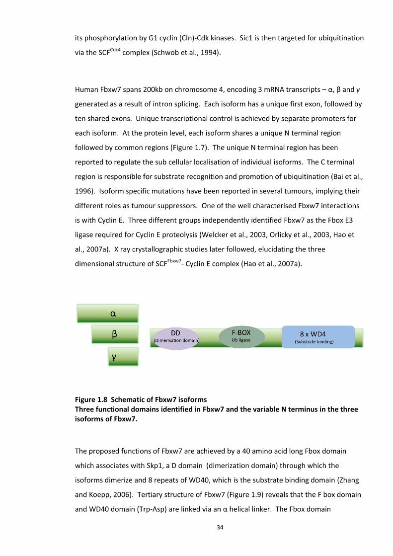
34
its phosphorylation by G1 cyclin (Cln)-Cdk kinases. Sic1 is then targeted for ubiquitination
via the SCFCdc4 complex (Schwob et al., 1994).
Human Fbxw7 spans 200kb on chromosome 4, encoding 3 mRNA transcripts – α, β and γ
generated as a result of intron splicing. Each isoform has a unique first exon, followed by
ten shared exons. Unique transcriptional control is achieved by separate promoters for
each isoform. At the protein level, each isoform shares a unique N terminal region
followed by common regions (Figure 1.7). The unique N terminal region has been
reported to regulate the sub cellular localisation of individual isoforms. The C terminal
region is responsible for substrate recognition and promotion of ubiquitination (Bai et al.,
1996). Isoform specific mutations have been reported in several tumours, implying their
different roles as tumour suppressors. One of the well characterised Fbxw7 interactions
is with Cyclin E. Three different groups independently identified Fbxw7 as the Fbox E3
ligase required for Cyclin E proteolysis (Welcker et al., 2003, Orlicky et al., 2003, Hao et
al., 2007a). X ray crystallographic studies later followed, elucidating the three
dimensional structure of SCFFbxw7- Cyclin E complex (Hao et al., 2007a).
Figure 1.8 Schematic of Fbxw7 isoforms Three functional domains identified in Fbxw7 and the variable N terminus in the three isoforms of Fbxw7.
The proposed functions of Fbxw7 are achieved by a 40 amino acid long Fbox domain
which associates with Skp1, a D domain (dimerization domain) through which the
isoforms dimerize and 8 repeats of WD40, which is the substrate binding domain (Zhang
and Koepp, 2006). Tertiary structure of Fbxw7 (Figure 1.9) reveals that the F box domain
and WD40 domain (Trp-Asp) are linked via an α helical linker. The Fbox domain
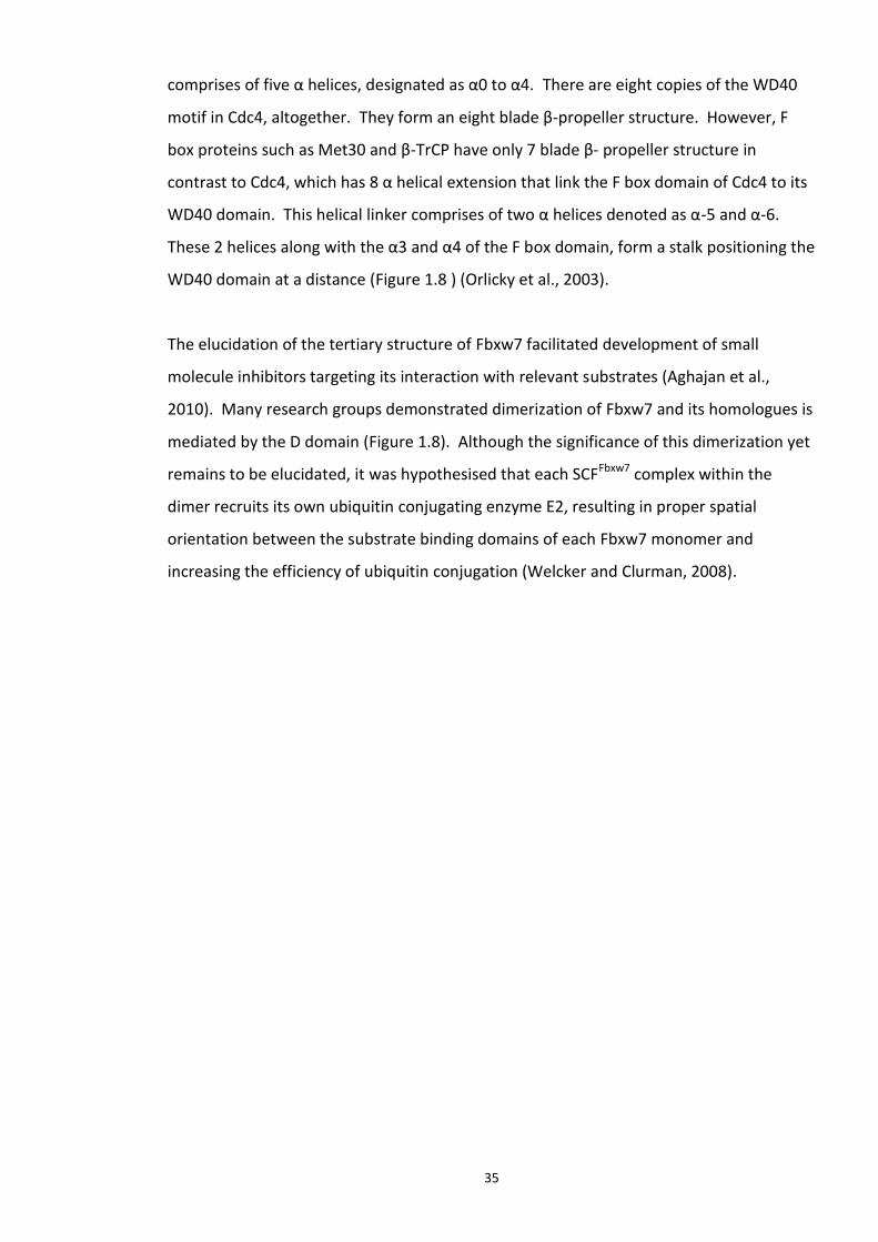
35
comprises of five α helices, designated as α0 to α4. There are eight copies of the WD40
motif in Cdc4, altogether. They form an eight blade β-propeller structure. However, F
box proteins such as Met30 and β-TrCP have only 7 blade β- propeller structure in
contrast to Cdc4, which has 8 α helical extension that link the F box domain of Cdc4 to its
WD40 domain. This helical linker comprises of two α helices denoted as α-5 and α-6.
These 2 helices along with the α3 and α4 of the F box domain, form a stalk positioning the
WD40 domain at a distance (Figure 1.8 ) (Orlicky et al., 2003).
The elucidation of the tertiary structure of Fbxw7 facilitated development of small
molecule inhibitors targeting its interaction with relevant substrates (Aghajan et al.,
2010). Many research groups demonstrated dimerization of Fbxw7 and its homologues is
mediated by the D domain (Figure 1.8). Although the significance of this dimerization yet
remains to be elucidated, it was hypothesised that each SCFFbxw7 complex within the
dimer recruits its own ubiquitin conjugating enzyme E2, resulting in proper spatial
orientation between the substrate binding domains of each Fbxw7 monomer and
increasing the efficiency of ubiquitin conjugation (Welcker and Clurman, 2008).
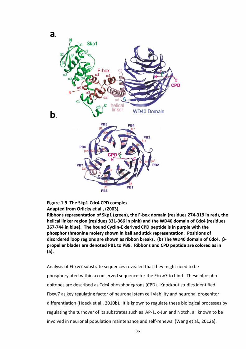
36
Figure 1.9 The Skp1-Cdc4 CPD complex Adapted from Orlicky et al., (2003). Ribbons representation of Skp1 (green), the F-box domain (residues 274-319 in red), the helical linker region (residues 331-366 in pink) and the WD40 domain of Cdc4 (residues 367-744 in blue). The bound Cyclin-E derived CPD peptide is in purple with the phosphor threonine moiety shown in ball and stick representation. Positions of disordered loop regions are shown as ribbon breaks. (b) The WD40 domain of Cdc4. β-propeller blades are denoted PB1 to PB8. Ribbons and CPD peptide are colored as in (a).
Analysis of Fbxw7 substrate sequences revealed that they might need to be
phosphorylated within a conserved sequence for the Fbxw7 to bind. These phospho-
epitopes are described as Cdc4 phosphodegrons (CPD). Knockout studies identified
Fbxw7 as key regulating factor of neuronal stem cell viability and neuronal progenitor
differentiation (Hoeck et al., 2010b). It is known to regulate these biological processes by
regulating the turnover of its substrates such as AP-1, c-Jun and Notch, all known to be
involved in neuronal population maintenance and self-renewal (Wang et al., 2012a).
37
As one of the characterised substrate of Fbxw7, Notch plays a crucial role in neuron fate
and differentiation. Notch is a transcription factor, which, upon proper extra cellular
stimulus, is processed and translocated in to the nucleus to regulate the expression of
genes that induce cell proliferation and prevent differentiation. Studies on Fbxw7
conditional knockout mice showed that Fbxw7 regulated the abundance of Notch
proteins by ubiquitin mediated proteolysis and thus played a pivotal role in regulating the
ratio of neurons and glial cells, a process critical in brain development (Matsumoto et al.,
2011).
1.5.4. UPS impairment in neurological diseases
In highly specialised cellular networks like that of neurons, their connectivity at the
synapse is critical in rapid information transfer in the brain. With millions of neurons
forming trillions of synapse, it is challenging to understand how each of these synapses is
individually modulated to fine-tune neural circuitry. It is understood that synaptic
stability and plasticity are maintained through finely co-ordinated intra neuronal
transport and post translational modification of synaptic proteins. Ubiquitination is one
such vital modifications that control synaptic protein homeostasis. Protein homeostasis
in post mitotic neurons is susceptible to functional disturbances while its abnormalities
result in the accumulation of insoluble and aggregated proteins in the cell. Studies
evaluating the incorporation of radio labelled amino acids into brain proteins suggest that
proteins are constantly replenished or up regulated in brain at different developmental
stages (Cajigas et al., 2010). Studies implicated the significance of tight and controlled
regulation of protein turn over in brain to enable normal brain processes (Lin and Man,
2013).
A few examples which highlight the significance of UPS mediated regulation of proteins
involved in neuronal signalling are as follows: β-catenin is one of the well-studied
substrates of the SCF E3 ligase beta βTrcp. This transcription factor has been shown to
regulate neural progenitor proliferation (Wang et al., 2004). Neural secretory proteins
like Bone morphogenic proteins (BMPs) and Sonic Hedgehog (Shh) are involved in
38
neurogenesis and development signalling pathways. The downstream effector proteins
of these regulatory proteins are tightly regulated by the UPS system. PSD95 (Postsynaptic
Density protein), an important scaffolding protein involved in localisation of receptors at
the post synaptic membrane (such as NMDARs : N-methyl-D-aspartate receptor and
AMPARs: α-amino-3-hydroxy-5-methyl-4-isoxazolepropionic acid receptor) are regulated
by UPS (Mabb and Ehlers, 2010). Another notable example to demonstrate the
importance of UPS regulation of signal transduction is that of Notch. Notch is a
membrane receptor protein that plays a significant role in neurogenesis in response to
extra cellular signals (Lathia et al., 2008). Recent studies discovered that Notch
degradation is mediated by Fbxw7, thus Fbxw7 action can serve to regulate neuronal
progenitor differentiation (Matsumoto et al., 2011).
The role UPS has in the cell cycle has been documented in actively dividing cells, however
not much is known about its role in non-dividing cells like neurons. Only a few CNS
disorders involving the UPS, directly or indirectly, are discussed here. Angelman
syndrome characterised by mental retardation, epilepsy, ataxia and speech problems was
the first human mental illness related to a genetic defect (defective UBE3A gene encoding
E6-AP, a Ubiquitin E3 ligase (Kishino et al., 1997)) in the UPS. A frame shift mutant form
of ubiquitin has been reported in patients with Alzheimer’s disease and Downs syndrome
(van Leeuwen et al., 1998). The inability of this mutant ubiquitin moiety to undergo
proteasomal degradation can be one of the possible disease mechanisms in at least one
of the above disease (Fischer et al., 2003). Huntington's chorea (HD) is one of the nine
PolyQ repeat neurodegenerative disorders characterised by the presence of increased tri
nucleotide repeats of CAG which are translated into long stretches of glutamine residues
increasing their tendency to aggregate. A mutant of the Htt protein (encoded by
Huntigton/Htt gene) is deposited in the form large microscopic inclusion bodies (IB) which
is the hallmark of HD. IBs interfere with the proteasome and impair the UPS function.
Though the exact disease mechanism is not yet elucidated, it is hypothesised the
impairment of UPS function may disturb the cellular protein dynamics resulting in
cytotoxicity leading to subsequent cell death (Finkbeiner and Mitra, 2008). Parkin
(PRKN2), a ubiquitin E3 ligase and UCHL1, a human DUB enzyme, have been implicated in
the pathology of autosomal recessive juvenile Parkinson’s disease (Segref and Hoppe,
2009). Spinocerebellar ataxia is caused by a mutant form of ataxin-1, which is resistant to
39
proteasomal degradation (Klimaschewski, 2003). Micro array analysis of transcripts from
peripheral blood revealed the UPS as one of the canonical pathway disrupted in SCZ and
bipolar disorder (Bousman et al., 2010). Finally, a more recent study performed on post
mortem brain samples of SCZ patients, uncovered abnormal levels of UPS enzymes and
protein ubiquitination (Rubio et al., 2013). The above-mentioned findings not only
emphasise the role of dysregulated protein degradation pathways in mental illnesses, but
also provide scope for new therapeutic drug targets.
1.5.5. UPS as drug target
UPS became a therapeutic attraction for the first time in 2003 following the approval of
Bortezomib (Velcade®), a proteasome inhibitor approved by the FDA for the treatment of
multiple myeloma. This was followed by the Nobel prize in chemistry awarded to Aaron
Ciechanover, Avram Hershko and Lewin Rose for their discovery of ubiquitin mediated
protein degradation in 2004. Since then, UPS components have become an attractive
drug targets and number of inhibitors targeting this signalling pathway are being
developed. As of 2013 quite a few of them have entered clinical trials. Examples include
Proteasome inhibitors - CEP-18770 and MLN-9708, E1 enzyme inhibitor – MLN4924, E3
enzyme inhibitor – RO5045337 (a Nutlin which antagonises Mdm2-p53 interaction). Till
date there is no report of a DUB inhibitor in clinical trials, although there are several
under preclinical development, reviewed in (Mattern et al., 2012).
The E1 enzyme has been described as a potential therapeutic target for treatment of
hematologic malignancies (Xu et al., 2010). Tertiary structure of the E2 has been solved
and provides insight in to three structural orientations of the catalytic site and the
interactions with other proteins. Structure activity relationship studies would help design
molecules targeting the active site of E2 enzyme. E2 and E3 enzymes meet the criteria for
an ideal drug target as they are enzymes, which can be screened for small molecule
inhibitors or specific antibodies. Since they are either expressed in low levels or not
expressed in normal cells, their inhibition may have minimal effect on normal cell growth
and function, thus minimizing toxic side effects as well as achieving maximal therapeutic
index. Different strategies of targeting the E3 ligases would include small molecule
40
inhibitors or peptides that can target E3-E2 interfaces, E3-substrate interfaces or E3 ligase
catalytic sites.
Proteasome inhibitors, though proven effective as a therapeutic strategy, have associated
toxicity and side effects due to the non-selective nature of their action (many proteins get
degraded by the 26S proteasome). As mentioned earlier, Bortezomib (velcade), was one
of the first drugs targeting proteasome for treatment of multiple myeloma, however,
excessive toxicity and drug resistance after prolonged use defined the need for the
development of new inhibitor classes, which are more specific to the target (Bedford et
al., 2011).
Ubiquitin Ligases (E3s) provide an alternative and attractive therapeutic target as their
actions are limited to a handful of protein substrates. The following scenario serves as a
best example of E3 ligase as a drug target. p53 is a tumour suppressor gene and
transcription factor which regulates cell cycle progression and induces apoptosis
depending on the stimuli, thus preventing malignant transformation. In nearly 50%
tumours p53 is inactivated and altering its activity has been considered as a strategy for
therapeutic intervention for cancer. p53 protein levels and activity are maintained under
strict control by its cognate E3 ligase Mdm2 (murine double minute 2). Mdm2 binds to
p53 at its transactivation domains and blocks its activation. Hence by inhibiting p53-
Mdm2 interaction, stabilizes p53 and activated p53 (Shinohara and Uesugi, 2007) (Ding et
al., 2013). X ray crystallographic structure of p53 (15-29) bound to Mdm2 (17-125)
reveals an interaction between hydrophobic N terminal stretch on Mdm2 and
amphipathic α helix of p53 peptide (Verkhivker, 2012, Vassilev et al., 2004). This
information led to the development of benzodiazephine analogues and polyaromatic
compounds, which disrupt the above complex. Several other compounds were later
identified which disrupt the p53-Mdm2 complex. These up regulated p53 protein levels,
activated the p53 pathway and induced cancer cell apoptosis (Wong et al., 2003). Other
examples of irreversible inhibition of E3 ligases are (1) Tetrazole compound (Ro106-9920)
which blocks ubiquitination of IκBα, (2) Ro106-9920 which reduces the serum levels of
TNFα and prostaglandins in lung (Wong et al., 2003). Allosteric inhibitors can also be used
to block the conformational changes or other post translational modification required for
the E3 ligase activation. Compounds with imidazole and benzosulfonamide nuclei
interfere with Mdm2 ubiquitination of p53 in this manner (Lai et al., 2002). Virtual ligand
41
screening (VLS) has also been used to screen for compounds with the ability to interfere
with E3 ligase activity. In silico based approaches have identified small pockets in the E3
ligase that are suitable for docking with small molecules that could act as inhibitors. This
approach has been used to inhibit Skp2 directed degradation of p27 (Wu et al., 2012).
High-throughput screens are another popular approach in drug discovery. Multiple steps
in the ubiquitination cascade can be targeted using this approach. Inhibitors can interfere
with kinase activity of the “priming kinase” (required for E3 ligase binding to the
substrate) or attenuate SCF complex assembly either by interfering with the interaction
directly or allosterically. An Inhibitor of the SKp1-Met30 interaction is an example for this
approach and is selective to the F box domain of Met30 and hence the inhibitor doesn’t
interfere with the binding of other Skp1 binding F Box proteins (Aghajan et al., 2010).
1.6. DISC1 as a possible therapeutic target for neurological disorders
Various genetic studies have implicated the DISC1 locus to be a predisposing genetic
factor in major mental illnesses including, SCZ, schizoaffective disorders, bipolar and
depression (Chubb et al., 2008, Song et al., 2010, Ishizuka et al., 2006). However, no
specific allele, mutation or SNPs has been proved to be a risk factor across various
studies. As discussed earlier, DISC1 has a conserved nuclear localisation signals (NLS),
which regulate its cytoplasmic/nuclear distribution. The nuclear form of DISC1 is
increased in patients with sporadic SCZ, major depression, and substance alcohol abuse
(Sawamura et al., 2005b). This study suggests the importance of the NLS domains on
DISC1 and proposes it as a putative drug target for the aforementioned diseases.
Protein aggregates are a hallmark of neurological diseases like Alzheimer’s disease (AD)
and Parkinson’s disease (PD) and DISC1 also forms aggregates both in vivo and in vitro.
These aggregates could possibly interfere with the physiological functions of DISC1 and
disrupting these aggregates can be considered as a potential therapeutic strategy.
cAMP specific PDE4 (phosphodiesterase) enzymes are potential interacting partners of
DISC1 which were identified in the yeast 2 hybrid screen (Camargo et al., 2007) PDE are
42
the enzymes, which degrade cAMP, a crucial secondary signalling molecule. Raised
cellular levels of cAMP caused the dissociation of DSIC1-PDE4 complex (Murdoch et al.,
2007a). Of various isoforms of PDE4, PDE4B locus has been identified as a possible risk
factor for SCZ (Millar et al., 2005). While genetic variants of PDE4B and PDE4D were
shown to be directly associated with SCZ (Pickard et al., 2007, Hennah and Porteous,
2009, Fatemi et al., 2008), DISC1 has been shown to specifically interact with certain
isoforms of PDE4B and PDE4D and this interaction is modulated by cellular cAMP levels
(Murdoch et al., 2007). Hence, the DISC1-PDE4 interaction has gained much attention in
the studies of pathophysiology of SCZ.
Studies show that shRNA knock- down of DISC1 in adult mice resulted in aberrant
dendritic morphology and mis-positioning of adult born dentate granule cells (Duan et al.,
2007), abnormalities in axonal targeting (Faulkner et al., 2008), abnormal development
of dentate gyrus neurons and dendritic abnormalities and induced mTOR signalling (Zhou
et al., 2013a). RNAi mediated knock down of DISC1 in differentiating PC12 cells caused
significant inhibition of neurite outgrowth and this could be rescued by the co
transfection of WT DISC1 (Kamiya et al., 2005a). In utero gene transfer of RNAi in mouse
embryos resulted in reduced neuronal migration and reduced dendritic arborisation of
cortical neurons (Kamiya et al., 2005b). In the absence of knockout DISC1 mice, the
above studies as well as transgenic DISC1 mouse models (reviewed in (Jaaro-Peled, 2009))
emphasize the importance of DISC1 in the mechanism behind neurological disease.
As proposed by the neurodevelopmental hypothesis of SCZ, the symptoms of SCZ
generally tend to appear in late adolescence and it has long been believed that the
biological events causing the disease may have begun at a much earlier stage (Ayhan et
al., 2011). Though other genes have been implicated in SCZ and neurogenesis, such as
neuregulin-1 and dysbindin, most of the studies were focussed on DISC1 due to its
increased association in linkage and association studies. The importance of the Scottish
translocation has long been a debate for several reasons. One such reason is that, since
the translocation is in the middle of an open reading frame, the protein even though
translated, may be unstable and eventually degraded leading to haploinsufficiency (both
alleles need to be functional for a wild type functional genotype). However, to date,
there is no proof of DISC1 truncated protein being translated. Truncated DISC1 protein
43
might interfere with function of the wild type protein product from the other allele,
resulting in total loss of function. This is known as dominant negative effect.
As discussed earlier, the phosphorylated form of DISC1 interacts with Bardet-Biedl
Syndrome (BBS) proteins at the centrosome, while non-phosphorylated forms up regulate
Wnt signalling via interaction with GSK3β which acts as a developmental switch between
progenitor proliferation to post mitotic neuronal migration (Ishizuka et al., 2011b). This
study emphasises the role of DISC1 as a scaffold protein function depends on several
interacting partners. One source kinase of the phosphorylation of DISC1 at Ser713 is Cdk5
(Ishizuka et al., 2011b). Cdk5 inhibitors CP-668863 and CP-681301, developed by Pfizer
have been considered as potential drugs to provide neuro protection in Alzheimer’s
disease (Hikida et al., 2012).
1.7. Aims of my Ph.D.
As detailed in my previous sections, genetic studies implicated DISC1, already associated
with SCZ and other mood disorders, but also in autism spectrum disorders, attention
deficit and hyperactivity disorder (ADHD) and Asperger syndrome. While lymphoblasts
cell lines of the Scottish family members carrying the translocation were shown to have
reduced expression of mRNA transcripts, no changes were detected in SCZ patients
outside this family which suggests the complexity of regulation of DISC1 expression and
protein processing. In addition, atypical antipsychotic drugs such as olanzapine and
risperidone were shown to increase DISC1 transcript levels in mouse frontal cortex and
hippocampus suggesting that they may exert their effect by modulating DISC1 expression
(Olincy et al., 2011, Chiba et al., 2006). Abnormal brain development and function has
been reported with RNAi mediated knockdown of DISC1 indicating that loss of DISC1
function is not desirable for appropriate brain development (Maher and LoTurco, 2012,
Hikida et al., 2012).
To date, little is known about the post translational modifications on DISC1.
Phosphorylation of DISC1 at Ser713 was shown to function as a molecular switch between
progenitor proliferation and post mitotic neuronal migration in the developing cortex.
44
From the above evidence it can be proposed that understanding how DISC1 protein levels
are modulated may hold the key to understand DISC1 associated pathology and uncover
key targets for therapeutic intervention. With recent studies implicating the
dysregulation of ubiquitin and Ubl signalling pathways in schizophrenia, it would be
interesting to investigate how DISC1 expression is regulated by these signalling pathways
and how it may contribute to disease development (Altar et al., 2005, Rubio et al., 2013).
Hence through my work I set out to address the following key questions:
1. Is DISC1 a potential SUMO substrate in cells? What are the key SUMO acceptor sites on
DISC1? What is physiological significance of DISC1 SUMOylation? As a scaffold protein
with many interacting partners, does SUMOylation effect its protein interactions such as
DIXDC1 and Ndel1 which have been implicated in neuronal precursor proliferation,
differentiation and migration. I addressed these aspects using in vitro peptide array
technology, UFDS (Ubc9 fusion directed SUMOylation) and Immunoprecipitation in
chapter 3.
2. In chapter 4, I investigated the ubiquitin modification of DISC1 protein using
proteasome inhibitor MG132. Immunoprecipitation, Western blotting and Mass
spectroscopy studies were used to identify potential lysine moieties which are modified
by ubiquitin conjugation. Human Fbox siRNA library screen was performed to identify
putative Fbox that regulates ubiquitination of DISC1 i.e., Fbxw7.
3. In chapter 5, utilising peptide array approach, I had mapped the crucial interaction sites
between DISC1 and the Skp1-Fbxw7 followed by a series of optimisation screens via
which I developed certain disruptor peptides to target DISC1- [Skp1-Fbxw7] complex.
Lead peptides were shortlisted following their ability to stabilise DISC1 protein levels in
HEK293 cells. Neuronal progenitors generated from iPS cells were also explored for their
potential as cell line models for psychiatric diseases.

45
4. In chapter 6, I discussed my work which involved developing a sensitive in vitro FP
assay to monitor DISC1- [Skp1-Fbxw7] interaction. This assay was adapted to perform a
high throughput chemical library screen to identify small molecules which disrupt this
complex.
46
2.
Materials and Methods
2.1 Materials
All chemicals used in this study were of analytical grade supplied by Thermo Fisher
scientific, Invitrogen or SIGMA Aldrich unless otherwise specified. All peptides used in the
present chapter were custom made by ProImmune, UK at >85% purity, assessed by Mass
spectroscopy and HPLC. Labelled peptides used as tracers in FP assays were tagged with
FITC (Fluorescein isothiocyanate) on the amino terminal end of the peptides. All the
drugs and disruptor peptides were dissolved in dimethly sulfoxide (DMSO) and used at a
final DMSO concentration of 0.001%.
2.2 Molecular Biology Methods
2.2.1 Transformation of competent cells
DH5α competent cells (Invitrogen) stored at -80ºC were carefully thawed on ice just
before use. 1-10 ng of DNA was added to 30 μl of competent cells in a sterile eppendorff
and incubated on ice for 10 min. The cells were then heat shocked at 42ºC for 45 sec
then placed on ice for 2 min. The cells were then added to 300 μl Luria Broth (LB) media
(1% (w/v) bacto-tryptone, 0.5% (w/v) bacto-yeast extract and 170 mM NaCl) and
incubated at 37ºC for 1 hr with shaking. 50-100 μl of transformation mix was then spread
on 100 mm petri dishes containing pre poured LB agar plates (L broth with 1.5% (w/v)
agar) containing appropriate antibiotic (ampicillin, 50 µg/ml, kanamycin 30 µg/ml) using a
47
sterile disposable spreader. These plates were incubated upside down overnight at 37ºC.
The growth of bacterial colonies indicated successful cell transformation.
2.2.2 Isolation of plasmid DNA from E coli
Single colonies were selected from the overnight incubated agar plates and grown
overnight in 5 ml LB media containing appropriate antibiotic in an orbital shaker at 37oC.
QIAprep Miniprep Kit (Qiagen) was used to isolate smaller amounts of plasmid DNA.
Alternatively, for a larger volume of bacterial culture, 500 ml LB media supplemented
with appropriate antibiotic was inoculated with 5ml starter culture grown for about 6-8hr
and incubated overnight at 37oC. For large scale plasmid DNA extraction the QIAprep
Maxiprep kit (Qiagen) was used according to the manufacturer’s instructions. The
purified DNA was then eluted with sterile double distilled H20 and stored at 4oC for short
term storage or -20oC for longer term.
2.2.3 Storage of plasmid DNA
Overnight incubated bacterial culture was mixed with 30% sterile glycerol solution at 1:1
ratio to give a final glycerol concentration of 15% in cryovials and snap frozen using dry
ice and stored at -80°C.
2.2.4 Quantification of DNA concentration
The concentration of purified DNA was determined using Nanodrop 3300
spectrophotometer (Thermo Scientific). Absorbance wavelength was set at 260 nm and
280 nm and the A260:A280 ratio determines the purity of the nucleic acid extract where a
value of 1.8 and 2.0 is considered pure for DNA and RNA, respectively. The ratio of
A260:A230 ideally should be between 2-2.3 and a lower reading may indicate organic
solvent contamination in the preparation.
2.2.5 Total RNA extraction
The protocols described in this section are described, according to Standard Operating
Procedure of Pfizer, for the work performed as part of a 10 week placement at Pfizer
Neuroscience research Unit, Cambridge, MA, USA).

48
Homogenisation: Culture medium was aspirated off culture dishes and 1ml TRIzol (per
each well of a 6 well culture plate) (Ambion 15596-026) was added.
Phase Separation: The homogenised samples were incubated for 5 min at RT to allow
complete dissociation of nucleoprotein complexes. 0.2 volumes of chloroform per 1ml
TRIzol reagent was then added and vigorously mixed followed by 2-3 min incubation. The
samples were centrifuged at no more than 1,000 g for 15 min at 4˚C. Following
centrifugation the mixture separates into a lower red phenol chloroform phase (which
constitutes protein), an interphase and a colourless aqueous phase. The aqueous phase,
which exclusively contains the RNA, was carefully transferred to a fresh tube without
disturbing the other phases.
RNA Precipitation: RNA in the separated aqueous phase is precipitated by adding 0.5
volumes of isopropanol per 1 ml of TRIzol reagent used. The samples are thoroughly
mixed and incubated for 10 min at RT.
RNA Wash: The RNA was pelleted by spinning at no more than 10,000 g. The supernatant
was removed and the pellet was washed with 1 ml of 75% ethanol. Samples were then
centrifuged at no more than 7,500 g for 5 min at 4˚C.
Redissolving the RNA: The ethanol was carefully aspirated off, the RNA pellet was air
dried and re-suspended initially in 100 µl followed by quantification.
DNase treatment of purified RNA: Purified RNA was further subjected to DNase
treatment to remove any DNA contamination. TURBO DNA free kit (Ambion AM1907)
was used and the DNase treatment was done according to the protocol mentioned in the
instruction manual.
RNA precipitations clean up: Re-suspended RNA was re-precipitated to remove any salts
or organic solvents that may have co-precipitated. NaCl was added to a final
concentration of 0.1M followed by the addition of 2.35 volumes of ethanol. The samples
were then thoroughly mixed and incubated at -20ºC for >30min or overnight. The RNA
was then precipitated at 4˚C by spinning at 10,000 g. The supernatant was carefully
aspirated and the RNA pellet was air dried. The RNA was re-suspended in water and
stored at -80˚C.
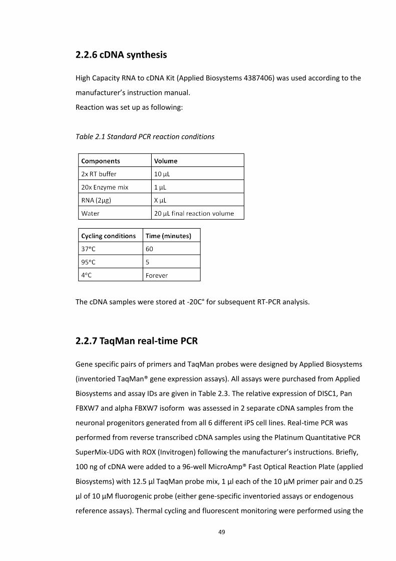
49
2.2.6 cDNA synthesis
High Capacity RNA to cDNA Kit (Applied Biosystems 4387406) was used according to the
manufacturer’s instruction manual.
Reaction was set up as following:
Table 2.1 Standard PCR reaction conditions
The cDNA samples were stored at -20C° for subsequent RT-PCR analysis.
2.2.7 TaqMan real-time PCR
Gene specific pairs of primers and TaqMan probes were designed by Applied Biosystems
(inventoried TaqMan® gene expression assays). All assays were purchased from Applied
Biosystems and assay IDs are given in Table 2.3. The relative expression of DISC1, Pan
FBXW7 and alpha FBXW7 isoform was assessed in 2 separate cDNA samples from the
neuronal progenitors generated from all 6 different iPS cell lines. Real-time PCR was
performed from reverse transcribed cDNA samples using the Platinum Quantitative PCR
SuperMix-UDG with ROX (Invitrogen) following the manufacturer’s instructions. Briefly,
100 ng of cDNA were added to a 96-well MicroAmp® Fast Optical Reaction Plate (applied
Biosystems) with 12.5 μl TaqMan probe mix, 1 μl each of the 10 μM primer pair and 0.25
μl of 10 μM fluorogenic probe (either gene-specific inventoried assays or endogenous
reference assays). Thermal cycling and fluorescent monitoring were performed using the
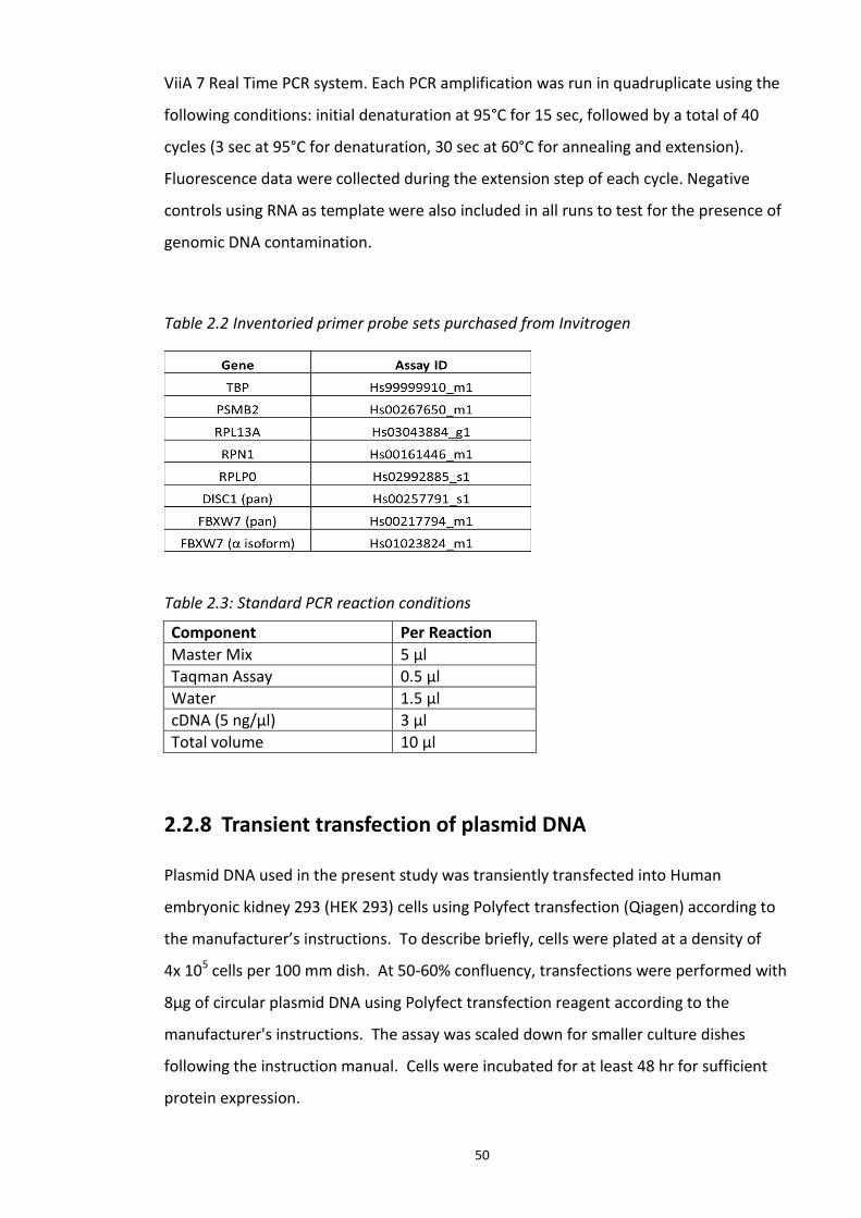
50
ViiA 7 Real Time PCR system. Each PCR amplification was run in quadruplicate using the
following conditions: initial denaturation at 95°C for 15 sec, followed by a total of 40
cycles (3 sec at 95°C for denaturation, 30 sec at 60°C for annealing and extension).
Fluorescence data were collected during the extension step of each cycle. Negative
controls using RNA as template were also included in all runs to test for the presence of
genomic DNA contamination.
Table 2.2 Inventoried primer probe sets purchased from Invitrogen
Table 2.3: Standard PCR reaction conditions
2.2.8 Transient transfection of plasmid DNA
Plasmid DNA used in the present study was transiently transfected into Human
embryonic kidney 293 (HEK 293) cells using Polyfect transfection (Qiagen) according to
the manufacturer’s instructions. To describe briefly, cells were plated at a density of
4x 10⁵ cells per 100 mm dish. At 50-60% confluency, transfections were performed with
8μg of circular plasmid DNA using Polyfect transfection reagent according to the
manufacturer's instructions. The assay was scaled down for smaller culture dishes
following the instruction manual. Cells were incubated for at least 48 hr for sufficient
protein expression.
Component Per Reaction
Master Mix 5 µl
Taqman Assay 0.5 µl
Water 1.5 µl
cDNA (5 ng/µl) 3 µl
Total volume 10 µl
51
2.2.9 si RNA transfection – Fbox siRNA library screen
ON Target PLUS siRNA library for Fbox (G-105625-05) was purchased from Dharmacon,
Thermofisher Scientific. HEK293 cells were plated at a density of 3 x 10³ cells/ well in 6-
well plates. 24 hr after incubation, the cells were transfected with control nonspecific
siRNA or the test siRNA from the above library at a final concentration of 25 nM/each well
using DharmaFECT (Dharmacon - Thermo Scientific) according to the manufacturer’s
protocol. The cells were incubated for 64 hr at 37°C in a CO2 incubator. Gene expression
knockdown was analysed by performing Western blotting.
2.3 Cell culture
All cell culture procedures were carried out in a Class II hoods using standard aseptic
techniques and sterile instruments. All culture reagents were supplied by Sigma. The
tissue culture flasks, dishes, and pipettes were supplied by Corning. All cultures were
examined regularly under a phase contrast inverted microscope (Leitz Diavert, Germany).
2.3.1 HEK293 cells
The HEK293 cells were used for the present study. These cells were maintained in growth
media containing DMEM (SIGMA) supplemented with 10% foetal bovine serum, 2 mM L-
glutamine, 100 U/μg penicillin-streptomycin at 37°C in a humidified atmosphere of 5%
CO2. The cells were passaged at about 80% confluency. For cell passage, growth medium
was aspirated and the cells were washed in sterile phosphate buffer saline (PBS) to
remove traces of media. The PBS was then aspirated off and the cells were treated with
2mL trypsin-EDTA solution per 75 cm2 culture flask of cells to dissociate cell monolayer.
Growth medium was added to quench the trypsin-EDTA solution and cells were collected
by centrifugation at 10,000 g for 2 min at room temperature. The supernatant was
removed without disturbing the cell pellet which was then re-suspended in appropriate
fresh growth media and re-plated according to the requirement. For cryopreserving the
cells, the cell pellet was re-suspended in 10% dimethyl sulfoxide (DMSO) containing
growth medium and stored at -80˚C.
52
2.3.2 iPS cell culture and neuronal progenitor differentiation
All the protocols described in this section were according to the SOP (standard operating
procedure) followed at Pfizer Neuroscience Research Unit, Cambridge, USA.
2.3.2.1 iPSc generation and culture
All the iPS cells (except Detroit 551 cell line) used in the present study were generated by
Chiang et al (Chiang et al., 2011). Detroit 551 iPS cell line was generated by Dr.Sandra
Engle, Pfizer Inc. Detroit 551 fibroblasts were obtained from ATCC (CCL-110) and
maintained in Knockout DMEM (Life Technologies 10829-018) with 15% FBS (Life
Technologies), 1x Nonessential amino acids (Life Technologies 11140-050), 2 mM L-
glutamine (Life Technologies 25030-081), 1x Gentamicin (Life Technologies 15710-072),
0.1 mM β-mercaptoethanol (Sigma M-7522). The cells were reprogrammed with
individual lentiviruses expressing c-Myc, KLF4, SOX2, Oct3/4, Nanog, and LIN28 using
standard techniques (Takahashi, Okita et al. 2007). One colony, Detroit 551-3, with ES-
like morphology, high expression of pluripotency markers (e.g. Oct3/4, TRA-1-60, TRA-1-
81, SSEA4), a normal karyotype, a normal karyotype and the ability to form all 3 germ
layers in a teratoma assay was used in all subsequent experiments. The cells were
maintained on mitomycin C (Sigma M0503)-treated mouse embryonic fibroblasts (MEF)
and grown in hPSC media [DMEM/F12 (Life Technologies 11330-057), 20% Knockout
serum replacement (Life Technologies, 10828-028), 2 mM L-glutamine (Life Technologies
25030-081), 1x Gentamicin (Life Technologies 15710-072), 0.1 mM β-mercaptoethanol
(Sigma M-7522), FGF2 (Life Technologies PHG0263)]. Cells were enzymatically passaged
using 1 mg/ml Dispase (Life Technologies 17105-0410) to every 4-6 days to fresh MEF.
2.3.2.2 Neuronal Progenitor differentiation
Neuronal differentiation protocol was adapted from the protocol of (Pankratz et al.,
2007). Embryoid body formation was induced on the first day, assigned as day 0. Culture
media was removed from the appropriate culture dishes with iPS cell colonies and
washed with warm (37°C) DMEM-F12 media. 1ml Dispase (Invitrogen- 17105-041) was
added per p100 culture plate and incubated for 37°C for 2-5 min until the edges of the
cell colony start curling up. Disapase was aspirated off and washed with DMEM-F12
53
media. Using Stempro EZPassage (Invitrogen 23181-010) the colonies were gently broken
until they were dissociated from the plate. The iPS cell colonies were transferred from
half of the p100 dish to 100 mm non coated, non-treated, non-adhesive cell culture dish
with hPSC media [DMEM/F12 (Life Technologies 11330-057), 20% Knockout serum
replacement (Life Technologies, 10828-028), 2 mM L-glutamine (Life Technologies 25030-
081), 1x Gentamicin (Life Technologies 15710-072), 0.1 mM β-mercaptoethanol (Sigma
M-7522), FGF2 (Life Technologies PHG0263)], to promote preferential adhesion between
cells over adhesion to the culture dish.
On day 2, the embryoid bodies were collected by centrifugation in appropriate centrifuge
tubes at 500 g for 5 min at RT. Supernatant was discarded while extreme care is taken not
to disturb the pellet which was re-suspended in 10 ml of GABA differentiation media [50%
Neurobasal Media (Life Technologies 21103-0490), 50% DMEM/F12 (Life Technologies
10565-018), 0.5x N2, 0.5x B27 (Life Technologies 17504-044), 1 mM L-glutamine (Life
Technologies 25030-081), 1x Gentamicin (Life Technologies 15710-072), 10µM SB431542,
0.2µM LDN-193189 (StemGent 04-0074), 20ng/ml FGF8b (R&D Systems 423-F8-025), Shh
(R&D Systems 1845-SH-025/CF), 10 ng/ml FGF2 (Invitrogen PHG0263)], supplemented
with ROCK inhibitor (10 µM) and transferred to cell culture plates pre-coated with
matrigel. Cell culture dishes were coated with matrigel according to manufacturer’s
instructions (BD Matrigel matrix- 354277). Fresh GABA Media change was done from day
4 to day 10.
Cells were passaged on day 10 by aspirating the media and washing 3 times with PBS. 1
ml of accutase (Invitrogen A1110501) was used to gently lift the cell off the dish. Equal
volume of DMEM-F12 media was added to neutralise the enzyme activity and the cells
were collected by gently pipetting the suspension and centrifuged at 1000 g for 5 min at
RT. Supernatant was disposed and the cell pellet was re-suspended in GABA media
supplemented with 10µM ROCK inhibitor and plated in 1:1 ratio on pre coated POL
coated plates (20 µg/ml poly-L-ornithine hydrobromide (Sigma P3655)/ 5 µg/ml natural
mouse laminin (Life Technologies 23017-015) (POL)-coated plates).
Fresh media change was performed every day with GABA media. Confluency and
morphology of the cells was microscopically observed on a daily basis to detect unwanted
54
differentiation. Cells can be passaged using the protocol described above depending on
the confluency after day 15. From this point onwards neuronal progenitors can be
maintained and expanded as required in NP media [50% Neurobasal Media (Life
Technologies 21103-0490), 50% DMEM/F12 (Life Technologies 10565-018), 0.5x N2, 0.5x
B27 (Life Technologies 17504-044), 1 mM L-glutamine (Life Technologies 25030-081), 1x
Gentamicin (Life Technologies 15710-072), 10ng/ml EGF (R&D Systems 236-EG), 10ng/ml
FGF2 (Invitrogen PHG0263)]. Cells were maintained in appropriate dishes or multi well
plates as required at 1.62 x 10⁵ per cm2 area of the culture vessel.
2.3.3 Preparation of Cell Lysates
Cells were briefly washed with PBS and then lysed in to ice cold cell lysis buffer 20 mM
Tris-HCl (pH 7.5), 150 mM NaCl, 1 mM Na2EDTA, 1 mM EGTA, 1% Triton, 2.5 mM sodium
pyrophosphate, 1 mM β-glycerophosphate, 1 mM Na3VO4, 1 µg/ml leupeptin, 10mM DTT
with Roche protease inhibitor, phosSTOP (Roche, 04906837001), 2mM NEM (N-
ethylmaleimide). The cell suspension was then incubated on an end-over-end rotor at
4°C to ensure proper cell lysis and then centrifuged at 13,000 g for 10 min at 4°C. The
protein concentration in the supernatant was analysed by Bradford method (Bradford,
1976).
2.4 Protein techniques
2.4.1 Co-Immunoprecipitation
Cell lysates were adjusted to equal protein amount (1 μg/μl) using cell lysis buffer. Cell
lysates normalised for the protein concentration were incubated with mouse IgG
(Millipore, 12487) Protein A agarose beads (life technologies, 15918-014) for 2 hr at 4°C.
Agarose beads were collected after a brief centrifugation and the collected supernatant
was added with HA tagged agarose (SIGMA; A2095) washed 3 times in cell lysis buffer and
the final volume is adjusted to 750 µl and incubated over night at 4°C. The beads were
precipitated and washed three times to remove non-specifically bound proteins. Immune
complexes were eluted from the beads by boiling them in SDS loading buffer. The
complexes were analysed on SDS PAGE. Tagged agarose beads used in the present study
include: Flag (Sigma), HA (Sigma), V5 (Abcam), IgG (Sigma).
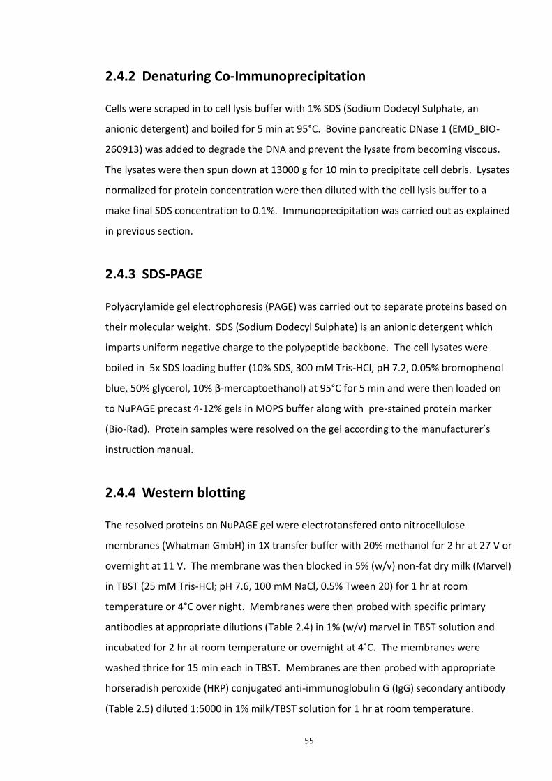
55
2.4.2 Denaturing Co-Immunoprecipitation
Cells were scraped in to cell lysis buffer with 1% SDS (Sodium Dodecyl Sulphate, an
anionic detergent) and boiled for 5 min at 95°C. Bovine pancreatic DNase 1 (EMD_BIO-
260913) was added to degrade the DNA and prevent the lysate from becoming viscous.
The lysates were then spun down at 13000 g for 10 min to precipitate cell debris. Lysates
normalized for protein concentration were then diluted with the cell lysis buffer to a
make final SDS concentration to 0.1%. Immunoprecipitation was carried out as explained
in previous section.
2.4.3 SDS-PAGE
Polyacrylamide gel electrophoresis (PAGE) was carried out to separate proteins based on
their molecular weight. SDS (Sodium Dodecyl Sulphate) is an anionic detergent which
imparts uniform negative charge to the polypeptide backbone. The cell lysates were
boiled in 5x SDS loading buffer (10% SDS, 300 mM Tris-HCl, pH 7.2, 0.05% bromophenol
blue, 50% glycerol, 10% β-mercaptoethanol) at 95°C for 5 min and were then loaded on
to NuPAGE precast 4-12% gels in MOPS buffer along with pre-stained protein marker
(Bio-Rad). Protein samples were resolved on the gel according to the manufacturer’s
instruction manual.
2.4.4 Western blotting
The resolved proteins on NuPAGE gel were electrotansfered onto nitrocellulose
membranes (Whatman GmbH) in 1X transfer buffer with 20% methanol for 2 hr at 27 V or
overnight at 11 V. The membrane was then blocked in 5% (w/v) non-fat dry milk (Marvel)
in TBST (25 mM Tris-HCl; pH 7.6, 100 mM NaCl, 0.5% Tween 20) for 1 hr at room
temperature or 4°C over night. Membranes were then probed with specific primary
antibodies at appropriate dilutions (Table 2.4) in 1% (w/v) marvel in TBST solution and
incubated for 2 hr at room temperature or overnight at 4˚C. The membranes were
washed thrice for 15 min each in TBST. Membranes are then probed with appropriate
horseradish peroxide (HRP) conjugated anti-immunoglobulin G (IgG) secondary antibody
(Table 2.5) diluted 1:5000 in 1% milk/TBST solution for 1 hr at room temperature.

56
Membranes were then washed 3 times for 10 min. Chemiluminescence (ECL) Western
blotting Substrate (Thermo Scientific) was used for detecting the HRP conjugates on the
membrane. Chemiluminescent images of immune detected bands were recorded on blue-
light sensitive autoradiography X-ray films which were then developed using the Kodak®
X-Omat Model 2000 processor. Band intensities were quantified using image J (Girish and
Vijayalakshmi, 2004).
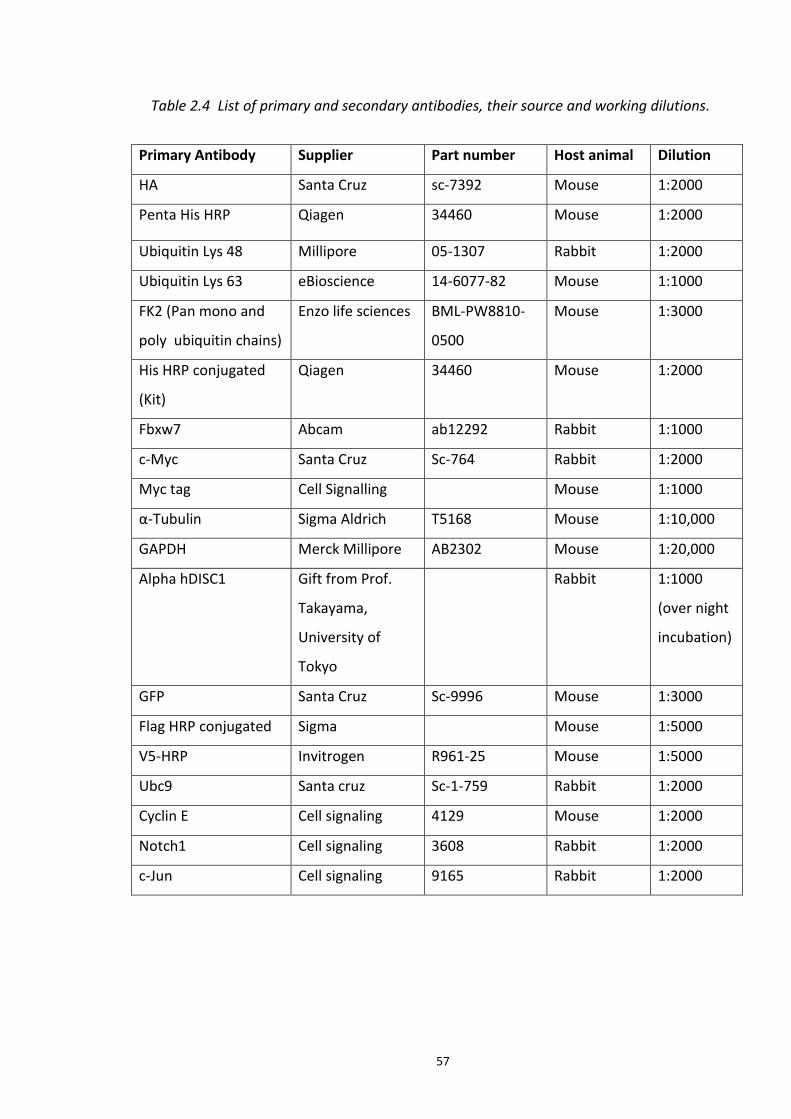
57
Table 2.4 List of primary and secondary antibodies, their source and working dilutions.
Primary Antibody Supplier Part number Host animal Dilution
HA Santa Cruz sc-7392 Mouse 1:2000
Penta His HRP Qiagen 34460 Mouse 1:2000
Ubiquitin Lys 48 Millipore 05-1307 Rabbit 1:2000
Ubiquitin Lys 63 eBioscience 14-6077-82 Mouse 1:1000
FK2 (Pan mono and
poly ubiquitin chains)
Enzo life sciences BML-PW8810-
0500
Mouse 1:3000
His HRP conjugated
(Kit)
Qiagen 34460 Mouse 1:2000
Fbxw7 Abcam ab12292 Rabbit 1:1000
c-Myc Santa Cruz Sc-764 Rabbit 1:2000
Myc tag Cell Signalling Mouse 1:1000
α-Tubulin Sigma Aldrich T5168 Mouse 1:10,000
GAPDH Merck Millipore AB2302 Mouse 1:20,000
Alpha hDISC1 Gift from Prof.
Takayama,
University of
Tokyo
Rabbit 1:1000
(over night
incubation)
GFP Santa Cruz Sc-9996 Mouse 1:3000
Flag HRP conjugated Sigma Mouse 1:5000
V5-HRP Invitrogen R961-25 Mouse 1:5000
Ubc9 Santa cruz Sc-1-759 Rabbit 1:2000
Cyclin E Cell signaling 4129 Mouse 1:2000
Notch1 Cell signaling 3608 Rabbit 1:2000
c-Jun Cell signaling 9165 Rabbit 1:2000
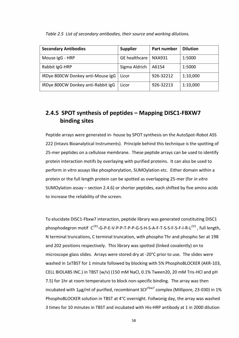
58
Table 2.5 List of secondary antibodies, their source and working dilutions.
Secondary Antibodies Supplier Part number Dilution
Mouse IgG - HRP GE healthcare NXA931 1:5000
Rabbit IgG-HRP Sigma Aldrich A6154 1:5000
IRDye 800CW Donkey anti-Mouse IgG Licor 926-32212 1:10,000
IRDye 800CW Donkey anti-Rabbit IgG Licor 926-32213 1:10,000
2.4.5 SPOT synthesis of peptides – Mapping DISC1-FBXW7 binding sites
Peptide arrays were generated in- house by SPOT synthesis on the AutoSpot-Robot ASS
222 (Intavis Bioanalytical Instruments). Principle behind this technique is the spotting of
25-mer peptides on a cellulose membrane. These peptide arrays can be used to identify
protein interaction motifs by overlaying with purified proteins. It can also be used to
perform in vitro assays like phosphorylation, SUMOylation etc. Either domain within a
protein or the full length protein can be spotted as overlapping 25-mer (for in vitro
SUMOylation assay – section 2.4.6) or shorter peptides, each shifted by five amino acids
to increase the reliability of the screen.
To elucidate DISC1-Fbxw7 interaction, peptide library was generated constituting DISC1
phosphodegron motif C191-G-P-E-V-P-P-T-P-P-G-S-H-S-A-F-T-S-S-F-S-F-I-R-L215 , full length,
N terminal truncations, C terminal truncation, with phospho Thr and phospho Ser at 198
and 202 positions respectively. This library was spotted (linked covalently) on to
microscope glass slides. Arrays were stored dry at -20°C prior to use. The slides were
washed in 1xTBST for 1 minute followed by blocking with 5% PhosphoBLOCKER (AKR-103,
CELL BIOLABS INC.) in TBST (w/v) (150 mM NaCl, 0.1% Tween20, 20 mM Tris-HCl and pH
7.5) for 1hr at room temperature to block non-specific binding. The array was then
incubated with 1μg/ml of purified, recombinant SCFFbw7 complex (Millipore, 23-030) in 1%
PhosphoBLOCKER solution in TBST at 4°C overnight. Follwonig day, the array was washed
3 times for 10 minutes in TBST and incubated with His-HRP antibody at 1 in 2000 dilution
59
in 1% blocking solution for 2 hours at room temperature. After a further 3 washes the
array was exposed to ECL and film (as in section 2.4.4).
Figure 2.1 Schematic of peptide array workflow Adapted from (Katz et al., 2011).
2.4.6 In vitro SUMOylation of hDISC1 peptide array
Human DISC1 (hDISC1) peptide array was pre-blocked in 5% (w/v) BSA (bovine serum
albumin, SIGMA) in TBST for 1hr at room temperature with gentle rocking. This was
followed by a brief washing in TBST for 5 min. SUMO kit (Enzo life sciences, UW8955) was
used to SUMOylate hDISC1 peptide array. This kit contains purified recombinant His
tagged human SUMO1, SUMO2, SUMO3 proteins, active E1, E2 enzymes, ATP (required
co factor for SUMO conjugation). A reaction mixture was prepared with all the above kit
60
components and made up to 2ml with sterile PBS (phosphate buffered saline). The array
was incubated at 30°C for 60 min and then washed in TBST, 3 times for 5 min each. This
was followed by probing with His HRP conjugated antibody at appropriate dilution
mentioned for 2hr at room temperature followed by 3 times, 5 min washes with TBST.
The array was ECL detected as explained in section 2.4.4.
2.4.7 Immunocytochemistry
All immunocytochemistry studies were performed at Pfizer NRU, Cambridge, MA, USA, as
a part of 10 week placement (discussed in chapter 5). iPS cell colonies and neuronal
progenitors (NPs) were stained for relevant markers for characterisation. NPs were
plated at 100 cells per cm2 area of 35mm MatTek dishes (Invitrogen). Cells were fixed
with 4% paraformaldehyde followed by three washes with TBS (Tris-buffered saline;
150mM NaCl, 20 mM Tris, pH 7.4). Cells were permeabilised by incubating with 0.2%
Triton dissolved in TBS. Cells were blocked for 1 hour with blocking solution (10% donkey
serum, 4% BSA in TBS). The primary antibody was diluted in antibody diluent (Dako,
S0809) to required concentration (dilutions were represented in the Table 2.6 and 2.7).
50 μl of diluted primary antibody was added to the coverslip for 2 hr or overnight
incubation at 4°C. The dishes were washed thrice with TBS. The fluorescent antibody
(Alexa 594-conjugated antibody or Alexa 488-conjugated antibody, depending on the
primary antibody choice, was diluted at 1:200 using diluent and added to the dish for 1
hour (this procedure was repeated if a second fluorescent probe was needed). The
coverslips were washed 5 times with TBS before mounting a cover slip with
Immunomount (sandon). Cells were visualized using the Zeiss LSM 710 confocal
microscope (Zeiss, Oberkochken, Germany).
Table 2.6 List of Stem cell marker antibodies used to characterise the iPS cell lines
Marker Localisation Host animal Dilution Source
Oct4A Nuclear Anti rabbit 1 : 800 Cell signalling, 2890
Tra-1-81 Surface Anti mouse 1:1000 Cell signalling, 4745S
Nanog Nuclear Anti goat 1:100 Millipore, MABD24
SSEA4 Surface Anti mouse 1:100 Millipore, MABD43034
DAPI Nuclear Invitrogen
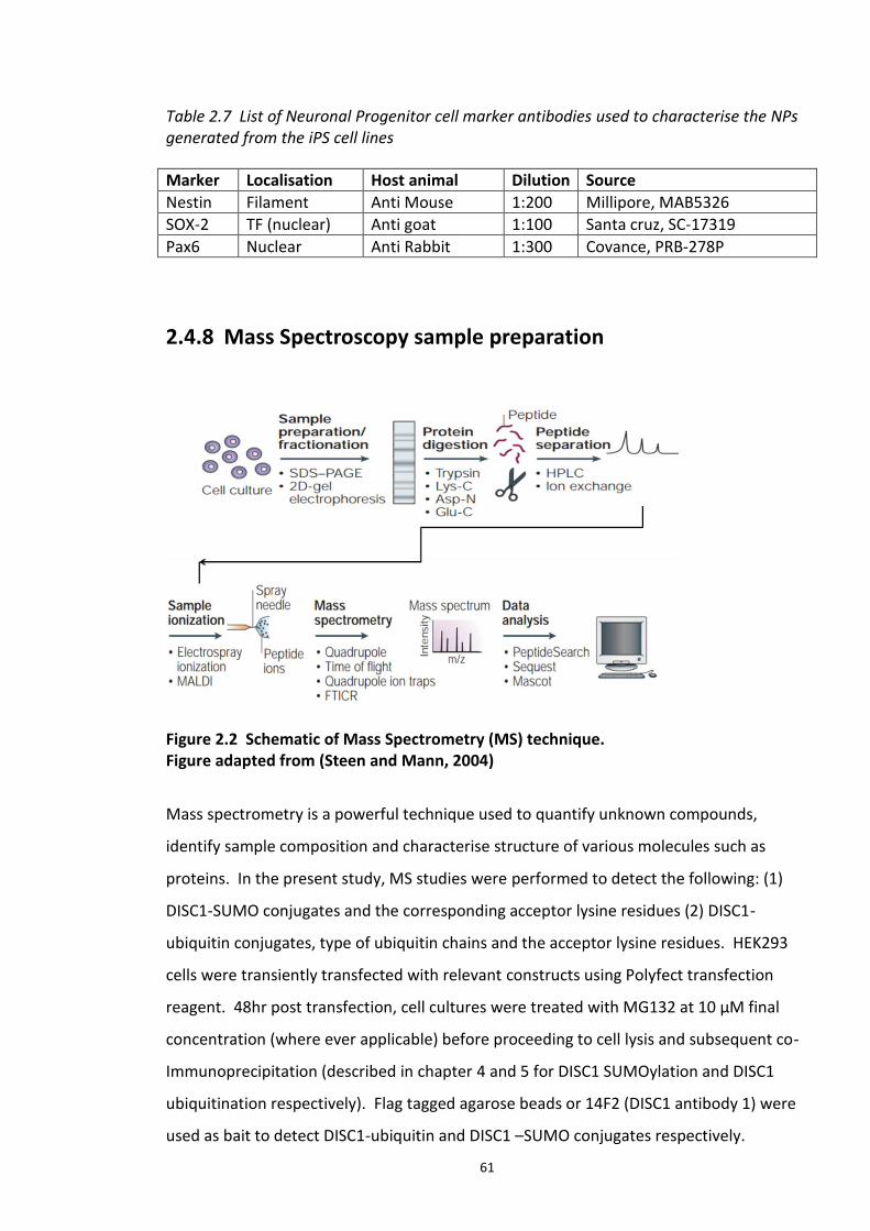
61
Table 2.7 List of Neuronal Progenitor cell marker antibodies used to characterise the NPs generated from the iPS cell lines
Marker Localisation Host animal Dilution Source
Nestin Filament Anti Mouse 1:200 Millipore, MAB5326
SOX-2 TF (nuclear) Anti goat 1:100 Santa cruz, SC-17319
Pax6 Nuclear Anti Rabbit 1:300 Covance, PRB-278P
2.4.8 Mass Spectroscopy sample preparation
Figure 2.2 Schematic of Mass Spectrometry (MS) technique. Figure adapted from (Steen and Mann, 2004)
Mass spectrometry is a powerful technique used to quantify unknown compounds,
identify sample composition and characterise structure of various molecules such as
proteins. In the present study, MS studies were performed to detect the following: (1)
DISC1-SUMO conjugates and the corresponding acceptor lysine residues (2) DISC1-
ubiquitin conjugates, type of ubiquitin chains and the acceptor lysine residues. HEK293
cells were transiently transfected with relevant constructs using Polyfect transfection
reagent. 48hr post transfection, cell cultures were treated with MG132 at 10 µM final
concentration (where ever applicable) before proceeding to cell lysis and subsequent co-
Immunoprecipitation (described in chapter 4 and 5 for DISC1 SUMOylation and DISC1
ubiquitination respectively). Flag tagged agarose beads or 14F2 (DISC1 antibody 1) were
used as bait to detect DISC1-ubiquitin and DISC1 –SUMO conjugates respectively.
62
Immunoprecipitate complexes were separated using SDS PAGE on bis tris acrylamide gel.
The gel was then stained with Colloidal blue stain, Invitrogen (part number: LC6025)
according to manufacturer’s instructions. This was followed by gel de-staining in ultra-
pure double distilled water over night. Relevant bands of interest (as indicated in the
respective results chapter 3 and 4) were then excised under sterile conditions. The Mass
spectroscopy analysis of the samples was out sourced to M.W. Keck Facility-Mass
Spectrometry and Protein Chemistry Services Yale University, Connecticut, USA. In brief,
the proteins in the excised gel bands were subjected to double digestion with Glu-C (a
serine protease which cleaves at the C-terminus of glutamic acid and aspartic acid
residues) and trypsin (a serine protease which primarily cleaves proteins and peptides at
the C terminal side of lysine and arginine). Protein digests were analysed using Thermo
Scientific LTQ-Orbitrap XL mass spectrometer.
The MS spectra were searched using the Mascot algorithm for MS spectra data (Hirosawa
et al., 1993). MASCOT software search engine identifies proteins by comparing the MS
data to peptide sequence databases. This program cleaves the protein in silico depending
on the cleavage enzyme used in MS and determines the theoretical mass of each peptide.
Each peptide is then scored based on the probability of a match between the sample
peptides and the protein database. The score is a measure of the reliability of
identification i.e., the number of peptides identified for each protein/peptide is
proportional to the MASCOT score for that protein /peptide. For details more details on
MASCOT score, refer to http://www.matrixscience.com/home.html.
2.4.9 Fluorescence polarization (FP) assay
Purified ‘His6-tagged Fbw7 (residues 263-707)-Skp1’ protein complex was a kind gift from
Bing Hao, Structural Biology Program, Memorial Sloan-Kettering Cancer Centre, New York,
USA. All the peptides used in the present study were custom made by Proimmune, UK.
FP assays were performed in 384-well black, low-binding, round-bottomed plate (Sigma
Aldrich) and read in Mithras LB 940 Multimode microplate Reader. Saturation
experiments were performed to determine the Kd values of fluorescently labelled traces
(peptides) with Skp1-Fbxw7 protein complex using 0.1 µM each of the peptides and serial
dilutions of Skp1-Fbxw7 (example: 0.1 µM – 50 µM) in assay buffer PBS to produce a total
63
reaction volume of 10 µL. Each assay plate was covered with adhesive aluminium foil
before reading polarization values using 485 nm and 535 nm excitation and emission
wavelengths with Mithras plate reader.
Competitive FP binding assays were performed with 5 µL total volume of the fluorescent
tracer (final concentration in the well - 0.1 µM) and Skp1-Fbxw7 complex (final
concentration in the well 0.4 µM) was pipetted in each well in quadruplicates. Disruptor
peptide or compound was titrated in a dose dependent manner using no fewer than 6
concentrations. Compounds and the disruptor peptides were serially diluted to minimise
pipetting errors. For every assay negative controls were performed with the Skp1-Fbxw7
complex. The output reading was obtained as mP units (milli polarisation units). mP
values were plotted against log concentration of the protein complex (for binding assays)
and compound/disruptor peptides (competition curves). The principle of this assay and
the Z′-factor for the competitive FP assay is further elucidated in chapter 6.
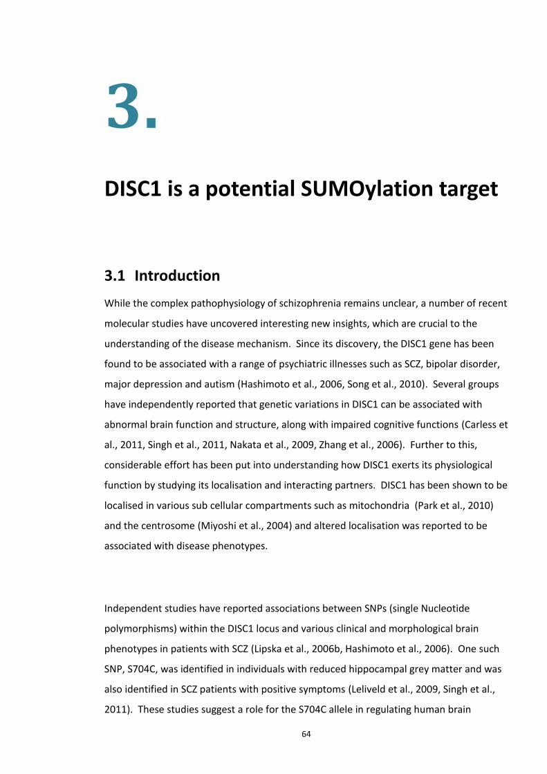
64
3.
DISC1 is a potential SUMOylation target
3.1 Introduction
While the complex pathophysiology of schizophrenia remains unclear, a number of recent
molecular studies have uncovered interesting new insights, which are crucial to the
understanding of the disease mechanism. Since its discovery, the DISC1 gene has been
found to be associated with a range of psychiatric illnesses such as SCZ, bipolar disorder,
major depression and autism (Hashimoto et al., 2006, Song et al., 2010). Several groups
have independently reported that genetic variations in DISC1 can be associated with
abnormal brain function and structure, along with impaired cognitive functions (Carless et
al., 2011, Singh et al., 2011, Nakata et al., 2009, Zhang et al., 2006). Further to this,
considerable effort has been put into understanding how DISC1 exerts its physiological
function by studying its localisation and interacting partners. DISC1 has been shown to be
localised in various sub cellular compartments such as mitochondria (Park et al., 2010)
and the centrosome (Miyoshi et al., 2004) and altered localisation was reported to be
associated with disease phenotypes.
Independent studies have reported associations between SNPs (single Nucleotide
polymorphisms) within the DISC1 locus and various clinical and morphological brain
phenotypes in patients with SCZ (Lipska et al., 2006b, Hashimoto et al., 2006). One such
SNP, S704C, was identified in individuals with reduced hippocampal grey matter and was
also identified in SCZ patients with positive symptoms (Leliveld et al., 2009, Singh et al.,
2011). These studies suggest a role for the S704C allele in regulating human brain
65
morphology and function. Other DISC1 variants such as L607F and R264Q have been
reported in both healthy individuals and in SCZ patients, and are known to affect cortical
morphology, though the exact mechanism by which these SNPs affect the brain
morphology is as yet unclear. It is interesting to note that no DISC1 mutations were
observed in certain cases of chronic mental disorders (Leliveld et al., 2008). Hence, in
this scenario it’s unclear how a healthy DISC1 gene contributes to the disease aetiology.
It may be hypothesised that post-translational modifications of DISC1 could be a reason
for the wild type and the mutant DISC1 proteins becoming dysfunctional in these
respective scenarios contributing to the pathophysiology of the disease.
3.1.1 Evidence of DISC1 aggregation contributing to impaired neuronal function
Carsten Korth and colleagues have conducted extensive studies of DISC1 aggregates in
various cell lines (Figure 3.1). They reported the presence of DISC1 in detergent insoluble
fraction prepared from post mortem brain tissue of individuals with chronic psychiatric
illness including SCZ (Korth, 2012). Studies in neuroblastoma cells demonstrate that the
100kDa DISC1 isoform can aggregate and that the resulting insoluble fraction fails to
interact with Ndel1, a well characterised DISC1 interacting partner (Leliveld et al., 2008).
DISC1 aggregation also influences the selection of the scaffold’s interacting partners. For
example, DISC1 aggregates have a reduced ability to interact with Ndel1 (Leliveld et al.,
2008) while they co-recruit soluble fractions of Dysbindin (Ottis et al., 2011). DISC1
aggregation was also found to be influenced by cellular stress (such as hydrogen
peroxide) and disease polymorphisms. DISC1 Ser704Cys variant was shown to have an
increased propensity to form aggregates (Korth, 2012).
DISC1 aggregates are also cell-invasive, a scenario mirrored in other psychiatric
aggregation illnesses with proteins such as α-synuclein and polyglutamine (Bader et al.,
2012). Independent studies by Kittler et al, reported the recruitment of GFP-DISC1
aggregates to aggresomes and subsequent degradation by the autophagy pathway.
Microscopic studies show GFP-DISC1 co-localised with autophagy markers such as LC3
66
(microtubule-associated protein light chain 3) in COS-7 cells and primary neuronal
cultures (Atkin et al., 2012). In COS7 cells, over-expressed GFP DISC1 co-recruits
endogenous DISC1 from soluble fractions under physiological stress. Mitochondrial
transport and neuronal function is also be impaired due to increased DISC1 aggregation
(Atkin et al., 2012). This may be attributed to yet unknown conformational changes in the
protein, which in turn may be influenced by post-translational modification events.
Figure 3.1 Laser scanning confocal microscopy of DISC1 aggregates in neuroblastoma cells. Figure taken from Korth et al. (2012). Mouse neuroblastoma cells (CAD cells) permanently transfected with monomeric red fluorescent protein (red) and transiently transfected with untagged, full length DISC1, stained with α-DISC1 mAb 14F2 and a secondary FITC-labelled antibody (green) and DAPI stained nucleus (blue). Scale : 10 µm
Furthermore characterisation of these DISC1 aggregates led to the identification of a
distinct oligomerization domain which lies towards the C-terminus (amino acids 640-854).
Genetic variants within this region such as S704 and C704 were reported to disrupt this
oligomerization of the DISC1 (Bader et al., 2012, Leliveld et al., 2009). It is also interesting
to note that this C terminal oligomerization domain on DISC1 corresponds to the
fragment lost in the Scottish translocation, and therefore it may have an underlying and
crucial role in the disease mechanism.
67
3.1.2 SUMOylation dependent regulation of protein aggregation in chronic mental disease
As discussed in chapter 1, post translational modifications such as phosphorylation and
SUMOylation have long been implicated in a number of neurodegenerative disorders,
including Alzheimer’s and Parkinson’s diseases (Bence et al., 2001). SUMOylation is a
post-translational modification characterised by the covalent conjugation of a small
ubiquitin like modifier (SUMO) to the target protein. The SUMO pathway is detailed in
section 1.4. The functional role of the SUMO pathway was emphasised by Ubc9 (SUMO
E2 enzyme) knockdown studies, which show that this pathway is crucial for eukaryotic cell
viability (Anderson et al., 2009). Studies on the role of SUMOylation in the central and
peripheral nervous system reveal a wide array of mechanisms by which this PTM can
contribute to the neuropathology of various diseases. Members of the SUMO cascade
regulate the activity of several signalling molecules contributing to cerebellar
development. One such example is MEF2A, whose activity has been shown to be
repressed by SUMOylation at K403 leading to post synaptic granule neuron
differentiation (Martin et al., 2007). Diseases such as NIID (Neuronal intranuclear
inclusion disease) are characterised by neuronal intra-nuclear inclusions (NIIs) carrying
SUMO-1 and SUMO substrates such as PML (promyelocytic leukaemia protein), RanGAP1
and HDAC4 (histone deacetylase 4) (Takahashi-Fujigasaki et al., 2006).
Emerging evidence describes SUMOylation as a powerful regulator of many signal
transduction pathways and gene expression cascades, with a majority of SUMOylated
substrates localised in the nucleus. As previously discussed (in section 1.4.1), the
functional consequences of SUMOylation of a substrate may include altered sub-cellular
and sub-nuclear localization, altered gene expression, as well as cross-regulation with
other post-translational modifications, such as ubiquitination. Such consequences are
often attained at the molecular levels via the alternation of protein - protein interactions
of the SUMO conjugated protein, by altered protein conformation, inhibition of an
existing binding site or the creation of a new binding site for the interacting proteins.
Pathological protein aggregates formed as a result of altered protein folding, stability or
targeting have been identified as a hallmark of many neurodegenerative disorders such as
68
Parkinson’s disease (aggregates of α-Synuclein constituting Lewy bodies) and Huntington
Disease (aggregates of Htt protein). However, recent studies have shown that deficiency
of a SUMO acceptor lysine in α-synuclein renders the Lewy bodies more toxic than the
wild type sequence (Krumova et al., 2011). Although, in some instances the converse was
true, where it was observed that SUMOylation reduced aggregation of mutant
Huntington protein and ataxin-7. There are also reports of SUMOylation dependent
enhancement of aggregate formation, as seen in super oxide dismutase 1 (SOD1), a
causative factor of ALS (Amyotrophic lateral sclerosis, a motor neuron disease) (Krumova
and Weishaupt, 2012). Progressive ataxia and dementia have also been demonstrated to
be associated with insoluble protein aggregates, showing strong immune reactivity for
SUMO1 (Martin et al., 2007).
3.2 Experimental aims
DISC1 has repeatedly been implicated as a candidate for susceptibility to SCZ and other
chronic mental disorders, and such studies have also shed light on the role of DISC1 in
neuronal development. Formation of DISC1 aggregates and reports of cell invasion by
these aggregates have provided proof of DISC1 mediated pathophysiology in mental
illnesses. However, the exact mechanistic relation of DISC1 to these diseases remains
unclear.
As several other aggregation prone proteins were known to be SUMO conjugated and
SUMOylation has been strongly implicated in several so called ‘aggregopathies’ such as
Parkinson’s disease, Huntington’s disease, it was hypothesised that SUMOylation may
play a role in regulating the formation of DISC1 aggregates.
My aims in the present section were:
1. To determine whether DISC1 is a substrate for SUMO conjugation.

69
2. To identify SUMO acceptor lysines (K) on DISC1.
3. To analyse the significance of my findings in light of what has already been published
and to explore the future perspectives.
70
3.3 Results
3.3.1 DISC1 is a putative SUMOylation target
SUMOylation usually occurs on a lysine residue entrenched in a core consensus motif
ψKxE/D (where ψ is a branched hydrophobic and x is any amino acid). Ubc9 (the SUMO
E2 enzyme) directly recognises this consensus motif and covalently bonds the SUMO
residue to the lysine residue within this motif (Bernier-Villamor et al., 2002). To add to
the complexity of this system, certain SUMO substrates also contain SUMO Interacting
Motifs (SIMs) which allow substrates to interact non-covalently with SUMO moieties
(Meulmeester and Melchior, 2008). The SUMOplot™ Analysis Program designed by
Abgent to predict putative SUMO consensus sequences was used to determine if DISC1
possessed SUMO consensus sequences. The SUMOplot Analysis Program predicts and
assigns probability scores to sites where SUMOylation may occur in a given protein. The
SUMOplot™ score system is based on the following criteria: direct amino acid match to
SUMO consensus sequence, and substitution of the consensus amino acid residues with
amino acid residues exhibiting similar hydrophobicity. The results of the analysis
suggested putative SUMO consensus sequences present within DISC1 (Figure 3.2).
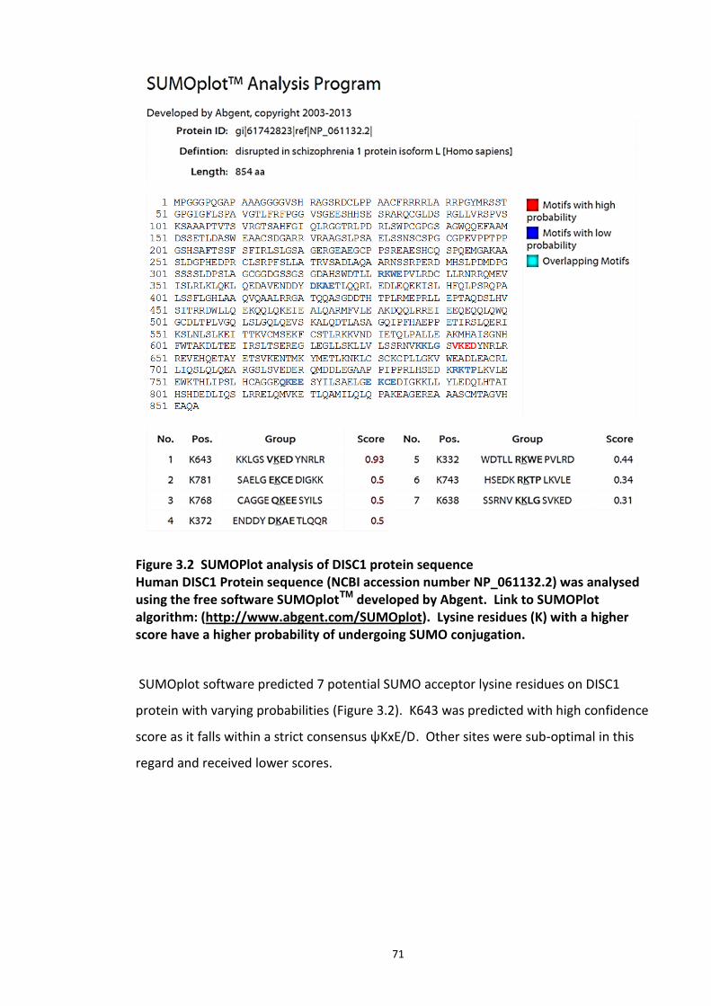
71
Figure 3.2 SUMOPlot analysis of DISC1 protein sequence Human DISC1 Protein sequence (NCBI accession number NP_061132.2) was analysed using the free software SUMOplotTM developed by Abgent. Link to SUMOPlot algorithm: (http://www.abgent.com/SUMOplot). Lysine residues (K) with a higher score have a higher probability of undergoing SUMO conjugation.
SUMOplot software predicted 7 potential SUMO acceptor lysine residues on DISC1
protein with varying probabilities (Figure 3.2). K643 was predicted with high confidence
score as it falls within a strict consensus ψKxE/D. Other sites were sub-optimal in this
regard and received lower scores.
72
3.3.2 DISC1 peptide array is selectively SUMOylated in vitro
Although search engine software such as SUMOplot is useful in identifying the SUMO
consensus sites on substrates, follow-up experiments are necessary to validate the sites.
Novel peptide array technology has been employed in number of studies to characterise
post-translational modifications such as Ubiquitination and SUMOylation. This approach
has been used to detect SUMO sites in substrates such as PDE4D5 (Li et al., 2010). A
study by K. Schwamborn and group describes a method by which peptide array
technology can be employed to identify SUMO target (Schwamborn et al., 2008). Peptide
arrays of overlapping 25-mer peptides sequentially shifted by five amino acids and
spanning the entire indicated DISC1 protein sequence (in Figure 3.1) were incubated with
SUMOylation assay mixture and this was followed by the detection of conjugated SUMO
with anti His antibody (to detect His-tagged SUMO moieties). Dark spots represent
positive areas of SUMO conjugation, while clear spots are negative for the modification
by SUMO (Figure 3.3). While no spots were detected on a control array that contained
the SUMOylation assay without the E2 ligase Ubc9, there were putative SUMO
conjugation sites identified on the test array (Figure 3.3).
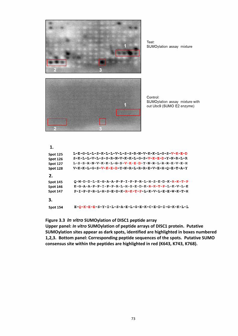
73
Figure 3.3 In vitro SUMOylation of DISC1 peptide array Upper panel: In vitro SUMOylation of peptide arrays of DISC1 protein. Putative SUMOylation sites appear as dark spots, identified are highlighted in boxes numbered 1,2,3. Bottom panel: Corresponding peptide sequences of the spots. Putative SUMO consensus site within the peptides are highlighted in red (K643, K743, K768).
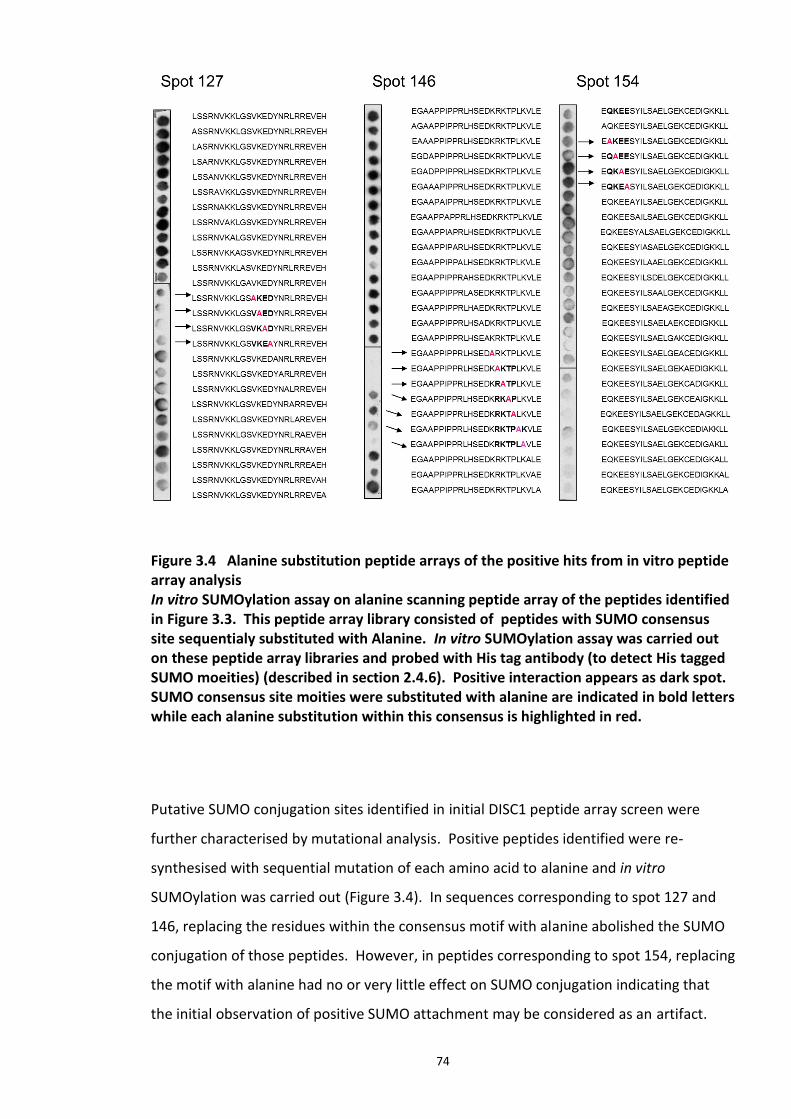
74
Figure 3.4 Alanine substitution peptide arrays of the positive hits from in vitro peptide array analysis In vitro SUMOylation assay on alanine scanning peptide array of the peptides identified in Figure 3.3. This peptide array library consisted of peptides with SUMO consensus site sequentialy substituted with Alanine. In vitro SUMOylation assay was carried out on these peptide array libraries and probed with His tag antibody (to detect His tagged SUMO moeities) (described in section 2.4.6). Positive interaction appears as dark spot. SUMO consensus site moities were substituted with alanine are indicated in bold letters while each alanine substitution within this consensus is highlighted in red.
Putative SUMO conjugation sites identified in initial DISC1 peptide array screen were
further characterised by mutational analysis. Positive peptides identified were re-
synthesised with sequential mutation of each amino acid to alanine and in vitro
SUMOylation was carried out (Figure 3.4). In sequences corresponding to spot 127 and
146, replacing the residues within the consensus motif with alanine abolished the SUMO
conjugation of those peptides. However, in peptides corresponding to spot 154, replacing
the motif with alanine had no or very little effect on SUMO conjugation indicating that
the initial observation of positive SUMO attachment may be considered as an artifact.
75
While SUMO prediction algorithms and in vitro peptide array screens are extremely useful
in identifying putative SUMOylation substrates, they are essentially artificial screening
tools that require validation to account for their limitations. It has been reported that
this motif is found in over a third of all characterised proteins, which leads to the real
possibility of identifying false positives using the SUMO prediction software (Song et al.,
2005). Many other proteins with the characterised consensus motif are not SUMO
targets, while many other proteins are SUMOylated at sites other than the consensus
motif. Hence, a range of biochemical validations are required before any physiological
importance can be attributed to the modification.
3.3.3 DISC1 is SUMOylated in HEK293 cells
3.3.3.1 Ubc9 and PIAS E3 ligases enhance DISC1 SUMOylation
SUMOylation is catalysed by Ubc9, an E2 conjugating enzyme and the E3 ligase, PIAS
(PIAS1, PIAS3, PIASy). Over expression of the SUMO E2 and E3 enzymes are known to
enhance SUMO conjugation of certain substrates, such as Smad4 (Lee et al., 2003). The
effect of co expression of the above mentioned SUMO E2 and E3 enzymes on DISC1
SUMOylation was therefore investigated. Figure 3.5 depicts the co-expression of Flag-
DISC1 with Ubc9, SUMO1/2 and various isoforms of PIAS (1/3/y). Over expression of
SUMO with the E2 and E3 ligase results in the retardation of the electrophoretic
migration of Flag-DISC1 compared to that co-expressed with empty vector. This suggests
that DISC1 has been modified by SUMOylation.
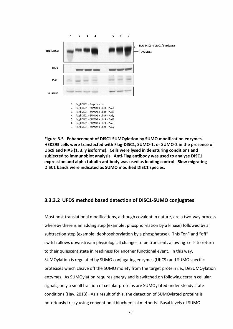
76
Figure 3.5 Enhancement of DISC1 SUMOylation by SUMO modification enzymes HEK293 cells were transfected with Flag-DISC1, SUMO-1, or SUMO-2 in the presence of Ubc9 and PIAS (1, 3, y isoforms). Cells were lysed in denaturing conditions and subjected to immunoblot analysis. Anti-Flag antibody was used to analyse DISC1 expression and alpha tubulin antibody was used as loading control. Slow migrating DISC1 bands were indicated as SUMO modified DISC1 species.
3.3.3.2 UFDS method based detection of DISC1-SUMO conjugates
Most post translational modifications, although covalent in nature, are a two-way process
whereby there is an adding step (example: phosphorylation by a kinase) followed by a
subtraction step (example: dephosphorylation by a phosphatase). This “on” and “off”
switch allows downstream physiological changes to be transient, allowing cells to return
to their quiescent state in readiness for another functional event. In this way,
SUMOylation is regulated by SUMO conjugating enzymes (UbC9) and SUMO specific
proteases which cleave off the SUMO moiety from the target protein i.e., DeSUMOylation
enzymes. As SUMOylation requires energy and is switched on following certain cellular
signals, only a small fraction of cellular proteins are SUMOylated under steady state
conditions (Hay, 2013). As a result of this, the detection of SUMOylated proteins is
notoriously tricky using conventional biochemical methods. Basal levels of SUMO
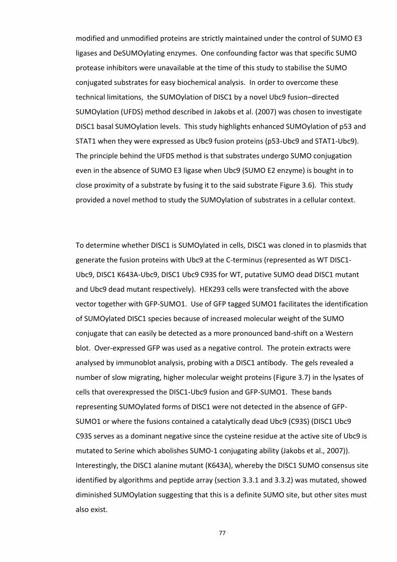
77
modified and unmodified proteins are strictly maintained under the control of SUMO E3
ligases and DeSUMOylating enzymes. One confounding factor was that specific SUMO
protease inhibitors were unavailable at the time of this study to stabilise the SUMO
conjugated substrates for easy biochemical analysis. In order to overcome these
technical limitations, the SUMOylation of DISC1 by a novel Ubc9 fusion–directed
SUMOylation (UFDS) method described in Jakobs et al. (2007) was chosen to investigate
DISC1 basal SUMOylation levels. This study highlights enhanced SUMOylation of p53 and
STAT1 when they were expressed as Ubc9 fusion proteins (p53-Ubc9 and STAT1-Ubc9).
The principle behind the UFDS method is that substrates undergo SUMO conjugation
even in the absence of SUMO E3 ligase when Ubc9 (SUMO E2 enzyme) is bought in to
close proximity of a substrate by fusing it to the said substrate Figure 3.6). This study
provided a novel method to study the SUMOylation of substrates in a cellular context.
To determine whether DISC1 is SUMOylated in cells, DISC1 was cloned in to plasmids that
generate the fusion proteins with Ubc9 at the C-terminus (represented as WT DISC1-
Ubc9, DISC1 K643A-Ubc9, DISC1 Ubc9 C93S for WT, putative SUMO dead DISC1 mutant
and Ubc9 dead mutant respectively). HEK293 cells were transfected with the above
vector together with GFP-SUMO1. Use of GFP tagged SUMO1 facilitates the identification
of SUMOylated DISC1 species because of increased molecular weight of the SUMO
conjugate that can easily be detected as a more pronounced band-shift on a Western
blot. Over-expressed GFP was used as a negative control. The protein extracts were
analysed by immunoblot analysis, probing with a DISC1 antibody. The gels revealed a
number of slow migrating, higher molecular weight proteins (Figure 3.7) in the lysates of
cells that overexpressed the DISC1-Ubc9 fusion and GFP-SUMO1. These bands
representing SUMOylated forms of DISC1 were not detected in the absence of GFP-
SUMO1 or where the fusions contained a catalytically dead Ubc9 (C93S) (DISC1 Ubc9
C93S serves as a dominant negative since the cysteine residue at the active site of Ubc9 is
mutated to Serine which abolishes SUMO-1 conjugating ability (Jakobs et al., 2007)).
Interestingly, the DISC1 alanine mutant (K643A), whereby the DISC1 SUMO consensus site
identified by algorithms and peptide array (section 3.3.1 and 3.3.2) was mutated, showed
diminished SUMOylation suggesting that this is a definite SUMO site, but other sites must
also exist.
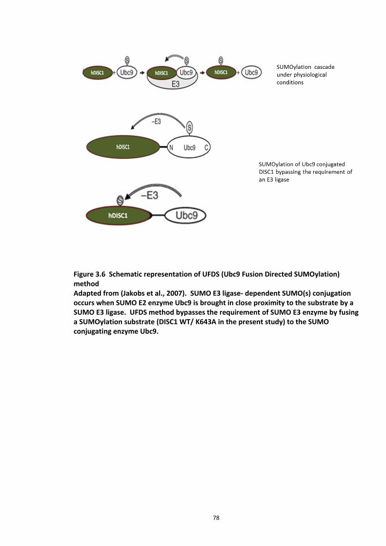
78
Figure 3.6 Schematic representation of UFDS (Ubc9 Fusion Directed SUMOylation) method Adapted from (Jakobs et al., 2007). SUMO E3 ligase- dependent SUMO(s) conjugation occurs when SUMO E2 enzyme Ubc9 is brought in close proximity to the substrate by a SUMO E3 ligase. UFDS method bypasses the requirement of SUMO E3 enzyme by fusing a SUMOylation substrate (DISC1 WT/ K643A in the present study) to the SUMO conjugating enzyme Ubc9.
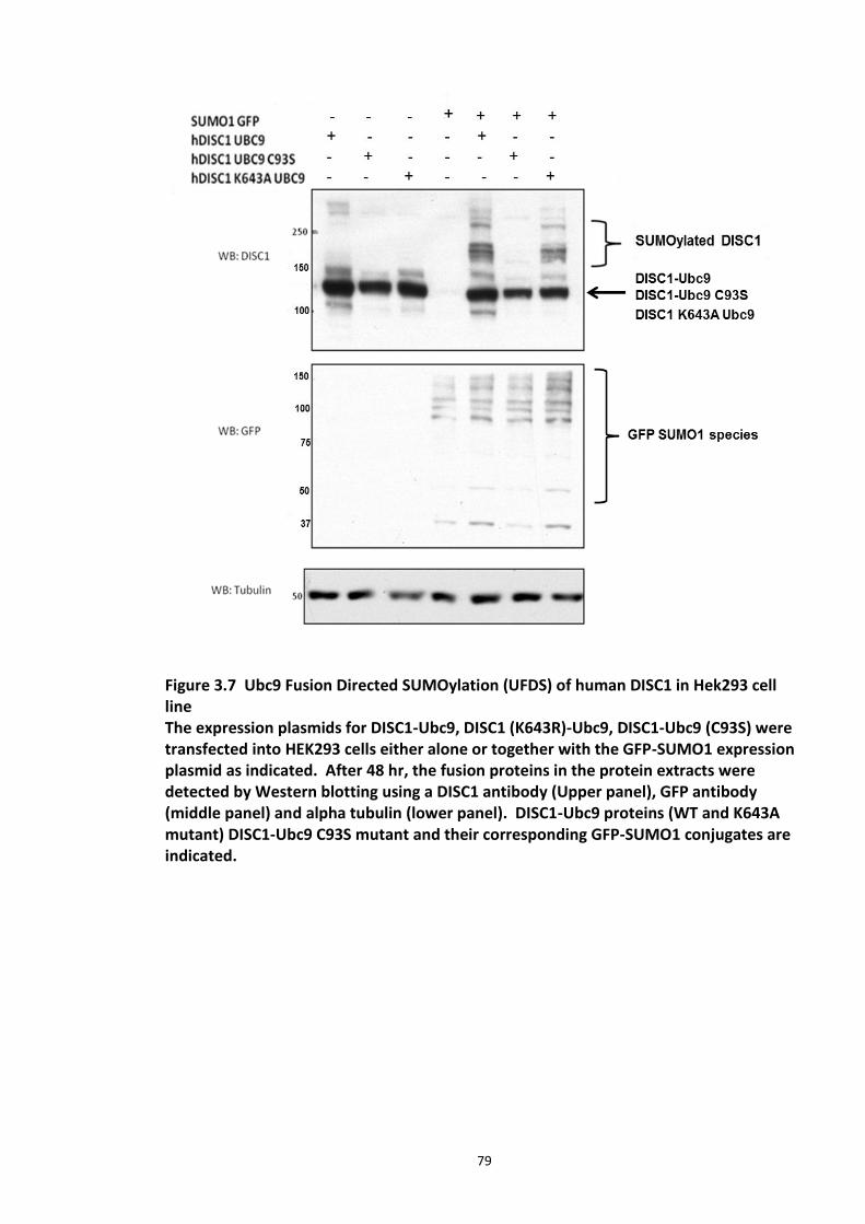
79
Figure 3.7 Ubc9 Fusion Directed SUMOylation (UFDS) of human DISC1 in Hek293 cell line The expression plasmids for DISC1-Ubc9, DISC1 (K643R)-Ubc9, DISC1-Ubc9 (C93S) were transfected into HEK293 cells either alone or together with the GFP-SUMO1 expression plasmid as indicated. After 48 hr, the fusion proteins in the protein extracts were detected by Western blotting using a DISC1 antibody (Upper panel), GFP antibody (middle panel) and alpha tubulin (lower panel). DISC1-Ubc9 proteins (WT and K643A mutant) DISC1-Ubc9 C93S mutant and their corresponding GFP-SUMO1 conjugates are indicated.
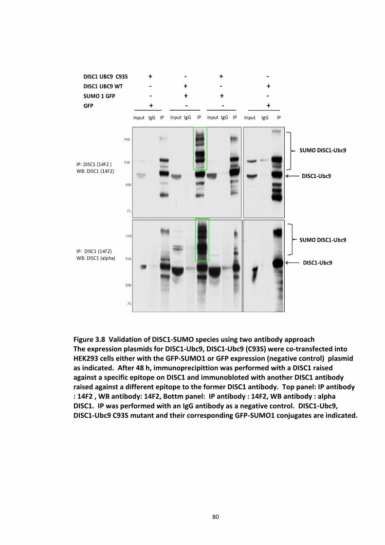
80
Figure 3.8 Validation of DISC1-SUMO species using two antibody approach The expression plasmids for DISC1-Ubc9, DISC1-Ubc9 (C93S) were co-transfected into HEK293 cells either with the GFP-SUMO1 or GFP expression (negative control) plasmid as indicated. After 48 h, immunoprecipittion was performed with a DISC1 raised against a specific epitope on DISC1 and immunobloted with another DISC1 antibody raised against a different epitope to the former DISC1 antibody. Top panel: IP antibody : 14F2 , WB antibody: 14F2, Bottm panel: IP antibody : 14F2, WB antibody : alpha DISC1. IP was performed with an IgG antibody as a negative control. DISC1-Ubc9, DISC1-Ubc9 C93S mutant and their corresponding GFP-SUMO1 conjugates are indicated.

81
By using the UFDS method, we established, for the first time, the novel post translational
modification on DISC1, SUMOylation. DISC1-Ubc9 fusion proteins were observed to be
migrating at about 125kDa. Co expression of these constructs with GFP-SUMO1 resulted
in the appearance of high molecular bands on the immunoblots, which correspond to the
DISC1–Ubc9 SUMO1 conjugates. DISC1-Ubc9 C93S (DISC1 fusion with the catalytically
inert form of UBC9) is completely devoid of these bands. This observation confirms the
specificity of SUMO conjugation. A high molecular weight band was observed when
DISC1-Ubc9 alone was overexpressed. This could likely be a result of endogenous SUMO
conjugation of the overexpressed DISC1 Ubc9 fusion construct (Figure 3.7).
To validate the high molecular weight bands appearing in the immunoblots as SUMO
DISC1 conjugates, immunoprecipitation was performed using UFDS lysates. DISC1
immunoprecipitation was performed using 14F2 (DISC1 antibody 1) and the immune
complexes were probed with alpha DISC1 antibody (DISC1 antibody 2). The presence of
DISC1 – SUMO conjugates in both the immunoblots rules out the possibility of non-
specific DISC1 bands (Figure 3.8).
The kinetics of SUMO conjugation on DISC1K643A Ubc9 mutant were also studied. K643
site was identified as a putative SUMO acceptor site on DISC1 in in vitro SUMOylation
studies. Ideally, alanine mutant at K643 should abolish the SUMO conjugation. However,
this was not the case, DISC1 SUMO conjugate bands can still be seen when an Alanine
mutant at K643 was overexpressed with GFP SUMO-1, which indicates the presence of
other potential SUMO acceptor lysine residues on DISC1 as predicted by SUMOPlot
software (figure 3.7). Hence, a mass spectrophotometry approach was employed to
uncover potential SUMO acceptor sites on DISC1 as detailed in section 3.3.4.
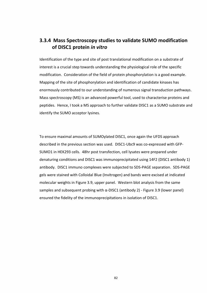
82
3.3.4 Mass Spectroscopy studies to validate SUMO modification of DISC1 protein in vitro
Identification of the type and site of post translational modification on a substrate of
interest is a crucial step towards understanding the physiological role of the specific
modification. Consideration of the field of protein phosphorylation is a good example.
Mapping of the site of phosphorylation and identification of candidate kinases has
enormously contributed to our understanding of numerous signal transduction pathways.
Mass spectroscopy (MS) is an advanced powerful tool, used to characterise proteins and
peptides. Hence, I took a MS approach to further validate DISC1 as a SUMO substrate and
identify the SUMO acceptor lysines.
To ensure maximal amounts of SUMOylated DISC1, once again the UFDS approach
described in the previous section was used. DISC1-Ubc9 was co-expressed with GFP-
SUMO1 in HEK293 cells. 48hr post transfection, cell lysates were prepared under
denaturing conditions and DISC1 was immunoprecipitated using 14F2 (DISC1 antibody 1)
antibody. DISC1 immuno complexes were subjected to SDS-PAGE separation. SDS-PAGE
gels were stained with Colloidal Blue (Invitrogen) and bands were excised at indicated
molecular weights in Figure 3.9, upper panel. Western blot analysis from the same
samples and subsequent probing with α-DISC1 (antibody 2) - Figure 3.9 (lower panel)
ensured the fidelity of the immunoprecipitations in isolation of DISC1.
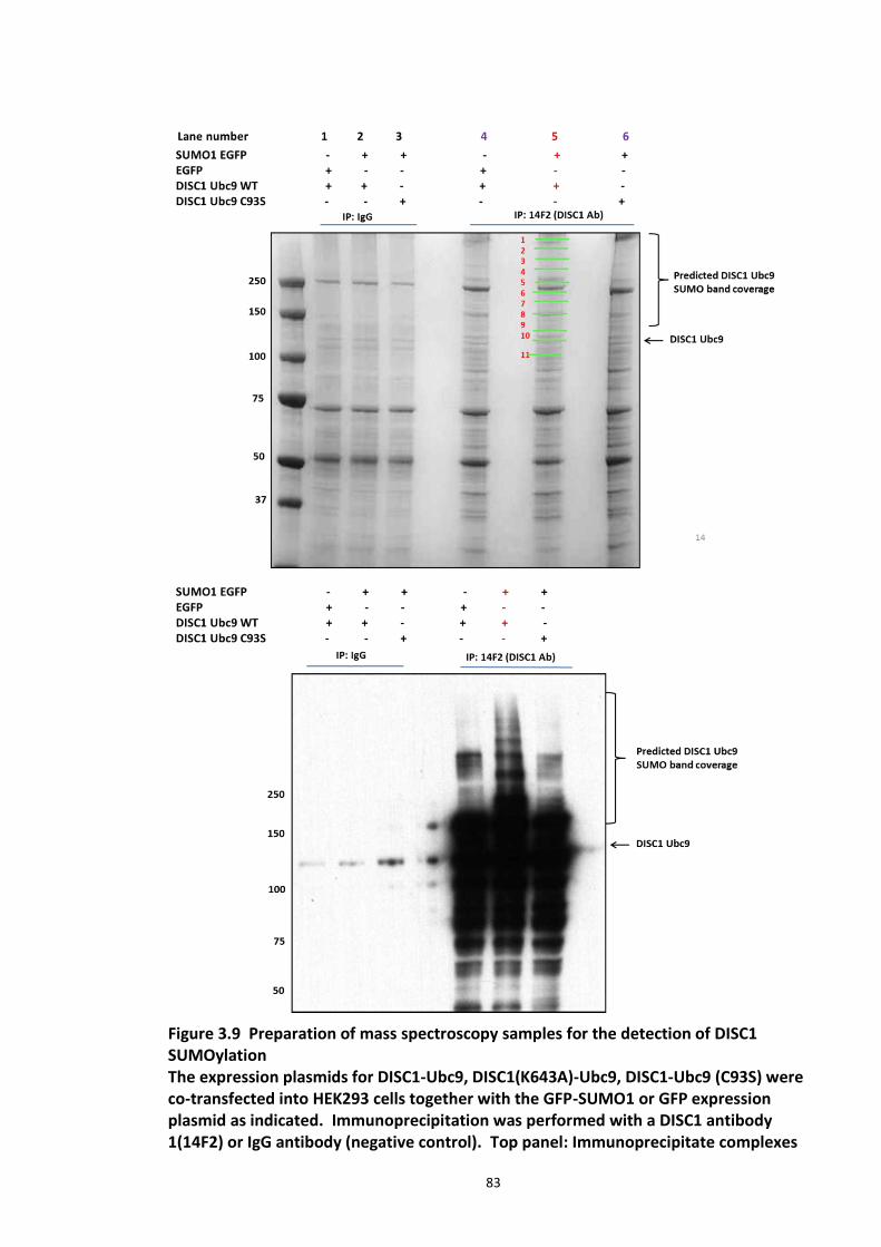
83
Figure 3.9 Preparation of mass spectroscopy samples for the detection of DISC1 SUMOylation The expression plasmids for DISC1-Ubc9, DISC1(K643A)-Ubc9, DISC1-Ubc9 (C93S) were co-transfected into HEK293 cells together with the GFP-SUMO1 or GFP expression plasmid as indicated. Immunoprecipitation was performed with a DISC1 antibody 1(14F2) or IgG antibody (negative control). Top panel: Immunoprecipitate complexes
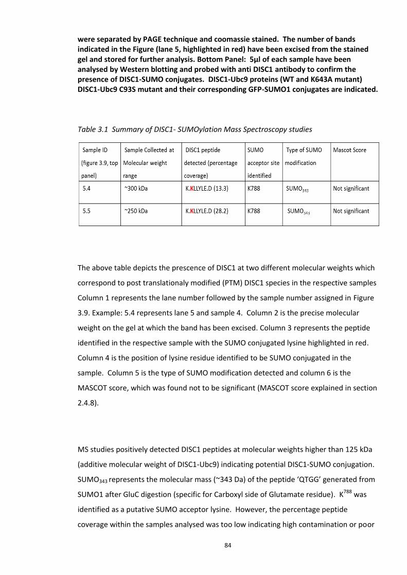
84
were separated by PAGE technique and coomassie stained. The number of bands indicated in the Figure (lane 5, highlighted in red) have been excised from the stained gel and stored for further analysis. Bottom Panel: 5µl of each sample have been analysed by Western blotting and probed with anti DISC1 antibody to confirm the presence of DISC1-SUMO conjugates. DISC1-Ubc9 proteins (WT and K643A mutant) DISC1-Ubc9 C93S mutant and their corresponding GFP-SUMO1 conjugates are indicated. Table 3.1 Summary of DISC1- SUMOylation Mass Spectroscopy studies
The above table depicts the prescence of DISC1 at two different molecular weights which
correspond to post translationaly modified (PTM) DISC1 species in the respective samples
Column 1 represents the lane number followed by the sample number assigned in Figure
3.9. Example: 5.4 represents lane 5 and sample 4. Column 2 is the precise molecular
weight on the gel at which the band has been excised. Column 3 represents the peptide
identified in the respective sample with the SUMO conjugated lysine highlighted in red.
Column 4 is the position of lysine residue identified to be SUMO conjugated in the
sample. Column 5 is the type of SUMO modification detected and column 6 is the
MASCOT score, which was found not to be significant (MASCOT score explained in section
2.4.8).
MS studies positively detected DISC1 peptides at molecular weights higher than 125 kDa
(additive molecular weight of DISC1-Ubc9) indicating potential DISC1-SUMO conjugation.
SUMO343 represents the molecular mass (~343 Da) of the peptide ‘QTGG’ generated from
SUMO1 after GluC digestion (specific for Carboxyl side of Glutamate residue). K788 was
identified as a putative SUMO acceptor lysine. However, the percentage peptide
coverage within the samples analysed was too low indicating high contamination or poor
85
sample quality. Although DISC1 peptides were detected in the other samples, the
confidence scoring was too low to be considered. (The principle of Mass spectroscopy
techniques is explained in section 2.4.8).
3.3.5 Consequences of DISC1 SUMOylation – effect on its protein interactions
SUMO modification of substrates has many physiological consequences. Since SUMO
proteins are about 10kDa size proteins, covalently conjugated to its substrate, this bulky
modification may either cause stearic hindrance thereby interfering with protein –
protein interactions or create additional binding surface on the substrate and promote or
strengthen protein interactions (Jentsch and Psakhye, 2013). Hence, we made an
attempt to understand the effect of SUMO modification on DISC1, on its protein
interactions. DISC1 is known to bind many proteins involved in several critical signalling
pathways reviewed in (Bradshaw and Porteous, 2010). Out of all these interacting
partners we were particularly interested in two crucial interactors – nuclear distribution
protein nudE-like 1 (Ndel1) and dishevelled axin domain containing 1 (DIXDC1) for the
following reasons: 1. Ndel1, a component of the dynein motor protein complex interacts
with DISC1 at the centrosome and mediates neurite outgrowth, migration and cortical
development (Ozeki et al., 2003a, Morris et al., 2003). 2. DISC1 interaction with DIXDC1
was shown to mediate embryonic neural progenitor proliferation and neuronal migration
(reviewed in section1.2.3) (Bradshaw and Porteous, 2010, Singh et al., 2010).
To investigate the effect of SUMO conjugation of DISC1 on its interaction with the above
mentioned proteins, we employed the UFDS method to enhance DISC1 SUMOylation
while co-expressing DISC1 binding partner of interest in HEK293 cells.
Immunoprecipitation was performed using an antibody specific for the tag on the
interacting partner as bait, followed by Western blot analysis of the immnuoprecipitate
complexes by probing for DISC1 antibody, to examine if the binding partner precipitated
only with unSUMOylated DISC1-Ubc9 or SUMOylated DISC1-Ubc9 or both.
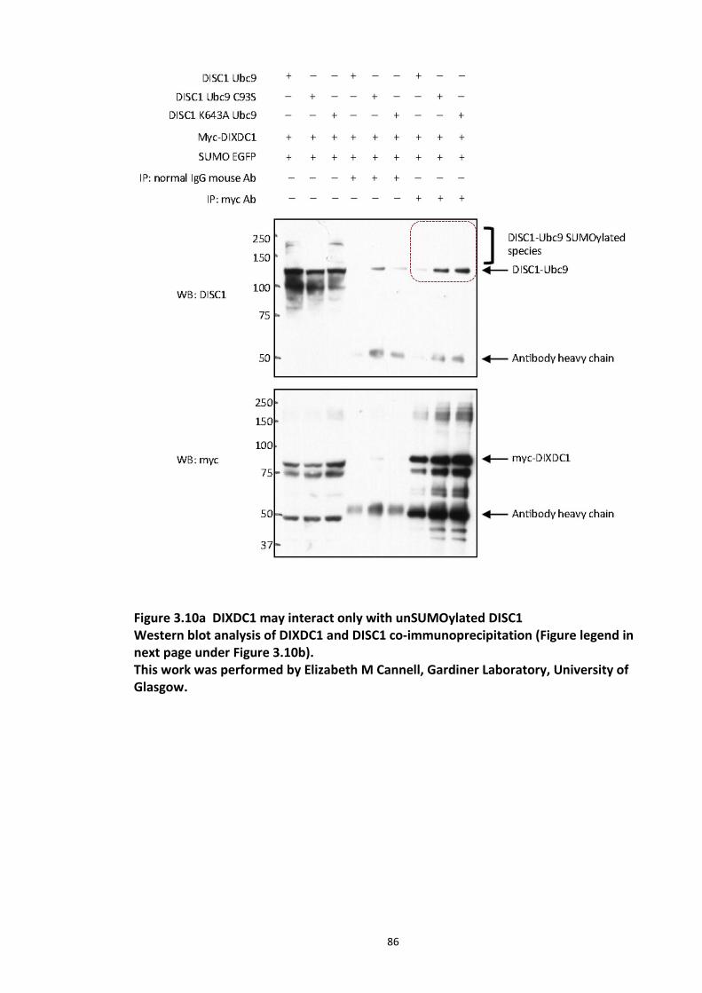
86
Figure 3.10a DIXDC1 may interact only with unSUMOylated DISC1 Western blot analysis of DIXDC1 and DISC1 co-immunoprecipitation (Figure legend in next page under Figure 3.10b). This work was performed by Elizabeth M Cannell, Gardiner Laboratory, University of Glasgow.
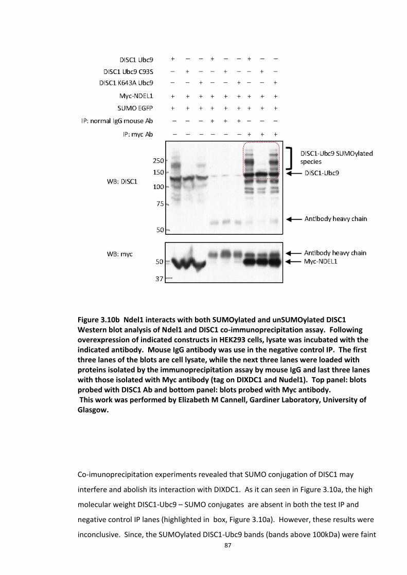
87
Figure 3.10b Ndel1 interacts with both SUMOylated and unSUMOylated DISC1 Western blot analysis of Ndel1 and DISC1 co-immunoprecipitation assay. Following overexpression of indicated constructs in HEK293 cells, lysate was incubated with the indicated antibody. Mouse IgG antibody was use in the negative control IP. The first three lanes of the blots are cell lysate, while the next three lanes were loaded with proteins isolated by the immunoprecipitation assay by mouse IgG and last three lanes with those isolated with Myc antibody (tag on DIXDC1 and Nudel1). Top panel: blots probed with DISC1 Ab and bottom panel: blots probed with Myc antibody. This work was performed by Elizabeth M Cannell, Gardiner Laboratory, University of Glasgow.
Co-imunoprecipitation experiments revealed that SUMO conjugation of DISC1 may
interfere and abolish its interaction with DIXDC1. As it can seen in Figure 3.10a, the high
molecular weight DISC1-Ubc9 – SUMO conjugates are absent in both the test IP and
negative control IP lanes (highlighted in box, Figure 3.10a). However, these results were
inconclusive. Since, the SUMOylated DISC1-Ubc9 bands (bands above 100kDa) were faint
88
in the input lanes (first three lanes) it could be possible that the SUMO DISC1-Ubc9 levels
in the immunoprecipitates were too low to be detected. More experimental repeats are
needed to ascertain this, which was not within the scope of this project.
Similar experiments with Ndel1 revealed that SUMOylation of DISC1 does not alter its
interaction with Ndel1 (Figure 3.10b) and the slow migrating, high molecular weight
DISC1-Ubc9-SUMO bands are present in the test IP lane but not the negative control lane
(highlighted with red box Figure 3.10b).
3.4 Discussion
3.4.1 DISC1 is modified by SUMO1
It has been established through the last two decades that post-translational modifications
(individually or co-operatively) regulate many signal transduction pathways (Prabakaran
et al., 2012). SUMOylation is one such kind of modification that is increasingly garnering
interest in the field of chronic mental disease research due to its role in protein
aggregation and solubility, a hallmark feature of many neurological disorders (Krumova
and Weishaupt, 2013, Krumova and Weishaupt, 2012).
It has been established here that there is a novel post translational modification on DISC1
in the form SUMOylation. DISC1 could be covalently modified by SUMO1 conjugation at
one or more site, as the SUMOplot algorithm identified number of putative consensus
SUMOylation motifs on DISC1 (Figure 3.2). In vitro peptide array studies identified three
of the predicted motifs K643, K768 and K743 (Figure 3.3, 3.4). However, Alanine
substitution scans of the putative sites identified in the initial peptide array screen
validated only two of those motifs, K643 and K743 (Figure 3.4). My preliminary studies
focused on the characterisation of the K643 site as it was predicted to be more likely to
be SUMO conjugated implied by highest score of 0.93 in the SUMOplot algorithm, while it
is also conserved in mouse and human. However, mutational studies on DISC1 K643R-
Ubc9 conjugate reduced but did not completely abolish SUMO modification. This may be
89
because of the presence of more than one SUMO acceptor site as predicted by SUMOplot
algorithm.
Studies show that the mere presence of consensus Lys residues in the SUMO motif is
often not sufficient for recognition by the SUMO conjugation machinery, there are other
criteria such as the presence of downstream acidic patches or pre-existing
phosphorylation (Yang et al., 2006). If the lysine residues are embedded in stable
structural helices, it may not be recognised by the SUMO conjugating enzyme, Ubc9.
However, it is important to note that certain substrates are SUMOylated at a non-
consensus lysine residue, for example, human E2-25K (Pichler et al., 2005). If this is the
case, it is difficult to predict such random sites. The UFDS method has proved to be
useful in efficient SUMOylation of DISC1 in my cell line model and this represents the first
reliable indication that DISC1 can be SUMOylated in vitro.
Mass spectroscopy, was also used in the present study to verify DISC1 as a SUMO
substrate and identify the site of post translational modification. DISC1-Ubc9 migrates at
about 125kDa on SDS-PAGE; however, as expected, the MS study detected DISC1
peptides in samples excised at molecular weights greater than 125kDa indicative the
putative post translational modification by SUMO. Though all modifications could not be
identified, SUMO343 modification was detected at K788 (SUMO343 refers to the molecular
weight of the putative adduct, 343.1492 Da molecular weight corresponds to the peptide
‘QTGG’ remaining after GluC cleavage of a SUMO1-modified lysine). Only one SUMO
acceptor lysine was identified in the MS studies while several bands are observed in UFDS
methods based immunoblots. From the MS spectral data it was evident that the sample
quality was not optimal. Absolute identification of SUMOylation sites on substrates has
always been challenging due to the reversible dynamic nature of SUMOylation (achieved
by the action of SUMO conjugation and deSUMOylating/SUMO protease enzymes in cells)
which deteriorates the sample quality. SUMO1 forms chains less efficiently in vitro since
it lacks the internal SUMOylation site which is present in SUMO 2 and SUMO 3 (Vertegaal,
2007). Also, successful detection of SUMO sites depends on the sensitivity of mass
spectrometer which can retrieve less abundant SUMOylated peptides from the MS
90
samples (Knuesel et al., 2005). Unfortunately technical and biological repeats for MS
studies were not feasible in this project due to time constraints and budgetary concerns.
It is also important to note that just as any other post translational modification,
SUMOylation is spatio temporally regulated which confers additional levels of regulation
(Loriol et al., 2012). It may be triggered by a physiological stimulus or other pre-existing
post translational modification such as phosphorylation as seen in case of c-Jun and p53
SUMOylation (which require a phosphorylation event) (Muller et al., 2000). With no
knowledge of the above stimuli nor the tertiary structure of DISC1, it would be difficult to
induce enough SUMOylation in cells to generate high quality samples for MS, which may
generate more information on this modification. However, my work provides a number
of different avenues of biochemical proof that the SUMO conjugation of DISC1 occurs in
vitro.
3.4.2 Significance of DISC1 as a SUMO substrate
Over the past few decades, there have been very few developments in psychiatric drug
development and most of the newer antipsychotics and anti-depressants have been
reported to cause severe side effects. The increasing characterisation and appreciation of
the DISC1 interactome is a factor that may provide novel opportunities to discover new
classes of brain function altering drugs (Hikida et al., 2012, Soares et al., 2011).
Demonstration of the SUMO conjugation of DISC1 opens up new opportunities to explore
its physiological functions. DISC1 is known as a scaffold protein with no reported
enzymatic activity. Several of its interactions have been studied and known to influence
various neuronal signalling pathways (Bradshaw and Porteous, 2010, Chubb et al., 2008).
One of the molecular consequences of SUMO conjugation of a substrate is a
conformational change leading to altered protein- protein interaction. Hence my studies
may prove useful in developing further understanding how DISC1 interactions are
modulated.
91
SUMO and other post translational modifications are known to regulate many aspects of
substrate proteins. Phosphorylation of DISC1 orchestrates neuronal progenitor cell
migration and proliferation in mice (Ishizuka et al., 2011c). Some substrate proteins
require a pre-existing phosphorylation to induce SUMO conjugation as seen in regulation
of heat shock factor HSFb and MEF2 (Hietakangas et al., 2006). SUMOylation and
ubiquitination are also known to cross-talk via regulation of each other’s respective
enzymatic cascade. This type of regulation is quite evident in E2-25K, an E2 enzyme ,
which loses its ubiquitin conjugating ability upon SUMOylation (Pichler et al., 2005). The
next chapter investigates another novel modification crucial in regulating DISC1 protein
turn over: ubiquitination.
Post translational modification by SUMO conjugation has been shown to influence
subcellular localisation of substrates such as RanGAP (a small GTPase activating protein).
While SUMO-RanGAP is co-localised with the nuclear pore complex, its unmodified
counterpart is predominantly cytoplasmic (Matunis et al., 1996). DISC1 is hypothesised to
exert its function by interaction with specific proteins in discrete sub cellular
compartments. For example, DISC1 is known to interact with NudE-like (NUDEL/NDEL1)
at the centrosome and activating transcription factor 4 (ATF4)/CREB2 in the nucleus
(Kamiya et al., 2005a, Morris et al., 2003). A 75kDa DISC1 isoform has been reported to
be specifically enriched in the nucleus in brain autopsies of patients with sporadic
schizophrenia and major depression as well as substance and alcohol abuse (Sawamura et
al., 2005a) suggesting its role in the pathophysiology of these major mental illnesses.
Hence, it would be interesting to investigate the role of SUMO conjugation on sub cellular
targeting of DISC1.
In an attempt to develop a fly model expressing human DISC1, Akira Sawa and colleagues
have shown that DISC1 appears in a punctate pattern co-localised with PML protein, a
core component of PML nuclear bodies (Sawamura et al., 2008). Another study has
shown that SUMO1 conjugation of PML protein is necessary for formation of PML bodies
(Zhong et al., 2000). Hence, it would be interesting to investigate if DISC1 aggregation is
regulated by SUMO modification.
92
In this project, the effect of SUMO modification of DISC1 on its interacting partner profile
was explored (section 3.3.5). Of the two binding proteins chosen, DIXDC1 was likely to be
interacting with non SUMOylated DISC1 although the results were inconclusive and
require further characterisation. DISC1 functionally interacts with DIXDC1 and regulates
neuronal progenitor proliferation during early brain development by modulating Wnt-
GSK3β/β-catenin pathway (Singh et al., 2010). Their role in neuronal migration was
suggested to occur through a pathway independent of Wnt/β-catenin signalling (Singh et
al., 2010). If SUMOylation of DISC1 indeed abolishes its interaction with DIXDC1, it is
likely that this PTM on DISC1 may be a crucial event during early brain development and
is influenced by DIXDC1-DISC1 complex formation.
Another interacting partner studied in this context is Nudel1, is a Cysteine protease
(endo-oligopeptidase A (EOPA)) whose activity is inhibited by DISC1 interaction in in vitro
assays (Hayashi et al., 2005). Nudel also regulates neuronal differentiation of PC12 cell
line (Ozeki et al., 2003a). Co-immunoprecipitation experiments show that Nudel1
interacts with both SUMOylated and non SUMOylated DISC1. However, these
experiments are only qualitative and do not provide any information on the strength of
binding or interaction. Presence of SUMO Interacting Motifs (SIMs) on proteins increases
their binding affinity to proteins which are covalently conjugated to SUMO proteins,
which is one of the possible consequence of protein SUMOylation (Jentsch and Psakhye,
2013).
Co-immunoprecipitation experiments performed in rodent brain lysates have shown that
binding occurs between the C terminal region (fragment spanning from 596 to 852
residues) of DISC1 and region between the calponin homology domain and the coiled-coil
domain of DIXDC1 (Singh et al., 2010). Ndel1 interacts with the C-terminal domain of
DISC1 (residues 727-854) (Ozeki et al., 2003a, Soares et al., 2011). As stated earlier, the
binding sites for DIXDC1 and Nudel1 on DISC1 overlap. It has been suggested that Cdk5
mediated phosphorylation on DIXDC1 results in the formation of DISC1/DIXDC11/Ndel1
complex which is indispensable in neuronal migration (Singh et al., 2010). Taking this in
93
to account, it is likely that SUMO conjugation of DISC1 may act as a trigger between
neuronal progenitor proliferation and migration and this event mimics the
phosphorylation event on DISC1 which acts as a molecular switch between proliferation
of mitotic progenitor cells enabling the migration of post-mitotic neurons. Putative
truncated protein in the DISC1 translocation in the Scottish pedigree (TrDISC1) lacks the
binding site for both these proteins as well as the identified SUMO acceptor lysine (K643
and K788). Hence, it is likely that this PTM on DISC1 influences various physiological
events possibly mediated by its interactions with various proteins.
Given the role SUMOylation in CNS related disease pathways (reviewed in section 1.4.2) it
can be considered as an attractive target for therapeutic intervention. For example the
increase in overall protein SUMOylation observed in neurons is a neuroprotective
response (Loftus et al., 2009) while SUMO-1 expression has been shown to rapidly
increase during hypoxia (Shao et al., 2004). It would be interesting to investigate if SUMO
conjugation of DISC1 has such a role to play in the disease mechanism of DISC1 associated
psychiatric illnesses.
To this end, future work should 1. Unequivocally identify the SUMO sites on DISC1 2. Use
site directed mutagenesis to block the SUMO acceptor sites 3.analysis of the aggregation
and cellular location of Wt vs mutant SUMO-null DISC1. Interesting insights into the
functionality of the SUMOylation of DISC1 in animal models could be gleaned by
generating transgenic mice carrying DISC1 mutations at SUMO conjugation sites.
3.5 Chapter summary
In summation, my work presented in this chapter uncovers a novel post translational
modification on DISC1 in the form of SUMOylation. In vitro SUMOylation of DISC1
peptide array identified K643 as a potential acceptor lysine. SUMOylation of arginine
mutant at K643 site on DISC1 was attenuated using the UFDS method suggesting that this
site as a principle SUMO conjugation site on DISC1. However, this site could not be
verified using MS studies due to poor sample quality. Our co-immnoprecipitation studies
also suggest that SUMO modification of DISC1 may abolish its binding with DIXDC1, while
its interaction with Nudel1 remained unaffected.
94
4.
Proteasomal regulation of human DISC1 protein
4.1 Introduction
Gene expression studies carried out on the post-mortem brain tissue of subjects with SCZ
has provided insight in to possible involvement of ubiquitin proteasome system
dysregulation in the SCZ disease mechanism. A number of studies used cDNA microarrays
to evaluate the profile of differential gene expression in these samples vs control.
Consequently, Ubiquitin carboxyl-terminal esterase L1 (UCHL1), a deubiquitinating
enzyme with predominant expression in CNS (known to be associated with neurological
disorders such as AD and PD) and PSMC6 (proteasome 26S subunit, ATPase, 6 encodes
one of the catalytic subunits of the 20S subunit of the proteasome) are two candidate
genes that were significantly down-regulated in subjects with SCZ (Altar et al., 2005,
Vawter et al., 2001). Another study used a blood based micro-array approach to screen
for the gene expression patterns from SCZ and BD patients belonging to two different
ethnic groups and in doing so, identified a number of UPS genes to be dysregulated
(Bousman et al., 2010). Various other reports of altered gene expression of UPS
components in SCZ and Bipolar disorder (Middleton et al., 2002) garnered my interest.
The functional role of DISC1 in neuronal development was initially investigated by
delineating its interacting partners using a yeast 2 hybrid screen (Millar et al., 2003, Ozeki
et al., 2003b). Key proteins involved in neuronal development, neuron migration and
synaptic signalling have been identified in these screens. These studies emphasise the
functional significance of DISC1 in brain development and strengthen its role in disease
95
mechanisms of psychiatric illnesses such as SCZ and certain mood disorders (Hashimoto
et al., 2006). The effects of DISC1 knockdown has been studied using various methods.
In utero gene transfer was used to transiently knockdown DISC1 in mouse models to
understand its role in growth and maturation of the brain (Niwa et al., 2010). This study
used GFP tagged DISC1 shRNA, which was injected in to the pre frontal cortex of mice
brain at E14. This resulted in morphological abnormalities in postnatal meso cortical
dopaminergic maturation, in turn, resulting in several behavioural abnormalities in
adulthood (Niwa et al., 2010). shRNA mediated DISC1 knockdown in cortical precursor
cells in ventricular zone, abolished the migration of cells in to the cortical plate (Kamiya
et al., 2005b).
Various independent studies focused on the functional consequences of the truncated
form of DISC1 protein (Millar et al., 2000c)) and revealed its dominant negative role. Over
expressed C-terminal truncated DISC1 (TrDISC1, amino acids 1-597) has been shown to
interfere with the wild type protein product of the functional allele affecting its function,
subcellular localisation and microtubular dynamics (Ozeki et al., 2003a, Kamiya et al.,
2005b). However, it should be noted that TrDISC1 protein has not been detected in cells.
Apart from the Scottish pedigree, several other groups independently reported many
other SNPs within the DISC1 locus in various ethnic groups to be associated with chronic
mental illnesses (Zhang et al., 2006, Saetre et al., 2008, Callicott et al., 2005). Exonic
polymorphisms such as S704 and C704 have been implicated in SCZ and major depression
respectively (Leliveld et al., 2009, Song et al., 2010). However, the effect of these SNPs or
truncations on the protein confirmation or stability has not been investigated.
The hallmark of many psychiatric illnesses is pathological aggregation of defective or
mutant proteins resulting in dysregulation of key signalling pathways, ultimately leading
to chronic neurological disorders such as Alzheimer’s disease, Creutzfeldt-Jakob’s
disease, Parkinson’s disease and Huntington’s disease (Leliveld et al., 2009). A study of
DISC1 dimerisation and oligomerisation, revealed interesting leads regarding the
pathological role of DISC1 (Leliveld et al., 2009). The presence of sarkosyl (detergent)
insoluble DISC1 aggregates in the brains of a sub set of cases with mental disease can be
96
triggered by subtle conformational changes and possibly yet unknown posttranslational
modifications (Leliveld et al., 2008). These putative post translational modifications in
turn might abolish or create new interacting partners leading to loss or gain in function.
In spite of the increasing evidence that DISC1 is a key susceptibility factor in SCZ, not
much is known about its turnover/half-life and whether its regulatory mechanisms at the
protein level depend on its cycling synthesis or degradation. Very few studies
investigated the levels of DISC1 at the transcript and protein level. Increased DISC1
transcript levels were reported in peripheral blood mononuclear cells of patients with
schizophrenia. In an effort to identify a suitable biomarker for SCZ, the above study
compared the transcript levels of various DISC1 isoforms in blood samples of patients
experiencing psychosis, to those samples collected after neuroleptic doses of haloperidol
was administered. A predominant decrease in DISC1 transcript levels was reported
following the treatment, suggesting a strong association of DISC1 levels to the disease
mechanism (Olincy et al., 2011). Another study used immunostaining techniques to
investigate the effect of epileptic seizures on DISC1 levels in adult rats (Fournier et al.,
2009). Electrically induced epileptic seizures significantly reduced DISC1 immunolabeling
within the dentate gyrus, while the width of the granule cell layers remained the same
between the control and induced (Fournier et al., 2009). This study by Fournier and
collegues suggests the crucial role of DISC1 levels in cytoarchitectural abnormalities of the
dentate gyrus observed in some SCZ subjects (Arnold, 1999).
Approximately 10-20% cellular proteins are short lived, with a half-life less than 3 hours.
Studies using proteasomal inhibitors show that up to 90% cellular proteins are degraded
by UPS (Lee and Goldberg, 1998). Ubiquitinated substrates are short lived and it is
challenging to capture the signal of ubiquitinated protein of interest using conventional
biochemical techniques. Proteasome inhibitors, hence, are very useful to stabilise cellular
levels of modified proteins. Proteins, whose turnover is regulated by UPS, should
accumulate, in a ubiquitin-conjugated form, upon proteasome inhibition. My initial
experiments were concerned with investigating the consequences of inhibiting the
proteasome on DISC1 and studying the dynamics of DISC1 ubiquitination (L isoform).
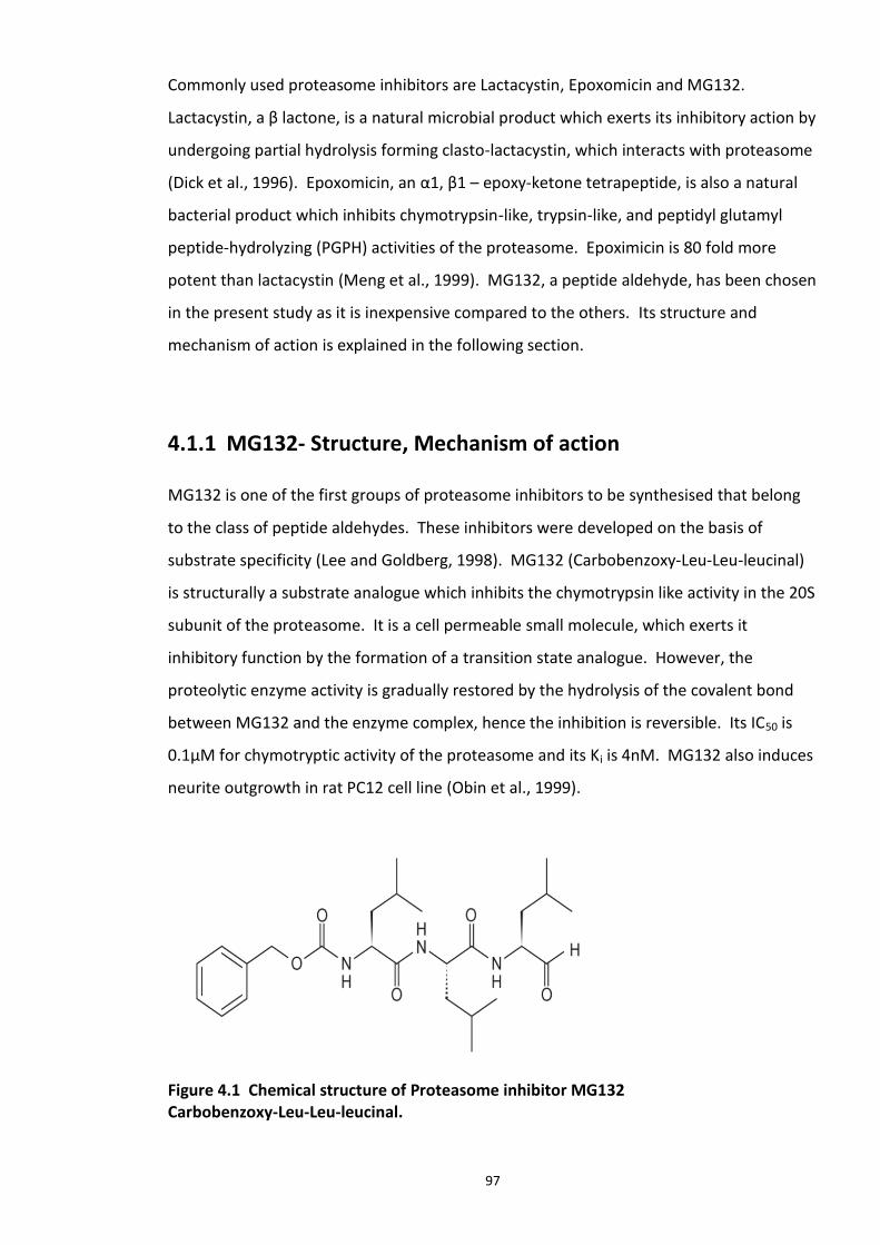
97
Commonly used proteasome inhibitors are Lactacystin, Epoxomicin and MG132.
Lactacystin, a β lactone, is a natural microbial product which exerts its inhibitory action by
undergoing partial hydrolysis forming clasto-lactacystin, which interacts with proteasome
(Dick et al., 1996). Epoxomicin, an α1, β1 – epoxy-ketone tetrapeptide, is also a natural
bacterial product which inhibits chymotrypsin-like, trypsin-like, and peptidyl glutamyl
peptide-hydrolyzing (PGPH) activities of the proteasome. Epoximicin is 80 fold more
potent than lactacystin (Meng et al., 1999). MG132, a peptide aldehyde, has been chosen
in the present study as it is inexpensive compared to the others. Its structure and
mechanism of action is explained in the following section.
4.1.1 MG132- Structure, Mechanism of action
MG132 is one of the first groups of proteasome inhibitors to be synthesised that belong
to the class of peptide aldehydes. These inhibitors were developed on the basis of
substrate specificity (Lee and Goldberg, 1998). MG132 (Carbobenzoxy-Leu-Leu-leucinal)
is structurally a substrate analogue which inhibits the chymotrypsin like activity in the 20S
subunit of the proteasome. It is a cell permeable small molecule, which exerts it
inhibitory function by the formation of a transition state analogue. However, the
proteolytic enzyme activity is gradually restored by the hydrolysis of the covalent bond
between MG132 and the enzyme complex, hence the inhibition is reversible. Its IC50 is
0.1μM for chymotryptic activity of the proteasome and its Ki is 4nM. MG132 also induces
neurite outgrowth in rat PC12 cell line (Obin et al., 1999).
Figure 4.1 Chemical structure of Proteasome inhibitor MG132 Carbobenzoxy-Leu-Leu-leucinal.
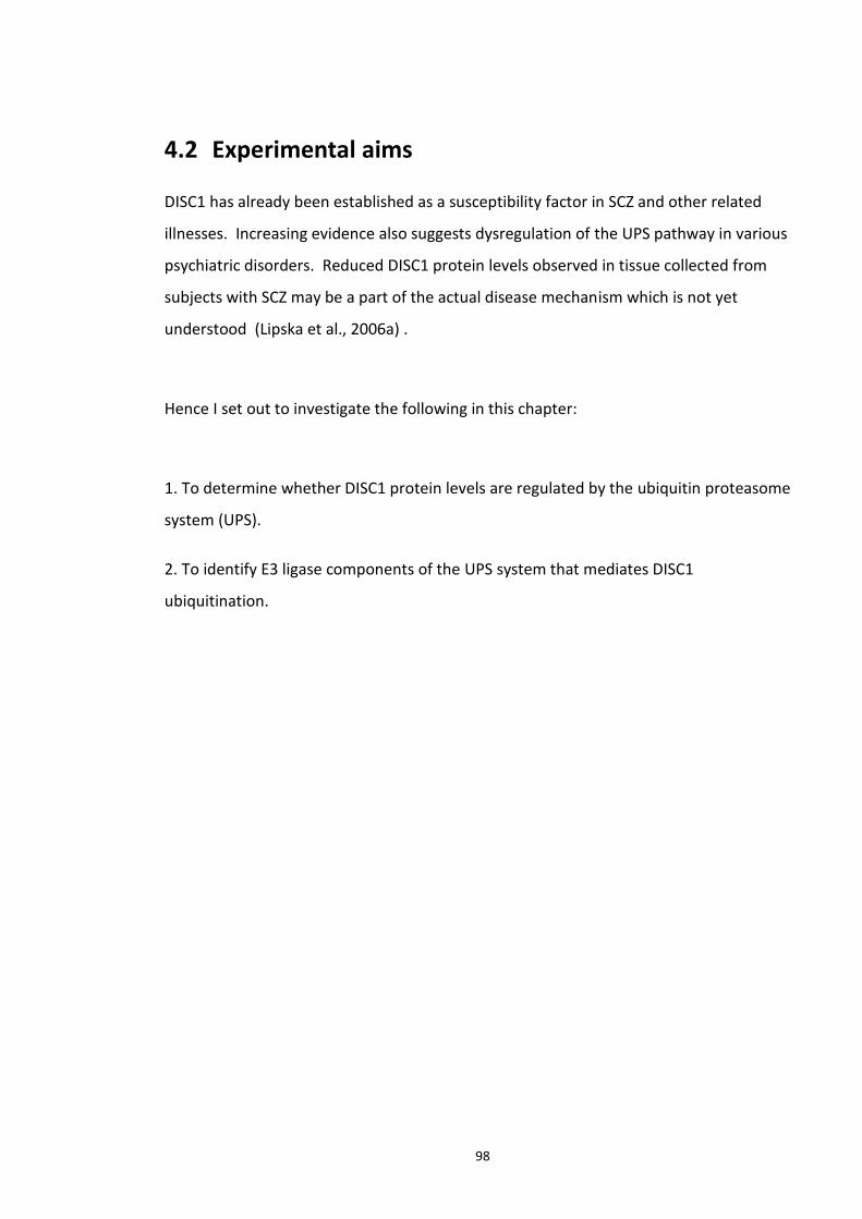
98
4.2 Experimental aims
DISC1 has already been established as a susceptibility factor in SCZ and other related
illnesses. Increasing evidence also suggests dysregulation of the UPS pathway in various
psychiatric disorders. Reduced DISC1 protein levels observed in tissue collected from
subjects with SCZ may be a part of the actual disease mechanism which is not yet
understood (Lipska et al., 2006a) .
Hence I set out to investigate the following in this chapter:
1. To determine whether DISC1 protein levels are regulated by the ubiquitin proteasome
system (UPS).
2. To identify E3 ligase components of the UPS system that mediates DISC1
ubiquitination.
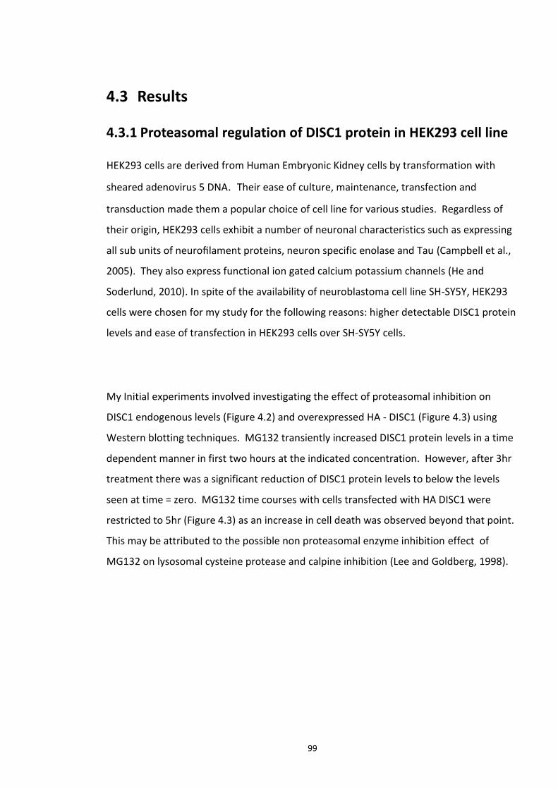
99
4.3 Results
4.3.1 Proteasomal regulation of DISC1 protein in HEK293 cell line
HEK293 cells are derived from Human Embryonic Kidney cells by transformation with
sheared adenovirus 5 DNA. Their ease of culture, maintenance, transfection and
transduction made them a popular choice of cell line for various studies. Regardless of
their origin, HEK293 cells exhibit a number of neuronal characteristics such as expressing
all sub units of neurofilament proteins, neuron specific enolase and Tau (Campbell et al.,
2005). They also express functional ion gated calcium potassium channels (He and
Soderlund, 2010). In spite of the availability of neuroblastoma cell line SH-SY5Y, HEK293
cells were chosen for my study for the following reasons: higher detectable DISC1 protein
levels and ease of transfection in HEK293 cells over SH-SY5Y cells.
My Initial experiments involved investigating the effect of proteasomal inhibition on
DISC1 endogenous levels (Figure 4.2) and overexpressed HA - DISC1 (Figure 4.3) using
Western blotting techniques. MG132 transiently increased DISC1 protein levels in a time
dependent manner in first two hours at the indicated concentration. However, after 3hr
treatment there was a significant reduction of DISC1 protein levels to below the levels
seen at time = zero. MG132 time courses with cells transfected with HA DISC1 were
restricted to 5hr (Figure 4.3) as an increase in cell death was observed beyond that point.
This may be attributed to the possible non proteasomal enzyme inhibition effect of
MG132 on lysosomal cysteine protease and calpine inhibition (Lee and Goldberg, 1998).
100
Figure 4.2 Proteasome inhibition transiently stabilises endogenous DISC1 levels in HEK293 cell line. Top panel: HEK293 cell cultures were treated with MG132 at 10µM concentration for indicated time points and endogenous DISC1 protein levels were analysed by Western blotting technique using Anti DISC1 antibody. Anti GAPDH antibody was used as a loading control. Bottom panel: Densitometric analysis of the amount of DISC1 protein normalised to GAPDH expression levels. Data is represented as ± S.E.M. of three biological repeats. Statistical significances between control (time = 0) and a time point was calculated using one way analysis of variance (ANOVA). *P < 0.05 values were considered significant.
101
Figure 4.3 Proteasome inhibition transiently stabilises over expressed DISC1 levels in He293 cell line. Top panel: HEK293 cell cultures were transiently transfected with HA (haemagglutinin)- DISC1. 48hr post incubation Ubiquitination cell cultures were treated with MG132 at 10 µM concentration for indicated time points and DISC1 protein levels were analysed by Western blotting technique with anti HA antibody. Anti GAPDH antibody was used as a loading control. Bottom panel: Densitometric analysis of the amount of DISC1 protein normalised to GAPDH expression levels. Data is represented as ± S.E.M. of three biological repeats. Statistical significances between control (time = 0) and a time point was calculated using one way analysis of variance (ANOVA). *P < 0.05 values were considered significant.
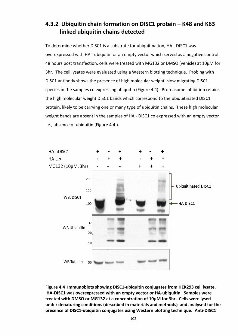
102
4.3.2 Ubiquitin chain formation on DISC1 protein – K48 and K63 linked ubiquitin chains detected
To determine whether DISC1 is a substrate for ubiquitination, HA - DISC1 was
overexpressed with HA - ubiquitin or an empty vector which served as a negative control.
48 hours post transfection, cells were treated with MG132 or DMSO (vehicle) at 10µM for
3hr. The cell lysates were evaluated using a Western blotting technique. Probing with
DISC1 antibody shows the presence of high molecular weight, slow migrating DISC1
species in the samples co expressing ubiquitin (Figure 4.4). Proteasome inhibition retains
the high molecular weight DISC1 bands which correspond to the ubiquitinated DISC1
protein, likely to be carrying one or many type of ubiquitin chains. These high molecular
weight bands are absent in the samples of HA - DISC1 co expressed with an empty vector
i.e., absence of ubiquitin (Figure 4.4.).
Figure 4.4 Immunoblots showing DISC1-ubiquitin conjugates from HEK293 cell lysate. HA-DISC1 was overexpressed with an empty vector or HA-ubiquitin. Samples were treated with DMSO or MG132 at a concentration of 10µM for 3hr. Cells were lysed under denaturing conditions (described in materials and methods) and analysed for the presence of DISC1-ubiquitin conjugates using Western blotting technique. Anti-DISC1
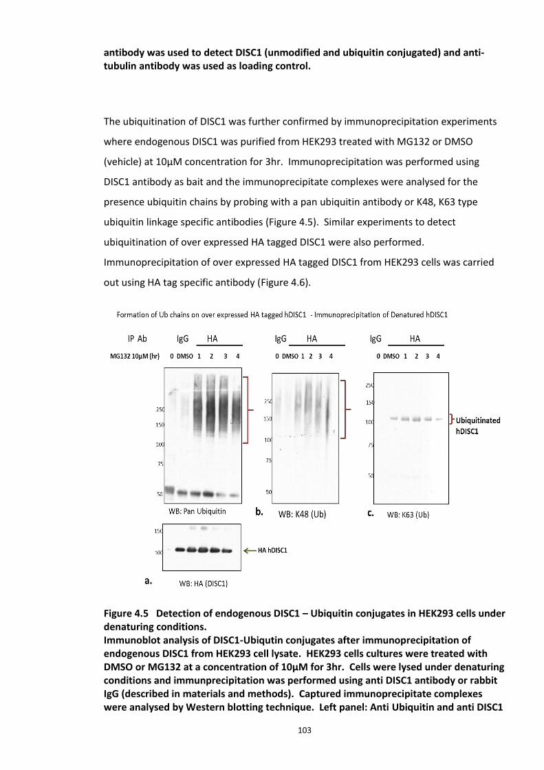
103
antibody was used to detect DISC1 (unmodified and ubiquitin conjugated) and anti-tubulin antibody was used as loading control.
The ubiquitination of DISC1 was further confirmed by immunoprecipitation experiments
where endogenous DISC1 was purified from HEK293 treated with MG132 or DMSO
(vehicle) at 10µM concentration for 3hr. Immunoprecipitation was performed using
DISC1 antibody as bait and the immunoprecipitate complexes were analysed for the
presence ubiquitin chains by probing with a pan ubiquitin antibody or K48, K63 type
ubiquitin linkage specific antibodies (Figure 4.5). Similar experiments to detect
ubiquitination of over expressed HA tagged DISC1 were also performed.
Immunoprecipitation of over expressed HA tagged DISC1 from HEK293 cells was carried
out using HA tag specific antibody (Figure 4.6).
Figure 4.5 Detection of endogenous DISC1 – Ubiquitin conjugates in HEK293 cells under denaturing conditions. Immunoblot analysis of DISC1-Ubiqutin conjugates after immunoprecipitation of endogenous DISC1 from HEK293 cell lysate. HEK293 cells cultures were treated with DMSO or MG132 at a concentration of 10µM for 3hr. Cells were lysed under denaturing conditions and immunprecipitation was performed using anti DISC1 antibody or rabbit IgG (described in materials and methods). Captured immunoprecipitate complexes were analysed by Western blotting technique. Left panel: Anti Ubiquitin and anti DISC1
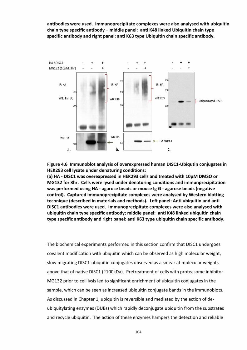
104
antibodies were used. Immunoprecipitate complexes were also analysed with ubiquitin chain type specific antibody – middle panel: anti K48 linked Ubiquitin chain type specific antibody and right panel: anti K63 type Ubiquitin chain specific antibody.
Figure 4.6 Immunoblot analysis of overexpressed human DISC1-Ubiqutin conjugates in HEK293 cell lysate under denaturing conditions: (a) HA - DISC1 was overexpressed in HEK293 cells and treated with 10µM DMSO or MG132 for 3hr. Cells were lysed under denaturing conditions and immunprecipitation was performed using HA - agarose beads or mouse Ig G - agarose beads (negative control). Captured immunoprecipitate complexes were analysed by Western blotting technique (described in materials and methods). Left panel: Anti ubiquitin and anti DISC1 antibodies were used. Immunoprecipitate complexes were also analysed with ubiquitin chain type specific antibody; middle panel: anti K48 linked ubiquitin chain type specific antibody and right panel: anti K63 type ubiquitin chain specific antibody.
The biochemical experiments performed in this section confirm that DISC1 undergoes
covalent modification with ubiquitin which can be observed as high molecular weight,
slow migrating DISC1-ubiquitin conjugates observed as a smear at molecular weights
above that of native DISC1 (~100kDa). Pretreatment of cells with proteasome inhibitor
MG132 prior to cell lysis led to significant enrichment of ubiquitin conjugates in the
sample, which can be seen as increased ubiquitin conjugate bands in the immunoblots.
As discussed in Chapter 1, ubiquitin is reversible and mediated by the action of de-
ubiquitylating enzymes (DUBs) which rapidly deconjugate ubiquitin from the substrates
and recycle ubiquitin. The action of these enzymes hampers the detection and reliable
105
characterisation of protein ubiquitination. One strategy that has been efficiently
employed was purifying the protein-ubiquitin conjugates under denaturing conditions i.e,
use of strong detergents and heat. Protein denaturation rapidly destroys enzyme activity
by disrupting protein secondary and tertiary structure by destroying hydrogen bonding,
salt bridges and disulphide bonds while the backbone peptide bond and other covalent
modifications remains intact (Ciechanover and Iwai, 2004). This strategy was successfully
employed in the present study which allowed efficient detection of post translation
medication on DISC1 and also aids further biophysical studies discussed in the next
section.
4.3.3 DISC1 ubiquitination - Mass spectroscopy evidence
Identification of ubiquitin modification sites on a substrate protein is crucial to the
facilitation of understanding the physiological role of ubiquitination in the context of that
protein. All PTMs direct specific functional consequences for the target protein. Most
often, ubiquitin receptor lysine residue(s) on the target is/are identified by site directed
mutagenesis experiments which block ubiquitination. Recent advances in technology
have enabled the use of Mass spectroscopy techniques to not only identify the target
lysine residue which undergoes the PTM but also the type of ubiquitin chain associated
with it. In this study, Flag DISC1 and HA Ubiquitin were co expressed in HEK293 cells
followed by MG132 treatment and cell lysis after 48hr incubation. Immunoprecipitate
complexes were collected using Flag tagged agarose beads as bait. Materials and
methods section 2.4.8 describes the protocol for the immunoprecipitation and
subsequent sample preparation for the Mass spectroscopy studies.
Mass spectroscopy samples were analysed at Yale School of Medicine, USA.
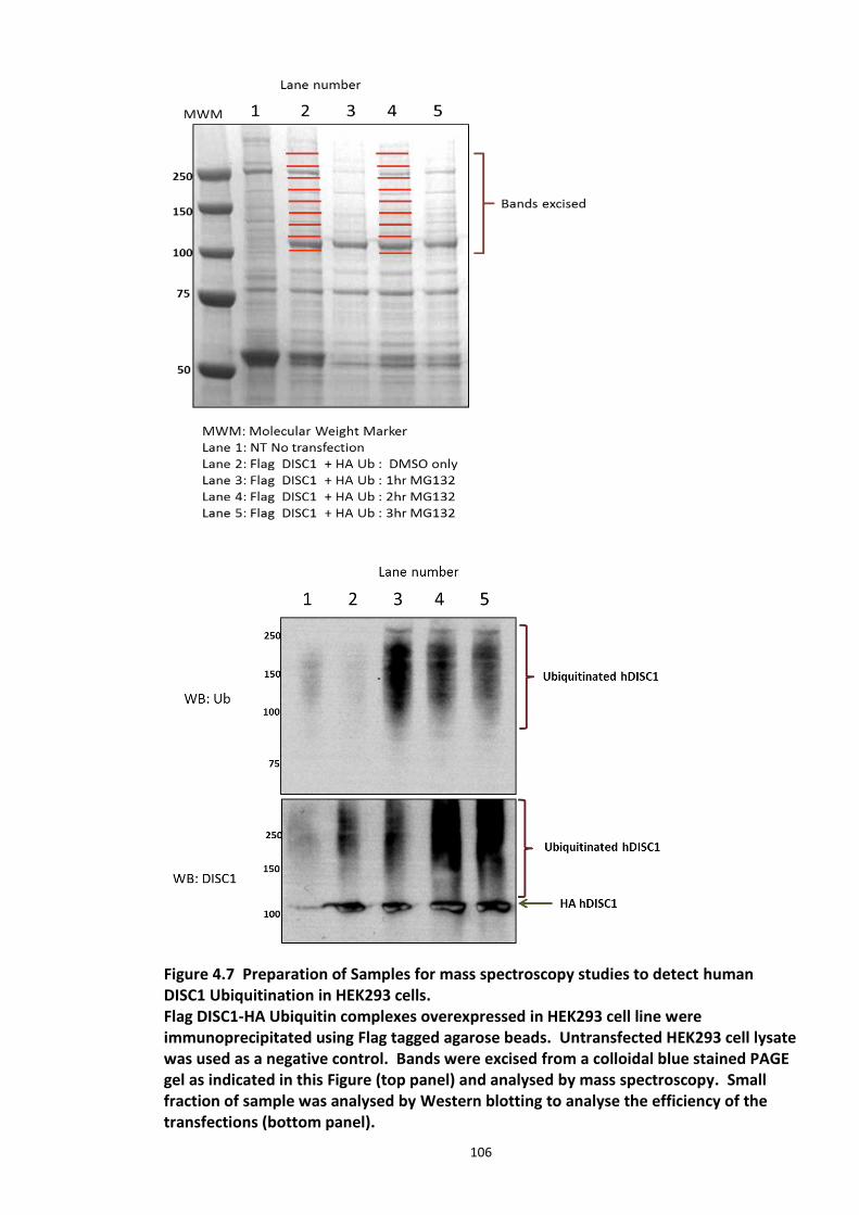
106
Figure 4.7 Preparation of Samples for mass spectroscopy studies to detect human DISC1 Ubiquitination in HEK293 cells. Flag DISC1-HA Ubiquitin complexes overexpressed in HEK293 cell line were immunoprecipitated using Flag tagged agarose beads. Untransfected HEK293 cell lysate was used as a negative control. Bands were excised from a colloidal blue stained PAGE gel as indicated in this Figure (top panel) and analysed by mass spectroscopy. Small fraction of sample was analysed by Western blotting to analyse the efficiency of the transfections (bottom panel).
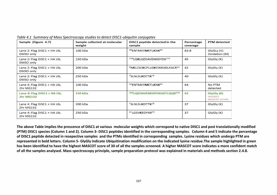
107
Table 4.1 Summary of Mass Spectroscopy studies to detect DISC1-ubiquitin conjugates
The above Table implies the prescence of DISC1 at various molecular weights which correspond to native DISC1 and post translationally modified (PTM) DISC1 species (Column 1 and 2). Column 3- DISC1 peptides identified in the corresponding samples. Column 4 and 5 indicate the percentage of DISC1 peptide detected in resepective samples and the PTMs identified in corresponding samples. Lysine residues which undergo PTM are represented in bold letters. Column 5- GlyGly indicate Ubiquitination modification on the indicated lysine residue.The sample highlighted in green has been identified to have the highest MASCOT score of 30 of all the samples screened. A higher MASCOT score indicates a more confident match of all the samples analysed. Mass spectroscopy principle, sample preparation protocol was explained in materials and methods section 2.4.8.
108
The mass spectroscopy studies on DISC1 Ubiquitnation can be summarised as follows:
DISC1- ubiquitin conjugates were detected at various molecular weights above 100kDa
(the weight at which native DISC1 migrates). These conjugates were represented as
glycine-glycine-lysine linkages (GlyGly(K)) in Table 4.1.
Six lysine residues were identified to be carrying G-G-K modification with variable
MASCOT scores. MASCOT scores were generated using a software package from Matrix
Science (www.matrixscience.com) that interprets mass spectral data into protein
identities. Mascot compares the spectral data to a database of known proteins and
scores based on confidence of the match.
Lysine 372 residue was chosen for subsequent work as it was predicted to be the site with
higher probability of ubiquitin modification implied by its highest MASCOT score of 30
(Table 4.1).
4.3.4 Analysis of ubiquitination kinetics of the DISC1 K372R mutant (site identified in the Mass spectroscopy studies)
V5 tagged DISC1 lysine to arginine mutant at position 372 (K) was generated and
evaluated for ubiquitination at basal levels and in the presence of the proteasome
inhibitor MG132. Lysine to arginine substitution mutants are ideal in this situation to
retain the net negative charge on the R group (side chain of the amino acid).
Immunoprecipitation studies were performed by over expressing V5 tagged DISC1 WT
and K372R mutant, to analyse the pattern of ubiquitin chain formation in response to
proteasomal inhibition. V5 tag antibody was used as bait to immunoprecipitate the
DISC1-ubiquitin protein conjugates.
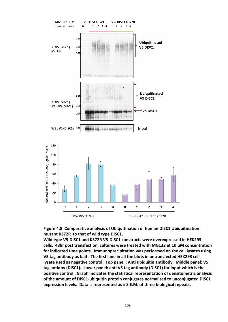
109
Figure 4.8 Comparative analysis of Ubiquitination of human DISC1 Ubiquitination mutant K372R to that of wild type DISC1. Wild type V5-DISC1 and K372R V5-DISC1 constructs were overexpressed in HEK293 cells. 48hr post transfection, cultures were treated with MG132 at 10 µM concentration for indicated time points. Immunoprecipitation was performed on the cell lysates using V5 tag antibody as bait. The first lane in all the blots in untransfected HEK293 cell lysate used as negative control. Top panel : Anti ubiquitin antibody. Middle panel: V5 tag antidoy (DISC1). Lower panel: anti V5 tag antibody (DISC1) for input which is the positive control . Graph indicates the statistical representation of densitometric analysis of the amount of DISC1-ubiquitin protein conjugates normalised to unconjugated DISC1 expression levels. Data is represented as ± S.E.M. of three biological repeats.
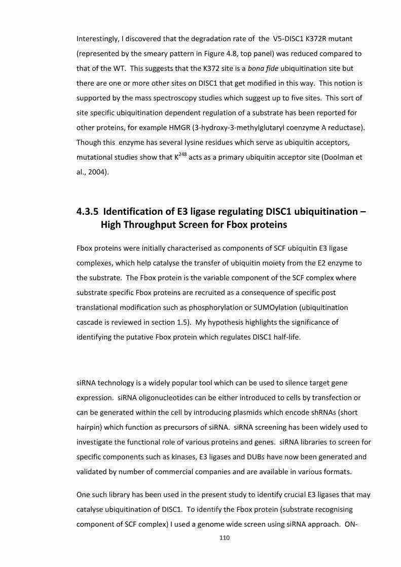
110
Interestingly, I discovered that the degradation rate of the V5-DISC1 K372R mutant
(represented by the smeary pattern in Figure 4.8, top panel) was reduced compared to
that of the WT. This suggests that the K372 site is a bona fide ubiquitination site but
there are one or more other sites on DISC1 that get modified in this way. This notion is
supported by the mass spectroscopy studies which suggest up to five sites. This sort of
site specific ubiquitination dependent regulation of a substrate has been reported for
other proteins, for example HMGR (3-hydroxy-3-methylglutaryl coenzyme A reductase).
Though this enzyme has several lysine residues which serve as ubiquitin acceptors,
mutational studies show that K248 acts as a primary ubiquitin acceptor site (Doolman et
al., 2004).
4.3.5 Identification of E3 ligase regulating DISC1 ubiquitination – High Throughput Screen for Fbox proteins
Fbox proteins were initially characterised as components of SCF ubiquitin E3 ligase
complexes, which help catalyse the transfer of ubiquitin moiety from the E2 enzyme to
the substrate. The Fbox protein is the variable component of the SCF complex where
substrate specific Fbox proteins are recruited as a consequence of specific post
translational modification such as phosphorylation or SUMOylation (ubiquitination
cascade is reviewed in section 1.5). My hypothesis highlights the significance of
identifying the putative Fbox protein which regulates DISC1 half-life.
siRNA technology is a widely popular tool which can be used to silence target gene
expression. siRNA oligonucleotides can be either introduced to cells by transfection or
can be generated within the cell by introducing plasmids which encode shRNAs (short
hairpin) which function as precursors of siRNA. siRNA screening has been widely used to
investigate the functional role of various proteins and genes. siRNA libraries to screen for
specific components such as kinases, E3 ligases and DUBs have now been generated and
validated by number of commercial companies and are available in various formats.
One such library has been used in the present study to identify crucial E3 ligases that may
catalyse ubiquitination of DISC1. To identify the Fbox protein (substrate recognising
component of SCF complex) I used a genome wide screen using siRNA approach. ON-
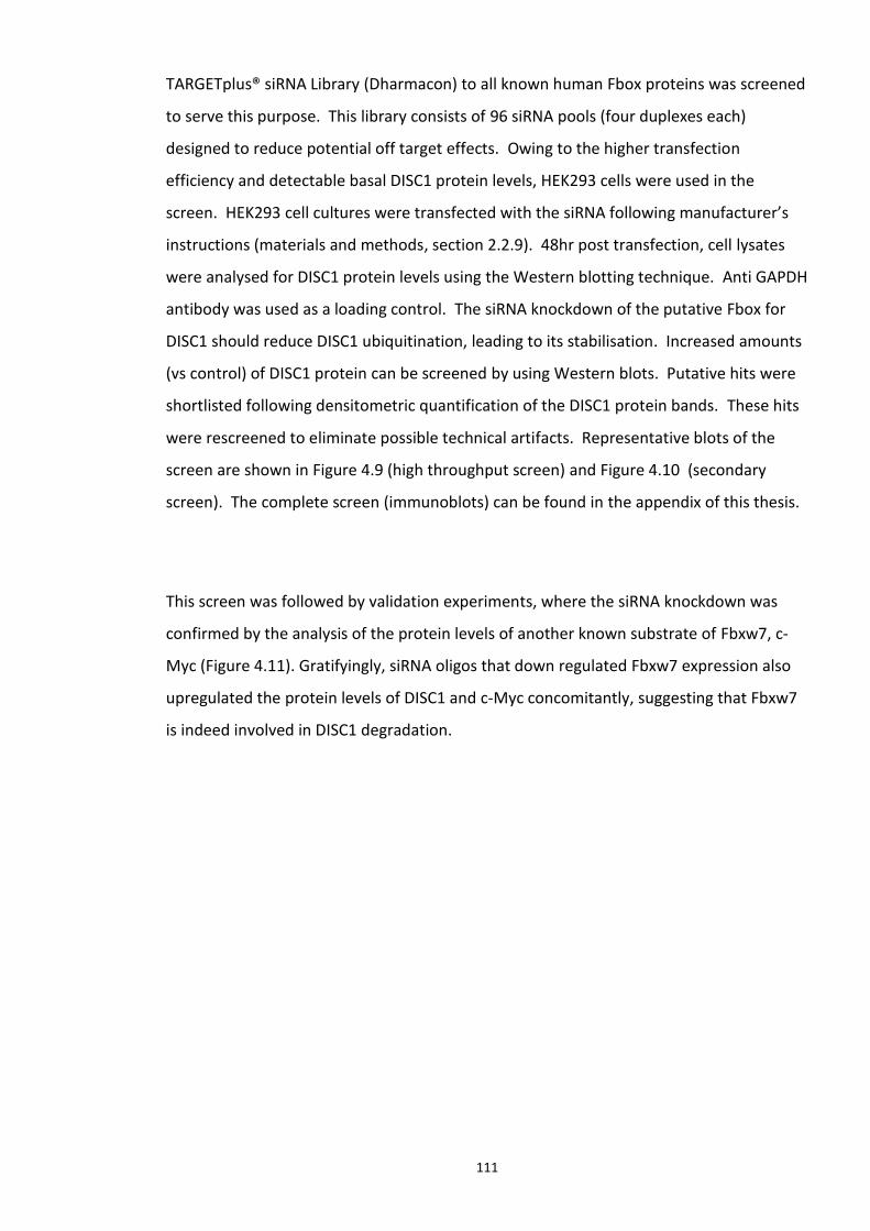
111
TARGETplus® siRNA Library (Dharmacon) to all known human Fbox proteins was screened
to serve this purpose. This library consists of 96 siRNA pools (four duplexes each)
designed to reduce potential off target effects. Owing to the higher transfection
efficiency and detectable basal DISC1 protein levels, HEK293 cells were used in the
screen. HEK293 cell cultures were transfected with the siRNA following manufacturer’s
instructions (materials and methods, section 2.2.9). 48hr post transfection, cell lysates
were analysed for DISC1 protein levels using the Western blotting technique. Anti GAPDH
antibody was used as a loading control. The siRNA knockdown of the putative Fbox for
DISC1 should reduce DISC1 ubiquitination, leading to its stabilisation. Increased amounts
(vs control) of DISC1 protein can be screened by using Western blots. Putative hits were
shortlisted following densitometric quantification of the DISC1 protein bands. These hits
were rescreened to eliminate possible technical artifacts. Representative blots of the
screen are shown in Figure 4.9 (high throughput screen) and Figure 4.10 (secondary
screen). The complete screen (immunoblots) can be found in the appendix of this thesis.
This screen was followed by validation experiments, where the siRNA knockdown was
confirmed by the analysis of the protein levels of another known substrate of Fbxw7, c-
Myc (Figure 4.11). Gratifyingly, siRNA oligos that down regulated Fbxw7 expression also
upregulated the protein levels of DISC1 and c-Myc concomitantly, suggesting that Fbxw7
is indeed involved in DISC1 degradation.
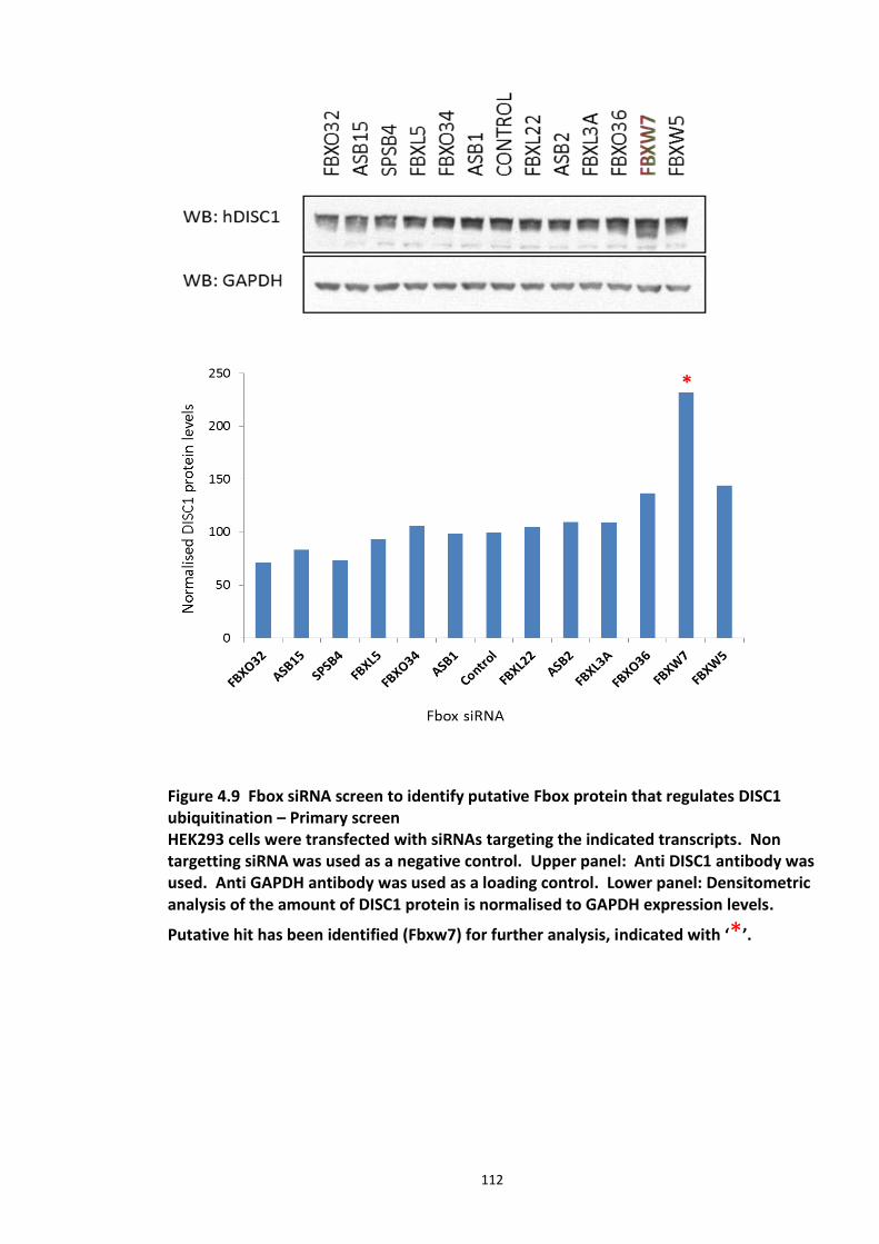
112
Figure 4.9 Fbox siRNA screen to identify putative Fbox protein that regulates DISC1 ubiquitination – Primary screen HEK293 cells were transfected with siRNAs targeting the indicated transcripts. Non targetting siRNA was used as a negative control. Upper panel: Anti DISC1 antibody was used. Anti GAPDH antibody was used as a loading control. Lower panel: Densitometric analysis of the amount of DISC1 protein is normalised to GAPDH expression levels.
Putative hit has been identified (Fbxw7) for further analysis, indicated with ‘*’.
113
Figure 4.10 Validation of Fbox siRNA screen hit - Secondary Screen Putative shortlisted Fbox proteins were rescreened. Upper panel: Anti DISC1 antibody was used. Anti GAPDH antibody was used as a loading control. Lower panel: Densitometric analysis of the amount of DISC1 protein is normalised to GAPDH expression levels. Complete information about Fbox siRNA screen with Figures is detailed in appendix. From the above screen, Fbxw7 was identified as a putative hit as its knock down consistently stabilised DISC1 protein levels.
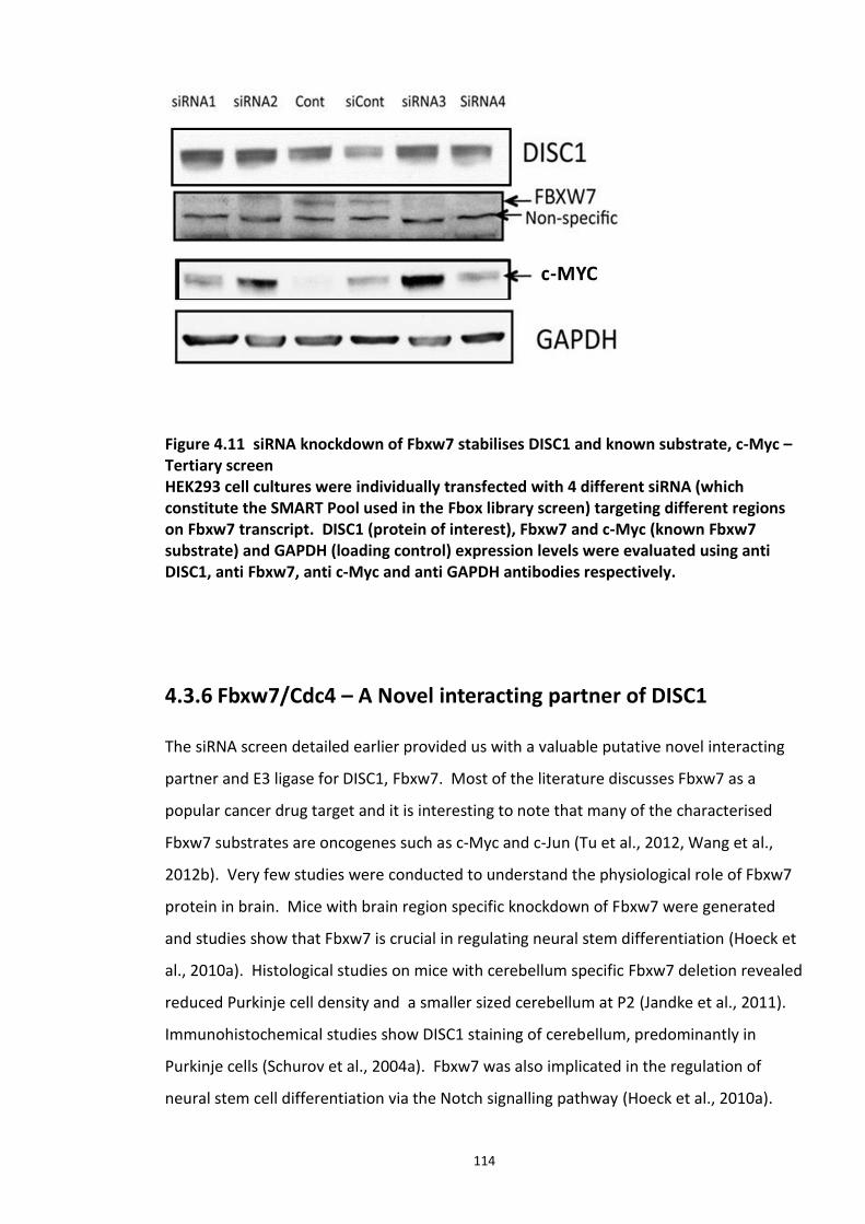
114
Figure 4.11 siRNA knockdown of Fbxw7 stabilises DISC1 and known substrate, c-Myc – Tertiary screen HEK293 cell cultures were individually transfected with 4 different siRNA (which constitute the SMART Pool used in the Fbox library screen) targeting different regions on Fbxw7 transcript. DISC1 (protein of interest), Fbxw7 and c-Myc (known Fbxw7 substrate) and GAPDH (loading control) expression levels were evaluated using anti DISC1, anti Fbxw7, anti c-Myc and anti GAPDH antibodies respectively.
4.3.6 Fbxw7/Cdc4 – A Novel interacting partner of DISC1
The siRNA screen detailed earlier provided us with a valuable putative novel interacting
partner and E3 ligase for DISC1, Fbxw7. Most of the literature discusses Fbxw7 as a
popular cancer drug target and it is interesting to note that many of the characterised
Fbxw7 substrates are oncogenes such as c-Myc and c-Jun (Tu et al., 2012, Wang et al.,
2012b). Very few studies were conducted to understand the physiological role of Fbxw7
protein in brain. Mice with brain region specific knockdown of Fbxw7 were generated
and studies show that Fbxw7 is crucial in regulating neural stem differentiation (Hoeck et
al., 2010a). Histological studies on mice with cerebellum specific Fbxw7 deletion revealed
reduced Purkinje cell density and a smaller sized cerebellum at P2 (Jandke et al., 2011).
Immunohistochemical studies show DISC1 staining of cerebellum, predominantly in
Purkinje cells (Schurov et al., 2004a). Fbxw7 was also implicated in the regulation of
neural stem cell differentiation via the Notch signalling pathway (Hoeck et al., 2010a).
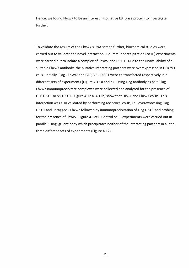
115
Hence, we found Fbxw7 to be an interesting putative E3 ligase protein to investigate
further.
To validate the results of the Fbxw7 siRNA screen further, biochemical studies were
carried out to validate the novel interaction. Co-immunoprecipitation (co-IP) experiments
were carried out to isolate a complex of Fbxw7 and DISC1. Due to the unavailability of a
suitable Fbxw7 antibody, the putative interacting partners were overexpressed in HEK293
cells. Initially, Flag - Fbxw7 and GFP, V5 - DISC1 were co transfected respectively in 2
different sets of experiments (Figure 4.12 a and b). Using Flag antibody as bait, Flag
Fbxw7 immunoprecipitate complexes were collected and analysed for the presence of
GFP DISC1 or V5 DISC1. Figure 4.12 a, 4.12b; show that DISC1 and Fbxw7 co-IP. This
interaction was also validated by performing reciprocal co-IP, i.e., overexpressing Flag
DISC1 and untagged - Fbxw7 followed by immunoprecipitation of Flag DISC1 and probing
for the presence of Fbxw7 (Figure 4.12c). Control co-IP experiments were carried out in
parallel using IgG antibody which precipitates neither of the interacting partners in all the
three different sets of experiments (Figure 4.12).
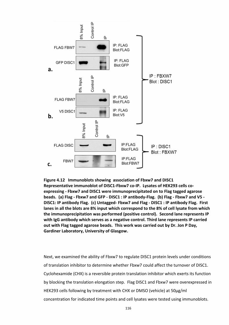
116
Figure 4.12 Immunoblots showing association of Fbxw7 and DISC1 Representative immunoblot of DISC1-Fbxw7 co-IP. Lysates of HEK293 cells co- expressing - Fbxw7 and DISC1 were immunoprecipitated on to Flag tagged agarose beads. (a) Flag - Fbxw7 and GFP - DISC1 : IP antibody-Flag. (b) Flag - Fbxw7 and V5 - DISC1: IP antibody Flag. (c) Untagged- Fbxw7 and Flag - DISC1 : IP antibody Flag. First lanes in all the blots are 8% input which correspond to the 8% of cell lysate from which the immunoprecipitation was performed (positive control). Second lane represents IP with IgG antibody which serves as a negative control. Third lane represents IP carried out with Flag tagged agarose beads. This work was carried out by Dr. Jon P Day, Gardiner Laboratory, University of Glasgow.
Next, we examined the ability of Fbxw7 to regulate DISC1 protein levels under conditions
of translation inhibitor to determine whether Fbxw7 could affect the turnover of DISC1.
Cyclohexamide (CHX) is a reversible protein translation inhibitor which exerts its function
by blocking the translation elongation step. Flag DISC1 and Fbxw7 were overexpressed in
HEK293 cells following by treatment with CHX or DMSO (vehicle) at 50µg/ml
concentration for indicated time points and cell lysates were tested using immunoblots.
117
Figure 4.13 Effect of Fbxw7 over-expression on the stability of Flag-DISC1 Flag - DISC1 was co transfected in HEK293 cells with untagged Fbxw7 or an empty vector (negative control). Cells were treated with Cyclohexamide (CHX) at indicated concentration and time and cell lysates are analysed by Western blotting technique using anti DISC1, Fbxw7 antibodies. Ku70 and GAPDH protein expression levels serve as loading controls as they are stable under Cyclohexamide treatment. n=2 This work was carried out by Dr. Jon P Day, Gardiner Laboratory, University of Glasgow.
The decrease in DISC1 levels, induced by CHX, was exacerbated following Fbxw7
expression, as suggested by preliminary studies (Baillie laboratory). This implies that the
E3 ligase complex with Fbxw7 promoted the degradation of DISC1. Other proteins which
are known for their stability under CHX treatment (Ku70 and GAPDH) were unaffected by
either CHX treatment or Fbxw7 expression. These data further validate my hypothesis
that Fbxw7 is the E3 ligase for DISC1 (Figure 4.14). These data were supported by the
observation that both proteins co-purify in DISC1 immunoprecipitations (Figure 4.12).
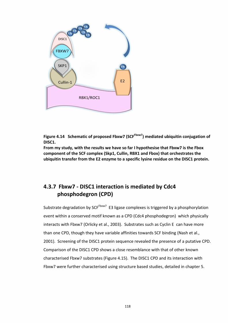
118
Figure 4.14 Schematic of proposed Fbxw7 (SCFFbxw7) mediated ubiquitin conjugation of DISC1. From my study, with the results we have so far I hypothesise that Fbxw7 is the Fbox component of the SCF complex (Skp1, Cullin, RBX1 and Fbox) that orchestrates the ubiquitin transfer from the E2 enzyme to a specific lysine residue on the DISC1 protein.
4.3.7 Fbxw7 - DISC1 interaction is mediated by Cdc4 phosphodegron (CPD)
Substrate degradation by SCFFbxw7 E3 ligase complexes is triggered by a phosphorylation
event within a conserved motif known as a CPD (Cdc4 phosphodegron) which physically
interacts with Fbxw7 (Orlicky et al., 2003). Substrates such as Cyclin E can have more
than one CPD, though they have variable affinities towards SCF binding (Nash et al.,
2001). Screening of the DISC1 protein sequence revealed the presence of a putative CPD.
Comparison of the DISC1 CPD shows a close resemblance with that of other known
characterised Fbxw7 substrates (Figure 4.15). The DISC1 CPD and its interaction with
Fbxw7 were further characterised using structure based studies, detailed in chapter 5.
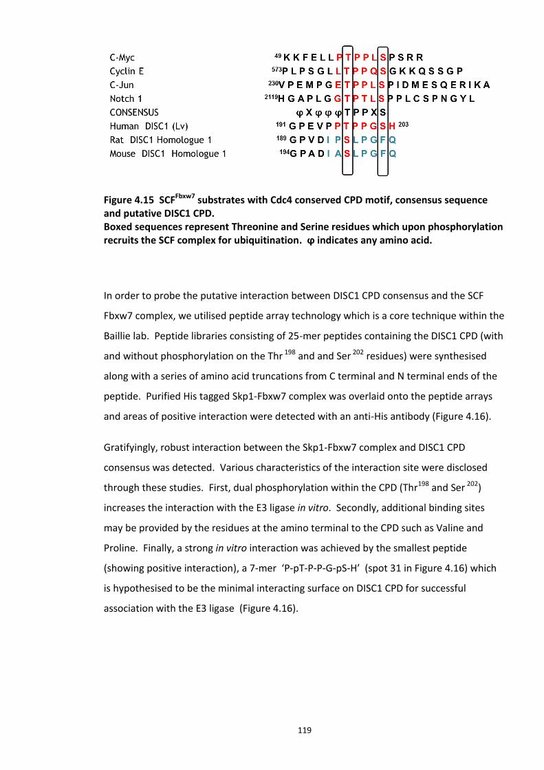
119
Figure 4.15 SCFFbxw7 substrates with Cdc4 conserved CPD motif, consensus sequence and putative DISC1 CPD. Boxed sequences represent Threonine and Serine residues which upon phosphorylation recruits the SCF complex for ubiquitination. ϕ indicates any amino acid.
In order to probe the putative interaction between DISC1 CPD consensus and the SCF
Fbxw7 complex, we utilised peptide array technology which is a core technique within the
Baillie lab. Peptide libraries consisting of 25-mer peptides containing the DISC1 CPD (with
and without phosphorylation on the Thr 198 and and Ser 202 residues) were synthesised
along with a series of amino acid truncations from C terminal and N terminal ends of the
peptide. Purified His tagged Skp1-Fbxw7 complex was overlaid onto the peptide arrays
and areas of positive interaction were detected with an anti-His antibody (Figure 4.16).
Gratifyingly, robust interaction between the Skp1-Fbxw7 complex and DISC1 CPD
consensus was detected. Various characteristics of the interaction site were disclosed
through these studies. First, dual phosphorylation within the CPD (Thr198 and Ser 202)
increases the interaction with the E3 ligase in vitro. Secondly, additional binding sites
may be provided by the residues at the amino terminal to the CPD such as Valine and
Proline. Finally, a strong in vitro interaction was achieved by the smallest peptide
(showing positive interaction), a 7-mer ‘P-pT-P-P-G-pS-H’ (spot 31 in Figure 4.16) which
is hypothesised to be the minimal interacting surface on DISC1 CPD for successful
association with the E3 ligase (Figure 4.16).
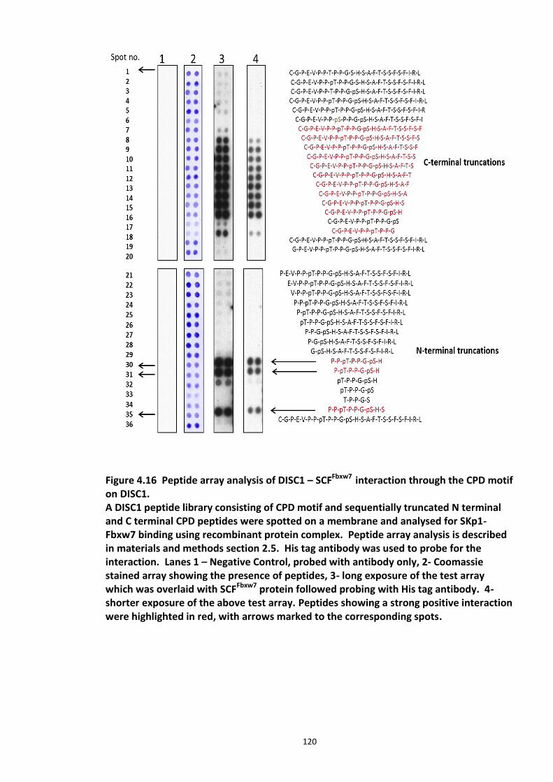
120
Figure 4.16 Peptide array analysis of DISC1 – SCFFbxw7 interaction through the CPD motif on DISC1. A DISC1 peptide library consisting of CPD motif and sequentially truncated N terminal and C terminal CPD peptides were spotted on a membrane and analysed for SKp1-Fbxw7 binding using recombinant protein complex. Peptide array analysis is described in materials and methods section 2.5. His tag antibody was used to probe for the interaction. Lanes 1 – Negative Control, probed with antibody only, 2- Coomassie stained array showing the presence of peptides, 3- long exposure of the test array which was overlaid with SCFFbxw7 protein followed probing with His tag antibody. 4- shorter exposure of the above test array. Peptides showing a strong positive interaction were highlighted in red, with arrows marked to the corresponding spots.
121
4.4 Discussion
4.4.1 Proteasomal regulation of DISC1 protein
The two main proteolytic pathways of mammalian cells, the lysosomal pathway and the
UPS, play an important role in maintaining cellular homeostasis of proteins. The role of
UPS in neurological disorders has been reviewed in section 1.5.4. Earlier studies have
proposed immunocytochemical staining for ubiquitin and its conjugates as a marker for
neuro-degeneration (Lowe et al., 1988). These were later identified as ubiquitin rich
inclusion bodies (IBs) enriched with deposits of ubiquitin conjugated misfolded proteins.
These accumulated proteins may lead to cellular stress and subsequent cell death. IBs are
a hallmark of neurodegenerative illnesses such as Alzheimer’s disease. Although so far
there has been no report of IB formation by DISC1 protein, DISC1 aggregate formation
has been widely reported in various cell types (Atkin et al., 2012). The role of these
aggregates in DISC1 associated diseases still remains unclear.
My study focused on how DISC1, a putative susceptibility factor of SCZ, is regulated in
cells, i.e. what controls the protein levels of DISC1? Initial studies involved
characterisation of DISC1 degradation kinetics in the presence of the 26S proteasome
inhibitor, MG132. DISC1 was shown to be stabilised and ubiquitin conjugated upon
inhibition of its proteasomal degradation. Mass spectroscopy analysis further
strengthened my suggested hypothesis that ubiquitin conjugation directs DISC1
proteasomal targeting.
My biochemical studies show the presence of K48 and K63 type ubiquitin chains on
DISC1. Antibodies which detect other types of ubiquitin chains (such as K3, K11) were
unavailable at the time of this study. Conjugated K48 linked ubiquitin chains are believed
to be the signal for proteasome degradation, while conjugation of K63 linked ubiquitin
chains is known to serve non proteolytic roles such as such as gene expression regulation
and inflammatory responses (Xu et al., 2009). Tetra ubiquitin has been identified as a
minimum (shortest) signal required for proteasomal targeting (Ciechanover and Ben-
Saadon, 2004). It has been suggested that specific receptors on the 19S complex of the
26S proteasome have the potential to recognise the poly ubiquitin chain assembly formed
122
by K48 linkage. It has also been proposed that longer ubiquitin chain length may increase
the residence time of the substrate. However, the exact mechanism by which the length
of ubiquitin chain influences the fate of ubiquitinated substrate has been a long standing
debate (Ciechanover and Ben-Saadon, 2004). A Variety of linkages, inter linkages
between chains and lengths which in turn assume various confirmations were all known
to contribute to the complexity and specificity of the ubiquitination of a substrate and
dictate the physiological response (Fushman and Wilkinson, 2011). Though the Mass
spectroscopy studies performed in the present study uncovered a putative ubiquitination
site on human DISC1 (K372), the type of ubiquitin chains nor the length could not be
verified due to the moderate quality of the samples. More investigation to uncover the
type of ubiquitin chains would be useful in understanding the physiological consequences
of chain formation on DISC1. Technical and biological replicates of Mass spectroscopy
studies would be useful, but were not within the scope of this project.
The immunoprecipitation data on ubiquitination of DISC1 K372R mutant suggest reduced
ubiquitin conjugation, if not completely abolished. This can be attributed to the fact that
substrates might have one or more ubiquitination targeted lysines. Ubiquitin conjugation
at different lysines on a substrate may be spatio temporally regulated as well as having
different physiological responses (Grabbe et al., 2011). Other experimental studies which
may be useful in the further characterising ubiquitination of DISC1 are ones involving
expression of an ubiquitin ‘lysineless’ mutant (Ubiquitin(K0)), in which all the seven lysine
residues are mutated to arginine residues. This mutant terminates ubiquitin chain
formation, and therefore stabilises the substrate preventing its proteasomal targeting.
These mutants should alter the pattern of slow migrating bands on a Western blot as a
result of less ubiquitination or shorter chain formation. Lysine at specific sites on
ubiquitin can also be mutated to arginines such as K48R or K63R. Overexpressing such
mutants would validate the type of chain formed on the substrate protein.
As discussed earlier, the extreme short life of ubiquitin modified substrates due to the
reversible nature of the modification (deubiquitination catalysed by DUBs) poses several
challenges in biochemical and biophysical characterisation of ubiquitin conjugated
123
substrates. Although cysteine protease inhibitors (example: Iodoactemide and NEM – N-
ethyl maleimide) have been widely used to inhibit DUBs, they were shown to interfere
with mass spectroscopy studies (Nielsen et al., 2008). Recent studies have focused on
developing strategies to overcome this issue and facilitate a more reliable detection and
characterisation of the protein ubiquitination. Tandem-repeated ubiquitin-binding
entities (TUBEs) are an example which was developed based on ubiquitin-associated
(UBA) domains. TUBEs specifically recognize, interact and thereby protect the tetra
ubiquitin by interacting with a higher affinity than individual UBA domains (present on
DUBs). TUBEs therefore function as molecular traps and were described as valuable tools
for studying PTMs (Hjerpe et al., 2009).
As discussed earlier, many studies have shown that loss of DISC1 expression (RNAi
knockdown of DISC1) results in abnormal brain development and function (Niwa et al.,
2010, Faulkner et al., 2008, Young-Pearse et al., 2010). Retroviral mediated shRNA
knockdown of DISC1 shows that it is a key factor regulating neuronal maturation in adult
brain (Faulkner et al., 2008). DISC1 knockdown studies in Zebra fish have revealed
abnormal oligodendrocyte and cerebellar neuronal development. RNAi mediated loss of
DISC1 expression has also affected neurite outgrowth in PC12 cells (Miyoshi et al., 2003b).
All these studies show that the loss of DISC1 function may be critical in the underlying
pathophysiology of DISC1 associated psychiatric illnesses. I see this uncovered novel PTM
on DISC1 as initial step towards understanding these disease mechanisms, and more
investigation and characterisation of this modification would provide more avenues for
therapeutic intervention.
4.4.2 Fbxw7 dependent degradation of DISC1
My screening studies uncovered a novel interacting partner for DISC1 i.e., Fbxw7. It is
interesting to note that none of the yeast 2 hybrid screens on DISC1 have identified
protein as a candidate interacting partner (Camargo et al., 2007). This may be attributed
to the transient nature of the DISC1-Fbxw7 interaction - many of the interactions that
characterise ubiquitination are extremely short lived. It may also be that a prior post
124
translational modification such as phosphorylation is required for this interaction, as
suggested by my study.
Literature mining revealed that Fbxw7 is mutated in several different types of cancers and
its inactivation leads to chromosomal instability (Jandke et al., 2011, Rajagopalan et al.,
2004). Recent studies have implicated Fbxw7 role in stem cell differentiation. Fbxw7
sustains hematopoietic stem cell quiescence and regulates their differentiation by
regulating transcription factors Notch, c-Jun and mTOR (Reavie et al., 2010, Iriuchishima
et al., 2011). Fbxw7 is crucial in neuronal stem cell maintenance and differentiation in
brain and is known to exert this function by controlling the abundance of Notch proteins
(Hoeck et al., 2010a, Matsumoto et al., 2011). Fbxw7 has been reviewed in section
1.5.3.5.
Several Fbox proteins recognise a short motif within a substrate which upon
phosphorylation facilitates recognition and interaction by Fbox protein, known as CPDs
(Ye et al., 2004). Many of these proteins have been crystallised with their respective
phosphopeptides. While some substrates have only one CPD, many other substrates such
as Sic1 and Cln2 have multiple degrons dispersed across the protein sequence. My
peptide array studies involving sequential truncation from the C terminal and N terminal
ends of the DISC1 CPD also identified the minimal interacting region of Fbxw7 on DISC1
within the CPD (Figure 4.16). As discussed earlier, as in the case of most of the other
Fbxw7 substrates, a phosphorylation event followed by an isomerisation might be crucial
in triggering this interaction (Zhang and Koepp, 2006, Hao et al., 2007b). Hence, peptide
array studies were used to further validate this hypothesis of Fbxw7 mediated DISC1
ubiquitination. As seen in Figure 4.16, SKp1-Fbxw7 complex specifically shows strong
interaction with DISC1 CPD peptides carrying dual phosphorylation (Thr198and Ser202).
This is an important finding which aligns with the observations made by other research
groups in other known Fbxw7 substrates. As can be seen in Figure 4.15, DISC1 CPD
consensus has a close similarity with Cyclin E’s CPD motif. Cyclin E (regulates cell cycle
transition from G1 to S phase) is a well characterised Fbxw7 substrate whose degradation
is triggered by phosphorylation within multiple degron motifs (Orlicky et al., 2003, Ye et
125
al., 2004). In vitro studies employing Cyclin E CPD peptides and site directed mutagenesis
studies have identified doubly phosphorylated degron (Thr380/Ser384) as a high affinity
motif and singly phosphorylated degron (Thr62) as a lower affinity motif. More studies are
to follow in the next section to further characterise the DISC1 CPD motif. Interestingly
the identified CPD is not conserved in mouse and Rat DISC1 homologues (Figure 4.15).
Another noteworthy aspect of Fbxw7 is its isoform specific regulation of its substrates
(Grim et al., 2008). As reviewed in section 1.5.3.5, three splice variants of Fbxw7 have
been characterised – α, β and γ isoforms, which vary at their amino terminal end. Fbw7-α
is predominantly nuclear; Fbw7-β is cytoplasmic; and Fbw7-γ is reported to be localised
in the nucleolus (Zhang and Koepp, 2006). Their differential localisation may also dictate
their isoform specific physiological role (Welcker et al., 2004). For example, it has been
demonstrated that p53 is regulated by the cytoplasmic β isoform (Galli et al., 2010), The
nucleolar pool of Fbxw7-γ regulates c-Myc (Welcker et al., 2004) and different degrons
with in Cyclin E were shown to differentially interact with three Fbxw7 isoforms (Zhang
and Koepp, 2006). The Fbxw7 siRNA used in the Fbox screen was designed to target a
common region for all the three isoforms. Fbxw7-γ isoform is absent in brain tissue
(embryonic and adult), β isoform is moderately expressed, while the α isoform is the most
abundant and ubiquitously expressed isoform of Fbxw7. α isoform is abundant in
neuronal stem cells and progenitors ((Hoeck et al., 2010a) (Akhoondi et al., 2010, Grim et
al., 2008) and HEK293 cells (cell line used in the present study). Hence I chose to
investigate the Fbxw7-α isoform interaction with DISC1 by performing
Immunoprecipitation studies. However, the involvement of the other isoforms
involvement in DISC1 regulation may not be ruled out and requires further investigation.
Positive interaction between FBXW-α isoform and DISC1 may also suggest that DISC1
could be localised in the nucleus, supporting its known centrosomal association (Morris et
al., 2003).
The following studies may shed light on the Fbxw7 and DISC1 interaction, and were not
within the scope of the present study:
126
1. Investigating DISC1 – Fbxw7 interaction studies (immunoprecipitation, pull down
assays, immunostaining techniques) and Isoform specific knockdown of Fbxw7 and its
effect on DISC1 ubiquitination would be useful in understanding if this interaction is
isoform specific.
2. The developmental expression profile of DISC1 protein in mice reveals that the 100kDa
form peaks at E13.5 and P35, which corresponds to periods of active neurogenesis,
supporting its role in active brain development (Lipska et al., 2006a). In humans, the
expression is predominant in hippocampus, while the expression is stronger in dentate
gyrus in early childhood and gradually rapidly declines with age into adulthood (Lipska et
al., 2006b). Fbxw7 has also been implicated in cerebellar development and neuronal
stem cell differentiation. However, the expression profile of Fbxw7 in human brain has
not been explored yet. It would be intriguing to investigate any expression based inverse
correlation between these two proteins in normal subjects and psychiatric patients in the
context of my study showing that DISC1 levels are regulated by Fbxw7. This also supports
the neurodevelopmental hypothesis of SCZ.
3. GSK3β has been known to be functionally associated with Fbxw7 (source kinase for its
substrates). GSK3β is a crucial interacting partner of DISC1 and studies in zebrafish,
mouse model systems and mammalian cell lines show that DISC1 variants (A83V, R264Q,
and L607F) could not activate Wnt/GSK3β signalling. In this context, it would be
interesting to assess the potential of GSK3β to directly phosphorylate DISC1 within the
CPD. However, this might prove challenging, as in general, a priming phosphorylation
event is required for GSK3β to phosphorylate its substrates.
4. It would be also interesting to investigate how DISC1 common variants and t (1; 11)
translocation (truncated DISC1) affect DISC1 stability in cells, which may explain the
molecular consequences of the underlying disease mechanism.
127
4.5 Chapter summary
To summarise this chapter’s results, I have provided novel and previously undiscovered
information on the regulation of DISC1 turnover in HEK293 cells. The data presented in
this chapter indicate that DISC1 protein levels are regulated by ubiquitin mediated
proteasomal degradation. Mass spectroscopy studies identified K372 as a potential
acceptor site while my biochemical data suggest the presence of more acceptor lysine
residues at other sites. This is a novel PTM identified on DISC1 and we speculate that this
modification has a potential role in the DISC1 pathway. I also propose that DISC1
ubiquitination may play a role in the mechanism of all DISC1 associated
neurodegenerative illnesses, as seen in Alzheimer’s and Parkinson’s diseases where UPS
dysfunction has been reported. My studies also uncovered Fbxw7 as a novel interacting
partner of DISC1 which regulates DISC1 protein levels by catalysing its ubiquitination as a
functional SCF component.
In the following chapter, my studies focused on targeting this newly uncovered PPI
(protein- protein interaction) with inhibitors to modulate DISC1 protein levels, exploring a
new avenue for therapeutic intervention.
128
5.
Development of disruptor peptide to modulate DISC1-SCFFbxw7 complex interaction
5.1 Introduction
Association studies, linkage studies and mouse models support DISC1 as one of the most
promising candidate susceptibility factors for SCZ and other major mental illnesses
(reviewed in detail in (Chubb et al., 2008)). Transgenic mice expressing a truncated
dominant-negative form of DISC1 (Tr DISC1), displayed several anatomical and
behavioural abnormalities consistent with those observed in patients with SCZ (Hikida et
al., 2007, Clapcote et al., 2007, Koike et al., 2006). It has been hypothesised that DISC1
SNPs and mutations such as that of the Scottish C terminal truncation may lead to loss of
DISC1 function, which may directly or indirectly contribute to the pathophysiology of
psychiatric illnesses. It has been difficult to study human DISC1 protein as the DISC1
protein levels in the lymphoblasts collected from the patients of the Scottish pedigree
were shown to be too low to be detected by Western blotting technique. These low
protein levels may be attributed to the instability or haploinsufficiency of TrDISC1 (Millar
et al., 2005b, Sawa and Snyder, 2005).
To date, there is no evidence that the DISC1 truncated protein (TrDISC1) is translated in
lymphocytes in which the translocation has been identified (Scottish translocation) (Millar
et al., 2000b). However, there are a few hypotheses for how DISC1 mutation can result in
psychiatric disease. Firstly, it has been hypothesized that the translated protein product
129
might not be stable and is rapidly degraded (Millar et al., 2005b). In a different scenario,
the truncated protein may be stable but may interfere with the function of the wild type
protein product from the other allele, resulting in loss of function known as a dominant
negative effect. Secondly, another hypothesis suggests that the DISC1 truncation could
result in haploinsufficiency (definition of haploinsufficiency- both alleles need to be
functional for a wild type functional genotype for a particular gene) (Thomson et al.,
2005). My proposed project aims to address the DISC1 haploinsufficiency hypothesis and
uncover the disease mechanisms involved.
Since DISC1 is ubiquitinated and the E3 ligase component Fbxw7 is crucial in regulating
DISC1 protein levels, I hypothesize that a disruptor peptide/small molecule targeting the
binding sites of DISC1 and Fbxw7 would abolish their interaction thus interfering with
DISC1 ubiquitination and increasing its stability. I propose that the identified and mapped
novel protein-protein interaction site could be a future drug target for SCZ and other
associated psychiatric illnesses with underlying DISC1 mutations.
5.1.1 Protein – protein interactions (PPI) as drug targets
The present drug market is mostly full of competitive small molecule inhibitors of various
enzymes, receptors and ion channels, and over the years the number of targets has
dwindled, whilst the developmental costs have soared. Protein-Protein interfaces of
large molecular complexes have become attractive drug targets as they have an added
advantage of extra specificity and can be easily identified through biochemical co-
purification (Arkin and Wells, 2004, Chène, 2006). Until recent years, they have been
considered to be ‘undruggable’ and complex targets. With drugs belonging to this new
class already on the market such as Tirofiban and Maraviroc and many others advancing
in clinical trials, there has been increasing scope to exploit this new class of drug targets
(Vlieghe et al., 2010). In essence, PPI inhibitors represent the next generation of drugs
and more of their kind are en route to the market in the near future. PPIs are involved in
a wide array of disease pathways that have become attractive drug targets lately, and the
ongoing elucidation of the human interactome is opening up avenues in this field of drug
discovery. The human interactome has been estimated to have approximately 650,000
130
interactions, providing an excellent source to develop novel targets for therapeutic drug
development (Chène, 2006, White et al., 2008). Having overcome initial technical hurdles
and experimental difficulties for characterising PPI drug targets, this class of therapeutic
intervention has gathered considerable pace.
5.1.2 DISC1 – Fbxw7 interaction as a drug target
It is estimated that nearly 80% of cellular proteins undergo irreversible ubiquitin
conjugated proteolysis by mediation of substrate specific ubiquitin E3 ligases, in response
to specific stimuli (Bernassola et al., 2010, Varshavsky, 1997). Most of the characterised
E3 ligases are modular protein complexes constituting a core scaffold unit with switchable
substrate targeting subunit (Joazeiro and Weissman, 2000, Kipreos and Pagano, 2000).
Cullin RING ligase (CRL) ligases, also known as SCF (Skp1-Cullin-Fbox) complexes, are the
best characterised complexes which have such architecture (reviewed in 1.5.3.3). The
crucial substrate recognition component of the SCF type E3 ligases is the Fbox protein
(Skowyra et al., 1997). To date, only 69 human Fbox proteins have their multiple specific
target substrates identified and it is hypothesised that there are a number of other Fbox
proteins whose substrates are yet to be identified. One Fbox protein which has been
implicated in the brain is Fbxw7, which plays a significant role in neural stem cell
differentiation (Matsumoto et al., 2011, Wang et al., 2012a). Cultured neurospheres
lacking Fbxw7 showed decreased neurogenesis and increased accumulation of cells
expressing radial glial markers (Hoeck et al., 2010a). From the above studies, the role of
Fbxw7 in neuronal proliferation and maturation is evident, though the precise mechanism
is not elucidated yet.
The present study so far uncovered a novel interacting partner of DISC1, Fbxw7, which
directly interacts and modulates DISC1 protein levels. This finding is highly significant in
the wake of recent reports which identify dysregulation of ubiquitin in neurodegenerative
diseases, such as Parkinson’s disease, where α-synuclein rich Lewy bodies stain positive
for ubiquitin and have been associated with dementia (Al-Mansoori et al., 2013). Since
‘haploinsufiency’ of DISC1 was one of the proposed disease mechanism of SCZ and other
associated chronic mental illnesses (Hikida et al., 2012), the DISC1-Fbxw7 interaction is
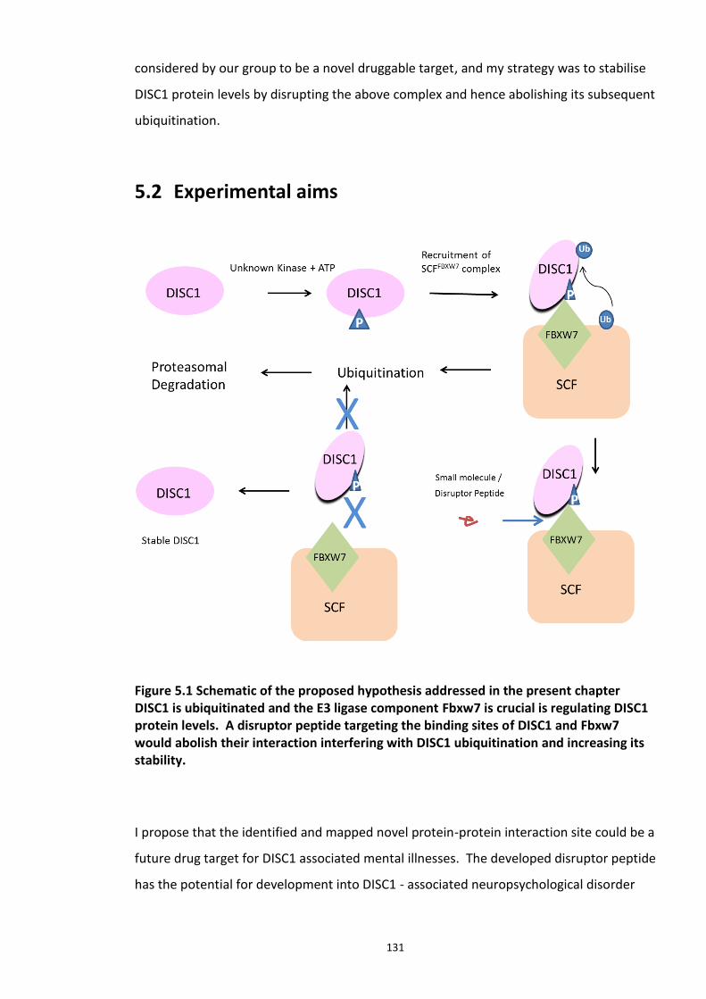
131
considered by our group to be a novel druggable target, and my strategy was to stabilise
DISC1 protein levels by disrupting the above complex and hence abolishing its subsequent
ubiquitination.
5.2 Experimental aims
Figure 5.1 Schematic of the proposed hypothesis addressed in the present chapter DISC1 is ubiquitinated and the E3 ligase component Fbxw7 is crucial is regulating DISC1 protein levels. A disruptor peptide targeting the binding sites of DISC1 and Fbxw7 would abolish their interaction interfering with DISC1 ubiquitination and increasing its stability.
I propose that the identified and mapped novel protein-protein interaction site could be a
future drug target for DISC1 associated mental illnesses. The developed disruptor peptide
has the potential for development into DISC1 - associated neuropsychological disorder
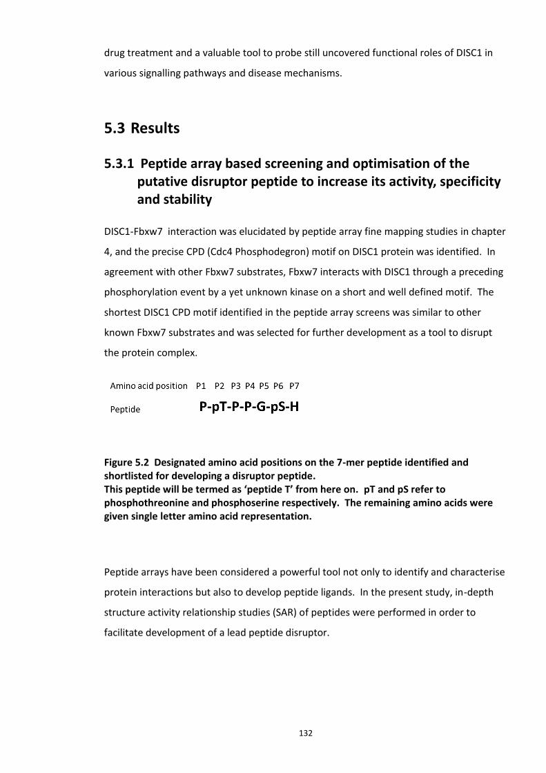
132
drug treatment and a valuable tool to probe still uncovered functional roles of DISC1 in
various signalling pathways and disease mechanisms.
5.3 Results
5.3.1 Peptide array based screening and optimisation of the putative disruptor peptide to increase its activity, specificity and stability
DISC1-Fbxw7 interaction was elucidated by peptide array fine mapping studies in chapter
4, and the precise CPD (Cdc4 Phosphodegron) motif on DISC1 protein was identified. In
agreement with other Fbxw7 substrates, Fbxw7 interacts with DISC1 through a preceding
phosphorylation event by a yet unknown kinase on a short and well defined motif. The
shortest DISC1 CPD motif identified in the peptide array screens was similar to other
known Fbxw7 substrates and was selected for further development as a tool to disrupt
the protein complex.
Figure 5.2 Designated amino acid positions on the 7-mer peptide identified and shortlisted for developing a disruptor peptide. This peptide will be termed as ‘peptide T’ from here on. pT and pS refer to phosphothreonine and phosphoserine respectively. The remaining amino acids were given single letter amino acid representation.
Peptide arrays have been considered a powerful tool not only to identify and characterise
protein interactions but also to develop peptide ligands. In the present study, in-depth
structure activity relationship studies (SAR) of peptides were performed in order to
facilitate development of a lead peptide disruptor.

133
Criteria considered while optimising and developing the disruptor peptide were as
follows:
1. Most of the naturally occurring amino acids are L isomers and the cellular proteases are
specific to peptide bonds linking L amino acids. Therefore, substitution with D isomers of
the amino acids increases the stability of the peptide in cells. Peptides flanked by non-
natural amino acids also resist proteolysis by exopeptidases.
2. Since side chains of the amino acids play a crucial role in the specificity of enzyme –
substrate complex formation, amino acids with capped or modified side chains serve as
better substitutes, as they evade proteolysis.
3. When the amino acids are substituted, it is important to maintain the overall charge to
mimic the natural peptide, so that the disruptor will act as a peptidomimetic with a higher
affinity towards Fbxw7 interaction than that of the natural peptide (DISC1 CPD).
Phosphomimetic amino acids such as Glu and Asp retain the charge while replacing the
phosphorylated residues pT and pS.
4. These substitution studies also provide information on the role of each amino acid
residue and whether the side chains participate in the interaction. Substitution with Ala,
which has a methyl group as its side chain, eliminates side chain interactions while the
peptide backbone confirmation is retained.
5. Pro with a cyclic side chain containing a nitrogen as a backbone atom, restricts protein
confirmation and makes it preferential for inducing β turns in polypeptides sheets
(MacArthur and Thornton, 1991). Hence, the amino acid substitutions considered should
have a similar backbone while they meet all the above criteria, such as the non-natural
amino acids - Pip, Tic, Sar.
6. Shifting the amino group on the peptide back bone, from the α carbon to the β carbon,
increases the length of the peptide backbone and increases their stability, since β peptide
bonds are non-natural.
7. While considering substitutions for His at P7 in the native peptide, amino acids such as
lys and the non-natural amino acid ornithine were considered to retain the negative
charge. With an imidazole ring in its side chain and its ability to exist in protonated and
de protonated forms depending on the pH, His is known to be crucial in protein
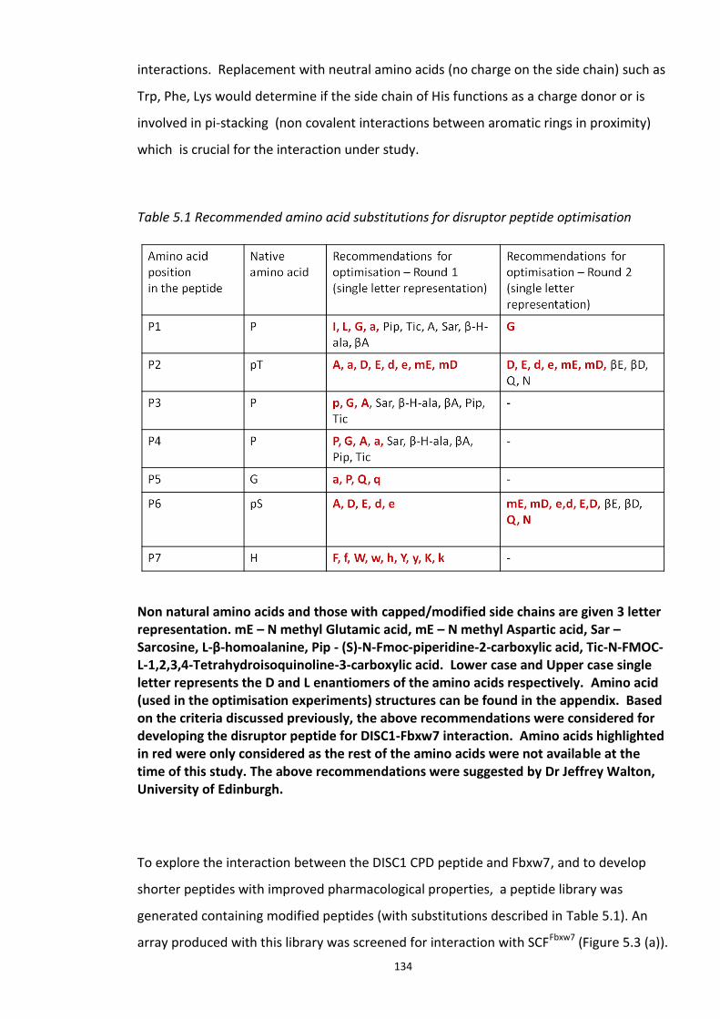
134
interactions. Replacement with neutral amino acids (no charge on the side chain) such as
Trp, Phe, Lys would determine if the side chain of His functions as a charge donor or is
involved in pi-stacking (non covalent interactions between aromatic rings in proximity)
which is crucial for the interaction under study.
Table 5.1 Recommended amino acid substitutions for disruptor peptide optimisation
Non natural amino acids and those with capped/modified side chains are given 3 letter representation. mE – N methyl Glutamic acid, mE – N methyl Aspartic acid, Sar – Sarcosine, L-β-homoalanine, Pip - (S)-N-Fmoc-piperidine-2-carboxylic acid, Tic-N-FMOC-L-1,2,3,4-Tetrahydroisoquinoline-3-carboxylic acid. Lower case and Upper case single letter represents the D and L enantiomers of the amino acids respectively. Amino acid (used in the optimisation experiments) structures can be found in the appendix. Based on the criteria discussed previously, the above recommendations were considered for developing the disruptor peptide for DISC1-Fbxw7 interaction. Amino acids highlighted in red were only considered as the rest of the amino acids were not available at the time of this study. The above recommendations were suggested by Dr Jeffrey Walton, University of Edinburgh.
To explore the interaction between the DISC1 CPD peptide and Fbxw7, and to develop
shorter peptides with improved pharmacological properties, a peptide library was
generated containing modified peptides (with substitutions described in Table 5.1). An
array produced with this library was screened for interaction with SCFFbxw7 (Figure 5.3 (a)).
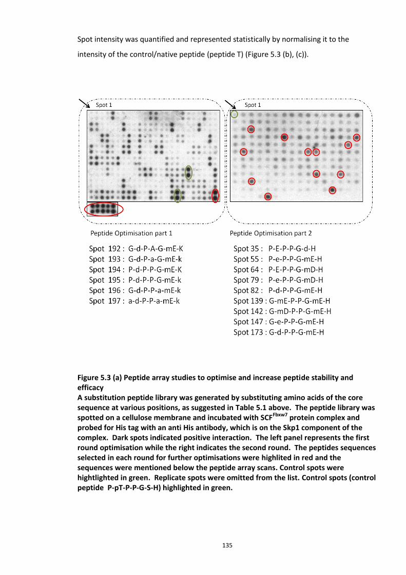
135
Spot intensity was quantified and represented statistically by normalising it to the
intensity of the control/native peptide (peptide T) (Figure 5.3 (b), (c)).
Figure 5.3 (a) Peptide array studies to optimise and increase peptide stability and efficacy A substitution peptide library was generated by substituting amino acids of the core sequence at various positions, as suggested in Table 5.1 above. The peptide library was spotted on a cellulose membrane and incubated with SCFFbxw7 protein complex and probed for His tag with an anti His antibody, which is on the Skp1 component of the complex. Dark spots indicated positive interaction. The left panel represents the first round optimisation while the right indicates the second round. The peptides sequences selected in each round for further optimisations were highlited in red and the sequences were mentioned below the peptide array scans. Control spots were hightlighted in green. Replicate spots were omitted from the list. Control spots (control peptide P-pT-P-P-G-S-H) highlighted in green.
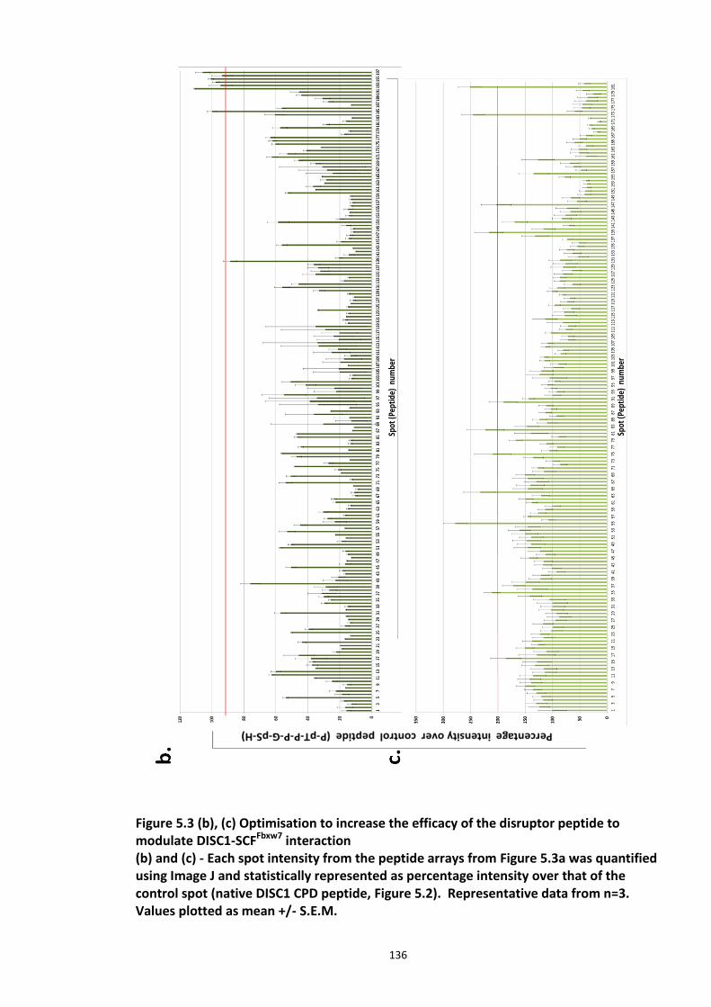
136
Figure 5.3 (b), (c) Optimisation to increase the efficacy of the disruptor peptide to modulate DISC1-SCFFbxw7 interaction (b) and (c) - Each spot intensity from the peptide arrays from Figure 5.3a was quantified using Image J and statistically represented as percentage intensity over that of the control spot (native DISC1 CPD peptide, Figure 5.2). Representative data from n=3. Values plotted as mean +/- S.E.M.
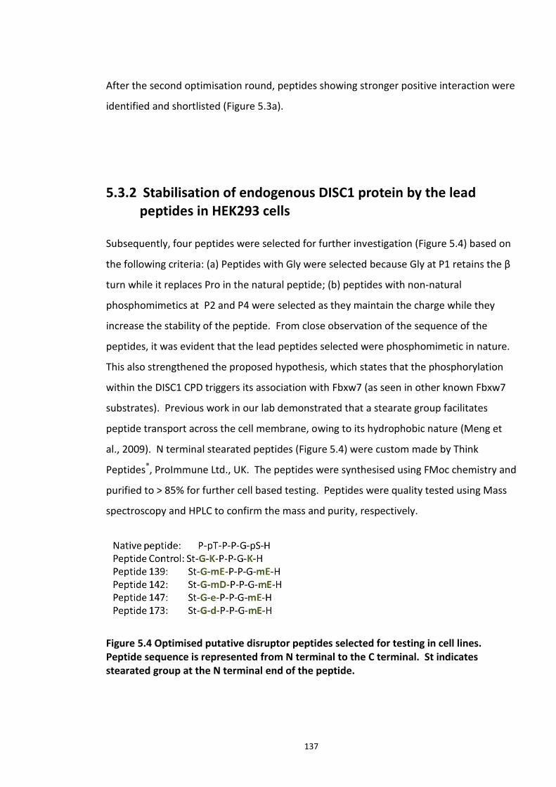
137
After the second optimisation round, peptides showing stronger positive interaction were
identified and shortlisted (Figure 5.3a).
5.3.2 Stabilisation of endogenous DISC1 protein by the lead peptides in HEK293 cells
Subsequently, four peptides were selected for further investigation (Figure 5.4) based on
the following criteria: (a) Peptides with Gly were selected because Gly at P1 retains the β
turn while it replaces Pro in the natural peptide; (b) peptides with non-natural
phosphomimetics at P2 and P4 were selected as they maintain the charge while they
increase the stability of the peptide. From close observation of the sequence of the
peptides, it was evident that the lead peptides selected were phosphomimetic in nature.
This also strengthened the proposed hypothesis, which states that the phosphorylation
within the DISC1 CPD triggers its association with Fbxw7 (as seen in other known Fbxw7
substrates). Previous work in our lab demonstrated that a stearate group facilitates
peptide transport across the cell membrane, owing to its hydrophobic nature (Meng et
al., 2009). N terminal stearated peptides (Figure 5.4) were custom made by Think
Peptides®, ProImmune Ltd., UK. The peptides were synthesised using FMoc chemistry and
purified to > 85% for further cell based testing. Peptides were quality tested using Mass
spectroscopy and HPLC to confirm the mass and purity, respectively.
Figure 5.4 Optimised putative disruptor peptides selected for testing in cell lines. Peptide sequence is represented from N terminal to the C terminal. St indicates stearated group at the N terminal end of the peptide.
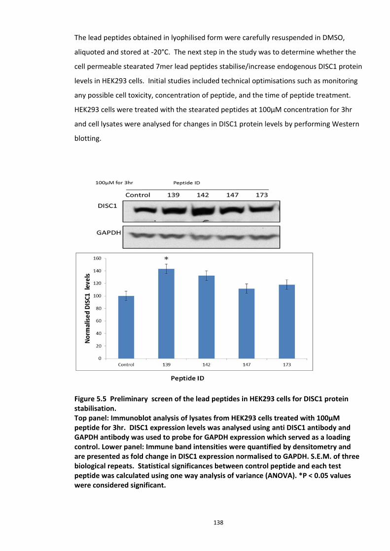
138
The lead peptides obtained in lyophilised form were carefully resuspended in DMSO,
aliquoted and stored at -20°C. The next step in the study was to determine whether the
cell permeable stearated 7mer lead peptides stabilise/increase endogenous DISC1 protein
levels in HEK293 cells. Initial studies included technical optimisations such as monitoring
any possible cell toxicity, concentration of peptide, and the time of peptide treatment.
HEK293 cells were treated with the stearated peptides at 100µM concentration for 3hr
and cell lysates were analysed for changes in DISC1 protein levels by performing Western
blotting.
Figure 5.5 Preliminary screen of the lead peptides in HEK293 cells for DISC1 protein stabilisation. Top panel: Immunoblot analysis of lysates from HEK293 cells treated with 100µM peptide for 3hr. DISC1 expression levels was analysed using anti DISC1 antibody and GAPDH antibody was used to probe for GAPDH expression which served as a loading control. Lower panel: Immune band intensities were quantified by densitometry and are presented as fold change in DISC1 expression normalised to GAPDH. S.E.M. of three biological repeats. Statistical significances between control peptide and each test peptide was calculated using one way analysis of variance (ANOVA). *P < 0.05 values were considered significant.
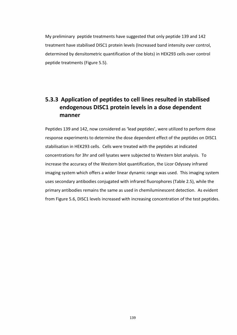
139
My preliminary peptide treatments have suggested that only peptide 139 and 142
treatment have stabilised DISC1 protein levels (Increased band intensity over control,
determined by densitometric quantification of the blots) in HEK293 cells over control
peptide treatments (Figure 5.5).
5.3.3 Application of peptides to cell lines resulted in stabilised endogenous DISC1 protein levels in a dose dependent manner
Peptides 139 and 142, now considered as ‘lead peptides’, were utilized to perform dose
response experiments to determine the dose dependent effect of the peptides on DISC1
stabilisation in HEK293 cells. Cells were treated with the peptides at indicated
concentrations for 3hr and cell lysates were subjected to Western blot analysis. To
increase the accuracy of the Western blot quantification, the Licor Odyssey infrared
imaging system which offers a wider linear dynamic range was used. This imaging system
uses secondary antibodies conjugated with infrared fluorophores (Table 2.5), while the
primary antibodies remains the same as used in chemiluminescent detection. As evident
from Figure 5.6, DISC1 levels increased with increasing concentration of the test peptides.
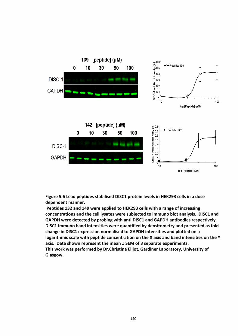
140
Figure 5.6 Lead peptides stabilised DISC1 protein levels in HEK293 cells in a dose dependent manner. Peptides 132 and 149 were applied to HEK293 cells with a range of increasing concentrations and the cell lysates were subjected to immuno blot analysis. DISC1 and GAPDH were detected by probing with anti DISC1 and GAPDH antibodies respectively. DISC1 immuno band intensities were quantified by densitometry and presented as fold change in DISC1 expression normalised to GAPDH intensities and plotted on a logarithmic scale with peptide concentration on the X axis and band intensities on the Y axis. Data shown represent the mean ± SEM of 3 separate experiments. This work was performed by Dr.Christina Elliot, Gardiner Laboratory, University of Glasgow.
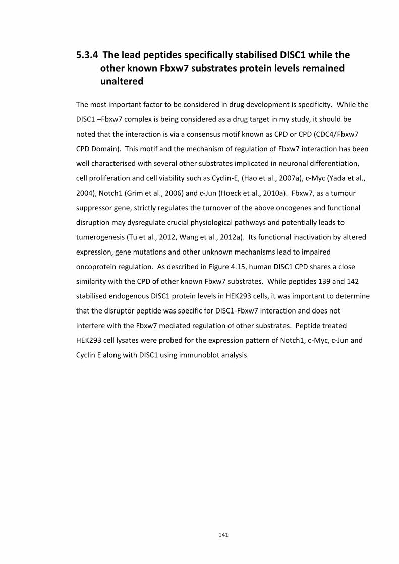
141
5.3.4 The lead peptides specifically stabilised DISC1 while the other known Fbxw7 substrates protein levels remained unaltered
The most important factor to be considered in drug development is specificity. While the
DISC1 –Fbxw7 complex is being considered as a drug target in my study, it should be
noted that the interaction is via a consensus motif known as CPD or CPD (CDC4/Fbxw7
CPD Domain). This motif and the mechanism of regulation of Fbxw7 interaction has been
well characterised with several other substrates implicated in neuronal differentiation,
cell proliferation and cell viability such as Cyclin-E, (Hao et al., 2007a), c-Myc (Yada et al.,
2004), Notch1 (Grim et al., 2006) and c-Jun (Hoeck et al., 2010a). Fbxw7, as a tumour
suppressor gene, strictly regulates the turnover of the above oncogenes and functional
disruption may dysregulate crucial physiological pathways and potentially leads to
tumerogenesis (Tu et al., 2012, Wang et al., 2012a). Its functional inactivation by altered
expression, gene mutations and other unknown mechanisms lead to impaired
oncoprotein regulation. As described in Figure 4.15, human DISC1 CPD shares a close
similarity with the CPD of other known Fbxw7 substrates. While peptides 139 and 142
stabilised endogenous DISC1 protein levels in HEK293 cells, it was important to determine
that the disruptor peptide was specific for DISC1-Fbxw7 interaction and does not
interfere with the Fbxw7 mediated regulation of other substrates. Peptide treated
HEK293 cell lysates were probed for the expression pattern of Notch1, c-Myc, c-Jun and
Cyclin E along with DISC1 using immunoblot analysis.
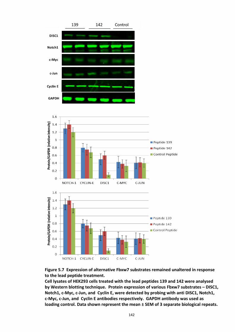
142
Figure 5.7 Expression of alternative Fbxw7 substrates remained unaltered in response to the lead peptide treatment. Cell lysates of HEK293 cells treated with the lead peptides 139 and 142 were analysed by Western blotting technique. Protein expression of various Fbxw7 substrates – DISC1, Notch1, c-Myc, c-Jun, and Cyclin E, were detected by probing with anti DISC1, Notch1, c-Myc, c-Jun, and Cyclin E antibodies respectively. GAPDH antibody was used as loading control. Data shown represent the mean ± SEM of 3 separate biological repeats.
143
Band intensities were quantified by using Odyssey software and statisticaly represented in the graphs. This work was carried out by Dr.Christina Elliot, Gardiner Laboratory, University of Glasgow.
Cell lysates from HEK293 cells treated with the lead peptides (139 and 142) were probed
for other known Fbxw7 substrates. Immunoblots (Figure 5.7) show that the peptides
specifically stabilised endogenous DISC1 protein levels while the other substrates
(Notch1, c-Myc, c-Jun, Cyclin E) largely remained unchanged implying that the lead
peptides specifically disrupt Fbxw7-DISC1 interaction and do not interfere with other
crucial interactions of Fbxw7.
144
5.3.5 Induced Pluripotent Stem cells as a cell line model
The exprimental work described in the section has been carried out as part of a 10 week
Ph.D. placement at Pfizer Neuroscience Research Unit, Cambridge, Boston, MA in
February – April 2013.
5.3.5.1 DISC1 animal models – Limitations
Identification of DISC1 in the Scottish pedigree has garnered much attention by scientists
who have tried to uncover the molecular mechanisms behind the biological phenomenon
in order to develop therapeutics from a most promising susceptibility gene for SCZ and
other associated mental illnesses. However, research in psychiatric illnesses is severely
hampered by the lack of availability of human brain tissue, especially live neurons from
patients.
The mouse has long been an attractive genetic model organism to study psychiatric
disease, as it has many advantages such as the availability of a range of inbred strains,
completed genome sequences and advancements in technology to genetically manipulate
mice in numerous ways. DISC1 mouse models were invaluable and provided
developmental and mechanistic insights, which could have been otherwise difficult to
obtain. A review by Hanna Jaaro-Peled discusses all the seven DISC1 mouse models
generated so far (Jaaro-Peled, 2009). These mouse models displayed a wide range of
phenotypes which include both abnormal anatomical changes (example: reduced brain
volume and neurite outgrowth) and behavioural phenotypes (example: diminished
working memory and pre pulse inhibition (PPI)). Q31L and L100P were two ENU (N-ethyl-
N-nitrosourea) induced mutants generated, which exhibited behavioural phenotypes-
depression like and SCZ like respectively (Clapcote et al., 2007). MRI studies in these mice
have also exhibited enlarged lateral ventricles, a common observation in SCZ patients
(Vita et al., 2006). Koike et al. (2006), reported that 129S6/SvEv mice strain harbour a
25bp deletion in the DISC1 gene which results in a termination codon in exon 7, which
abolishes the translation of full length DISC1 protein. Working memory was shown to be
145
impaired when this variant was transferred to the C57BL/6 genetic background (Koike et
al., 2006). A DISC1 knockout mouse would the most useful tool; however, generation of a
‘true null’ has been challenging so far, as all DISC1 isoforms have not been identified and
characterised yet (Brandon et al., 2004, Miyoshi et al., 2003a, James et al., 2004a). Since
DISC1 was shown to be crucial in neurodevelopment processes, there is also a possible
risk of lethality during development of KO mice (Schurov et al., 2004a) (Ishizuka et al.,
2006). A conditional knockout mice in Cre/loxP system was another alternative suggested
by experts to circumvent the complexity surrounding the KO mice generation (Ishizuka et
al., 2006). Kuroda et al, generated transgenic mice lacking exons 2 and 3 (also known as
Disc1 (∆2-3)) which were shown to have higher threshold for the induction of long-term
potentiation (LTP) and showed PPI deficits which are a well-known phenomenon
observed in schizophrenia patients and other neuropsychiatric disorders (Kuroda et al.,
2011).
However, it should be noted that animal models do not completely translate to the
human condition and their use has always been limited in psychiatric research. It is
impractical to generate schizophrenic mice and extremely difficult to study the psychotic
symptoms such as hallucinations and delusion. To add to the complexity, most of these
illnesses are polygenic and involve gene - environment interactions. Research over the
last decade failed to develop a cell line/animal disease model for SCZ, which has been a
major obstacle in drug development, as drug toxicity may vary with the variable ability of
different species to metabolise drugs.
Since most of the drugs are targeted to regulate protein expression or enzyme activity, it
is important that the protein of interest is well conserved in the species model. In light of
DISC1 being implicated in brain dysfunction, it should be noted that DISC1 protein is
poorly conserved between various species which may be considered as a major drawback
in considering the use of rodent derived cell lines for DISC1 studies. Primate cerebral
cortex differs substantially from that of rodents in many features, such as cerebral cortex
size, complexity and the nature and range of stem cell population. Drug discovery for
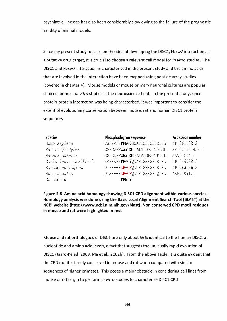
146
psychiatric illnesses has also been considerably slow owing to the failure of the prognostic
validity of animal models.
Since my present study focuses on the idea of developing the DISC1/Fbxw7 interaction as
a putative drug target, it is crucial to choose a relevant cell model for in vitro studies. The
DISC1 and Fbxw7 interaction is characterised in the present study and the amino acids
that are involved in the interaction have been mapped using peptide array studies
(covered in chapter 4). Mouse models or mouse primary neuronal cultures are popular
choices for most in vitro studies in the neuroscience field. In the present study, since
protein-protein interaction was being characterised, it was important to consider the
extent of evolutionary conservation between mouse, rat and human DISC1 protein
sequences.
Figure 5.8 Amino acid homology showing DISC1 CPD alignment within various species. Homology analysis was done using the Basic Local Alignment Search Tool (BLAST) at the NCBI website (http://www.ncbi.nlm.nih.gov/blast). Non conserved CPD motif residues in mouse and rat were highlighted in red.
Mouse and rat orthologues of DISC1 are only about 56% identical to the human DISC1 at
nucleotide and amino acid levels, a fact that suggests the unusually rapid evolution of
DISC1 (Jaaro-Peled, 2009, Ma et al., 2002b). From the above Table, it is quite evident that
the CPD motif is barely conserved in mouse and rat when compared with similar
sequences of higher primates. This poses a major obstacle in considering cell lines from
mouse or rat origin to perform in vitro studies to characterise DISC1 CPD.
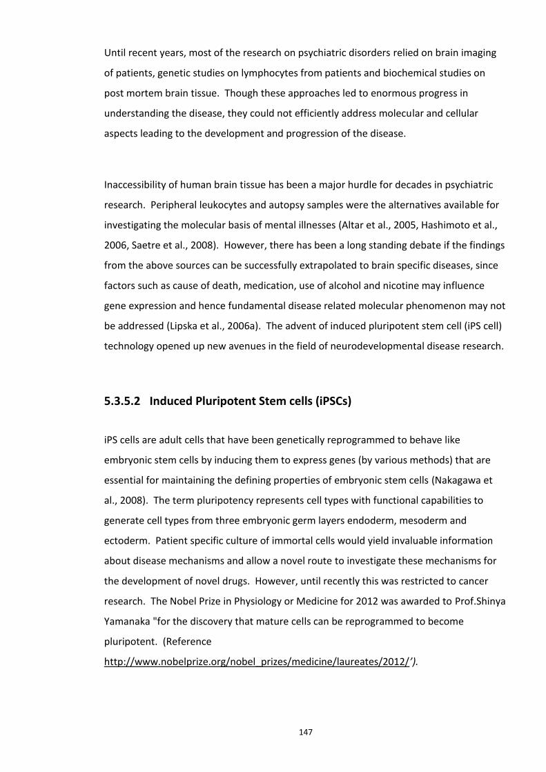
147
Until recent years, most of the research on psychiatric disorders relied on brain imaging
of patients, genetic studies on lymphocytes from patients and biochemical studies on
post mortem brain tissue. Though these approaches led to enormous progress in
understanding the disease, they could not efficiently address molecular and cellular
aspects leading to the development and progression of the disease.
Inaccessibility of human brain tissue has been a major hurdle for decades in psychiatric
research. Peripheral leukocytes and autopsy samples were the alternatives available for
investigating the molecular basis of mental illnesses (Altar et al., 2005, Hashimoto et al.,
2006, Saetre et al., 2008). However, there has been a long standing debate if the findings
from the above sources can be successfully extrapolated to brain specific diseases, since
factors such as cause of death, medication, use of alcohol and nicotine may influence
gene expression and hence fundamental disease related molecular phenomenon may not
be addressed (Lipska et al., 2006a). The advent of induced pluripotent stem cell (iPS cell)
technology opened up new avenues in the field of neurodevelopmental disease research.
5.3.5.2 Induced Pluripotent Stem cells (iPSCs)
iPS cells are adult cells that have been genetically reprogrammed to behave like
embryonic stem cells by inducing them to express genes (by various methods) that are
essential for maintaining the defining properties of embryonic stem cells (Nakagawa et
al., 2008). The term pluripotency represents cell types with functional capabilities to
generate cell types from three embryonic germ layers endoderm, mesoderm and
ectoderm. Patient specific culture of immortal cells would yield invaluable information
about disease mechanisms and allow a novel route to investigate these mechanisms for
the development of novel drugs. However, until recently this was restricted to cancer
research. The Nobel Prize in Physiology or Medicine for 2012 was awarded to Prof.Shinya
Yamanaka "for the discovery that mature cells can be reprogrammed to become
pluripotent. (Reference
http://www.nobelprize.org/nobel_prizes/medicine/laureates/2012/’).
148
Yamanaka and colleagues generated mouse iPS cells for the first time in 2006, from
mouse embryonic and adult fibroblasts by viral transduction of Oct3/4, Sox2, c-Myc, and
Klf4, under ES cell culture conditions (Takahashi and Yamanaka, 2006). These four factors
were later used to generate iPS cells from human skin fibroblasts (Takahashi et al., 2007).
However, there were major drawbacks to the protocols used in these studies, with c-Myc
being an onco gene and the associated increased risk of tumor development at a later
stage. The same group went on to develop a method, whereby, iPS cells were made
without having to transduce the c-myc gene and use drugs for selection (Nakagawa et al.,
2008). The method described in the above study also increased the quality of iPS cells
and reduced the back ground of non iPS cells (Nakagawa et al., 2008). In later years,
integration free reprogramming methods were developed which increased the quality
and yield of iPS cells (Cahan and Daley, 2013). These pioneering studies were followed by
the successful generation of iPS cells from patients with various genetic disorders
including neurological disorders like Huntington’s and Parkinson’s disease (Zhang et al.,
2010, Park et al., 2008). Patient specific stem cells offer an extraordinary opportunity to
reiterate both normal and pathological human tissue formation in vitro, particularly in
case of brain diseases, thereby enabling disease investigation and drug target
development (Park et al., 2008). It can be understood that after well over a decade of
initial constraints these area have now been overcome and research in this area is
currently flourishing.
5.3.5.3 iPS cell technology in brain research
iPS cell technology offers an excellent alternative and is invaluable in neuropsychiatric
research. Their ability to differentiate in to three germ layers (ectoderm, endoderm and
mesoderm) renders iPS cells as ideal patient specific model cell lines for SCZ, where
aetiology still remains unclear. Generation of human and patient specific cell models is
advantageous in developing drug targets in that they allow a limitless supply of human
patient specific cells that can be differentiated into the cell type of choice where the
disease specific phenotype can be recapitulated. This novel research approach facilitates
the design and verification of disease models which are otherwise not feasible for
psychiatric illnesses.
149
A number of groups reported the successful generation and use of iPS cells in disease
modelling to investigate neurological disorders (Pedrosa et al., 2011, Chiang et al., 2011,
Zhang et al., 2010, Wernig et al., 2008, Cherry and Daley, 2013). Brennand et al, (2011)
employed this principle and reprogrammed fibroblasts from SCZ patients to human iPSC
and subsequently differentiated these pluripotent cells into glutamatergic neurons. They
performed extensive gene expression profiling and looked for morphological changes in
these disorder specific neurons to investigate the molecular defects in SCZ. Compared to
the control neurons, they reported reduced PSD95-protein levels, glutamate receptor
expression, abnormal Wnt and cAMP signalling along with morphological changes such as
reduced neuronal connectivity and neurite number in SCZ neurons (Brennand et al.,
2011).
The ability of iPS cells to efficiently differentiate into any desired cell type such as neurons
has been successfully demonstrated (Gao et al., 2013), (Pedrosa et al., 2011). With the
addition of specific factors to the culture media, iPS cells have been further differentiated
into brain region specific identities including hindbrain, forebrain and spinal cord,
permitting large scale production of specific neurons carrying the genetic background
from the disease carriers (Brennand and Gage, 2012). This also facilitates analysis of the
differentiated cells under controlled conditions. iPSC derived neurons are ideal
particularly for neurodevelopmental disorders like SCZ, since they facilitate recapitulation
of the early stages of neuronal differentiation in genetic backgrounds which may increase
the risk of disease. They also allow the study of the cellular and molecular events
triggering the onset of these disorders.
For the purpose of screening the lead peptides (developed in the present study) within a
short time frame, expandable neuronal progenitors generated from the iPS cells were
used.
150
Figure 5.9 Application of iPS cell technology in research and drug development Adapted from (Robinton and Daley, 2012). This novel technology facilitates reprogramming of easily available patient specific skin fibroblasts into affected neuronal subtypes. These differentiated neurons can be used for patient genotype specific disease modelling in vitro and serve as a tool to study drug targets and develop potential drugs.
5.3.5.4 Experimental aims
The aims of this section of the chapter are as follows:
1. Explore iPSc technology for disease modelling by differentiating the accessible iPS cells
reprogrammed from SCZ patients and differentiating them to neuronal progenitor cells
(NPs).
2. Characterisation of the neuroprogenitor cells (NPs) by screening of pluripotent and
neuronal markers.
3. Study DISC1 and Fbxw7 expression profile in NPs.
4. Test the novel hypothesis that DISC1-Fbxw7 interaction controls DISC1 protein levels in
NPs.
151
5.3.5.5 Results
iPS cells generated from skin biopsies of patients diagnosed with SCZ have been used in
the present study. The cells were taken from healthy controls (with no known mutations
in DISC1 gene) and SCZ patients harbouring a 4bp deletion at the 12th exon-intron
boundary in the C-terminus of the DISC1 gene. Neuronal progenitors differentiated from
these cells were considered suitable for in vitro testing of the putative disruptor peptides
for the following reasons:
1. Availability: Recent advancements in the field of iPSc research have enabled the
production of these cell lines without specialised equipment.
2. Origin: Since the iPS cells used in the present study were generated from patients
diagnosed with SCZ, they allow close recapitulation of the cellular phenomena related to
SCZ and are apt for use in investigating the underlying molecular events.
3. Disrupted DISC1 gene: One of the major genetic risk factor of SCZ, DISC1 gene carries a
4bp deletion, resulting in a frame shift mutation which could be translated to a truncated
protein. It would be intriguing to investigate if this mutation has a role to play in DISC1
signalling pathway in these cells.
The iPS cells (except Detroit) used in the present study have been described and
characterized previously (Chiang et al., 2011, Pedrosa et al., 2011). iPS Detroit cell line
were generated by Dr.Sandra Engle, Pfizer Inc. All the cell lines used in this study were
karyotyped (for chromosomal aberrations) and screened for the expression markers
(stem cells) at Pfizer.
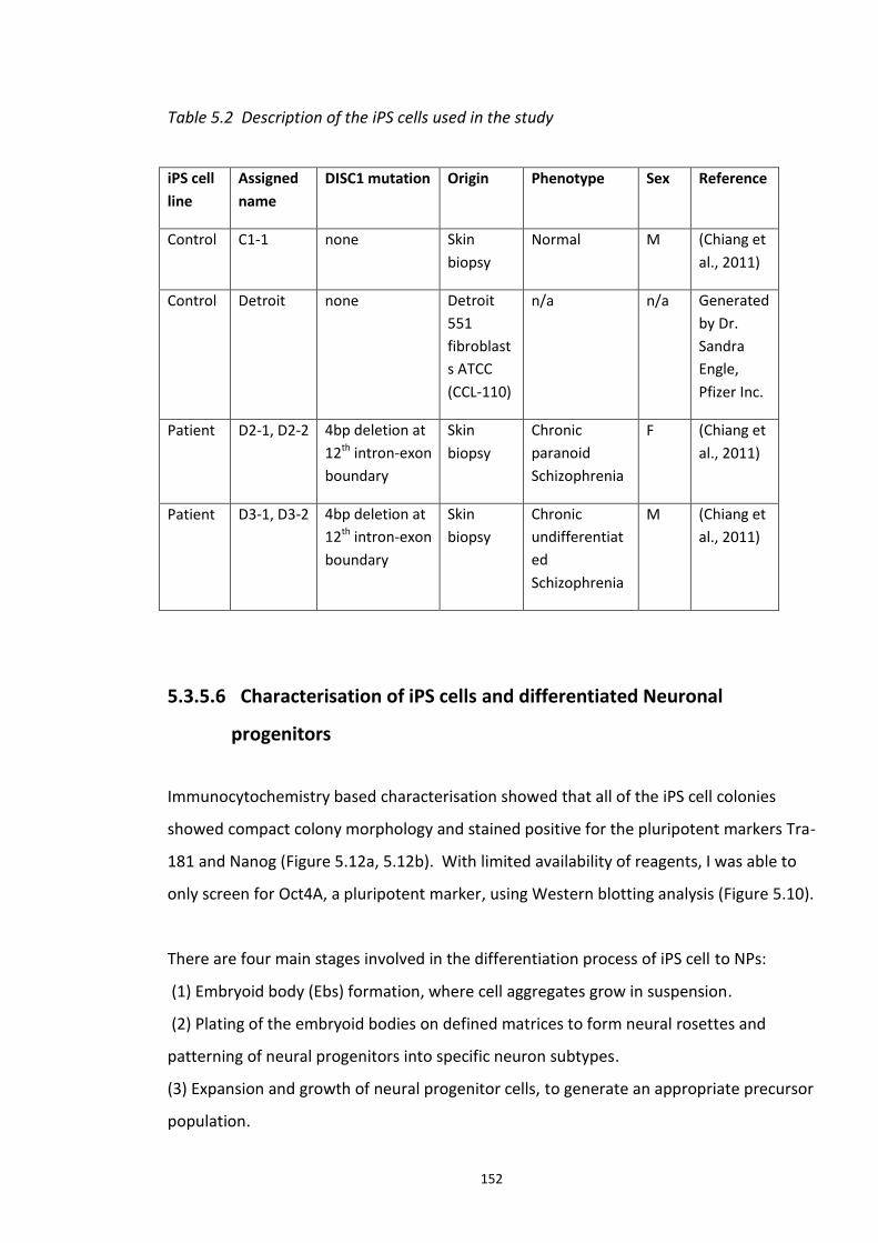
152
Table 5.2 Description of the iPS cells used in the study
iPS cell
line
Assigned
name
DISC1 mutation Origin Phenotype Sex Reference
Control C1-1 none Skin
biopsy
Normal M (Chiang et
al., 2011)
Control Detroit none Detroit
551
fibroblast
s ATCC
(CCL-110)
n/a n/a Generated
by Dr.
Sandra
Engle,
Pfizer Inc.
Patient D2-1, D2-2 4bp deletion at
12th intron-exon
boundary
Skin
biopsy
Chronic
paranoid
Schizophrenia
F (Chiang et
al., 2011)
Patient D3-1, D3-2 4bp deletion at
12th intron-exon
boundary
Skin
biopsy
Chronic
undifferentiat
ed
Schizophrenia
M (Chiang et
al., 2011)
5.3.5.6 Characterisation of iPS cells and differentiated Neuronal
progenitors
Immunocytochemistry based characterisation showed that all of the iPS cell colonies
showed compact colony morphology and stained positive for the pluripotent markers Tra-
181 and Nanog (Figure 5.12a, 5.12b). With limited availability of reagents, I was able to
only screen for Oct4A, a pluripotent marker, using Western blotting analysis (Figure 5.10).
There are four main stages involved in the differentiation process of iPS cell to NPs:
(1) Embryoid body (Ebs) formation, where cell aggregates grow in suspension.
(2) Plating of the embryoid bodies on defined matrices to form neural rosettes and
patterning of neural progenitors into specific neuron subtypes.
(3) Expansion and growth of neural progenitor cells, to generate an appropriate precursor
population.
153
(4) Differentiation to specific cell types where neural progenitor cells can develop into
neurons of specific subtype (This step was not performed in the present study).
The various stages of neuronal differentiation from iPSc are schematically represented in
Figure 5.11. Supplementing minimal growth media with fibroblast growth factor 2
(FGF2), L-glutamine and ß-mercaptoethanol leads to neural stem cell generation, which is
characterised by expression of neural markers such as Nestin (Li and Zhang, 2006, Kim et
al., 2011). The neuronal progenitors generated, have the potential to be further
differentiated in to neurons, astrocytes or oligodendrocytes using specific culture
medium supplements (Chojnacki and Weiss, 2008). iPS cells described in Table 5.3, were
karyotyped to check for any possible mutations or carcinogenic gene activation which
might be possible as a result of episomal gene transfer (Chiang et al., 2011).
The maintenance and differentiation protocol has been optimized and standardized at
Pfizer by Dr. Lindsay Wilson, to generate GABAergic neuronal progenitors cells from iPS
cells by reprogramming. The detailed protocol has been explained in section 2.3.2. In
brief, iPSCs were maintained on Mitomycin C treated (mitotically arrested) MEFs (Mouse
Embryonic Fibroblasts) in the presence of 10 ng/mL FGF2. On day 0, iPSc colonies were
detached from feeder layers and grown on ultra-low adherence plates in suspension
culture and supplemented with ROCK inhibitor to induce embyoid body (EBs) formation in
the absence of FGF2. On day 2, EBs were transferred on to matrigel coated plates. The
morphology changes with the progression of differentiation were monitored and
representative images were detailed in Figure 5.11. The phenotype of the progenitors
was also analysed by immunocytochemistry using various neural markers (Figure 5.13
a,b).
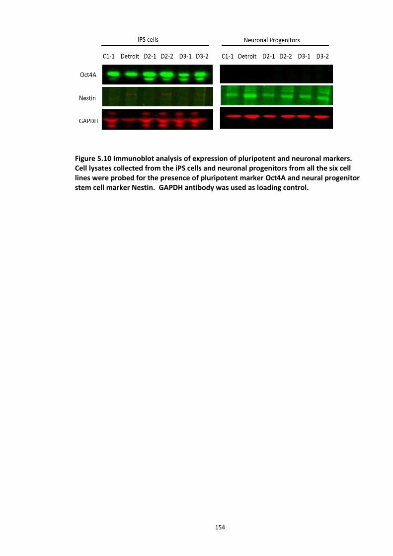
154
Figure 5.10 Immunoblot analysis of expression of pluripotent and neuronal markers. Cell lysates collected from the iPS cells and neuronal progenitors from all the six cell lines were probed for the presence of pluripotent marker Oct4A and neural progenitor stem cell marker Nestin. GAPDH antibody was used as loading control.
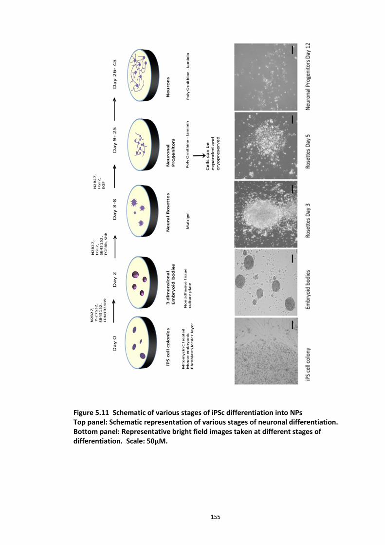
155
Figure 5.11 Schematic of various stages of iPSc differentiation into NPs Top panel: Schematic representation of various stages of neuronal differentiation. Bottom panel: Representative bright field images taken at different stages of differentiation. Scale: 50µM.
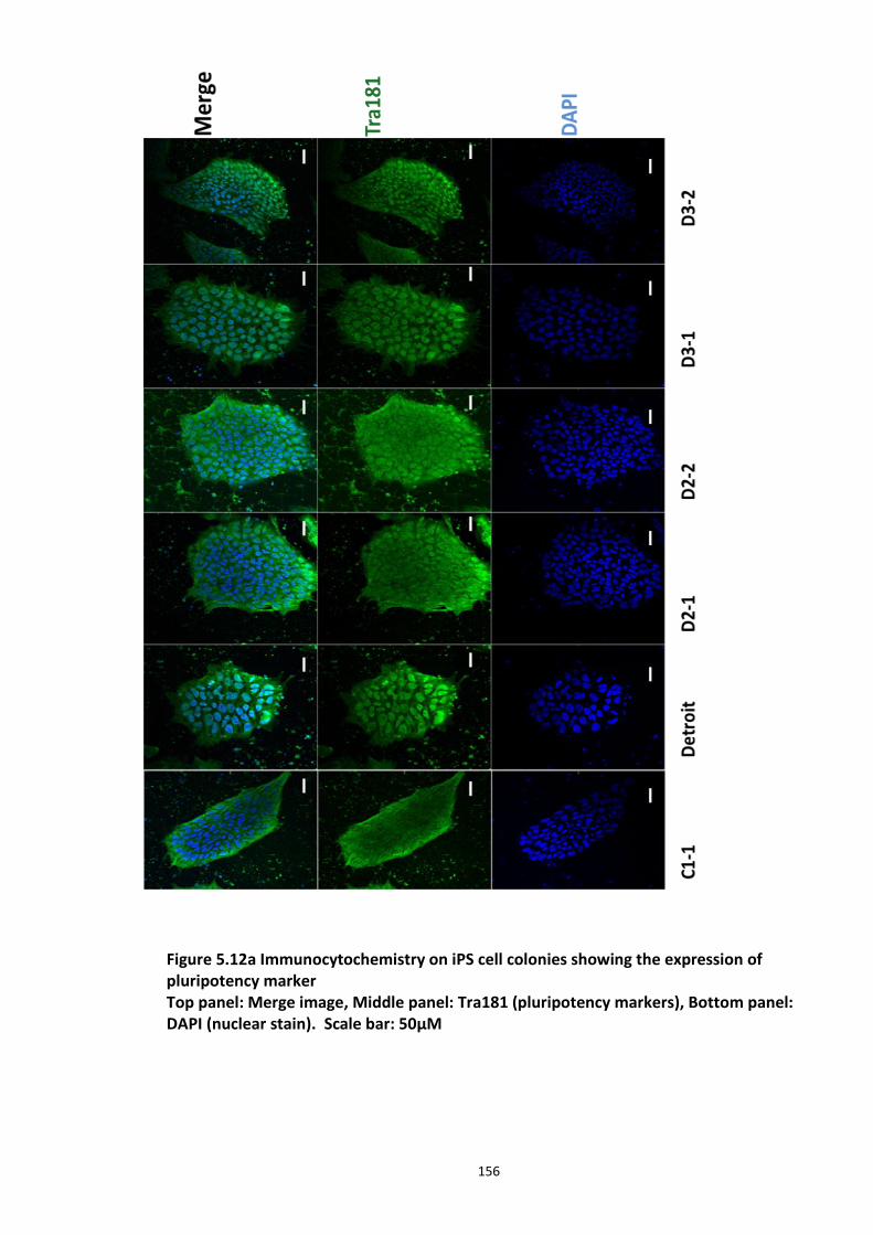
156
Figure 5.12a Immunocytochemistry on iPS cell colonies showing the expression of pluripotency marker Top panel: Merge image, Middle panel: Tra181 (pluripotency markers), Bottom panel: DAPI (nuclear stain). Scale bar: 50µM
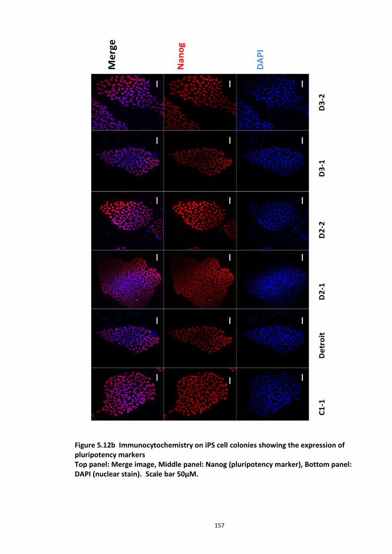
157
Figure 5.12b Immunocytochemistry on iPS cell colonies showing the expression of pluripotency markers Top panel: Merge image, Middle panel: Nanog (pluripotency marker), Bottom panel: DAPI (nuclear stain). Scale bar 50µM.
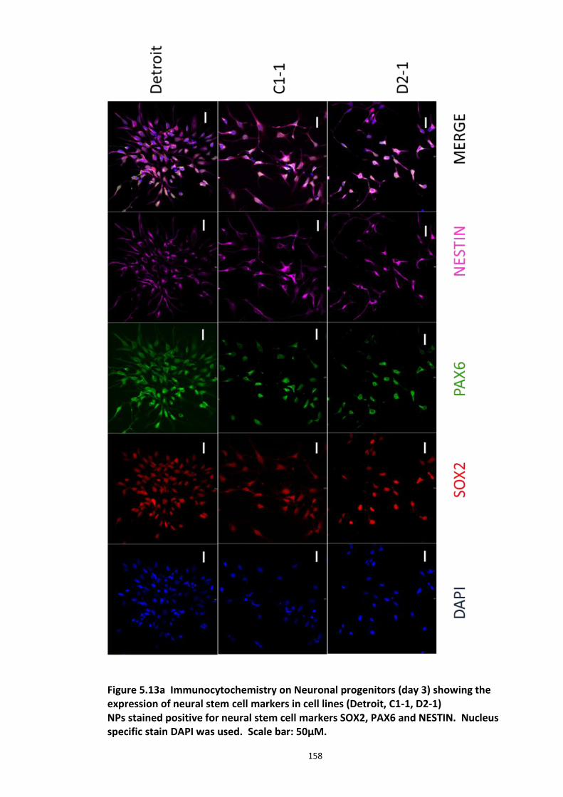
158
Figure 5.13a Immunocytochemistry on Neuronal progenitors (day 3) showing the expression of neural stem cell markers in cell lines (Detroit, C1-1, D2-1) NPs stained positive for neural stem cell markers SOX2, PAX6 and NESTIN. Nucleus specific stain DAPI was used. Scale bar: 50µM.
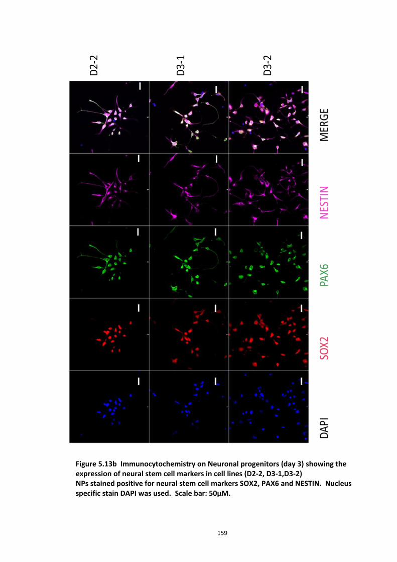
159
Figure 5.13b Immunocytochemistry on Neuronal progenitors (day 3) showing the expression of neural stem cell markers in cell lines (D2-2, D3-1,D3-2) NPs stained positive for neural stem cell markers SOX2, PAX6 and NESTIN. Nucleus specific stain DAPI was used. Scale bar: 50µM.
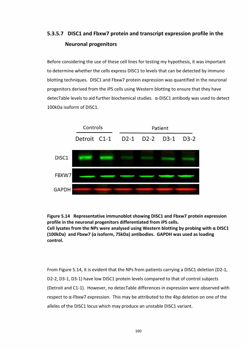
160
5.3.5.7 DISC1 and Fbxw7 protein and transcript expression profile in the
Neuronal progenitors
Before considering the use of these cell lines for testing my hypothesis, it was important
to determine whether the cells express DISC1 to levels that can be detected by immuno
blotting techniques. DISC1 and Fbxw7 protein expression was quantified in the neuronal
progenitors derived from the iPS cells using Western blotting to ensure that they have
detecTable levels to aid further biochemical studies. α-DISC1 antibody was used to detect
100kDa isoform of DISC1.
Figure 5.14 Representative immunoblot showing DISC1 and Fbxw7 protein expression profile in the neuronal progenitors differentiated from iPS cells. Cell lysates from the NPs were analysed using Western blotting by probing with α DISC1 (100kDa) and Fbxw7 (α isoform, 75kDa) antibodies. GAPDH was used as loading control.
From Figure 5.14, it is evident that the NPs from patients carrying a DISC1 deletion (D2-1,
D2-2, D3-1, D3-1) have low DISC1 protein levels compared to that of control subjects
(Detroit and C1-1). However, no detecTable differences in expression were observed with
respect to α-Fbxw7 expression. This may be attributed to the 4bp deletion on one of the
alleles of the DISC1 locus which may produce an unstable DISC1 variant.
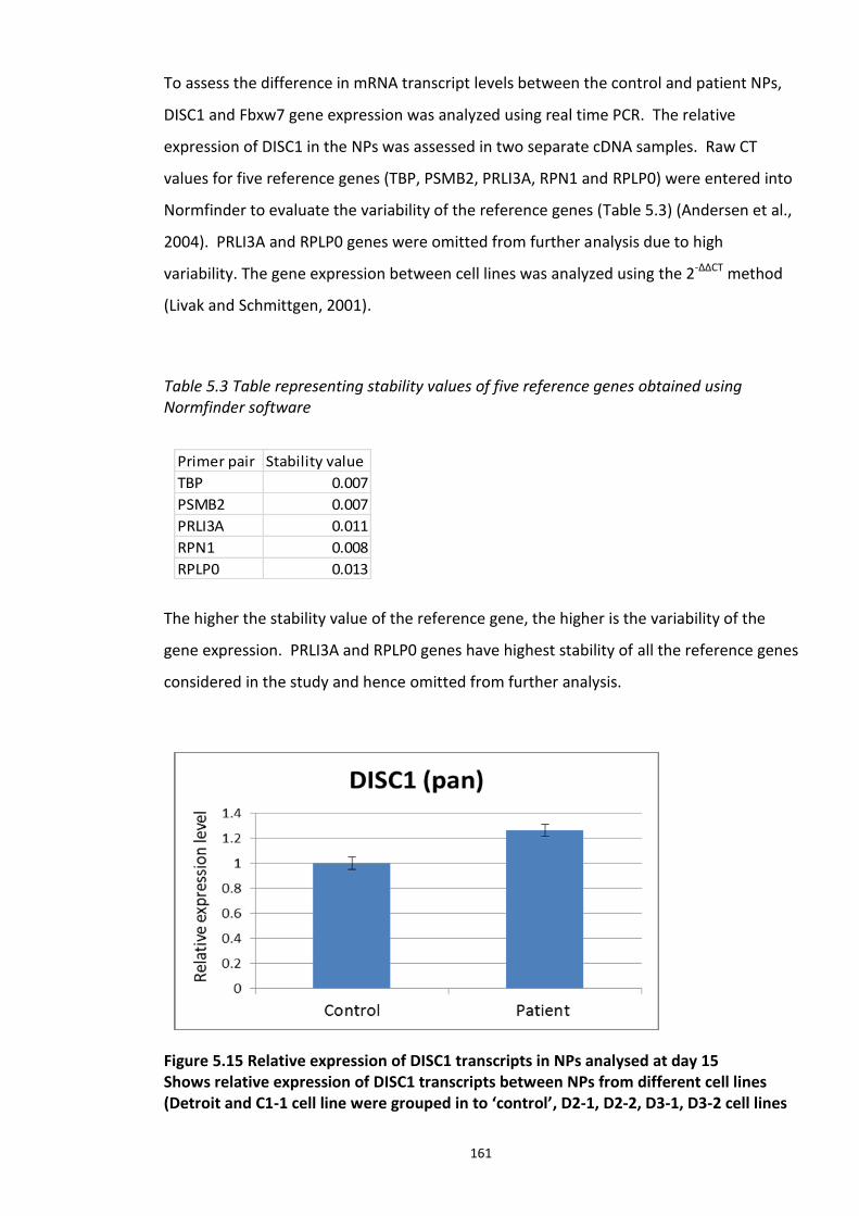
161
To assess the difference in mRNA transcript levels between the control and patient NPs,
DISC1 and Fbxw7 gene expression was analyzed using real time PCR. The relative
expression of DISC1 in the NPs was assessed in two separate cDNA samples. Raw CT
values for five reference genes (TBP, PSMB2, PRLI3A, RPN1 and RPLP0) were entered into
Normfinder to evaluate the variability of the reference genes (Table 5.3) (Andersen et al.,
2004). PRLI3A and RPLP0 genes were omitted from further analysis due to high
variability. The gene expression between cell lines was analyzed using the 2-∆∆CT method
(Livak and Schmittgen, 2001).
Table 5.3 Table representing stability values of five reference genes obtained using Normfinder software
The higher the stability value of the reference gene, the higher is the variability of the
gene expression. PRLI3A and RPLP0 genes have highest stability of all the reference genes
considered in the study and hence omitted from further analysis.
Figure 5.15 Relative expression of DISC1 transcripts in NPs analysed at day 15 Shows relative expression of DISC1 transcripts between NPs from different cell lines (Detroit and C1-1 cell line were grouped in to ‘control’, D2-1, D2-2, D3-1, D3-2 cell lines
Primer pair Stability value
TBP 0.007
PSMB2 0.007
PRLI3A 0.011
RPN1 0.008
RPLP0 0.013
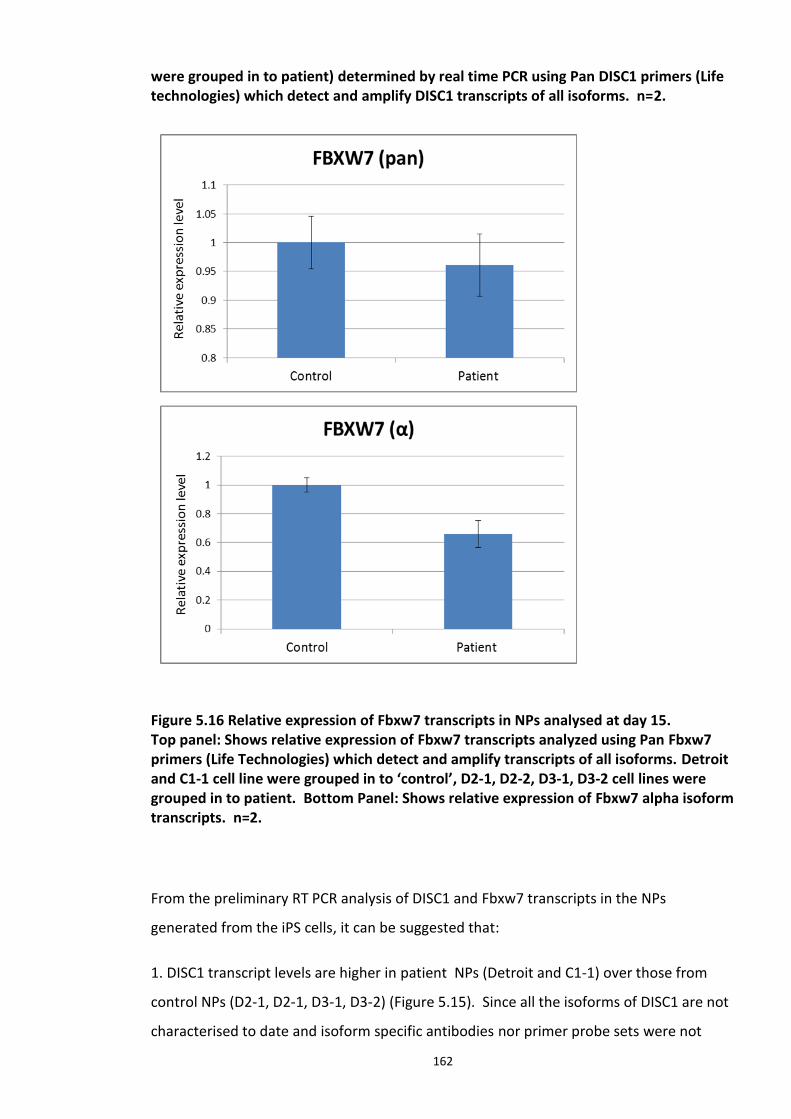
162
were grouped in to patient) determined by real time PCR using Pan DISC1 primers (Life technologies) which detect and amplify DISC1 transcripts of all isoforms. n=2.
Figure 5.16 Relative expression of Fbxw7 transcripts in NPs analysed at day 15. Top panel: Shows relative expression of Fbxw7 transcripts analyzed using Pan Fbxw7 primers (Life Technologies) which detect and amplify transcripts of all isoforms. Detroit and C1-1 cell line were grouped in to ‘control’, D2-1, D2-2, D3-1, D3-2 cell lines were grouped in to patient. Bottom Panel: Shows relative expression of Fbxw7 alpha isoform transcripts. n=2.
From the preliminary RT PCR analysis of DISC1 and Fbxw7 transcripts in the NPs
generated from the iPS cells, it can be suggested that:
1. DISC1 transcript levels are higher in patient NPs (Detroit and C1-1) over those from
control NPs (D2-1, D2-1, D3-1, D3-2) (Figure 5.15). Since all the isoforms of DISC1 are not
characterised to date and isoform specific antibodies nor primer probe sets were not
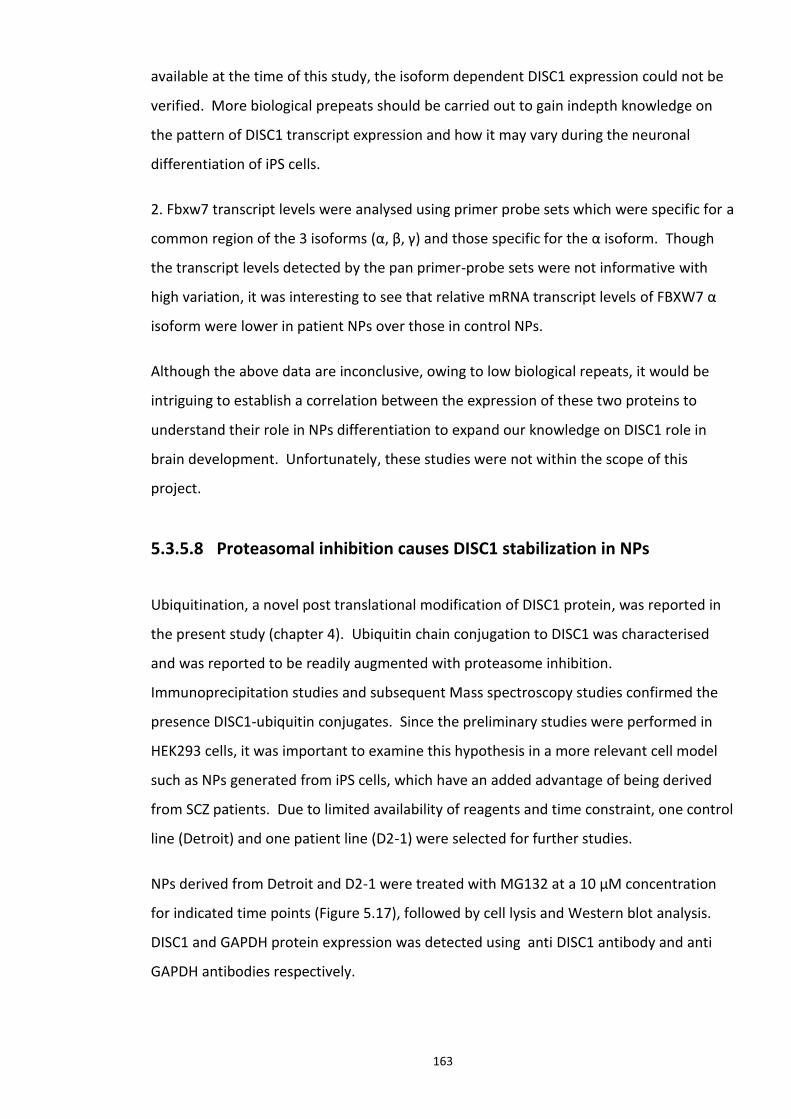
163
available at the time of this study, the isoform dependent DISC1 expression could not be
verified. More biological prepeats should be carried out to gain indepth knowledge on
the pattern of DISC1 transcript expression and how it may vary during the neuronal
differentiation of iPS cells.
2. Fbxw7 transcript levels were analysed using primer probe sets which were specific for a
common region of the 3 isoforms (α, β, γ) and those specific for the α isoform. Though
the transcript levels detected by the pan primer-probe sets were not informative with
high variation, it was interesting to see that relative mRNA transcript levels of FBXW7 α
isoform were lower in patient NPs over those in control NPs.
Although the above data are inconclusive, owing to low biological repeats, it would be
intriguing to establish a correlation between the expression of these two proteins to
understand their role in NPs differentiation to expand our knowledge on DISC1 role in
brain development. Unfortunately, these studies were not within the scope of this
project.
5.3.5.8 Proteasomal inhibition causes DISC1 stabilization in NPs
Ubiquitination, a novel post translational modification of DISC1 protein, was reported in
the present study (chapter 4). Ubiquitin chain conjugation to DISC1 was characterised
and was reported to be readily augmented with proteasome inhibition.
Immunoprecipitation studies and subsequent Mass spectroscopy studies confirmed the
presence DISC1-ubiquitin conjugates. Since the preliminary studies were performed in
HEK293 cells, it was important to examine this hypothesis in a more relevant cell model
such as NPs generated from iPS cells, which have an added advantage of being derived
from SCZ patients. Due to limited availability of reagents and time constraint, one control
line (Detroit) and one patient line (D2-1) were selected for further studies.
NPs derived from Detroit and D2-1 were treated with MG132 at a 10 µM concentration
for indicated time points (Figure 5.17), followed by cell lysis and Western blot analysis.
DISC1 and GAPDH protein expression was detected using anti DISC1 antibody and anti
GAPDH antibodies respectively.
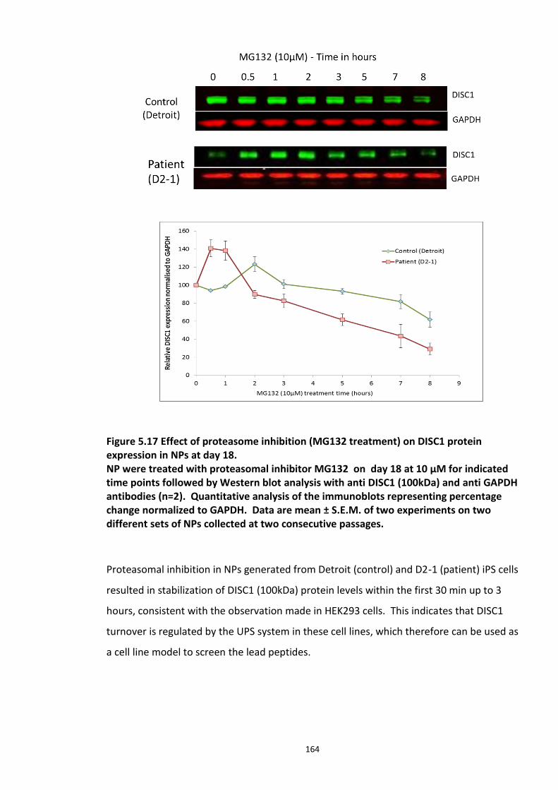
164
Figure 5.17 Effect of proteasome inhibition (MG132 treatment) on DISC1 protein expression in NPs at day 18. NP were treated with proteasomal inhibitor MG132 on day 18 at 10 µM for indicated time points followed by Western blot analysis with anti DISC1 (100kDa) and anti GAPDH antibodies (n=2). Quantitative analysis of the immunoblots representing percentage change normalized to GAPDH. Data are mean ± S.E.M. of two experiments on two different sets of NPs collected at two consecutive passages.
Proteasomal inhibition in NPs generated from Detroit (control) and D2-1 (patient) iPS cells
resulted in stabilization of DISC1 (100kDa) protein levels within the first 30 min up to 3
hours, consistent with the observation made in HEK293 cells. This indicates that DISC1
turnover is regulated by the UPS system in these cell lines, which therefore can be used as
a cell line model to screen the lead peptides.
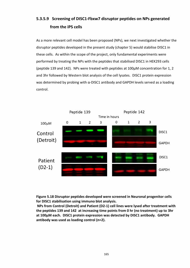
165
5.3.5.9 Screening of DISC1-Fbxw7 disruptor peptides on NPs generated
from the iPS cells
As a more relevant cell model has been proposed (NPs), we next investigated whether the
disruptor peptides developed in the present study (chapter 5) would stabilise DISC1 in
these cells. As within the scope of the project, only fundamental experiments were
performed by treating the NPs with the peptides that stabilised DISC1 in HEK293 cells
(peptide 139 and 142). NPs were treated with peptides at 100µM concentration for 1, 2
and 3hr followed by Western blot analysis of the cell lysates. DISC1 protein expression
was determined by probing with α-DISC1 antibody and GAPDH levels served as a loading
control.
Figure 5.18 Disruptor peptides developed were screened in Neuronal progenitor cells for DISC1 stabilisation using immuno blot analysis. NPs from Control (Detroit) and Patient (D2-1) cell lines were lysed after treatment with the peptides 139 and 142 at increasing time points from 0 hr (no treatment) up to 3hr at 100µM each. DISC1 protein expression was detected by DISC1 antibody. GAPDH antibody was used as loading control (n=2).
166
Figure 5.19 Quantitative analysis of immuno blots (from Figure 5.18) showing fold ratio of DISC1 normalized GAPDH. Data are mean ± S.E.M. of two experiments. Statistical differences could not be determined due to low replicate experiments (n=2).
Lead peptide 139 stabilised DISC1 in both the cells lines as seen in Figure 5.18, left panel
within 3hr of peptide treatment at a 100µM concentration. Although peptide 142
stabilised DISC1 protein levels in the Detroit cell line (control) at the 3hr time point, the
effect was negligible in D2-1. While the results were promising, more technical and
biological repeats could not be performed to ascertain this effect of the lead disruptor
peptides due to time and budget restraints.
167
5.4 Discussion
In summary, the present chapter explores the potential of (1) DISC1 protein complexes
for drug discovery and development; (2) peptides that act as substrate-specific Fbox E3
ligase inhibitors; (3) iPS cell derived NPs as a patient specific cell model to investigate
disease mechanism and role of DISC1.
A single gene locus may produce multi compartmentalised proteins by mechanisms such
as post translational modification, alternative splicing, alternative transcription and
translation initiation (Danpure, 1995). Genetic studies on psychiatric patients identified a
SCZ proband as carrying a balanced t(1;16)(p31.2;q21) translocation, who had a history
of psychotic episodes with hallucinations and delusions (Millar et al., 2005b). Biochemical
studies in cells from different individuals with the translocation revealed that DISC1
expression was nearly 50% less both at transcript and protein levels in subjects with the
translocation. It has been proposed that the translocation is the likely reason for the
observed reduction in the DISC1 expression and for the individuals with the translocation
being susceptible to SCZ (Millar et al., 2005b, James et al., 2004a). The haploinsufficiency
disease model is one of the widely accepted disease mechanisms in which an individual is
heterozygous for a gene mutation or a truncation at a particular locus. RNAi mediated
knockdown of DISC1 in mouse embryonic brains delayed neuronal migration and
decreased dendritic arborisation (Kamiya et al., 2005b).
Co-Immunoprecipitation and co-crystal structure (data shown in appendix) confirmed
complex formation of Fbxw7 and DISC1 (chapter 4). Having identified Fbxw7 as a putative
E3 ligase involved in regulating DISC1 turn over, my studies focused on developing the
Fbxw7 – DISC1 interaction as a putative drug target. Using advanced peptide array
technology, it has been possible to map potential Fbxw7 docking sites on the DISC1 CPD
that aligns with a characterised consensus Φ-X-Φ-Φ-T- P-P-X-S (Φ- hydrophobic amino
acid, X – any amino acid) (Orlicky et al., 2003). Phosphorylation dependent substrate
recognition and SCF E3 ligase recruitment has been a paradigm confirmed by several
studies (Orlicky et al., 2003, Hao et al., 2007a, Nash et al., 2001, Skowyra et al., 1997). My
168
peptide array studies show that in vitro DISC1 CPD interaction with Fbxw7 is enhanced
following dual phosphorylation (chapter 4), however, the kinase responsible for the
phosphorylation is not yet known. As seen in other Fbxw7 substrates it is likely that
phosphorylation within the CPD may trigger a conformational change, which makes it
accessible to Fbxw7 (Orlicky et al., 2003).
In the present chapter, we explored the potential of peptides as a proof of concept in
drug development for psychiatric illness. I proposed that by targeting DISC1-Fbxw7
complex formation and interfering with ubiquitination mediated degradation we could
stabilise DISC1 protein. Using structure activity relationship studies, a peptide was
developed which had a potential to stabilise DISC1 protein levels in the cell by modulating
DISC1-Fbxw7 interaction. Of the 4 lead peptides, two peptides (139 and 142) have shown
a significant effect. Intriguingly, these two peptides had mE and mD substitutions at the
phopsho sites. It can be hypothesised that the phospho groups are crucial in the
interaction and the N methyl capping of the phosphomemitic amino acids (mE and mD)
therefore retain the negative charge, increase membrane permeability as well as increase
resistance to proteolytic degradation in cells. My studies also show that the peptide is
specific to the DISC1-Fbxw7 interaction as the peptide treatments did not alter
endogenous levels of other known Fbxw7 substrates such as c-Myc, c-Jun, Notch1 and
Cyclin-E. It is crucial to demonstrate this specificity as altering the stability of the other
substrates may be tumorigenic which is not desirable. The specificity of the peptide is
probably due to the differences in affinity that are seen between Fbxw7 and DISC1
compared with the other substrates. The Baillie lab has calculated the affinity of Fbxw7
for the dually phosphorylated DISC1 as 1µM (using isothermal calorimetry) and this is
weak compared with the 65 nM affinity of Fbxw7 for cyclin E (Orlicky et al., 2003). So, it is
possible that the weak DISC1-Fbxw7 interaction can be disrupted by the peptide but the
others cannot.
It would be interesting to investigate the ability of the peptides to disrupt the DISC1-
Fbxw7 interaction in vivo, however, this was not within the scope of this project as
peptides are thought not to cross the blood-brain barrier. It should also be noted that all
169
the recommendations made for amino acid substitutions to increase the peptide efficacy
were not possible to screen (in my peptide optimisation screens) as some of the non-
natural amino acids (Sar, β-H-ala, βA, Pip, Tic) were not available at the time of this study.
Implementation and characterisation of these substitutions may further increase the
potency of the peptide.
Synthesis and purification of these synthetic peptides is extremely time consuming and
extreme care should be taken while handling and storage as they are susceptible to
oxidation and bacterial degradation. Despite the generic complications in developing
peptide drugs, they do come with certain advantages such as specificity, potency, smaller
size and their cost of production has been improving with advancements in the field of
synthetic chemistry making them more accessible. They are also less immunogenic and
can have higher tissue penetrance (but probably do not cross blood-brain barrier (Vlieghe
and Khrestchatisky, 2010). Since the degradation products of the peptides are amino
acids, they pose a minimum risk of systemic toxicity and side effects. The rapid increase
in the marketing of synthetic therapeutic peptides is a positive signal, which points to the
potential of more research into and preference for this class of drugs by pharmaceutical
companies.
The following review provides a list of all the peptide drugs released in to the market to
date which include peptides ranging from the size of 2 amino acids (Naaxia for
conjunctivitis ) to 41 amino acids (Acthrel for Cushing’s syndrome) (Vlieghe et al., 2010).
However, it is important to consider that a potential CNS drug targeting psychiatric
illnesses should ideally cross BBB (blood brain barrier) either by passive diffusion or
carrier mediated active transport. Research over the past decade focused on developing
a novel drug delivery system to evade BBB, reviewed (Chen and Liu, 2012).
As the present study sheds light on new DISC1 interactions and the significance of DISC1
turnover regulation, it also opens new exciting avenues of research. DISC1 directly
interacts and negatively regulates the enzyme activity of GSK3β, preventing it from
170
phosphorylating β-catenin (De Rienzo et al., 2011). Two different domains (amino acids
1-220 and 356-595) on DISC1 have been mapped to directly interact with purified GSK3β
protein using in vitro binding assays (Mao et al., 2009b). This finding is crucial, as the
DISC1 CPD (amino acids 196-203) lays well within the proposed GSK3β interacting
domain. It is worth noting that GSK3β phosphorylates within the CPD of the other known
Fbxw7 substrates triggering subsequent ubiquitin conjugation (Pérez-Benavente et al.,
2013, Welcker et al., 2003, Wei et al., 2005, Kitagawa et al., 2009). GSK3β activity has
already been implicated in several psychiatric diseases such as SCZ, autism and
Alzheimer’s disease and has been a common therapeutic target for few decades. 40%
reduction in GSK3β protein, transcript levels and activity has been observed in post-
mortem frontal cortex of schizophrenic patients (Kozlovsky et al., 2000, Kozlovsky et al.,
2001, Kozlovsky et al., 2004). As a kinase with a large number of substrates, it can be
hypothesised that GSK3β may also play a crucial role as an upstream regulator of DISC1
ubiquitination; however, much more work is needed to verify this. Further
characterisation of the lead peptides developed in the present study is absolutely
necessary and may provide insight in to the DISC1 pathway. Exploring how these peptide
affect DISC1 interactions with other proteins identified as risk factors for neurological
diseases such as DIXDC1, GSK3β, Ndel1 will provide molecular insights which is otherwise
impossible to obtain in genetic or clinical studies.
5.4.1 Use of iPS cells and differentiated NPs and neurons as models to facilitate drug discovery
In the last section, my study explored the potential use of iPS cell technology in disease
modelling and drug discovery. Until recent years, iPS cell generation and differentiation
has been perceived as a strenuous, long and expensive procedure. It should be noted
that the first publication on generation of human iPS cells was only 6 years ago and there
is still much speculation about their potential in drug development. Considerable
advancements have been made lately to improve iPS reprogramming techniques and this
new advanced technology can be considered as a boost for neuroscience research. Apart
from serving as indefinite supply of human neurons, these cells also serve as a medium
that enables the study of human psychiatric illnesses in a neuronal context. Researchers
171
have successfully employed this approach in modelling other neuronal diseases such as
Parkinson’s, Alzheimer’s, and Huntington diseases (Wernig et al., 2008, Zhang et al., 2010,
Yahata et al., 2011). Non clinical drug evaluation is extremely important before
advancement into human clinical trials. Employing iPS cells and differentiated cell lines of
interest serve this purpose efficiently as they can compare disease and patient specific
profiles from a similar genetic makeup.
The present chapter describes the results of a study conducted to utilise iPS cells for
studying disease mechanisms involving a DISC1 mutation. I also highlight the possible
downstream application of iPSc and their diiferentiated derivatves for drug screening.
Interestingly, the proteasome inhibitor MG132 stabilised endogenous DISC1 protein
levels in the control and patient neuronal progenitors (NPs). Although, we had seen this
in a model line (HEK293), it was reassuring that the phenomenon was recapitulated in the
NPs. Immunoprecipitation experiments to show ubiquitin chains in the NPs were not
successful and required further technical optimisation to increase the concentration of
DISC1-ubiquitin conjugates in the sample to levels that could be detected by biochemical
methods.
Gratifyingly, immunoblots and quantitative real time PCR data indicated that Fbxw7 and
DISC1 displayed reasonable expression levels, though comparative expression patterns
between the control and patient NPs could not be established because of the low number
of replicates. Lead peptides which successfully stabilised DISC1 in HEK293 cells (139 and
142), were tested on the NPs generated from control and patient iPS cells. Due to time
constraints, only two replicates were possible. These preliminary set of experiments
show increased DISC1 levels. However, more biological replicates originating from
different sets of neuronal differentiation are crucial in establishing the effect of the
peptides.
While the potential use of iPS cell technology is an exciting advance for neuroscience
research, crucial challenges need to be addressed. First and foremost, is the possible
172
genetic variability between iPS cell clones which may be attributed to genetic
recombination or integration of episomal vectors or virus in to the chromosomal DNA.
Still in its infancy, methods to improve the quality and efficiency of iPS cell generation
(reprogramming) are still under way. The protocols employed to differentiate iPS cells
into desired cell type (media, serum and growth factors) also plays a crucial role in
replicating the cell properties. Future research into development of cell type specific
differentiation protocols, phenotype and genotype characterisation is necessary.
5.5 Chapter summary
To summarize, the data presented in this chapter explores the potential of PPIs
(substrate-E3 ligase) as therapeutic drug targets. The lead peptides developed, efficiently
and specifically disrupted DISC1-Fbxw7 interaction in HEK293 cells. iPSCs differentiated
NPs were explored as cell line model as they represent a powerful new approach to
capture the genetic background from human individuals with confirmed diagnoses of SCZ.
With the provided framework, future experiments should involve complete
characterisation of the pateint derived iPS cells and their differentiated derivatives (such
as morphology, DISC1 and Fbxw7 relative expression and how their pattern of expression
alters in relation to the various stages of differentiaon). I speculate that this may provide
crucial information and possibly recapitulate disease onset physiology at the molecular
level. Investigating the physiological effects (such as neurite outgrowth, number) of the
lead peptides in terminally differentiated neurons derived from the iPS cells may also
expand our knowledege on how DISC1 function contributes to neuronal function.
173
6.
HTS for small molecules that modulate DISC1-Fbxw7 interaction
6.1 Introduction
PPIs (Protein-Protein Interactions) mediate many cellular signal transduction systems, yet
notoriously challenging to target due to their large molecular surfaces, lack of profound
pockets and paucity of appropriate screening libraries, which can target these
interactions. One strategy to overcome these difficulties, is to target so-called hot spots,
a recently evolved concept in PPIs (Mullard, 2012). Although the complex nature of PPIs
is dictated by physical features and forces that hold binding partners together are
challenging to target, certain interactions have a small binding regions, often termed as
‘hot spots’, on at least one of the partners that can be singled out. The concept of the
‘hot spot’ was based on an observation that the bulk of free energy is not evenly
distributed on the protein surface but concentrated on certain residues (amino acids),
which are crucial for the interaction (Lea and Simeonov, 2011). Advancements in protein
mapping, crystallography and computational analysis led to identification of PPIs as more
approachable and promising drug targets, opposing to the conventional view.
‘Small molecules” are organic molecules with a molecular weight less than 900 Daltons ,
which target certain biological events such as PPI or enzyme activity (Arkin and Wells,
2004). The upper molecular weight limit for the small molecules increases the likelihood
of cell permeability and their potential to target intracellular sites. Most of the commonly
used drugs are small molecules, as they have an added advantage of metabolic stability
and bioavailability over peptide drugs. As an example, the drug RG7388, developed by
174
Roche Pharmaceuticals to target p53-Mdm2 interaction (RG7388 is a selective, potent
Mdm2 antagonist) stabilises and activates p53 and is currently in clinical development for
cancer therapy (Ding et al., 2013). Projects such as this, have garnered enthusiasm of
pharmaceutical firms for small molecule inhibitors of PPIs.
Once a suiable drug target is identified, different approaches can be employed to identify
compound modulators. HTS is one such widely used approach discussed in section 6.2.
Functional assays are used in the first round to identify compounds from large chemical
libraries which give positive or negative response known as ‘hits’ that become the starting
points of drug discovery. A chemical or compound library consists of stored compounds
with associated information about each compound such as structure, molecular weight,
purity, and other biophysical properties. Follow up secondary assays are used to test for
specific artefacts and false positives identified in the primary screens.
In the previous chapter, using advanced peptide array technology and structure activity
relationship studies, we successfully developed and optimised lead peptides, which
stabilise DISC1 levels in HEK293 cells. My work so far explored the potential of the novel
PPI, DISC1-Fbxw7 as a drug target for psychiatric illnesses. I see this as a promising drug
target, as loss of DISC1 function is one of the proposed disease mechanisms (Hikida et al.,
2012).
A similar approach has been taken to target Fbxw7 mediated regulation of Cyclin E
signalling as a therapeutic intervention for cancer (Hao et al., 2007a). A FP assay was
constructed from pure protein and peptides that correspond to the binding interface.
This assay was used to perform a HTS on a 50,000 compound library which identified
SCF-I2 (assigned name) to cause inhibition of Sic1 (yeast Cdk1 inhibitor) ubiquitination
(Orlicky et al., 2010). Crystal structure elucidation revealed that SCFI2 elicits its inhibitory
effect by an allosteric mechanism (WD40 domain inhibitor) which was the first of its kind
to be identified (Orlicky et al., 2010). Another well-known example where a substrate-E3
ligase interaction was targeted for drug development was that of the p53-Mdm2
175
complex. Ubiquitination of p53, a tumour suppressor and transcription factor, is tightly
regulated by the Mdm2 protein (E3 ligase). Mdm2 gene overexpression leading to
inhibition of p53 has been shown in many malignancies. Hence, the disruption of p53-
Mdm2 complex represents a suitable drug target for cancer therapy (Vassilev et al.,
2004). Close analysis of the crystal structure of Mdm2 revealed the presence of a
hydrophobic pocket that was involved in the interaction with the p53 peptide. This area
constituted the transactivation domain and was ideal for targeting with small molecules.
To this end, a number of HTS studies were undertaken that led to the development of
Nutlins (imidazole analogues), which successfully displaced p53 from the Mdm2 leading
to subsequent stabilisation of p53 protein (Vassilev et al., 2004).
In the present study, one of the shortest DISC1 peptides identified to be interacting with
the SCFFbxw7 complex in the peptide array studies was an 7-residue stretch of DISC1, which
is dually phosphorylated at T198 and S202. This DISC1 peptide was chosen for in vitro assay
development in light of the structural model of Skp1-Fbw7-Cyclin E elucidated by Bing
Hao and colleagues (Hao et al., 2007a). As a part of this study by Hao et al, purified Skp1-
Fbxw7 was co-crystallised with Cyclin E peptides - CyclinEdegC peptide ((residues 360–390,
phosphorylated at Ser372, Thr380, and Ser384) and CyclinEdegN (residues 55–68,
phosphorylated at Thr62). Crystallography studies uncovered the residues of these
peptides that make contact with the WD40 domain of Fbxw7. Since the DISC1 CPD motif
is remarkably similar to that of Cyclin E, the labelled DISC1 peptides to be used as tracer
molecules were designed in order to test whether a similar interaction may also be
possible with DISC1. I developed an in vitro FP assay to validate binding of the identified
and optimised DISC1 CPD peptide. The assay was also fit to screen small molecule
libraries; a wide spread approach taken in drug discovery and development. Ultimately,
identified and validated hits from this approach could then be used to test the hypothesis
that by targeting DISC1-Fbxw7, DISC1 protein levels can be regulated.
176
6.2 High Throughput Screening (HTS)
High Throughput Screening (HTS) is a process of assaying libraries containing large
numbers of compounds to identify potential effectors of biological activity against
biological events or targets. HTS based studies are widely employed by the
pharmaceutical industry in the initial stages of drug discovery and development. They
enable testing of large chemical libraries of compounds categorised by their backbone
structure, size or specific chemical groups, for their activity in a range of cellular and non-
cellular assays. Testing up to 100,000 compounds per day has become feasible by the
gradual replacement of cuvettes and dishes with 96 to 3456 well micro well plates and
the recent advent of automated robotic liquid handling workstations. This approach has
garnered much popularity in recent years due to the increased availability of chemical
libraries and the miniaturization of the sensitive assays (Shinohara and Uesugi, 2007)
(Duffy et al., 2013). HTS constitutes a primary screen to identify ‘hits’ (positive response)
from the compound library, with suitable positive and negative controls. In general,
primary screens are less quantitative, tested only in singlets or doublets, usually within a
concentration range of 1-10 µM (which may again vary depending on the type of assay
and the biological event being monitored). The short listed compound hits are then re-
tested in secondary assays, which are more accurate and used for quantification and to
determine the IC50 following construction of dose response curves and time response
read outs depending on the assays. A confirmed hit exhibits the same activity in the
secondary assays, which is then taken forward to tertiary screens or counter screens for
optimisation. Compounds that hold biological activity after this process are often termed
as ’lead compounds’. Secondary screens play a crucial role in excluding false positive hits.
Fluorescence polarization/anisotropy (FP) and Fluorescence/Foerster Resonance Energy
Transfer (FRET) are homogenous assays preferred by screening groups, as they are
solution-phase assays. Having fewer assay components, simple assay steps and
protocols, they are easier to develop than heterogeneous assays such as ELISA (Enzyme
Linked Immuno Sorbent Assay) which has multiple components, incubations and wash
steps. Following the identification of a suitable drug target, it is important that the assay
selected for HTS meets certain criteria, such as being executable in 384 or 1536 well plate
format and response readout via a single parameter (Lea and Simeonov, 2011).
177
6.2.1 Fluorescence Polarisation Assay (FP)
This assay was first described by Jean Perrin in1926 (F, 1926) (Lea and Simeonov, 2011)
and gradually expanded by Gregorio Weber and colleagues in the 1950’s. This technique
has since been in use to characterise antigen-antibody, hormone –receptor and ligand-
protein interactions. Since mid-1990 FP assay has been adapted for use in high
throughput screening.
Fluorescence based screening methods are rapidly gaining attention in assay
development. In recent years, FP assays have become increasingly popular and been
used for quantitative analysis of enzyme activity (Zhang et al., 2007) and various
molecular interactions (Kimple et al., 2008). The format of this assay, which is mix-read
with no wash steps, also makes it a popular choice in the field of drug development. This
assay is homogenous, solution based and highly sensitive and sometimes can use just Pico
molar concentrations of the fluorescent probes.
6.2.1.1 Principle of FP assay
The principle of a FP assay is the measurement of the molecular size-dependent change in
the polarisation of plane polarised light. When plane polarised light is passed through a
sample, the fluorescent molecules in the sample are excited and emit light in the same
plane if the fluorescent probe is stationary. However, if the fluorescent molecule tumbles
and rotates in various planes in its excitation state, it tends to emit light in a different
plane than the one in which it was excited. Larger molecules tend to rotate and tumble at
a slower rate, hence the emitted light, following their excitation remains highly polarised.
If a molecule is smaller, rotation and tumbling is faster and hence the light is depolarised
relative to the excitation plane. The extent of polarisation is read out as polarisation
units/ milli polarisation units (mP) (Lea and Simeonov, 2011).
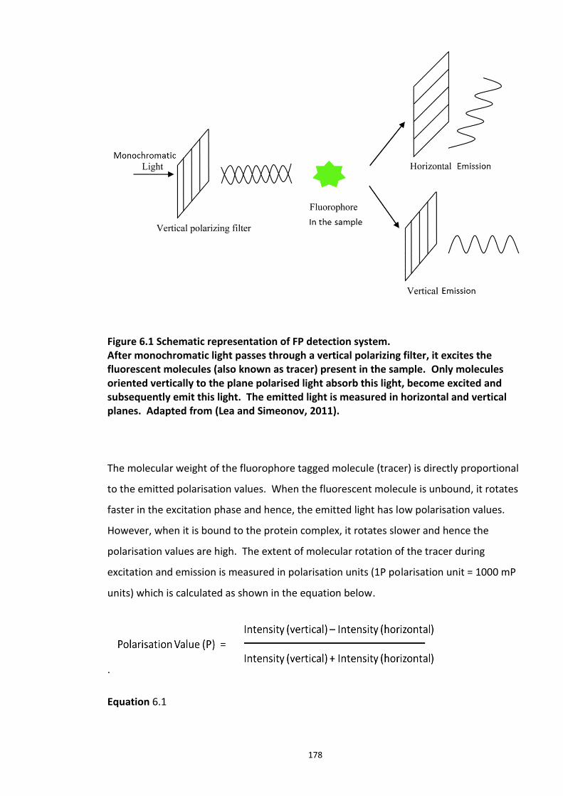
178
Figure 6.1 Schematic representation of FP detection system. After monochromatic light passes through a vertical polarizing filter, it excites the fluorescent molecules (also known as tracer) present in the sample. Only molecules oriented vertically to the plane polarised light absorb this light, become excited and subsequently emit this light. The emitted light is measured in horizontal and vertical planes. Adapted from (Lea and Simeonov, 2011).
The molecular weight of the fluorophore tagged molecule (tracer) is directly proportional
to the emitted polarisation values. When the fluorescent molecule is unbound, it rotates
faster in the excitation phase and hence, the emitted light has low polarisation values.
However, when it is bound to the protein complex, it rotates slower and hence the
polarisation values are high. The extent of molecular rotation of the tracer during
excitation and emission is measured in polarisation units (1P polarisation unit = 1000 mP
units) which is calculated as shown in the equation below.
.
Equation 6.1
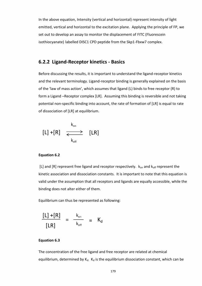
179
In the above equation, Intensity (vertical and horizontal) represent intensity of light
emitted, vertical and horizontal to the excitation plane. Applying the principle of FP, we
set out to develop an assay to monitor the displacement of FITC (Fluorescein
isothiocyanate) labelled DISC1 CPD peptide from the Skp1-Fbxw7 complex.
6.2.2 Ligand-Receptor kinetics - Basics
Before discussing the results, it is important to understand the ligand-receptor kinetics
and the relevant terminology. Ligand-receptor binding is generally explained on the basis
of the ‘law of mass action’, which assumes that ligand (L) binds to free receptor (R) to
form a Ligand –Receptor complex [LR]. Assuming this binding is reversible and not taking
potential non-specific binding into account, the rate of formation of [LR] is equal to rate
of dissociation of [LR] at equilibrium.
Equation 6.2
[L] and [R] represent free ligand and receptor respectively. kon and koff represent the
kinetic association and dissociation constants. It is important to note that this equation is
valid under the assumption that all receptors and ligands are equally accessible, while the
binding does not alter either of them.
Equilibrium can thus be represented as following:
Equation 6.3
The concentration of the free ligand and free receptor are related at chemical
equilibrium, determined by Kd. Kd is the equilibrium dissociation constant, which can be
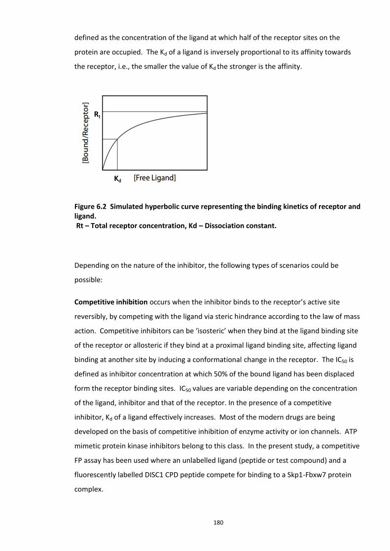
180
defined as the concentration of the ligand at which half of the receptor sites on the
protein are occupied. The Kd of a ligand is inversely proportional to its affinity towards
the receptor, i.e., the smaller the value of Kd the stronger is the affinity.
Figure 6.2 Simulated hyperbolic curve representing the binding kinetics of receptor and ligand. Rt – Total receptor concentration, Kd – Dissociation constant.
Depending on the nature of the inhibitor, the following types of scenarios could be
possible:
Competitive inhibition occurs when the inhibitor binds to the receptor’s active site
reversibly, by competing with the ligand via steric hindrance according to the law of mass
action. Competitive inhibitors can be ‘isosteric’ when they bind at the ligand binding site
of the receptor or allosteric if they bind at a proximal ligand binding site, affecting ligand
binding at another site by inducing a conformational change in the receptor. The IC50 is
defined as inhibitor concentration at which 50% of the bound ligand has been displaced
form the receptor binding sites. IC50 values are variable depending on the concentration
of the ligand, inhibitor and that of the receptor. In the presence of a competitive
inhibitor, Kd of a ligand effectively increases. Most of the modern drugs are being
developed on the basis of competitive inhibition of enzyme activity or ion channels. ATP
mimetic protein kinase inhibitors belong to this class. In the present study, a competitive
FP assay has been used where an unlabelled ligand (peptide or test compound) and a
fluorescently labelled DISC1 CPD peptide compete for binding to a Skp1-Fbxw7 protein
complex.
181
Non-competitive inhibition is also a type of reversible inhibition, which occurs when the
inhibitor binds to a different site on the receptor than the natural ligand and inhibits the
biological function of the receptor. This type of inhibition results in formation of [LRI] and
[RI] complexes which are non-functional; (R Receptor, L ligand, I inhibitor).
The following references would be useful for a review of this concept in depth - (Krohn
and Link, 2003, Prystay et al., 2001, Fang, 2012).
6.2 Experimental aims
Studies outlined previously in this thesis have demonstrated that the conserved CPD
motif in DISC1 is responsible for DISC1 interaction with the Fbxw7 protein of the Skp1-
Fbxw7 protein complex (chapter 5). In addition, peptide array studies also revealed that
dual phosphorylation within this motif enhances Fbxw7 interaction in vitro. As we
targeted this novel PPI with disruptor peptides in the previous chapter, here we
investigated if this can be targeted by small molecules.
The aims of the experimental work detailed in this chapter were:
1. To develop and optimise a quantitative, reliable FP assay to quantify and compare the
binding affinities of DISC1 CPD peptides (singly and double phosphorylated).
2. Adapt the optimised FP assay to screen large compound libraries in High throughput
screens (HTS) to identify inhibitors and activators targeting DISC1-Fbxw7 interaction.
3. Perform secondary screens to confirm the hits identified in the HTS.
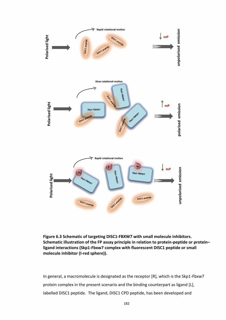
182
Figure 6.3 Schematic of targeting DISC1-FBXW7 with small molecule inhibitors. Schematic illustration of the FP assay principle in relation to protein-peptide or protein–ligand interactions (Skp1-Fbxw7 complex with fluorescent DISC1 peptide or small molecule inhibitor (I-red sphere)).
In general, a macromolecule is designated as the receptor [R], which is the Skp1-Fbxw7
protein complex in the present scenario and the binding counterpart as ligand [L],
labelled DISC1 peptide. The ligand, DISC1 CPD peptide, has been developed and
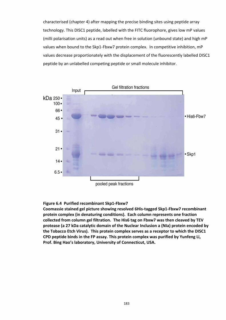
183
characterised (chapter 4) after mapping the precise binding sites using peptide array
technology. This DISC1 peptide, labelled with the FITC fluorophore, gives low mP values
(milli polarisation units) as a read out when free in solution (unbound state) and high mP
values when bound to the Skp1-Fbxw7 protein complex. In competitive inhibition, mP
values decrease proportionately with the displacement of the fluorescently labelled DISC1
peptide by an unlabelled competing peptide or small molecule inhibitor.
Figure 6.4 Purified recombinant Skp1-Fbxw7 Coomassie stained gel picture showing resolved 6His-tagged Skp1-Fbxw7 recombinant protein complex (in denaturing conditions). Each column represents one fraction collected from column gel filtration. The His6 tag on Fbxw7 was then cleaved by TEV protease (a 27 kDa catalytic domain of the Nuclear Inclusion a (NIa) protein encoded by the Tobacco Etch Virus). This protein complex serves as a receptor to which the DISC1 CPD peptide binds in the FP assay. This protein complex was purified by Yunfeng Li, Prof. Bing Hao’s laboratory, University of Connecticut, USA.
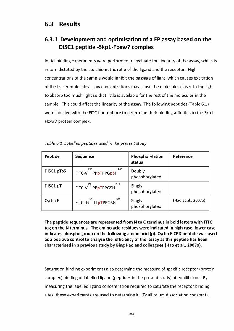
184
6.3 Results
6.3.1 Development and optimisation of a FP assay based on the DISC1 peptide -Skp1-Fbxw7 complex
Initial binding experiments were performed to evaluate the linearity of the assay, which is
in turn dictated by the stoichiometric ratio of the ligand and the receptor. High
concentrations of the sample would inhibit the passage of light, which causes excitation
of the tracer molecules. Low concentrations may cause the molecules closer to the light
to absorb too much light so that little is available for the rest of the molecules in the
sample. This could affect the linearity of the assay. The following peptides (Table 6.1)
were labelled with the FITC fluorophore to determine their binding affinities to the Skp1-
Fbxw7 protein complex.
Table 6.1 Labelled peptides used in the present study
Peptide Sequence Phosphorylation status
Reference
DISC1 pTpS FITC-V195
PPpTPPGpSH203
Doubly phosphorylated
DISC1 pT FITC-V195
PPpTPPGSH203
Singly phosphorylated
Cyclin E FITC- G377
LLpTPPQSG385
Singly phosphorylated
(Hao et al., 2007a)
The peptide sequences are represented from N to C terminus in bold letters with FITC tag on the N terminus. The amino acid residues were indicated in high case, lower case indicates phospho group on the following amino acid (p). Cyclin E CPD peptide was used as a positive control to analyse the efficiency of the assay as this peptide has been characterised in a previous study by Bing Hao and colleagues (Hao et al., 2007a).
Saturation binding experiments also determine the measure of specific receptor (protein
complex) binding of labelled ligand (peptides in the present study) at equilibrium. By
measuring the labelled ligand concentration required to saturate the receptor binding
sites, these experiments are used to determine Kd (Equilibrium dissociation constant).
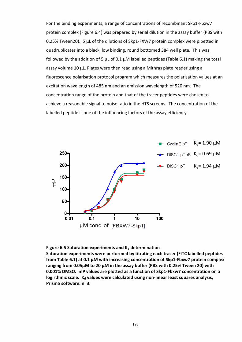
185
For the binding experiments, a range of concentrations of recombinant Skp1-Fbxw7
protein complex (Figure 6.4) was prepared by serial dilution in the assay buffer (PBS with
0.25% Tween20). 5 µL of the dilutions of Skp1-FXW7 protein complex were pipetted in
quadruplicates into a black, low binding, round bottomed 384 well plate. This was
followed by the addition of 5 µL of 0.1 µM labelled peptides (Table 6.1) making the total
assay volume 10 µL. Plates were then read using a Mithras plate reader using a
fluorescence polarisation protocol program which measures the polarisation values at an
excitation wavelength of 485 nm and an emission wavelength of 520 nm. The
concentration range of the protein and that of the tracer peptides were chosen to
achieve a reasonable signal to noise ratio in the HTS screens. The concentration of the
labelled peptide is one of the influencing factors of the assay efficiency.
Figure 6.5 Saturation experiments and Kd determination Saturation experiments were performed by titrating each tracer (FITC labelled peptides from Table 6.1) at 0.1 µM with increasing concentration of Skp1-Fbxw7 protein complex ranging from 0.05µM to 20 µM in the assay buffer (PBS with 0.25% Tween 20) with 0.001% DMSO. mP values are plotted as a function of Skp1-Fbxw7 concentration on a logirthmic scale. Kd values were calculated using non-linear least squares analysis, Prism5 software. n=3.
186
From the saturation binding experiments, a Kd value was determined for each tracer
molecule as represented in Figure 6.5. Previous in vitro studies identified the doubly
phosphorylated pThr380/pSer384 Cyclin E motif as high-affinity degron and a singly
phosphorylated pThr62 motif as a low-affinity degron (Hao et al., 2007). Since Cyclin E
CPD has close sequence similarity to that of DISC1, we expected similar binding affinities
with DISC1 peptides. As expected, dually phosphorylated DISC1 CPD peptide
(pT198/pS202) (Kd 0.69) had 2.8 times more binding affinity over the singly phosphorylated
DISC1 CPD peptide (pT198/S202) (Kd 1.94). The binding affinity of Cyclin E (pT) (Kd 1.90)
peptide was also close to that of DISC1 pT peptide (Kd 1.94).
6.3.2 Competitive inhibition experiments
Competitive binding studies were performed to determine the concentration of a
ligand/inhibitor required to reduce the specific binding of the labelled ligand to the
receptor by half (50%) represented by the term Inhibitory Concentration or IC50. IC50 is
inversely proportional to the receptor binding affinity of the ligand. By determining and
comparing the IC50 values of various ligands (compounds) determined in a particular
experiment, the compounds can be ranked based on their relative inhibitory capacity and
this measure can be used to identify the lead compounds for further optimisation and
development. My initial experiments involved testing the ability of unlabelled DISC1
peptide T (P197pTPPGpSH203 - peptide from which disruptor peptides was developed,
chapter 5) to displace the labelled DISC1 peptide (DISC1pTpS) from Skp1-Fbxw7 protein
complex. Competitive inhibitory effects of the lead disruptor peptides 139 and 142
(discussed in chapter 5) were also investigated using the principle discussed below.
[Peptide 139 – G-mE-P-P-G-mE-H; Peptide 142 - G-mD-P-P-G-mE-H].
A disruptor peptide or small molecule inhibitor (represented as ‘I’ in the Figure 6.3)
displaces the labelled peptide (bound) from the complex which then goes back in to
solution (unbound) indicated by low mP values (schematic representation in Figure 6.3).
Inhibitors that bind specifically to the protein complex at the labelled peptide binding
site, as well as allosteric inhibitors, can be identified when they result in low polarisation
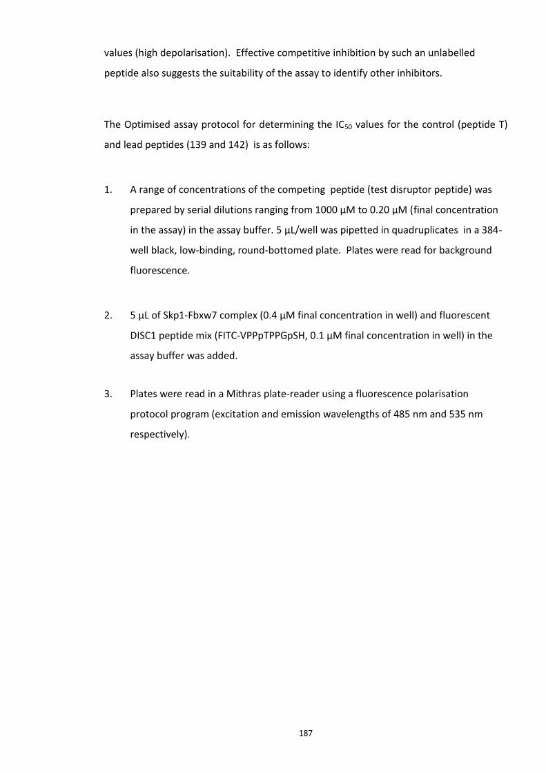
187
values (high depolarisation). Effective competitive inhibition by such an unlabelled
peptide also suggests the suitability of the assay to identify other inhibitors.
The Optimised assay protocol for determining the IC50 values for the control (peptide T)
and lead peptides (139 and 142) is as follows:
1. A range of concentrations of the competing peptide (test disruptor peptide) was
prepared by serial dilutions ranging from 1000 µM to 0.20 µM (final concentration
in the assay) in the assay buffer. 5 µL/well was pipetted in quadruplicates in a 384-
well black, low-binding, round-bottomed plate. Plates were read for background
fluorescence.
2. 5 µL of Skp1-Fbxw7 complex (0.4 µM final concentration in well) and fluorescent
DISC1 peptide mix (FITC-VPPpTPPGpSH, 0.1 µM final concentration in well) in the
assay buffer was added.
3. Plates were read in a Mithras plate-reader using a fluorescence polarisation
protocol program (excitation and emission wavelengths of 485 nm and 535 nm
respectively).
188
Figure 6.6 Competitive binding experiments Competitive binding FP assay experiments were performed with unlabelled test peptide T, 139 and 142. A range of concentrations chosen for titration of competitive disruptor peptides were represented on X axis on logarithmic scale. IC50 values were determined by non-linear least squares analysis, Prism5 Software.
From Figure 6.6, it is evident that unlabelled DISC1 peptide T competitively inhibited the
binding of labelled DISC1 pTpS peptide in a dose dependent manner as expected, with an
IC50 of 18.6±0.4 µM (top graph). However, the lead peptides 139 and 142 (middle and
bottom graphs) (discussed in chapter 5) did not disrupt the DISC1 peptide tracer from the
Skp1-Fbxw7 complex. Though its unclear at this point why these peptides couldn’t
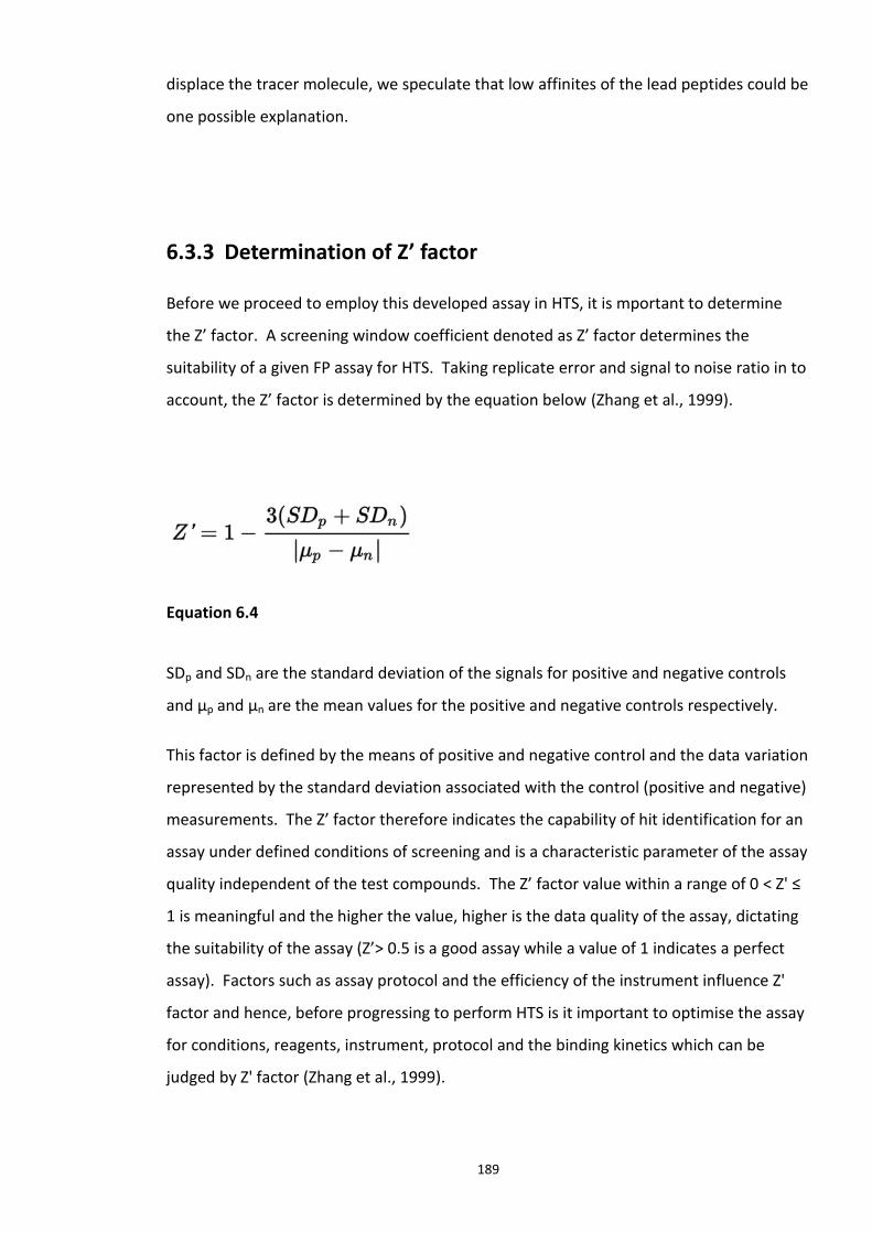
189
displace the tracer molecule, we speculate that low affinites of the lead peptides could be
one possible explanation.
6.3.3 Determination of Z’ factor
Before we proceed to employ this developed assay in HTS, it is mportant to determine
the Z’ factor. A screening window coefficient denoted as Z’ factor determines the
suitability of a given FP assay for HTS. Taking replicate error and signal to noise ratio in to
account, the Z’ factor is determined by the equation below (Zhang et al., 1999).
Equation 6.4
SDp and SDn are the standard deviation of the signals for positive and negative controls
and µp and µn are the mean values for the positive and negative controls respectively.
This factor is defined by the means of positive and negative control and the data variation
represented by the standard deviation associated with the control (positive and negative)
measurements. The Z’ factor therefore indicates the capability of hit identification for an
assay under defined conditions of screening and is a characteristic parameter of the assay
quality independent of the test compounds. The Z’ factor value within a range of 0 < Z' ≤
1 is meaningful and the higher the value, higher is the data quality of the assay, dictating
the suitability of the assay (Z’> 0.5 is a good assay while a value of 1 indicates a perfect
assay). Factors such as assay protocol and the efficiency of the instrument influence Z'
factor and hence, before progressing to perform HTS is it important to optimise the assay
for conditions, reagents, instrument, protocol and the binding kinetics which can be
judged by Z' factor (Zhang et al., 1999).
190
Figure 6.7 Calculated Z′-factor for competitive FP-based assays Controls and Z’-factor for original competitive FP assay using labelled DISC1pTpS peptide (0.1µM) and Skp1-Fbxw7 protein complex (0.4µM). Positive controls representing 100% inhibition contain unbound labelled DISC1 peptide, while the Skp1-Fbxw7 protein bound labelled DISC1 peptide representing 0% inhibition was used as negative control. Each point represents a replicate assay to determine Z' factor.
Solving the equation 6.4 with the data from Figure 6.7, a Z' factor value of 0.74 was
obtained which indicates that the designed assay and the conditions optimised are
suiTable for the use of it for HTS (Zhang et al., 1999).
6.3.4 Adapted FP assay protocol for HTS
For the above described optimisation experiments, the following protocol was designed
to perform HTS, also taking into consideration, reagent availability (purified recombinant
protein and peptide) and the size of the compound library.
1) 5µL of each compound (10 µM final concentration in 1% DMSO) was added to each
well (with a liquid handler availabe at respective screening facilities) in a 384-well black,
low-binding, round-bottomed plate using a liquid handler. Plates were read for
background Fluorescence.
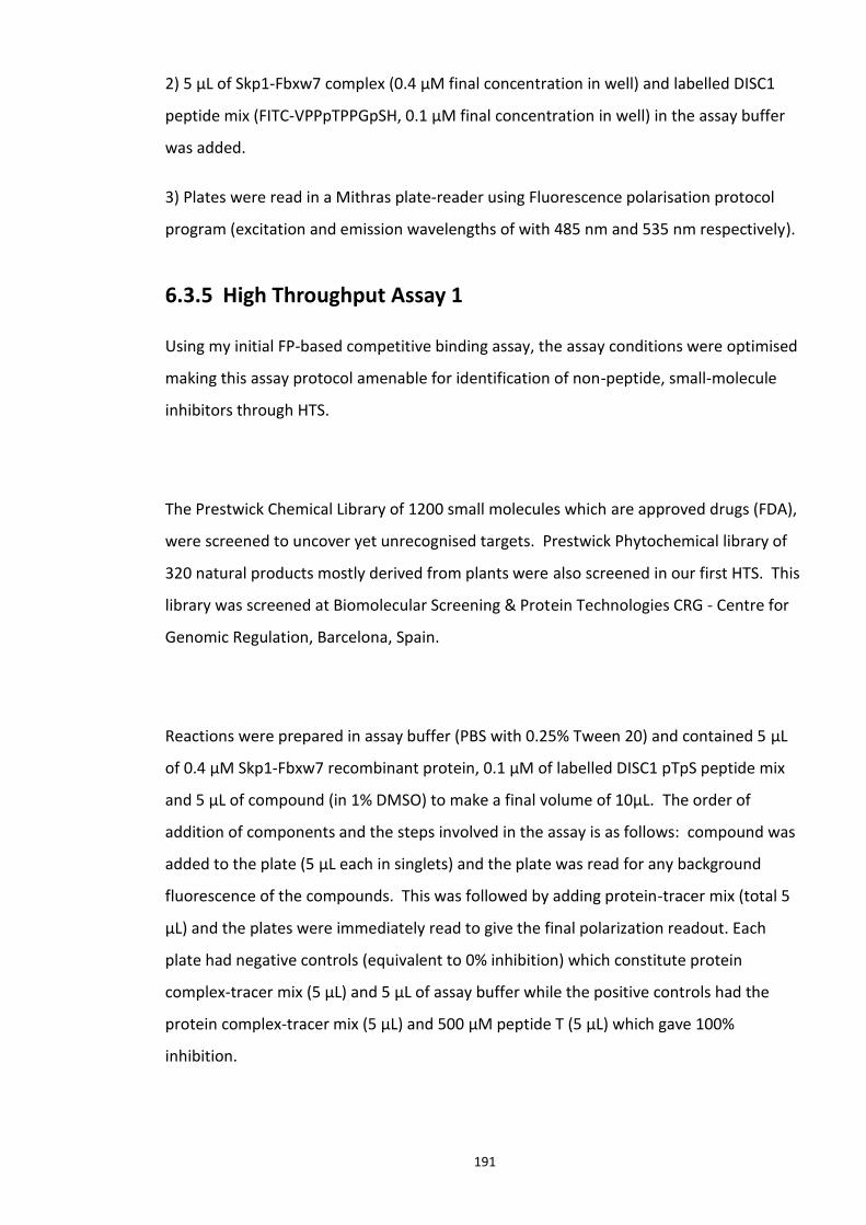
191
2) 5 µL of Skp1-Fbxw7 complex (0.4 µM final concentration in well) and labelled DISC1
peptide mix (FITC-VPPpTPPGpSH, 0.1 µM final concentration in well) in the assay buffer
was added.
3) Plates were read in a Mithras plate-reader using Fluorescence polarisation protocol
program (excitation and emission wavelengths of with 485 nm and 535 nm respectively).
6.3.5 High Throughput Assay 1
Using my initial FP-based competitive binding assay, the assay conditions were optimised
making this assay protocol amenable for identification of non-peptide, small-molecule
inhibitors through HTS.
The Prestwick Chemical Library of 1200 small molecules which are approved drugs (FDA),
were screened to uncover yet unrecognised targets. Prestwick Phytochemical library of
320 natural products mostly derived from plants were also screened in our first HTS. This
library was screened at Biomolecular Screening & Protein Technologies CRG - Centre for
Genomic Regulation, Barcelona, Spain.
Reactions were prepared in assay buffer (PBS with 0.25% Tween 20) and contained 5 µL
of 0.4 µM Skp1-Fbxw7 recombinant protein, 0.1 µM of labelled DISC1 pTpS peptide mix
and 5 µL of compound (in 1% DMSO) to make a final volume of 10µL. The order of
addition of components and the steps involved in the assay is as follows: compound was
added to the plate (5 µL each in singlets) and the plate was read for any background
fluorescence of the compounds. This was followed by adding protein-tracer mix (total 5
µL) and the plates were immediately read to give the final polarization readout. Each
plate had negative controls (equivalent to 0% inhibition) which constitute protein
complex-tracer mix (5 µL) and 5 µL of assay buffer while the positive controls had the
protein complex-tracer mix (5 µL) and 500 µM peptide T (5 µL) which gave 100%
inhibition.
192
The screen was performed twice on the library on two different days using the same
protocol to give two technical replicates. mP values of all the compounds were plotted
on an XY scatter plot against the respective assigned compound ID (well number).
Figure 6.8 Summary of the HTS screen 1 (Prestwick chemical library and Prestwick phytochemical library). Plot (a) represents data for day 1; plot (b) represents data on day 2. Plot (c) summary of the screen from both the days. mP values were plotted against the corresponding Drug IDs/well numbers on a XY scatter plot. The compound highlighted with green circle was shortlisted as a hit in the replicate screens was identified as Merbromin (CAS number - 129-16-8). The compounds highlighted in blue circle were the hits obtained only on day 2 screen identified as Cefsulodin (CAS number - 52152-93-9) and Fusaric acid (CAS number - 536-69-6). This screen was performed by Dr.Tamara Martin and Jane Evelyn Findlay, University of Glasgow at Biomolecular Screening & Protein Technologies CRG - Centre for Genomic Regulation, Barcelona, Spain.
193
CAS number (Chemical Abstracts Service) is a unique numerical code assigned to every
known chemical, independent of its structure, chemical nature nor properties. It is more
useful in identifying a compound or chemical than its chemical name. Stereoisomers of a
compound have different assigned CAS number (http://www.cas.org/content/chemical-
substances). After the screen, the mP values were plotted against each well number (also
the assigned compound ID) starting from 1. In Figure 6.8, the replicate screens
performed were also summarised in a single graph to identify the hits. Compounds that
have displaced labelled DISC1 peptide from the Skp1-Fbxw7 complex represented by a
drop in mP value by at least 20 units (compared to the positive control) were considered
for further analysis. Unfortunately, some distortions in the readouts of 5 plates were
observed on the day 2 screen. This can be seen in the plots presented in Figure 6.8. The
several other parameters that dictate the efficiency of FP assay have been discussed in
detail in section 6.4.
6.3.5.1 Secondary screens
Secondary screens are necessary to identify false positive hits or compounds with low
affinities. Dose dependent experiments were performed to generate dose response
curves which illustrate the relation between the inhibitor dose and the magnitude of the
inhibition.
The identified compounds were shortlisted as putative hits (Figure 6.8) from the screen
and were purchased from Sigma. Secondary screens which include experiments to
identify any quenchers (substances which decrease fluorescence intensity) or false
positives (example: auto fluorescent compounds) were undertaken. Compounds showing
dose dependent inhibition needed to be identified for further characterisation.
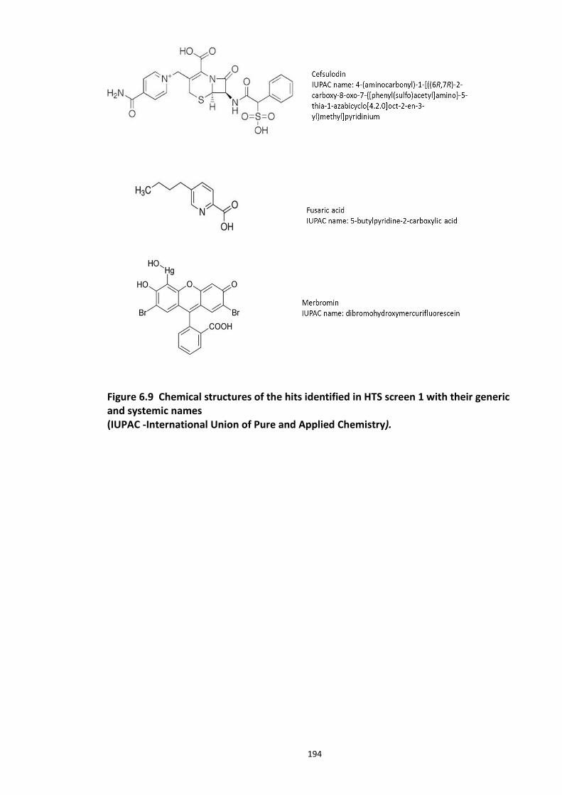
194
Figure 6.9 Chemical structures of the hits identified in HTS screen 1 with their generic and systemic names (IUPAC -International Union of Pure and Applied Chemistry).
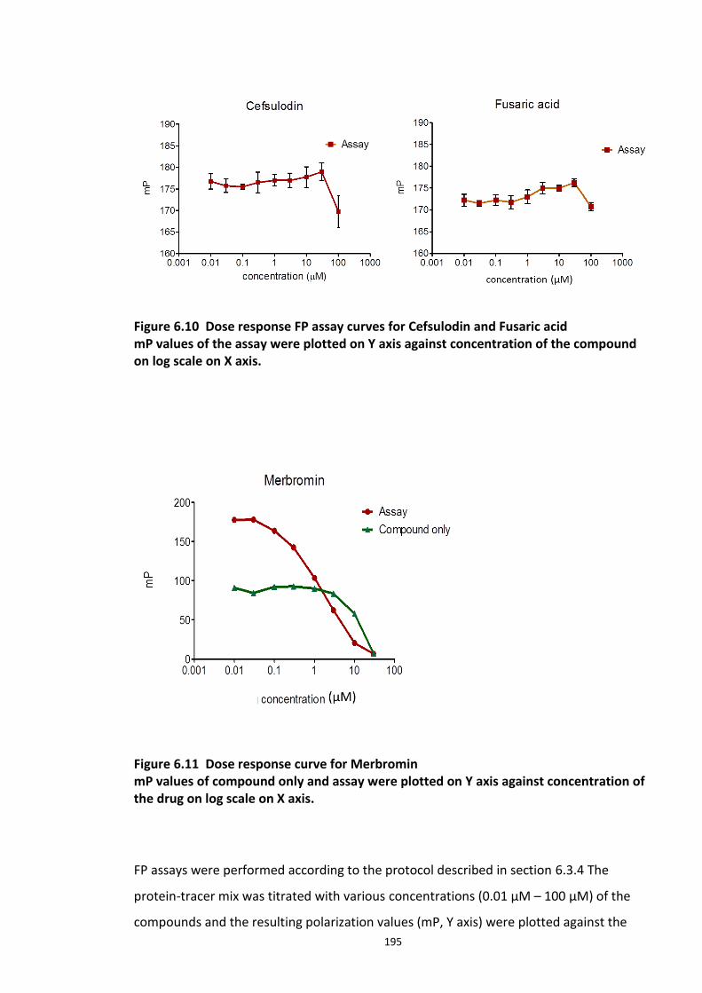
195
Figure 6.10 Dose response FP assay curves for Cefsulodin and Fusaric acid mP values of the assay were plotted on Y axis against concentration of the compound on log scale on X axis.
Figure 6.11 Dose response curve for Merbromin mP values of compound only and assay were plotted on Y axis against concentration of the drug on log scale on X axis.
FP assays were performed according to the protocol described in section 6.3.4 The
protein-tracer mix was titrated with various concentrations (0.01 µM – 100 µM) of the
compounds and the resulting polarization values (mP, Y axis) were plotted against the
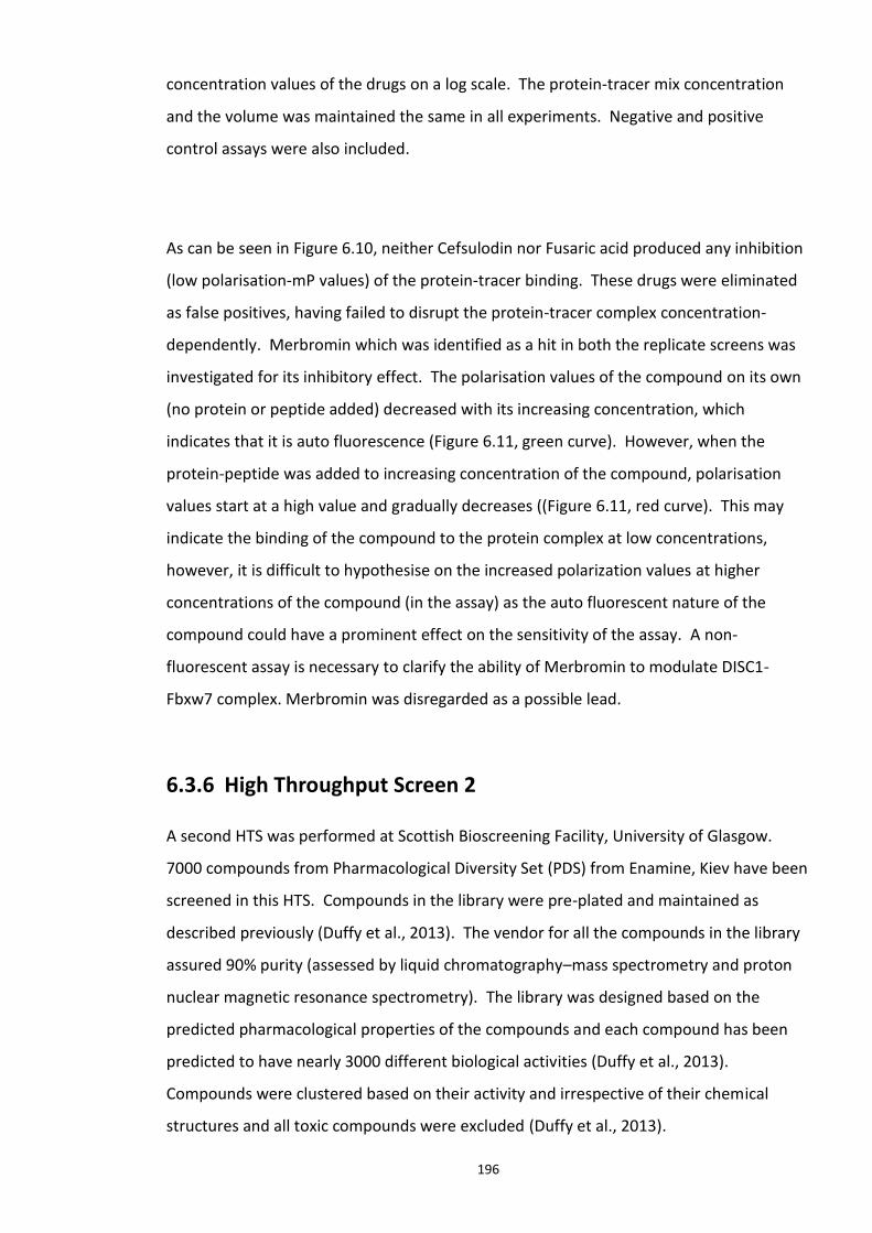
196
concentration values of the drugs on a log scale. The protein-tracer mix concentration
and the volume was maintained the same in all experiments. Negative and positive
control assays were also included.
As can be seen in Figure 6.10, neither Cefsulodin nor Fusaric acid produced any inhibition
(low polarisation-mP values) of the protein-tracer binding. These drugs were eliminated
as false positives, having failed to disrupt the protein-tracer complex concentration-
dependently. Merbromin which was identified as a hit in both the replicate screens was
investigated for its inhibitory effect. The polarisation values of the compound on its own
(no protein or peptide added) decreased with its increasing concentration, which
indicates that it is auto fluorescence (Figure 6.11, green curve). However, when the
protein-peptide was added to increasing concentration of the compound, polarisation
values start at a high value and gradually decreases ((Figure 6.11, red curve). This may
indicate the binding of the compound to the protein complex at low concentrations,
however, it is difficult to hypothesise on the increased polarization values at higher
concentrations of the compound (in the assay) as the auto fluorescent nature of the
compound could have a prominent effect on the sensitivity of the assay. A non-
fluorescent assay is necessary to clarify the ability of Merbromin to modulate DISC1-
Fbxw7 complex. Merbromin was disregarded as a possible lead.
6.3.6 High Throughput Screen 2
A second HTS was performed at Scottish Bioscreening Facility, University of Glasgow.
7000 compounds from Pharmacological Diversity Set (PDS) from Enamine, Kiev have been
screened in this HTS. Compounds in the library were pre-plated and maintained as
described previously (Duffy et al., 2013). The vendor for all the compounds in the library
assured 90% purity (assessed by liquid chromatography–mass spectrometry and proton
nuclear magnetic resonance spectrometry). The library was designed based on the
predicted pharmacological properties of the compounds and each compound has been
predicted to have nearly 3000 different biological activities (Duffy et al., 2013).
Compounds were clustered based on their activity and irrespective of their chemical
structures and all toxic compounds were excluded (Duffy et al., 2013).
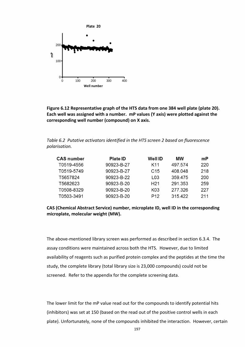
197
Figure 6.12 Representative graph of the HTS data from one 384 well plate (plate 20). Each well was assigned with a number. mP values (Y axis) were plotted against the corresponding well number (compound) on X axis.
Table 6.2 Putative activators identified in the HTS screen 2 based on fluorescence polarisation.
CAS (Chemical Abstract Service) number, microplate ID, well ID in the corresponding microplate, molecular weight (MW).
The above-mentioned library screen was performed as described in section 6.3.4. The
assay conditions were maintained across both the HTS. However, due to limited
availability of reagents such as purified protein complex and the peptides at the time the
study, the complete library (total library size is 23,000 compounds) could not be
screened. Refer to the appendix for the complete screening data.
The lower limit for the mP value read out for the compounds to identify potential hits
(inhibitors) was set at 150 (based on the read out of the positive control wells in each
plate). Unfortunately, none of the compounds inhibited the interaction. However, certain
Plate 20
0 100 200 300 4000
100
200
Well number
mP
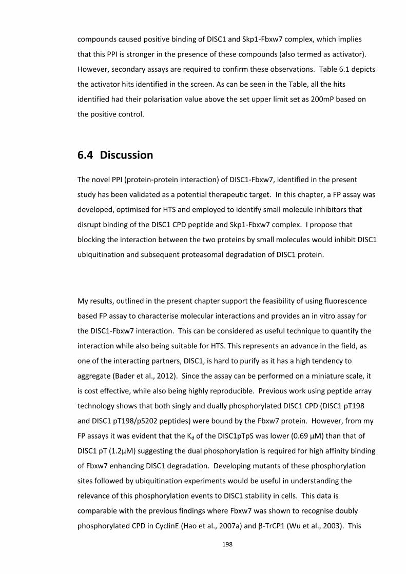
198
compounds caused positive binding of DISC1 and Skp1-Fbxw7 complex, which implies
that this PPI is stronger in the presence of these compounds (also termed as activator).
However, secondary assays are required to confirm these observations. Table 6.1 depicts
the activator hits identified in the screen. As can be seen in the Table, all the hits
identified had their polarisation value above the set upper limit set as 200mP based on
the positive control.
6.4 Discussion
The novel PPI (protein-protein interaction) of DISC1-Fbxw7, identified in the present
study has been validated as a potential therapeutic target. In this chapter, a FP assay was
developed, optimised for HTS and employed to identify small molecule inhibitors that
disrupt binding of the DISC1 CPD peptide and Skp1-Fbxw7 complex. I propose that
blocking the interaction between the two proteins by small molecules would inhibit DISC1
ubiquitination and subsequent proteasomal degradation of DISC1 protein.
My results, outlined in the present chapter support the feasibility of using fluorescence
based FP assay to characterise molecular interactions and provides an in vitro assay for
the DISC1-Fbxw7 interaction. This can be considered as useful technique to quantify the
interaction while also being suitable for HTS. This represents an advance in the field, as
one of the interacting partners, DISC1, is hard to purify as it has a high tendency to
aggregate (Bader et al., 2012). Since the assay can be performed on a miniature scale, it
is cost effective, while also being highly reproducible. Previous work using peptide array
technology shows that both singly and dually phosphorylated DISC1 CPD (DISC1 pT198
and DISC1 pT198/pS202 peptides) were bound by the Fbxw7 protein. However, from my
FP assays it was evident that the Kd of the DISC1pTpS was lower (0.69 µM) than that of
DISC1 pT (1.2µM) suggesting the dual phosphorylation is required for high affinity binding
of Fbxw7 enhancing DISC1 degradation. Developing mutants of these phosphorylation
sites followed by ubiquitination experiments would be useful in understanding the
relevance of this phosphorylation events to DISC1 stability in cells. This data is
comparable with the previous findings where Fbxw7 was shown to recognise doubly
phosphorylated CPD in CyclinE (Hao et al., 2007a) and β-TrCP1 (Wu et al., 2003). This
199
data also supports the proposed hypothesis of multi phosphorylated CPD mediated DISC1
interaction with Fbxw7and its subsequent degradation. Fbxw7 has a binding pocket
within the WD40 domain and additional pockets on the surface exposed to the solvent.
These pockets were found to make crucial intermolecular contacts with the phospho
residues and the moieties flanking them (Wu et al., 2003, Hao et al., 2007a). Since the
DISC1 CPD has a close resemblance to that of Cyclin E, we propose a similar binding
model for DISC1 involving the phospho-residues.
Orlicky et al employed a FP assay to identify small molecule inhibitors of substrate
recognition component of CDC4 (yeast homologue of Fbxw7) (Orlicky et al., 2010). A
similar approach was taken in the present study to identify small molecule inhibitors of
DISC1-Fbxw7 complex. During my optimisation experiments, control peptide (Peptide T)
and the lead peptides (139 and 142) were tested for their ability to displace the labelled
peptide from the receptor (Skp1-Fbxw7). Although, the control peptide nicely displaced
the labelled DISC1 peptide in a dose dependent manner with an IC50 of 18.6±0.4 µM, lead
peptides 139 and 142, which stabilised DISC1 levels in HEK293 cells (chapter 5), failed to
disrupt the peptide-protein interaction. While this does not necessarily mean that the
peptides do not target the PPI under study, we propose that more intense
characterisation is needed and it is necessary to explore the factors that may interfere
with the assay. In this context, it is important to note that FP may not be ideal for
identifying weak interactions/inhibitors and is dependent on the peptide-protein Kd
(Huang, 2003). This may be difficult to overcome by using high inhibitor or disruptor
peptide concentrations, as it may lead to abnormal polarisation values due to aggregation
or nonspecific binding (Lea and Simeonov, 2011). Hydrophobicity of the peptides may
also have an influence on their activity and it has been shown that at high concentrations
they tend to form micelle like particles (Lea and Simeonov, 2011). While peptide
purification is a laborious task, it is important to note that their purity is crucial in
dictating the sensitivity of an in vitro assay. The labelled peptides were obtained at the
purity of about 85% and hence experiments using higher purity grade should be
performed to evaluate the efficiency of the HTS (determined by Z' factor). Practical
limitations such as limited purified protein availability restricted further in vitro
characterisation of the lead peptides 139 and 142 in the present study.
200
In our first HTS (HTS1 – section-6.3.5) three compounds (Cefsulodin, Fusaric acid,
Merbromin) were identified as hits and taken forward for secondary screens. As a
complementary approach, a literature search was carried out which revealed that
Cefsulodin is a narrow spectrum third generation analogue of cephalosporin, which is
active only against Pseudomonas aeruginosa (Tsuda et al., 1988). It has been proposed
that penicillin binding protein 3 (PBP3) is the principle target for this drug though it has
not been characterised properly. Earlier studies implicated its use in combination with
other antibiotics in treating haematological malignancies; other potential therapeutic
uses could not be established (Tsuda et al., 1988). Fusaric acid is a mycotoxin produced
by several species in genus Fusarium (belong to the family of filamentous fungi). It has
been known to decrease plant viability and toxic to certain gram positive bacteria and
mycobacterium (Fischer et al., 1980). However, its clinical use as antimicrobial is
restricted. Its mode of action has not been established yet. A review by Hexiang Wang
and T.B.Ng detailed the pharmacological effects of Fusaric acid on various physiological
systems. One such effect is its potential to elevate dopamine levels while decreasing
norepinephrine levels supporting its role as a dopamine β-hydroxylase (DBH) inhibitor
(Wang and Ng, 1999, Fischer et al., 1980). DBH is a key enzyme involved in catalysing the
conversion of dopamine to noradrenalin. Clinical studies have established an association
between DBH gene polymorphism and smoking status within SCZ population (Zhang et al.,
2012). I considered this as attractive hit as dopamine signalling dysfunction has been
reported to play a key role in the aetiology of SCZ associated disorders as proposed by
Stein and Wise, also known as the Dopamine hypothesis (Stein and Wise, 1971, Davis et
al., 1991, Cubells et al., 2011, Ella et al., 2012, Zhang et al., 2012). The compound
shortlisted as a hit in the two replicates of HTS1 is Merbromin (trade name
mercurochrome). It is used as a topical antiseptic. It has been identified as a pan active
inhibitor of various Protein methyltransferases (PMT) in an independent HTS screen
(Ibanez et al., 2012).
In the secondary screens, Cefsulodin and Fusaric acid failed to displace the labelled DISC1
peptide from the protein complex and were considered as false positives. However,
Merbromin was identified as an autofluorescent compound, evident from low mP values
201
with increasing concentration of compound on its own (with no protein-peptide mix
added). As suggested earlier, a non-fluorescent assay should be performed before
omitting this compound as a potential hit.
In the second HTS (HTS2, section 6.3.6) 7000 compounds were screened from
Pharmacological Diversity Set (PDS) from Enamine (Kiev). Unfortunately, none of the
compounds inhibited Skp1-Fbxw7 binding of DISC1. However, certain compounds (listed
in Table 6.1) have shown increased polarisation (compared to the positive control), which
may infer that in the presence of these compounds, the binding was stronger (or more
receptor sites were occupied by the ligand). Assuming that these compounds may
increase the DISC1-Fbxw7 interaction, they were shortlisted. It can be hypothesised that
the hits may have a potential to directly or allosterically enhance DISC1-Fbxw7
interaction, which can only be confirmed after further secondary screens and
characterisation. Due to financial restrictions and time constraint further secondary
screens for the putative hits could not be performed. A gradual fall in the polarisation
values was observed with increasing well number in all the plates in HTS screen 2 (data
presented in the appendix of this thesis). I speculate that as it took nearly 20 minutes to
read each plate, time drift may have caused a temperature increase leading to assay
reagent evaporation which may have inturn resulted in a fall in the mP values.
From the two HTS campaigns carried out for this study, no dose-dependently active and
chemically meaningful hits were obtained. Possible reasons for this are: Quality of the
chemical library has a crucial role to play in the number of the hits of the screen. Only
8500 compounds were screened (total in screen 1 and 2) in this study, which is relatively
small compared to the libraries screened by pharmaceutical companies constituting
millions of compounds. Screening compound libraries posed major challenges such as
auto fluorescence, quenchers, temperature control and evaporation due to low assay
volumes.
202
A possible alternative to resource-demanding HTS of small molecule libraries is ‘drug
design’. Increasing availability of crystallographic protein structures and advancements in
computers technology allows for virtual screening and in silico modelling. There has been
much focus on developing algorithms which allow one to identify possible ligands. These
can then be chemically synthesised or purchased if commercially available, and their
binding properties analysed using conventional assays. Hits may then be optimized using
further rounds of in silico screening/modelling and conventional assays. In simple terms,
computer aided drug design has successfully led to the development of a number of FDA
approved drugs (Durrant et al., 2009). This approach could be an alternative way to
develop DISC1-Fbxw7 inhibitors, given that Skp1-Fbxw7 crystal structure has been solved
(Hao et al., 2007a, Wang et al., 2004, Orlicky et al., 2010) and a suitable assay to test
possible hits has been developed in this work. DISC1 expression levels were shown to be
high during active brain developmental and to regulate neurogenesis and this supports
the role of abnormal levels of DISC1 in the pathophysiology of associated
neurodevelopmental disorders. Hence, I propose that modulation of DISC1 protein levels
is an attractive potential mechanism.
6.5 Chapter summary
To summarize, in this chapter, we developed a reliable, quantitative FP assay using which
we characterised and quantified DISC1- [Skp1-Fbxw7] interaction. Using this assay, we
have shown that doubly phosphorylated DISC1 CPD has a higher affinity over the singly
phosphorylated CPD peptide providing in depth information on DISC1-Fbxw7 binding
affinity. Furthermore, the FP assay was used in two HTS campaigns to screen a total of
8520 small molecules to identify potential modulators of the interaction. Although no
confident inhibitors of the interaction were found, contemplated potential activators
were shortlisted for future analysis. I propose that more intense optimisation of the
assay and screening of more libraries would be fruitful.
203
7.
Final Discussion
7.1 Background
Since the discovery of DISC1 in a Scottish pedigree, carrying a highly penetrant balanced
translocation (1q42.1; 11q14.3), there has been accumulating evidence to support its
multiple roles in neurogenesis, migration and synapse signalling. These aspects make it a
promising candidate gene for SCZ (Bradshaw et al., 2008, Bradshaw and Porteous, 2010,
Maher and LoTurco, 2012). DISC1’s multi compartmental localisation and a broad
interactome support its involvement in multiple signalling pathways. Recent studies have
associated DISC1 with other major chronic mental disorders such as autism, depression
and bipolar disorder and it has been proposed that all DISC1 related illnesses should be
categorised as ‘DISCopathies’ (Korth, 2009, Korth, 2012).
Although several DISC1 SNPs and haplotypes were identified to be associated with these
neurological disorders, there has been no established consensus as to the disease causing
mutation (Lipska et al., 2006b, Saetre et al., 2008). Moreover, the role of DISC1 in
subjects with no known mutations has garnered much attention. While, DISC1
haploinsufficiency was one of the initially proposed mechanisms in the Scottish
translocation, there have been no reports of low levels of mRNA transcripts or protein
levels in SCZ patients (Leliveld et al., 2008, Lipska et al., 2006b). However, short splice
variants were shown to be specifically upregulated in SCZ patients (Nakata et al., 2009).
This is intriguing as these reported short isoforms resemble the Scottish truncation gene
product, which may suggest that regulation of specific alternative splicing of these splice
variants could be one of underlying disease mechanisms.
204
A classical hallmark of neurological disorders is abnormal processing or misfolding of
proteins, which in some cases, leads to toxic aggregate formation (Taylor et al., 2002).
DISC1 protein aggregates have been reported by several groups and high expression of
heat shock proteins (molecular chaperones) have been found to be up regulated in SCZ
brains. Hence disruption in DISC1 processing was one of the other disease mechanisms
suggested (Hikida et al., 2012, Leliveld et al., 2008).
Post translational modifications are crucial in maintaining protein integrity, folding and
cellular turnover, and UPS dysfunction has been previously reported in psychiatric
illnesses (Lehman, 2009, Layfield et al., 2005). A more recent study also reported UPS
dysfunction and brain specific abnormalities in UPS related mRNA transcripts in patients
suffering from SCZ (Rubio et al., 2013, Bousman et al., 2010, Altar et al., 2005). Owing to
the overlapping nature of UPS related abnormalities and phenotypes observed with loss
of DISC1 function, we proposed that exploring and uncovering the yet unknown PTMs on
DISC1 would provide answers, as well as open new avenues to understand the underlying
pathophysiology on DISC1 associated disorders.
In the following sections, I briefly review my results and discuss the wider implications of
my intriguing findings.
7.2 Consequences of DISC1 SUMOylation on its neurological functions
My initial work (results chapter 3) focussed on investigating DISC1 as a putative SUMO
substrate. SUMOylation is a Ubl (ubiquitin Like). Posttranslational modification, which
plays a vital role in neuronal signalling pathways contributing to synapse formation, ion
channel activity, spine morphogenesis and synaptic transmission; reviewed in (Martin et
al., 2007, Wilkinson and Henley, 2010). For the first time I provide the experimental
205
evidence that DISC1 can be SUMOylated. I identified potential SUMO acceptor lysine
(K643), although the presence of other sites may not be ruled out. From previous
reports, mitochondrial and neuronal fractions had enriched SUMOylation of substrates
and since DISC1 localisation and regulation of mitochondrial dynamics has been studied
and its dysfunction has been observed in SCZ and other neuropsychiatric disorders (James
et al., 2004b, Millar et al., 2005a), I propose that in-depth characterisation of this PTM
may provide answers to questions concerning the mechanistic behind DISC1’s
involvement in mitochondrial dysfunction.
Transgenic mice expressing mutant DISC1 (C terminal truncated DISC1), under control of
CaMKII promoter, displayed abnormal phenotypes consistent with those observed in SCZ
patients, such as enlarged ventricles, altered spine density and fewer parvalbumin-
positive neurons (Hikida et al., 2007, Ayhan et al., 2011). This established a strong
association between DISC1 with post synaptic physiology. A direct correlation has been
established between the presynaptic neuronal expression of DISC1 and presynaptic
glutamate release (Maher and LoTurco, 2012). SUMOylation of synapse associated
substrates regulates presynaptic glutamate release depending on the stimulus. Different
stimuli may promote SUMOylation or deSUMOylation of different subsets of proteins,
which in turn affects the release the neurotransmitter (Feligioni et al., 2009). It would be
interesting to investigate the SUMOylation status of DISC1 at the synapse (one approach
could be the use of SUMO “dead” mutant of DISC1) to understand the precise role of
DISC1 in synapse signalling.
I also provide preliminary evidence of one of many possible consequences of this
modification on DISC1, such as altered protein interactions, in this case with DIXDC1.
Although inconclusive, if this indeed is true, this could have major physiological
consequences as this protein complex regulates neurogenesis and cortical development
(Singh et al., 2010). This PTM event on DISC1 may act to regulate neuronal progenitor
proliferation via its interaction with DIXDC1. As a multifunctional scaffold protein, DISC1
has the ability to orchestrate multiple signalling pathways via its broad “interactome” and
we speculate that SUMO conjugation would have a major impact by either creating new
206
binding surfaces or inhibiting interactions possibly mediated by conformational changes
on DISC1.
One of the other consequences, is altered sub cellular localisation regulated by reversible
dynamic PTMs such as SUMOylation and Ubiquitination. Another well studied protein
SUMO substrate is PML (Promyelocytic leukemia protein), which is SUMO conjugated and
localised in a sub nuclear structures known as PML nuclear bodies. Many other SUMO-
modified proteins have been shown to be co-localised in these transcription PML nuclear
bodies (Zhong et al., 2000, Gill, 2004). Since DISC1 also partially co-localises with PML
nuclear bodies (Sawamura et al., 2008), I speculate that SUMO modified DISC1 may be
targeted to these PML bodies which are associated with cell proliferation and
differentiation (Zhong et al., 2000). DISC1 interacts with ATF4 (Activating Transcription
Factor) and modulates cAMP-dependent cAMP-response element (CRE)-mediated
transcription. DISC1 variants 37W and 607F disrupted nuclear targeting of WT DISC1 in a
dominant negative manner (Malavasi et al., 2012). It can be speculated that these
variants could have an altered propensity to undergo PTM (either SUMOylation or
ubiquitination) which may alter their sub cellular targeting. The above mentioned are just
a few of examples of the possible consequences of DISC1 SUMOylation and I propose that
it may have a more complex role in neuronal signalling.
The above points may be addressed by further experimental work which could not be
done during my studies due to time constraints:
1. Detection of SUMOylated DISC1 using the UFDS method in a more relevant cell line,
such as primary neuronal cultures or iPSc originated neurons. Immunostaining could be
performed to identify the localisation and distribution pattern of SUMO conjugated DISC1
and its tendency to form aggregates.
2. Mass spectroscopy studies with high quality samples followed by site directed
mutagenesis would provide firm evidence of all possible SUMO acceptor sites on DISC1.
207
3. Although DISC1 is less conserved in human and rodents, the SUMO site (K643) site is
conserved in both. Hence, investigating the SUMOylation status of DISC1 in DISC1 mouse
models would be useful. It is intriguing that this site is missing in the C terminal
truncation (Scottish translocation) and hence the lack of this PTM may contribute to its
dominant negative character.
Other PTMs such as phosphorylation may be required as a triggering event to initiate
DISC1 SUMOylation. PKA mediated phosphorylation at S710 (S713 in human) has been
shown to regulate neuronal migration during cortical development (Ishizuka et al.,
2011b). SUMOylation on phosphor dead (S710A) and phosphor mimicking (S710D) DISC1
mutants, using the UFDS method, may identify a possible physiological link between
these two PTM events.
7.3 DISC1 turn over regulation via ubiquitin mediated proteasomal degradation
As discussed in chapter 5, I demonstrate for the first time that the turnover of DISC1 is
mediated by the UPS pathway. My data suggests that DISC1 is ubiquitinated and its
degradation is inhibited in the presence of proteasome inhibitor, MG132 in a time and
dose dependent manner. Previous studies have reported that DISC1 antibodies
consistently detect high molecular weight bands (above 100kDa), especially in developing
cerebral cortex (Morris et al., 2003, James et al., 2004a, Sawamura et al., 2005a).
Although, these DISC1 associated high molecular weight bands were proposed to be
DISC1 dimers, from my findings, these bands may also relate to the SUMOylated or
ubiquitinated DISC1 species. Spatial and temporal regulation of DISC1 protein expression
during brain development also suggest that complex regulatory mechanisms may be
involved in regulating DISC1 turnover (Brandon et al., 2004, Miyoshi et al., 2003a, Ozeki et
al., 2003a). Since, many of the proteins functioning at the synapse are either activated,
inactivated or degraded upon suitable stimuli; an upstream deficiency in the UPS would
208
disrupt the synapse signalling reported in SCZ (Vawter et al., 2001, Altar et al., 2005). I
speculate DISC1 could be one such crucial substrate involved.
Although, my work primarily focused on the proteolytic role of the UPS, it is important to
note that ubiquitin conjugation to DISC1 may have other potential functional
consequences. Until recently, a common notion existed that substrates with K48 chains
are normally targeted for proteasomal degradation while those with K63 chains to
lysosomes and DNA repair mechanisms (Nagao et al., 2006) (Nathan et al., 2013).
However, there has been emerging evidence supporting the role of these chain
formations in other cellular events. For example, Met4 (transcriptional activator) is
conjugated by single K48 type Ubiquitin chain catalysed by E3 ligase, Cdc34–SCFMet30 (Flick
et al., 2004). This conjugation inactivates Met4 activity instead of targeting it for
degradation. As such, this study provided the first line of evidence for the non proteolytic
role of K48 type ubiquitin chain conjugation (Flick et al., 2004). Several mechanisms have
been proposed to explain the differential fate of the ub conjugated substrates dictated by
the type of chains (Nathan et al., 2013). One mechanism that was widely accepted was
the presence of various classes of DUB’s which specifically recognise the substrates and
promote their deubiquitination (Finley, 2009).
Although through my findings, I address some important aspects of DISC1 protein
regulation, my work also raises some interesting questions such as:
1. What is the stimulus that triggers DISC1 ubiquitination? How does the developmental
profile of ubiquitinated DISC1 vary? What is the role of DISC1 ubiquitination in
neurogenesis mediated by its protein interactions?
2. Is there cross-talk between the UPS and SUMOylation pathways with respect to DISC1?
Do they compete for same acceptor lysine residues or promote or inhibit other PTMs?
These are questions that should be addressed if further time and funding can be sought.
209
7.3.1. Significance of Fbxw7 dependent DISC1 protein turn over
As discussed in chapter 4, siRNA library screen identified Fbxw7 as a potential Fbox
protein, which regulated DISC1 ubiquitination as a component of an SCF E3 ligase
complex. Also known as hCdc4 and hSel-10, scientific literature emphasizes Fbxw7 as a
therapeutic target for cancer therapy (Kanei-Ishii et al., 2008, Wang et al., 2012a,
Iriuchishima et al., 2011). With the increasing identification of neuronal proteins as
Fbxw7 substrates, its role brain specific physiological pathways have garnered much
attention lately.
High levels of Fbxw7 mRNA transcripts were detected from E13.5 with predominant
expression in granule cell (GC) layers (Jandke et al., 2011). Fbxw7 expression was also
identified to be significantly high in adult brain regions such as hippocampus and
cerebellum. This is interesting in the context of strong expression and association of
DISC1 with hippocampus structure and function (Lipska et al., 2006a, Faulkner et al.,
2008, Meyer and Morris, 2008, Austin et al., 2004). Regional specific shRNA knockdown
of DISC1 hampers the migration of early born GC in the dentate gyrus (Meyer and Morris,
2009), while its knockdown was shown to enhance migration of adult born granule cells in
the dentate gyrus (Kvajo et al., 2008). However, literature searches did not reveal any
inverse correlation of DISC1 and Fbxw7 expression during development as we expected.
While the complexity surrounding the isoform specific regulation of these proteins is one
possible reason, it can also be speculated that Fbxw7 may also target yet unknown
upstream substrates of the DISC1 pathway. Hence, the role played by DISC1 may be
spatio-temporally distinct, varying with the stimuli which dictates its function.
Two important substrates of Fbxw7 are c-Jun and Notch, which regulate neuronal
apoptosis and neurogenesis respectively (Lütolf et al., 2002, Hoeck et al., 2010a).
Cerebellum specific deletion of Fbxw7 reduced the size of the cerebellar vermis, with
increased Notch 1 and phosphorylated c-Jun levels, implying a crucial role of Fbxw7 in
cerebellar morphogenesis (Jandke et al., 2011). Studies also reported that Fbxw7−/−
embryos die in utero at E10.5-11.5 with reported abnormalities in brain (Zhou et al.,
2013b). I speculate that the abnormal anatomical changes observed in Fbxw7 conditional
210
knockout mice could also be associated with DISC1 turnover, whose protein levels are
tightly regulated during early development. My findings support the critical role of Fbxw7
in brain development by identifying DISC1 as its substrate and I suggest a complex
interaction between these proteins in regulating various cellular pathways. However,
further work is required to elucidate how FBXW-DISC1 interaction contributes to the
development of the brain.
7.4 Targeting novel PPI, DISC1-Fbxw7 interaction: scope for therapeutic intervention
Upon identification of a conserved phosphodegron (CPD) on DISC1, I performed peptide
array studies and identified the shortest stretch of the CPD domain that might be involved
in the interaction with Fbxw7. This sequence within the DISC1 CPD, was optimised using
peptide array studies by various substitutions with non-natural amino acids while the net
charge of the peptide remained unaltered, to increase the peptides physiological stability
and affinity and towards the Fbxw7 substrate binding domain (WD40). Following a series
of optimisation rounds, lead peptides 139 and 142 were selected for testing on cells, to
investigate their ability to stabilise DISC1 as per my hypothesis. These peptides stabilised
DISC1 in a dose and time dependent manner in the HEK293 cell line, while the other
substrates of Fbxw7 (c-Myc, Cyclin E and Notch) were unaltered. I propose that the
differences in affinity between Fbxw7 and DISC1 (low) compared to other substrates
including Cyclin E (high) allows the peptides to be substrate specific.
I suggest that the peptides can be a starting point for the development of novel
therapeutics. The lead cell permeable peptides which specifically stabilise DISC1 in cells
offer an attractive alternative to proteasome inhibitors which affect a larger pool of
substrates leading to side effects. Performing physiological assays of DISC1 function, such
as assessing neurite outgrowth and neurite number, following peptide treatment in a
relevant neuronal cell line (such as iPS derived neurons) may explain the mechanism of
reported disease phenotypes.
211
Furthermore, the Skp1-Fbxw7 protein complex (Schulman et al., 2000), was crystallized
bound to a 15 residue, doubly phosphorylated DISC1 peptide P193EVPPpTPPGpSHSAFT207
(X ray crystallography work was performed by Yunfeng Li, Prof. Bing Hao’s laboratory,
University of Connecticut, USA; Figures in appendix section 7). This crystal structure
(Figure in appendix) shows a striking resemblance to that of Cyclin EdegronC - residues 360–
390, phosphorylated at Ser372, Thr380, and Ser384) (Hao et al., 2007a). My findings
therefore suggest that the doubly phosphorylated DISC1 is recognised by Fbxw7 following
Cyclin E paradigm.
I also explored the use of patient specific iPS differentiated neurons for potential as
disease models. As an emerging revolutionary tool, iPS technology holds a therapeutic
application for drug screening. For neurodevelopmental disorders such as SCZ, which
involve complex gene environment interactions, the use of the human neurons
developed by this method offer a considerable advantage over animal models, as human
DISC1 is significantly different from those in rodents. They also offer a unique
opportunity to recapitulate the disease phenotype and elucidate the disease mechanism.
Patient specific iPS cells have been generated by various groups (Brennand et al., 2011,
Brennand and Gage, 2012, Chiang et al., 2011). In the present study, I used neuronal
progenitors but not fully differentiated neurons due to the limitations of cell number and
time constraints. DISC1 protein levels in the patient derived neuronal progenitors were
low compared to those in controls subjects, and were ideal to test out hypothesis of
targeting DISC1-Fbxw7 interaction to rescue DISC1 protein levels. My preliminary results
were promising as the lead peptides stabilised DISC1 levels, however, more experimental
repeats are required to validate this effect. Assessing the pattern of expression of DISC1
and Fbxw7 at various stages of differentiation from iPS cells to neurons and comparative
analysis of how this profile varies between control subjects and patients would be
intriguing. Although similar studies were performed in post mortem brain samples
(Lipska et al., 2006b), they may not resemble the physiological levels in live neurons.
212
I suggest that the following list of experiments would further enhance our understanding
of DISC1 function:
1. Site specific mutational studies within DISC1 CPD, followed by assessment of DISC1
ubiquitination and turnover.
2. Phosphorylation has been shown to be a prerequisite for β-TrCP1 and Fbxw7 to
recognise their cognate substrates-β catenin and Cyclin E respectively (Wu et al., 2003,
Hao et al., 2007a). My findings of phosphorylated CPD mediated DISC1 ubiquitination is
comparable to the models proposed in the above studies. However, I speculate that
GSK3β could be the source kinase as for the other Fbxw7 substrates. This may be
challenging to explore as a priming phosphorylation of the substrate by a different kinase
is often required for GSK3β phosphorylation.
3. It may be useful to investigate the effect of GSK3β activators and inhibitors on DISC1-
Fbxw7 interaction since Wnt/GSK3β signalling has been identified as a critical signalling
pathway in psychiatric disorders.
4. It would be interesting to identify any DISC1 common or rare mutants falling within
DISC1 CPD region. Certain DISC1 variants (A83V, R264Q and L607F) failed to activate Wnt
pathway and diminished neuronal progenitor proliferation (Singh et al., 2011). Similarly,
a recent study reported that patients carrying R264Q variant were resistant to anti-
psychotic medication (Mouaffak et al., 2011). Although the precise mechanism is not
known, I speculate that these variants affect DISC1 protein stability and my findings
provide a frame work and open new avenues to explore the underlying disease
mechanism.
7.4.1. Small molecule modulators of DISC1-Fbxw7 interaction
Experimental work discussed in chapter 6, concerned the process of screening for small
molecule modulators that affect via DISC1-Fbxw7 protein interaction. A reliable and
quantitative FP assays was developed based on DISC1 CPD mediated interaction with
Fbxw7, which was adapted to perform HTS on two different compound libraries to
identify modulators of this interaction. The hit identified in the first HTS, a fluorescent
213
compound, Merbromin requires intense characterisation in secondary screens using non
fluorescence based techniques to corroborate its potential to be developed as a drug.
Certain compounds in the second screen were identified to increase the DISC1-Fbxw7
interaction (activators). Assessment of their potential to modulate this PPI may identify
compounds which may be developed further to act as DISC1 down regulators. These
would be useful for the investigation of DISC1 function in animal studies.
7.5 Final Conclusion
My study identified novel post translational modifications (SUMOylation and
Ubiquitination) on DISC1 which would advance the understanding of how DISC1 protein
function in various cellular pathways is regulated. With the identification of SCFFbxw7 as
the E3 ligase that regulates DISC1 ubiquitination and subsequent elucidation of their
binding sites, I uncovered the DISC1-Fbxw7 PPI as a promising drug target. I optimised
two lead peptides (139 and 142) to inhibit this interaction and in so doing arrived at a
new class of therapeutic approaches directed at the up regulation of DISC1 via inhibition
of its destruction by the proteasome. I also developed an in vitro assay to identify small
molecule disruptors which could provide a novel, targeted therapeutic strategy for the
treatment of SCZ and associated disorders which involve loss or gain of DISC1 function. I
propose that the modulation of this interaction may in turn modulate its function,
targeting the neurodevelopmental processes, underlying the pathophysiology of DISC1
associated illnesses that delay or prevent the onset of the psychiatric illness.
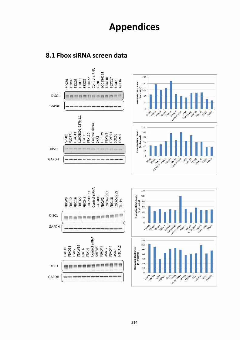
214
Appendices
8.1 Fbox siRNA screen data
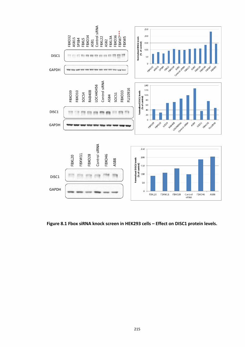
215
Figure 8.1 Fbox siRNA knock screen in HEK293 cells – Effect on DISC1 protein levels.
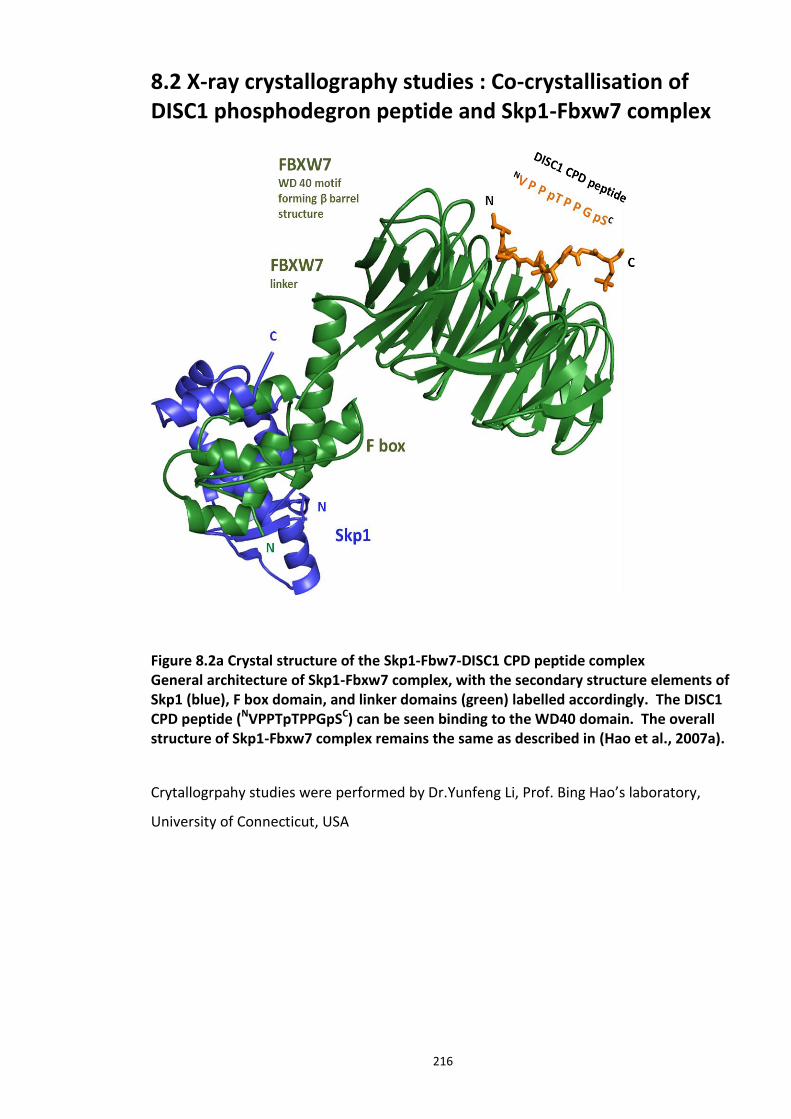
216
8.2 X-ray crystallography studies : Co-crystallisation of DISC1 phosphodegron peptide and Skp1-Fbxw7 complex
Figure 8.2a Crystal structure of the Skp1-Fbw7-DISC1 CPD peptide complex General architecture of Skp1-Fbxw7 complex, with the secondary structure elements of Skp1 (blue), F box domain, and linker domains (green) labelled accordingly. The DISC1 CPD peptide (NVPPTpTPPGpSC) can be seen binding to the WD40 domain. The overall structure of Skp1-Fbxw7 complex remains the same as described in (Hao et al., 2007a).
Crytallogrpahy studies were performed by Dr.Yunfeng Li, Prof. Bing Hao’s laboratory,
University of Connecticut, USA
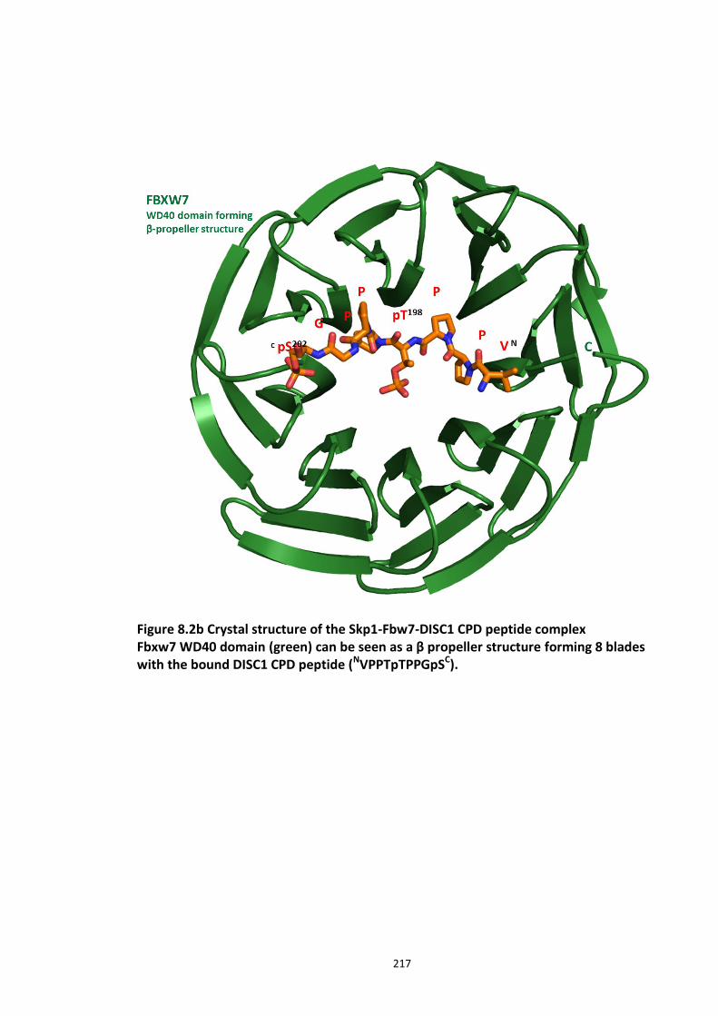
217
Figure 8.2b Crystal structure of the Skp1-Fbw7-DISC1 CPD peptide complex Fbxw7 WD40 domain (green) can be seen as a β propeller structure forming 8 blades with the bound DISC1 CPD peptide (NVPPTpTPPGpSC).
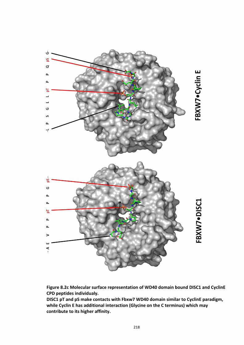
218
Figure 8.2c Molecular surface representation of WD40 domain bound DISC1 and CyclinE CPD peptides individualy. DISC1 pT and pS make contacts with Fbxw7 WD40 domain similar to CyclinE paradigm, while Cyclin E has additional interaction (Glycine on the C terminus) which may contribute to its higher affinity.
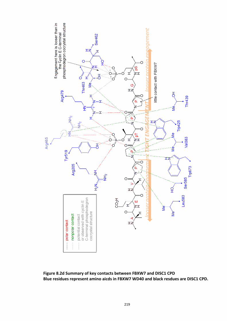
219
Figure 8.2d Summary of key contacts between FBXW7 and DISC1 CPD Blue residues represent amino aicds in FBXW7 WD40 and black resdues are DISC1 CPD.
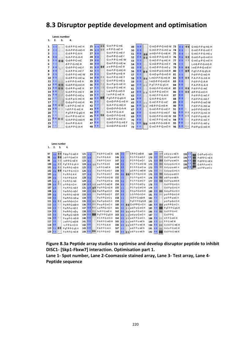
220
8.3 Disruptor peptide development and optimisation
Figure 8.3a Peptide array studies to optimise and develop disruptor peptide to inhibit DISC1- [Skp1-Fbxw7] interaction. Optimisation part 1. Lane 1- Spot number, Lane 2-Coomassie stained array, Lane 3- Test array, Lane 4- Peptide sequence
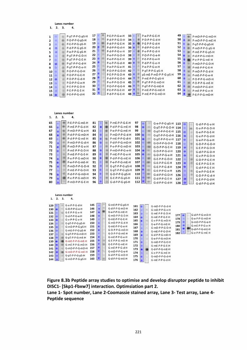
221
Figure 8.3b Peptide array studies to optimise and develop disruptor peptide to inhibit DISC1- [Skp1-Fbxw7] interaction. Optimsiation part 2. Lane 1- Spot number, Lane 2-Coomassie stained array, Lane 3- Test array, Lane 4- Peptide sequence
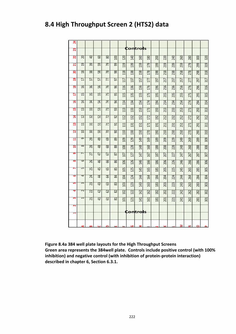
222
8.4 High Throughput Screen 2 (HTS2) data
Figure 8.4a 384 well plate layouts for the High Throughput Screens Green area represents the 384well plate. Controls include positive control (with 100% inhibition) and negative control (with inhibition of protein-protein interaction) described in chapter 6, Section 6.3.1.
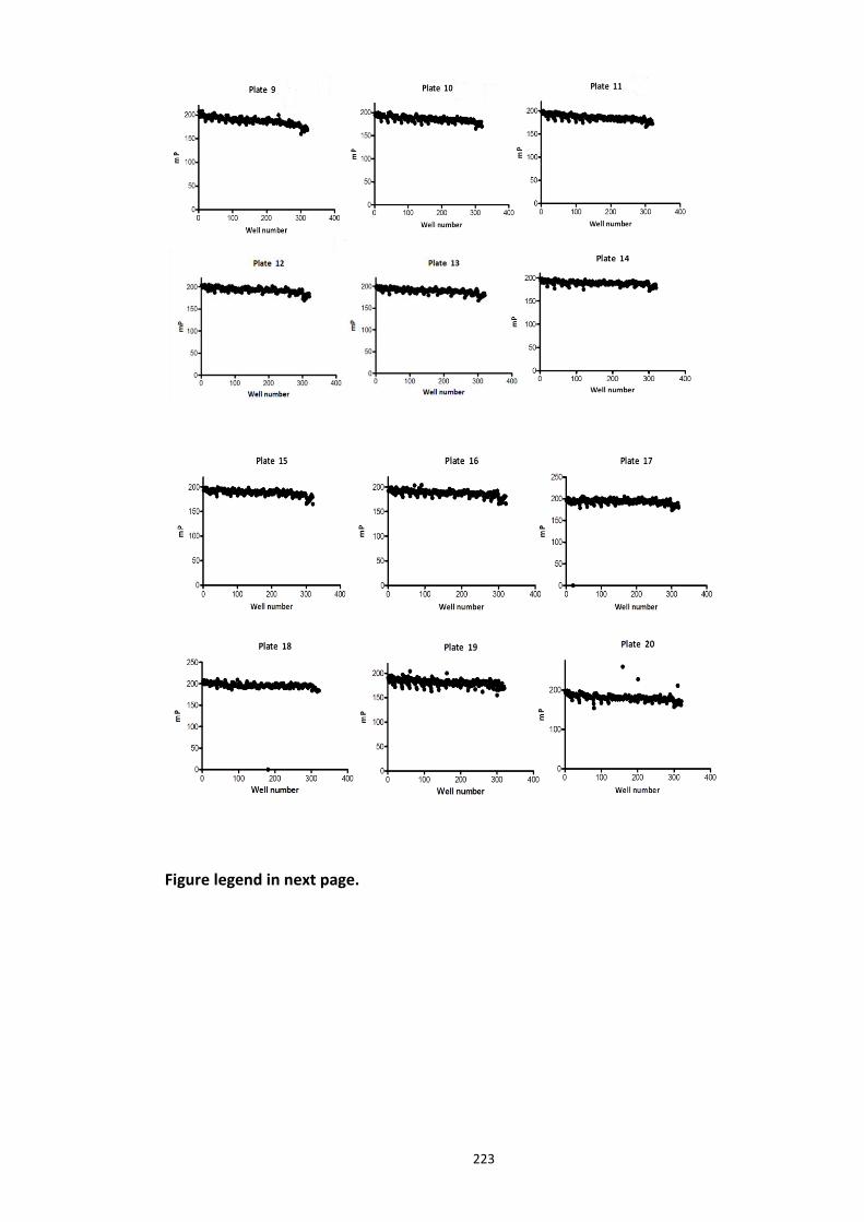
223
Figure legend in next page.
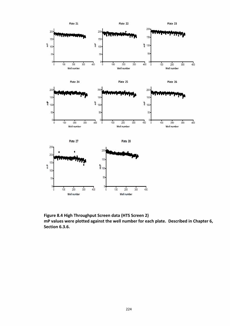
224
Figure 8.4 High Throughput Screen data (HTS Screen 2) mP values were plotted against the well number for each plate. Described in Chapter 6, Section 6.3.6.
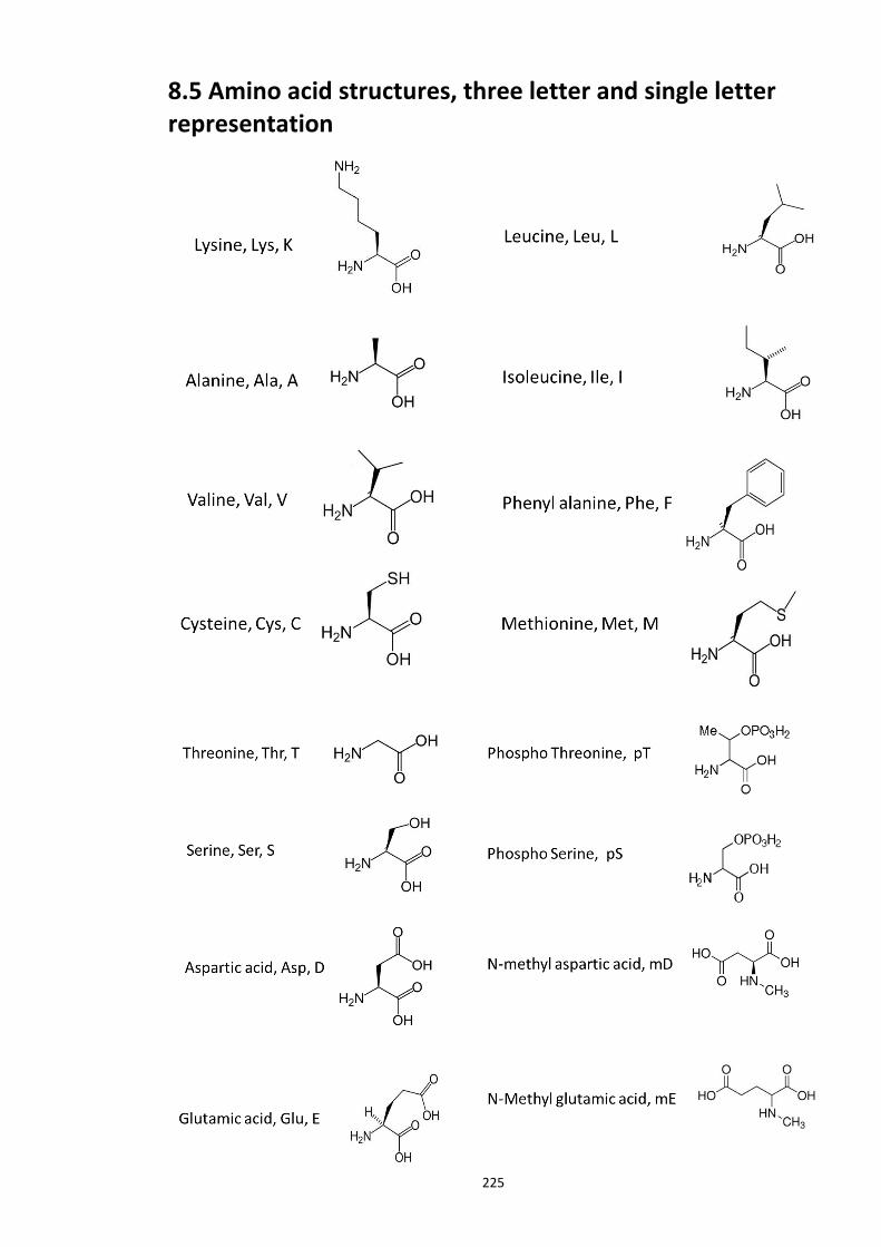
225
8.5 Amino acid structures, three letter and single letter representation
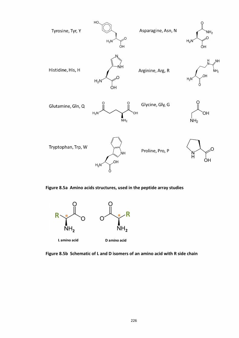
226
Figure 8.5a Amino acids structures, used in the peptide array studies
Figure 8.5b Schematic of L and D isomers of an amino acid with R side chain
227
Bibliography
ADAMS, J. 2003. The proteasome: structure, function, and role in the cell. Cancer Treat Rev, 29 Suppl 1, 3-9.
AGHAJAN, M., JONAI, N., FLICK, K., FU, F., LUO, M., CAI, X., OUNI, I., PIERCE, N., TANG, X., LOMENICK, B., DAMOISEAUX, R., HAO, R., DEL MORAL, P. M., VERMA, R., LI, Y., LI, C., HOUK, K. N., JUNG, M. E., ZHENG, N., HUANG, L., DESHAIES, R. J., KAISER, P. & HUANG, J. 2010. Chemical genetics screen for enhancers of rapamycin identifies a specific inhibitor of an SCF family E3 ubiquitin ligase. Nat Biotechnol, 28, 738-42.
AHN, K., SONG, J. H., KIM, D. K., PARK, M. H., JO, S. A. & KOH, Y. H. 2009. Ubc9 gene polymorphisms and late-onset Alzheimer's disease in the Korean population: a genetic association study. Neurosci Lett, 465, 272-5.
AKHOONDI, S., LINDSTRÖM, L., WIDSCHWENDTER, M., CORCORAN, M., BERGH, J., SPRUCK, C., GRANDÉR, D. & SANGFELT, O. 2010. Inactivation of Fbxw7/hCDC4-β expression by promoter hypermethylation is associated with favorable prognosis in primary breast cancer. Breast Cancer Res, 12, R105.
AL-MANSOORI, K. M., HASAN, M. Y., AL-HAYANI, A. & EL-AGNAF, O. M. 2013. The Role of α-Synuclein in Neurodegenerative Diseases: From Molecular Pathways in Disease to Therapeutic Approaches. Curr Alzheimer Res, 10, 559-68.
ALTAR, C. A., JURATA, L. W., CHARLES, V., LEMIRE, A., LIU, P., BUKHMAN, Y., YOUNG, T. A., BULLARD, J., YOKOE, H., WEBSTER, M. J., KNABLE, M. B. & BROCKMAN, J. A. 2005. Deficient hippocampal neuron expression of proteasome, ubiquitin, and mitochondrial genes in multiple schizophrenia cohorts. Biol Psychiatry, 58, 85-96.
ANDERSEN, C. L., JENSEN, J. L. & ØRNTOFT, T. F. 2004. Normalization of real-time quantitative reverse transcription-PCR data: a model-based variance estimation approach to identify genes suited for normalization, applied to bladder and colon cancer data sets. Cancer Res, 64, 5245-50.
ANDERSON, D. B., WILKINSON, K. A. & HENLEY, J. M. 2009. Protein SUMOylation in neuropathological conditions. Drug News Perspect, 22, 255-65.
ARKIN, M. R. & WELLS, J. A. 2004. Small-molecule inhibitors of protein-protein interactions: progressing towards the dream. Nat Rev Drug Discov, 3, 301-17.
ARNOLD, O. H. 1999. Schizophrenia - A disturbance of signal interaction between the entorhinal cortex and the dentate gyrus? The contribution of experimental dibenamine
228
psychosis to the pathogenesis of schizophrenia: A hypothesis. Neuropsychobiology, 40, 21-32.
ATKIN, T. A., BRANDON, N. J. & KITTLER, J. T. 2012. Disrupted in Schizophrenia 1 forms pathological aggresomes that disrupt its function in intracellular transport. Hum Mol Genet, 21, 2017-28.
ATKIN, T. A., MACASKILL, A. F., BRANDON, N. J. & KITTLER, J. T. 2011. Disrupted in Schizophrenia-1 regulates intracellular trafficking of mitochondria in neurons. Mol Psychiatry, 16, 122-4, 121.
AUSTIN, C. P., KY, B., MA, L., MORRIS, J. A. & SHUGHRUE, P. J. 2004. Expression of Disrupted-In-Schizophrenia-1, a schizophrenia-associated gene, is prominent in the mouse hippocampus throughout brain development. Neuroscience, 124, 3-10.
AYHAN, Y., ABAZYAN, B., NOMURA, J., KIM, R., LADENHEIM, B., KRASNOVA, I. N., SAWA, A., MARGOLIS, R. L., CADET, J. L., MORI, S., VOGEL, M. W., ROSS, C. A. & PLETNIKOV, M. V. 2011. Differential effects of prenatal and postnatal expressions of mutant human DISC1 on neurobehavioral phenotypes in transgenic mice: evidence for neurodevelopmental origin of major psychiatric disorders. Mol Psychiatry, 16, 293-306.
BADER, V., OTTIS, P., PUM, M., HUSTON, J. P. & KORTH, C. 2012. Generation, purification, and characterization of cell-invasive DISC1 protein species. J Vis Exp, e4132.
BAI, C., SEN, P., HOFMANN, K., MA, L., GOEBL, M., HARPER, J. W. & ELLEDGE, S. J. 1996. SKP1 connects cell cycle regulators to the ubiquitin proteolysis machinery through a novel motif, the F-box. Cell, 86, 263-74.
BAUMEISTER, W., WALZ, J., ZÜHL, F. & SEEMÜLLER, E. 1998. The proteasome: paradigm of a self-compartmentalizing protease. Cell, 92, 367-80.
BEDFORD, L., LOWE, J., DICK, L., MAYER, R. & BROWNELL, J. 2011. Ubiquitin-like protein conjugation and the ubiquitin-proteasome system as drug targets. Nature Reviews Drug Discovery, 10, 29-46.
BEG, A. A. & SCHEIFFELE, P. 2006. Neuroscience. SUMO wrestles the synapse. Science, 311, 962-3.
BENCE, N., SAMPAT, R. & KOPITO, R. 2001. Impairment of the ubiquitin-proteasome system by protein aggregation. Science, 292, 1552-1555.
BERNASSOLA, F., CIECHANOVER, A. & MELINO, G. 2010. The ubiquitin proteasome system and its involvement in cell death pathways. Cell Death and Differentiation, 17, 1-3.
BERNASSOLA, F., KARIN, M., CIECHANOVER, A. & MELINO, G. 2008. The HECT family of E3 ubiquitin ligases: multiple players in cancer development. Cancer Cell, 14, 10-21.
229
BERNIER-VILLAMOR, V., SAMPSON, D. A., MATUNIS, M. J. & LIMA, C. D. 2002. Structural basis for E2-mediated SUMO conjugation revealed by a complex between ubiquitin-conjugating enzyme Ubc9 and RanGAP1. Cell, 108, 345-56.
BERTOLINO, A. 1999. Dysregulation of dopamine and pathology of prefrontal neurons: neuroimaging studies in schizophrenia and related animal models. Epidemiol Psichiatr Soc, 8, 248-54.
BERTOLINO, A., NAWROZ, S., MATTAY, V. S., BARNETT, A. S., DUYN, J. H., MOONEN, C. T., FRANK, J. A., TEDESCHI, G. & WEINBERGER, D. R. 1996. Regionally specific pattern of neurochemical pathology in schizophrenia as assessed by multislice proton magnetic resonance spectroscopic imaging. Am J Psychiatry, 153, 1554-63.
BLACKSHAW, S., HARPAVAT, S., TRIMARCHI, J., CAI, L., HUANG, H., KUO, W. P., WEBER, G., LEE, K., FRAIOLI, R. E., CHO, S. H., YUNG, R., ASCH, E., OHNO-MACHADO, L., WONG, W. H. & CEPKO, C. L. 2004. Genomic analysis of mouse retinal development. PLoS Biol, 2, E247.
BLACKWOOD, D. H., FORDYCE, A., WALKER, M. T., ST CLAIR, D. M., PORTEOUS, D. J. & MUIR, W. J. 2001. Schizophrenia and affective disorders--cosegregation with a translocation at chromosome 1q42 that directly disrupts brain-expressed genes: clinical and P300 findings in a family. Am J Hum Genet, 69, 428-33.
BLOMSTER, H. A., IMANISHI, S. Y., SIIMES, J., KASTU, J., MORRICE, N. A., ERIKSSON, J. E. & SISTONEN, L. 2010. In vivo identification of sumoylation sites by a signature tag and cysteine-targeted affinity purification. J Biol Chem, 285, 19324-9.
BORDEN, K. L. & FREEMONT, P. S. 1996. The RING finger domain: a recent example of a sequence-structure family. Curr Opin Struct Biol, 6, 395-401.
BOUSMAN, C. A., CHANA, G., GLATT, S. J., CHANDLER, S. D., LUCERO, G. R., TATRO, E., MAY, T., LOHR, J. B., KREMEN, W. S., TSUANG, M. T. & EVERALL, I. P. 2010. Preliminary evidence of ubiquitin proteasome system dysregulation in schizophrenia and bipolar disorder: convergent pathway analysis findings from two independent samples. Am J Med Genet B Neuropsychiatr Genet, 153B, 494-502.
BRADFORD, M. M. 1976. A rapid and sensitive method for the quantitation of microgram quantities of protein utilizing the principle of protein-dye binding. Anal Biochem, 72, 248-54.
BRADSHAW, N. J. & PORTEOUS, D. J. 2010. DISC1-binding proteins in neural development, signalling and schizophrenia. Neuropharmacology.
BRADSHAW, N. J., CHRISTIE, S., SOARES, D. C., CARLYLE, B. C., PORTEOUS, D. J. & MILLAR, J. K. 2009. NDE1 and NDEL1: multimerisation, alternate splicing and DISC1 interaction. Neurosci Lett, 449, 228-33.
230
BRADSHAW, N. J., OGAWA, F., ANTOLIN-FONTES, B., CHUBB, J. E., CARLYLE, B. C., CHRISTIE, S., CLAESSENS, A., PORTEOUS, D. J. & MILLAR, J. K. 2008. DISC1, PDE4B, and NDE1 at the centrosome and synapse. Biochem Biophys Res Commun, 377, 1091-6.
BRANDON, N. J., HANDFORD, E. J., SCHUROV, I., RAIN, J. C., PELLING, M., DURAN-JIMENIZ, B., CAMARGO, L. M., OLIVER, K. R., BEHER, D., SHEARMAN, M. S. & WHITING, P. J. 2004. Disrupted in Schizophrenia 1 and Nudel form a neurodevelopmentally regulated protein complex: implications for schizophrenia and other major neurological disorders. Mol Cell Neurosci, 25, 42-55.
BRANDON, N. J., SCHUROV, I., CAMARGO, L. M., HANDFORD, E. J., DURAN-JIMENIZ, B., HUNT, P., MILLAR, J. K., PORTEOUS, D. J., SHEARMAN, M. S. & WHITING, P. J. 2005. Subcellular targeting of DISC1 is dependent on a domain independent from the Nudel binding site. Mol Cell Neurosci, 28, 613-24.
BRAUN, B. C., GLICKMAN, M., KRAFT, R., DAHLMANN, B., KLOETZEL, P. M., FINLEY, D. & SCHMIDT, M. 1999. The base of the proteasome regulatory particle exhibits chaperone-like activity. Nat Cell Biol, 1, 221-6.
BRENNAND, K. J. & GAGE, F. H. 2012. Modeling psychiatric disorders through reprogramming. Dis Model Mech, 5, 26-32.
BRENNAND, K. J., SIMONE, A., JOU, J., GELBOIN-BURKHART, C., TRAN, N., SANGAR, S., LI, Y., MU, Y., CHEN, G., YU, D., MCCARTHY, S., SEBAT, J. & GAGE, F. H. 2011. Modelling schizophrenia using human induced pluripotent stem cells. Nature, 473, 221-5.
BURDICK, K. E., KAMIYA, A., HODGKINSON, C. A., LENCZ, T., DEROSSE, P., ISHIZUKA, K., ELASHVILI, S., ARAI, H., GOLDMAN, D., SAWA, A. & MALHOTRA, A. K. 2008. Elucidating the relationship between DISC1, NDEL1 and NDE1 and the risk for schizophrenia: evidence of epistasis and competitive binding. Hum Mol Genet, 17, 2462-73.
CADWELL, K. & COSCOY, L. 2005. Ubiquitination on nonlysine residues by a viral E3 ubiquitin ligase. Science, 309, 127-30.
CAHAN, P. & DALEY, G. Q. 2013. Origins and implications of pluripotent stem cell variability and heterogeneity. Nat Rev Mol Cell Biol, 14, 357-68.
CAJIGAS, I. J., WILL, T. & SCHUMAN, E. M. 2010. Protein homeostasis and synaptic plasticity. EMBO J, 29, 2746-52.
CALLICOTT, J. H., STRAUB, R. E., PEZAWAS, L., EGAN, M. F., MATTAY, V. S., HARIRI, A. R., VERCHINSKI, B. A., MEYER-LINDENBERG, A., BALKISSOON, R., KOLACHANA, B., GOLDBERG, T. E. & WEINBERGER, D. R. 2005. Variation in DISC1 affects hippocampal structure and function and increases risk for schizophrenia. Proc Natl Acad Sci U S A, 102, 8627-32.
CAMARGO, L. M., COLLURA, V., RAIN, J. C., MIZUGUCHI, K., HERMJAKOB, H., KERRIEN, S., BONNERT, T. P., WHITING, P. J. & BRANDON, N. J. 2007. Disrupted in Schizophrenia 1
231
Interactome: evidence for the close connectivity of risk genes and a potential synaptic basis for schizophrenia. Mol Psychiatry, 12, 74-86.
CAMPBELL, S. A., LIN, J., DOBRIKOVA, E. Y. & GROMEIER, M. 2005. Genetic determinants of cell type-specific poliovirus propagation in HEK 293 cells. J Virol, 79, 6281-90.
CARLESS, M. A., GLAHN, D. C., JOHNSON, M. P., CURRAN, J. E., BOZAOGLU, K., DYER, T. D., WINKLER, A. M., COLE, S. A., ALMASY, L., MACCLUER, J. W., DUGGIRALA, R., MOSES, E. K., GÖRING, H. H. & BLANGERO, J. 2011. Impact of DISC1 variation on neuroanatomical and neurocognitive phenotypes. Mol Psychiatry, 16, 1096-104, 1063.
CARVALHO, T., ALMEIDA, F., CALAPEZ, A., LAFARGA, M., BERCIANO, M. T. & CARMO-FONSECA, M. 1999. The spinal muscular atrophy disease gene product, SMN: A link between snRNP biogenesis and the Cajal (coiled) body. J Cell Biol, 147, 715-28.
CENCIARELLI, C., CHIAUR, D. S., GUARDAVACCARO, D., PARKS, W., VIDAL, M. & PAGANO, M. 1999. Identification of a family of human F-box proteins. Curr Biol, 9, 1177-9.
CHAU, V., TOBIAS, J. W., BACHMAIR, A., MARRIOTT, D., ECKER, D. J., GONDA, D. K. & VARSHAVSKY, A. 1989. A multiubiquitin chain is confined to specific lysine in a targeted short-lived protein. Science, 243, 1576-83.
CHEN, C. Y., TSAI, M. S., LIN, C. Y., YU, I. S., CHEN, Y. T., LIN, S. R., JUAN, L. W., HSU, H. M., LEE, L. J. & LIN, S. W. 2012. Rescue of the genetically engineered Cul4b mutant mouse as a potential model for human X-linked mental retardation. Hum Mol Genet, 21, 4270-85.
CHEN, Y. & LIU, L. 2012. Modern methods for delivery of drugs across the blood-brain barrier. Adv Drug Deliv Rev, 64, 640-65.
CHÈNE, P. 2006. Drugs targeting protein-protein interactions. ChemMedChem, 1, 400-11.
CHERRY, A. B. & DALEY, G. Q. 2013. Reprogrammed cells for disease modeling and regenerative medicine. Annu Rev Med, 64, 277-90.
CHIANG, C. H., SU, Y., WEN, Z., YORITOMO, N., ROSS, C. A., MARGOLIS, R. L., SONG, H. & MING, G. L. 2011. Integration-free induced pluripotent stem cells derived from schizophrenia patients with a DISC1 mutation. Mol Psychiatry, 16, 358-60.
CHIBA, S., HASHIMOTO, R., HATTORI, S., YOHDA, M., LIPSKA, B., WEINBERGER, D. R. & KUNUGI, H. 2006. Effect of antipsychotic drugs on DISC1 and dysbindin expression in mouse frontal cortex and hippocampus. J Neural Transm, 113, 1337-46
CHOJNACKI, A. & WEISS, S. 2008. Production of neurons, astrocytes and oligodendrocytes from mammalian CNS stem cells. Nat Protoc, 3, 935-40.
CHUBB, J. E., BRADSHAW, N. J., SOARES, D. C., PORTEOUS, D. J. & MILLAR, J. K. 2008. The DISC locus in psychiatric illness. Mol Psychiatry, 13, 36-64.
232
CIECHANOVER, A. & BEN-SAADON, R. 2004. N-terminal ubiquitination: more protein substrates join in. Trends Cell Biol, 14, 103-6.
CIECHANOVER, A. & IWAI, K. 2004. The ubiquitin system: From basic mechanisms to the patient bed. Iubmb Life, 56, 193-201.
CIECHANOVER, A., HOD, Y. & HERSHKO, A. 2012. A heat-stable polypeptide component of an ATP-dependent proteolytic system from reticulocytes. 1978. Biochem Biophys Res Commun, 425, 565-70.
CLAPCOTE, S. J., LIPINA, T. V., MILLAR, J. K., MACKIE, S., CHRISTIE, S., OGAWA, F., LERCH, J. P., TRIMBLE, K., UCHIYAMA, M., SAKURABA, Y., KANEDA, H., SHIROISHI, T., HOUSLAY, M. D., HENKELMAN, R. M., SLED, J. G., GONDO, Y., PORTEOUS, D. J. & RODER, J. C. 2007. Behavioral phenotypes of Disc1 missense mutations in mice. Neuron, 54, 387-402.
COSTAS, J., SUÁREZ-RAMA, J. J., CARRERA, N., PAZ, E., PÁRAMO, M., AGRA, S., BRENLLA, J., RAMOS-RÍOS, R. & ARROJO, M. 2013. Role of DISC1 Interacting Proteins in Schizophrenia Risk from Genome-Wide Analysis of Missense SNPs. Ann Hum Genet.
CUBELLS, J. F., SUN, X., LI, W., BONSALL, R. W., MCGRATH, J. A., AVRAMOPOULOS, D., LASSETER, V. K., WOLYNIEC, P. S., TANG, Y. L., MERCER, K., PULVER, A. E. & ELSTON, R. C. 2011. Linkage analysis of plasma dopamine β-hydroxylase activity in families of patients with schizophrenia. Hum Genet, 130, 635-43.
DANPURE, C. J. 1995. How can the products of a single gene be localized to more than one intracellular compartment? Trends Cell Biol, 5, 230-8.
DAVIS, K. L., KAHN, R. S., KO, G. & DAVIDSON, M. 1991. Dopamine in schizophrenia: a review and reconceptualization. Am J Psychiatry, 148, 1474-86.
DE MOT, R., NAGY, I. & BAUMEISTER, W. 1998. A self-compartmentalizing protease in Rhodococcus: the 20S proteasome. Antonie Van Leeuwenhoek, 74, 83-7.
DE RIENZO, G., BISHOP, J. A., MAO, Y., PAN, L., MA, T. P., MOENS, C. B., TSAI, L. H. & SIVE, H. 2011. Disc1 regulates both β-catenin-mediated and noncanonical Wnt signaling during vertebrate embryogenesis. FASEB J, 25, 4184-97.
DESHAIES, R. J. & JOAZEIRO, C. A. 2009. RING domain E3 ubiquitin ligases. Annu Rev Biochem, 78, 399-434.
DEVON, R. S., ANDERSON, S., TEAGUE, P. W., BURGESS, P., KIPARI, T. M., SEMPLE, C. A., MILLAR, J. K., MUIR, W. J., MURRAY, V., PELOSI, A. J., BLACKWOOD, D. H. & PORTEOUS, D. J. 2001. Identification of polymorphisms within Disrupted in Schizophrenia 1 and Disrupted in Schizophrenia 2, and an investigation of their association with schizophrenia and bipolar affective disorder. Psychiatr Genet, 11, 71-8.
233
DI FIORE, P. P., POLO, S. & HOFMANN, K. 2003. When ubiquitin meets ubiquitin receptors: a signalling connection. Nat Rev Mol Cell Biol, 4, 491-7.
DICK, L. R., CRUIKSHANK, A. A., GRENIER, L., MELANDRI, F. D., NUNES, S. L. & STEIN, R. L. 1996. Mechanistic studies on the inactivation of the proteasome by lactacystin: a central role for clasto-lactacystin beta-lactone. J Biol Chem, 271, 7273-6.
DING, Q., ZHANG, Z., LIU, J. J., JIANG, N., ZHANG, J., ROSS, T. M., CHU, X. J., BARTKOVITZ, D., PODLASKI, F., JANSON, C., TOVAR, C., FILIPOVIC, Z. M., HIGGINS, B., GLENN, K., PACKMAN, K., VASSILEV, L. T. & GRAVES, B. 2013. Discovery of RG7388, a Potent and Selective p53-Mdm2 Inhibitor in Clinical Development. J Med Chem.
DOOLMAN, R., LEICHNER, G. S., AVNER, R. & ROITELMAN, J. 2004. Ubiquitin is conjugated by membrane ubiquitin ligase to three sites, including the N terminus, in transmembrane region of mammalian 3-hydroxy-3-methylglutaryl coenzyme A reductase: implications for sterol-regulated enzyme degradation. J Biol Chem, 279, 38184-93.
DUAN, X., CHANG, J. H., GE, S., FAULKNER, R. L., KIM, J. Y., KITABATAKE, Y., LIU, X. B., YANG, C. H., JORDAN, J. D., MA, D. K., LIU, C. Y., GANESAN, S., CHENG, H. J., MING, G. L., LU, B. & SONG, H. 2007. Disrupted-In-Schizophrenia 1 regulates integration of newly generated neurons in the adult brain. Cell, 130, 1146-58.
DUFFY, M. R., PARKER, A. L., KALKMAN, E. R., WHITE, K., KOVALSKYY, D., KELLY, S. M. & BAKER, A. H. 2013. Identification of novel small molecule inhibitors of adenovirus gene transfer using a high throughput screening approach. J Control Release, 170, 132-40.
DUNCAN, K., SCHÄFER, G., VAVA, A., PARKER, M. I. & ZERBINI, L. F. 2012. Targeting neddylation in cancer therapy. Future Oncol, 8, 1461-70.
DURRANT, J. D., AMARO, R. E. & MCCAMMON, J. A. 2009. AutoGrow: a novel algorithm for protein inhibitor design. Chem Biol Drug Des, 73, 168-78.
ELLA, E., SATO, N., NISHIZAWA, D., KAGEYAMA, S., YAMADA, H., KURABE, N., ISHINO, K., TAO, H., TANIOKA, F., NOZAWA, A., RENYIN, C., SHINMURA, K., IKEDA, K. & SUGIMURA, H. 2012. Association between dopamine beta hydroxylase rs5320 polymorphism and smoking behaviour in elderly Japanese. J Hum Genet, 57, 385-90.
ETLINGER, J. D. & GOLDBERG, A. L. 1977. A soluble ATP-dependent proteolytic system responsible for the degradation of abnormal proteins in reticulocytes. Proc Natl Acad Sci U S A, 74, 54-8.
EUN JEOUNG, L., SUNG HEE, H., JAESUN, C., SUNG HWA, S., KWANG HUM, Y., MIN KYOUNG, K., TAE YOON, P. & SANG SUN, K. 2008. Regulation of glycogen synthase kinase 3beta functions by modification of the small ubiquitin-like modifier. Open Biochem J, 2, 67-76.
234
F, P. 1926. Polarization of light of fluorescence, average life of molecules. J Phys Radium, 2, 390-401.
FANG, Y. 2012. Ligand-receptor interaction platforms and their applications for drug discovery. Expert Opin Drug Discov, 7, 969-88.
FATEMI, S. H., KING, D. P., REUTIMAN, T. J., FOLSOM, T. D., LAURENCE, J. A., LEE, S., FAN, Y. T., PACIGA, S. A., CONTI, M. & MENNITI, F. S. 2008. PDE4B polymorphisms and decreased PDE4B expression are associated with schizophrenia. Schizophr Res, 101, 36-49.
FAULKNER, R. L., JANG, M. H., LIU, X. B., DUAN, X., SAILOR, K. A., KIM, J. Y., GE, S., JONES, E. G., MING, G. L., SONG, H. & CHENG, H. J. 2008. Development of hippocampal mossy fiber synaptic outputs by new neurons in the adult brain. Proc Natl Acad Sci U S A, 105, 14157-62.
FELIGIONI, M., NISHIMUNE, A. & HENLEY, J. M. 2009. Protein SUMOylation modulates calcium influx and glutamate release from presynaptic terminals. Eur J Neurosci, 29, 1348-56.
FINKBEINER, S. & MITRA, S. 2008. The ubiquitin-proteasome pathway in Huntington's disease. ScientificWorldJournal, 8, 421-33.
FINLEY, D. 2009. Recognition and processing of ubiquitin-protein conjugates by the proteasome. Annu Rev Biochem, 78, 477-513.
FINLEY, D., BARTEL, B. & VARSHAVSKY, A. 1989. The tails of ubiquitin precursors are ribosomal proteins whose fusion to ubiquitin facilitates ribosome biogenesis. Nature, 338, 394-401.
FISCHER, D. F., DE VOS, R. A., VAN DIJK, R., DE VRIJ, F. M., PROPER, E. A., SONNEMANS, M. A., VERHAGE, M. C., SLUIJS, J. A., HOBO, B., ZOUAMBIA, M., STEUR, E. N., KAMPHORST, W., HOL, E. M. & VAN LEEUWEN, F. W. 2003. Disease-specific accumulation of mutant ubiquitin as a marker for proteasomal dysfunction in the brain. FASEB J, 17, 2014-24.
FISCHER, H. D., RUDOLPH, E., SCHMIDT, J., HILSE, H., OEHME, P. & PIESCHE, L. 1980. Interaction at the synaptic level of fusaric acid with neurotransmitters. Acta Biol Med Ger, 39, 935-40.
FLICK, K., OUNI, I., WOHLSCHLEGEL, J. A., CAPATI, C., MCDONALD, W. H., YATES, J. R. & KAISER, P. 2004. Proteolysis-independent regulation of the transcription factor Met4 by a single Lys 48-linked ubiquitin chain. Nat Cell Biol, 6, 634-41.
FOURNIER, N. M., CARUNCHO, H. J. & KALYNCHUK, L. E. 2009. Decreased levels of disrupted-in-schizophrenia 1 (DISC1) are associated with expansion of the dentate granule cell layer in normal and kindled rats. Neurosci Lett, 455, 134-9.
235
FUSHMAN, D. & WILKINSON, K. D. 2011. Structure and recognition of polyubiquitin chains of different lengths and linkage. F1000 Biol Rep, 3, 26.
GALLI, F., ROSSI, M., D'ALESSANDRA, Y., DE SIMONE, M., LOPARDO, T., HAUPT, Y., ALSHEICH-BARTOK, O., ANZI, S., SHAULIAN, E., CALABRÒ, V., LA MANTIA, G. & GUERRINI, L. 2010. Mdm2 and Fbw7 cooperate to induce p63 protein degradation following DNA damage and cell differentiation. J Cell Sci, 123, 2423-33.
GAO, A., PENG, Y., DENG, Y. & QING, H. 2013. Potential therapeutic applications of differentiated induced pluripotent stem cells (iPSCs) in the treatment of neurodegenerative diseases. Neuroscience, 228, 47-59.
GILL, G. 2004. SUMO and ubiquitin in the nucleus: different functions, similar mechanisms? Genes Dev, 18, 2046-59.
GIRDWOOD, D., BUMPASS, D., VAUGHAN, O. A., THAIN, A., ANDERSON, L. A., SNOWDEN, A. W., GARCIA-WILSON, E., PERKINS, N. D. & HAY, R. T. 2003. P300 transcriptional repression is mediated by SUMO modification. Mol Cell, 11, 1043-54.
GIRISH, V. & VIJAYALAKSHMI, A. 2004. Affordable image analysis using NIH Image/ImageJ. Indian J Cancer, 41, 47.
GLICKMAN, M. H., RUBIN, D. M., FRIED, V. A. & FINLEY, D. 1998. The regulatory particle of the Saccharomyces cerevisiae proteasome. Mol Cell Biol, 18, 3149-62.
GOLDSTEIN, G., SCHEID, M., HAMMERLING, U., SCHLESINGER, D. H., NIALL, H. D. & BOYSE, E. A. 1975. Isolation of a polypeptide that has lymphocyte-differentiating properties and is probably represented universally in living cells. Proc Natl Acad Sci U S A, 72, 11-5.
GONG, B., CHEN, F., PAN, Y., ARRIETA-CRUZ, I., YOSHIDA, Y., HAROUTUNIAN, V. & PASINETTI, G. M. 2010. SCFFbx2-E3-ligase-mediated degradation of BACE1 attenuates Alzheimer's disease amyloidosis and improves synaptic function. Aging Cell, 9, 1018-31.
GOTTESMAN, I. I. & ERLENMEYER-KIMLING, L. 2001. Family and twin strategies as a head start in defining prodromes and endophenotypes for hypothetical early-interventions in schizophrenia. Schizophr Res, 51, 93-102.
GRABBE, C., HUSNJAK, K. & DIKIC, I. 2011. The spatial and temporal organization of ubiquitin networks. Nat Rev Mol Cell Biol, 12, 295-307.
GRIM, J. E., GUSTAFSON, M. P., HIRATA, R. K., HAGAR, A. C., SWANGER, J., WELCKER, M., HWANG, H. C., ERICSSON, J., RUSSELL, D. W. & CLURMAN, B. E. 2008. Isoform- and cell cycle-dependent substrate degradation by the Fbw7 ubiquitin ligase. J Cell Biol, 181, 913-20.
236
GRIM, J., SALA, O., CHUNG, N., RADICH, J., VARNUM-FINNEY, B., BERNSTEIN, I. & CLURMAN, B. 2006. Notch regulation by the Fbw7/hcdc4/Sel-10 ubiquitin ligase. Blood, 108, 413A-413A.
GRIMA, G., BENZ, B., PARPURA, V., CUENOD, M. & DO, K. Q. 2003. Dopamine-induced oxidative stress in neurons with glutathione deficit: implication for schizophrenia. Schizophr Res, 62, 213-24.
HAO, B., OEHLMANN, S., SOWA, M. E., HARPER, J. W. & PAVLETICH, N. P. 2007. Structure of a Fbw7-Skp1-cyclin E complex: multisite-phosphorylated substrate recognition by SCF ubiquitin ligases. Mol Cell, 26, 131-43.
HARRISON, P. J. & WEINBERGER, D. R. 2005. Schizophrenia genes, gene expression, and neuropathology: on the matter of their convergence. Mol Psychiatry, 10, 40-68; image 5.
HARRISON, P. J. 2000. Dopamine and schizophrenia--proof at last? Lancet, 356, 958-9.
HARTWELL, L. H., MORTIMER, R. K., CULOTTI, J. & CULOTTI, M. 1973. Genetic Control of the Cell Division Cycle in Yeast: V. Genetic Analysis of cdc Mutants. Genetics, 74, 267-86.
HASHIMOTO, R., NUMAKAWA, T., OHNISHI, T., KUMAMARU, E., YAGASAKI, Y., ISHIMOTO, T., MORI, T., NEMOTO, K., ADACHI, N., IZUMI, A., CHIBA, S., NOGUCHI, H., SUZUKI, T., IWATA, N., OZAKI, N., TAGUCHI, T., KAMIYA, A., KOSUGA, A., TATSUMI, M., KAMIJIMA, K., WEINBERGER, D. R., SAWA, A. & KUNUGI, H. 2006. Impact of the DISC1 Ser704Cys polymorphism on risk for major depression, brain morphology and ERK signaling. Hum Mol Genet, 15, 3024-33.
HAY, R. T. 2001. Protein modification by SUMO. Trends Biochem Sci, 26, 332-3.
HAY, R. T. 2005. SUMO: a history of modification. Mol Cell, 18, 1-12.
HAY, R. T. 2013. Decoding the SUMO signal. Biochem Soc Trans, 41, 463-73.
HAYASHI, M. A., PORTARO, F. C., BASTOS, M. F., GUERREIRO, J. R., OLIVEIRA, V., GORRÃO, S. S., TAMBOURGI, D. V., SANT'ANNA, O. A., WHITING, P. J., CAMARGO, L. M., KONNO, K., BRANDON, N. J. & CAMARGO, A. C. 2005. Inhibition of NUDEL (nuclear distribution element-like)-oligopeptidase activity by disrupted-in-schizophrenia 1. Proc Natl Acad Sci U S A, 102, 3828-33.
HAYS, P. 2000. Does smoking cause schizophrenia? Can J Psychiatry, 45, 940-1.
HE, B. & SODERLUND, D. M. 2010. Human embryonic kidney (HEK293) cells express endogenous voltage-gated sodium currents and Na v 1.7 sodium channels. Neurosci Lett, 469, 268-72.
HECKER, C. M., RABILLER, M., HAGLUND, K., BAYER, P. & DIKIC, I. 2006. Specification of SUMO1- and SUMO2-interacting motifs. J Biol Chem, 281, 16117-27.
237
HENNAH, W. & PORTEOUS, D. 2009. The DISC1 pathway modulates expression of neurodevelopmental, synaptogenic and sensory perception genes. PLoS One, 4, e4906.
HERSHKO, A. & CIECHANOVER, A. 1998. The ubiquitin system. Annual Review of Biochemistry, 67, 425-479.
HERSHKO, A., CIECHANOVER, A. & ROSE, I. A. 1979. Resolution of the ATP-dependent proteolytic system from reticulocytes: a component that interacts with ATP. Proc Natl Acad Sci U S A, 76, 3107-10.
HERSHKO, A., CIECHANOVER, A., HELLER, H., HAAS, A. L. & ROSE, I. A. 1980. Proposed role of ATP in protein breakdown: conjugation of protein with multiple chains of the polypeptide of ATP-dependent proteolysis. Proc Natl Acad Sci U S A, 77, 1783-6.
HIETAKANGAS, V., ANCKAR, J., BLOMSTER, H. A., FUJIMOTO, M., PALVIMO, J. J., NAKAI, A. & SISTONEN, L. 2006. PDSM, a motif for phosphorylation-dependent SUMO modification. Proc Natl Acad Sci U S A, 103, 45-50.
HIKIDA, T., GAMO, N. J. & SAWA, A. 2012. DISC1 as a therapeutic target for mental illnesses. Expert Opin Ther Targets, 16, 1151-60.
HIKIDA, T., JAARO-PELED, H., SESHADRI, S., OISHI, K., HOOKWAY, C., KONG, S., WU, D., XUE, R., ANDRADÉ, M., TANKOU, S., MORI, S., GALLAGHER, M., ISHIZUKA, K., PLETNIKOV, M., KIDA, S. & SAWA, A. 2007. Dominant-negative DISC1 transgenic mice display schizophrenia-associated phenotypes detected by measures translatable to humans. Proc Natl Acad Sci U S A, 104, 14501-6.
HILL, J. J., HASHIMOTO, T. & LEWIS, D. A. 2006. Molecular mechanisms contributing to dendritic spine alterations in the prefrontal cortex of subjects with schizophrenia. Mol Psychiatry, 11, 557-66.
HIROSAWA, M., HOSHIDA, M., ISHIKAWA, M. & TOYA, T. 1993. MASCOT: multiple alignment system for protein sequences based on three-way dynamic programming. Comput Appl Biosci, 9, 161-7.
HJERPE, R., AILLET, F., LOPITZ-OTSOA, F., LANG, V., ENGLAND, P. & RODRIGUEZ, M. S. 2009. Efficient protection and isolation of ubiquitylated proteins using tandem ubiquitin-binding entities. EMBO Rep, 10, 1250-8.
HOCHSTRASSER, M. 2006. Lingering mysteries of ubiquitin-chain assembly. Cell, 124, 27-34.
HOECK, J., JANDKE, A., BLAKE, S., NYE, E., SPENCER-DENE, B., BRANDNER, S. & BEHRENS, A. 2010. Fbw7 controls neural stem cell differentiation and progenitor apoptosis via Notch and c-Jun. Nature Neuroscience, 13, 1365-U95.
238
HOEGE, C., PFANDER, B., MOLDOVAN, G. L., PYROWOLAKIS, G. & JENTSCH, S. 2002. RAD6-dependent DNA repair is linked to modification of PCNA by ubiquitin and SUMO. Nature, 419, 135-41.
HOPPE, T. 2005. Multiubiquitylation by E4 enzymes: 'one size' doesn't fit all. Trends Biochem Sci, 30, 183-7.
HOUSLAY, M. D. 2001. PDE4 cAMP-specific phosphodiesterases. Prog Nucleic Acid Res Mol Biol, 69, 249-315.
HOUSLAY, M. D., SCHAFER, P. & ZHANG, K. Y. 2005. Keynote review: phosphodiesterase-4 as a therapeutic target. Drug Discov Today, 10, 1503-19.
IBANEZ, G., SHUM, D., BLUM, G., BHINDER, B., RADU, C., ANTCZAK, C., LUO, M. & DJABALLAH, H. 2012. A high throughput scintillation proximity imaging assay for protein methyltransferases. Comb Chem High Throughput Screen, 15, 359-71.
IKEDA, M., ALEKSIC, B., KINOSHITA, Y., OKOCHI, T., KAWASHIMA, K., KUSHIMA, I., ITO, Y., NAKAMURA, Y., KISHI, T., OKUMURA, T., FUKUO, Y., WILLIAMS, H. J., HAMSHERE, M. L., IVANOV, D., INADA, T., SUZUKI, M., HASHIMOTO, R., UJIKE, H., TAKEDA, M., CRADDOCK, N., KAIBUCHI, K., OWEN, M. J., OZAKI, N., O'DONOVAN, M. C. & IWATA, N. 2011. Genome-wide association study of schizophrenia in a Japanese population. Biol Psychiatry, 69, 472-8.
IRIUCHISHIMA, H., TAKUBO, K., MATSUOKA, S., ONOYAMA, I., NAKAYAMA, K. I., NOJIMA, Y. & SUDA, T. 2011. Ex vivo maintenance of hematopoietic stem cells by quiescence induction through Fbxw7α overexpression. Blood, 117, 2373-7.
ISHIKURA, S., WEISSMAN, A. M. & BONIFACINO, J. S. 2010. Serine residues in the cytosolic tail of the T-cell antigen receptor alpha-chain mediate ubiquitination and endoplasmic reticulum-associated degradation of the unassembled protein. J Biol Chem, 285, 23916-24.
ISHIZUKA, K., KAMIYA, A., OH, E. C., KANKI, H., SESHADRI, S., ROBINSON, J. F., MURDOCH, H., DUNLOP, A. J., KUBO, K.-I., FURUKORI, K., HUANG, B., ZELEDON, M., HAYASHI-TAKAGI, A., OKANO, H., NAKAJIMA, K., HOUSLAY, M. D., KATSANIS, N. & SAWA, A. 2011. DISC1-dependent switch from progenitor proliferation to migration in the developing cortex. Nature, 473, 92-96.
ISHIZUKA, K., PAEK, M., KAMIYA, A. & SAWA, A. 2006. A review of Disrupted-In-Schizophrenia-1 (DISC1): neurodevelopment, cognition, and mental conditions. Biol Psychiatry, 59, 1189-97.
JAARO-PELED, H. 2009. Gene models of schizophrenia: DISC1 mouse models. Prog Brain Res, 179, 75-86.
239
JABLENSKY, A. 2000. Epidemiology of schizophrenia: the global burden of disease and disability. Eur Arch Psychiatry Clin Neurosci, 250, 274-85.
JABLENSKY, A., SARTORIUS, N., ERNBERG, G., ANKER, M., KORTEN, A., COOPER, J. E., DAY, R. & BERTELSEN, A. 1992. Schizophrenia: manifestations, incidence and course in different cultures. A World Health Organization ten-country study. Psychol Med Monogr Suppl, 20, 1-97.
JAKOBS, A., KOEHNKE, J., HIMSTEDT, F., FUNK, M., KORN, B., GAESTEL, M. & NIEDENTHAL, R. 2007. Ubc9 fusion-directed SUMOylation (UFDS): a method to analyze function of protein SUMOylation. Nat Methods, 4, 245-50.
JAMES, R., ADAMS, R. R., CHRISTIE, S., BUCHANAN, S. R., PORTEOUS, D. J. & MILLAR, J. K. 2004. Disrupted in Schizophrenia 1 (DISC1) is a multicompartmentalized protein that predominantly localizes to mitochondria. Mol Cell Neurosci, 26, 112-22.
JANDKE, A., DA COSTA, C., SANCHO, R., NYE, E., SPENCER-DENE, B. & BEHRENS, A. 2011. The F-box protein Fbw7 is required for cerebellar development. Developmental Biology, 358, 201-212.
JENTSCH, S. & PSAKHYE, I. 2013. Control of Nuclear Activities by Substrate-Selective and Protein-Group SUMOylation. Annu Rev Genet.
JOAZEIRO, C. A. & WEISSMAN, A. M. 2000. RING finger proteins: mediators of ubiquitin ligase activity. Cell, 102, 549-52.
KAMIYA, A., KUBO, K., TOMODA, T., TAKAKI, M., YOUN, R., OZEKI, Y., SAWAMURA, N., PARK, U., KUDO, C., OKAWA, M., ROSS, C. A., HATTEN, M. E., NAKAJIMA, K. & SAWA, A. 2005. A schizophrenia-associated mutation of DISC1 perturbs cerebral cortex development. Nat Cell Biol, 7, 1167-78.
KAMURA, T., KOEPP, D. M., CONRAD, M. N., SKOWYRA, D., MORELAND, R. J., ILIOPOULOS, O., LANE, W. S., KAELIN, W. G., ELLEDGE, S. J., CONAWAY, R. C., HARPER, J. W. & CONAWAY, J. W. 1999. Rbx1, a component of the VHL tumor suppressor complex and SCF ubiquitin ligase. Science, 284, 657-61.
KANEI-ISHII, C., NOMURA, T., TAKAGI, T., WATANABE, N., NAKAYAMA, K. I. & ISHII, S. 2008. Fbxw7 acts as an E3 ubiquitin ligase that targets c-Myb for nemo-like kinase (NLK)-induced degradation. J Biol Chem, 283, 30540-8.
KATZ, C., LEVY-BELADEV, L., ROTEM-BAMBERGER, S., RITO, T., RÜDIGER, S. G. & FRIEDLER, A. 2011. Studying protein-protein interactions using peptide arrays. Chem Soc Rev, 40, 2131-45.
KELLENDONK, C., SIMPSON, E. H. & KANDEL, E. R. 2009. Modeling cognitive endophenotypes of schizophrenia in mice. Trends Neurosci, 32, 347-58.
240
KIM, J. Y., LIU, C. Y., ZHANG, F., DUAN, X., WEN, Z., SONG, J., FEIGHERY, E., LU, B., RUJESCU, D., ST CLAIR, D., CHRISTIAN, K., CALLICOTT, J. H., WEINBERGER, D. R., SONG, H. & MING, G. L. 2012. Interplay between DISC1 and GABA signaling regulates neurogenesis in mice and risk for schizophrenia. Cell, 148, 1051-64.
KIM, K. Y., HYSOLLI, E. & PARK, I. H. 2011. Neuronal maturation defect in induced pluripotent stem cells from patients with Rett syndrome. Proc Natl Acad Sci U S A, 108, 14169-74.
KIMPLE, A. J., YASGAR, A., HUGHES, M., JADHAV, A., WILLARD, F. S., MULLER, R. E., AUSTIN, C. P., INGLESE, J., IBEANU, G. C., SIDEROVSKI, D. P. & SIMEONOV, A. 2008. A high throughput fluorescence polarization assay for inhibitors of the GoLoco motif/G-alpha interaction. Comb Chem High Throughput Screen, 11, 396-409.
KIPREOS, E. T. & PAGANO, M. 2000. The F-box protein family. Genome Biol, 1, REVIEWS3002.
KIRISAKO, T., KAMEI, K., MURATA, S., KATO, M., FUKUMOTO, H., KANIE, M., SANO, S., TOKUNAGA, F., TANAKA, K. & IWAI, K. 2006. A ubiquitin ligase complex assembles linear polyubiquitin chains. EMBO J, 25, 4877-87.
KIRKIN, V., MCEWAN, D. G., NOVAK, I. & DIKIC, I. 2009. A role for ubiquitin in selective autophagy. Mol Cell, 34, 259-69.
KISHINO, T., LALANDE, M. & WAGSTAFF, J. 1997. UBE3A/E6-AP mutations cause Angelman syndrome. Nat Genet, 15, 70-3.
KITAGAWA, K., HIRAMATSU, Y., UCHIDA, C., ISOBE, T., HATTORI, T., ODA, T., SHIBATA, K., NAKAMURA, S., KIKUCHI, A. & KITAGAWA, M. 2009. Fbw7 promotes ubiquitin-dependent degradation of c-Myb: involvement of GSK3-mediated phosphorylation of Thr-572 in mouse c-Myb. Oncogene, 28, 2393-405.
KLEINHAUS, K., HARLAP, S., PERRIN, M., MANOR, O., WEISER, M., LICHTENBERG, P. & MALASPINA, D. 2011. Age, sex and first treatment of schizophrenia in a population cohort. J Psychiatr Res, 45, 136-41.
KLIMASCHEWSKI, L. 2003. Ubiquitin-dependent proteolysis in neurons. News Physiol Sci, 18, 29-33.
KNUESEL, M., CHEUNG, H. T., HAMADY, M., BARTHEL, K. K. & LIU, X. 2005. A method of mapping protein sumoylation sites by mass spectrometry using a modified small ubiquitin-like modifier 1 (SUMO-1) and a computational program. Mol Cell Proteomics, 4, 1626-36.
KOIKE, H., ARGUELLO, P. A., KVAJO, M., KARAYIORGOU, M. & GOGOS, J. A. 2006. Disc1 is mutated in the 129S6/SvEv strain and modulates working memory in mice. Proc Natl Acad Sci U S A, 103, 3693-7.
241
KORTH, C. 2009. DISCopathies: brain disorders related to DISC1 dysfunction. Rev Neurosci, 20, 321-30.
KORTH, C. 2012. Aggregated proteins in schizophrenia and other chronic mental diseases: DISC1opathies. Prion, 6, 134-41.
KOZLOVSKY, N., BELMAKER, R. H. & AGAM, G. 2000. Low GSK-3beta immunoreactivity in postmortem frontal cortex of schizophrenic patients. Am J Psychiatry, 157, 831-3.
KOZLOVSKY, N., BELMAKER, R. H. & AGAM, G. 2001. Low GSK-3 activity in frontal cortex of schizophrenic patients. Schizophr Res, 52, 101-5.
KOZLOVSKY, N., SHANON-WEICKERT, C., TOMASKOVIC-CROOK, E., KLEINMAN, J. E., BELMAKER, R. H. & AGAM, G. 2004. Reduced GSK-3beta mRNA levels in postmortem dorsolateral prefrontal cortex of schizophrenic patients. J Neural Transm, 111, 1583-92.
KROHN, K. A. & LINK, J. M. 2003. Interpreting enzyme and receptor kinetics: keeping it simple, but not too simple. Nucl Med Biol, 30, 819-26.
KRUMOVA, P. & WEISHAUPT, J. H. 2012. Sumoylation fights "aggregopathies". Cell Cycle, 11, 641-2.
KRUMOVA, P. & WEISHAUPT, J. H. 2013. Sumoylation in neurodegenerative diseases. Cell Mol Life Sci, 70, 2123-38.
KRUMOVA, P., MEULMEESTER, E., GARRIDO, M., TIRARD, M., HSIAO, H. H., BOSSIS, G., URLAUB, H., ZWECKSTETTER, M., KÜGLER, S., MELCHIOR, F., BÄHR, M. & WEISHAUPT, J. H. 2011. Sumoylation inhibits alpha-synuclein aggregation and toxicity. J Cell Biol, 194, 49-60.
KURODA, K., YAMADA, S., TANAKA, M., IIZUKA, M., YANO, H., MORI, D., TSUBOI, D., NISHIOKA, T., NAMBA, T., IIZUKA, Y., KUBOTA, S., NAGAI, T., IBI, D., WANG, R., ENOMOTO, A., ISOTANI-SAKAKIBARA, M., ASAI, N., KIMURA, K., KIYONARI, H., ABE, T., MIZOGUCHI, A., SOKABE, M., TAKAHASHI, M., YAMADA, K. & KAIBUCHI, K. 2011. Behavioral alterations associated with targeted disruption of exons 2 and 3 of the Disc1 gene in the mouse. Hum Mol Genet, 20, 4666-83.
KVAJO, M., MCKELLAR, H., ARGUELLO, P. A., DREW, L. J., MOORE, H., MACDERMOTT, A. B., KARAYIORGOU, M. & GOGOS, J. A. 2008. A mutation in mouse Disc1 that models a schizophrenia risk allele leads to specific alterations in neuronal architecture and cognition. Proc Natl Acad Sci U S A, 105, 7076-81.
LAI, Z., YANG, T., KIM, Y. B., SIELECKI, T. M., DIAMOND, M. A., STRACK, P., ROLFE, M., CALIGIURI, M., BENFIELD, P. A., AUGER, K. R. & COPELAND, R. A. 2002. Differentiation of Hdm2-mediated p53 ubiquitination and Hdm2 autoubiquitination activity by small molecular weight inhibitors. Proc Natl Acad Sci U S A, 99, 14734-9.
242
LAUWERS, E., JACOB, C. & ANDRÉ, B. 2009. K63-linked ubiquitin chains as a specific signal for protein sorting into the multivesicular body pathway. J Cell Biol, 185, 493-502.
LAYFIELD, R., LOWE, J., BEDFORD, L. & MAYER, R. 2005. The ubiquitin-proteasome system and neurodegenerative disorders. Essays in Biochemistry, Vol 41: the Ubiquitin-Proteasome System, 41, 157-171.
LEA, W. A. & SIMEONOV, A. 2011. Fluorescence polarization assays in small molecule screening. Expert Opin Drug Discov, 6, 17-32.
LEE, D. H. & GOLDBERG, A. L. 1998. Proteasome inhibitors: valuable new tools for cell biologists. Trends Cell Biol, 8, 397-403.
LEE, J. & ZHOU, P. 2010. Cullins and cancer. Genes Cancer, 1, 690-9.
LEE, P. S., CHANG, C., LIU, D. & DERYNCK, R. 2003. Sumoylation of Smad4, the common Smad mediator of transforming growth factor-beta family signaling. J Biol Chem, 278, 27853-63.
LEHMAN, N. 2009. The ubiquitin proteasome system in neuropathology. Acta Neuropathologica, 118, 329-347.
LELIVELD, S. R., BADER, V., HENDRIKS, P., PRIKULIS, I., SAJNANI, G., REQUENA, J. R. & KORTH, C. 2008. Insolubility of disrupted-in-schizophrenia 1 disrupts oligomer-dependent interactions with nuclear distribution element 1 and is associated with sporadic mental disease. J Neurosci, 28, 3839-45.
LELIVELD, S. R., HENDRIKS, P., MICHEL, M., SAJNANI, G., BADER, V., TROSSBACH, S., PRIKULIS, I., HARTMANN, R., JONAS, E., WILLBOLD, D., REQUENA, J. R. & KORTH, C. 2009. Oligomer assembly of the C-terminal DISC1 domain (640-854) is controlled by self-association motifs and disease-associated polymorphism S704C. Biochemistry, 48, 7746-55.
LENROOT, R. K., GOGTAY, N., GREENSTEIN, D. K., WELLS, E. M., WALLACE, G. L., CLASEN, L. S., BLUMENTHAL, J. D., LERCH, J., ZIJDENBOS, A. P., EVANS, A. C., THOMPSON, P. M. & GIEDD, J. N. 2007. Sexual dimorphism of brain developmental trajectories during childhood and adolescence. Neuroimage, 36, 1065-73.
LEWIS, D. A. & LEVITT, P. 2002. Schizophrenia as a disorder of neurodevelopment. Annu Rev Neurosci, 25, 409-32.
LI, W., ZHOU, Y., JENTSCH, J. D., BROWN, R. A., TIAN, X., EHNINGER, D., HENNAH, W., PELTONEN, L., LÖNNQVIST, J., HUTTUNEN, M. O., KAPRIO, J., TRACHTENBERG, J. T., SILVA, A. J. & CANNON, T. D. 2007. Specific developmental disruption of disrupted-in-schizophrenia-1 function results in schizophrenia-related phenotypes in mice. Proc Natl Acad Sci U S A, 104, 18280-5.
243
LI, X. J. & ZHANG, S. C. 2006. In vitro differentiation of neural precursors from human embryonic stem cells. Methods Mol Biol, 331, 169-77.
LI, X., VADREVU, S., DUNLOP, A., DAY, J., ADVANT, N., TROEGER, J., KLUSSMANN, E., JAFFREY, E., HAY, R. T., ADAMS, D. R., HOUSLAY, M. D. & BAILLIE, G. S. 2010. Selective SUMO modification of cAMP-specific phosphodiesterase-4D5 (PDE4D5) regulates the functional consequences of phosphorylation by PKA and ERK. Biochem J, 428, 55-65.
LIN, A. W. & MAN, H. Y. 2013. Ubiquitination of neurotransmitter receptors and postsynaptic scaffolding proteins. Neural Plast, 2013, 432057.
LIPSKA, B. K., MITKUS, S. N., MATHEW, S. V., FATULA, R., HYDE, T. M., WEINBERGER, D. R. & KLEINMAN, J. E. 2006. Functional genomics in postmortem human brain: abnormalities in a DISC1 molecular pathway in schizophrenia. Dialogues Clin Neurosci, 8, 353-7.
LIPSKA, B. K., PETERS, T., HYDE, T. M., HALIM, N., HOROWITZ, C., MITKUS, S., WEICKERT, C. S., MATSUMOTO, M., SAWA, A., STRAUB, R. E., VAKKALANKA, R., HERMAN, M. M., WEINBERGER, D. R. & KLEINMAN, J. E. 2006. Expression of DISC1 binding partners is reduced in schizophrenia and associated with DISC1 SNPs. Human Molecular Genetics, 15, 1245-1258.
LIU, H. C., ENIKOLOPOV, G. & CHEN, Y. 2012. Cul4B regulates neural progenitor cell growth. BMC Neurosci, 13, 112.
LIVAK, K. J. & SCHMITTGEN, T. D. 2001. Analysis of relative gene expression data using real-time quantitative PCR and the 2(-Delta Delta C(T)) Method. Methods, 25, 402-8.
LOFTUS, L. T., GALA, R., YANG, T., JESSICK, V. J., ASHLEY, M. D., ORDONEZ, A. N., THOMPSON, S. J., SIMON, R. P. & MELLER, R. 2009. Sumo-2/3-ylation following in vitro modelled ischemia is reduced in delayed ischemic tolerance. Brain Res, 1272, 71-80.
LORIOL, C., PARISOT, J., POUPON, G., GWIZDEK, C. & MARTIN, S. 2012. Developmental regulation and spatiotemporal redistribution of the sumoylation machinery in the rat central nervous system. PLoS One, 7, e33757.
LOWE, J., BLANCHARD, A., MORRELL, K., LENNOX, G., REYNOLDS, L., BILLETT, M., LANDON, M. & MAYER, R. J. 1988. Ubiquitin is a common factor in intermediate filament inclusion bodies of diverse type in man, including those of Parkinson's disease, Pick's disease, and Alzheimer's disease, as well as Rosenthal fibres in cerebellar astrocytomas, cytoplasmic bodies in muscle, and mallory bodies in alcoholic liver disease. J Pathol, 155, 9-15.
LÜTOLF, S., RADTKE, F., AGUET, M., SUTER, U. & TAYLOR, V. 2002. Notch1 is required for neuronal and glial differentiation in the cerebellum. Development, 129, 373-85.
244
MA, L., LIU, Y., KY, B., SHUGHRUE, P. J., AUSTIN, C. P. & MORRIS, J. A. 2002. Cloning and characterization of Disc1, the mouse ortholog of DISC1 (Disrupted-in-Schizophrenia 1). Genomics, 80, 662-72.
MABB, A. M. & EHLERS, M. D. 2010. Ubiquitination in postsynaptic function and plasticity. Annu Rev Cell Dev Biol, 26, 179-210.
MACARTHUR, M. W. & THORNTON, J. M. 1991. Influence of proline residues on protein conformation. J Mol Biol, 218, 397-412.
MAHER, B. J. & LOTURCO, J. J. 2012. Disrupted-in-schizophrenia (DISC1) functions presynaptically at glutamatergic synapses. PLoS One, 7, e34053.
MALAVASI, E. L., OGAWA, F., PORTEOUS, D. J. & MILLAR, J. K. 2012. DISC1 variants 37W and 607F disrupt its nuclear targeting and regulatory role in ATF4-mediated transcription. Hum Mol Genet, 21, 2779-92.
MANDELA, P., YANKOVA, M., CONTI, L. H., MA, X. M., GRADY, J., EIPPER, B. A. & MAINS, R. E. 2012. Kalrn plays key roles within and outside of the nervous system. BMC Neurosci, 13, 136.
MANIATIS, T. 1999. A ubiquitin ligase complex essential for the NF-kappaB, Wnt/Wingless, and Hedgehog signaling pathways. Genes Dev, 13, 505-10.
MAO, Y., GE, X., FRANK, C. L., MADISON, J. M., KOEHLER, A. N., DOUD, M. K., TASSA, C., BERRY, E. M., SODA, T., SINGH, K. K., BIECHELE, T., PETRYSHEN, T. L., MOON, R. T., HAGGARTY, S. J. & TSAI, L. H. 2009. Disrupted in schizophrenia 1 regulates neuronal progenitor proliferation via modulation of GSK3beta/beta-catenin signaling. Cell, 136, 1017-31.
MARBLESTONE, J. G., EDAVETTAL, S. C., LIM, Y., LIM, P., ZUO, X. & BUTT, T. R. 2006. Comparison of SUMO fusion technology with traditional gene fusion systems: enhanced expression and solubility with SUMO. Protein Sci, 15, 182-9.
MARÍN, I. 2009. Diversification of the cullin family. BMC Evol Biol, 9, 267.
MARTIN, S., WILKINSON, K. A., NISHIMUNE, A. & HENLEY, J. M. 2007. Emerging extranuclear roles of protein SUMOylation in neuronal function and dysfunction. Nat Rev Neurosci, 8, 948-59.
MATSUI, S., SANDBERG, A. A., NEGORO, S., SEON, B. K. & GOLDSTEIN, G. 1982. Isopeptidase: a novel eukaryotic enzyme that cleaves isopeptide bonds. Proc Natl Acad Sci U S A, 79, 1535-9.
MATSUMOTO, A., ONOYAMA, I., SUNABORI, T., KAGEYAMA, R., OKANO, H. & NAKAYAMA, K. I. 2011. Fbxw7-dependent degradation of Notch is required for control of "stemness" and neuronal-glial differentiation in neural stem cells. J Biol Chem, 286, 13754-64.
245
MATTERN, M. R., WU, J. & NICHOLSON, B. 2012. Ubiquitin-based anticancer therapy: carpet bombing with proteasome inhibitors vs surgical strikes with E1, E2, E3, or DUB inhibitors. Biochim Biophys Acta, 1823, 2014-21.
MATUNIS, M. J., COUTAVAS, E. & BLOBEL, G. 1996. A novel ubiquitin-like modification modulates the partitioning of the Ran-GTPase-activating protein RanGAP1 between the cytosol and the nuclear pore complex. J Cell Biol, 135, 1457-70.
MAXWELL, P. H., WIESENER, M. S., CHANG, G. W., CLIFFORD, S. C., VAUX, E. C., COCKMAN, M. E., WYKOFF, C. C., PUGH, C. W., MAHER, E. R. & RATCLIFFE, P. J. 1999. The tumour suppressor protein VHL targets hypoxia-inducible factors for oxygen-dependent proteolysis. Nature, 399, 271-5.
MCGRATH, J., SAHA, S., WELHAM, J., EL SAADI, O., MACCAULEY, C. & CHANT, D. 2004. A systematic review of the incidence of schizophrenia: the distribution of rates and the influence of sex, urbanicity, migrant status and methodology. BMC Med, 2, 13.
MEGUMI, Y., MIYAUCHI, Y., SAKURAI, H., NOBEYAMA, H., LORICK, K., NAKAMURA, E., CHIBA, T., TANAKA, K., WEISSMAN, A. M., KIRISAKO, T., OGAWA, O. & IWAI, K. 2005. Multiple roles of Rbx1 in the VBC-Cul2 ubiquitin ligase complex. Genes Cells, 10, 679-91.
MENG, D., LYNCH, M. J., HUSTON, E., BEYERMANN, M., EICHHORST, J., ADAMS, D. R., KLUSSMANN, E., KLUSMANN, E., HOUSLAY, M. D. & BAILLIE, G. S. 2009. MEK1 binds directly to betaarrestin1, influencing both its phosphorylation by ERK and the timing of its isoprenaline-stimulated internalization. J Biol Chem, 284, 11425-35.
MENG, L., MOHAN, R., KWOK, B. H., ELOFSSON, M., SIN, N. & CREWS, C. M. 1999. Epoxomicin, a potent and selective proteasome inhibitor, exhibits in vivo antiinflammatory activity. Proc Natl Acad Sci U S A, 96, 10403-8.
MEULMEESTER, E. & MELCHIOR, F. 2008. Cell biology: SUMO. Nature, 452, 709-11.
MEYER, K. D. & MORRIS, J. A. 2008. Immunohistochemical analysis of Disc1 expression in the developing and adult hippocampus. Gene Expr Patterns, 8, 494-501.
MEYER, K. D. & MORRIS, J. A. 2009. Disc1 regulates granule cell migration in the developing hippocampus. Hum Mol Genet, 18, 3286-97.
MIDDLETON, F. A., MIRNICS, K., PIERRI, J. N., LEWIS, D. A. & LEVITT, P. 2002. Gene expression profiling reveals alterations of specific metabolic pathways in schizophrenia. J Neurosci, 22, 2718-29.
MILLAR, J. K., CHRISTIE, S. & PORTEOUS, D. J. 2003. Yeast two-hybrid screens implicate DISC1 in brain development and function. Biochem Biophys Res Commun, 311, 1019-25.
MILLAR, J. K., CHRISTIE, S., ANDERSON, S., LAWSON, D., HSIAO-WEI LOH, D., DEVON, R. S., ARVEILER, B., MUIR, W. J., BLACKWOOD, D. H. & PORTEOUS, D. J. 2001. Genomic
246
structure and localisation within a linkage hotspot of Disrupted In Schizophrenia 1, a gene disrupted by a translocation segregating with schizophrenia. Mol Psychiatry, 6, 173-8.
MILLAR, J. K., CHRISTIE, S., SEMPLE, C. A. & PORTEOUS, D. J. 2000. Chromosomal location and genomic structure of the human translin-associated factor X gene (TRAX; TSNAX) revealed by intergenic splicing to DISC1, a gene disrupted by a translocation segregating with schizophrenia. Genomics, 67, 69-77.
MILLAR, J. K., JAMES, R., BRANDON, N. J. & THOMSON, P. A. 2004. DISC1 and DISC2: discovering and dissecting molecular mechanisms underlying psychiatric illness. Ann Med, 36, 367-78.
MILLAR, J. K., JAMES, R., CHRISTIE, S. & PORTEOUS, D. J. 2005a. Disrupted in schizophrenia 1 (DISC1): subcellular targeting and induction of ring mitochondria. Mol Cell Neurosci, 30, 477-84.
MILLAR, J. K., PICKARD, B. S., MACKIE, S., JAMES, R., CHRISTIE, S., BUCHANAN, S. R., MALLOY, M. P., CHUBB, J. E., HUSTON, E., BAILLIE, G. S., THOMSON, P. A., HILL, E. V., BRANDON, N. J., RAIN, J. C., CAMARGO, L. M., WHITING, P. J., HOUSLAY, M. D., BLACKWOOD, D. H., MUIR, W. J. & PORTEOUS, D. J. 2005. DISC1 and PDE4B are interacting genetic factors in schizophrenia that regulate cAMP signaling. Science, 310, 1187-91.
MILLAR, J. K., WILSON-ANNAN, J. C., ANDERSON, S., CHRISTIE, S., TAYLOR, M. S., SEMPLE, C. A., DEVON, R. S., ST CLAIR, D. M., MUIR, W. J., BLACKWOOD, D. H. & PORTEOUS, D. J. 2000. Disruption of two novel genes by a translocation co-segregating with schizophrenia. Hum Mol Genet, 9, 1415-23.
MING, G. L. & SONG, H. 2009. DISC1 partners with GSK3beta in neurogenesis. Cell, 136, 990-2.
MINTY, A., DUMONT, X., KAGHAD, M. & CAPUT, D. 2000. Covalent modification of p73alpha by SUMO-1. Two-hybrid screening with p73 identifies novel SUMO-1-interacting proteins and a SUMO-1 interaction motif. J Biol Chem, 275, 36316-23.
MIYOSHI, K., ASANUMA, M., MIYAZAKI, I., DIAZ-CORRALES, F. J., KATAYAMA, T., TOHYAMA, M. & OGAWA, N. 2004. DISC1 localizes to the centrosome by binding to kendrin. Biochem Biophys Res Commun, 317, 1195-9.
MIYOSHI, K., HONDA, A., BABA, K., TANIGUCHI, M., OONO, K., FUJITA, T., KURODA, S., KATAYAMA, T. & TOHYAMA, M. 2003. Disrupted-In-Schizophrenia 1, a candidate gene for schizophrenia, participates in neurite outgrowth. Mol Psychiatry, 8, 685-94.
MORRIS, J. A., KANDPAL, G., MA, L. & AUSTIN, C. P. 2003. DISC1 (Disrupted-In-Schizophrenia 1) is a centrosome-associated protein that interacts with MAP1A, MIPT3, ATF4/5 and NUDEL: regulation and loss of interaction with mutation. Hum Mol Genet, 12, 1591-608.
247
MOUAFFAK, F., KEBIR, O., CHAYET, M., TORDJMAN, S., VACHERON, M. N., MILLET, B., JAAFARI, N., BELLON, A., OLIÉ, J. P. & KREBS, M. O. 2011. Association of Disrupted in Schizophrenia 1 (DISC1) missense variants with ultra-resistant schizophrenia. Pharmacogenomics J, 11, 267-73.
MULLARD, A. 2012. Protein-protein interaction inhibitors get into the groove. Nat Rev Drug Discov, 11, 173-5.
MULLER, S., BERGER, M., LEHEMBRE, F., SEELER, J. S., HAUPT, Y. & DEJEAN, A. 2000. c-Jun and p53 activity is modulated by SUMO-1 modification. J Biol Chem, 275, 13321-9.
MURDOCH, H., MACKIE, S., COLLINS, D. M., HILL, E. V., BOLGER, G. B., KLUSSMANN, E., PORTEOUS, D. J., MILLAR, J. K. & HOUSLAY, M. D. 2007. Isoform-selective susceptibility of DISC1/phosphodiesterase-4 complexes to dissociation by elevated intracellular cAMP levels. J Neurosci, 27, 9513-24.
NAGAO, R., IKEDA, A. & KISHI, T. 2006. Transcriptional regulation by the ubiquitin system. Genes & Genetic Systems, 81, 437-437.
NAKAGAWA, M., KOYANAGI, M., TANABE, K., TAKAHASHI, K., ICHISAKA, T., AOI, T., OKITA, K., MOCHIDUKI, Y., TAKIZAWA, N. & YAMANAKA, S. 2008. Generation of induced pluripotent stem cells without Myc from mouse and human fibroblasts. Nat Biotechnol, 26, 101-6.
NAKATA, K., LIPSKA, B. K., HYDE, T. M., YE, T., NEWBURN, E. N., MORITA, Y., VAKKALANKA, R., BARENBOIM, M., SEI, Y., WEINBERGER, D. R. & KLEINMAN, J. E. 2009. DISC1 splice variants are upregulated in schizophrenia and associated with risk polymorphisms. Proc Natl Acad Sci U S A, 106, 15873-8.
NARAYANAN, S., ARTHANARI, H., WOLFE, M. S. & WAGNER, G. 2011. Molecular characterization of disrupted in schizophrenia-1 risk variant S704C reveals the formation of altered oligomeric assembly. J Biol Chem, 286, 44266-76.
NASH, P., TANG, X., ORLICKY, S., CHEN, Q., GERTLER, F. B., MENDENHALL, M. D., SICHERI, F., PAWSON, T. & TYERS, M. 2001. Multisite phosphorylation of a CDK inhibitor sets a threshold for the onset of DNA replication. Nature, 414, 514-21.
NATHAN, J. A., KIM, H. T., TING, L., GYGI, S. P. & GOLDBERG, A. L. 2013. Why do cellular proteins linked to K63-polyubiquitin chains not associate with proteasomes? EMBO J, 32, 552-65.
NELSON, D. E., RANDLE, S. J. & LAMAN, H. 2013. Beyond ubiquitination: the atypical functions of Fbxo7 and other F-box proteins. Open Biol, 3, 130131.
NIELSEN, M. L., VERMEULEN, M., BONALDI, T., COX, J., MORODER, L. & MANN, M. 2008. Iodoacetamide-induced artifact mimics ubiquitination in mass spectrometry. Nat Methods, 5, 459-60.
248
NIWA, M., KAMIYA, A., MURAI, R., KUBO, K., GRUBER, A. J., TOMITA, K., LU, L., TOMISATO, S., JAARO-PELED, H., SESHADRI, S., HIYAMA, H., HUANG, B., KOHDA, K., NODA, Y., O'DONNELL, P., NAKAJIMA, K., SAWA, A. & NABESHIMA, T. 2010. Knockdown of DISC1 by in utero gene transfer disturbs postnatal dopaminergic maturation in the frontal cortex and leads to adult behavioral deficits. Neuron, 65, 480-9.
OBIN, M., MESCO, E., GONG, X., HAAS, A. L., JOSEPH, J. & TAYLOR, A. 1999. Neurite outgrowth in PC12 cells. Distinguishing the roles of ubiquitylation and ubiquitin-dependent proteolysis. J Biol Chem, 274, 11789-95.
OHTA, T., MICHEL, J. J. & XIONG, Y. 1999. Association with cullin partners protects ROC proteins from proteasome-dependent degradation. Oncogene, 18, 6758-66.
OLINCY, A., HOUSE, R., GAO, B., RECKSIEK, P., PHANG, T. L., SULLIVAN, B., HOLLIS, J. P., HOPKINS, J., SHADE, T., EDWARDS, M. G., VIANZON, R., GRIFFITHS, C., CEILLEY, J., HELFRICH, R. W., RITVO, J., WEIS, E., WEISS, D. & GAULT, J. 2011. Elevated DISC1 transcript levels in PBMCs during acute psychosis in patients with schizophrenia. Transl Biomed, 2.
ONISHI, A., PENG, G. H., HSU, C., ALEXIS, U., CHEN, S. & BLACKSHAW, S. 2009. Pias3-dependent SUMOylation directs rod photoreceptor development. Neuron, 61, 234-46.
ORLICKY, S., TANG, X., NEDUVA, V., ELOWE, N., BROWN, E. D., SICHERI, F. & TYERS, M. 2010. An allosteric inhibitor of substrate recognition by the SCF(Cdc4) ubiquitin ligase. Nat Biotechnol, 28, 733-7.
ORLICKY, S., TANG, X., WILLEMS, A., TYERS, M. & SICHERI, F. 2003. Structural basis for phosphodependent substrate selection and orientation by the SCFCdc4 ubiquitin ligase. Cell, 112, 243-56.
OTTIS, P., BADER, V., TROSSBACH, S. V., KRETZSCHMAR, H., MICHEL, M., LELIVELD, S. R. & KORTH, C. 2011. Convergence of two independent mental disease genes on the protein level: recruitment of dysbindin to cell-invasive disrupted-in-schizophrenia 1 aggresomes. Biol Psychiatry, 70, 604-10.
OZEKI, Y., TOMODA, T., KLEIDERLEIN, J., KAMIYA, A., BORD, L., FUJII, K., OKAWA, M., YAMADA, N., HATTEN, M. E., SNYDER, S. H., ROSS, C. A. & SAWA, A. 2003. Disrupted-in-Schizophrenia-1 (DISC-1): mutant truncation prevents binding to NudE-like (NUDEL) and inhibits neurite outgrowth. Proc Natl Acad Sci U S A, 100, 289-94.
PANKRATZ, M. T., LI, X. J., LAVAUTE, T. M., LYONS, E. A., CHEN, X. & ZHANG, S. C. 2007. Directed neural differentiation of human embryonic stem cells via an obligated primitive anterior stage. Stem Cells, 25, 1511-20.
PARK, I. H., ARORA, N., HUO, H., MAHERALI, N., AHFELDT, T., SHIMAMURA, A., LENSCH, M. W., COWAN, C., HOCHEDLINGER, K. & DALEY, G. Q. 2008. Disease-specific induced pluripotent stem cells. Cell, 134, 877-86.
249
PARK, Y. U., JEONG, J., LEE, H., MUN, J. Y., KIM, J. H., LEE, J. S., NGUYEN, M. D., HAN, S. S., SUH, P. G. & PARK, S. K. 2010. Disrupted-in-schizophrenia 1 (DISC1) plays essential roles in mitochondria in collaboration with Mitofilin. Proc Natl Acad Sci U S A, 107, 17785-90.
PATTON, E. E., WILLEMS, A. R. & TYERS, M. 1998. Combinatorial control in ubiquitin-dependent proteolysis: don't Skp the F-box hypothesis. Trends Genet, 14, 236-43.
PAUL, S. 2008. Dysfunction of the ubiquitin-proteasome system in multiple disease conditions: therapeutic approaches. Bioessays, 30, 1172-1184.
PEDROSA, E., SANDLER, V., SHAH, A., CARROLL, R., CHANG, C., ROCKOWITZ, S., GUO, X., ZHENG, D. & LACHMAN, H. M. 2011. Development of patient-specific neurons in schizophrenia using induced pluripotent stem cells. J Neurogenet, 25, 88-103.
PERÄLÄ, J., SUVISAARI, J., SAARNI, S. I., KUOPPASALMI, K., ISOMETSÄ, E., PIRKOLA, S., PARTONEN, T., TUULIO-HENRIKSSON, A., HINTIKKA, J., KIESEPPÄ, T., HÄRKÄNEN, T., KOSKINEN, S. & LÖNNQVIST, J. 2007. Lifetime prevalence of psychotic and bipolar I disorders in a general population. Arch Gen Psychiatry, 64, 19-28.
PÉREZ-BENAVENTE, B., GARCÍA, J. L., RODRÍGUEZ, M. S., PINEDA-LUCENA, A., PIECHACZYK, M., FONT DE MORA, J. & FARRÀS, R. 2013. GSK3-SCF(Fbxw7) targets JunB for degradation in G2 to preserve chromatid cohesion before anaphase. Oncogene, 32, 2189-99.
PETROSKI, M. 2008. The ubiquitin system, disease, and drug discovery. Bmc Biochemistry, 9.
PICHLER, A., GAST, A., SEELER, J. S., DEJEAN, A. & MELCHIOR, F. 2002. The nucleoporin RanBP2 has SUMO1 E3 ligase activity. Cell, 108, 109-20.
PICHLER, A., KNIPSCHEER, P., OBERHOFER, E., VAN DIJK, W. J., KÖRNER, R., OLSEN, J. V., JENTSCH, S., MELCHIOR, F. & SIXMA, T. K. 2005. SUMO modification of the ubiquitin-conjugating enzyme E2-25K. Nat Struct Mol Biol, 12, 264-9.
PICKARD, B. S., THOMSON, P. A., CHRISTOFOROU, A., EVANS, K. L., MORRIS, S. W., PORTEOUS, D. J., BLACKWOOD, D. H. & MUIR, W. J. 2007. The PDE4B gene confers sex-specific protection against schizophrenia. Psychiatr Genet, 17, 129-33.
PICKART, C. 2000. Ubiquitin in chains. Trends in Biochemical Sciences, 25, 544-548.
PICKART, C. M. 2001. Mechanisms underlying ubiquitination. Annu Rev Biochem, 70, 503-33.
PIVA, R., LIU, J., CHIARLE, R., PODDA, A., PAGANO, M. & INGHIRAMI, G. 2002. In vivo interference with Skp1 function leads to genetic instability and neoplastic transformation. Mol Cell Biol, 22, 8375-87.
250
PLANT, L. D., RAJAN, S. & GOLDSTEIN, S. A. 2005. K2P channels and their protein partners. Curr Opin Neurobiol, 15, 326-33.
PLETNIKOV, M. V., AYHAN, Y., NIKOLSKAIA, O., XU, Y., OVANESOV, M. V., HUANG, H., MORI, S., MORAN, T. H. & ROSS, C. A. 2008. Inducible expression of mutant human DISC1 in mice is associated with brain and behavioral abnormalities reminiscent of schizophrenia. Mol Psychiatry, 13, 173-86, 115.
POUNTNEY, D. L., VOELCKER, N. H. & GAI, W. P. 2005. Annular alpha-synuclein oligomers are potentially toxic agents in alpha-synucleinopathy. Hypothesis. Neurotox Res, 7, 59-67.
PRABAKARAN, S., LIPPENS, G., STEEN, H. & GUNAWARDENA, J. 2012. Post-translational modification: nature's escape from genetic imprisonment and the basis for dynamic information encoding. Wiley Interdiscip Rev Syst Biol Med, 4, 565-83.
PRYSTAY, L., GOSSELIN, M. & BANKS, P. 2001. Determination of equilibrium dissociation constants in fluorescence polarization. J Biomol Screen, 6, 141-50.
PULVER, A. E. 2000. Search for schizophrenia susceptibility genes. Biol Psychiatry, 47, 221-30.
RAJAGOPALAN, H., JALLEPALLI, P. V., RAGO, C., VELCULESCU, V. E., KINZLER, K. W., VOGELSTEIN, B. & LENGAUER, C. 2004. Inactivation of hCDC4 can cause chromosomal instability. Nature, 428, 77-81.
RAVID, T. & HOCHSTRASSER, M. 2007. Autoregulation of an E2 enzyme by ubiquitin-chain assembly on its catalytic residue. Nat Cell Biol, 9, 422-7.
REAVIE, L., DELLA GATTA, G., CRUSIO, K., ARANDA-ORGILLES, B., BUCKLEY, S. M., THOMPSON, B., LEE, E., GAO, J., BREDEMEYER, A. L., HELMINK, B. A., ZAVADIL, J., SLECKMAN, B. P., PALOMERO, T., FERRANDO, A. & AIFANTIS, I. 2010. Regulation of hematopoietic stem cell differentiation by a single ubiquitin ligase-substrate complex. Nat Immunol, 11, 207-15.
ROBINTON, D. A. & DALEY, G. Q. 2012. The promise of induced pluripotent stem cells in research and therapy. Nature, 481, 295-305.
ROBLEDO, O. 1961. [Serotonin and schizophrenia]. Antioquia Med, 11, 254-60.
RODRIGUEZ, M. S., DARGEMONT, C. & HAY, R. T. 2001. SUMO-1 conjugation in vivo requires both a consensus modification motif and nuclear targeting. J Biol Chem, 276, 12654-9.
RUBIO, M. D., WOOD, K., HAROUTUNIAN, V. & MEADOR-WOODRUFF, J. H. 2013. Dysfunction of the ubiquitin proteasome and ubiquitin-like systems in schizophrenia. Neuropsychopharmacology, 38, 1910-20.
251
SAETRE, P., AGARTZ, I., DE FRANCISCIS, A., LUNDMARK, P., DJUROVIC, S., KÄHLER, A., ANDREASSEN, O. A., JAKOBSEN, K. D., RASMUSSEN, H. B., WERGE, T., HALL, H., TERENIUS, L. & JÖNSSON, E. G. 2008. Association between a disrupted-in-schizophrenia 1 (DISC1) single nucleotide polymorphism and schizophrenia in a combined Scandinavian case-control sample. Schizophr Res, 106, 237-41.
SAWA, A. & SNYDER, S. H. 2005. Genetics. Two genes link two distinct psychoses. Science, 310, 1128-9.
SAWAMURA, N., ANDO, T., MARUYAMA, Y., FUJIMURO, M., MOCHIZUKI, H., HONJO, K., SHIMODA, M., TODA, H., SAWAMURA-YAMAMOTO, T., MAKUCH, L., HAYASHI, A., ISHIZUKA, K., CASCELLA, N., KAMIYA, A., ISHIDA, N., TOMODA, T., HAI, T., FURUKUBO-TOKUNAGA, K. & SAWA, A. 2008. Nuclear DISC1 regulates CRE-mediated gene transcription and sleep homeostasis in the fruit fly. Molecular Psychiatry, 13, 1138-1148.
SAWAMURA, N., SAWAMURA-YAMAMOTO, T., OZEKI, Y., ROSS, C. & SAWA, A. 2005. A form of DISC1 enriched in nucleus: Altered subcellular distribution in orbitofrontal cortex in psychosis and substance/alcohol abuse. Proceedings of the National Academy of Sciences of the United States of America, 102, 1187-1192.
SAWAMURA, N., SAWAMURA-YAMAMOTO, T., OZEKI, Y., ROSS, C. A. & SAWA, A. 2005. A form of DISC1 enriched in nucleus: altered subcellular distribution in orbitofrontal cortex in psychosis and substance/alcohol abuse. Proc Natl Acad Sci U S A, 102, 1187-92.
SCHEFFNER, M., NUBER, U. & HUIBREGTSE, J. M. 1995. Protein ubiquitination involving an E1-E2-E3 enzyme ubiquitin thioester cascade. Nature, 373, 81-3.
SCHESCHONKA, A., TANG, Z. & BETZ, H. 2007. Sumoylation in neurons: nuclear and synaptic roles? Trends Neurosci, 30, 85-91.
SCHULMAN, B. A., CARRANO, A. C., JEFFREY, P. D., BOWEN, Z., KINNUCAN, E. R., FINNIN, M. S., ELLEDGE, S. J., HARPER, J. W., PAGANO, M. & PAVLETICH, N. P. 2000. Insights into SCF ubiquitin ligases from the structure of the Skp1-Skp2 complex. Nature, 408, 381-6.
SCHUROV, I. L., HANDFORD, E. J., BRANDON, N. J. & WHITING, P. J. 2004. Expression of disrupted in schizophrenia 1 (DISC1) protein in the adult and developing mouse brain indicates its role in neurodevelopment. Mol Psychiatry, 9, 1100-10.
SCHWAMBORN, K., KNIPSCHEER, P., VAN DIJK, E., VAN DIJK, W. J., SIXMA, T. K., MELOEN, R. H. & LANGEDIJK, J. P. 2008. SUMO assay with peptide arrays on solid support: insights into SUMO target sites. J Biochem, 144, 39-49.
SCHWARTZ, A. L., CIECHANOVER, A., BRANDT, R. A. & GEUZE, H. J. 1988. Immunoelectron microscopic localization of ubiquitin in hepatoma cells. EMBO J, 7, 2961-6.
SCHWOB, E., BOHM, T., MENDENHALL, M. D. & NASMYTH, K. 1994. The B-type cyclin kinase inhibitor p40SIC1 controls the G1 to S transition in S. cerevisiae. Cell, 79, 233-44.
252
SEGREF, A. & HOPPE, T. 2009. Think locally: control of ubiquitin-dependent protein degradation in neurons. EMBO Rep, 10, 44-50.
SEOL, J. H., FELDMAN, R. M., ZACHARIAE, W., SHEVCHENKO, A., CORRELL, C. C., LYAPINA, S., CHI, Y., GALOVA, M., CLAYPOOL, J., SANDMEYER, S., NASMYTH, K. & DESHAIES, R. J. 1999. Cdc53/cullin and the essential Hrt1 RING-H2 subunit of SCF define a ubiquitin ligase module that activates the E2 enzyme Cdc34. Genes Dev, 13, 1614-26.
SHALIZI, A., GAUDILLIERE, B., YUAN, Z., STEGMULLER, J., SHIROGANE, T., GE, Q., TAN, Y., SCHULMAN, B., HARPER, J. W. & BONNI, A. 2006. A calcium-regulated MEF2 sumoylation switch controls postsynaptic differentiation. Science, 311, 1012-7.
SHAO, R., ZHANG, F. P., TIAN, F., ANDERS FRIBERG, P., WANG, X., SJÖLAND, H. & BILLIG, H. 2004. Increase of SUMO-1 expression in response to hypoxia: direct interaction with HIF-1alpha in adult mouse brain and heart in vivo. FEBS Lett, 569, 293-300.
SHENTON, M. E., DICKEY, C. C., FRUMIN, M. & MCCARLEY, R. W. 2001. A review of MRI findings in schizophrenia. Schizophr Res, 49, 1-52.
SHINOHARA, T. & UESUGI, M. 2007. [In-vivo activation of the p53 pathway by small-molecule antagonists of Mdm2]. Tanpakushitsu Kakusan Koso, 52, 1816-7.
SIEPKA, S. M., YOO, S. H., PARK, J., SONG, W., KUMAR, V., HU, Y., LEE, C. & TAKAHASHI, J. S. 2007. Circadian mutant Overtime reveals F-box protein FBXL3 regulation of cryptochrome and period gene expression. Cell, 129, 1011-23.
SIMONS, A. M., HORWITZ, A. A., STARITA, L. M., GRIFFIN, K., WILLIAMS, R. S., GLOVER, J. N. & PARVIN, J. D. 2006. BRCA1 DNA-binding activity is stimulated by BARD1. Cancer Res, 66, 2012-8.
SINGH, K. K., DE RIENZO, G., DRANE, L., MAO, Y., FLOOD, Z., MADISON, J., FERREIRA, M., BERGEN, S., KING, C., SKLAR, P., SIVE, H. & TSAI, L. H. 2011. Common DISC1 polymorphisms disrupt Wnt/GSK3β signaling and brain development. Neuron, 72, 545-58.
SINGH, K. K., GE, X., MAO, Y., DRANE, L., MELETIS, K., SAMUELS, B. A. & TSAI, L. H. 2010. Dixdc1 is a critical regulator of DISC1 and embryonic cortical development. Neuron, 67, 33-48.
SKOWYRA, D., CRAIG, K. L., TYERS, M., ELLEDGE, S. J. & HARPER, J. W. 1997. F-box proteins are receptors that recruit phosphorylated substrates to the SCF ubiquitin-ligase complex. Cell, 91, 209-19.
SLOWIK, S. 1967. [Role of serotonin in the pathogenesis of schizophrenia]. Psychiatr Pol, 1, 95-102.
253
SOARES, D. C., CARLYLE, B. C., BRADSHAW, N. J. & PORTEOUS, D. J. 2011. DISC1: Structure, Function, and Therapeutic Potential for Major Mental Illness. ACS Chem Neurosci, 2, 609-632.
SODA, T., FRANK, C., ISHIZUKA, K., BACCARELLA, A., PARK, Y. U., FLOOD, Z., PARK, S. K., SAWA, A. & TSAI, L. H. 2013. DISC1-ATF4 transcriptional repression complex: dual regulation of the cAMP-PDE4 cascade by DISC1. Mol Psychiatry, 18, 898-908.
SONG, J., DURRIN, L. K., WILKINSON, T. A., KRONTIRIS, T. G. & CHEN, Y. 2004. Identification of a SUMO-binding motif that recognizes SUMO-modified proteins. Proc Natl Acad Sci U S A, 101, 14373-8.
SONG, J., ZHANG, Z., HU, W. & CHEN, Y. 2005. Small ubiquitin-like modifier (SUMO) recognition of a SUMO binding motif: a reversal of the bound orientation. J Biol Chem, 280, 40122-9.
SONG, W., LI, W., FENG, J., HESTON, L. L., SCARINGE, W. A. & SOMMER, S. S. 2008. Identification of high risk DISC1 structural variants with a 2% attributable risk for schizophrenia. Biochem Biophys Res Commun, 367, 700-6.
SONG, W., LI, W., NOLTNER, K., YAN, J., GREEN, E., GROZEVA, D., JONES, I. R., CRADDOCK, N., LONGMATE, J., FENG, J. & SOMMER, S. S. 2010. Identification of high risk DISC1 protein structural variants in patients with bipolar spectrum disorder. Neurosci Lett, 486, 136-40.
STEEN, H. & MANN, M. 2004. The ABC's (and XYZ's) of peptide sequencing. Nat Rev Mol Cell Biol, 5, 699-711.
STEGMÜLLER, J. & BONNI, A. 2010. Destroy to create: E3 ubiquitin ligases in neurogenesis. F1000 Biol Rep, 2.
STEIN, L. & WISE, C. D. 1971. Possible etiology of schizophrenia: progressive damage to the noradrenergic reward system by 6-hydroxydopamine. Science, 171, 1032-6.
STEMMANN, O. & LECHNER, J. 1996. The Saccharomyces cerevisiae kinetochore contains a cyclin-CDK complexing homologue, as identified by in vitro reconstitution. EMBO J, 15, 3611-20.
TAI, H. C. & SCHUMAN, E. M. 2008. Ubiquitin, the proteasome and protein degradation in neuronal function and dysfunction. Nat Rev Neurosci, 9, 826-38.
TAKAHASHI, K. & YAMANAKA, S. 2006. Induction of pluripotent stem cells from mouse embryonic and adult fibroblast cultures by defined factors. Cell, 126, 663-76.
TAKAHASHI, K., TANABE, K., OHNUKI, M., NARITA, M., ICHISAKA, T., TOMODA, K. & YAMANAKA, S. 2007. Induction of pluripotent stem cells from adult human fibroblasts by defined factors. Cell, 131, 861-72.
254
TAKAHASHI-FUJIGASAKI, J., ARAI, K., FUNATA, N. & FUJIGASAKI, H. 2006. SUMOylation substrates in neuronal intranuclear inclusion disease. Neuropathol Appl Neurobiol, 32, 92-100.
TANAKA, K. 1998. Proteasomes: structure and biology. J Biochem, 123, 195-204.
TAYLOR, J. P., HARDY, J. & FISCHBECK, K. H. 2002. Toxic proteins in neurodegenerative disease. Science, 296, 1991-5.
THOMSON, P. A., HARRIS, S. E., STARR, J. M., WHALLEY, L. J., PORTEOUS, D. J. & DEARY, I. J. 2005. Association between genotype at an exonic SNP in DISC1 and normal cognitive aging. Neurosci Lett, 389, 41-5.
TSUDA, S., NISHIGAKI, H., OKUDA, T., HORIIKE, S., YOKOTA, S., YASHIGE, H., TANIWAKI, M., MISAWA, S., TAKINO, T. & INAZAWA, J. 1988. [Clinical evaluation of a combination treatment with cefmenoxime and cefsulodin of severe infections in leukemia and related disorders]. Jpn J Antibiot, 41, 577-87.
TU, K., ZHENG, X., ZAN, X., HAN, S., YAO, Y. & LIU, Q. 2012. Evaluation of Fbxw7 expression and its correlation with the expression of c-Myc, cyclin E and p53 in human hepatocellular carcinoma. Hepatol Res, 42, 904-10.
VAN LEEUWEN, F. W., DE KLEIJN, D. P., VAN DEN HURK, H. H., NEUBAUER, A., SONNEMANS, M. A., SLUIJS, J. A., KOYCU, S., RAMDJIELAL, R. D., SALEHI, A., MARTENS, G. J., GROSVELD, F. G., PETER, J., BURBACH, H. & HOL, E. M. 1998. Frameshift mutants of beta amyloid precursor protein and ubiquitin-B in Alzheimer's and Down patients. Science, 279, 242-7.
VARSHAVSKY, A. 1997. The ubiquitin system. Trends in Biochemical Sciences, 22, 383-387.
VASSILEV, L. T., VU, B. T., GRAVES, B., CARVAJAL, D., PODLASKI, F., FILIPOVIC, Z., KONG, N., KAMMLOTT, U., LUKACS, C., KLEIN, C., FOTOUHI, N. & LIU, E. A. 2004. In vivo activation of the p53 pathway by small-molecule antagonists of MDM2. Science, 303, 844-8.
VAWTER, M. P., BARRETT, T., CHEADLE, C., SOKOLOV, B. P., WOOD, W. H., DONOVAN, D. M., WEBSTER, M., FREED, W. J. & BECKER, K. G. 2001. Application of cDNA microarrays to examine gene expression differences in schizophrenia. Brain Res Bull, 55, 641-50.
VERKHIVKER, G. M. 2012. Simulating molecular mechanisms of the MDM2-mediated regulatory interactions: a conformational selection model of the MDM2 lid dynamics. PLoS One, 7, e40897.
VERTEGAAL, A. 2007. Small ubiquitin-related modifiers in chains. Biochemical Society Transactions, 35, 1422-1423.
255
VITA, A., DE PERI, L., SILENZI, C. & DIECI, M. 2006. Brain morphology in first-episode schizophrenia: a meta-analysis of quantitative magnetic resonance imaging studies. Schizophr Res, 82, 75-88.
VLIEGHE, P. & KHRESTCHATISKY, M. 2010. Peptide-based vectors for blood-brain barrier targeting and delivery of drugs to the central nervous system. Ther Deliv, 1, 489-94.
VLIEGHE, P., LISOWSKI, V., MARTINEZ, J. & KHRESTCHATISKY, M. 2010. Synthetic therapeutic peptides: science and market. Drug Discov Today, 15, 40-56.
WALCZAK, H., IWAI, K. & DIKIC, I. 2012. Generation and physiological roles of linear ubiquitin chains. Bmc Biology, 10.
WALSH, C. T., GARNEAU-TSODIKOVA, S. & GATTO, G. J. 2005. Protein posttranslational modifications: the chemistry of proteome diversifications. Angew Chem Int Ed Engl, 44, 7342-72.
WANG, H. & NG, T. B. 1999. Pharmacological activities of fusaric acid (5-butylpicolinic acid). Life Sci, 65, 849-56.
WANG, X., ADHIKARI, N., LI, Q., GUAN, Z. & HALL, J. L. 2004. The role of [beta]-transducin repeat-containing protein ([beta]-TrCP) in the regulation of NF-[kappa]B in vascular smooth muscle cells. Arterioscler Thromb Vasc Biol, 24, 85-90.
WANG, X., WANG, J. & JIANG, X. 2011. MdmX protein is essential for Mdm2 protein-mediated p53 polyubiquitination. J Biol Chem, 286, 23725-34.
WANG, Z., INUZUKA, H., FUKUSHIMA, H., WAN, L., GAO, D., SHAIK, S., SARKAR, F. & WEI, W. 2012. Emerging roles of the FBW7 tumour suppressor in stem cell differentiation. Embo Reports, 13, 36-43.
WANG, Z., INUZUKA, H., ZHONG, J., WAN, L., FUKUSHIMA, H., SARKAR, F. H. & WEI, W. 2012. Tumor suppressor functions of FBW7 in cancer development and progression. FEBS Lett, 586, 1409-18.
WATANABE, M., TAKAHASHI, K., TOMIZAWA, K., MIZUSAWA, H. & TAKAHASHI, H. 2008. Developmental regulation of Ubc9 in the rat nervous system. Acta Biochim Pol, 55, 681-6.
WEI, W., JIN, J., SCHLISIO, S., HARPER, J. W. & KAELIN, W. G. 2005. The v-Jun point mutation allows c-Jun to escape GSK3-dependent recognition and destruction by the Fbw7 ubiquitin ligase. Cancer Cell, 8, 25-33.
WELCKER, M. & CLURMAN, B. 2008. FBW7 ubiquitin ligase: a tumour suppressor at the crossroads of cell division, growth and differentiation. Nature Reviews Cancer, 8, 83-93.
256
WELCKER, M., ORIAN, A., GRIM, J. E., GRIM, J. A., EISENMAN, R. N. & CLURMAN, B. E. 2004. A nucleolar isoform of the Fbw7 ubiquitin ligase regulates c-Myc and cell size. Curr Biol, 14, 1852-7.
WELCKER, M., SINGER, J., LOEB, K. R., GRIM, J., BLOECHER, A., GURIEN-WEST, M., CLURMAN, B. E. & ROBERTS, J. M. 2003. Multisite phosphorylation by Cdk2 and GSK3 controls cyclin E degradation. Mol Cell, 12, 381-92.
WERNIG, M., ZHAO, J. P., PRUSZAK, J., HEDLUND, E., FU, D., SOLDNER, F., BROCCOLI, V., CONSTANTINE-PATON, M., ISACSON, O. & JAENISCH, R. 2008. Neurons derived from reprogrammed fibroblasts functionally integrate into the fetal brain and improve symptoms of rats with Parkinson's disease. Proc Natl Acad Sci U S A, 105, 5856-61.
WHITE, A. W., WESTWELL, A. D. & BRAHEMI, G. 2008. Protein-protein interactions as targets for small-molecule therapeutics in cancer. Expert Rev Mol Med, 10, e8.
WILKINSON, K. A. & HENLEY, J. M. 2010. Mechanisms, regulation and consequences of protein SUMOylation. Biochem J, 428, 133-45.
WILKINSON, K. A., NAKAMURA, Y. & HENLEY, J. M. 2010. Targets and consequences of protein SUMOylation in neurons. Brain Res Rev, 64, 195-212.
WONG, B. R., PARLATI, F., QU, K., DEMO, S., PRAY, T., HUANG, J., PAYAN, D. G. & BENNETT, M. K. 2003. Drug discovery in the ubiquitin regulatory pathway. Drug Discov Today, 8, 746-54.
WU, G., XU, G., SCHULMAN, B. A., JEFFREY, P. D., HARPER, J. W. & PAVLETICH, N. P. 2003. Structure of a beta-TrCP1-Skp1-beta-catenin complex: destruction motif binding and lysine specificity of the SCF(beta-TrCP1) ubiquitin ligase. Mol Cell, 11, 1445-56.
WU, L., GRIGORYAN, A. V., LI, Y. F., HAO, B., PAGANO, M. & CARDOZO, T. J. 2012. Specific Small Molecule Inhibitors of Skp2-Mediated p27 Degradation. Chemistry & Biology, 19, 1515-1524.
XU, G. W., ALI, M., WOOD, T. E., WONG, D., MACLEAN, N., WANG, X., GRONDA, M., SKRTIC, M., LI, X., HURREN, R., MAO, X., VENKATESAN, M., BEHESHTI ZAVAREH, R., KETELA, T., REED, J. C., ROSE, D., MOFFAT, J., BATEY, R. A., DHE-PAGANON, S. & SCHIMMER, A. D. 2010. The ubiquitin-activating enzyme E1 as a therapeutic target for the treatment of leukemia and multiple myeloma. Blood, 115, 2251-9.
XU, P., DUONG, D. M., SEYFRIED, N. T., CHENG, D., XIE, Y., ROBERT, J., RUSH, J., HOCHSTRASSER, M., FINLEY, D. & PENG, J. 2009. Quantitative proteomics reveals the function of unconventional ubiquitin chains in proteasomal degradation. Cell, 137, 133-45.
YADA, M., HATAKEYAMA, S., KAMURA, T., NISHIYAMA, M., TSUNEMATSU, R., IMAKI, H., ISHIDA, N., OKUMURA, F., NAKAYAMA, K. & NAKAYAMA, K. 2004. Phosphorylation-
257
dependent degradation of c-Myc is mediated by the F-box protein Fbw7. Embo Journal, 23, 2116-2125.
YAHATA, N., ASAI, M., KITAOKA, S., TAKAHASHI, K., ASAKA, I., HIOKI, H., KANEKO, T., MARUYAMA, K., SAIDO, T. C., NAKAHATA, T., ASADA, T., YAMANAKA, S., IWATA, N. & INOUE, H. 2011. Anti-Aβ drug screening platform using human iPS cell-derived neurons for the treatment of Alzheimer's disease. PLoS One, 6, e25788.
YANG, S. H., GALANIS, A., WITTY, J. & SHARROCKS, A. D. 2006. An extended consensus motif enhances the specificity of substrate modification by SUMO. EMBO J, 25, 5083-93.
YE, X., NALEPA, G., WELCKER, M., KESSLER, B. M., SPOONER, E., QIN, J., ELLEDGE, S. J., CLURMAN, B. E. & HARPER, J. W. 2004. Recognition of phosphodegron motifs in human cyclin E by the SCF(Fbw7) ubiquitin ligase. J Biol Chem, 279, 50110-9.
YI, J. J. & EHLERS, M. D. 2007. Emerging roles for ubiquitin and protein degradation in neuronal function. Pharmacol Rev, 59, 14-39.
YOUNG-PEARSE, T. L., SUTH, S., LUTH, E. S., SAWA, A. & SELKOE, D. J. 2010. Biochemical and functional interaction of disrupted-in-schizophrenia 1 and amyloid precursor protein regulates neuronal migration during mammalian cortical development. J Neurosci, 30, 10431-40.
ZHANG, F., SARGINSON, J., CROMBIE, C., WALKER, N., ST CLAIR, D. & SHAW, D. 2006. Genetic association between schizophrenia and the DISC1 gene in the Scottish population. Am J Med Genet B Neuropsychiatr Genet, 141B, 155-9.
ZHANG, H. T., HUANG, Y., JIN, S. L., FRITH, S. A., SUVARNA, N., CONTI, M. & O'DONNELL, J. M. 2002. Antidepressant-like profile and reduced sensitivity to rolipram in mice deficient in the PDE4D phosphodiesterase enzyme. Neuropsychopharmacology, 27, 587-95.
ZHANG, J. H., CHUNG, T. D. & OLDENBURG, K. R. 1999. A Simple Statistical Parameter for Use in Evaluation and Validation of High Throughput Screening Assays. J Biomol Screen, 4, 67-73.
ZHANG, N., AN, M. C., MONTORO, D. & ELLERBY, L. M. 2010. Characterization of Human Huntington's Disease Cell Model from Induced Pluripotent Stem Cells. PLoS Curr, 2, RRN1193.
ZHANG, S., CHEN, L., KUMAR, S., WU, L., LAWRENCE, D. S. & ZHANG, Z. Y. 2007. An affinity-based fluorescence polarization assay for protein tyrosine phosphatases. Methods, 42, 261-7.
ZHANG, W. & KOEPP, D. M. 2006. Fbw7 isoform interaction contributes to cyclin E proteolysis. Mol Cancer Res, 4, 935-43.
258
ZHANG, X. Y., CHEN, D. C., XIU, M. H., HUI, L., LIU, H., LUO, X., ZUO, L., ZHANG, H., KOSTEN, T. A. & KOSTEN, T. R. 2012. Association of functional dopamine-beta-hydroxylase (DBH) 19 bp insertion/deletion polymorphism with smoking severity in male schizophrenic smokers. Schizophr Res, 141, 48-53.
ZHENG, N., SCHULMAN, B. A., SONG, L., MILLER, J. J., JEFFREY, P. D., WANG, P., CHU, C., KOEPP, D. M., ELLEDGE, S. J., PAGANO, M., CONAWAY, R. C., CONAWAY, J. W., HARPER, J. W. & PAVLETICH, N. P. 2002. Structure of the Cul1-Rbx1-Skp1-F boxSkp2 SCF ubiquitin ligase complex. Nature, 416, 703-9.
ZHONG, S., MÜLLER, S., RONCHETTI, S., FREEMONT, P. S., DEJEAN, A. & PANDOLFI, P. P. 2000. Role of SUMO-1-modified PML in nuclear body formation. Blood, 95, 2748-52.
ZHOU, M., LI, W., HUANG, S., SONG, J., KIM, J. Y., TIAN, X., KANG, E., SANO, Y., LIU, C., BALAJI, J., WU, S., ZHOU, Y., PARIVASH, S. N., EHNINGER, D., HE, L., SONG, H., MING, G. L. & SILVA, A. J. 2013. mTOR Inhibition ameliorates cognitive and affective deficits caused by Disc1 knockdown in adult-born dentate granule neurons. Neuron, 77, 647-54.
ZHOU, W., WEI, W. & SUN, Y. 2013. Genetically engineered mouse models for functional studies of SKP1-CUL1-F-box-protein (SCF) E3 ubiquitin ligases. Cell Res, 23, 599-619.