Embed Size (px)
Citation preview
UNDERSTANDING THE
INTRACELLULAR REGULATION OF
INTERLEUKIN-1
A thesis submitted to the University of Manchester for the degree of
Doctor of Philosophy in the Faculty of Life Sciences
2015
Joseph S Ainscough
2
Table of Contents
1 Introduction 15
1.1 General Introduction 15
1.1.1 The importance of inflammation 15
1.1.2 Mechanisms of inflammation 15
1.1.3 Inflammation and interleukin-1 16
1.1.4 IL-1 production 19
1.1.5 Mechanisms of IL-1 processing 21
1.1.6 Mechanisms of IL-1 release 24
1.1.7 Metal ions in IL-1 secretion 27
1.1.8 IL-1 in disease 29
1.1.9 Post-translational modification 35
1.1.10 Ubiquitination: protein degradation and beyond 37
1.2 Aims 42
1.3 Alternative format 45
2 Paper 1: Dendritic Cell IL-1α and IL-1β are Polyubiquitinated and Degraded by
the Proteasome 49
2.1 Abbreviations 49
2.2 JBC Standard abbreviations 49
2.3 Capsule 50
2.4 Abstract 50
2.5 Introduction 51
2.6 Experimental procedures 54
2.7 Results 58
2.8 Discussion 71
2.9 Acknowledgements 75
3
2.10 References 75
2.11 Supplementary data for chapter 2 81
3 Paper 2: TLR Stimulation Inhibits pro-IL-1β Polyubiquitination and Degradation
86
3.1 Abbreviations 86
3.2 Abstract 87
3.3 Introduction 88
3.4 Methods 90
3.5 Results 96
3.6 Discussion 107
3.7 Acknowledgements 111
3.8 References 111
4 Paper 3: Interleukin-1β Processing is Dependent Upon a Calcium-Mediated
Interaction With Calmodulin 117
4.1 Abbreviations 117
4.2 JBC standard abbreviations 117
4.3 Capsule 118
4.4 Abstract 118
4.5 Introduction 119
4.6 Experimental procedures 122
4.7 Results 127
4.8 Discussion 139
4.9 Acknowledgements 143
4.10 Conflicts of interest 143
4.11 Author contributions 143
4.12 References 143
4
4.13 Supplementary data for chapter 4 148
5 Supplementary chapter 152
5.1 Abbreviations 152
5.2 Introduction 152
5.3 Methods 153
5.4 Results 160
5.5 Discussion 170
5.6 References 173
6 Discussion 176
6.1 General discussion 176
6.1.1 Revisiting the initial aims 176
6.1.2 IL-1: Regulation by degradation 176
6.1.3 Analysing the interactome of pro-IL-1β 183
6.2 Future work 188
6.3 Concluding remarks 191
7 Bibliography 194
8 Appendices 217
8.1 Danger, Intracellular Signaling, and the Orchestration of Dendritic Cell Function
in Skin Sensitization 217
8.2 Dendritic Cell IL-1α and IL-1β are Polyubiquitinated and Degraded by the
Proteasome 217
Word count = 63971
5
Table of figures
1 Introduction 15
Figure 1.1. A summary of IL-1 receptors 17
Figure 1.2. A summary of production and processing of IL-1 22
Figure 1.3. Inflammasome assembly and caspase-1 activation 24
Figure 1.4. The mechanisms of IL-1 secretion 27
Figure 1.5. The role of IL-1 in disease 35
Figure 1.6. The process of protein ubiquitination 38
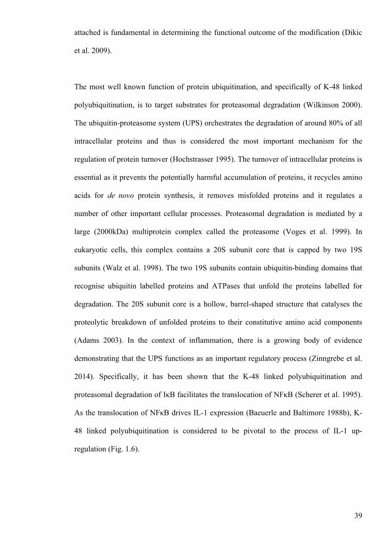
Figure 1.7. The role of protein ubiquitination in the NFκB signaling pathway 40
2 Paper 1: Dendritic Cell IL-1α and IL-1β are Polyubiquitinated and Degraded by
the Proteasome 49
Figure 1. Intracellular expression and secretion of IL-1 by BMDC: impact of LPS and
ATP 59
Figure 2. IL-1 is degraded intracellularly in BMDC by a process up regulated during
DC activation 61
Figure 3. IL-1 degradation in BMDC is dependent upon the proteasome 63
Figure 4. IL-1 degradation is dependent on the proteasome in macrophages 64
Figure 5. IL-1 degradation in BMDC is not dependent upon autophagy 66
Figure 6. IL-1 is polyubiquitinated in DC 68
Figure 7. Ubiquitinated IL-1 expression following LPS priming and ATP challenge 70
Figure S2.1 Characterisation of the IL-1β ELISA 81
Figure S2.2. XS106 cells produce IL-1β intracellularly in response to LPS, but do not
secrete this in response to ATP stimulation 81
Figure S2.3. Characterisation of bone marrow-derived dendritic cell membrane marker
expression 82
6
Figure S2.4. Characterisation of IL-1β secretion in bone marrow-derived dendritic cells
83
Figure S2.5. Viability profile of bone marrow-derived dendritic cells in response to ATP
83
Fig S2.6 Ubiquitination of secreted IL-1 84
3 Paper 2: TLR Stimulation Inhibits pro-IL-1β Polyubiquitination and Degradation
86
Figure 1. VenusIL-1β is polyubiquitinated and degraded by the proteasome in iBMDM
and THP-1 cells 97
Figure 2. The rate of venusIL-1β degradation can be measured using fluorescence as an
output in in iBMDM and THP-1 cells 100
Figure 3. Rate of IL-1β degradation in iBMDM and THP-1 is reduced upon TLR
stimulation 101
Figure 4. TLR stimulation inhibits the polyubiquitination of IL-1β 104
Figure 5. The TLR induced inhibition of IL-1 degradation is temporal 106
4 Paper 3: Interleukin-1β Processing is Dependent Upon a Calcium-Mediated
Interaction With Calmodulin 117
Figure 1. Identification of pro-IL-1β interacting proteins on a human proteome
microarray 128
Figure 2. Calmodulin interacts with pro-IL-1β and not mature IL-1β, and this
interaction is dependent on calcium 131
Figure 3. Calcium chelation and calmodulin inhibition attenuates the cleavage of pro-
IL-1β in THP-1 cells 134
Figure 4. Calmodulin is also required for secretion of IL-1β in human monocytes 136
Figure 5. Calmodulin inhibition has no effect on the assembly of the inflammasome 138
Figure S4.1. Characterisation of IL-1β secretion in the human THP-1 cell line 148
7
Figure S4.2. Characterisation of IL-1β secretion in primary human monocytes 149
Figure S4.3.Effect of BAPTA-AM and E6 berbamine on IL-1β processing (full blot) 150
5 Supplementary chapter 152
Figure 5.1. Characterisation of the IL-1β ELISA 161
Figure 5.2. XS106 cells produce IL-1β intracellularly in response to LPS, but do not
secrete this in response to ATP stimulation 162
Figure 5.3. Characterisation of bone marrow-derived dendritic cell membrane marker
expression 163
Figure 5.4. Characterisation of IL-1β secretion in bone marrow-derived dendritic cells
165
Figure 5.5. Viability profile of bone marrow-derived dendritic cells in response to ATP
166
Figure 5.6. Characterisation of IL-1β secretion in the human THP-1 cell line 169
Figure 5.7. Characterisation of IL-1β secretion in primary human monocytes 170
6 Discussion 176
Figure 7.1. A new paradigm of IL-1 processing and release 177
Figure 7.2. The effect of TLR stimulation on IL-1β ubiquitination 179
Figure 7.3. The proposed role for Calmodulin in the processing of IL-1β 186
7 Bibliography 194
8 Appendices 217
8
Abstract
Institution: The University of Manchester Name: Joseph S Ainscough Degree title: PhD Immunology Thesis Title: Understanding the Intracellular Regulation of Interleukin-1 Date: 2015
Interleukin (IL)-1α and IL-1β are pivotal to the initiation and orchestration of inflammation. Unlike most cytokines, IL-1 does not have a signal peptide and therefore secretion requires 2 independent processes; an initial signal to induce the up-regulation of the inactive precursor (pro-IL-1) and a second signal to drive cleavage and subsequent secretion. Whereas many previous studies have focused on the mechanisms that drive IL-1 secretion, the aims of this thesis were to investigate the processes that regulate the intracellular precursors of IL-1 (pro-IL-1α and pro-IL-1β). The hypothesis here was that regulation of these precursors may serve to control the vigour of IL-1 secretion and, ultimately, may influence the potency of pro-inflammatory responses. Post-translational modifications were of particular interest in this thesis, as these modifications are becoming increasingly important to immune system function. Ubiquitination is an important post-translational modification whereby ubiquitin, an 8.5kDa protein, is covalently bound to lysine residues on substrate proteins. In chapter 2, evidence was provided to show that in murine DC, IL-1α and IL-1β are polyubiquitinated and that, in both DC and macrophages, this polyubiquitination drives the proteasomal degradation of IL-1. In addition, these data demonstrated that in the presence of a second signal, polyubiquitinated IL-1 is still available for secretion. Overall, these investigations highlight that the polyubiquitination and proteasomal degradation of IL-1 serves as an essential process in the regulation of IL-1 and, therefore, should be considered as an extra dimension to the current two-signal paradigm of IL-1 release. To support this work, an immortalized bone marrow derived murine macrophage cell line and a human monocyte cell line that both stably express fluorescent IL-1β were employed to measure the rate of IL-1β degradation. In these investigations, it was shown that fluorescence is a reliable readout for measuring IL-1β degradation in these cell lines. In addition, it was demonstrated that that TLR-stimulation leads to an inhibition in IL-1β ubiquitination and degradation. Together, the work presented herein highlights that ubiquitination actively regulates the vigour of IL-1β protein expression and thus may be an important regulator of inflammation. To complement this work, a broader approach was taken, whereby the interactome of pro-IL-1β was explored using a human protein microarray. In these investigations, a human proteome microarray containing 19,951 unique proteins was used to identify proteins that bind human recombinant pro-IL-1β. In these analyses, calmodulin was identified as a particularly strong hit, with a SNR of ~11. Using an ELISA-based protein-binding assay, the interaction of recombinant calmodulin with pro-IL-1β, but not mature IL-1β, was confirmed and shown to be calcium dependent. Finally, using small molecule inhibitors it was demonstrated that both calcium and calmodulin were required for nigericin induced IL-1β secretion in human monocytes. Collectively, the evidence presented in these investigations suggests that following calcium influx, pro-IL-1β interacts with intracellular calmodulin and that this interaction is central for IL-1β processing and release. In addition,
9
a number of other potentially important pro-IL-1β-interacting proteins were also identified in this work, including IL22RA2 and PLCXD3. Overall, the work presented in this thesis serves to highlight that IL-1 is regulated by a broad range of potentially important intracellular processes. We postulate that these processes may be pivotal in the regulation of inflammation and thus the maintenance of homeostasis.
10
Declaration
No portion of the work referred to in the thesis has been submitted in support of an
application for another degree or qualification of this or any other university or other
institute of learning.
Copyright statement
i. The author of this thesis (including any appendices and/or schedules to this thesis) owns
certain copyright or related rights in it (the “Copyright”) and he has given The University
of Manchester certain rights to use such Copyright, including for administrative purposes.
ii. Copies of this thesis, either in full or in extracts and whether in hard or electronic copy,
may be made only in accordance with the Copyright, Designs and Patents Act 1988 (as
amended) and regulations issued under it or, where appropriate, in accordance with
licensing agreements which the University has from time to time. This page must form part
of any such copies made.
iii. The ownership of certain Copyright, patents, designs, trade marks and other intellectual
property (the “Intellectual Property”) and any reproductions of copyright works in the
thesis, for example graphs and tables (“Reproductions”), which may be described in this
thesis, may not be owned by the author and may be owned by third parties. Such
Intellectual Property and Reproductions cannot and must not be made available for use
without the prior written permission of the owner(s) of the relevant Intellectual Property
and/or Reproductions.
iv. Further information on the conditions under which disclosure, publication and
commercialisation of this thesis, the Copyright and any Intellectual Property and/or
Reproductions described in it may take place is available in the University IP Policy (see
http://documents.manchester.ac.uk/DocuInfo.aspx?DocID=487), in any relevant Thesis
restriction declarations deposited in the University Library, The University Library’s
regulations (see http://www.manchester.ac.uk/library/aboutus/regulations) and in The
University’s policy on Presentation of Theses
11
Abbreviations
AIM2 Absent in melanoma 2 ATP Adenosine triphosphate APC Antigen presenting cells ASC Apoptosis speck-like protein containing a CARD ~ Approximately EAE Autoimmune encephalomyelitis BM Bone marrow BSA Bovine serum albumin Ca2+ Calcium ions CARD Caspase activation and recruitment domain cDNA Complementary deoxyribonucleic acid CAPS Cryopin associated periodic syndrome CHX Cycloheximide DAMP Damage associated molecular patterns DC Dendritic cells DMSO Dimethyl sulfoxide ELISA Enzyme-linked immunosorbent assay EDTA Ethylenediaminetetraacetic acid FITC Fluorescein isothiocyanate FCS Foetal calf serum GPCR G-protein coupled receptors GM-CSF Granulocyte macrophage-colony stimulating factor GFP Green fluorescent protein HRP Horseradish peroxidase HRPT Hypoxanthine-guanine phosphoribosyltransferase IBD Inflammatory bowel disease IκB Inhibitor of κB IP3 Inositol trisphosphate IL Interleukin IL-1RA Interleukin-1 receptor antagonist IRAK Interleukin-1 receptor kinase IL-1RI Interleukin-1 receptor type I IL-1RII Interleukin-1 receptor type II JIA Juvenile idiopathic arthritis KO Knockout LC3 Light chain 3 LUBAC Linear ubiquitin assembly complex LPS Lipopolysaccharide K Lysine NOD Nucleotide-binding oligomerisation domain NLR NLR-like receptor NLRC4 NLR family CARD-containing protein 4 NLRP3 NLR family PYD-containing protein 3 NFκB Nuclear factor kappa-light-chain-enhancer of activated B cells mRNA Messenger Ribonucleic acid MS Multiple sclerosis MVB Multivesicular bodies PAMP Pathogen associated molecular patterns PRR Pattern recognition receptors

12
PBMC Peripheral blood mononuclear cells PMSF Phenylmethanesulfonylfluoride PO4
3− Phosphate PBS Phosphate buffered saline POGZ Pogo transposable element containing a zinc finger domain PCR Polymerase chain reaction K+ Potassium ions PI Propidium iodide PYD Pyrin domain RT-PCR Reverse transcription polymerase chain reaction RNA Ribonucleic acid SNR Signal to noise ratio SUMO Small ubiquitin-like modifier SP1 Specificity protein 1 SEM Standard error of the mean TH T helper TIR TOLL interleukin receptor TLR TOLL-like receptors TNF Tumour necrosis factor TRAF TNFR-associated factor 6 UPS Ubiquitin-proteasome system VCAM-1 Vascular cell adhesion molecule 1 venusIL-1βTHP1 venuspro-IL-1β human THP-l cells venusIL-1βiBMDM venuspro-IL-1β immortalized Murine bone marrow derived macrophages WCL Whole cell lysate YFP Yellow fluorescent protein Zn2+ Zinc ions

13
Acknowledgements Firstly, I would like to thank my supervisors Dr Frank Gerberick, Dr Rebecca Dearman
and Professor Ian Kimber for their advice and guidance during my PhD. The effort that
they contributed was beyond the call, and was pivotal in my development throughout this
project. Special thanks go to Rebecca for the time and effort she has put into my written
and experimental work, and also to Ian for his invaluable contributions to experimental
design. In addition, I would like to thank my collaborators, especially Dr David Brough
and Dr Gloria Lopez-Castejon for their specialist input.
Secondly, I would like to thank all past and present members of the lab. I am especially
grateful for the provision of caffeine and food, without which the work in this thesis would
not be possible. I would also like to thank everyone for making my time in the lab such an
enjoyable one.
In addition, I would like to thank all my friends and family. Special thanks go to my
parents Kerris and Stephen Ainscough, who have provided constant support and
encouragement. I am also especially grateful to Kirsty Ratanji, who has played an
important part in my life throughout this process.
Finally, I would like to thank my granddad, Kenny Morland, who has taught me so much.
Whilst I may never make it as a professional footballer, the advice he has given me
throughout my life has been invaluable. I would therefore, like to dedicate this thesis to
him.

14
CHAPTER 1:
INTRODUCTION
15
1 Introduction
1.1 General Introduction
1.1.1 The importance of inflammation
‘rubor et tumor cum calore et dolore’ (Celsus, 25AD)
Inflammation, from the Latin ‘inflamere’ (to inflame), was first described by Aulus
Cornelius Celsus nearly 2 millennia ago (Tracy 2006). Writing in a collection of medical
books called De Medicina, he defined inflammation by its four cardinal signs; ‘rubor
(redness), calor (heat), dolor (pain) and tumor (swelling)’ (Medzhitov 2010). Today, it is
known that these signs are the result of a complex series of molecular, cellular and
vascular events. These changes underpin a broad range of critical physiological processes,
and therefore the capacity to induce inflammation is fundamental to the maintenance of
homeostasis. In response to infection, inflammation facilitates an adaptive immune
response and in the response to injury and trauma, inflammation plays a vital role in repair
of tissue (Frantz et al. 2009). Whilst this acute inflammatory response is typically
beneficial to the host, a chronic inflammatory condition can be severely detrimental
(Medzhitov 2008). This is apparent in disorders such as diabetes and rheumatoid arthritis,
where chronic inflammation is a significant cause of morbidity (Dalbeth and Haskard
2005, Dandona et al. 2004). Thus, the mechanisms that are responsible for the induction,
regulation and resolution of inflammation are not only of great interest academically, but
are also of significant importance therapeutically.
1.1.2 Mechanisms of inflammation

16
Although the signs of inflammation are clear and well defined, the underlying mechanisms
are extremely complex. As mentioned previously, inflammation can be induced by a broad
range of triggers, including invading pathogens and trauma. In general, the induction of an
inflammatory response depends upon the release of specific molecular signatures, termed
danger signals (Matzinger 1994). These danger signals can be either endogenous or
exogenous and serve to alert the immune system of a potentially pathogenic challenge
(Gallucci and Matzinger 2001). There are a multitude of potential danger signals and these
can be detected by a broad range of specific receptors, expressed both on and in cells such
as dendritic cells (DC), macrophages and mast cells (Takeuchi and Akira 2010).
Ultimately, the detection of these signals results in the expression and release of a battery
of pro-inflammatory cytokines, chemokines and other mediators such as prostaglandins
(Lundberg 2000). These mediators act both locally and systemically to propagate and
orchestrate inflammatory responses. In a typical inflammatory response, antigen presenting
cells (APC) become activated and migrate to local lymph nodes, tissue-resident
macrophages and mast cells are also activated and function to seek and destroy invading
pathogens and vasodilation is induced in local blood vessels to facilitate the extravasation
of circulating leukocytes and plasma (Serhan and Savill 2005).
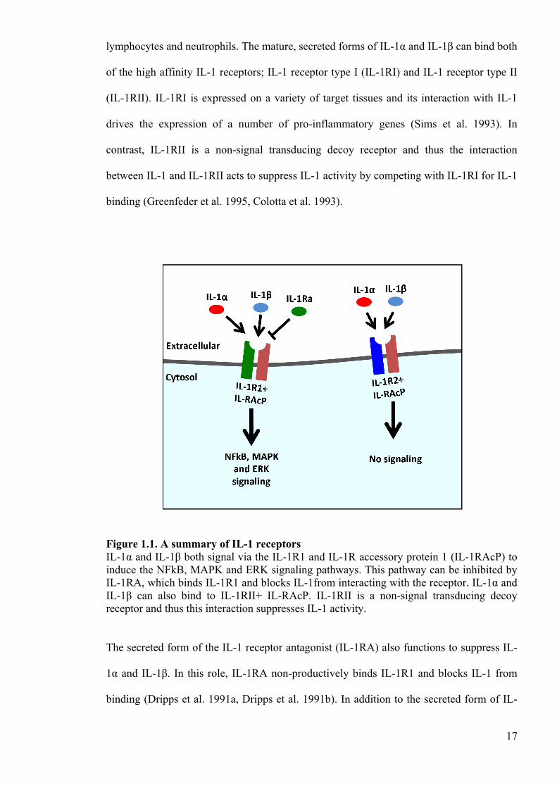
1.1.3 Inflammation and interleukin-1
One of the most important family of cytokines in the inflammatory response is the
interleukin (IL)-1 family (IL-1F) (Dinarello 2009). In total, there are 11 IL-1F members, of
which IL-1α and IL-1β are by far the best characterised. Although IL-1α and IL-1β share a
very similar tertiary structure, the amino acid sequence homology between the two proteins
is only 27% (Cameron et al. 1985). These cytokines are produced by a wide range of cell
types, including macrophages, DC, monocytes, natural killer cells, T-lymphocytes, B-
17
lymphocytes and neutrophils. The mature, secreted forms of IL-1α and IL-1β can bind both
of the high affinity IL-1 receptors; IL-1 receptor type I (IL-1RI) and IL-1 receptor type II
(IL-1RII). IL-1RI is expressed on a variety of target tissues and its interaction with IL-1
drives the expression of a number of pro-inflammatory genes (Sims et al. 1993). In
contrast, IL-1RII is a non-signal transducing decoy receptor and thus the interaction
between IL-1 and IL-1RII acts to suppress IL-1 activity by competing with IL-1RI for IL-1
binding (Greenfeder et al. 1995, Colotta et al. 1993).
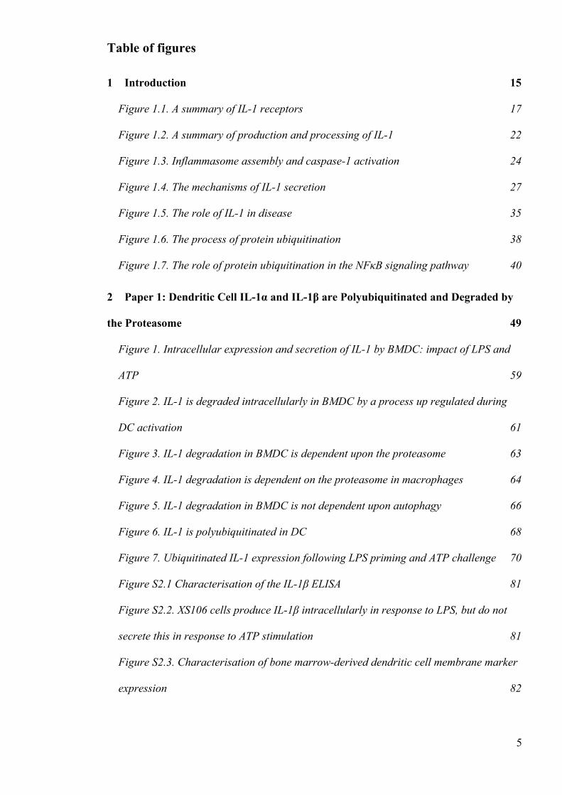
Figure 1.1. A summary of IL-1 receptors IL-1α and IL-1β both signal via the IL-1R1 and IL-1R accessory protein 1 (IL-1RAcP) to induce the NFkB, MAPK and ERK signaling pathways. This pathway can be inhibited by IL-1RA, which binds IL-1R1 and blocks IL-1from interacting with the receptor. IL-1α and IL-1β can also bind to IL-1RII+ IL-RAcP. IL-1RII is a non-signal transducing decoy receptor and thus this interaction suppresses IL-1 activity.
The secreted form of the IL-1 receptor antagonist (IL-1RA) also functions to suppress IL-
1α and IL-1β. In this role, IL-1RA non-productively binds IL-1R1 and blocks IL-1 from
binding (Dripps et al. 1991a, Dripps et al. 1991b). In addition to the secreted form of IL-
18
1RA, which contains a leader sequence for secretion via the classical secretory pathway,
there are 3 intracellular forms of IL-1RA produced as a result of alternative splicing. It is
postulated that these intracellular isoforms have inhibitory effects on the NFκB signaling
pathway (Wolf et al. 2001). In addition, Watson et al. demonstrated that intracellular IL-
1RA is secreted following activation of the P2X7 receptor (Wilson et al. 2004). Like the
classically secreted isoform, intracellular IL-1RA also inhibits IL-1 signaling via the IL-
1R1 receptor.
The importance of IL-1β in the initiation and orchestration of inflammation is well
established. IL-1β is one of the first cytokines produced in response to danger, and its
secretion is sufficient to induce a potent and vigourous inflammatory response (Dinarello
2011). This has been demonstrated experimentally in variety of tissues, using a broad
range of animal models. Many of these studies have shown that the administration of
recombinant IL-1β alone is sufficient to drive the necessary molecular and cellular changes
required for a localised inflammatory response. This is evident in the skin, for example,
where the intradermal administration of recombinant IL-1β results in an increase in the
expression of a number of pro-inflammatory mediators (Enk et al. 1993), the activation and
migration of epidermis-resident APC (Cumberbatch et al. 1997), the infiltration of
circulating leukocytes (Papini and Bruni 1996) and ultimately, the induction of
inflammation. Other studies have employed transgenic mice and IL-1 inhibitors to
demonstrate the importance of IL-1 in inflammation. In early experiments, it was shown
that, unlike wild type controls, IL-1β-deficient mice do not develop an acute inflammatory
response when injected subcutaneously with turpentine (Fantuzzi and Dinarello 1996). In
similar experiments, IL-1RI deficient mice have been shown to have an impaired acute
inflammatory response in a number of experimentally induced disease states (Labow et al.
1997). In humans, a recombinant version of the IL-1 receptor antagonist (IL-1RA) has

19
been developed for use therapeutically, and has been used to treat successfully the
inflammatory manifestations associated with a number of diseases, including rheumatoid
arthritis and pericarditis (reviewed in (Dinarello et al. 2012)). Therefore, the experimental
evidence indicates that IL-1β is not only capable of inducing inflammation, but is often a
requisite for an acute inflammatory response.
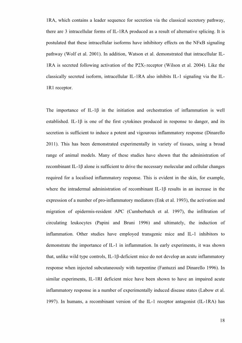
1.1.4 IL-1 production
In the induction of inflammation, the initial signal for IL-1 up-regulation is provided
typically by a specific set of danger signals called pathogen associated molecular patterns
(PAMP). These PAMP are molecular patterns derived from invading pathogens and are
detected by a range of pattern recognition receptors (PRR), most commonly of the TOLL-
like receptor (TLR) family. The first TLR to be identified was TLR4, in an investigation
that demonstrated that TLR4 recognises a membrane component of Gram-negative
bacteria called lipopolysaccharide (LPS) (Medzhitov et al. 1997). The TLR family has
since grown to 10 receptors in humans and 12 receptors in mice. Like TLR4, these other
TLR can be activated by various PAMP, including the bacterial products peptidoglycan
(TLR2) (Schwandner et al. 1999) and flagellin (TLR5) (Hayashi et al. 2001), double
stranded viral RNA (TLR3) (Alexopoulou et al. 2001) and single stranded viral RNA
(TLR7) (Lund et al. 2004). Broadly speaking, the type of ligand that is recognised by a
TLR is dependent upon the cellular location of the TLR. TLR1, TLR2, TLR4, TLR5,
TLR6, and TLR11 are all expressed on the cell surface and, thus, typically recognise
microbial membrane products (Kawai and Akira 2010). In contrast, TLR3, TLR7, TLR8,
and TLR9 are all expressed in endolysosomes and, thus, typically recognise microbial
nucleic acids (Ozinsky et al. 2000).
20
In general, there are 2 main signaling pathways implicated in the up-regulation of IL-1; the
MyD88-dependent pathway and the TRIF-dependent pathway (reviewed in (Ainscough et
al. 2013)). MyD88 is a cytosolic TOLL IL-1 receptor (TIR) domain-containing protein
required for the downstream signaling of all TLR except TLR3 (O'Neill and Bowie 2007).
Upon activation of these TLR, MyD88 binds to the intracellular TIR domain on TLR and
recruits the IL-1 receptor kinases (IRAK) 1, 2 and 4 (Wesche et al. 1997). Once activated,
these kinases recruit, activate and then release the E3 ubiquitin ligase TNFR-associated
factor 6 (TRAF6) into the cytosol, where it forms a complex with TAK1, TAB2 and
TAB2/3 (Qian et al. 2001). This complex then activates another complex called the IKK
complex. The IKK complex comprises of 2 kinases (IKKα and IKKβ) and a regulatory
subunit called NEMO (Kawai and Akira 2010). The activation of IKK drives the
phosphorylation and degradation of the inhibitor of κB (IκB) (Hacker and Karin 2006). As
IκB acts to inhibit nuclear factor kappa-light-chain-enhancer of activated B cells (NFκB)
translocation in the steady state, degradation of IκB facilitates NFκB translocation into the
nucleus (Baeuerle and Baltimore 1988b). Once in the nucleus, NFκB promotes the
transcription of a number of pro-inflammatory proteins, including the precursors pro-IL-1α
and pro-IL-1β (Baeuerle and Baltimore 1988a).
As mentioned above, IL-1 expression can also be induced via the less well-characterised
TRIF-dependent pathway. Like MyD88, TRIF is a TIR domain containing protein that
binds to the intracellular TIR on TLR. Unlike MyD88, however, TRIF is only required for
the downstream signaling of TLR3 and TLR4 (Yamamoto et al. 2004). Following
activation of these TLR, TRIF is recruited and associates with TRAF6 via RIP-1, TRADD
and pellino-1 (Sato et al. 2003, Chang et al. 2009). As in the MyD88-dependent pathway,
TRAF6 is activated and forms a complex with TAK1, TAB2 and TAB2/3. Again, the
21
activation of this complex ultimately leads to the translocation of NFκB, and an increase in
pro-IL-1α and pro-IL-1β expression (fig. 1.1).
1.1.5 Mechanisms of IL-1 processing
Unlike most other cytokines, IL-1α and IL-1β are expressed without a signal peptide and
are, therefore, not secreted via the classic secretory pathway (March et al. 1985, Rubartelli
et al. 1990, Stevenson et al. 1992). Instead, both cytokines are expressed as 31kDa
precursors (March et al. 1985). The IL-1β precursor is cleaved into the mature bioactive
17kDa cytokine by the protease caspase-1(Thornberry et al. 1992). Caspase-1 is abundant
in all hematopoietic cells, and is expressed as a 45kDa proenzyme called procaspase-1.
Upon activation, this proenzyme is cleaved into a 10kDa subunit and a 20kDa subunit,
which come together to form part of the active heterotetrameric enzyme (two 10kDa
subunits and two 20kDa subunits) (Wilson et al. 1994). The activation of caspase-1 is
dependent upon the formation of a large molecular complex called the inflammasome
(Ogura et al. 2006).
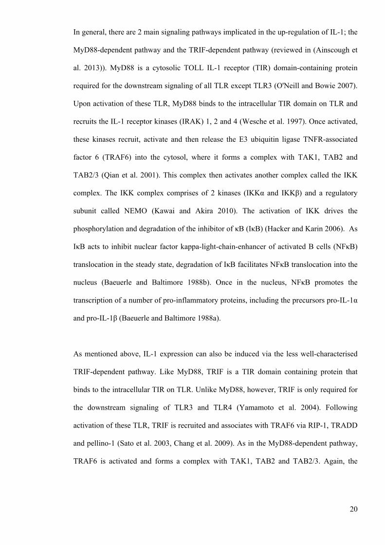
The assembly of the inflammasome is driven by cytosolic PRR, most commonly of the
nucleotide-binding oligomerisation domain (NOD)-like receptor family (Franchi et al.
2009). As with TLR, these NLR function to detect the presence of a variety of PAMP, as
well as damage associated molecular patterns (DAMP) (Shaw et al. 2010). DAMP are
important endogenous molecules that are released in response to injury. NLR family pyrin
domain (PYD) containing 3 (NLRP3) is the most comprehensively studied NLR and is
activated by a diverse range of DAMP/PAMP, including adenosine triphosphate (ATP)
(Ferrari et al. 1997), the crystalline compounds silica (Hornung et al. 2008), asbestos
(Dostert et al. 2008), and uric acid (Martinon et al. 2007), the bacterial product listeriolysin
O (Meixenberger et al. 2010) and the potassium ionophore nigericin (Mariathasan et al.
22
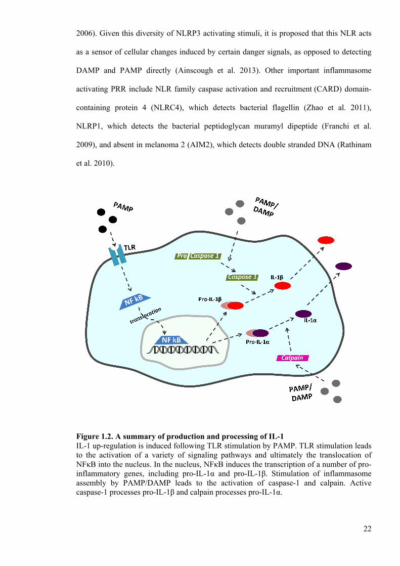
2006). Given this diversity of NLRP3 activating stimuli, it is proposed that this NLR acts
as a sensor of cellular changes induced by certain danger signals, as opposed to detecting
DAMP and PAMP directly (Ainscough et al. 2013). Other important inflammasome
activating PRR include NLR family caspase activation and recruitment (CARD) domain-
containing protein 4 (NLRC4), which detects bacterial flagellin (Zhao et al. 2011),
NLRP1, which detects the bacterial peptidoglycan muramyl dipeptide (Franchi et al.
2009), and absent in melanoma 2 (AIM2), which detects double stranded DNA (Rathinam
et al. 2010).
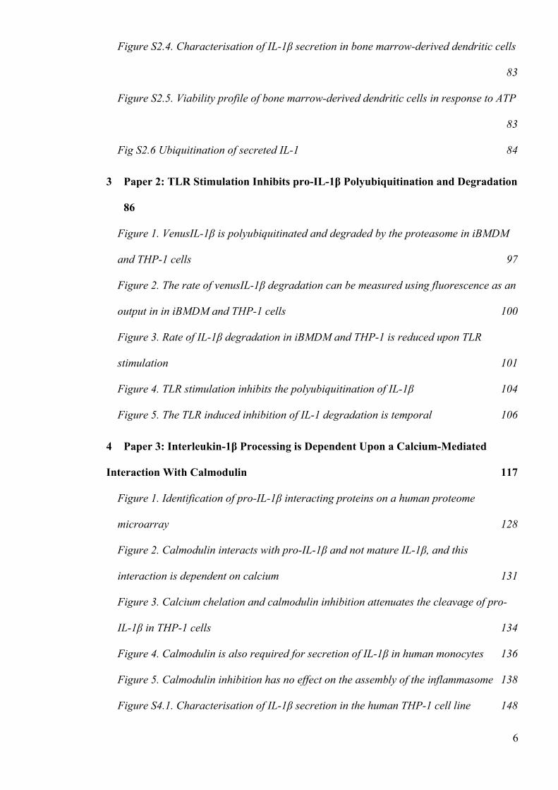
Figure 1.2. A summary of production and processing of IL-1 IL-1 up-regulation is induced following TLR stimulation by PAMP. TLR stimulation leads to the activation of a variety of signaling pathways and ultimately the translocation of NFκB into the nucleus. In the nucleus, NFκB induces the transcription of a number of pro-inflammatory genes, including pro-IL-1α and pro-IL-1β. Stimulation of inflammasome assembly by PAMP/DAMP leads to the activation of caspase-1 and calpain. Active caspase-1 processes pro-IL-1β and calpain processes pro-IL-1α.
23
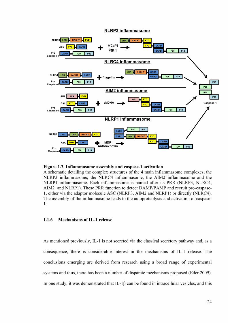
The ability of these cytosolic PRR to drive the assembly of the inflammasome is dependent
upon a complex molecular structure (fig. 1.2). NLRP3, NLRP1 and NLRC4 all have a
leucine-rich repeat domain for sensing PAMP and DAMP and a nucleotide-binding
domain for oligomerisation (Martinon et al. 2002). NLRP3 and NLRP1 have a PYD
domain for recruitment of the adaptor molecule apoptosis speck-like protein containing a
CARD (ASC) (Agostini et al. 2004). AIM2 detects DNA via a HIN domain but still uses a
PYD domain for recruiting ASC (Jin et al. 2013). As its name suggests, ASC has a CARD
domain that recruits procaspase-1 to the inflammasome complex via a CARD-CARD
interaction. Interestingly, NLRP1 and NLRC4 also have their own CARD domain and so
can recruit caspase-1 independently of ASC (Bryant and Fitzgerald 2009). Once
procaspase-1 has formed part of this inflammasome complex, the zymogen undergoes
autoproteolysis to form an active heterodimer capable of proteolytic cleavage of pro-IL-1β
into its bioactive form (Yang et al. 1998).
Despite its importance in inflammation, the processing of IL-1α is not as well
characterised. In contrast to pro-IL-1β, pro-IL-1α can bind IL-1RI and therefore is active in
its precursor form (Mosley et al. 1987). Nevertheless, mature IL-1α is significantly more
potent than its proprotein and thus processing is still an important event in the development
of inflammation associated with IL-1α (Afonina et al. 2011). Pro-IL-1α is most commonly
cleaved by the calcium-dependent protease calpain, but can also be processed by other
proteases such as granzyme-B, mast cell chymase or neutrophil elastase (Rider et al. 2013).
Although caspase-1 does not cleave pro-IL-1α directly, caspase -1 knockout (KO) mice do
exhibit reduced IL-1α secretion, suggesting that caspase-1 does play a role (Kuida et al.
1995). As IL-1α is protected from processing by intracellular IL-1RII it is postulated that
caspase-1 acts to cleave IL-1RII, thereby facilitating calpain proteolysis of the precursor to
active IL-1α (Zheng et al. 2013).
24
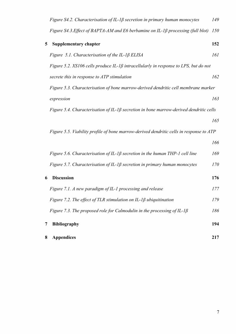
Figure 1.3. Inflammasome assembly and caspase-1 activation A schematic detailing the complex structures of the 4 main inflammasome complexes; the NLRP3 inflammasome, the NLRC4 inflammasome, the AIM2 inflammasome and the NLRP1 inflammasome. Each inflammasome is named after its PRR (NLRP3, NLRC4, AIM2 and NLRP1). These PRR function to detect DAMP/PAMP and recruit pro-caspase-1, either via the adaptor molecule ASC (NLRP3, AIM2 and NLRP1) or directly (NLRC4). The assembly of the inflammasome leads to the autoproteolysis and activation of caspase-1.
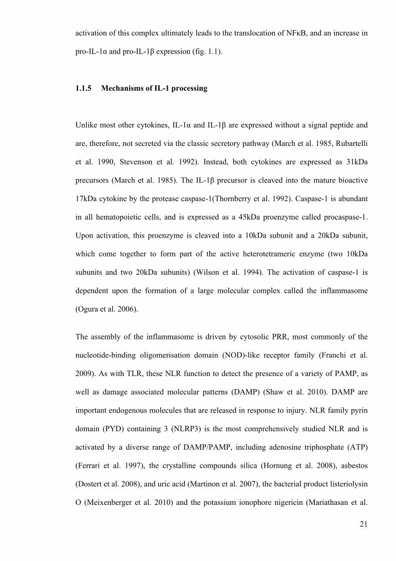
1.1.6 Mechanisms of IL-1 release
As mentioned previously, IL-1 is not secreted via the classical secretory pathway and, as a
consequence, there is considerable interest in the mechanisms of IL-1 release. The
conclusions emerging are derived from research using a broad range of experimental
systems and thus, there has been a number of disparate mechanisms proposed (Eder 2009).
In one study, it was demonstrated that IL-1β can be found in intracellular vesicles, and this
25
fraction can be protected from lysosomal degradation by triggering IL-1 secretion (Andrei
et al. 1999). In more recent investigations, it has been shown that a fraction of IL-1β is
sequestered into autophagosomes, suggesting that autophagy provides a route of IL-1
secretion (Harris et al. 2011). Interestingly, IL-1β is secreted when autophagy is inhibited
and therefore it is postulated that IL-1β can, in certain circumstances, be released via this
“rescue and redirect” mechanism. Specifically, it is proposed that LPS stimulates the
formation of IL-1β-containing autophagosomes that exocytose upon autophagy inhibition,
releasing IL-1β from the cytoplasm.
In addition, IL-1β can be secreted in vesicles, either in the form of microvesicles (100-
600nm) or in the form of exosomes (50- 80nm) (Pizzirani et al. 2007). This was initially
observed in the human monocyte cell line THP-1 cells, whereby stimulation of the P2X7
receptor with ATP caused a rapid release of bioactive IL-1β-containing microvesicles
(MacKenzie et al. 2001). The P2X7 receptor is a transmembrane ionotropic receptor that is
expressed on immune cells (Wiley et al. 2011). The stimulation of this receptor causes the
opening of a cation channel, facilitating the rapid efflux of potassium ions (K+). The
release of IL-1β via exosomes was suggested in a more recent study using murine
macrophages (Qu et al. 2007). In this investigation, rapid ATP-induced IL-1β secretion
correlated with exosome release. The mechanism proposed in this study was that
stimulation of the P2X7 receptor causes the formation of an endosome. It was suggested
that this endosome entraps cytosolic IL-1β through a process of inward budding, forming
IL-1β-containing exosomes within multivesicular bodies (MVB) (Record et al. 2011).
Ultimately, the authors in this study postulated that IL-1β release was facilitated by the
exocytosis of these IL-1β-containing exosomes. However, as IL-1β has yet to be found
contained in exosomes, this hypothesis is purely speculative at this stage.
26
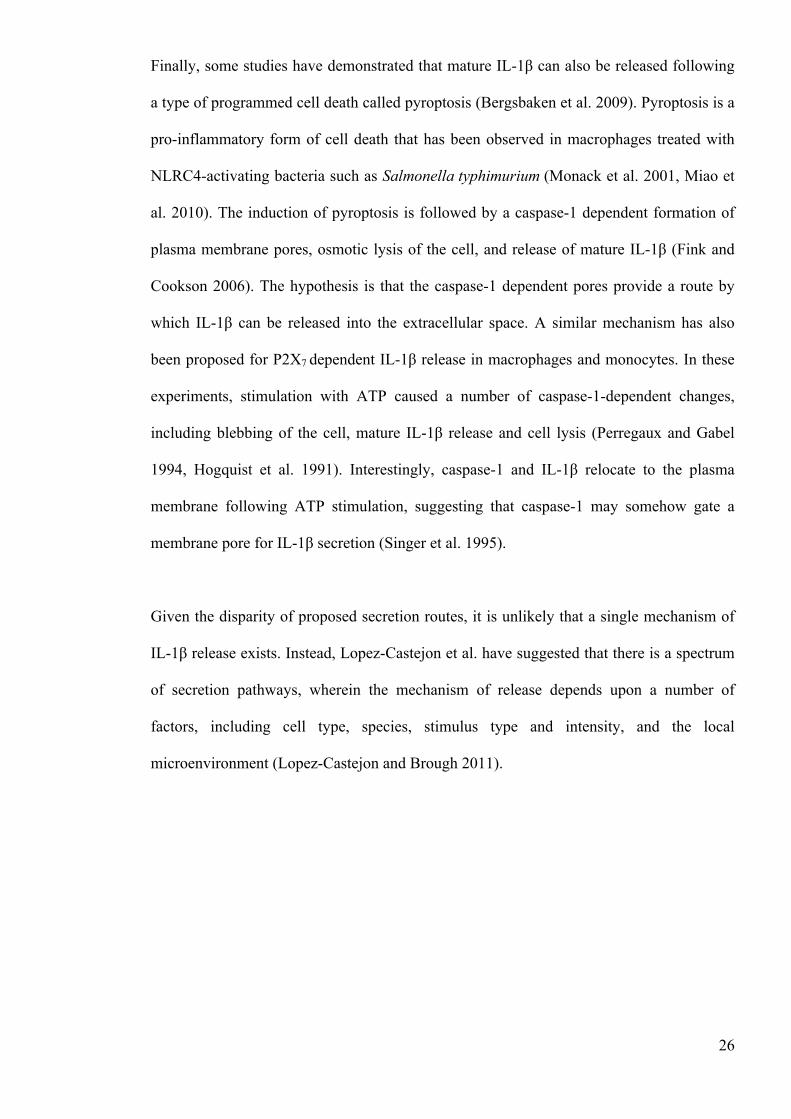
Finally, some studies have demonstrated that mature IL-1β can also be released following
a type of programmed cell death called pyroptosis (Bergsbaken et al. 2009). Pyroptosis is a
pro-inflammatory form of cell death that has been observed in macrophages treated with
NLRC4-activating bacteria such as Salmonella typhimurium (Monack et al. 2001, Miao et
al. 2010). The induction of pyroptosis is followed by a caspase-1 dependent formation of
plasma membrane pores, osmotic lysis of the cell, and release of mature IL-1β (Fink and
Cookson 2006). The hypothesis is that the caspase-1 dependent pores provide a route by
which IL-1β can be released into the extracellular space. A similar mechanism has also
been proposed for P2X7 dependent IL-1β release in macrophages and monocytes. In these
experiments, stimulation with ATP caused a number of caspase-1-dependent changes,
including blebbing of the cell, mature IL-1β release and cell lysis (Perregaux and Gabel
1994, Hogquist et al. 1991). Interestingly, caspase-1 and IL-1β relocate to the plasma
membrane following ATP stimulation, suggesting that caspase-1 may somehow gate a
membrane pore for IL-1β secretion (Singer et al. 1995).
Given the disparity of proposed secretion routes, it is unlikely that a single mechanism of
IL-1β release exists. Instead, Lopez-Castejon et al. have suggested that there is a spectrum
of secretion pathways, wherein the mechanism of release depends upon a number of
factors, including cell type, species, stimulus type and intensity, and the local
microenvironment (Lopez-Castejon and Brough 2011).
27
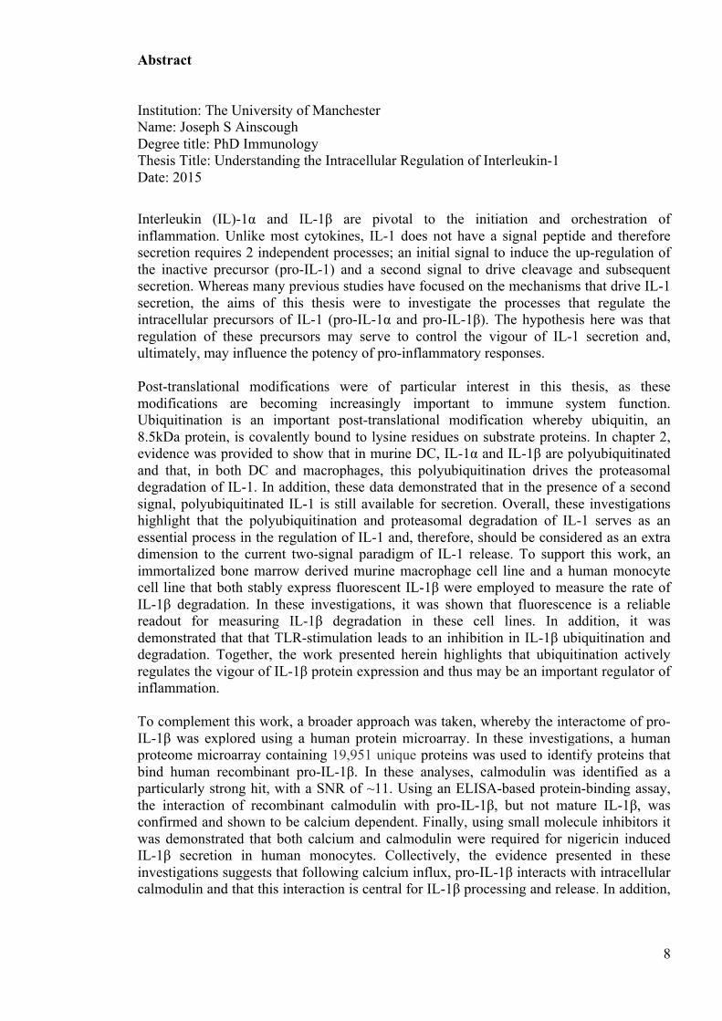
Figure 1.4. The mechanisms of IL-1 secretion A schematic showing the various, proposed mechanisms of IL-1 secretion, including rescue and redirect, protected release and terminal release. In the rescue and redirect pathway, it is postulated that IL-1β is sequestered into autophagosomes and consequently secreted. In the protected release pathway, it is proposed that IL-1β can be secreted via the release of IL-1β-containing exosomes or microvesicles. In the terminal release pathway, it is proposed that caspase-1 dependent pores are formed and that IL-1β is secreted via these pores. This diagram was adapted from Lopez-Castejon and Brough (Lopez-Castejon and Brough 2011).
1.1.7 Metal ions in IL-1 secretion
There are a number of metal ions involved in the intracellular processing of pro-IL-1
(Ogura et al. 2006). One of the most important and well-established metal ions in the
secretion of IL-1 is K+. A role for K+ in IL-1 processing was first postulated in a study by
Perregaux and Gabel, where it was shown that ATP and nigericin-induced IL-1β release
28
correlated with a net decrease in intracellular K+ concentration (Perregaux and Gabel
1994). Importantly, this study also demonstrated that the decrease in intracellular K+
concentration was necessary for nigericin and ATP-induced IL-1β maturation. Whereas
nigericin is a K+ ionophore and thus functions as a K+ channel directly (Daniele et al.
1978), ATP acts via P2X7 receptors to facilitate K+ efflux (Ferrari et al. 1997). In addition,
more recent investigations have established that the efflux of K+ is a feature induced by all
known NLRP3-activating stimuli, including ATP and nigericin, as well as the NLRP1
activator anthrax lethal toxin (Munoz-Planillo et al. 2013). However, it is not yet clear
whether K+efflux alone is sufficient to drive NLRP3 or NLRP1 inflammasome-dependent
IL-1β processing.
In addition to K+, zinc ions (Zn2+) are also implicated in IL-1β secretion. Zn2+ is an
important nutrient and is essential for innate and adaptive immune system function
(Terpilowska and Siwicki 2011). In macrophages, the depletion of intracellular Zn2+ is
associated with NLRP3 inflammasome activation and the release of bioactive IL-1β
(Summersgill et al. 2014). Although the sensors of Zn2+ depletion are currently unknown,
Zn2+ depletion-induced NLRP3 activation is dependent upon a destabilisation of the
lysosome membrane, suggesting that this is an important event in Zn2+-induced IL-1β
secretion. This mechanism of IL-1β processing may be particularly relevant to the
inflammation associated with Alzheimer’s Disease, as this disease is associated with both
Zn2+ depletion and NLRP3 inflammasome activation (Brewer et al. 2010, Heneka et al.
2013).
The importance of calcium (Ca2+) in IL-1β processing and release is also well established.
In Brough et al., it was shown that ATP and nigericin both induce the release of
intracellular calcium stores, leading to an increase in cytosolic Ca2+ concentration (Brough
29
et al. 2003). Crucially, the chelation of intracellular Ca2+ was shown to inhibit IL-1β
processing, suggesting that the release of Ca2+ from intracellular stores is required for IL-
1β activation. To support these data, more recent investigations have targeted the signaling
pathways that lead to the release of intracellular Ca2+ stores. In these studies, the inhibition
of either phospholipase C, IP3-gated Ca2+ release channels or store-operated Ca2+ entry
abrogated ATP-induced IL-1β processing in macrophages (Murakami et al. 2012). In
addition, it has recently been shown that extracellular Ca2+ can also act as a danger signal
and induce IL-1β secretion (Rossol et al. 2012). Specifically, experiments using monocytes
demonstrated that extracellular Ca2+ can signal via G-protein coupled receptors (GPCR) to
drive the release of intracellular Ca2+, the activation of NLRP3 and the processing of pro-
IL-1β (Rossol et al. 2012). However, despite continuing efforts, the precise mechanisms of
intracellular Ca2+-induced IL-1β processing have yet to be determined.
1.1.8 IL-1 in disease
Although the appropriate expression and secretion of IL-1 is central to inflammation and
the maintenance of health, the improper regulation of IL-1 is an important factor in a broad
range of diseases (Dinarello 2011, Dinarello 2009). Cryopin-associated periodic
syndromes (CAPS) are a group of inherited autoimmune disorders including Muckle-Wells
syndrome, neonatal-onset multisystem inflammatory disease and familial cold
autoinflammatory syndrome (Gabay et al. 2010). These disorders occur as a result of
mutations in the genes encoding cryopin or NLRP3, and are characterised by spontaneous
inflammasome assembly and caspase-1 activity (Agostini et al. 2004). Ultimately, this
raised caspase-1 activity causes an increase in IL-1 secretion, leading to recurrent and
systemic inflammatory episodes. Importantly, the administration of the recombinant IL-
1RA therapeutic Anakinra inhibits the development of symptoms, indicating that the
30
dysregulation of IL-1 is central to the development of these disorders (Hoffman et al. 2004,
Hawkins et al. 2003).
IL-1 is also thought to be involved in the progression of multiple sclerosis (MS). MS is a
debilitating autoimmune disease in which the myelin sheaths surrounding the neurones are
damaged, leading to disruption in the ability of the nervous system to communicate (Wu
and Alvarez 2011). In the experimental autoimmune encephalomyelitis (EAE) model of
MS, the inhibition of IL-1 reduces the severity and delays the onset of the disease (Sutton
et al. 2006, Matsuki et al. 2006). Moreover, IL-1R1 KO mice do not develop EAE,
suggesting that IL-1 may play a crucial role in the development of MS. As MS is mediated
by T Helper (TH) 17 cells, it is postulated that IL-1 contributes to the diesease onset by
promoting the TH 17 cell function (Chung et al. 2009). TH cells play important roles in
regulating the adaptive immune system. The most well characterised subsets of TH cells are
TH 1 cells, which play important roles in driving effector responses against intracellular
bacteria, TH 2 cells, which function to drive effector responses to extracellular parasites,
and TH 17 cells, which are important in the host defence against extracellular bacteria and
funghi.
Ulcerative colitis and Crohn’s disease are both common forms of chronic inflammatory
bowel disease (IBD), with the former affecting the large intestine and the latter affecting
the entire gastrointestinal tract (El-Salhy 2012). Casini-Raggi et al. found that IL-1α and
IL-1β expression was significantly higher in the freshly isolated intestinal mucosal cells of
IBD patients, relative to healthy control tissue (Casiniraggi et al. 1995). Interestingly, the
ratio of IL-1 expression to IL-1RA expression was found to correlate closely with the
severity of IBD, indicating that IL-1 is a crucial component of the disease. To support this,
31
the administration of IL-1 inhibitors has been shown to have a positive effect in a number
of experimental IBD models (Sims and Smith 2010).
In addition to CAPS, MS and IBD, there are a range of other autoimmune diseases to
which IL-1 may contribute. In the onset of type-1 diabetes, elevated IL-1β production has
been observed in serum of patients with the disease, relative to healthy individuals
(Mandrup-Poulsen et al. 2010). As IL-1β has been shown to exert toxic effects on insulin-
producing β-cells, it is postulated that the cytokine could, in some circumstances at least,
be a causative factor (Maedler et al. 2002). IL-1 is also crucial for the development of
certain forms of arthritis, including rheumatoid arthritis, juvenile idiopathic arthritis (JIA)
and gout (Kay and Calabrese 2004). In the murine collagen induced arthritis model of
rheumatoid arthritis, the inhibition of IL-1 prevents disease progression and reverses the
symptoms (Joosten et al. 1999). In the systemic onset form of JIA, treatment with
Anakinra (recombinant IL-1RA) significantly alleviates symptoms, suggesting that the
disease is dependent upon IL-1 (Verbsky and White 2004, Pascual et al. 2005, Gattorno et
al. 2008). Although it has been known for some time that gout is caused by the
accumulation of uric acid crystals (Schumacher 2008), a role for IL-1 has only recently
gained attention. This is following the discovery that uric acid crystals activate the NLRP3
inflammasome (Martinon et al. 2007). Encouragingly, pilot studies have shown that
Anakinra has a positive effect in gout patients, indicating that IL-1 is central to the
development of gout (So et al. 2007).
In addition to autoimmune disease, IL-1 has been implicated in a variety of other disorders.
Asthma is a common respiratory syndrome associated with aberrant and excessive
pulmonary inflammation (Holgate 2011). IL-1 levels are significantly higher in individuals
with status asthmaticus, a severe acute asthmatic attack that is unresponsive to treatment
32
(Mao et al. 2000). Moreover, in the murine model of ovalbumin-induced asthma, mice
overexpressing IL-1RA exhibit reduced pulmonary inflammation relative to the wild type
control (Wang et al. 2006). As the goblet cell hyperplasia and eosinophilic inflammation
associated with the disease were strongly reduced in these KO mice specifically, it is
proposed that these IL-1-induded processes are required for the development of asthma. In
the skin, IL-1 is also involved in a number of disorders including contact hypersensitivity
and atopic dermatitis (Sims and Smith 2010). Contact hypersensitivity is an inflammatory
condition caused by an inappropriate immune response to specific chemicals, termed
contact allergens when encountered on the skin surface (Kimber et al. 2011). After skin
exposure to contact allergens, IL-1 is rapidly up-regulated by epidermal Langerhans cells
and this up-regulation is required for the development of skin sensitisation (Enk and Katz
1992). Atopic dermatitis is also an inflammatory skin disease and this is driven by TH 2
cells (Bieber 2008). Interestingly, patients with atopic dermatitis present with increased IL-
1RI expression (Shimizu et al. 2005), and IL-1 KO mice exhibit delayed onset in an atopic
dermatitis model (Konishi et al. 2002), suggesting that IL-1 is required for the initiation
phase of the disease.
IL-1 is also emerging as an important factor in the development of a number of
cardiovascular diseases. Atherosclerosis is a disease whereby the diameter of the lumen is
reduced due to a hardening of the artery. Recently, this disease has been associated with a
systemic increase in the expression of proinflammatory cytokines, including IL-1
(Vicenova et al. 2009). An increased expression of IL-1 has also been found at the site of
atheromatous plaques, implicating IL-1 as an important mediator in the development of
such plaques (Tipping and Hancock 1993). Importantly, atherosclerosis is a leading cause
of myocardial infarction, a cardiac event whereby the muscles in the heart are blocked
(Guillen et al. 1995). Interestingly, it has been shown that IL-1 expression is also up
33
regulated during myocardial infarction. As IL-1β has been shown to enhance expression of
tissue factor and induce procoagulant activity, it is suggested that this cytokine may be an
important contributory factor to the development of a myocardial infarction (Schwager and
Jungi 1993).
IL-1 also contributes to the progression of a number of brain disorders (Rothwell and
Luheshi 2000). Increased IL-1 production has been observed in some important
neurodegenerative diseases, including Alzheimer’s Disease, Parkinson’s disease and
Downs syndrome (Cacabelos et al. 1994, Cacabelos et al. 1991, Griffin et al. 1989). An
elevation in IL-1 expression is also observed in acute brain injury and stroke (Griffin et al.
1994). The use of IL-1RA as a therapeutic has therefore been explored. In an
experimentally-induced murine model of stroke, intracerebroventricular or peripheral
administration of IL-1RA markedly reduces neuronal tissue injury, resulting in a vastly
improved behavioural outcome (Rothwell 2003). In a clinical trial, administration of IL-
1RA was also shown to benefit patients with acute stroke, relative to a placebo control
(Emsley et al. 2005). Therefore, IL-1 may represent an attractive therapeutic target in the
treatment of a number of neurological disorders.
As mentioned previously, the use of the recombinant IL-1RA therapeutic Anakinra has
already shown to be of benefit in a number of inflammatory diseases. In addition to
Anakinra, there exists a number of other clinical tools designed to target IL-1 signalling.
Canakinumab is an antibody developed to neutralise the activity of IL-1β. Importantly, this
therapeutic has been approved as a treatment for CAPS and systemic-onset juvenile
idiopathic arthritis by both the US food and drug administration and the European
medicines agency (Molto and Olive 2010). Moreover, it is postulated that Canakinumab
could also be used in the treatment of other complex inflammatory diseases, such as
34
rheumatoid arthritis and gout (Church and McDermott 2009). Finally, a therapeutic
comprised of both the IL-1RAcP and IL-1R1 has also been developed and termed IL-1 trap
(Ratner 2008). Like Anakinra, this therapeutic targets the signalling of both IL-1α and IL-
1β and thus has the potential to treat a broader range of diseases compared with
Canakinumab. Again, this therapeutic is already approved for use as a treatment for CAPS,
and has the potential to be used as a treatment for a number of clinically important
inflammatory disorders.

35
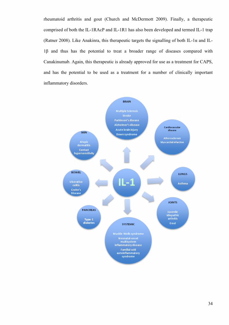
Figure 1.5. The role of IL-1 in disease A diagram detailing the variety of diseases in which IL-1 has been shown to contribute. Diseases can be split into 8 categories: brain, skin, joints cardiovascular disease, bowel, pancreas, lungs and systemic. 1.1.9 Post-translational modification
From the evidence presented above, it is clear that the regulation of IL-1 is vital to the
maintenance of homeostasis. As a result, it is important that the processes involved are
understood. Unlike most cytokines, both pro-IL-1α and pro-IL-1β are cytosolic and thus
may be regulated by a variety of intracellular mechanisms. Of particular interest is the
potential that the IL-1 cytokines are regulated by post-translational modification (Perkins
2006). Post-translational modifications are covalent processing events involving either
proteolytic cleavage of proteins or the addition of modifying groups (such as glycans,
phosphorylation etc.) to proteins following biosynthesis. These modifications are an
important set of mechanisms whereby cells can regulate the characteristics and function of
specific proteins. Depending upon the modification, post-translational modification can
regulate the activity or determine the localisation of a protein, can modify the potential for
protein-protein interaction and can affect the turnover rate of the protein.
Phosphorylation and dephosphorylation are considered to be some of the most important
and well-studied post-translational modifications, implicated in the regulation of a large
number of proteins and cell signaling pathways (Cohen 2000). In brief, phosphorylation is
dependent on kinases and involves the addition of a phosphate group (PO43−) to serine,
threonine or tyrosine residues on target proteins. Conversely, dephosphorylation, involves
the removal of PO43− and is mediated by phosphatases. In general, a change in the
phosphorylation state of a protein causes a conformational change, altering the function of
the protein. In the context of enzymes, phosphorylation and dephosphorylation can modify
36
the catalytic activity of a protein, thereby activating or deactivating the enzyme (Krebs and
Beavo 1979). As discussed previously, phosphorylation and dephosphorylation are central
to the transduction of NFκB signaling pathways and therefore represent important
regulators of IL-1 expression (Viatour et al. 2005). Interestingly, both pro-IL-1α and pro-
IL-1β are phosphorylated directly, and this phosphorylation is thought to enhance the
conversion of pro-IL-1 to mature IL-1 (Kobayashi et al. 1988). Although further
investigation is required, it is evident that phosphorylation and dephosphorylation are
significant processes in the regulation of IL-1.
There are also a number of other important post-translational modifications, including
glycosylation, SUMOlyation, S-Nitrosylation, methylation and ubiquitination.
Glycosylation is one of the most common forms of post-translational modification and
involves the addition of carbohydrate moieties to target proteins (Ohtsubo and Marth
2006). This process is implicated in a wide range of cellular mechanisms and its function is
dependent upon the target protein (Schwarz and Aebi 2011). Although IL-1 cytokines are
not directly glycosylated, glycosylation is required for IL-1 signaling. Specifically, IL-1R1
is glycosylated and this glycosylation is pivotal for optimal IL-1 binding (Mancilla et al.
1992). SUMOlyation is a post-translational modification whereby small ubiquitin-like
modifier (SUMO) proteins are conjugated to target proteins (Wilson and Rangasamy
2001). Like glycosylation, SUMOlyation plays important roles in the transduction of the
NFκB signaling pathways (reviewed in (Geiss-Friedlander and Melchior 2007)).
Therefore, this process is also considered to be important in the up-regulation of IL-1
expression. Although processes of S-nitrosylation and methylation have not yet been
linked to IL-1 regulation directly, these are both important post-translational modifications
and so may play a role here. S-nitrosylation involves the addition of nitric oxides to
cytosine residues, and this process can have important effects on protein activity, protein
37
interactions, or the subcellular location of target proteins (Hess et al. 2005). Methylation
involves the addition of methyl groups to amino acid side chains and this has the effect of
increasing protein hydrophobicity (Paik et al. 2007). Ubiquitination is also a crucial post
translational modification that has been implicated strongly in IL-1 regulation (Komander
2009). The importance and roles of ubiquitination are discussed in greater detail in the next
section.
1.1.10 Ubiquitination: protein degradation and beyond
Protein ubiquitination is an important and versatile post-translational modification that
regulates a broad range of eukaryotic cell functions (Sun and Chen 2004). In short,
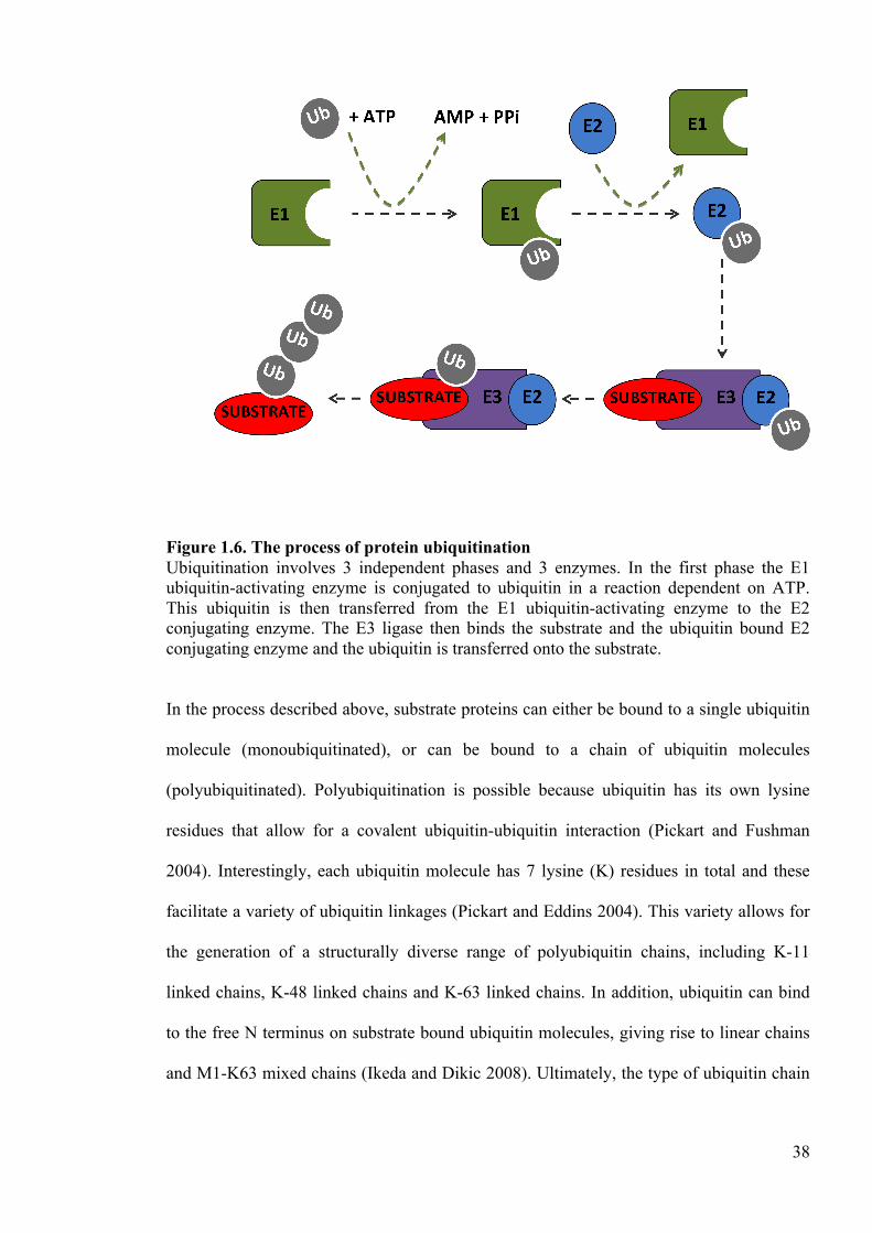
ubiquitination is a process whereby ubiquitin, an 8.5kDa protein, is covalently bound to
lysine residues on target proteins (Fig. 1.5). The mechanism of ubiquitination is a complex
one, involving 3 types of enzymes; namely an E1 ubiquitin-activating enzyme, an E2
ubiquitin-conjugating enzyme and an E3 ubiquitin ligase (Pickart 2001). In this multistep
process, ubiquitin is loaded onto the E1 ubiquitin-activating enzyme in a reaction requiring
ATP. The E2 ubiquitin-conjugating enzyme then binds both the E1 enzyme and the loaded
ubiquitin, and this ubiquitin is transferred onto the active site cysteine residue of the E2
enzyme. In the final step, the E3 ubiquitin ligase binds to both the ubiquitin-loaded E2
ubiquitin-conjugating enzyme and the substrate protein, and this results in the transfer of
ubiquitin from the E2 enzyme to the lysine residues on the target protein. In an additional
layer of regulation, the process of ubiquitination can be reversed by another set of enzymes
called deubiquitinases (Love et al. 2007).
38
Figure 1.6. The process of protein ubiquitination Ubiquitination involves 3 independent phases and 3 enzymes. In the first phase the E1 ubiquitin-activating enzyme is conjugated to ubiquitin in a reaction dependent on ATP. This ubiquitin is then transferred from the E1 ubiquitin-activating enzyme to the E2 conjugating enzyme. The E3 ligase then binds the substrate and the ubiquitin bound E2 conjugating enzyme and the ubiquitin is transferred onto the substrate.
In the process described above, substrate proteins can either be bound to a single ubiquitin
molecule (monoubiquitinated), or can be bound to a chain of ubiquitin molecules
(polyubiquitinated). Polyubiquitination is possible because ubiquitin has its own lysine
residues that allow for a covalent ubiquitin-ubiquitin interaction (Pickart and Fushman
2004). Interestingly, each ubiquitin molecule has 7 lysine (K) residues in total and these
facilitate a variety of ubiquitin linkages (Pickart and Eddins 2004). This variety allows for
the generation of a structurally diverse range of polyubiquitin chains, including K-11
linked chains, K-48 linked chains and K-63 linked chains. In addition, ubiquitin can bind
to the free N terminus on substrate bound ubiquitin molecules, giving rise to linear chains
and M1-K63 mixed chains (Ikeda and Dikic 2008). Ultimately, the type of ubiquitin chain
39
attached is fundamental in determining the functional outcome of the modification (Dikic
et al. 2009).
The most well known function of protein ubiquitination, and specifically of K-48 linked
polyubiquitination, is to target substrates for proteasomal degradation (Wilkinson 2000).
The ubiquitin-proteasome system (UPS) orchestrates the degradation of around 80% of all
intracellular proteins and thus is considered the most important mechanism for the
regulation of protein turnover (Hochstrasser 1995). The turnover of intracellular proteins is
essential as it prevents the potentially harmful accumulation of proteins, it recycles amino
acids for de novo protein synthesis, it removes misfolded proteins and it regulates a
number of other important cellular processes. Proteasomal degradation is mediated by a
large (2000kDa) multiprotein complex called the proteasome (Voges et al. 1999). In
eukaryotic cells, this complex contains a 20S subunit core that is capped by two 19S
subunits (Walz et al. 1998). The two 19S subunits contain ubiquitin-binding domains that
recognise ubiquitin labelled proteins and ATPases that unfold the proteins labelled for
degradation. The 20S subunit core is a hollow, barrel-shaped structure that catalyses the
proteolytic breakdown of unfolded proteins to their constitutive amino acid components
(Adams 2003). In the context of inflammation, there is a growing body of evidence
demonstrating that the UPS functions as an important regulatory process (Zinngrebe et al.
2014). Specifically, it has been shown that the K-48 linked polyubiquitination and
proteasomal degradation of IκB facilitates the translocation of NFκB (Scherer et al. 1995).
As the translocation of NFκB drives IL-1 expression (Baeuerle and Baltimore 1988b), K-
48 linked polyubiquitination is considered to be pivotal to the process of IL-1 up-
regulation (Fig. 1.6).
40
Figure 1.7. The role of protein ubiquitination in the NFκB signaling pathway Ubiquitination is implicated throughout the NFκB pathway. Following TLR stimulation, the E3 ligase TRAF-6 induces the formation of K-63-linked polyubiquitin chains on IRAK2. These chains function as a scaffold for the recruitment of the TAK1–TAB1–TAB2/3 complex and the IKK complex. Activation of these complexes leads to the K-48 polyubiquitination and subsequent degradation of IκB, facilitating NFκB translocation and pro-IL-1 expression. The functional implications of the other forms of protein ubiquitination are not as well
defined as they are for K-48 linked polyubiquitination. This is mainly because the roles of
these modifications are dependent on a variety of factors such as the nature and subcellular
location of the substrate. Intriguingly, it is becoming increasingly apparent that these other
forms of protein ubiquitination are also important regulators of the innate immune system,
and more specifically, the inflammatory response (Malynn and Ma 2010). Like K-48
41
linked polyubiquitination, these other modifications are also involved in the transduction
of the NFκB signaling pathway and thus the up-regulation of IL-1. As discussed
previously, the ubiquitin ligase TRAF6 is particularly important in the canonical NFκB
signaling pathway (Qian et al. 2001). In this role, the TRAF6-induced K-63-linked
polyubiquitin chains on IRAK2 function as a scaffold for the recruitment of the TAK1–
TAB1–TAB2/3 complex and the IKK complex (Kanayama et al. 2004b, Deng et al. 2000).
Ultimately, the ubiquitin-dependent recruitment of these complexes to IRAK1 is central to
NFκB induced IL-1 expression.
In addition, other forms of polyubiquitination have also been associated with the NFκB
signaling pathways. Of particular interest is the linear ubiquitin assembly complex
(LUBAC), which has recently been shown to be required for NFκB activation (Verhelst et
al. 2011) (Iwai and Tokunaga 2009). As its name suggests, LUBAC is an important
ubiquitin ligase complex that generates linear polyubiquitin chains (Stieglitz et al. 2012).
Interestingly, Ikeda et al. demonstrated that murine macrophages that do not express the
SHARPIN subunit of LUBAC are unable to phosphorylate IκB in response to LPS and are
therefore unable to induce NFκB translocation (Ikeda et al. 2011). In support of this,
Sasaki et al. also showed that when the E3 ligase activity of LUBAC is removed, LPS-
induced NFκB activation is impaired in murine B cells (Sasaki et al. 2013). Although it is
clear from the evidence presented above that linear ubiquitin chains are required for the
transduction of NFκB signaling pathways, more work is needed to identify the precise
roles of this modification.
A role for ubiquitination in the assembly of the inflammasome has also been described. In
these studies, NLRP3 was shown to be bound to K-63 linked polyubiquitin chains in
resting macrophages, but not in LPS and ATP treated macrophages (Py et al. 2013).
42
Importantly, this investigation identified BRCC3 as the deubiquitinase that deubiquitinates
NLRP3, and showed that this deubiquitination was required for NLRP3 activation and IL-
1β maturation. In addition, studies have demonstrated that caspase-1 is also subject to K-63
polyubiquitination, and have shown that this ubiquitination facilitates the activation of
caspase-1 and thus the maturation of IL-1 (Labbe et al. 2011). Overall, the process of
ubiquitination is rapidly emerging as a key event in both the up-regulation and the
processing of IL-1, and is therefore a subject of increasing interest.
1.2 Aims
From the evidence presented above, it is clear that IL-1 cytokines function as central
mediators of innate immunity and inflammation. Therefore, the mechanisms that serve to
regulate the potency and vigour of IL-1 release are of great academic and therapeutic
interest and importance. Whereas many previous studies have focused on the mechanisms
that drive IL-1 secretion, the overall aims of this thesis were to investigate the processes
that regulate the intracellular precursors of IL-1 (pro-IL-1α and pro-IL-1β). The hypothesis
here was that regulation of these precursors may serve to control the vigour of IL-1
secretion and, ultimately, may influence the potency of pro-inflammatory responses.
The first aim of the current project was to investigate the role of protein ubiquitination in
the regulation of pro-IL-1α and pro-IL-1β expression. This aim has been addressed in
paper 1, which is entitled “Dendritic Cell IL-1α and IL-1β Are Polyubiquitinated and
Degraded by the Proteasome.” In this paper, the ubiquitination and degradation of IL-1
was investigated in murine bone marrow derived DC, murine bone marrow derived
macrophages and a murine macrophage cell line. In these investigations, LPS and
polyinosinic-polycytidylic acid were used to up-regulate pro-IL-1 and ATP was used to
43
induce IL-1 secretion. IL-1 degradation was assessed using small molecule inhibitors of
the proteasome, and ubiquitination was assessed using co-immunoprecipitation and
Western blotting techniques.
The second aim was to determine whether the rate of pro-IL-1β ubiquitination and
degradation changes depending upon local circumstance, and if so, whether these changes
serve to regulate the vigour of IL-1 protein expression. This aim has been addressed in
paper 2, which is entitled “TLR Stimulation Inhibits pro-IL-1β Polyubiquitination and
Degradation.” This study employed 2 stably expressing fluorescent pro-IL-1β cell- lines;
venus pro-IL-1β expressing murine immortalised bone marrow derived macrophages and
venus pro-IL-1β expressing human THP-l cells. As venus IL-1β is stably expressed in these
cell lines, changes in the levels of cellular fluorescence could be used to track changes in
the rate of IL-1β degradation. Thus, in this investigation, these cells were used to examine
the factors that modulate the rate of pro-IL-1β degradation.
The final aim of this PhD was to investigate the interactome of pro-IL-1β. The hypothesis
here was that the identification of pro-IL-1β-interacting proteins may elucidate novel
mechanisms of pro-IL-1β regulation. It was also postulated that this approach may identify
the specific proteins responsible for the ubiquitination of IL-1β. This aim has been
addressed in paper 3, which is entitled “Interleukin-1β Processing is Dependent upon a
Calcium-Mediated Interaction with Calmodulin.” In these experiments, a human proteome
microarray containing 19,951 unique proteins was used to identify proteins that bind
human recombinant pro-IL-1β. Protein binding assays were also used to confirm
interactions and to investigate their nature in more detail. Finally, the functional
implications of these interactions were analysed in primary human monocytes and the
human THP-1 cell line. In these experiments, LPS was used to induce pro-IL-1β

44
expression, nigericin was utilised to induce processing and secretion, and small molecule
inhibitors were used to investigate the functional impact of the interactions.
45
1.3 Alternative format
The thesis is being presented in the alternative format in accordance with the rules and
regulations of the University of Manchester. The rational for presenting these results in the
alternative format is that we believe that the work done forms 3 independent research
papers of publication quality. The three results chapters presented herein are in manuscript
form, and are presented in the style of the publishing journal or intended journal of
submission. However, elements have been reformatted to ensure these chapters form a
cohesive body of work. In addition, a chapter containing supplemental data has also been
added. Below are the details of each manuscript, its publishing journal or intended journal
of submission, and contribution of each author to the work presented.
Chapter 2: Dendritic Cell IL-1α and IL-1β Are Polyubiquitinated and Degraded by the
Proteasome
Authors: Joseph S Ainscough, G. Frank Gerberick, Maryam Zahedi-Nejad, Gloria Lopez-
Castejon, David Brough, Ian Kimber and Rebecca J Dearman
Publishing journal: Journal of Biological Chemistry
Contribution of authors: This manuscript is representative of experiments of which I
contributed the vast majority. Work to investigate the degradation of IL-1 in macrophages
was completed by the third and fourth authors (Maryam Zahedi-Nejad and Gloria Lopez-
Castejon). My three supervisors Dr Frank Gerberick, Dr. Rebecca Dearman and Prof. Ian
Kimber, provided advice and guidance on all experimental work. Dr. David Brough and
Dr. Gloria Lopez-Castejon provided advice and helped with the interpretation of the
46
results. As first author on this paper, I was fully responsible for writing the text of the
manuscript. The manuscript was reviewed and commented on by all co-authors. These
comments were then synthesised by myself to produce the final manuscript.
Chapter 3: TLR Stimulation Inhibits pro-IL-1β Polyubiquitination and Degradation
Authors: Joseph S Ainscough, James Bagnall, Pawel Paszek, G. Frank Gerberick, Ian
Kimber and Rebecca J Dearman
Intended journal: Immunology
Contribution of authors: This manuscript is representative of experiments for which I am
solely responsible. My three supervisors Dr. Gerberick, Dr. Dearman and Prof. Kimber
provided advice and guidance on all experimental work. Dr. Paszek provided advice and
guidance with the transgenic work and Dr. Bagnall helped to genereate the transgenic
venus IL-1βTHP-1 cell line. As first author on this paper, I was fully responsible for
writing the text of the manuscript. The manuscript was reviewed and commented on by all
co-authors. These comments were then synthesised by myself to produce the final
manuscript.
Chapter 4: Interleukin-1β Processing is Dependent upon a Calcium-Mediated Interaction
with Calmodulin
Authors: Joseph S Ainscough, G. Frank Gerberick, Ian Kimber and Rebecca J Dearman
47
Journal of submission: Journal of Biological Chemistry
Contribution of authors: This manuscript is representative of experiments for which I am
solely responsible. My three supervisors Dr. Gerberick, Dr. Dearman and Prof. Kimber
provided advice and guidance on all experimental work. As first author on this paper, I was
fully responsible for writing the text of the manuscript. The manuscript was reviewed and
commented on by all co-authors. These comments were then synthesised by myself to
produce the final manuscript.

48
CHAPTER 2:
DENDRITIC CELL IL-1α AND IL-1β ARE POLYUBIQUITINATED
AND DEGRADED BY THE PROTEASOME
49
2 Paper 1: Dendritic Cell IL-1α and IL-1β are Polyubiquitinated and
Degraded by the Proteasome
Joseph S. Ainscough1, G. Frank Gerberick2, Maryam Zahedi-Nejad1, Gloria Lopez-
Castejon1, David Brough1, Ian Kimber1 and Rebecca J. Dearman1
1 Faculty of Life Sciences, University of Manchester, Manchester.
2The Procter & Gamble Company Co., Cincinnati, OH 45253, USA
Running title: Ubiquitination and proteasomal degradation of IL-1 in DC
Key words: Dendritic cell, inflammation, proteasome, ubiquitination, IL-1α, IL-1β
Paper published in the Journal of Biological chemistry (JBC; 2014)
2.1 Abbreviations
BM Bone marrow CHX Cycloheximide DAMP Damage Associated Molecular Patterns DC Dendritic cells HRPT Hypoxanthine-guanine phosphoribosyltransferase NF-κB Nuclear factor kappa-light-chain-enhancer of activated B cells PAMP Pathogen Associated Molecular Patterns TLR TOLL like receptor WCL Whole cell lysate
2.2 JBC Standard abbreviations
ATP Adenosine triphosphate cDNA Complementary deoxyribonucleic acid DMSO Dimethyl sulfoxide ELISA Enzyme-linked immunosorbent assay EDTA Ethylenediaminetetraacetic acid FCS Fetal calf serum GM-CSF Granulocyte macrophage-colony stimulating factor
50
Ig Immunoglobulin IL Interleukin LC3 Light chain 3 LPS Lipopolysaccharide mRNA Messenger Ribonucleic acid PMSF Phenylmethanesulfonylfluoride PBS Phosphate buffered saline PCR Polymerase chain reaction RT-PCR Reverse transcription polymerase chain reaction RNA Ribonucleic acid SEM Standard error of the mean TNF Tumor necrosis factor
2.3 Capsule
Background: Interleukin-1 secretion is an important process in inflammation and thus, the
intracellular regulation of these cytokines is of interest.
Results: Inhibition of the proteasome in dendritic cells inhibits Interleukin-1 degradation
and leads to an accumulation of polyubiquitinated Interleukin-1.
Conclusion: Interleukin-1 cytokines are regulated by polyubiquitination and proteasomal
degradation.
Significance: Polyubiquitination and degradation are important processes in the
intracellular regulation of Interleukin-1.
2.4 Abstract
IL-1α and β are key players in the innate immune system. The secretion of these cytokines
by dendritic cells (DC) is integral to the development of proinflammatory responses. These
cytokines are not secreted via the classical secretory pathway. Instead, 2 independent
processes are required; an initial signal to induce up-regulation of the precursor pro-IL-1α
and β, and a second signal to drive cleavage and consequent secretion. Pro-IL-1α and β are
both cytosolic and thus, are potentially subject to post-translational modifications. These
51
modifications may, in turn, have a functional outcome in the context of IL-1α and β
secretion and hence inflammation. We report here that IL-1α and β were degraded
intracellularly in murine bone marrow derived DC and that this degradation was dependent
on active cellular processes. In addition, we demonstrate that degradation was ablated
when the proteasome was inhibited, whereas autophagy did not appear to play a major
role. Further, inhibition of the proteasome led to an accumulation of polyubiquitinated IL-
1α and β, indicating that IL-1α and β were polyubiquitinated prior to proteasomal
degradation. Finally, our investigations suggest that polyubiquitination and proteasomal
degradation are not continuous processes but instead are upregulated following DC
activation. Overall, these data highlight that IL-1α and β polyubiquitination and
proteasomal degradation are central mechanisms in the regulation of intracellular IL-1
levels in DC.
2.5 Introduction
Dendritic cells (DC) are of fundamental importance to the immune system, playing pivotal
roles in the initiation and orchestration of immune responses (Steinman and Banchereau
2007, Banchereau and Steinman 1998). They serve as dynamic antigen presenting cells
that bridge the innate and adaptive immune systems. Thus, DC survey the local
microenvironment, discriminating between a broad range of pathogenic and non-
pathogenic cues and initiating immune responses, including inflammation (Mellman and
Steinman 2001). Inflammation is a complex response of the innate immune system that is
associated with 5 characteristic features; erythema, edema, heat, pain, and loss of function
(Medzhitov 2010, Netea et al. 2009). These symptoms, which are crucial for the resolution
of infection and injury, occur as a result of a series of changes driven by the production of
proinflammatory cytokines, including members of the interleukin-1 (IL-1) family. IL-1α
52
and IL-1β are closely related potent proinflammatory members of the IL-1 family and are
produced by DC (Dinarello 2009). The secretion of these cytokines is an integral
component of the role of DC in orchestrating immune and inflammatory responses.
Therefore, the transcriptional and post-transcriptional regulation of IL-1 by DC is of
considerable importance, not only in the context of the resolution of infection and injury,
but also in the context of preventing inappropriate or excessive inflammatory reactions.
This is evident in pathologies such as gout (Chen et al. 2006), rheumatoid arthritis (Buchan
et al. 1988), cancer (Apte et al. 2006) and dementia (Cacabelos et al. 1994), where the
dysregulation of IL-1 is implicated strongly.
Unlike most cytokines, IL-1α and IL-1β are not secreted via the classical secretory
pathway. Instead, IL-1 is secreted via a non-conventional pathway and its release requires
two independent signals. The first signal is typically provided by Pathogen Associated
Molecular Patterns (PAMP) that act via pattern recognition receptors, such as members of
the TOLL-like receptor (TLR) family, to stimulate complex signaling pathways (Takeuchi
and Akira 2010, Devaraj et al. 2008). Ultimately, the activation of these pathways results
in the translocation of the transcription factor nuclear factor kappa-light-chain-enhancer of
activated B cells (NF-κB) to the nucleus. This drives the transcription of a variety of pro-
inflammatory proteins, including IL-1α, IL-1β and IL-6 (Cogswell et al. 1994), with IL-1α
and IL-1β being transcribed as 31kd precursors (pro-IL-1). The secretion of these
cytokines requires a second signal, which is provided usually by additional PAMP or
molecules associated with tissue trauma or damage (Damage Associated Molecular
Patterns; DAMP). These DAMP signal via cytosolic pattern recognition receptors,
typically of the NOD-like receptor family, to stimulate assembly of inflammasome
complexes (Franchi et al. 2009). Formation of the inflammasome complex induces
activation of the enzyme caspase-1 (Latz et al. 2013). Active caspase-1 cleaves pro-IL-1β
53
to the 17kd bioactive form, facilitating its secretion (Watanabe et al. 2007). The
inflammasome is also thought to be involved in IL-1α secretion, although this process is
less well characterized and is also dependent on the calcium-dependent protease calpain.
Similarly, the processing and secretion of pro-IL-1α involves cleavage into its 17kd form
(Mikami et al. 2011, Watanabe and Kobayashi 1994).
Although many studies have focused on the regulation of IL-1 secretion, the intracellular
control of pro-IL-1 remains poorly understood. Both pro-IL-1α and pro-IL-1β are cytosolic
and may therefore be subject to a number of post-translational modifications. Such
modifications represent an important mechanism by which cells can regulate the
characteristics and function of proteins. For example, post-translational modifications have
been shown to act as regulators at various stages of the NF-κB signaling pathway (Perkins
2006). Ubiquitination involves the addition of ubiquitin, an 8.5kd protein, to a given
substrate (Komander 2009). This process is mediated by a series of enzymes; E1, E2 and
E3, that act sequentially to bind ubiquitin covalently to a Lys residue on the substrate
protein. It is the final E3 ubiquitin ligase that confers the substrate specificity for
ubiquitination. Substrate proteins may remain monoubiquitinated or may have further
ubiquitin molecules added (polyubiquitination). In the formation of polyubiquitin chains,
the carboxyl group on ubiquitin can bind to a number of different residues on the following
ubiquitin, thus giving rise to various forms of polyubiquitination. Ultimately, the type of
ubiquitin chain bound has a fundamental impact on the functional outcome of the
modification (Welchman et al. 2005). As an example, Lys 48-linked polyubiquitin chains
serve to target proteins for proteasomal degradation (Thrower et al. 2000) whereas Lys-63-
linked polyubiquitin chains function in the regulation of the NF-κB pathway (Wu et al.
2006).
54
Here, we provide evidence that in murine DC, IL-1α and IL-1β are polyubiquitinated and
that, in both DC and macrophages, this polyubiquitination drives the proteasomal
degradation of IL-1. Furthermore, these data demonstrate that in the presence of a second
signal, polyubiquitinated IL-1 is still available for secretion. Collectively, our results
demonstrate that in DC, the polyubiquitination and proteasomal degradation of IL-1 serves
as an essential process in the regulation of IL-1 and, therefore, should be considered as an
extra dimension to the current two-signal paradigm of IL-1 release.
2.6 Experimental procedures
Animals
Female BALB/c mice (6-8 weeks old) were used throughout these experiments (Harlan
Olac, Bicester, UK). Mice were provided with environmental stimuli (bedding and nesting
materials); food (SDS PCD pelleted diet; Special Diets Services Ltd, Witham, UK) and
water were available ad libitum. Relative humidity was 55 ± 10% with a 12h light/dark
cycle and ambient temperature maintained at 21 ± 2°C. Maintenance and treatment of
animals were conducted as specified by the U.K. Animals (Scientific Procedures) Act
1986. Mice were sacrificed by exposure to CO2 gas in rising concentration followed by
dislocation of the neck in concordance with schedule 1 (U.K. Animals [Scientific
Procedures] Act 1986).
Antibodies and reagents
LPS from Escherichia coli serotype 055:B5 (TLR2/4), Poly(I:C), ATP, the autophagy
inhibitor wortmanin and the translation inhibitor cycloheximide (CHX) were purchased
55
from Sigma Chemical Co (Poole, UK). The proteasome inhibitor MG132 was obtained
from Merck Millipore (Billerica, MA, USA). Recombinant murine pro-IL-1β was
purchased from Affymetrix eBioscience (San Diego, CA). For Western-blot analysis, the
primary antibodies were goat anti-mouse IL-1α antibody (AF401-NA0, goat anti-mouse
IL-1β antibody (AF401-NA; both R&D systems; Minneapolis, MN, USA) or mouse anti-
ubiquitin antibody (SC8017; Santa Cruz Biotechnology; Santa Cruz, CA, USA). The HRP-
conjugated secondary antibodies were rabbit anti-goat IgG antibody (P0449; DAKO;
Copenhagen, Denmark) and goat anti-mouse light chain antibody (AP200P; Millipore).
Generation and culture of murine bone marrow derived DC
Murine bone marrow derived (BM)DC were generated following a previously described
method (Lutz et al. 1999). Briefly, bone marrow was extracted by flushing the tibias and
femurs with PBS. The cell suspension was centrifuged at 200g for 5 min at room
temperature. The remaining pellet was resuspended in pre-warmed, FCS-supplemented
culture medium (RPMI 1640; GIBCO, Paisley, UK), containing 400 µg/ml penicillin/
streptomycin, 292 µg/ml L-glutamine, 0.05 mM 2- mercaptoethanol, 4 ng/ml GM-CSF
(Miltenyi Biotech, Bisley, UK) and 10% FCS (GIBCO). A viable cell count was
performed by trypan blue exclusion (0.5%; Sigma). Cells were cultured at approximately
2x10⁶ cells/ml in petri dishes and incubated at 37oC. The cultures were fed on day 3 by
addition of 10 ml of fresh culture medium, and again on day 6 by gentle aspiration of 10
ml of medium followed by the addition of 10 ml fresh culture medium.
BMDC treatments
56
BMDC were plated on day 8, in culture medium without GM-CSF, at 106 cells/well (24-
well plate) or 107 cells/well (6-well plate; 106 cells/ml). Following an initial 24h dose
response experiment to determine the optimum dose of LPS to induce IL-1 production,
cells were primed using 0.1 µg/ml LPS. BMDC were primed with LPS as indicated in the
text, and were activated with various concentrations of ATP for 30 min at the end of the
culture. MG132, wortmanin or a DMSO control was added for the final 4h of incubation.
CHX was added for the final 1h of incubation. After incubation, supernatants were
harvested and frozen at -80°C. Cell lysates were harvested in 200 µl of lysis buffer (20 mM
Tris HCl, 137 mM NaCl, 20 mM EDTA, 10% glycerol, 0.5% Ipegal, 1 mM PMSF,
protease inhibitor cocktail [1:100]) and frozen at -80°C. For PCR analysis, lysates were
prepared for RNA extraction following the manufacturer’s instructions (Purelink RNA
mini kit; Life Technologies, Carlsbad, CA, USA).
Immunoprecipitation of IL-1
To prepare lysates for immunoprecipitation, supernatants were removed and cells washed
twice with PBS. Cells were incubated on ice with wash buffer (20 mM N-
ethylmaleimide in PBS). After a final wash with PBS, cell lysates were prepared in 500µl
of a specialized lysis buffer, formulated to prevent deubiquitination (25 mM Tris pH 7.4,
150 mM NaCl, 0.5% Na-deoxycholate, 1% TritonX-100, 0.1% sodium dodecyl sulfate, 5
mM N-ethylmaleimide , 1 mM PMSF). An aliquot of lysate (50 µl) was retained as whole
cell lysate (WCL) and the remainder was immunoprecipitated using an anti-IL-1α or anti-
IL-1β antibody (both antibodies were capture antibodies supplied in the R&D ELISA
Duosets). Briefly, samples were incubated overnight with 2 µg of antibody at 4°C. Protein
G sepharose beads (50 µl; Sigma) were added to each sample for 2h at 4°C. After
incubation, the samples were washed three times by centrifugation at 10000xg for 30 s and
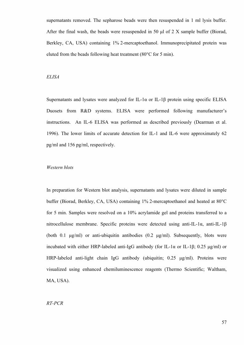
57
supernatants removed. The sepharose beads were then resuspended in 1 ml lysis buffer.
After the final wash, the beads were resuspended in 50 µl of 2 X sample buffer (Biorad,
Berkley, CA, USA) containing 1% 2-mercaptoethanol. Immunoprecipitated protein was
eluted from the beads following heat treatment (80°C for 5 min).
ELISA
Supernatants and lysates were analyzed for IL-1α or IL-1β protein using specific ELISA
Duosets from R&D systems. ELISA were performed following manufacturer’s
instructions. An IL-6 ELISA was performed as described previously (Dearman et al.
1996). The lower limits of accurate detection for IL-1 and IL-6 were approximately 62
pg/ml and 156 pg/ml, respectively.
Western blots
In preparation for Western blot analysis, supernatants and lysates were diluted in sample
buffer (Biorad, Berkley, CA, USA) containing 1% 2-mercaptoethanol and heated at 80°C
for 5 min. Samples were resolved on a 10% acrylamide gel and proteins transferred to a
nitrocellulose membrane. Specific proteins were detected using anti-IL-1α, anti-IL-1β
(both 0.1 µg/ml) or anti-ubiquitin antibodies (0.2 µg/ml). Subsequently, blots were
incubated with either HRP-labeled anti-IgG antibody (for IL-1α or IL-1β; 0.25 µg/ml) or
HRP-labeled anti-light chain IgG antibody (ubiquitin; 0.25 µg/ml). Proteins were
visualized using enhanced chemiluminescence reagents (Thermo Scientific; Waltham,
MA, USA).
RT-PCR
58
Total RNA was purified from samples using Purelink RNA mini kit and converted to
cDNA using a high capacity RNA to cDNA kit (Life Technologies). mRNA expression
levels of mouse IL-1β were determined by RT-PCR using a Taqman primer obtained from
Life Technologies using a RT-PCR machine (StepOne plus). Expression was normalized
using untreated cells (control) and to hypoxanthine-guanine phosphoribosyltransferase
(HRPT), with the ΔΔ cyclic threshold method used to calculate relative fold change.
Statistical analysis
Statistical analysis was performed using the software Graphpad Prism 6. Data were
analyzed by one-way ANOVA to determine overall differences and a Tukey post-hoc test
was performed to determine statistically significant differences between treatment groups.
* = p<0.05, ** = p< 0.01.
2.7 Results
Pro-IL-1α and pro-IL-1β are degraded by an active cellular process
In initial experiments, it was confirmed that the regulation of IL-1 production in DC was
consistent with the current paradigm of IL-1 secretion (Lopez-Castejon and Brough 2011),
requiring two signals. Stimulation of BMDC with the TLR 4 ligand LPS resulted in an up-
regulation in intracellular (lysate) expression of both pro-IL-1α and pro-IL-1β protein,
without inducing detectable secretion (fig. 1A). The intracellular IL-1 induced by LPS was
exclusively 31kd in size (corresponding to the IL-1 precursor, pro-IL-1) as determined by
Western blot analysis (fig. 1D). In subsequent experiments signal 2 was provided, by ATP.
59
ATP acts via the P2X7 receptor to induce activation of the NLRP3 inflammasome
(Ghiringhelli et al. 2009, Ferrari et al. 1997, Mariathasan et al. 2006) and thus the cleavage
and secretion of IL-1. Here, challenge of LPS-primed BMDC with ATP (1 to 10 mM)
resulted in IL-1α (fig. 1B & E) and IL-1β (fig. 1C & F) processing and release.
Figure 1. Intracellular expression and secretion of IL-1 by BMDC: impact of LPS and ATP BMDC (106 cells/ml) were cultured for 24h in the presence of media alone or increasing doses of LPS (A, D) or for 4h with media alone or LPS (0.1µg/ml) and then challenged with ATP (0-10mM) for 30 min (B, C, E, F). Supernatants and cell lysates were harvested and analyzed for the presence of IL-1α or IL-1β using cytokine specific ELISA. IL-1 content of cell lysates is displayed in A (!, IL-1α; □, IL-1β) and supernatants (secreted IL-1) in B (IL-1α) and C (IL-1β) as mean ± SEM (n=3). Supernatants (SN) and lysates (Lys) were also analyzed by Western blotting using either anti-IL-1α (D, E) or anti-IL-1β (D, F) antibodies. A protein marker lane on each gel was used to determine molecular weight. Representative blots are shown in each case.
One interesting observation made was that the total amount of IL-1β appeared to increase
dramatically following optimal ATP challenge (from ~5ng/106 cells to 100ng/106 cells).
Given that the ATP is only added for the final 30 min of incubation, it seemed unlikely that
this increase reflected an actual increase in protein expression. Indeed, under these
60
conditions IL-1β mRNA levels were unaffected by ATP stimulation (data not shown).
Consequently, it was hypothesized that the IL-1β ELISA used has a greater avidity for the
mature cytokine, relative to the 31kd precursor. To investigate this, recombinant pro-IL-1β
and the recombinant mature IL-1β were analyzed in parallel in the ELISA over a
concentration range of 300 to 1.2 pM, It was demonstrated that the ELISA used does
indeed have a considerably greater avidity for the mature cytokine, relative to the 31kd
precursor (data not shown).
To investigate the intracellular regulation of pro-IL-1α and β in BMDC, the kinetics of IL-
1 protein expression in response to LPS alone was examined. Here, stimulation with LPS
caused an upregulation of both pro-IL-1 forms (fig. 2A and B). The expression of both IL-
1α and IL-1β were transient, peaking at 4h post-stimulation and decreasing thereafter. In
contrast, the classically secreted cytokine IL-6 (Rubartelli et al. 1990) was secreted almost
as soon as it was produced and accumulated in the supernatant (fig. 2C). Despite the
reduction in intracellular cytokine, IL-1 secretion was not detected at any time point,
indicating that both Il-1α and IL-1β were degraded intracellularly. Repeat experiments
confirmed that the loss of IL-1 between 4 and 48h was statistically significant (Fig. 2D;
p<0.01). Further, the process was temperature-dependent, such that incubation of LPS-
primed BMDC at 4oC, rather than 37oC, largely abrogated the effect (fig. 2D). As
incubation at 4oC effectively inhibits cellular metabolic activity, these data show that IL-1
degradation is dependent on an active, cellular process.
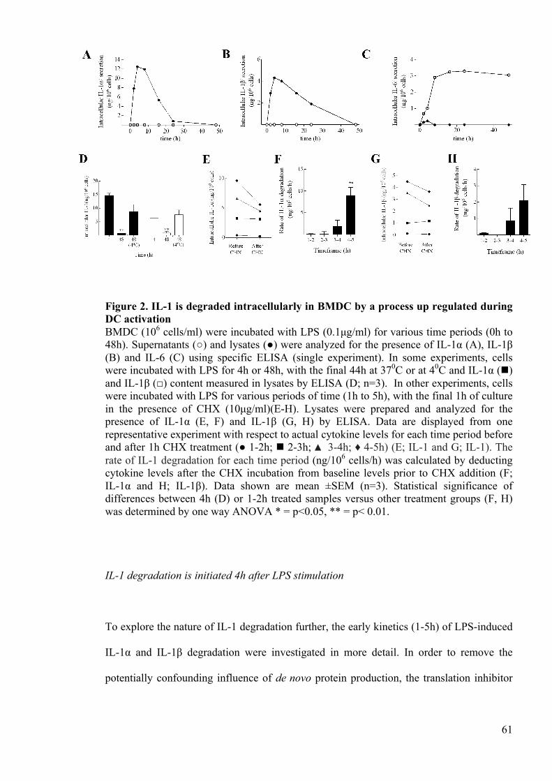
61
Figure 2. IL-1 is degraded intracellularly in BMDC by a process up regulated during DC activation BMDC (106 cells/ml) were incubated with LPS (0.1µg/ml) for various time periods (0h to 48h). Supernatants (○) and lysates (●) were analyzed for the presence of IL-1α (A), IL-1β (B) and IL-6 (C) using specific ELISA (single experiment). In some experiments, cells were incubated with LPS for 4h or 48h, with the final 44h at 370C or at 40C and IL-1α (!) and IL-1β (□) content measured in lysates by ELISA (D; n=3). In other experiments, cells were incubated with LPS for various periods of time (1h to 5h), with the final 1h of culture in the presence of CHX (10µg/ml)(E-H). Lysates were prepared and analyzed for the presence of IL-1α (E, F) and IL-1β (G, H) by ELISA. Data are displayed from one representative experiment with respect to actual cytokine levels for each time period before and after 1h CHX treatment (● 1-2h; ! 2-3h; ▲ 3-4h; ♦ 4-5h) (E; IL-1 and G; IL-1). The rate of IL-1 degradation for each time period (ng/106 cells/h) was calculated by deducting cytokine levels after the CHX incubation from baseline levels prior to CHX addition (F; IL-1α and H; IL-1β). Data shown are mean ±SEM (n=3). Statistical significance of differences between 4h (D) or 1-2h treated samples versus other treatment groups (F, H) was determined by one way ANOVA * = p<0.05, ** = p< 0.01.
IL-1 degradation is initiated 4h after LPS stimulation
To explore the nature of IL-1 degradation further, the early kinetics (1-5h) of LPS-induced
IL-1α and IL-1β degradation were investigated in more detail. In order to remove the
potentially confounding influence of de novo protein production, the translation inhibitor
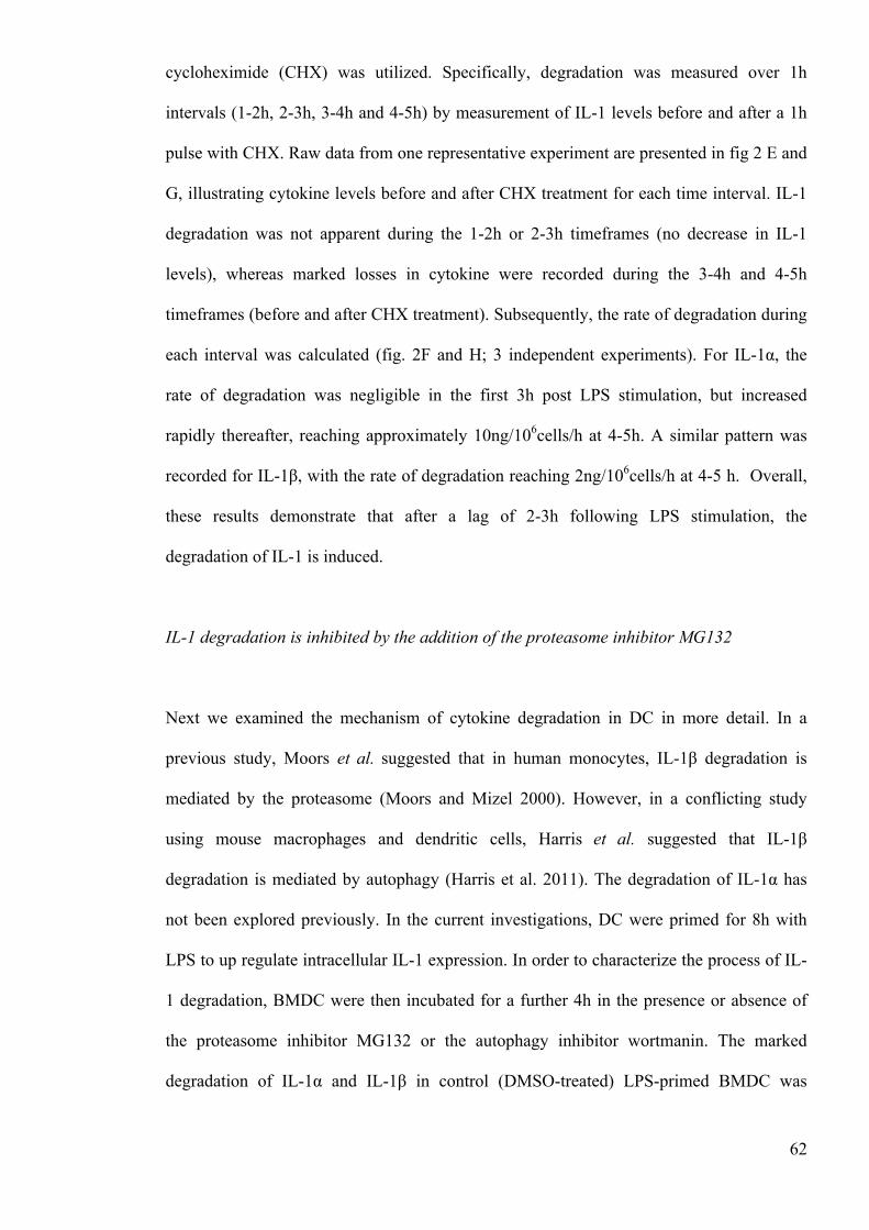
62
cycloheximide (CHX) was utilized. Specifically, degradation was measured over 1h
intervals (1-2h, 2-3h, 3-4h and 4-5h) by measurement of IL-1 levels before and after a 1h
pulse with CHX. Raw data from one representative experiment are presented in fig 2 E and
G, illustrating cytokine levels before and after CHX treatment for each time interval. IL-1
degradation was not apparent during the 1-2h or 2-3h timeframes (no decrease in IL-1
levels), whereas marked losses in cytokine were recorded during the 3-4h and 4-5h
timeframes (before and after CHX treatment). Subsequently, the rate of degradation during
each interval was calculated (fig. 2F and H; 3 independent experiments). For IL-1α, the
rate of degradation was negligible in the first 3h post LPS stimulation, but increased
rapidly thereafter, reaching approximately 10ng/106cells/h at 4-5h. A similar pattern was
recorded for IL-1β, with the rate of degradation reaching 2ng/106cells/h at 4-5 h. Overall,
these results demonstrate that after a lag of 2-3h following LPS stimulation, the
degradation of IL-1 is induced.
IL-1 degradation is inhibited by the addition of the proteasome inhibitor MG132
Next we examined the mechanism of cytokine degradation in DC in more detail. In a
previous study, Moors et al. suggested that in human monocytes, IL-1β degradation is
mediated by the proteasome (Moors and Mizel 2000). However, in a conflicting study
using mouse macrophages and dendritic cells, Harris et al. suggested that IL-1β
degradation is mediated by autophagy (Harris et al. 2011). The degradation of IL-1α has
not been explored previously. In the current investigations, DC were primed for 8h with
LPS to up regulate intracellular IL-1 expression. In order to characterize the process of IL-
1 degradation, BMDC were then incubated for a further 4h in the presence or absence of
the proteasome inhibitor MG132 or the autophagy inhibitor wortmanin. The marked
degradation of IL-1α and IL-1β in control (DMSO-treated) LPS-primed BMDC was
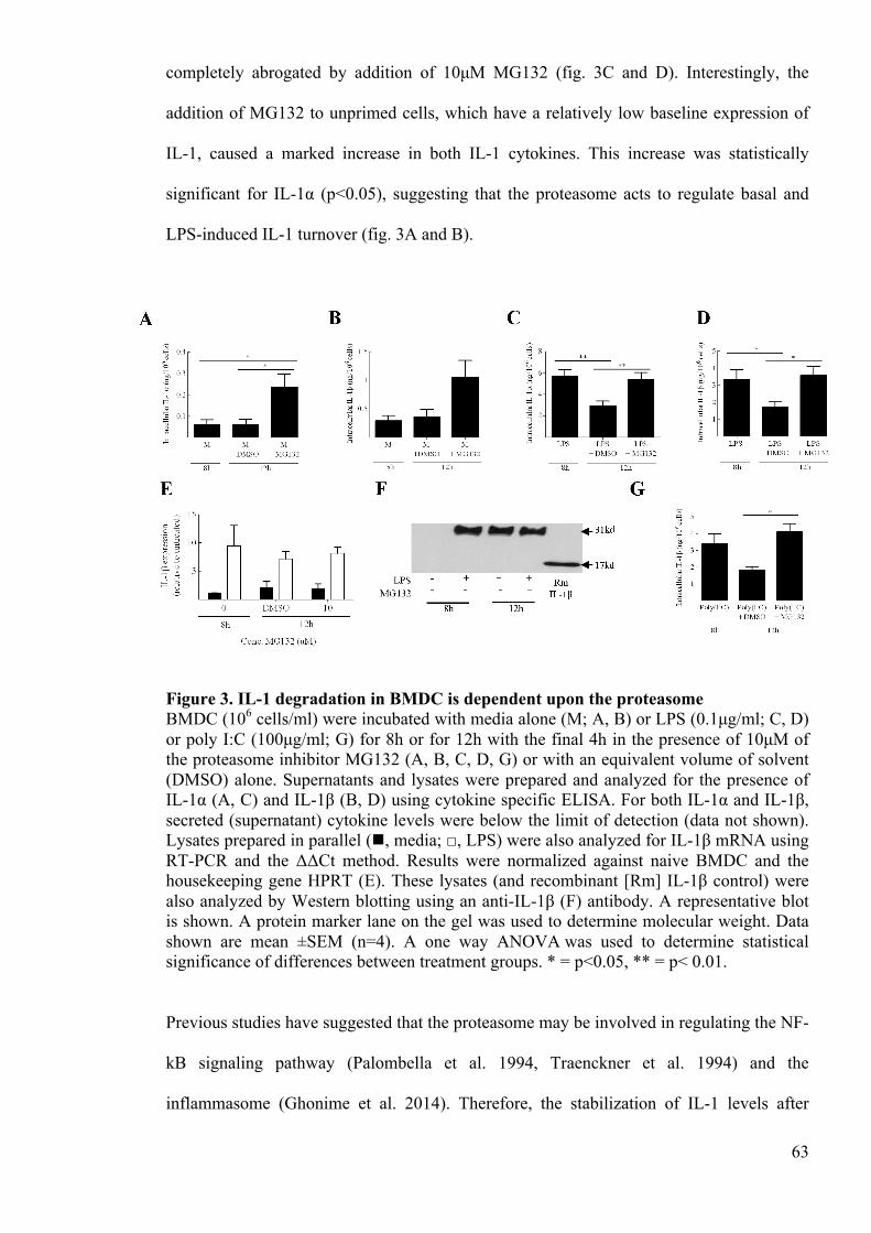
63
completely abrogated by addition of 10µM MG132 (fig. 3C and D). Interestingly, the
addition of MG132 to unprimed cells, which have a relatively low baseline expression of
IL-1, caused a marked increase in both IL-1 cytokines. This increase was statistically
significant for IL-1α (p<0.05), suggesting that the proteasome acts to regulate basal and
LPS-induced IL-1 turnover (fig. 3A and B).
Figure 3. IL-1 degradation in BMDC is dependent upon the proteasome BMDC (106 cells/ml) were incubated with media alone (M; A, B) or LPS (0.1µg/ml; C, D) or poly I:C (100µg/ml; G) for 8h or for 12h with the final 4h in the presence of 10µM of the proteasome inhibitor MG132 (A, B, C, D, G) or with an equivalent volume of solvent (DMSO) alone. Supernatants and lysates were prepared and analyzed for the presence of IL-1α (A, C) and IL-1β (B, D) using cytokine specific ELISA. For both IL-1α and IL-1β, secreted (supernatant) cytokine levels were below the limit of detection (data not shown). Lysates prepared in parallel (!, media; □, LPS) were also analyzed for IL-1β mRNA using RT-PCR and the ΔΔCt method. Results were normalized against naive BMDC and the housekeeping gene HPRT (E). These lysates (and recombinant [Rm] IL-1β control) were also analyzed by Western blotting using an anti-IL-1β (F) antibody. A representative blot is shown. A protein marker lane on the gel was used to determine molecular weight. Data shown are mean ±SEM (n=4). A one way ANOVA was used to determine statistical significance of differences between treatment groups. * = p<0.05, ** = p< 0.01.
Previous studies have suggested that the proteasome may be involved in regulating the NF-
kB signaling pathway (Palombella et al. 1994, Traenckner et al. 1994) and the
inflammasome (Ghonime et al. 2014). Therefore, the stabilization of IL-1 levels after
64
inhibition of the proteasome observed here may reflect increased IL-1 expression or
processing rather than an inhibition of IL-1 degradation. However, analysis of IL-1β
mRNA expression showed that the addition of MG132 to both untreated or LPS primed
BMDC was without effect on IL-1β mRNA levels (fig. 3E). Western blot analysis
confirmed that the IL-1 was the 31kd, precursor form regardless of addition of the
proteosomal inhibitor (fig. 3F). Thus, MG132 did not affect IL-1 processing or
transcription.
Figure 4. IL-1 degradation is dependent on the proteasome in macrophages J774 (A, B, C, E, F, G) or BMDM (D, H) (106 cells/ml) were incubated with LPS (1µg/ml) for 16h with final 4h in the presence of CHX (10µg/ml) and either DMSO, MG262 (A, E), ALLN (B, F) or β lactone (C, G) (all 0-50µM for J775 cells). For BM derived macrophages a single concentration of each proteasome inhibitor was used (50µM for ALLN; 30µM for both MG262 and β lactone). Supernatants and lysates were prepared and analyzed for the presence of IL-1α (A, B, C, D) and IL-1β (E, F, G, H) using cytokine specific ELISA. For both IL-1α and IL-1β, secreted cytokine levels were below the limit of detection (data not shown). Data shown are mean ±SEM (n=3). A one way ANOVA was used to determine statistical significance of differences between the DMSO treated samples versus the other treatment groups. * = p<0.05, ** = p< 0.01.
Harris et al. suggested that the sequestration of IL-1β into autophagosomes was dependent
on the TRIF signaling pathway. As LPS signals via both TRIF and MyD88 signaling
pathways, it was important to explore IL-1β degradation where the IL-1 expression was up

65
regulated via the TRIF signaling pathway only. TLR3 has been shown to signal via the
TRIF signaling pathway exclusively and so the TLR3 ligand poly(I:C) was used to
investigate degradation here. Once again, there was a marked degradation of IL-1β
between 8h and 12h (fig. 3G). Importantly, this degradation was completely inhibited by
proteasome inhibition, suggesting that IL-1 degradation under these conditions is
proteasomal, even when the upregulation is dependent on the TRIF signaling pathway.
In order to examine whether proteosomal degradation was a general feature of IL-1
production in cell types other than DC, parallel experiments were conducted using murine
BM derived macrophages and the macrophage cell line J774 cells (fig. 4). Cells were
primed with LPS for 16h, with the last 4h being in the presence of CHX (to inhibit further
IL-1 translation) alone or in the presence of various proteosomal inhibitors: MG262 (an
inhibitor from the same chemical series as MG132 (Kisselev and Goldberg 2001)), ALLN
or Β lactone (all at 1-50 µM formulated in DMSO, or DMSO alone control) and IL-1α or
IL-1β expression measured. There was no detectable secretion of cytokine, but LPS
priming of both primary macrophages and the cell line resulted in intracellular cytokine
expression (~2.5 ng/106 cells). For J774 cells, a dose dependent increase in cytokine
content was recorded in the presence of each of the three proteosomal inhibitors. Similar
increases in IL-1 were recorded at optimal doses (30-50 mM) of all three inhibitors in
parallel experiments conducted with BM derived macrophages. Thus, IL-1 degradation in
macrophages is also dependent upon the proteasome.
To investigate whether autophagy was implicated in IL-1 degradation, it was first
confirmed that under the conditions utilized, wortmanin was indeed an autophagy inhibitor
in BMDC. The addition of wortmanin to LPS-primed cells resulted in a dose-dependent
reduction in the conversion of LC3-I to LC3-II with complete inhibition observed at 10 µM
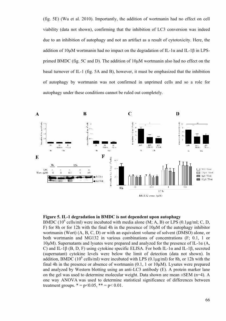
66
(fig. 5E) (Wu et al. 2010). Importantly, the addition of wortmanin had no effect on cell
viability (data not shown), confirming that the inhibition of LC3 conversion was indeed
due to an inhibition of autophagy and not an artifact as a result of cytotoxicity. Here, the
addition of 10µM wortmanin had no impact on the degradation of IL-1α and IL-1β in LPS-
primed BMDC (fig. 5C and D). The addition of 10µM wortmanin also had no effect on the
basal turnover of IL-1 (fig. 5A and B), however, it must be emphasized that the inhibition
of autophagy by wortmanin was not confirmed in unprimed cells and so a role for
autophagy under these conditions cannot be ruled out completely.
Figure 5. IL-1 degradation in BMDC is not dependent upon autophagy BMDC (106 cells/ml) were incubated with media alone (M; A, B) or LPS (0.1µg/ml; C, D, F) for 8h or for 12h with the final 4h in the presence of 10µM of the autophagy inhibitor wortmanin (Wort) (A, B, C, D) or with an equivalent volume of solvent (DMSO) alone, or both wortmanin and MG132 in various combinations of concentrations (F; 0.1, 1 or 10µM). Supernatants and lysates were prepared and analyzed for the presence of IL-1α (A, C) and IL-1β (B, D, F) using cytokine specific ELISA. For both IL-1α and IL-1β, secreted (supernatant) cytokine levels were below the limit of detection (data not shown). In addition, BMDC (106 cells/ml) were incubated with LPS (0.1µg/ml) for 8h, or 12h with the final 4h in the presence or absence of wortmanin (0.1, 1 or 10µM). Lysates were prepared and analyzed by Western blotting using an anti-LC3 antibody (E). A protein marker lane on the gel was used to determine molecular weight. Data shown are mean ±SEM (n=4). A one way ANOVA was used to determine statistical significance of differences between treatment groups. * = p<0.05, ** = p< 0.01.
67
To test whether autophagic and proteasomal pathways were synergistic in IL-1
degradation, we explored degradation under conditions of both proteasome and autophagy
inhibition (fig. 5F). However, these data indicate that IL-1 degradation under these
conditions was unaffected by autophagy inhibition, even under conditions of partial (1µM
MG132) or total proteasome inhibition (10µM MG132). Together, these data suggest that
degradation of both IL-1α and IL-1β over the period studied here was dependent upon the
proteasome in DC and macrophages.
Intracellular IL-1 is polyubiquitinated
To explore whether IL-1α and IL-1β conform to the classic paradigm of proteasomal
degradation, the ubiquitination status of IL-1 was investigated. Here, stimulation of BMDC
with LPS, or LPS in the presence of MG132 to block degradation, resulted in a strong pro-
IL-1α and pro-IL-1β signal, as determined by Western blot analysis of the WCL (fig. 6A
and B). Pro-IL-1α and IL-1β were successfully immunoprecipitated from these LPS-
primed BMDC lysates using appropriate antibodies, again as determined by Western
blotting. In both the anti-IL-1α and anti-IL-1β immunoprecipitated fractions, a band of
approximately 25kd was observed in control lanes (unprimed BMDC lysates and lysis
buffer-negative control). The size of this band is consistent with light chain IgG antibody
fragments and is therefore likely to be due cross reactivity between the
immunoprecipitation antibodies and the anti-IgG secondary antibody used for detection.
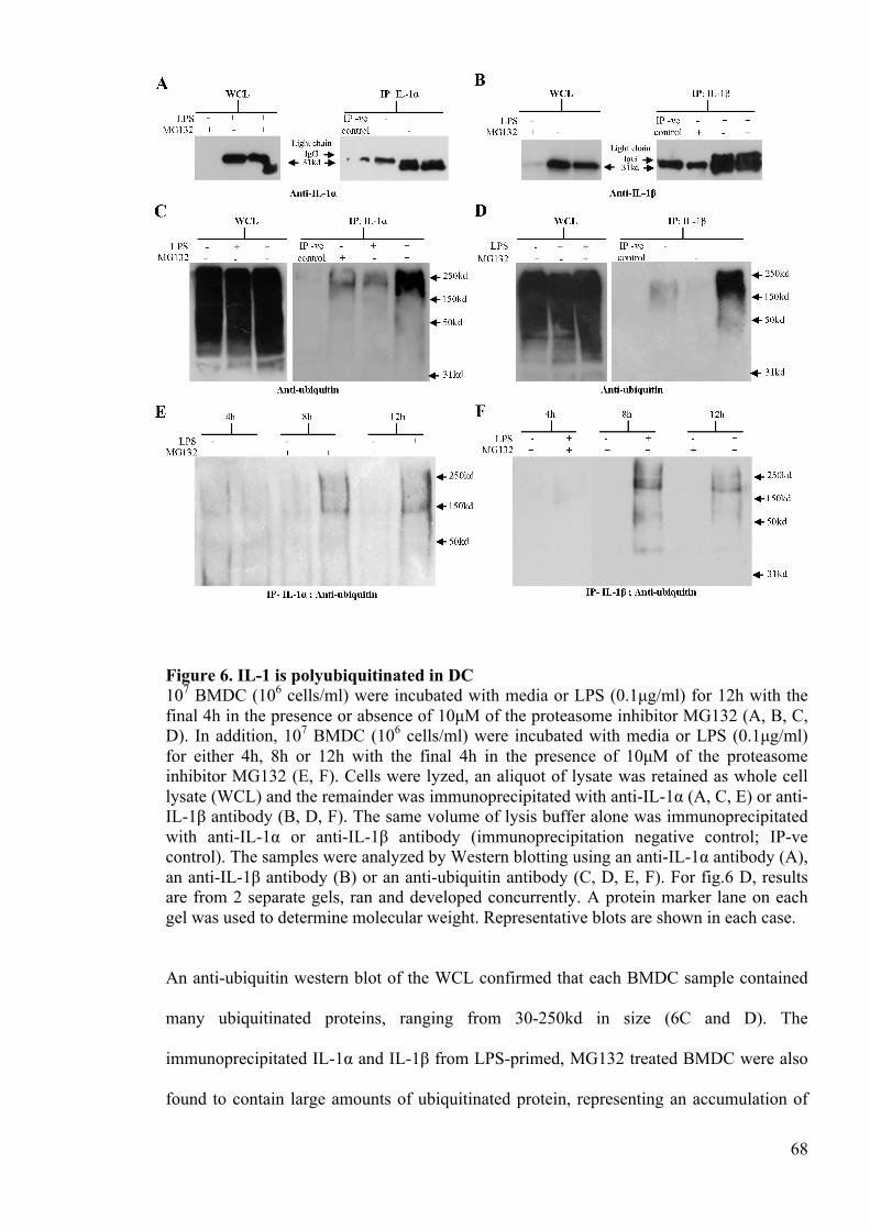
68
Figure 6. IL-1 is polyubiquitinated in DC 107 BMDC (106 cells/ml) were incubated with media or LPS (0.1µg/ml) for 12h with the final 4h in the presence or absence of 10µM of the proteasome inhibitor MG132 (A, B, C, D). In addition, 107 BMDC (106 cells/ml) were incubated with media or LPS (0.1µg/ml) for either 4h, 8h or 12h with the final 4h in the presence of 10µM of the proteasome inhibitor MG132 (E, F). Cells were lyzed, an aliquot of lysate was retained as whole cell lysate (WCL) and the remainder was immunoprecipitated with anti-IL-1α (A, C, E) or anti-IL-1β antibody (B, D, F). The same volume of lysis buffer alone was immunoprecipitated with anti-IL-1α or anti-IL-1β antibody (immunoprecipitation negative control; IP-ve control). The samples were analyzed by Western blotting using an anti-IL-1α antibody (A), an anti-IL-1β antibody (B) or an anti-ubiquitin antibody (C, D, E, F). For fig.6 D, results are from 2 separate gels, ran and developed concurrently. A protein marker lane on each gel was used to determine molecular weight. Representative blots are shown in each case.
An anti-ubiquitin western blot of the WCL confirmed that each BMDC sample contained
many ubiquitinated proteins, ranging from 30-250kd in size (6C and D). The
immunoprecipitated IL-1α and IL-1β from LPS-primed, MG132 treated BMDC were also
found to contain large amounts of ubiquitinated protein, representing an accumulation of
69
ubiquitinated IL-1. Ubiquitinated IL-1 ranged in size from 40-250kd. Given that ubiquitin
is only 8.5kd (Goldstein et al. 1975), the size of the ubiquitinated IL-1 indicates that both
IL-1α and IL-1β are polyubiquitinated. A much weaker smear of ubiquitinated IL-1 was
also detected in the immunoprecipitated IL-1 from unprimed MG132-treated samples,
supporting previous evidence that proteasomeal degradation also regulates the basal
turnover of IL-1. Interestingly, in LPS-primed BMDC in the absence of proteosomal
inhibition, a weak smear of ubiquitinated IL-1 was also detected in the immunoprecipitated
IL-1α but not the IL-1β. To provide further support that the process of IL-1 degradation is
initiated some 4h after LPS stimulation, the kinetics of IL-1 ubiquitination were
investigated. BMDC were incubated with media or LPS and incubated for 4h, 8h or 12h,
with the final 4h in the presence MG132 (fig 6E and F). Here, an anti-ubiquitin Western
blot of the IL-1 immunoprecipitated from 4h LPS-primed BMDC did not detect
ubiquitinated protein whereas samples from 8 or 12h primed cells contained ubiquitinated
IL-1.
Intracellular polyubiquitinated IL-1 is lost following ATP activation
Having shown that IL-1α and IL-1β are polyubiquitinated in DC following signal 1, the
impact of signal 2 (ATP) on ubiquitination was investigated. Here, Western blot analysis
of the WCL showed that LPS-primed BMDC expressed large amounts of pro-IL-1,
regardless of challenge with ATP (fig. 7A and B). As before, pro-IL-1α and pro-IL-1β
were successfully immunoprecipitated from the LPS-primed BMDC lysates. Parallel
analysis of the supernatants by ELISA confirmed that ATP induced the secretion of IL-1
from LPS primed BMDC, despite the presence of MG132 (data not shown). Anti-ubiquitin
Western blot analysis of the WCL revealed that all samples contained a range of different
polyubiquitinated proteins (30-150kd). As previously shown, IL-1 immunoprecipitated
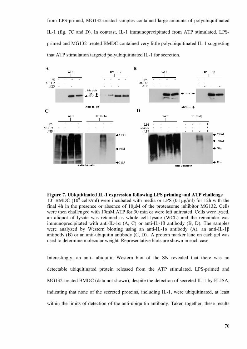
70
from LPS-primed, MG132-treated samples contained large amounts of polyubiquitinated
IL-1 (fig. 7C and D). In contrast, IL-1 immunoprecipitated from ATP stimulated, LPS-
primed and MG132-treated BMDC contained very little polyubiquitinated IL-1 suggesting
that ATP stimulation targeted polyubiquitinated IL-1 for secretion.
Figure 7. Ubiquitinated IL-1 expression following LPS priming and ATP challenge 107 BMDC (106 cells/ml) were incubated with media or LPS (0.1µg/ml) for 12h with the final 4h in the presence or absence of 10µM of the proteasome inhibitor MG132. Cells were then challenged with 10mM ATP for 30 min or were left untreated. Cells were lyzed, an aliquot of lysate was retained as whole cell lysate (WCL) and the remainder was immunoprecipitated with anti-IL-1α (A, C) or anti-IL-1β antibody (B, D). The samples were analyzed by Western blotting using an anti-IL-1α antibody (A), an anti-IL-1β antibody (B) or an anti-ubiquitin antibody (C, D). A protein marker lane on each gel was used to determine molecular weight. Representative blots are shown in each case.
Interestingly, an anti- ubiquitin Western blot of the SN revealed that there was no
detectable ubiquitinated protein released from the ATP stimulated, LPS-primed and
MG132-treated BMDC (data not shown), despite the detection of secreted IL-1 by ELISA,
indicating that none of the secreted proteins, including IL-1, were ubiquitinated, at least
within the limits of detection of the anti-ubiquitin antibody. Taken together, these results
71
suggest that the sequence of events is that ubiquitinated pro-IL-1 is targeted for cleavage,
and that the ubiquitin chain is removed prior to secretion, most likely in the same event
that cleaves the pro-IL-1 into its mature form. However, Lopez-Castejon et al. (2013)
recently reported that deubiquitinases play a role in regulating IL-1 release by affecting
inflammasome function (Lopez-Castejon et al. 2013). Therefore, the possibility that ATP
influences, directly or indirectly, the deubiquitination of IL-1 cannot be discounted and
thus, should be the subject of further investigation.
2.8 Discussion
The release of IL-1 from DC is an important step in the initiation of the inflammatory
response (Gabay et al. 2010). Consistent with previous publications (Masters et al. 2010),
the current studies have shown that IL-1 secretion by DC requires two signals; one signal
to increase precursor expression, and a second signal to stimulate caspase-1 activation and
to facilitate cytokine release. Given the potency of these cytokines, the complexity of this
mechanism reflects the need for tight control of IL-1α and IL-1β production. Here we
report that both IL-1α and IL-1β are degraded by the proteasome and suggest that this
degradation mechanism acts as an important regulator of intracellular IL-1.
The degradation of IL-1α had not been previously investigated, and the degradation of IL-
1β is poorly understood. It has been suggested that in human monocytes, IL-1β
degradation is mediated by the proteasome (Moors and Mizel 2000). However, a
conflicting paper using human and mouse macrophages, as well as mouse DC, indicated
that IL-1β degradation is controlled by autophagy (Harris et al. 2011). In the current
investigations, the degradation of both IL-1α and IL-1β has been shown to be dependent on
72
the proteasome in mouse DC. In addition, we provide evidence suggesting that IL-1
degradation in macrophages is also dependent on the proteasome.
Ubiquitination is rapidly emerging as a key player in the regulation of immune responses
and thus the direct polyubiquitination of IL-1 as demonstrated herein is of great interest
(Malynn and Ma 2010, Lopez-Castejon et al. 2013). Ubiquitination is implicated as a
fundamental regulator at various stages of the canonical NF-κB signalling pathway. Recent
evidence has highlighted the importance of ubiquitination in this pathway by showing that
the activation of the ubiquitin ligase TNF receptor associated factor 6 induces the Lys 63-
linked polyubiquitin of NF-κB essential modulator and receptor-interacting protein 1.
These Lys 63-linked polyubiquitin chains then serve as a scaffold for the recruitment of a
kinase complex, which, when assembled, drives the activation of NF-κB (Wu et al. 2006,
Kanayama et al. 2004a). In addition, we recently reported that in macrophages,
ubiquitination may also be implicated in the assembly of the inflammasome (Lopez-
Castejon et al. 2013, Juliana et al. 2012). As the inhibition of the proteasome caused a
marked increase in levels of polyubiquitinated IL-1, and as the classical role of
ubiquitination is to target a protein for degradation (Wilkinson 2000), the probable role of
IL-1 polyubiquitination is to target these cytokines for degradation. However, given that it
is also known that ubiquitination is a diverse modification with a range of
immunoregulatory functions, it is tempting to speculate that polyubiquitinated IL-1 may
also have an immunoregulatory role, potentially modulating the pathways that drive IL-1
processing and secretion.
Having shown that IL-1 is polyubiquitinated and consequently degraded in DC, it is of
interest to consider the function of this degradation in vivo. One obvious role for IL-1
degradation is to prevent the intracellular accumulation of pro-IL-1. The importance of this
73
is clear when it is considered that the purpose of the 2-signal system of IL-1 release is to
allow secretion only when a perceived threat is significant enough to warrant
inflammation. A stimulus that induces IL-1 expression without concurrent inflammasome
activation or vice versa is not likely to be sufficiently dangerous and thus, will not induce
secretion in DC. However, without rapid degradation in DC, these cytokines would persist
intracellularly as inactive precursors and therefore the need for 2 signals to induce IL-1
secretion would be ablated. Thus, relatively innocuous threats could drive unnecessary and
potentially damaging inflammatory responses. Another role for IL-1 degradation may be in
regulating the amount of IL-1 available for secretion. The current investigations
demonstrate that the process of ubiquitination and proteasomal degradation is induced by
DC activation and that basal turnover of IL-1 is regulated by the proteasome. Together,
these data suggest that there is a basal level of ubiquitination and that DC activation drives
the up regulation or the activation of proteins that mediate IL-1 ubiquitination. As the E3
ubiquitin ligases are relatively specific for the protein that they ubiquitinate (Pickart 2001),
the hypothesis is that it is the E3 ubiquitin ligase of IL-1 that is up regulated or activated as
a consequence of DC priming. If this is true, expression levels of the E3 ubiquitin ligase of
IL-1 may be modulated to control the amount of IL-1 in the cell at any one time. This
regulation may play a crucial role in vivo, acting to constrain the amount of IL-1 available
for secretion, both in the resting state and during the inflammatory response. This is
however speculative and thus requires further investigation.
As previously discussed, the dysregulation of IL-1 is implicated in a number of debilitating
diseases. In Alzheimer’s disease, IL-1 has been shown to be markedly overproduced in
both experimental animal models such as the rat (Cacabelos et al. 1994) and in humans
(Cacabelos et al. 1991, Griffin et al. 1989). Likewise, IL-1 has also been shown to be
elevated in the cerebrospinal fluid of patients with Parkinson’s disease (Mogi et al. 1996).
74
IL-1 also exacerbates acute brain injury such as stroke (Brough et al. 2011) and thus, its
dysregulation is of significance clinically. Although data exist to suggest the
overproduction of IL-1 in these diseases is associated with an increase in IL-1 mRNA
expression (Nicoll et al. 2000), there is strong evidence to suggest that the proteasome is
impaired in both Alzheimer’s and Parkinson’s disease (Riederer et al. 2011). Thus, given
that the proteasome appears to be necessary to regulate IL-1 levels, the breakdown in
proteasome functionality may be an important contributor to the observed elevation in IL-1
levels in neurodegenerative disease.
Intriguingly, the proteasome is an important therapeutic target in a variety of cancer
treatments. One particularly successful therapeutic is the proteasome inhibitor Bortezomib,
which has been used to treat a variety of cancers including myeloma, chronic lymphocytic
leukaemia, prostate cancer, pancreatic cancer and colon cancer (Lim and Tan 2007). As
demonstrated herein, inhibition of the proteasome causes a significant spike in IL-1 levels
in vitro. If the effect of proteasomal inhibition is mirrored in vivo, Bortezomib may also
cause an elevation in the IL-1 available for secretion and therefore an increase in
inflammation. Although previous evidence appears to negate this hypothesis, suggesting
that Bortezomib has an anti-inflammatory effect due to inhibition of the NF-κB pathway
(Chen et al. 2012), there may still be an impact from increased polyubiquitinated IL-1.
Thus, the spike in IL-1 observed here in vitro may still be of relevance in vivo and thus,
may still have a considerable impact upon the efficacy of Bortezomib, and potentially any
other proteasome inhibitors, as anti-cancer treatments.
In the current investigations, the regulation of IL-1α and IL-1β in DC appear to mirror each
other. Although both cytokines serve as proinflammatory cytokines, the exact roles that IL-
1α and IL-1β play in vivo are distinct. In the initiation of allergic contact dermatitis, for
75
example, IL-1β but not IL-1α appears to be important whereas in irritant contact dermatitis
IL-1α appears to play a more important role (Cumberbatch et al. 2002). Thus, the
similarities in IL-1α and IL-1β observed here may not be paralleled in other cell types,
other tissue types or in response to other stimuli. Indeed, in human and murine skin,
preformed IL-1α is detected in large amounts whereas IL-1β is not detectable (Mee et al.
2005, Kupper 1990). Although this could be due to differences in transcription, it is
plausible that the mechanism of ubiquitination and degradation described herein could be a
factor. Specifically, IL-1α degradation may be inhibited in keratinocytes whereas IL-1β
degradation may be functional, effectively resulting in an accumulation of IL-1α.
To conclude, the current investigations show that both IL-1α and IL-1β are tightly
regulated by polyubiquitination and proteasomal degradation, adding to the growing body
of work that demonstrates that ubiquitination is a key regulator of inflammation and
immunity. These findings not only suggest that the mechanism of IL-1 degradation could
represent an important therapeutic target, but also highlight that degradation may be
pivotal to the maintenance of tissue homeostasis.
2.9 Acknowledgements
JS Ainscough was funded by the Medical Research Council (MRC) and The Procter &
Gamble Company.
2.10 References
Apte, R. N., Dotan, S., Elkabets, M., White, M. R., Reich, E., Carmi, Y., Song, X., Dvozkin, T., Krelin, Y. and Voronov, E. (2006) 'The involvement of IL-1 in tumorigenesis, tumor invasiveness, metastasis and tumor-host interactions', Cancer and Metastasis Reviews, 25(3), 387-408.
76
Banchereau, J. and Steinman, R. M. (1998) 'Dendritic cells and the control of immunity',
Nature, 392(6673), 245-252. Brough, D., Tyrrell, P. J. and Allan, S. M. (2011) 'Regulation of interleukin-1 in acute
brain injury', Trends in Pharmacological Sciences, 32(10), 617-622. Buchan, G., Barrett, K., Turner, M., Chantry, D., Maini, R. N. and Feldmann, M. (1988)
'Interleukin-1 and Tumor Necrosis Factor messenger-RNA expression in Rheumatoid-Arthritis - Prolonged production of IL-1α', Clinical and Experimental Immunology, 73(3), 449-455.
Cacabelos, R., Alvarez, X. A., Fernandeznovoa, L., Franco, A., Mangues, R., Pellicer, A.
and Nishimura, T. (1994) 'Brain Interleukin-1β in Alzheimers-Disease and vascular dementia', Methods and Findings in Experimental and Clinical Pharmacology, 16(2), 141-151.
Cacabelos, R., Francomaside, A. and Alvarez, X. A. (1991) 'Interleukin-1 in Alzheimers-
Disease and Multiinfarct Dementia - neuropsychological correlations', Methods and Findings in Experimental and Clinical Pharmacology, 13(10), 703-708.
Chen, C.-J., Shi, Y., Hearn, A., Fitzgerald, K., Golenbock, D., Reed, G., Akira, S. and
Rock, K. L. (2006) 'MyD88-dependent IL-1 receptor signaling is essential for gouty inflammation stimulated by monosodium urate crystals', Journal of Clinical Investigation, 116(8), 2262-2271.
Chen, F.-T., Liu, Y.-C., Yang, C.-M. and Yang, C.-H. (2012) 'Anti-inflammatory effect of
the proteasome inhibitor bortezomib on endotoxin-induced uveitis in rats', Investigative Ophthalmology & Visual Science, 53(7), 3682-3694.
Cogswell, J. P., Godlevski, M. M., Wisely, G. B., Clay, W. C., Leesnitzer, L. M., Ways, J.
P. and Gray, J. G. (1994) 'NF-κB regulates IL-1β transcription through a consensus NF-κB binding-site and a nonconsensus Cre-like site', Journal of Immunology, 153(2), 712-723.
Cumberbatch, M., Dearman, R. J., Groves, R. W., Antonopoulos, C. and Kimber, I. (2002)
'Differential regulation of epidermal Langerhans cell migration by interleukins (IL)-1α and IL-1β during irritant- and allergen-induced cutaneous immune responses', Toxicology and Applied Pharmacology, 182(2), 126-135.
Dearman, R. J., Hope, J. C., Hopkins, S. J. and Kimber, I. (1996) 'Antigen-induced
unresponsiveness in contact sensitivity: Association of depressed T lymphocyte proliferative responses with decreased interleukin 6 secretion', Immunology Letters, 50(1-2), 29-34.
Devaraj, S., Dasu, M. R., Rockwood, J., Winter, W., Griffen, S. C. and Jialal, I. (2008)
'Increased toll-like receptor (TLR)2 and TLR4 expression in monocytes from patients with type 1 diabetes: Further evidence of a proinflammatory state', Journal of Clinical Endocrinology & Metabolism, 93(2), 578-583.
Dinarello, C. A. (2009) 'Immunological and inflammatory functions of the Interleukin-1
family', Annual Review of Immunology, 27, 519-550.
77
Ferrari, D., Chiozzi, P., Falzoni, S., DalSusino, M., Melchiorri, L., Baricordi, O. R. and
DiVirgilio, F. (1997) 'Extracellular ATP triggers IL-1β release by activating the purinergic P2Z receptor of human macrophages', Journal of Immunology, 159(3), 1451-1458.
Franchi, L., Eigenbrod, T., Munoz-Planillo, R. and Nunez, G. (2009) 'The inflammasome:
a caspase-1-activation platform that regulates immune responses and disease pathogenesis', Nature Immunology, 10(3), 241-247.
Gabay, C., Lamacchia, C. and Palmer, G. (2010) 'IL-1 pathways in inflammation and
human diseases', Nature Reviews Rheumatology, 6(4), 232-241. Ghiringhelli, F., Apetoh, L., Tesniere, A., Aymeric, L., Ma, Y., Ortiz, C., Vermaelen, K.,
Panaretakis, T., Mignot, G., Ullrich, E., Perfettini, J.-L., Schlemmer, F., Tasdemir, E., Uhl, M., Genin, P., Civas, A., Ryffel, B., Kanellopoulos, J., Tschopp, J., Andre, F., Lidereau, R., McLaughlin, N. M., Haynes, N. M., Smyth, M. J., Kroemer, G. and Zitvogel, L. (2009) 'Activation of the NLRP3 inflammasome in dendritic cells induces IL-1β-dependent adaptive immunity against tumors', Nature Medicine, 15(10), 1170-U99.
Ghonime, M. G., Shamaa, O. R., Das, S., Eldomany, R. A., Fernandes-Alnemri, T.,
Alnemri, E. S., Gavrilin, M. A. and Wewers, M. D. (2014) 'Inflammasome priming by lipopolysaccharide is dependent upon ERK signaling and proteasome function', Journal of Immunology, 192(8), 3881-3888.
Goldstein, G., Scheid, M., Hammerling, U., Boyse, E. A., Schlesinger, D. H. and Niall, H.
D. (1975) 'Isolation of a polypeptide that has lymphocyte-differentiating properties and is probably represented universally in living cells', Proceedings of the National Academy of Sciences of the United States of America, 72(1), 11-15.
Griffin, W. S. T., Stanley, L. C., Ling, C., White, L., Macleod, V., Perrot, L. J., White, C.
L. and Araoz, C. (1989) 'Brain Interleukin-1 and s-100 immunoreactivity are elevated in Down Syndrome and Alzheimer-Disease', Proceedings of the National Academy of Sciences of the United States of America, 86(19), 7611-7615.
Harris, J., Hartman, M., Roche, C., Zeng, S. G., O'Shea, A., Sharp, F. A., Lambe, E. M.,
Creagh, E. M., Golenbock, D. T., Tschopp, J., Kornfeld, H., Fitzgerald, K. A. and Lavelle, E. C. (2011) 'Autophagy controls IL-1β secretion by targeting pro-IL-1β for degradation', Journal of Biological Chemistry, 286(11), 9587-9597.
Juliana, C., Fernandes-Alnemri, T., Kang, S., Farias, A., Qin, F. and Alnemri, E. S. (2012)
'Non-transcriptional priming and deubiquitination regulate NLRP3 inflammasome activation', Journal of Biological Chemistry, 287(43), 36617-36622.
Kanayama, A., Seth, R. B., Sun, L. J., Ea, C. K., Hong, M., Shaito, A., Chiu, Y. H., Deng,
L. and Chen, Z. J. (2004) 'TAB2 and TAB3 activate the NF-κBpathway through binding to polyubiquitin chains', Molecular Cell, 15(4), 535-548.
Kisselev, A. F. and Goldberg, A. L. (2001) 'Proteasome inhibitors: from research tools to
drug candidates', Chemistry & Biology, 8(8), 739-758.
78
Komander, D. (2009) 'The emerging complexity of protein ubiquitination', Biochemical Society Transactions, 37, 937-953.
Kupper, T. S. (1990) 'Immune and inflammatory processes in cutaneous tissues -
mechanisms and speculations', Journal of Clinical Investigation, 86(6), 1783-1789. Latz, E., Xiao, T. S. and Stutz, A. (2013) 'Activation and regulation of the
inflammasomes', Nature Reviews Immunology, 13(6), 397-411. Lim, K.-L. and Tan, J. M. M. (2007) 'Role of the ubiquitin proteasome system in
Parkinson's disease', Bmc Biochemistry, 8. Lopez-Castejon, G. and Brough, D. (2011) 'Understanding the mechanism of IL-
1β secretion', Cytokine & Growth Factor Reviews, 22(4), 189-195. Lopez-Castejon, G., Luheshi, N. M., Compan, V., High, S., Whitehead, R. C., Flitsch, S.,
Kirov, A., Prudovsky, I., Swanton, E. and Brough, D. (2013) 'Deubiquitinases Regulate the activity of Caspase-1 and Interleukin-1β secretion via assembly of the inflammasome', Journal of Biological Chemistry, 288(4), 2721-2733.
Lutz, M. B., Kukutsch, N., Ogilvie, A. L. J., Rossner, S., Koch, F., Romani, N. and
Schuler, G. (1999) 'An advanced culture method for generating large quantities of highly pure dendritic cells from mouse bone marrow', Journal of Immunological Methods, 223(1), 77-92.
Malynn, B. A. and Ma, A. (2010) 'Ubiquitin makes its mark on immune regulation',
Immunity, 33(6), 843-852. Mariathasan, S., Weiss, D. S., Newton, K., McBride, J., O'Rourke, K., Roose-Girma, M.,
Lee, W. P., Weinrauch, Y., Monack, D. M. and Dixit, V. M. (2006) 'Cryopyrin activates the inflammasome in response to toxins and ATP', Nature, 440(7081), 228-232.
Masters, S. L., Dunne, A., Subramanian, S. L., Hull, R. L., Tannahill, G. M., Sharp, F. A.,
Becker, C., Franchi, L., Yoshihara, E., Chen, Z., Mullooly, N., Mielke, L. A., Harris, J., Coll, R. C., Mills, K. H. G., Mok, K. H., Newsholme, P., Nunez, G., Yodoi, J., Kahn, S. E., Lavelle, E. C. and O'Neill, L. A. J. (2010) 'Activation of the NLRP3 inflammasome by islet amyloid polypeptide provides a mechanism for enhanced IL-1β in type 2 diabetes', Nature Immunology, 11(10), 897-U1501.
Medzhitov, R. (2010) 'Inflammation 2010: New adventures of an old flame', Cell, 140(6),
771-776. Mee, J. B., Antonopoulos, C., Poole, S., Kupper, T. S. and Groves, R. W. (2005) 'Counter-
regulation of interleukin-1α (IL-1α) and IL-1 receptor antagonist in murine keratinocytes', Journal of Investigative Dermatology, 124(6), 1267-1274.
Mellman, I. and Steinman, R. M. (2001) 'Dendritic cells: Specialized and regulated antigen
processing machines', Cell, 106(3), 255-258. Mikami, N., Matsushita, H., Kato, T., Kawasaki, R., Sawazaki, T., Kishimoto, T., Ogitani,
Y., Watanabe, K., Miyagi, Y., Sueda, K., Fukada, S.-i., Yamamoto, H. and
79
Tsujikawa, K. (2011) 'Calcitonin gene-related peptide is an important regulator of cutaneous immunity: effect on dendritic cell and T cell functions', Journal of Immunology, 186(12), 6886-6893.
Mogi, M., Harada, M., Narabayashi, H., Inagaki, H., Minami, M. and Nagatsu, T. (1996)
'Interleukin (IL)-1β, IL-2, IL-4, IL-6 and transforming growth factor-α levels are elevated in ventricular cerebrospinal fluid in juvenile parkinsonism and Parkinson's disease', Neuroscience Letters, 211(1), 13-16.
Moors, M. A. and Mizel, S. B. (2000) 'Proteasome-mediated regulation of interleukin-
1β turnover and export in human monocytes', Journal of Leukocyte Biology, 68(1), 131-136.
Netea, M. G., Nold-Petry, C. A., Nold, M. F., Joosten, L. A. B., Opitz, B., van der Meer, J.
H. M., van de Veerdonk, F. L., Ferwerda, G., Heinhuis, B., Devesa, I., Funk, C. J., Mason, R. J., Kullberg, B. J., Rubartelli, A., van der Meer, J. W. M. and Dinarello, C. A. (2009) 'Differential requirement for the activation of the inflammasome for processing and release of IL-1β in monocytes and macrophages', Blood, 113(10), 2324-2335.
Nicoll, J. A. R., Mrak, R. E., Graham, D. I., Stewart, J., Wilcock, G., MacGowan, S., Esiri,
M. M., Murray, L. S., Dewar, D., Love, S., Moss, T. and Griffin, W. S. T. (2000) 'Association of interleukin-1 gene polymorphisms with Alzheimer's disease', Annals of Neurology, 47(3), 365-368.
Palombella, V. J., Rando, O. J., Goldberg, A. L. and Maniatis, T. (1994) 'The ubiquitin-
proteasome pathway is required for Processing the NF-κB1 precursor protein and the activation of NF-κB', Cell, 78(5), 773-785.
Perkins, N. D. (2006) 'Post-translational modifications regulating the activity and function
of the NF-κB pathway', Oncogene, 25(51), 6717-6730. Pickart, C. M. (2001) 'Mechanisms underlying ubiquitination', Annual Review of
Biochemistry, 70, 503-533. Riederer, B. M., Leuba, G., Vernay, A. and Riederer, I. M. (2011) 'The role of the
ubiquitin proteasome system in Alzheimer's disease', Experimental Biology and Medicine, 236(3), 268-276.
Rubartelli, A., Cozzolino, F., Talio, M. and Sitia, R. (1990) 'A novel secretory pathway for
Interleukin-1β, a protein lacking A signal sequence', Embo Journal, 9(5), 1503-1510.
Steinman, R. M. and Banchereau, J. (2007) 'Taking dendritic cells into medicine', Nature,
449(7161), 419-426. Takeuchi, O. and Akira, S. (2010) 'Pattern recognition receptors and inflammation', Cell,
140(6), 805-820. Thrower, J. S., Hoffman, L., Rechsteiner, M. and Pickart, C. M. (2000) 'Recognition of the
polyubiquitin proteolytic signal', Embo Journal, 19(1), 94-102.
80
Traenckner, E. B. M., Wilk, S. and Baeuerle, P. A. (1994) 'A proteasome inhibitor prevents activation of NF-κB and stabilizes a newly phosphorylated form of I-κB that is still bound to NF-κB', Embo Journal, 13(22), 5433-5441.
Watanabe, H., Gaide, O., Petrilli, V., Martinon, F., Contassot, E., Roques, S., Kummer, J.
A., Tschopp, J. and French, L. E. (2007) 'Activation of the IL-1β processing inflammasome is involved in contact hypersensitivity', Journal of Investigative Dermatology, 127(8), 1956-1963.
Watanabe, N. and Kobayashi, Y. (1994) 'Selective release of a processed form of
Interleukin-1α', Cytokine, 6(6), 597-601. Welchman, R. L., Gordon, C. and Mayer, R. J. (2005) 'Ubiquitin and ubiquitin-like
proteins as multifunctional signals', Nature Reviews Molecular Cell Biology, 6(8), 599-609.
Wilkinson, K. D. (2000) 'Ubiquitination and deubiquitination: Targeting of proteins for
degradation by the proteasome', Seminars in Cell & Developmental Biology, 11(3), 141-148.
Wu, C. J., Conze, D. B., Li, T., Srinivasula, S. M. and Ashwell, J. D. (2006) 'NEMO is a
sensor of Lys 63-linked polyubiquitination and functions in NF-κB activation', Nature Cell Biology, 8(4), 398-U58.
Wu, Y.-T., Tan, H.-L., Shui, G., Bauvy, C., Huang, Q., Wenk, M. R., Ong, C.-N.,
Codogno, P. and Shen, H.-M. (2010) 'Dual Role of 3-Methyladenine in modulation of autophagy via different temporal patterns of inhibition on Class I and III Phosphoinositide 3-Kinase', Journal of Biological Chemistry, 285(14), 10850-10861.

81
2.11 Supplementary data for chapter 2
Figure S2.1 Characterisation of the IL-1β ELISA Analysis of the relative avidity of the IL-1β ELISA for pro and mature IL-1β. Mouse recombinant pro-IL-1β and recombinant mature IL-1β were analysed in parallel using an IL-1β ELISA Duoset kit over a concentration range of 300 to 1.2pM. Optical density was measured at 450nm using an automated plate reader. Data shown are from one experiment, representing the mean of a triplicate determination in each case.
Figure S2.2. XS106 cells produce IL-1β intracellularly in response to LPS, but do not secrete this in response to ATP stimulation 106 XS106 cells (106 cells/ml) were cultured for 4h in the presence of media alone or increasing doses of LPS (0.01 to 100µg/ml)(A). In addition, 106 XS106 cells (106 cells/ml) were incubated with medium alone (○) or with LPS (10µg/ml; ●) for various time periods (0h to 48h) (B; single experiment). 106 XS106 cells or 106 BMDC (both 106 cells/ml) were also primed with LPS (BMDC incubated with 0.1 µg/ml and XS106 cells incubated with 10 µg/ml) for 4h and then challenged with ATP (0-10mM) for 30 min (C). Supernatants and cell lysates were harvested and analysed for the presence of IL-1β using cytokine specific ELISA. IL-1β content of cell lysates is displayed in A and B, and supernatants (secreted IL-1) in C. Data shown in A and C are mean ± SEM (n=3). A one-way ANOVA was used to determine statistical significance of differences between untreated and treated groups (A, C). *, p < 0.05; **, p < 0.01.
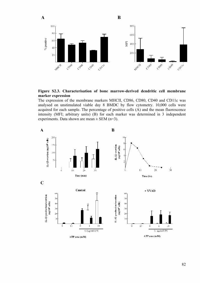
82
Figure S2.3. Characterisation of bone marrow-derived dendritic cell membrane marker expression The expression of the membrane markers MHCII, CD86, CD80, CD40 and CD11c was analysed on unstimulated viable day 8 BMDC by flow cytometry. 10,000 cells were acquired for each sample. The percentage of positive cells (A) and the mean fluorescence intensity (MFI; arbitrary units) (B) for each marker was determined in 3 independent experiments. Data shown are mean ± SEM (n=3).
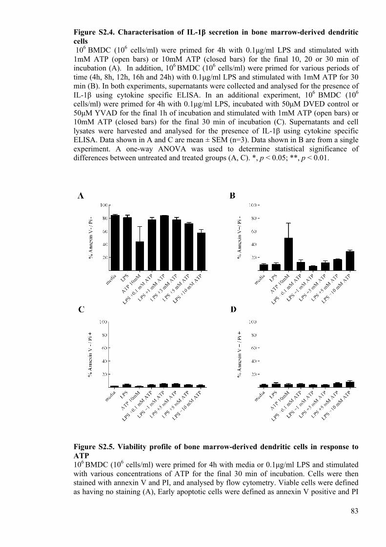
83
Figure S2.4. Characterisation of IL-1β secretion in bone marrow-derived dendritic cells 106 BMDC (106 cells/ml) were primed for 4h with 0.1µg/ml LPS and stimulated with 1mM ATP (open bars) or 10mM ATP (closed bars) for the final 10, 20 or 30 min of incubation (A). In addition, 106 BMDC (106 cells/ml) were primed for various periods of time (4h, 8h, 12h, 16h and 24h) with 0.1µg/ml LPS and stimulated with 1mM ATP for 30 min (B). In both experiments, supernatants were collected and analysed for the presence of IL-1β using cytokine specific ELISA. In an additional experiment, 106 BMDC (106 cells/ml) were primed for 4h with 0.1µg/ml LPS, incubated with 50µM DVED control or 50µM YVAD for the final 1h of incubation and stimulated with 1mM ATP (open bars) or 10mM ATP (closed bars) for the final 30 min of incubation (C). Supernatants and cell lysates were harvested and analysed for the presence of IL-1β using cytokine specific ELISA. Data shown in A and C are mean ± SEM (n=3). Data shown in B are from a single experiment. A one-way ANOVA was used to determine statistical significance of differences between untreated and treated groups (A, C). *, p < 0.05; **, p < 0.01.
Figure S2.5. Viability profile of bone marrow-derived dendritic cells in response to ATP 106 BMDC (106 cells/ml) were primed for 4h with media or 0.1µg/ml LPS and stimulated with various concentrations of ATP for the final 30 min of incubation. Cells were then stained with annexin V and PI, and analysed by flow cytometry. Viable cells were defined as having no staining (A), Early apoptotic cells were defined as annexin V positive and PI
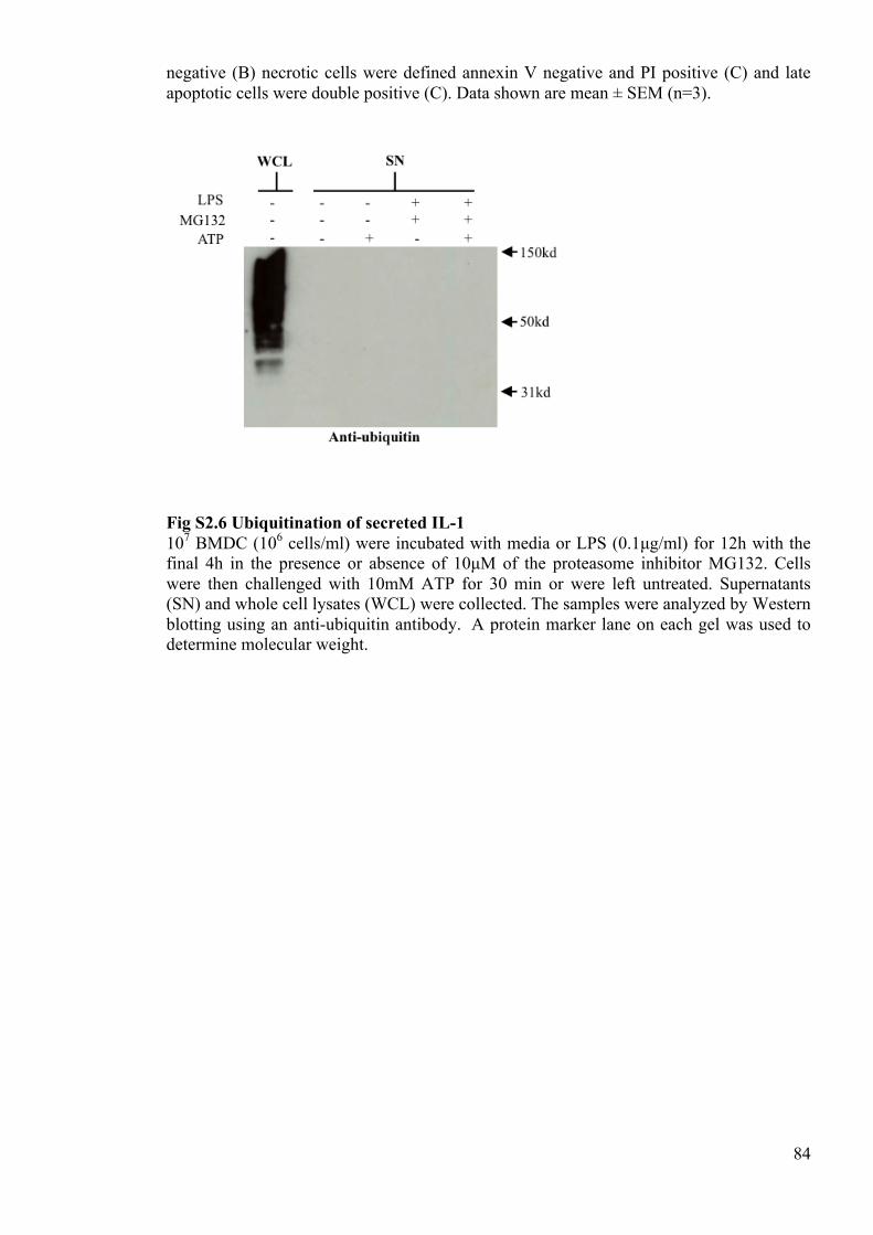
84
negative (B) necrotic cells were defined annexin V negative and PI positive (C) and late apoptotic cells were double positive (C). Data shown are mean ± SEM (n=3).
Fig S2.6 Ubiquitination of secreted IL-1 107 BMDC (106 cells/ml) were incubated with media or LPS (0.1µg/ml) for 12h with the final 4h in the presence or absence of 10µM of the proteasome inhibitor MG132. Cells were then challenged with 10mM ATP for 30 min or were left untreated. Supernatants (SN) and whole cell lysates (WCL) were collected. The samples were analyzed by Western blotting using an anti-ubiquitin antibody. A protein marker lane on each gel was used to determine molecular weight.
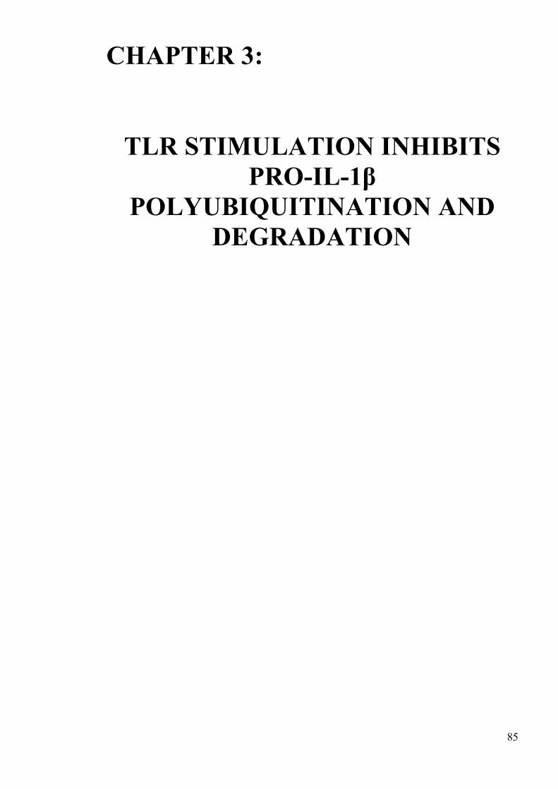
85
CHAPTER 3:
TLR STIMULATION INHIBITS
PRO-IL-1β POLYUBIQUITINATION AND
DEGRADATION !
!
!
!
!
!
!
!
!
!
!
!
!
!
!
!
!
!
!
86
3 Paper 2: TLR Stimulation Inhibits pro-IL-1β Polyubiquitination
and Degradation
Joseph S. Ainscough, James Bagnall, Pawel Paszek, G. Frank Gerberick*, Ian Kimber and
Rebecca J. Dearman
Faculty of Life Sciences, University of Manchester, Michael Smith Building, Oxford
Road, Manchester, M13 9PT, (+44)161 275 5368
Faculty of Life Sciences, University of Manchester, Manchester.
* The Procter & Gamble Company Co., Cincinnati, OH 45253, USA
Running title: TLR Stimulation Inhibits pro-IL-1β Ubiquitination
Key words: IL-1β, ubiquitin, proteasome, inflammation, ubiquitin ligase
3.1 Abbreviations
ATP Adenosine triphosphate cDNA Complementary deoxyribonucleic acid CHX Cycloheximide DAMP Damage Associated Molecular Patterns!ELISA Enzyme-linked immunosorbent assay EDTA Ethylenediaminetetraacetic acid FCS Foetal calf serum GFP Green fluorescent protein HRPT Hypoxanthine-guanine phosphoribosyltransferase Ig Immunoglobulin IBD Inflammatory bowel disease IκB Inhibitor of κB IL Interleukin LPS Lipopolysaccharide MFI Mean fluorescent intensity mRNA Messenger Ribonucleic acid NLRP3 NOD-like receptor family pyrin domain–containing protein 3
87
NF-κB Nuclear factor kappa-light-chain-enhancer of activated B cells PAMP Pathogen Associated Molecular Patterns PRR Pattern recognition receptor PMSF Phenylmethanesulfonylfluoride PBS Phosphate buffered saline PCR Polymerase chain reaction RT-PCR Reverse transcription polymerase chain reaction RNA Ribonucleic acid TLR TOLL like receptor venusIL-1βTHP1 venuspro-IL-1β human THP-l cells venusIL-1βiBMDM venuspro-IL-1β immortalized Murine bone marrow derived macrophages WCL Whole cell lysate YFP Yellow fluorescent protein
3.2 Abstract
Interleukin (IL)-1β is a key player in the initiation and orchestration of inflammation. The
secretion of IL-1β requires 2 independent processes; an initial signal to induce the up-
regulation of the inactive precursor (pro-IL-1β) and, a second signal to drive cleavage and
subsequent secretion. In previous investigations using murine dendritic cells, we have
demonstrated that in lipopolysaccharide-stimulated cells, pro-IL-1β is polyubiquitinated
and degraded by the proteasome, resulting in a rapid reduction in the amount of
intracellular IL-1β available for secretion. The current study has employed an
immortalized bone marrow derived murine macrophage cell line and a human monocyte
cell line that both stably express fluorescent IL-1β, and used these to measure the rate of
IL-1β degradation. The results presented herein demonstrate that fluorescence is a reliable
readout for measuring IL-1β degradation in these cell lines. In addition, these data show
that TLR-stimulation leads to an inhibition in IL-1β ubiquitination and degradation.
Finally, the current investigations indicate that this inhibition in IL-1β ubiquitination and
degradation is transient, peaking at ~4-8 h and returning to baseline thereafter. Overall,
these data demonstrate that ubiquitination actively regulates the vigour of IL-1β protein
expression and thus may be an important regulator of inflammation.
88
3.3 Introduction
Interleukin (IL)-1β is a proinflammatory cytokine that is critical to the function of the
innate immune system (Garlanda et al. 2013) (Dinarello 2009). Within this role, the
secreted, mature cytokine drives a myriad of changes, culminating in the initiation and
propagation of inflammation (Dinarello 2011). To orchestrate this potent response, IL-1β
modulates the expression of a variety of other cytokines, chemokines and adhesion
molecules. Ultimately, these changes allow both the migration of antigen presenting cells
away from the affected site and the infiltration of immune cells towards the affected site
(Tracy 2006). The resultant proinflammatory response is a powerful one, and thus IL-1β
needs to be tightly regulated to ensure that the vigour of secretion is appropriate.
To allow for greater control of secretion, IL-1β is produced as an inactive 31kDa precursor
that must be cleaved to be activated (March et al. 1985, Thornberry et al. 1992). The initial
upregulation of the pro-protein is induced by specific molecular motifs that are derived
from pathogens, namely pathogen associated molecular patterns (PAMP) (Gallucci and
Matzinger 2001). These PAMP are sensed by TOLL-like receptors (TLR) and initiate the
expression of a battery of proinflammatory cytokines by the transcription factor nuclear
factor kappa-light-chain-enhancer of activated B cells (NFκB) (Baeuerle and Baltimore
1988a, Cogswell et al. 1994). Typically, the proteolytic cleavage of pro-IL-1β to mature
IL-1β is independent of upregulation and requires the assembly of the inflammasome
complex. When assembled, the inflammasome is a large molecular scaffold comprising of
adaptor molecules, a cytosolic pattern recognition receptor (PRR), and pro-caspase-1
(Bryant and Fitzgerald 2009). Here, cytosolic PRR detect the presence of additional
PAMP, or damage associated molecular patterns (DAMP) and induce the formation of the
inflammasome complex (Latz 2010). The assembly of the inflammasome activates
89
caspase-1, which in turn cleaves pro-IL-1β into the active 17kDa cytokine (Wilson et al.
1994).
In recent investigations, ubiquitination has emerged as a key player in the regulation of IL-
1β upregulation and secretion (Ainscough et al. 2014). Ubiquitination is a post-
translational modification whereby ubiquitin, an 8.5kDa protein, is covalently bound to
lysine residues on substrate proteins (Pickart 2001). These proteins may remain
monoubiquitinated or may have additional ubiquitin molecules added in a process called
polyubiquitination (Pickart and Fushman 2004). The most common role for ubiquitination,
and specifically lysine (K)-48 ubiquitination, is to target proteins for proteasomal
degradation (Wilkinson 2000). However, more recently, it has been shown that these
modifications also play roles in a variety of cell signaling pathways. This is particularly
evident in the TLR signaling pathways that induce IL-1β expression. The ubiquitin ligase
TRAF6 plays a particularly prominent role in these signalling pathways (Qian et al. 2001).
Here TRAF6-induced K-63-linked polyubiquitin chains on IRAK1 function as a scaffold
for the recruitment of the TAK1–TAB1–TAB2/3 complex and the IKK complex (Deng et
al. 2000, Kanayama et al. 2004b). Ultimately, the interactions at this complex result in the
phosphorylation, ubiquitination and proteasomal degradation of the inhibitor of κB (IκB)
(Baeuerle and Baltimore 1988a). As the name suggests, IκB inhibits NFκB translocation
and so the degradation of this facilitates NFκB translocation and the consequent
transcription of pro-IL-1β (Baeuerle and Baltimore 1988a). Polyubiquitination is also
implicated in the assembly of the inflammasome and caspase-1 activation, and therefore is
also important in IL-1β processing. The NOD-like receptor family pyrin domain–
containing protein 3 (NLRP3) inflammasome is ubiquitinated in resting cells and the
stimulation of these cells with lipopolysaccharide (LPS) and adenosine triphosphate (ATP)
leads to the removal of the ubiquitin chains and consequent activation of the complex (Py
90
et al. 2013). In addition to the NLRP3 inflammasome, caspase-1 is also polyubiquitinated
and this polyubiquitination facilitates activation of the enzyme (Labbe et al. 2011).
As well as regulating the upregulation and processing of pro-IL-1β, a recent report has
shown that pro-IL-1β is itself polyubiquitinated and degraded by the proteasome in murine
dendritic cells and macrophages (Ainscough et al. 2014). In this study, it was hypothesised
that the rate of pro-IL-1β polyubiquitination and proteasomal degradation may be dynamic,
and that these processes may function to regulate the amount of pro-IL-1β available for
secretion. Given that IL-1β is so important in inflammation, it is also suggested that these
processes could ultimately regulate the vigour of the inflammatory response. To test these
hypotheses, the current study has utilised 2 stably expressing fluorescent pro-IL-1β cell-
lines; venus pro-IL-1β immortalized Murine bone marrow derived macrophages (venusIL-
1βiBMDM) and venus pro-IL-1β Human THP-l cells (venusIL-1βTHP1; both expressing
the fluorescent probe on the C terminus). In these investigations, venus pro-IL-1β was
shown to be polyubiquitinated and degraded by the proteasome in both cell lines.
Importantly, changes in the rate of degradation of pro-IL-1β could be investigated by
tracking the changes in cellular fluorescence. Using this, we have demonstrated for the first
time that LPS inhibits pro-IL-1β polyubiquitination and degradation, leading to an
augmented pro-IL-1β protein expression. We speculate here that this mechanism may be
vital in enhancing TLR-induced inflammation.
3.4 Methods
Antibodies and reagents
91
LPS from Escherichia coli serotype 055:B5 (TLR2/4), Poly(I:C) and cycloheximide
(CHX) were purchased from Sigma Chemical Co (Poole, UK). The proteasome inhibitor
MG132 was obtained from Merck Millipore (Billerica, MA, USA). For Western blot
analysis, the primary antibodies used were a goat anti-human IL-1β antibody (AF200-NA),
a goat anti-mouse IL-1β antibody (AF201-NA) both R&D systems; Minneapolis, MN,
USA) or a mouse anti-ubiquitin antibody (SC8017; Santa Cruz Biotechnology; Santa Cruz,
CA, USA). The horseradish peroxidase-conjugated secondary antibodies were rabbit anti-
goat IgG antibody (P0449; DAKO; Copenhagen, Denmark) and goat anti-mouse light
chain antibody (AP200P; Millipore).
Development of the IL1B-venus expression vector
The human IL1B coding sequence (Accession Number NM_000576) was synthesised with
flanking Gateway® att recombination sites and inserted into pUC57 vector, resulting in the
pUC57-IL1B vector (GenScript, Township, NJ, USA). The IL1B gene was then transferred
to the 3rd generation ‘pLNT-#-Venus’ lentiviral transfer by LR clonase recombination
reaction (Katzen 2007, Nagai et al. 2002). The resultant vector was termed pLNT-IL1B-
venus and allowed for ubiquitin-ligase C promoter-mediated constitutive expression of N-
terminally fused target sequences (Bagnall et al. 2015).
Lentivirus Production
1.25x10⁷ HEK293T cells were seeded in a 15cm dish then transfected by polyethylenimine
“Max” (Polysciences Inc., Warrington, PA, USA) with 10.5µg total DNA made up by the
packaging vectors pMDLg-RRE, pCMV-VSVG, pRSV-REV and the pLNT-IL1B-venus
transfer vector at a ratio of 2:1:2:4. After 6h the transfection mix was removed and
92
replaced with fresh media. After 48h, the cell media was collected and the virus
concentrated by ultracentrifugation. The supernatant was then removed and the virus
resuspended in small volumes of PBS.
Lentiviral Transduction
IL1B-venus lentivirus was applied to a culture of 1.5x10⁴ cells THP1 cells in 2ml of
media. After 2 days, the virally loaded media was replaced with fresh media and the
transduced culture propagated for several passages. Transduction efficiency was
determined by confocal microscopy. Subsequently, the transduced cell culture was frozen
into stocks for use in all future experiments.
Maintenance of cell lines
VenusIL-1βiBMDM (obtained from Dr David Brough) were cultured in FCS-
supplemented culture medium (DMEM; Life technologies), containing 400 µg/ml
penicillin/ streptomycin, 292 µg/ml L-glutamine and 10% FCS (Life technologies).
VenusIL-1β THP-1 cells and HEK293T cells were cultured in FCS-supplemented culture
medium (RPMI 1640; Life technologies), containing 400 µg/ml penicillin/ streptomycin,
292 µg/ml L-glutamine, 0.05 mM 2-mercaptoethanol and 10% FCS (Life technologies).
Cell treatments
VenusIL-1βiBMDM or venusIL-1β THP-1 cells were cultured at 106 cells/well (24-well
plate) or 107 cells/well (6-well plate; 106 cells/ml). In initial experiments, cells were
incubated with 10 µg/ml CHX for 4 h in the presence or absence of the proteasome
93
inhibitor MG132. Cells were also incubated for 4 h in media or increasing concentrations
of LPS or Poly(I:C). A kinetics experiment was also performed, whereby cells were
incubated with media, LPS (1 µg/ml) or Poly(I:C) (100 µg/ml) for various lengths of time.
After incubation, supernatants were harvested and frozen at -80°C. Cell lysates were
harvested in 200 µl of lysis buffer (20 mM Tris HCl, 137 mM NaCl, 20 mM EDTA, 10%
glycerol, 0.5% Ipegal, 1 mM phenylmethylsulfonyl fluoride (PMSF) , protease inhibitor
cocktail [1:100]) and frozen at -80°C. For PCR analysis, lysates were prepared for RNA
extraction following the manufacturer’s instructions (Purelink RNA mini kit; Life
Technologies, Carlsbad, CA, USA).
Flow cytometric analysis
Following treatments as described above, cells were washed in FACS buffer (5%
FCS/PBS) and resuspended in Sodium azide buffer (0.05% sodium azide in 1% FCS/PBS).
Samples were analysed using a FACScalibur flow cytometer and CellQuest Pro software
(both BD biosciences). 25,000 cells were acquired from each sample. Cells were initially
gated on forward scatter and size scatter and gates drawn based upon the position of the
cells in the untreated samples. The mean fluorescence intensity (MFI) was calculated for
each sample using Flowjo analysis software (Flowjo; Ashland, OR, USA). In some
experiments, the MFI was normalised by deducting the MFI of untreated controls from the
MFI of the treated samples.
ELISA
94
Supernatants and lysates were analysed for the presence of IL-1β protein using specific
ELISA Duosets from R&D systems. ELISA were performed following the manufacturer’s
instructions.
Western blots
In preparation for Western blot analysis, supernatants and lysates were diluted in sample
buffer (Biorad, Berkley, CA, USA) containing 1% 2-mercaptoethanol and heated for 5 min
at 80°C. Samples were resolved on a 10% acrylamide gel and proteins transferred to a
nitrocellulose membrane. Specific proteins were detected using a goat anti-mouse IL-1β
antibody (0.1 µg/ml) or an anti-ubiquitin antibody (0.2 µg/ml). Finally, blots were
incubated with a horseradish peroxidase-labeled anti-goat IgG antibody or a horseradish
peroxidase-labeled anti-light chain IgG antibody (both 0.25 µg/ml), and proteins visualised
using enhanced chemiluminescence reagents (Thermo Scientific; Waltham, MA, USA).
Imaging of venusIL-1βiBMDM
After treatment as described above, cells were fixed with 4% formaldehyde for 20 minutes
at room temperature. Cells were then washed with PBS and reconstituted at 5x10⁵ cells/ ml
in PBS. Cells were fixed onto glass slides by transferring 200 µl of the cell suspension into
cytospin cartridges and spinning in the cytospin centrifuge at 700g for 5 min. Finally,
slides were mounted using Vectashield (Vector Laboratories Ltd, Peterborough, UK),
sealed with nail varnish and imaged using fluorescence microscopy. In these experiments,
the operator was blinded as to the identity of the samples.
Immunoprecipitation of venusIL-1β
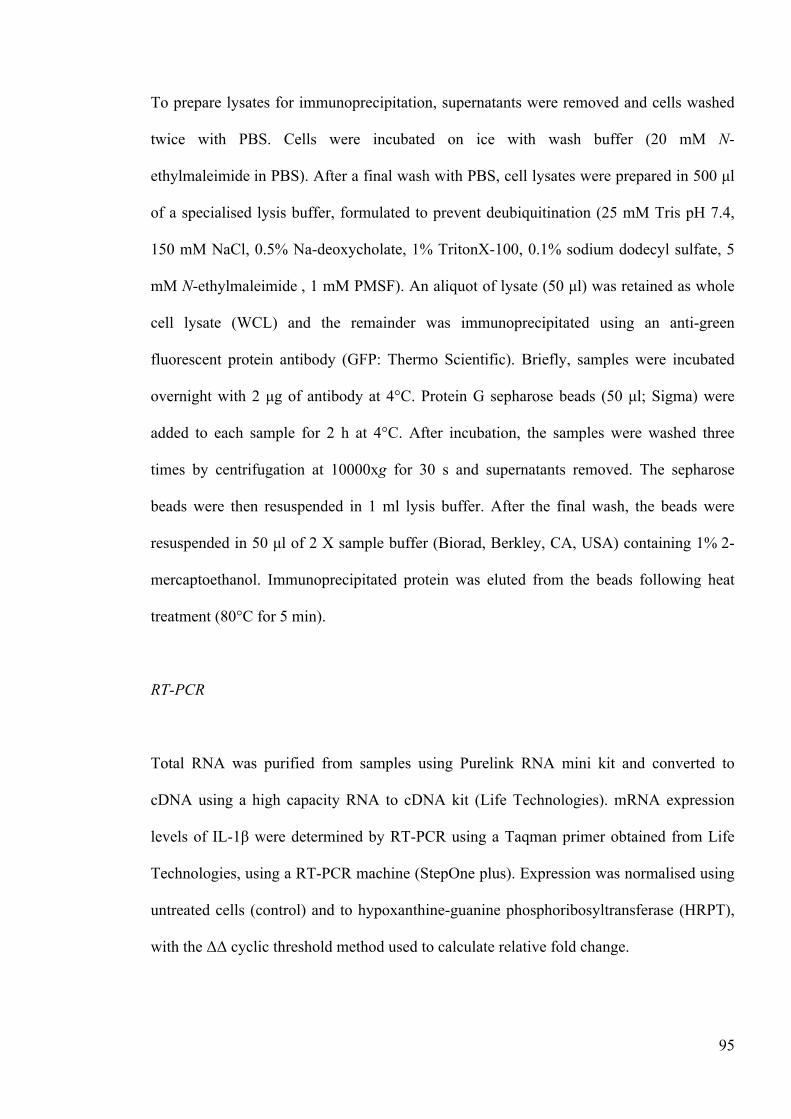
95
To prepare lysates for immunoprecipitation, supernatants were removed and cells washed
twice with PBS. Cells were incubated on ice with wash buffer (20 mM N-
ethylmaleimide in PBS). After a final wash with PBS, cell lysates were prepared in 500 µl
of a specialised lysis buffer, formulated to prevent deubiquitination (25 mM Tris pH 7.4,
150 mM NaCl, 0.5% Na-deoxycholate, 1% TritonX-100, 0.1% sodium dodecyl sulfate, 5
mM N-ethylmaleimide , 1 mM PMSF). An aliquot of lysate (50 µl) was retained as whole
cell lysate (WCL) and the remainder was immunoprecipitated using an anti-green
fluorescent protein antibody (GFP: Thermo Scientific). Briefly, samples were incubated
overnight with 2 µg of antibody at 4°C. Protein G sepharose beads (50 µl; Sigma) were
added to each sample for 2 h at 4°C. After incubation, the samples were washed three
times by centrifugation at 10000xg for 30 s and supernatants removed. The sepharose
beads were then resuspended in 1 ml lysis buffer. After the final wash, the beads were
resuspended in 50 µl of 2 X sample buffer (Biorad, Berkley, CA, USA) containing 1% 2-
mercaptoethanol. Immunoprecipitated protein was eluted from the beads following heat
treatment (80°C for 5 min).
RT-PCR
Total RNA was purified from samples using Purelink RNA mini kit and converted to
cDNA using a high capacity RNA to cDNA kit (Life Technologies). mRNA expression
levels of IL-1β were determined by RT-PCR using a Taqman primer obtained from Life
Technologies, using a RT-PCR machine (StepOne plus). Expression was normalised using
untreated cells (control) and to hypoxanthine-guanine phosphoribosyltransferase (HRPT),
with the ΔΔ cyclic threshold method used to calculate relative fold change.
96
Statistical analyses
Statistical analyses were performed using the software Graphpad Prism 6. Data were
analysed by one-way ANOVA to determine overall differences and a Tukey post-hoc test
was performed to determine statistically significant differences between treatment groups.
* = p<0.05, ** = p< 0.01.
3.5 Results
Venus pro-IL-1β is polyubiquitinated and degraded by the proteasome in iBMDM and
THP-1 cells
In previous investigations, pro-IL-1β has been shown to be polyubiquitinated and degraded
by the proteasome in murine dendritic cells (Ainscough et al. 2014). To examine whether
venus proIL-1β is under the same mechanisms of regulation in the transgenic iBMDM and
THP-1 cell lines, the proteasome inhibitor MG132 was utilised. In these experiments, cells
were pretreated with CHX to inhibit de novo protein synthesis and incubated with
increasing concentrations of MG132 for 4 h (Fig. 1A and B). Here, the addition of MG132
caused a significant, dose-dependent increase in intracellular IL-1β levels in both cell lines.
As protein translation was inhibited in these experiments, this increase could only be the
result of an inhibition of degradation. Thus, these data demonstrate that venus pro-IL-1β is
degraded by the proteasome in both iBMDM and THP-1 cells.
97
Figure 1. VenusIL-1β is polyubiquitinated and degraded by the proteasome in iBMDM and THP-1 cells 106 venusIL-1βiBMDM cells (106 cells/ml; A,C) or 106 venusIL-1βTHP-1 cells (106
cells/ml; B) were incubated with 10 µg/ml CHX for 4 h in the presence or absence of the proteasome inhibitor MG132 (0.1, 1 or 10 µM). Cell lysates were collected and analysed for the presence of IL-1β using a cytokine specific ELISA (A, B). Lysates were also analysed by Western blotting using an anti-IL-1β antibody (C). In addition, 107 venusIL-1βiBMDM (D) or 107 venusIL-1βTHP-1 (E) (both 106 cells/ml) were incubated for 4 h in the presence or absence of 10 µM of the proteasome inhibitor MG132. Cells were lyzed, an aliquot of lysate was retained as whole cell lysate (WCL) and the remainder was immunoprecipitated with an anti-GFP antibody. The same volume of lysis buffer alone was immunoprecipitated with anti-GFP antibody (immunoprecipitation negative control; IP-). The samples were analysed by Western blotting using an anti-IL-1β antibody or an anti-ubiquitin antibody. A protein marker lane on each gel was used to determine molecular weight. Representative blots are shown in each case. Data shown is mean ± SEM (n=3). Statistical significance between samples treated with CHX alone and CHX and MG132 was determined by one way ANOVA. *= p<0.05, **=p<0.01.
To investigate whether venus pro-IL-1β is also polyubiquitinated, the lysates from the
MG132-treated iBMDM were analysed using an anti-IL-1β Western blot. In this
experiment, venus pro-IL-1β appears as a 57kDa band in all four samples (Fig. 1C).
Importantly, the addition of either 1 or 10 µM of MG132 resulted in the appearance of a
98
distinct smear on the blot (60-250kDa). This smear is indicative of polyubiquitinated
protein and suggests that venus pro-IL-1β undergoes polyubiquitination prior to
proteasomal degradation. To support these data, a co-immunoprecipitation experiment was
performed. In this experiment, venusIL-1βiBMDM (Fig. 1D) or venusIL-1β THP-1 cells
(Fig. 1E) were incubated for 4 h in media with or without 10µM of MG132. These cells
were then lysed and venus pro-IL-1β immunoprecipitated using an anti-GFP antibody.
Venus Pro-IL-1β was successfully immunoprecipitated from these lysates, as demonstrated
using an anti-IL-1β Western blot. An anti-ubiquitin Western blot of the WCL confirmed
that each sample contained many ubiquitinated proteins, ranging from 50-250kDa in size
(Fig. 1D and E). The immunoprecipitated venus IL-1β from MG132 treated iBMDM and
THP-1 cells were also found to contain large amounts of ubiquitinated protein,
representing an accumulation of polyubiquitinated venus IL-1β in these samples. Levels of
polyubiquitinated venus IL-1β appear to be markedly higher in the MG132 treated
venusIL-1β THP-1 cells, relative to the MG132 treated venusIL-1βiBMDM. The most
likely reason for this is that basal venus IL-1β expression is much higher in the venusIL-
1βTHP-1 cells.
Together these data provide strong evidence that venus IL-1β is polyubiquitinated and
degraded in both iBMDM and THP-1 cells. Importantly, previous studies have shown that
the GFP fluorophore is extremely stable in a variety of cells (Corish and Tyler-Smith 1999,
Cubitt et al. 1995). As venus yellow fluorescent protein is just a simple derivative of GFP,
it is highly likely that this protein is also very stable (Nagai et al. 2002). Furthermore,
given that ubiquitination is such a protein specific process, it is highly unlikely that the
foreign venus yellow fluorescent protein fluorophore is processed by these mechanisms.
Thus, we can be confident that the proteasomal degradation of venus pro-IL-1β is driven
by the ubiquitination of IL-1β and not the venus fluorophore.
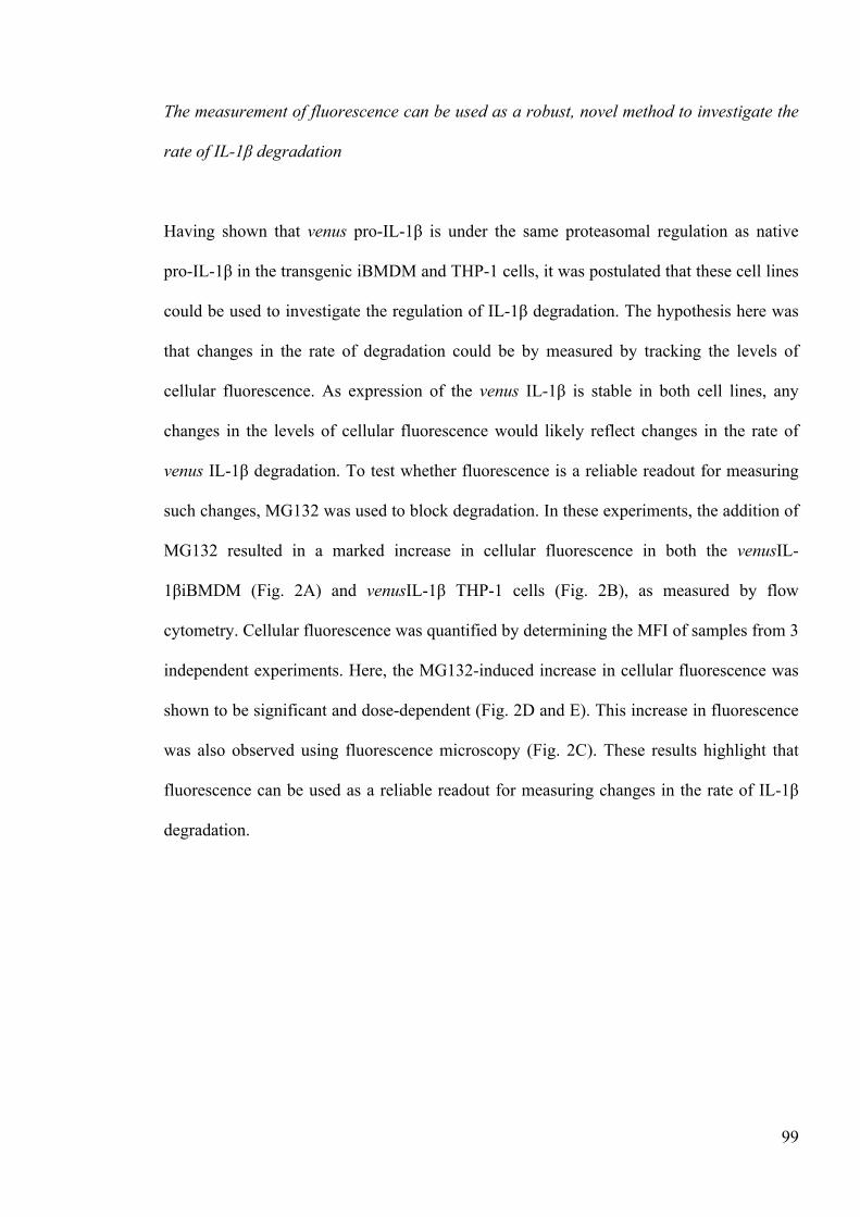
99
The measurement of fluorescence can be used as a robust, novel method to investigate the
rate of IL-1β degradation
Having shown that venus pro-IL-1β is under the same proteasomal regulation as native
pro-IL-1β in the transgenic iBMDM and THP-1 cells, it was postulated that these cell lines
could be used to investigate the regulation of IL-1β degradation. The hypothesis here was
that changes in the rate of degradation could be by measured by tracking the levels of
cellular fluorescence. As expression of the venus IL-1β is stable in both cell lines, any
changes in the levels of cellular fluorescence would likely reflect changes in the rate of
venus IL-1β degradation. To test whether fluorescence is a reliable readout for measuring
such changes, MG132 was used to block degradation. In these experiments, the addition of
MG132 resulted in a marked increase in cellular fluorescence in both the venusIL-
1βiBMDM (Fig. 2A) and venusIL-1β THP-1 cells (Fig. 2B), as measured by flow
cytometry. Cellular fluorescence was quantified by determining the MFI of samples from 3
independent experiments. Here, the MG132-induced increase in cellular fluorescence was
shown to be significant and dose-dependent (Fig. 2D and E). This increase in fluorescence
was also observed using fluorescence microscopy (Fig. 2C). These results highlight that
fluorescence can be used as a reliable readout for measuring changes in the rate of IL-1β
degradation.
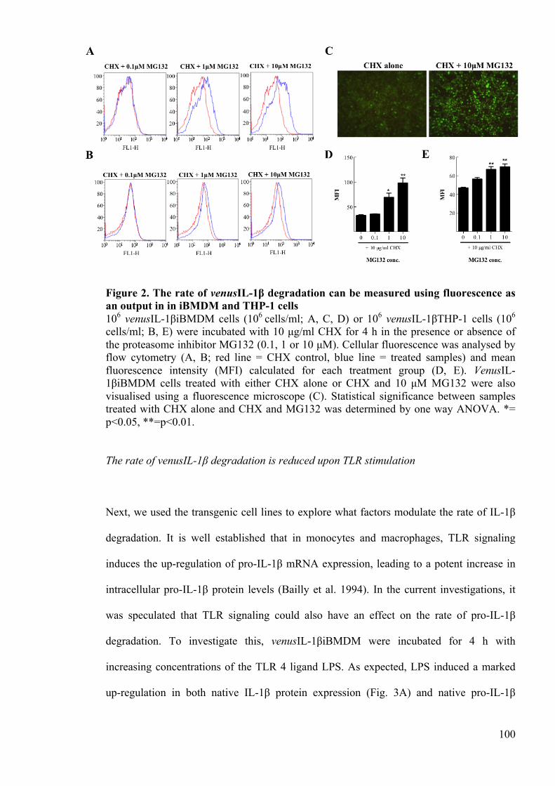
100
Figure 2. The rate of venusIL-1β degradation can be measured using fluorescence as an output in in iBMDM and THP-1 cells 106 venusIL-1βiBMDM cells (106 cells/ml; A, C, D) or 106 venusIL-1βTHP-1 cells (106
cells/ml; B, E) were incubated with 10 µg/ml CHX for 4 h in the presence or absence of the proteasome inhibitor MG132 (0.1, 1 or 10 µM). Cellular fluorescence was analysed by flow cytometry (A, B; red line = CHX control, blue line = treated samples) and mean fluorescence intensity (MFI) calculated for each treatment group (D, E). VenusIL-1βiBMDM cells treated with either CHX alone or CHX and 10 µM MG132 were also visualised using a fluorescence microscope (C). Statistical significance between samples treated with CHX alone and CHX and MG132 was determined by one way ANOVA. *= p<0.05, **=p<0.01.
The rate of venusIL-1β degradation is reduced upon TLR stimulation
Next, we used the transgenic cell lines to explore what factors modulate the rate of IL-1β
degradation. It is well established that in monocytes and macrophages, TLR signaling
induces the up-regulation of pro-IL-1β mRNA expression, leading to a potent increase in
intracellular pro-IL-1β protein levels (Bailly et al. 1994). In the current investigations, it
was speculated that TLR signaling could also have an effect on the rate of pro-IL-1β
degradation. To investigate this, venusIL-1βiBMDM were incubated for 4 h with
increasing concentrations of the TLR 4 ligand LPS. As expected, LPS induced a marked
up-regulation in both native IL-1β protein expression (Fig. 3A) and native pro-IL-1β
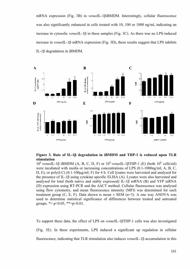
101
mRNA expression (Fig. 3B) in venusIL-1βiBMDM. Interestingly, cellular fluorescence
was also significantly enhanced in cells treated with 10, 100 or 1000 ng/ml, indicating an
increase in cytosolic venusIL-1β in these samples (Fig. 3C). As there was no LPS-induced
increase in venusIL-1β mRNA expression (Fig. 3D), these results suggest that LPS inhibits
IL-1β degradation in iBMDM.
Figure 3. Rate of IL-1β degradation in iBMDM and THP-1 is reduced upon TLR stimulation 106 venusIL-1β iBMDM (A, B, C, D, F) or 106 venusIL-1βTHP-1 (E) (both 106 cells/ml) were incubated with media or increasing concentrations of LPS (0.1-1000ng/ml; A, B, C, D, E), or poly(I:C) (0.1-100µg/ml; F) for 4 h. Cell lysates were harvested and analysed for the presence of IL-1β using cytokine specific ELISA (A). Lysates were also harvested and analysed for total (both native and stably expressed) IL-1β mRNA (B) and YFP mRNA (D) expression using RT-PCR and the ΔΔCT method. Cellular fluorescence was analysed using flow cytometry, and mean fluorescence intensity (MFI) was determined for each treatment group (C, E, F). Data shown is mean ± SEM (n=3). A one way ANOVA was used to determine statistical significance of differences between treated and untreated groups. *= p<0.05, **=p<0.01.
To support these data, the effect of LPS on venusIL-1βTHP-1 cells was also investigated
(Fig. 3E). In these experiments, LPS induced a significant up regulation in cellular
fluorescence, indicating that TLR stimulation also induces venusIL-1β accumulation in this
102
cell line. Again, LPS had no effect on venusIL-1β mRNA expression (data not shown),
suggesting that TLR stimulation also inhibits IL-1β degradation in THP-1 cells. To
confirm that this was an effect of TLR ligands other than LPS, the same experiments were
performed using the TLR 3 ligand Poly(I:C) on iBMDM (Fig. 3F). Here, the addition of
Poly(I:C) also induced a significant increase in cellular fluorescence at the highest dose
tested (100 µg/ml). Together, these data demonstrate that TLR ligands inhibit the
degradation of IL-1β in murine and human cells, at least in the first 4 h following
exposure.
TLR stimulation inhibits IL-1β polyubiquitination
Having shown that TLR stimulation appears to inhibit venus IL-1β degradation, the
imperative was to decipher the mechanisms behind this observation. One suggestion was
that the TLR ligand-induced up-regulation in native IL-1β protein overloads the IL-1β
degradation machinery, resulting in an accumulation of the stably expressed protein.
Another possibility was that TLR signaling actively inhibits polyubiquitination or
proteasomal degradation, either specifically or globally. To investigate these hypotheses,
the lysates from LPS-stimulated iBMDM were analysed using an anti IL-1β Western blot
(Fig. 4A). In this experiment, LPS induced a dose dependent increase in the native 31kDa
pro-IL-1β protein expression. LPS also induced a dose dependent increase in the amount of
stably-expressed 57kDa venus pro-IL-1β, supporting the evidence that TLR stimulation
inhibits degradation of venus pro-IL-1β. In the untreated cells, a smear of IL-1β protein
was observed between 50-250kDa, indicating the presence of ubiquitinated IL-1β in this
sample. This observation suggests that there is a basal level of IL-1β polyubiquitination
and proteasomal degradation in iBMDM, functioning to maintain a steady rate of IL-1β
turnover.
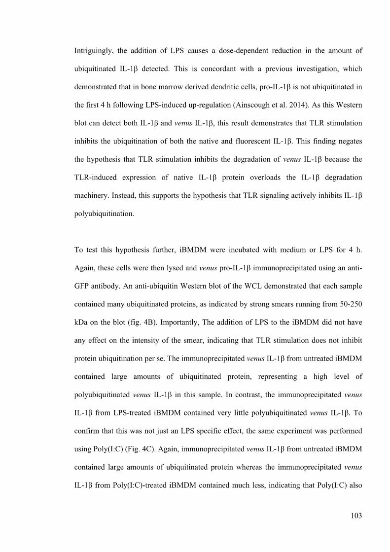
103
Intriguingly, the addition of LPS causes a dose-dependent reduction in the amount of
ubiquitinated IL-1β detected. This is concordant with a previous investigation, which
demonstrated that in bone marrow derived dendritic cells, pro-IL-1β is not ubiquitinated in
the first 4 h following LPS-induced up-regulation (Ainscough et al. 2014). As this Western
blot can detect both IL-1β and venus IL-1β, this result demonstrates that TLR stimulation
inhibits the ubiquitination of both the native and fluorescent IL-1β. This finding negates
the hypothesis that TLR stimulation inhibits the degradation of venus IL-1β because the
TLR-induced expression of native IL-1β protein overloads the IL-1β degradation
machinery. Instead, this supports the hypothesis that TLR signaling actively inhibits IL-1β
polyubiquitination.
To test this hypothesis further, iBMDM were incubated with medium or LPS for 4 h.
Again, these cells were then lysed and venus pro-IL-1β immunoprecipitated using an anti-
GFP antibody. An anti-ubiquitin Western blot of the WCL demonstrated that each sample
contained many ubiquitinated proteins, as indicated by strong smears running from 50-250
kDa on the blot (fig. 4B). Importantly, The addition of LPS to the iBMDM did not have
any effect on the intensity of the smear, indicating that TLR stimulation does not inhibit
protein ubiquitination per se. The immunoprecipitated venus IL-1β from untreated iBMDM
contained large amounts of ubiquitinated protein, representing a high level of
polyubiquitinated venus IL-1β in this sample. In contrast, the immunoprecipitated venus
IL-1β from LPS-treated iBMDM contained very little polyubiquitinated venus IL-1β. To
confirm that this was not just an LPS specific effect, the same experiment was performed
using Poly(I:C) (Fig. 4C). Again, immunoprecipitated venus IL-1β from untreated iBMDM
contained large amounts of ubiquitinated protein whereas the immunoprecipitated venus
IL-1β from Poly(I:C)-treated iBMDM contained much less, indicating that Poly(I:C) also
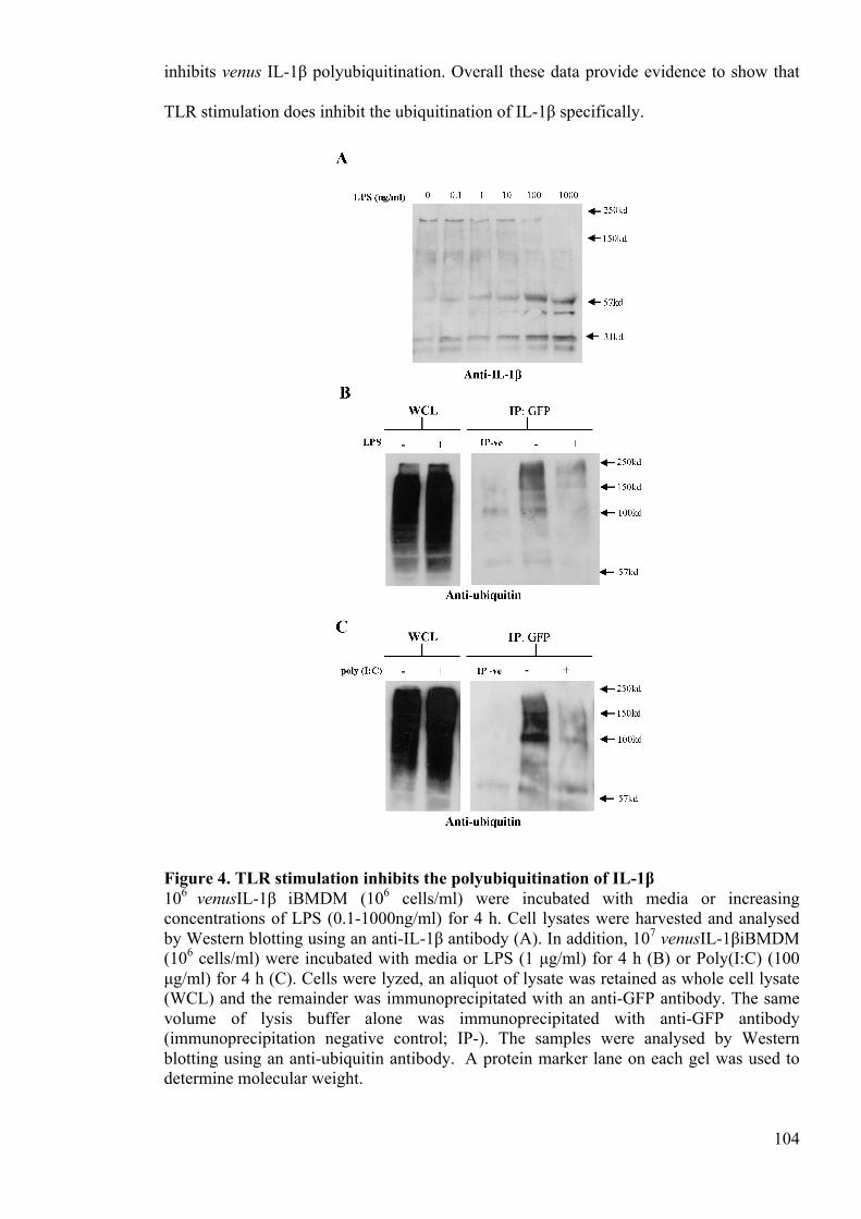
104
inhibits venus IL-1β polyubiquitination. Overall these data provide evidence to show that
TLR stimulation does inhibit the ubiquitination of IL-1β specifically.
Figure 4. TLR stimulation inhibits the polyubiquitination of IL-1β 106 venusIL-1β iBMDM (106 cells/ml) were incubated with media or increasing concentrations of LPS (0.1-1000ng/ml) for 4 h. Cell lysates were harvested and analysed by Western blotting using an anti-IL-1β antibody (A). In addition, 107 venusIL-1βiBMDM (106 cells/ml) were incubated with media or LPS (1 µg/ml) for 4 h (B) or Poly(I:C) (100 µg/ml) for 4 h (C). Cells were lyzed, an aliquot of lysate was retained as whole cell lysate (WCL) and the remainder was immunoprecipitated with an anti-GFP antibody. The same volume of lysis buffer alone was immunoprecipitated with anti-GFP antibody (immunoprecipitation negative control; IP-). The samples were analysed by Western blotting using an anti-ubiquitin antibody. A protein marker lane on each gel was used to determine molecular weight.
105
The TLR induced inhibition of IL-1β polyubiquitination and degradation is transient
Having shown that IL-1β ubiquitination is inhibited 4 h after LPS stimulation, it was of
interest explore the kinetics of this mechanism. A previous study in this laboratory has
shown that TLR-induced IL-1 protein expression is characterised typically by a potent up-
regulation, followed by rapid degradation (Ainscough et al. 2014). It was postulated in this
work that the rate and timing of IL-1 ubiquitination could have a significant effect on both
the vigour of IL-1 up-regulation and the rate of IL-1 clearance. To test these hypotheses,
iBMDM were incubated with media, LPS or Poly(I:C) for 2 h, 4 h, 8 h or 12 h and the
changes in IL-1β protein expression assessed. Here, stimulation of iBMDM with LPS or
Poly(I:C) caused an up-regulation in intracellular IL-1β expression, without inducing
secretion. As observed previously, the expression of IL-1β was transient, peaking at 8 h
post-stimulation and decreasing thereafter (Fig. 5A). Furthermore, the treatment of
iBMDM with LPS or Poly(I:C) also caused an increase in cellular fluorescence, supporting
the results indicating that TLR stimulation inhibits IL-1β degradation (Fig. 5B).
Interestingly, cellular fluorescence is also transient, peaking at 8 h and decreasing
thereafter. Thus, it is postulated that TLR-induced inhibition of IL-1β degradation is also
transient.
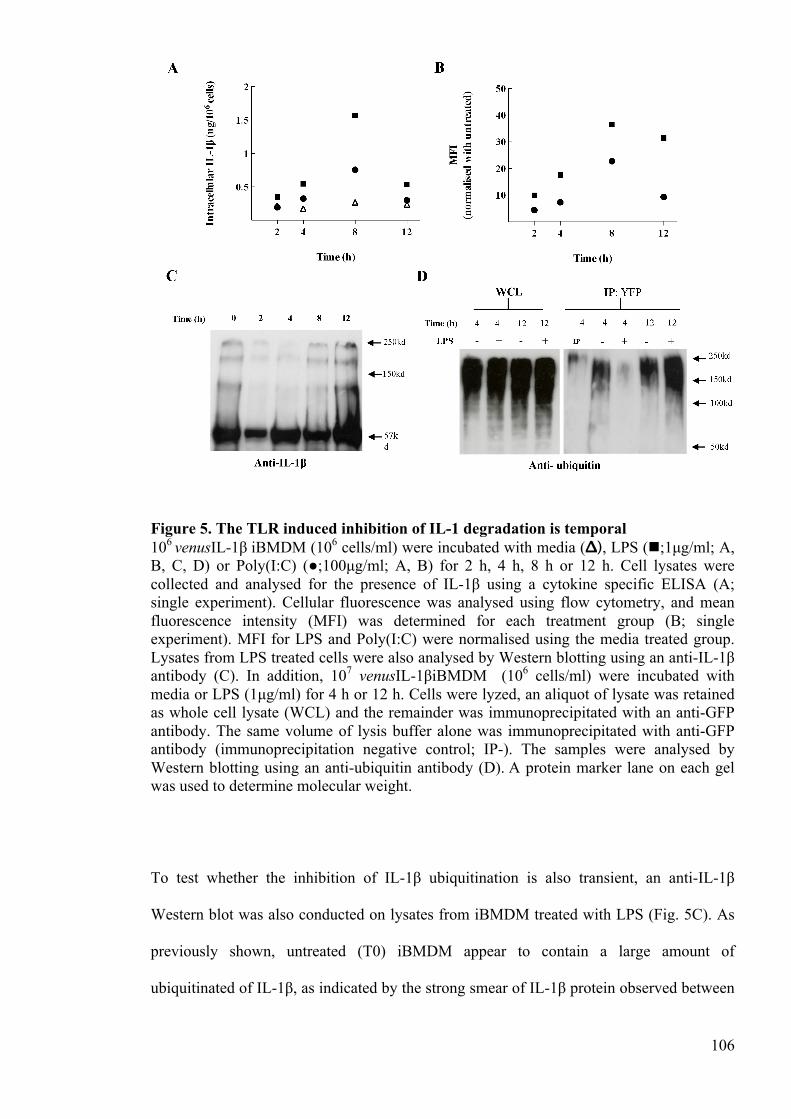
106
Figure 5. The TLR induced inhibition of IL-1 degradation is temporal 106 venusIL-1β iBMDM (106 cells/ml) were incubated with media (Δ), LPS (!;1µg/ml; A, B, C, D) or Poly(I:C) (●;100µg/ml; A, B) for 2 h, 4 h, 8 h or 12 h. Cell lysates were collected and analysed for the presence of IL-1β using a cytokine specific ELISA (A; single experiment). Cellular fluorescence was analysed using flow cytometry, and mean fluorescence intensity (MFI) was determined for each treatment group (B; single experiment). MFI for LPS and Poly(I:C) were normalised using the media treated group. Lysates from LPS treated cells were also analysed by Western blotting using an anti-IL-1β antibody (C). In addition, 107 venusIL-1βiBMDM (106 cells/ml) were incubated with media or LPS (1µg/ml) for 4 h or 12 h. Cells were lyzed, an aliquot of lysate was retained as whole cell lysate (WCL) and the remainder was immunoprecipitated with an anti-GFP antibody. The same volume of lysis buffer alone was immunoprecipitated with anti-GFP antibody (immunoprecipitation negative control; IP-). The samples were analysed by Western blotting using an anti-ubiquitin antibody (D). A protein marker lane on each gel was used to determine molecular weight.
To test whether the inhibition of IL-1β ubiquitination is also transient, an anti-IL-1β
Western blot was also conducted on lysates from iBMDM treated with LPS (Fig. 5C). As
previously shown, untreated (T0) iBMDM appear to contain a large amount of
ubiquitinated of IL-1β, as indicated by the strong smear of IL-1β protein observed between
107
50-250kDa. The incubation of iBMDM with LPS for 2 or 4 h caused a marked reduction in
the amount of ubiquitinated IL-1β detected, in concordance with the results in figure 4.
Again, this reduction is transient, with the level of ubiquitinated IL-1β recovering partly at
8 h and completely by 12 h. To support these data, a co-immunoprecipitation experiment
was conducted, whereby iBMDM were incubated with and without LPS for 4 h or 12 h
(Fig. 5D). An anti-ubiquitin Western blot of the WCL confirmed that each iBMDM sample
contained many ubiquitinated proteins, ranging from 50 to 250 kDa in size. As observed in
Figure 4, the venus IL-1β immunoprecipitated from untreated iBMDM contained large
amounts of ubiquitinated protein, representing an accumulation of ubiquitinated IL-1β. The
venus IL-1β immunoprecipitated from iBMDM treated with LPS for 4 h did not contain a
smear of ubiquitinated IL-1β, whereas the venus IL-1β immunoprecipitated from iBMDM
treated with LPS for 12 h contained more ubiquitinated IL-1β than the untreated iBMDM
samples. Together, these results indicate that the observed TLR-ligand induced inhibition
of IL-1β ubiquitination and degradation is transient.
3.6 Discussion
IL-1β is a potent pro-inflammatory cytokine, and thus the mechanisms that serve to
regulate this protein intracellularly are of great interest. In a previous investigation, we
showed that IL-1β is polyubiquitinated and degraded by the proteasome in murine
dendritic cells and macrophages (Ainscough et al. 2014). The current study demonstrates
that IL-1β tagged with the venus fluorophore is also polyubiquitinated and degraded by the
proteasome in murine macrophages and human monocytes. Primarily, these findings
support the work presented in our previous investigations and indicate that mechanisms of
IL-1β polyubiquitination and proteasomal degradation serve to regulate IL-1β protein
expression in a range of cell subsets and species.
108
The ubiquitin-proteasome system is a well-established cellular process that regulates the
turnover of around 80% of all intracellular proteins (Hochstrasser 1995). This turnover is
important in a number of ways; it serves to inhibit the potentially harmful accumulation of
proteins, it recycles amino acids for de novo protein synthesis and it removes misfolded
proteins (Lecker et al. 2006). In the context of IL-1, it is clear that ubiquitination plays an
important role in preventing the intracellular accumulation of pro-IL-1. The importance of
this clearance is obvious when the effects of IL-1 dysregulation are considered (Dinarello
et al. 2012). However, given that the requirement for IL-1 regulation is so great, it was
hypothesised that IL-1 ubiquitination and degradation may also function beyond this role.
The findings of this study support this hypothesis, showing that TLR stimulation inhibits
transiently the ubiquitination and degradation of pro-IL-1β. It is proposed here that this
inhibition serves to facilitate a more potent TLR-induced IL-1β protein up-regulation.
Thus, these data show for the first time that IL-1 ubiquitination functions in concert with
mRNA expression to regulate the strength of IL-1β up-regulation.
Given that the TLR-induced inhibition of IL-1β ubiquitination may serve a number of
important purposes, it is of interest to consider the mechanisms that regulate this process.
In the current investigations, we show that TLR stimulation inhibits IL-1β ubiquitination
but does not inhibit protein ubiquitination per se. Therefore, it is likely that TLR-
stimulation inhibits a mechanism that drives the specific ubiquitination of IL-1β. As the E3
ubiquitin ligases are relatively specific for the protein that they ubiquitinate (Laney and
Hochstrasser 1999), one hypothesis is that TLR-stimulation inhibits the E3 ubiquitin-ligase
dependent ubiquitination of IL-1β, either by inhibiting the expression or the activity of the
enzyme. An alternative hypothesis is that TLR-stimulation induces the expression or
activity of deubiquitinase enzymes. Deubiquitinases are an important family of enzymes
that function to deubiquitinate specific proteins (Love et al. 2007). Thus, a TLR-induced
109
increase in the expression or activity of an IL-1β-specific deubiquitinase could explain the
results aforementioned. This is, however, speculative and thus requires further
investigation.
Having determined that IL-1β ubiquitination serves to regulate the vigour of IL-1β protein
expression, it is important to consider its purpose of this mechanism in vivo. It is clear that
a rapid pro-inflammatory response is vital to host defence against invading pathogens. The
speed of the inflammatory response is evident in Lu et al, whereby the injection of an
aluminium adjuvant resulted in inflammatory cell infiltration within 2 h of the insult (Lu
and HogenEsch 2013). Given that IL-1β is such an important inducer of inflammation, it is
postulated here that the speed of IL-1β secretion is of great importance to the effectiveness
of the pro-inflammatory response. As discussed previously, pro-IL-1β protein levels must
be up-regulated intracellularly before the cytokine can be processed and released
(Rubartelli et al. 1990, Stevenson et al. 1992). Thus, in light of the evidence presented, it is
proposed that the TLR-induced inhibition of IL-1β degradation serves not only to augment
a more potent IL-1β production, but also to increase the speed of IL-1β protein production.
This mechanism may be of particular benefit in cells that constitutively produce IL-1
cytokines. This is because, in these cells, the TLR-induced inhibition of IL-1 degradation
could serve to up-regulate pro-IL-1 levels before TLR-induced expression of the precursor.
From a broader perspective, the mechanisms of IL-1β ubiquitination may have additional
roles to play in the regulation of inflammation. It is well established that some cells and
tissues are better at producing and secreting IL-1β than others (Netea et al. 2009). This
differential in the capacity to produce and release IL-1β is important, as the inflammatory
response induced by a cell must reflect its environment and its role within that
environment. The ability of a cell to produce and secrete IL-1β is determined by a number
110
of factors, including TLR expression (Muzio et al. 2000), NLR expression (Guarda et al.
2011), basal caspase-1 activity (Netea et al. 2009) and post-transcriptional modification
(Kobayashi et al. 1988). Here, we hypothesise that the mechanisms of IL-1β ubiquitination
may also serve to regulate differentially the vigour of IL-1β up-regulation in different cells
and tissues. One potential benefit of this mode of regulation is that it would allow
regulation of IL-1β independently of other pro-inflammatory proteins.
The dysregulation of IL-1β is associated with a number of important diseases, thus the
findings presented here are of great interest. Ulcerative colitis and Crohns disease are both
common forms of chronic inflammatory bowel disease (IBD) (El-Salhy 2012). In both of
these disorders, the expression of IL-1β is significantly higher in the intestines of IBD
patients, relative to expression in the intestines of healthy controls (Casiniraggi et al.
1995). Importantly, the administration of IL-1 inhibitors has a positive impact on a number
of experimental models of IBD (Sims and Smith 2010). Likewise, an increase in IL-1
expression is also associated with a number of other important diseases, including asthma
(Mao et al. 2000), Parkinson’s Disease (McGeer and McGeer 2004), Alzheimer’s Disease
(Cacabelos et al. 1991), arthritis (Kay and Calabrese 2004) and multiple sclerosis (Rossi et
al. 2014). Therefore, there is an imperative to develop therapeutics that limit IL-1β protein
expression. In light of the findings in the current study, it is proposed that IL-1β protein
levels could be down-regulated by increasing the ubiquitination of the cytokine.
Furthermore, it is proposed that the dysregulation of IL-1β ubiquitination could be a
causative factor in the development of certain inflammatory disorders.
To conclude, the current investigations demonstrate that IL-1β ubiquitination is inhibited
transiently following TLR stimulation, facilitating a potent up-regulation of the precursor.
These findings highlight that IL-1β ubiquitination can function as an active regulator of IL-
111
1β up-regulation, adding to a growing body of work that suggests that ubiquitination serves
as a fundamental mediator of innate immunity.
3.7 Acknowledgements
JS Ainscough was funded by the Medical Research Council (MRC) and The Procter &
Gamble Company. We would like to thank Dr. David Brough for the provision of the
venus iBMDM cell line.
3.8 References
Ainscough, J. S., Gerberick, G. F., Zahedi-Nejad, M., Lopez-Castejon, G., Brough, D., Kimber, I. and Dearman, R. J. (2014) 'Dendritic Cell IL-1 alpha and IL-1 beta Are Polyubiquitinated and Degraded by the Proteasome', Journal of Biological Chemistry, 289(51), 35582-35592.
Baeuerle, P. A. and Baltimore, D. (1988) 'Activation of dna-binding activity in an
apparently cytoplasmic precursor of the nf-kappa-b transcription factor', Cell, 53(2), 211-217.
Bagnall, J., Boddington, C., Boyd, J., Brignall, R., Rowe, W., Jones, N. A., Schmidt, L.,
Spiller, D. G., White, M. R. H. and Paszek, P. (2015) 'Quantitative dynamic imaging of immune cell signalling using lentiviral gene transfer', Integrative Biology, 7(6), 713-725.
Bailly, S., Fay, M., Ferrua, B. and Gougerotpocidalo, M. A. (1994) 'Comparative
production of Il-1-beta and Il-1-alpha LPS-stimulated human monocytes- ELISAs measurement revisited', Cytokine, 6(2), 111-115.
Bryant, C. and Fitzgerald, K. A. (2009) 'Molecular mechanisms involved in inflammasome
activation', Trends in Cell Biology, 19(9), 455-464. Cacabelos, R., Francomaside, A. and Alvarez, X. A. (1991) 'Interleukin-1 in Alzheimers-
Disease and Multiinfarct Dementia - neuropsychological correlations', Methods and Findings in Experimental and Clinical Pharmacology, 13(10), 703-708.
Casiniraggi, V., Kam, L., Chong, Y. J. T., Fiocchi, C., Pizarro, T. T. and Cominelli, F.
(1995) 'Mucosal imbalance of Il-1 and Il-1 receptor antagonist in inflammatory bowel-disease- a novel mechanism of chronic intestinal inflammation', Journal of Immunology, 154(5), 2434-2440.
Cogswell, J. P., Godlevski, M. M., Wisely, G. B., Clay, W. C., Leesnitzer, L. M., Ways, J.
P. and Gray, J. G. (1994) 'NF-κB regulates IL-1β transcription through a
112
consensus NF-κB binding-site and a nonconsensus Cre-like site', Journal of Immunology, 153(2), 712-723.
Corish, P. and Tyler-Smith, C. (1999) 'Attenuation of green fluorescent protein half-life in
mammalian cells', Protein Engineering, 12(12), 1035-1040. Cubitt, A. B., Heim, R., Adams, S. R., Boyd, A. E., Gross, L. A. and Tsien, R. Y. (1995)
'Understanding, improving and using green fluorescent proteins', Trends in Biochemical Sciences, 20(11), 448-455.
Deng, L., Wang, C., Spencer, E., Yang, L. Y., Braun, A., You, J. X., Slaughter, C., Pickart,
C. and Chen, Z. J. (2000) 'Activation of the I kappa B kinase complex by TRAF6 requires a dimeric ubiquitin-conjugating enzyme complex and a unique polyubiquitin chain', Cell, 103(2), 351-361.
Dinarello, C. A. (2009) 'Immunological and inflammatory functions of the Interleukin-1
family', Annual Review of Immunology, 27, 519-550. Dinarello, C. A. (2011) 'Interleukin-1 in the pathogenesis and treatment of inflammatory
diseases', Blood, 117(14), 3720-3732. Dinarello, C. A., Simon, A. and van der Meer, J. W. M. (2012) 'Treating inflammation by
blocking interleukin-1 in a broad spectrum of diseases', Nature Reviews Drug Discovery, 11(8), 633-652.
El-Salhy, M. (2012) 'Irritable bowel syndrome: Diagnosis and pathogenesis', World
Journal of Gastroenterology, 18(37), 5151-5163. Gallucci, S. and Matzinger, P. (2001) 'Danger signals: SOS to the immune system',
Current Opinion in Immunology, 13(1), 114-119. Garlanda, C., Dinarello, C. A. and Mantovani, A. (2013) 'The Interleukin-1 Family: Back
to the Future', Immunity, 39(6), 1003-1018. Guarda, G., Zenger, M., Yazdi, A. S., Schroder, K., Ferrero, I., Menu, P., Tardivel, A.,
Mattmann, C. and Tschopp, J. (2011) 'Differential Expression of NLRP3 among Hematopoietic Cells', Journal of Immunology, 186(4), 2529-2534.
Hochstrasser, M. (1995) 'Ubiquitin, proteasomes, and the regulation of intracellular protein
degradation', Current Opinion in Cell Biology, 7(2), 215-223. Kanayama, A., Seth, R. B., Sun, L. J., Ea, C. K., Hong, M., Shaito, A., Chiu, Y. H., Deng,
L. and Chen, Z. J. (2004) 'TAB2 and TAB3 activate the NF-κB pathway through binding to polyubiquitin chains', Molecular Cell, 15(4), 535-548.
Katzen, F. (2007) 'Gateway (R) recombinational cloning: a biological operating system',
Expert Opinion on Drug Discovery, 2(4), 571-589. Kay, J. and Calabrese, L. (2004) 'The role of interleukin-1 in the pathogenesis of
rheumatoid arthritis', Rheumatology, 43, 2-9.
113
Kobayashi, Y., Appella, E., Yamada, M., Copeland, T. D., Oppenheim, J. J. and Matsushima, K. (1988) 'Phosphorylation of intracellular precursors of IL-1', Journal of Immunology, 140(7), 2279-2287.
Labbe, K., McIntire, C. R., Doiron, K., Leblanc, P. M. and Saleh, M. (2011) 'Cellular
Inhibitors of Apoptosis Proteins cIAP1 and cIAP2 Are Required for Efficient Caspase-1 Activation by the Inflammasome', Immunity, 35(6), 897-907.
Laney, J. D. and Hochstrasser, M. (1999) 'Substrate targeting in the ubiquitin system', Cell,
97(4), 427-430. Latz, E. (2010) 'The inflammasomes: mechanisms of activation and function', Current
Opinion in Immunology, 22(1), 28-33. Lecker, S. H., Goldberg, A. L. and Mitch, W. E. (2006) 'Protein degradation by the
ubiquitin-proteasome pathway in normal and disease states', Journal of the American Society of Nephrology, 17(7), 1807-1819.
Love, K. R., Catic, A., Schlieker, C. and Ploegh, H. L. (2007) 'Mechanisms, biology and
inhibitors of deubiquitinating enzymes', Nature Chemical Biology, 3(11), 697-705. Lu, F. and HogenEsch, H. (2013) 'Kinetics of the inflammatory response following
intramuscular injection of aluminum adjuvant', Vaccine, 31(37), 3979-3986. Mao, X. Q., Kawai, M., Yamashita, T., Enomoto, T., Dake, Y., Sasaki, S., Kataoka, Y.,
Fukuzumi, T., Endo, K., Sano, H., Aoki, T., Kurimoto, F., Adra, C. N., Shirakawa, T. and Hopkin, J. M. (2000) 'Imbalance production between interleukin-1 beta (IL-1 beta) and IL-1 receptor antagonist (IL-1Ra) in bronchial asthma', Biochemical and Biophysical Research Communications, 276(2), 607-612.
March, C. J., Mosley, B., Larsen, A., Cerretti, D. P., Braedt, G., Price, V., Gillis, S.,
Henney, C. S., Kronheim, S. R., Grabstein, K., Conlon, P. J., Hopp, T. P. and Cosman, D. (1985) 'Cloning, sequence and expression of 2 distinct human interleukin-1 complementary DNAs', Nature, 315(6021), 641-647.
McGeer, P. L. and McGeer, E. G. (2004) 'Inflammation and neurodegeneration in
Parkinson's disease', Parkinsonism & Related Disorders, 10, S3-S7. Muzio, M., Bosisio, D., Polentarutti, N., D'Amico, G., Stoppacciaro, A., Mancinelli, R.,
van't Veer, C., Penton-Rol, G., Ruco, L. P., Allavena, P. and Mantovani, A. (2000) 'Differential expression and regulation of toll-like receptors (TLR) in human leukocytes: Selective expression of TLR3 in dendritic cells', Journal of Immunology, 164(11), 5998-6004.
Nagai, T., Ibata, K., Park, E. S., Kubota, M., Mikoshiba, K. and Miyawaki, A. (2002) 'A
variant of yellow fluorescent protein with fast and efficient maturation for cell-biological applications', Nature Biotechnology, 20(1), 87-90.
Netea, M. G., Nold-Petry, C. A., Nold, M. F., Joosten, L. A. B., Opitz, B., van der Meer, J.
H. M., van de Veerdonk, F. L., Ferwerda, G., Heinhuis, B., Devesa, I., Funk, C. J., Mason, R. J., Kullberg, B. J., Rubartelli, A., van der Meer, J. W. M. and Dinarello, C. A. (2009) 'Differential requirement for the activation of the inflammasome for
114
processing and release of IL-1β in monocytes and macrophages', Blood, 113(10), 2324-2335.
Pickart, C. M. (2001) 'Mechanisms underlying ubiquitination', Annual Review of
Biochemistry, 70, 503-533. Pickart, C. M. and Fushman, D. (2004) 'Polyubiquitin chains: polymeric protein signals',
Current Opinion in Chemical Biology, 8(6), 610-616. Py, B. F., Kim, M.-S., Vakifahmetoglu-Norberg, H. and Yuan, J. (2013) 'Deubiquitination
of NLRP3 by BRCC3 Critically Regulates Inflammasome Activity', Molecular Cell, 49(2), 331-338.
Qian, Y. C., Commane, M., Ninomiya-Tsuji, J., Matsumoto, K. and Li, X. X. (2001)
'IRAK-mediated translocation of TRAF6 and TAB2 in the interleukin-1-induced activation of N-F kappa B', Journal of Biological Chemistry, 276(45), 41661-41667.
Rossi, S., Studer, V., Motta, C., Germani, G., Macchiarulo, G., Buttari, F., Mancino, R.,
Castelli, M., De Chiara, V., Weiss, S., Martino, G., Furlan, R. and Centonze, D. (2014) 'Cerebrospinal fluid detection of interleukin-1 beta in phase of remission predicts disease progression in multiple sclerosis', Journal of Neuroinflammation, 11.
Rubartelli, A., Cozzolino, F., Talio, M. and Sitia, R. (1990) 'A novel secretory pathway for
Interleukin-1β, a protein lacking A signal sequence', Embo Journal, 9(5), 1503-1510.
Sims, J. E. and Smith, D. E. (2010) 'The IL-1 family: regulators of immunity', Nature
Reviews Immunology, 10(2), 89-102. Stevenson, F. T., Torrano, F., Locksley, R. M. and Lovett, D. H. (1992) 'Interleukin-1- the
patterns of translation and intracellular distribution support alternative secretory mechanisms', Journal of Cellular Physiology, 152(2), 223-231.
Thornberry, N. A., Bull, H. G., Calaycay, J. R., Chapman, K. T., Howard, A. D., Kostura,
M. J., Miller, D. K., Molineaux, S. M., Weidner, J. R., Aunins, J., Elliston, K. O., Ayala, J. M., Casano, F. J., Chin, J., Ding, G. J. F., Egger, L. A., Gaffney, E. P., Limjuco, G., Palyha, O. C., Raju, S. M., Rolando, A. M., Salley, J. P., Yamin, T. T., Lee, T. D., Shively, J. E., Maccross, M., Mumford, R. A., Schmidt, J. A. and Tocci, M. J. (1992) 'A novel heterodimeric cysteine protease is required for interleukin-1-beta processing in monocytes', Nature, 356(6372), 768-774.
Tracy, R. P. (2006) 'The five cardinal signs of inflammation: Calor, dolor, rubor, tumor...
and penuria (apologies to Aulus Cornelius Celsus, De medicina, c. AD 25)', Journals of Gerontology Series a-Biological Sciences and Medical Sciences, 61(10), 1051-1052.
Wilkinson, K. D. (2000) 'Ubiquitination and deubiquitination: Targeting of proteins for
degradation by the proteasome', Seminars in Cell & Developmental Biology, 11(3), 141-148.

115
Wilson, K. P., Black, J. A. F., Thomson, J. A., Kim, E. E., Griffith, J. P., Navia, M. A., Murcko, M. A., Chambers, S. P., Aldape, R. A., Raybuck, S. A. and Livingston, D. J. (1994) 'Structure and mechanism of interleukin-1-beta converting-enzyme', Nature, 370(6487), 270-275.
116
CHAPTER 4:
INTERLEUKIN-1β PROCESSING IS DEPENDENT UPON A CALCIUM-MEDIATED INTERACTION WITH
CALMODULIN
117
4 Paper 3: Interleukin-1β Processing is Dependent Upon a Calcium-
Mediated Interaction With Calmodulin
Joseph S. Ainscough1, G. Frank Gerberick2, Ian Kimber1 and Rebecca J. Dearman1
1 Faculty of Life Sciences, University of Manchester, Manchester
2 The Procter & Gamble Company Co., Cincinnati, OH 45253, USA
Running title: Interleukin-1β Processing is Dependent Upon Calmodulin
Key words: Interleukin-1, IL-1β, Calmodulin, Inflammasome, Calcium, Caspase-1
Paper submitted to the Journal of Biological Chemistry
4.1 Abbreviations
ASC Apoptosis speck like protein containing a CARD DAMP Damage associated molecular patterns NLR NOD-like receptor
NF-κB Nuclear factor kappa-light-chain-enhancer of activated B cells PAMP Pathogen associated molecular patterns PRR Pattern recognition receptors POGZ Pogo transposable element containing a zinc finger domain SNR Signal to noise ratio SP1 Specificity protein 1 4.2 JBC standard abbreviations
ATP Adenosine triphosphate BSA Bovine serum albumin CARD Caspase activation and recruitment domain DMSO Dimethyl sulfoxide ELISA Enzyme-linked immunosorbent assay EDTA Ethylenediaminetetraacetic acid EGTA Ethylene glycol tetraacetic acid FCS Fetal calf serum HRP Horseradish peroxidase Ig Immunoglobulin IL Interleukin
118
LPS Lipopolysaccharide PMSF Phenylmethanesulfonylfluoride PBS Phosphate buffered saline SEM Standard error of the mean
4.3 Capsule
Background: iThe processing and secretion of IL-1β is an important process in the
induction of inflammation.
Results: Pro-IL-1β interacts with calmodulin, and the inhibition of calmodulin abrogates
IL-1β processing.
Conclusion: The interaction between calmodulin and IL-1β is required for IL-1β
processing.
Significance: Calmodulin is an important mediator of IL-1β processing and therefore may
be important in the development of inflammation.
4.4 Abstract
The secretion of IL-1β is a central event in the initiation of inflammation. Unlike most
other cytokines, the secretion of IL-1β requires two signals; one signal to induce the
intracellular up-regulation of pro-IL-1β, and a second signal to drive secretion of the
bioactive molecule. The release of pro-IL-1β is a complex process involving proteolytic
cleavage by caspase-1. However, the exact mechanism of secretion is poorly understood.
Here, we sought to identify novel proteins involved in IL-1β secretion and intracellular
processing in order to gain further insight into the mechanism of IL-1 release. A human
proteome microarray containing 19,951 unique proteins was used to identify proteins that
bind human recombinant pro-IL-1β. Probes with a signal to noise ration of >3 were
defined as relevant biologically. In these analyses, calmodulin was identified as a
119
particularly strong hit, with a SNR of ~11. Using an ELISA-based protein-binding assay,
the interaction of recombinant calmodulin with pro-IL-1β, but not mature IL-1β, was
confirmed and shown to be calcium dependent. Finally, using small molecule inhibitors it
was demonstrated that both calcium and calmodulin were required for nigericin induced
IL-1β secretion in THP-1 cells and primary human monocytes. Together, these data
suggest that following calcium influx into the cell, pro-IL-1β interacts with calmodulin and
that this interaction is important for IL-1β processing and release.
4.5 Introduction
IL-1β is a potent pro-inflammatory cytokine of the IL-1 cytokine family, functioning as a
key mediator in the response to infection and injury (Dinarello 2011, Gabay et al. 2010).
As such, the mechanisms involved in IL-1β secretion are of some significance. However,
despite considerable interest, the processes involved in IL-1β processing and release are
complex and remain poorly understood.
Unlike most other cytokines, IL-1β does not have a signal peptide and therefore is not
secreted via the classical secretory pathway (Rubartelli et al. 1990). Instead, secretion is a
multistep process, requiring both the up-regulation of the precursor pro-IL-1β and
proteolytic cleavage to yield the bioactive molecule. Up-regulation of pro-IL-1β is a well-
defined process and is induced typically by the detection of pattern associated molecular
patterns (PAMP) by pattern recognition receptors (PRR) (Kawai and Akira 2010, Takeuchi
and Akira 2010). The detection of PAMP by PRR drives the induction of complex
signaling pathways, culminating in the translocation of nuclear factor kappa-light-chain-
enhancer of activated B cells (NF-κB) into the nucleus (Moynagh 2005). In turn, NF-κB
120
initiates the transcription of a number of pro-inflammatory proteins, including pro-IL-1β
(Cogswell et al. 1994).
At this stage, secretion is dependent on the proteolytic cleavage of the 31kD precursor
molecule into its 17kD bioactive form. Without this step, pro-IL-1β is polyubiquitinated
and degraded by the proteasome (Ainscough et al. 2014). Pro-IL-1β cleavage is dependent
on the activation of caspase-1, which is driven by the assembly of the inflammasome
(Thornberry et al. 1992, Ogura et al. 2006). Typically, the inflammasome complex is
comprised of an inflammasome sensor molecule, caspase-1 and an adaptor protein called
apoptosis speck like protein containing a CARD (ASC) (Giles et al. 2014, Schroder and
Tschopp 2010). There are many known inflammasome sensor molecules, most commonly
of the NOD-like receptor (NLR) family, and these serve to detect the presence of an array
of PAMP and damage associated molecular patterns (DAMP) (Benko et al. 2008). The
most comprehensively studied NLR is the NLRP3 (Latz 2010). The study of NLRP3 has
elucidated a diverse range of stimuli that are capable of inducing the assembly of the
inflammasome, including ATP (Ferrari et al. 1997), the crystalline compounds silica
(Hornung et al. 2008), asbestos (Dostert et al. 2008), and uric acid (Martinon et al. 2007),
the bacterial product listeriolysin O (Meixenberger et al. 2010) and the potassium
ionophore nigericin (Mariathasan et al. 2006). Given this diversity of stimuli, it is unlikely
that a single, unifying mechanism for NLRP3 activation exists. Instead, it is proposed that
NLRP3 acts as a sensor of cellular changes induced by certain danger signals (Ainscough
et al. 2014).
The importance of fluctuating intracellular ion concentrations for the assembly of the
NLRP3 inflammasome is well established. Potassium efflux is observed in the response to
many NLRP3 stimulants, including ATP, bacterial pore-forming toxins and nigericin
121
(Munoz-Planillo et al. 2013). Crucially, Pétrilli et al. demonstrated that NLRP3
inflammasome assembly, caspase-1 activation and IL-1β maturation were inhibited when
potassium efflux was inhibited (Petrilli et al. 2007). It is not clear, however, whether
potassium efflux alone is sufficient for inflammasome assembly and IL-1β processing. In
addition to potassium, calcium is also implicated in NLRP3-dependent IL-1β secretion.
Specifically, ATP and nigericin have both been shown to induce release of intracellular
calcium stores, leading to an increase in cytosolic calcium concentration (Brough et al.
2003). Importantly, the same study also demonstrated that the chelation of intracellular
calcium inhibits the processing and release of pro-IL-1β in murine macrophages,
suggesting that an increase in cytosolic calcium concentration is required for this process.
However, despite continuing efforts, the exact role of calcium in IL-1β secretion remains
unknown.
Calmodulin is a calcium binding protein that is found in all eukaryotic cells (Chin and
Means 2000). Upon increasing intracellular calcium concentrations, each calmodulin can
bind up to 4 calcium ions via its EF hand domain (Ikura 1996). These interactions result in
a conformational change in the calmodulin, allowing it to bind to its target protein(s).
Using a human proteome microarray comprising 19,951 unique proteins to identify those
that bind human recombinant pro-IL-1β, we show here for the first time that pro-IL-1β
binds calmodulin. We have also confirmed that this interaction is specific for pro-IL-1β,
but not mature IL-1β, and is dependent on the presence of calcium ions. Finally, we show
that calcium and calmodulin are required for IL-1β secretion by both the human THP-1
monocytic cell line and primary human monocytes. Taken together, these data provide
strong evidence that the direct interaction between calmodulin and pro-IL-1β is pivotal in
driving IL-1β processing.
122
4.6 Experimental procedures
Antibodies and reagents
LPS from Escherichia coli serotype 055:B5 (Toll like receptors 2/4) and nigericin were
purchased from Sigma Chemical Co (Poole, UK). The recombinant proteins used were
human pro-IL-1β, human calmodulin (both Sino Biological; North Wales, Philadelphia,
PA, USA) and human IL-1β (R&D systems; Minneapolis, MN, USA). The calcium
chelator BAPTA-AM was purchased from Life Technologies (Carlsbad, CA, USA) and the
calmodulin inhibitors E6 berbamine and W7 were purchased from Enzo Life Sciences
(Exeter, UK) and Santa Cruz Biotechnology (CA, USA) respectively. For Western-blot
analysis, the primary antibody used was a goat anti-human IL-1β antibody (AF201NA
R&D systems) and the secondary antibody used was a sheep anti-mouse IgG antibody
(103001; AbD Serotech; Kidlington, UK). For immunofluorescence analysis, the primary
antibodies used were a rabbit anti-ASC antibody (SC33958; Santa Cruz Biotechnology)
and a rabbit anti-calmodulin antibody (AB45689; AbCam, Cambridge, UK). The
secondary antibody used was an Alexa Fluor 488 conjugated goat anti-rabbit IgG antibody
(A11008; Life Technologies).
Identification of pro-IL-1β interacting proteins using HuProt human proteome
microarrays
Two HuProt human protein microarray slides (v.2.0) containing 19,951 probe sets spotted
in duplicate were purchased from CDI Laboratories (Mayaguez, PR, USA). Microarray
slides were pre-incubated in block buffer (2% BSA and 0.1% tween in PBS) for 2 h at
room temperature. Slides were then aspirated and incubated with recombinant human pro-
123
IL-1β (10 µg/ml) diluted in reagent diluent (PBS (containing Ca2+) with 0.1% tween) or
reagent diluent alone (negative control) for 1 h at room temperature. Both slides were
aspirated and washed three times with reagent diluent. The microarray slides were then
incubated with mouse anti-human IL-1β antibody (200ng/ml; R&D systems) diluted in
reagent diluent. As before, slides were aspirated and washed three times. Finally, slides
were incubated with an Alexa Fluor 633 conjugated goat anti-mouse secondary antibody (2
µg/ml) in reagent diluent for 1 h at room temperature, followed by a final 3 washes. The
microarrays were spun dry at 700g for 3 min and scanned using a GenePix 4000B
microarray scanner (Molecular Devices; Sunnyvale, CA, USA).
Proteome microarray analysis
The images acquired by the microarray scanner were analyzed using Genepix Pro 6.0
microarray analysis software (Molecular Devices). Probe signals were acquired from the
slides using GenePix Pro 6.0 software (Molecular Devices). The Genepix pro software also
calculated noise, using an algorithm to determine the background signal of each block. The
proteins were considered to be a hit when the average signal-to-noise ratio (SNR) of each
duplicate was above 3.
Protein binding assay
Ninety-six-well immunoplates (Nunc, Thermo Fisher Scientific, Waltham, MA) were
coated with PBS alone or various concentrations of either BSA or recombinant calmodulin
diluted in PBS and incubated overnight at 4°C. Plates were washed 3 times with wash
buffer (0.05% Tween-20/PBS). Wells were blocked with 1% BSA/PBS for 1 h at room
temperature. Plates were washed a further 3 times and 3 nM recombinant human pro-IL-1β
124
or recombinant human IL-1β in reagent diluent (PBS with 0.1% tween) was added to the
wells. Plates were then incubated for 2 h at room temperature. After another 3 washes,
biotinylated goat anti-human IL-1β (R&D Systems; 200,ng/ml in reagent diluent) was
added to the wells and plates incubated for 2 h at room temperature. Again, plates were
washed 3 times and 100 µl streptavidin-HRP (1:40; R&D Systems) diluted in reagent
diluent was added to each well. Plates were then incubated in the dark for 20 min at room
temperature. Following a final washing step, tetramethylbenzidine stabilized chromogen
(Life technologies) was added to the wells and plates incubated in the dark for 10 min. The
reaction was stopped with 0.5 M sulphuric acid. Plates were read at 450,nm using a BioTek
ELX800 plate reader and Gen5 data analysis software (both BioTeK, Winooski, VT,
USA).
To examine whether calcium is a requirement in the interaction between calmodulin and
pro-IL-1β, the protein-binding assay was repeated using PBS without calcium in the
reagent diluent. To investigate the role of calcium further, 10 mM EDTA or 10 mM EGTA
was added for either the final 10 min or the full 2 hours of the pro-IL-1β incubation. In
experiments designed to investigate the relative affinity of the detection antibody for pro
and mature IL-1β, the plates were coated with the primary antibody from the human IL-1β
duoset (4.0µg/ml; R&D Systems). These plates were then processed as described above.
Maintenance and treatment of THP-1 cell line
THP-1 cells were cultured in FCS-supplemented culture medium (RPMI 1640; Life
technologies), containing 400 µg/ml penicillin/ streptomycin, 292 µg/ml L-glutamine, 0.05
mM 2-mercaptoethanol and 10% FCS (Life technologies). THP-1 cells (1 ml of 1x10⁶
cells/ml) were cultured in 24 well tissue culture plates and primed with LPS (1 µg/ml) for
125
4 h to induce up-regulation of pro-IL-1β. To induce pro-IL-1β processing and secretion,
cells were treated with 10 µM nigericin for a further hour. E6 berbamine was added for the
final 30 min of THP-1 cell priming. BAPTA-AM and W7 inhibitors were both added 15
min prior to the addition of nigericin. After incubation, supernatants were harvested and
frozen at 80°C. Cell lysates were harvested in 200 µl of lysis buffer (20 mM Tris HCl, 137
mM NaCl, 20 mM EDTA, 10% glycerol, 0.5% Ipegal, 1 mM PMSF, protease inhibitor
cocktail [1:100]) and frozen at -80°C.
Isolation and treatment of human primary peripheral blood monocytes
Buffy coats from the peripheral venous blood of healthy donors were acquired from a
National Health Service Blood and Transplant Service (Manchester Donor
Centre, Plymouth Grove, Manchester). The collection and use of this tissue was approved
by a blanket material transfer agreement. Peripheral blood mononuclear cells were isolated
from whole blood by density centrifugation using Histopaque (Sigma). Monocytes were
enriched from peripheral blood mononuclear cells using the negative magnetic selection
monocyte isolation kit II (Miltenyi Biotec, Bergisch Gladbach, Germany). Monocyte
purity was determined by staining with an allophycocyanin labelled anti- human CD14
antibody (BD Biosciences, Mountain View, CA) and analysis by flow cytometry
(FACSCalibur flow cytometer; BD Biosciences, Mountain View, CA). Monocytes (1 ml of
1x10⁶ cells/ml) were cultured in 24 well tissue culture plates in FCS-supplemented culture
medium (IMDM; Life technologies), containing 400 µg/ml penicillin/ streptomycin and
10% FCS (Life technologies). Cells were primed with LPS (1 ng/ml) for 4 h to induce up-
regulation of pro-IL-1β. To induce processing and secretion of pro-IL-1β, cells were
treated with 10 µM nigericin for a further hour. E6 berbamine was added for the final 30

126
min of cell priming. After incubation, supernatants were harvested and frozen at 80°C. Cell
lysates were harvested in 200 µl of lysis buffer and frozen at -80°C.
ELISA
Supernatants and lysates were analyzed for the presence of IL-1β or IL-8 protein using
specific ELISA Duosets from R&D systems. ELISA were performed following the
manufacturer’s instructions.
Western blots
In preparation for Western blot analysis, supernatants and lysates were diluted in sample
buffer (Biorad, Berkley, CA, USA) containing 1% 2-mercaptoethanol and heated for 5 min
at 80°C. Samples were resolved on a 10% acrylamide gel and proteins transferred to a
nitrocellulose membrane. Specific proteins were detected using goat anti-human IL-1β (1
µg/ml). Finally, blots were incubated with a HRP-labeled anti-goat IgG antibody (0.25
µg/ml) and proteins visualized using enhanced chemiluminescence reagents (Thermo
Scientific; Waltham, MA, USA).
Immunofluorescence staining of THP-1 cells
After treatment as described above, THP-1cells were fixed with 4% formaldehyde for 20
minutes at room temperature. Cells were then washed with PBS and reconstituted at 5x10⁵
cells/ml in PBS. Cells were fixed onto glass slides by transferring 200 µl of the cell
suspension into cytospin cartridges and spinning in the cytospin centrifuge at 700g for 5
min. The glass slides were incubated in 0.2 M glycine (Sigma) for 20 min and then washed
127
with PBS. THP-1 cells were permeabilized by incubating the slides in 0.5% Triton X-
100/PBS solution (Sigma) for 8 min at room temperature and blocked in block buffer (3%
BSA in PBS) overnight at 4°C. Slides were then incubated with anti-ASC antibody (5
µg/ml) or anti-calmodulin antibody (1:100), diluted in block buffer. After washing with
PBS, slides were incubated with Alexa Fluor 488 conjugated goat anti-rabbit IgG antibody
(10 µg/ml). Finally, slides were mounted using Vectashield (Vector Laboratories Ltd,
Peterborough, UK), sealed with nail varnish and imaged using fluorescence microscopy. In
these experiments, the operator was blinded as to the identity of the samples.
Statistical analyses
Statistical analyses were performed using the software Graphpad Prism 6. Data were
analyzed by one-way ANOVA to determine overall differences and a Tukey post-hoc test
was performed to determine statistically significant differences between treatment groups.
* = p<0.05, ** = p< 0.01.
4.7 Results
Identification of proteins interacting with pro-IL-1β using human proteome microarrays
The HuProt microarray was used to assess the interaction between human recombinant
pro-IL-1β and 19,951 individual probe sets. Probe sets were full-length recombinant
proteins expressed as glutathione-S-transferase-His6 fusions in the yeast S. cerevisiae. In
parallel, a microarray was processed without the addition of pro-IL-1β and used as the
negative control. Of the 19,951 probe sets, only 6 qualified as positive hits (displayed an
average SNR of >3) and were thus identified as potential pro-IL-1β interacting proteins.
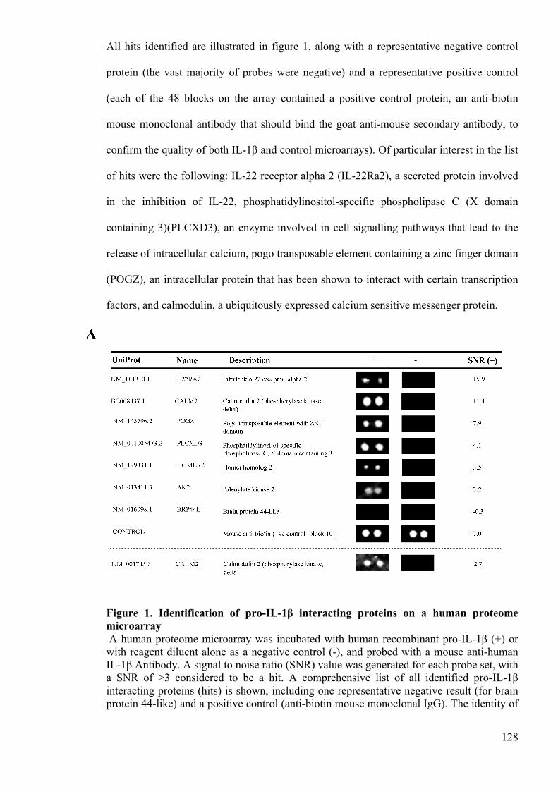
128
All hits identified are illustrated in figure 1, along with a representative negative control
protein (the vast majority of probes were negative) and a representative positive control
(each of the 48 blocks on the array contained a positive control protein, an anti-biotin
mouse monoclonal antibody that should bind the goat anti-mouse secondary antibody, to
confirm the quality of both IL-1β and control microarrays). Of particular interest in the list
of hits were the following: IL-22 receptor alpha 2 (IL-22Ra2), a secreted protein involved
in the inhibition of IL-22, phosphatidylinositol-specific phospholipase C (X domain
containing 3)(PLCXD3), an enzyme involved in cell signalling pathways that lead to the
release of intracellular calcium, pogo transposable element containing a zinc finger domain
(POGZ), an intracellular protein that has been shown to interact with certain transcription
factors, and calmodulin, a ubiquitously expressed calcium sensitive messenger protein.
Figure 1. Identification of pro-IL-1β interacting proteins on a human proteome microarray A human proteome microarray was incubated with human recombinant pro-IL-1β (+) or with reagent diluent alone as a negative control (-), and probed with a mouse anti-human IL-1β Antibody. A signal to noise ratio (SNR) value was generated for each probe set, with a SNR of >3 considered to be a hit. A comprehensive list of all identified pro-IL-1β interacting proteins (hits) is shown, including one representative negative result (for brain protein 44-like) and a positive control (anti-biotin mouse monoclonal IgG). The identity of

129
each protein, the corresponding images for each duplicate set of proteins on the array and the mean SNR for the duplicate protein spots are shown.
Given the pre-existing links between calcium signalling and IL-1β secretion, and given that
calmodulin was such a strong hit on the microarray (SNR= 11.4), the interaction between
calmodulin and pro-IL-1β was examined in further detail. Interestingly, the microarray also
included an additional probe set representing the same CALM2 gene, encoded by a slightly
different mRNA sequence (NM_001743.3). Although this probe set scored an average
SNR of 2.7 and therefore is just below the arbitrary threshold, this SNR is still high and
thus supports data suggesting that calmodulin interacts with pro-IL-1β. Curiously, there are
3 different genes (designated CALM1, CALM2 and CALM3) that encode for the same
protein (Toutenhoofd and Strehler 2000). As the protein encoded by CALM3 was also
present as a probe set on the microarray (BC006182.1), it was of concern that this probe
set was negative on the array (SNR of -1.1). However, a closer analysis of the mRNA
sequence used for the CALM3 probe set revealed that the protein encoded by this sequence
does not share complete protein sequence homology with native calmodulin. As the
CALM2 (BC008437.1) sequence does encode a protein that shares 100% protein sequence
homology with native calmodulin, it is likely that the subtle differences in the proteins
encoded by CALM2 (BC008437.1) and CALM3 (BC006182.1) cause the observed
differences in their capacity to interact with pro-IL-1β. Together, these data provide strong
evidence that calmodulin interacts with pro-IL-1β.
The interaction between IL-1β and calmodulin is robust, specific for the pro-protein only
and is dependent upon calcium
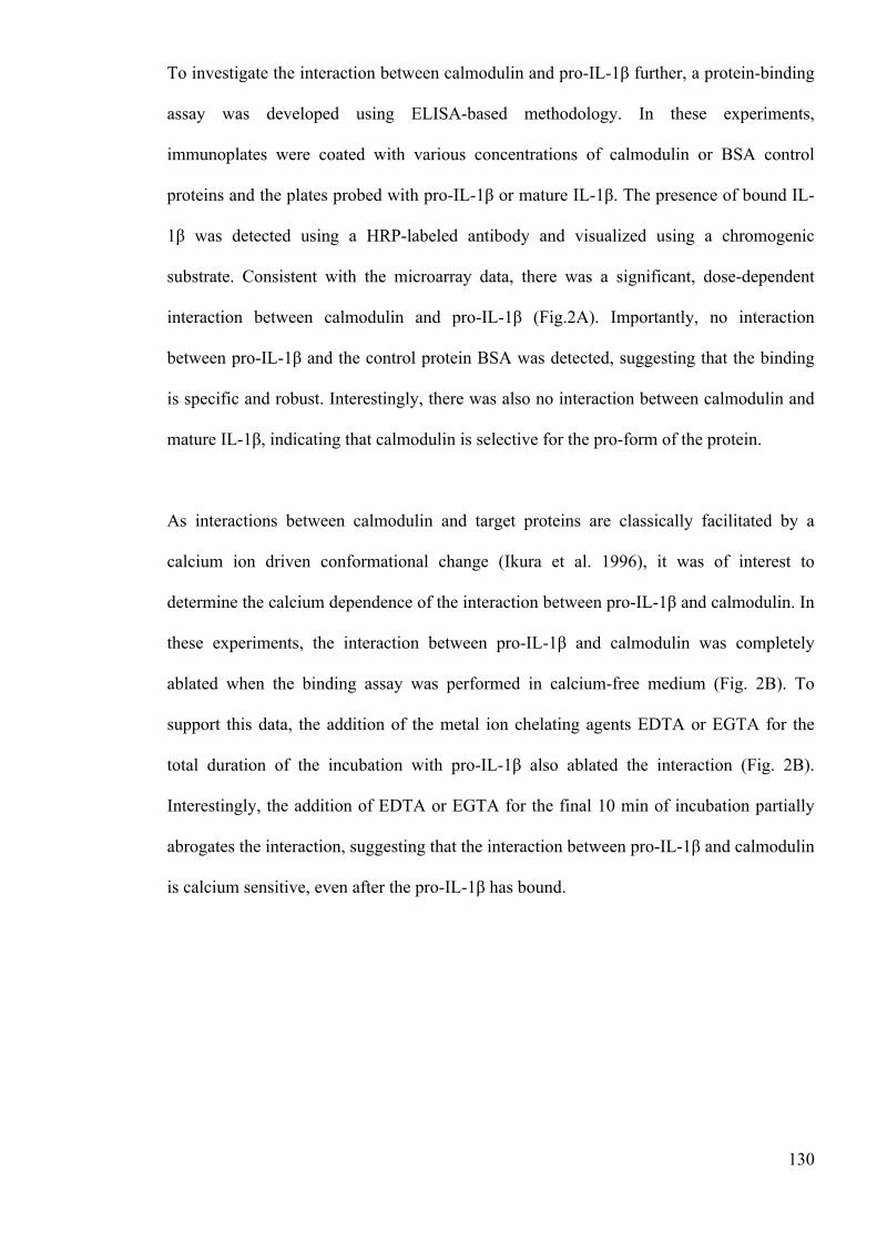
130
To investigate the interaction between calmodulin and pro-IL-1β further, a protein-binding
assay was developed using ELISA-based methodology. In these experiments,
immunoplates were coated with various concentrations of calmodulin or BSA control
proteins and the plates probed with pro-IL-1β or mature IL-1β. The presence of bound IL-
1β was detected using a HRP-labeled antibody and visualized using a chromogenic
substrate. Consistent with the microarray data, there was a significant, dose-dependent
interaction between calmodulin and pro-IL-1β (Fig.2A). Importantly, no interaction
between pro-IL-1β and the control protein BSA was detected, suggesting that the binding
is specific and robust. Interestingly, there was also no interaction between calmodulin and
mature IL-1β, indicating that calmodulin is selective for the pro-form of the protein.
As interactions between calmodulin and target proteins are classically facilitated by a
calcium ion driven conformational change (Ikura et al. 1996), it was of interest to
determine the calcium dependence of the interaction between pro-IL-1β and calmodulin. In
these experiments, the interaction between pro-IL-1β and calmodulin was completely
ablated when the binding assay was performed in calcium-free medium (Fig. 2B). To
support this data, the addition of the metal ion chelating agents EDTA or EGTA for the
total duration of the incubation with pro-IL-1β also ablated the interaction (Fig. 2B).
Interestingly, the addition of EDTA or EGTA for the final 10 min of incubation partially
abrogates the interaction, suggesting that the interaction between pro-IL-1β and calmodulin
is calcium sensitive, even after the pro-IL-1β has bound.
131
Figure 2. Calmodulin interacts with pro-IL-1β and not mature IL-1β, and this interaction is dependent on calcium Recombinant pro-IL-1β (3 nM; closed bars) or recombinant mature IL-1β (3 nM; open bars) was added to wells of 96 well immunoplates coated with either recombinant calmodulin or BSA control (both at concentrations of 5, 25, 50 or 100nM). After washing, the amount of IL-1β bound to the wells was detected using a HRP-labeled anti-IL-1β antibody and a chromogenic substrate, with absorbance used as a measure of interaction between the IL-1β and the coating proteins. The protein-binding assay was repeated using 50 nM of calmodulin (closed bars) or 50 nM of BSA (open bars) and a reagent diluent with
132
and without Ca2+ for the duration of the incubation, or with and without 10 mM EDTA or EGTA for either the full 2 h incubation or the final 10 min of incubation (B). To determine how these treatments affected IL-1β detection by the HRP labeled anti-IL-1β secondary antibody, and to determine the relative affinity of this detection antibody for pro and mature IL-1β, 96 well immunoplates were coated with an anti-IL-1β antibody and incubated with either recombinant mature IL-1β (●), recombinant pro-IL-1β in standard diluents (�) or recombinant pro-IL-1β diluted in reagent diluent without Ca2+(▲), diluted using a 9-fold serial dilution (C; no differences observed here). Data shown are mean ± SEM of 3 independent analyses. Statistical significance of differences between BSA and calmodulin treated samples were determined by one way ANOVA **=p<0.01 (A). Statistical significance of differences between samples treated with Ca2+ and other treatment groups were also determined by one way ANOVA **=p<0.01 (B).
To confirm the apparent selectivity of calmodulin for pro-IL-1β and not the mature form, it
was important to demonstrate that the detection antibody used in the protein-binding assay
could detect both pro-IL-1β and mature IL-1β with equal avidity. It was also important to
demonstrate that the removal of calcium from the reagent diluent was without nonspecific
effects on the detection of pro-IL-1β. In order to assess this, plates were coated with an
anti-IL-1β antibody overnight. The plates were then incubated with serial doubling
dilutions (0.58 to 300 pM) of recombinant mature IL-1β, recombinant pro-IL-1β,
recombinant pro-IL-1β in reagent diluent without calcium, recombinant pro-IL-1β in
reagent diluent with EDTA or recombinant pro-IL-1β in reagent diluent with EGTA. The
amount of cytokine bound was detected using the standard secondary anti-IL-1 β reagents.
In these experiments, neither the addition of the chelators (data not shown) nor the removal
of calcium had any effect on the avidity of the binding assay for pro-IL-1β (Fig. 2C).
However, these data did demonstrate that the anti-IL-1β reagents have greater avidity for
the mature cytokine, relative to the 31kD precursor, on a mole per mole basis. Given that it
is pro-IL-1β and not mature IL-1β that has been shown to interact with calmodulin, the
observed interaction with the precursor form cannot be reconciled on the basis of
differential binding of the cytokine detection reagents. Together these data provide strong
evidence to suggest that pro-IL-1β, and not mature IL-1β, interacts with calmodulin in the
presence of high calcium ion concentrations.
133
IL-1β processing is attenuated by calmodulin inhibition
To explore the function of the pro-IL-1β and calmodulin interaction, the role of calcium
and calmodulin in IL-1β processing and secretion was investigated in vitro. Initial
experiments confirmed that the human THP-1 monocytic cell line conformed to the current
paradigm of IL-1 secretion, requiring 2 signals. Here, a 4 h incubation with the TLR4
ligand LPS induced a potent up-regulation of intracellular pro-IL-1β (Fig. 3A).
Furthermore, stimulation of LPS-primed THP-1 cells with the ionophore nigericin for 1 h
induced the processing and release of the cytokine. Using this experimental system, and
the intracellular calcium chelator BAPTA-AM, we first explored the role of calcium in IL-
1β up-regulation and release. In these experiments, the addition of BAPTA-AM had no
effect on the intracellular up-regulation of the pro-protein (Fig. 3A). However, the addition
of BAPTA-AM (100 µM) almost completely ablated nigericin-stimulated IL-1β secretion
(Fig. 3B). Importantly, the addition of BAPTA-AM had no significant impact on the
viability of the THP-1 cells (data not shown). These data support the previous evidence
(Brough et al. 2003), which suggests that IL-1β secretion requires the release of
intracellular calcium stores.
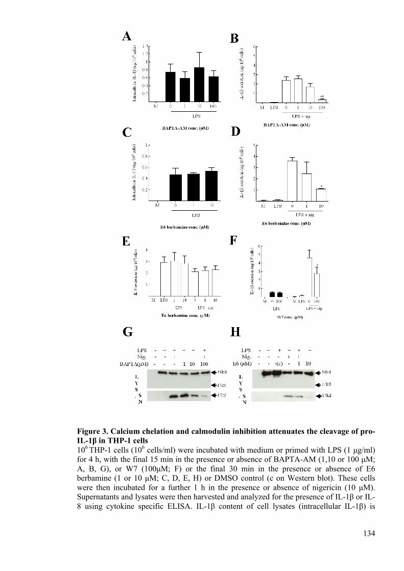
134
Figure 3. Calcium chelation and calmodulin inhibition attenuates the cleavage of pro-IL-1β in THP-1 cells 106 THP-1 cells (106 cells/ml) were incubated with medium or primed with LPS (1 µg/ml) for 4 h, with the final 15 min in the presence or absence of BAPTA-AM (1,10 or 100 µM; A, B, G), or W7 (100µM; F) or the final 30 min in the presence or absence of E6 berbamine (1 or 10 µM; C, D, E, H) or DMSO control (c on Western blot). These cells were then incubated for a further 1 h in the presence or absence of nigericin (10 µM). Supernatants and lysates were then harvested and analyzed for the presence of IL-1β or IL-8 using cytokine specific ELISA. IL-1β content of cell lysates (intracellular IL-1β) is
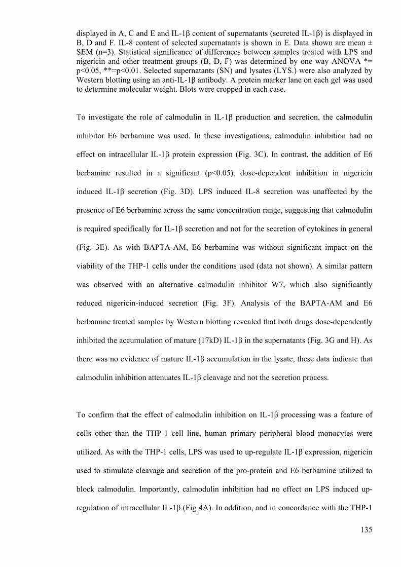
135
displayed in A, C and E and IL-1β content of supernatants (secreted IL-1β) is displayed in B, D and F. IL-8 content of selected supernatants is shown in E. Data shown are mean ± SEM (n=3). Statistical significance of differences between samples treated with LPS and nigericin and other treatment groups (B, D, F) was determined by one way ANOVA *= p<0.05, **=p<0.01. Selected supernatants (SN) and lysates (LYS.) were also analyzed by Western blotting using an anti-IL-1β antibody. A protein marker lane on each gel was used to determine molecular weight. Blots were cropped in each case.
To investigate the role of calmodulin in IL-1β production and secretion, the calmodulin
inhibitor E6 berbamine was used. In these investigations, calmodulin inhibition had no
effect on intracellular IL-1β protein expression (Fig. 3C). In contrast, the addition of E6
berbamine resulted in a significant (p<0.05), dose-dependent inhibition in nigericin
induced IL-1β secretion (Fig. 3D). LPS induced IL-8 secretion was unaffected by the
presence of E6 berbamine across the same concentration range, suggesting that calmodulin
is required specifically for IL-1β secretion and not for the secretion of cytokines in general
(Fig. 3E). As with BAPTA-AM, E6 berbamine was without significant impact on the
viability of the THP-1 cells under the conditions used (data not shown). A similar pattern
was observed with an alternative calmodulin inhibitor W7, which also significantly
reduced nigericin-induced secretion (Fig. 3F). Analysis of the BAPTA-AM and E6
berbamine treated samples by Western blotting revealed that both drugs dose-dependently
inhibited the accumulation of mature (17kD) IL-1β in the supernatants (Fig. 3G and H). As
there was no evidence of mature IL-1β accumulation in the lysate, these data indicate that
calmodulin inhibition attenuates IL-1β cleavage and not the secretion process.
To confirm that the effect of calmodulin inhibition on IL-1β processing was a feature of
cells other than the THP-1 cell line, human primary peripheral blood monocytes were
utilized. As with the THP-1 cells, LPS was used to up-regulate IL-1β expression, nigericin
used to stimulate cleavage and secretion of the pro-protein and E6 berbamine utilized to
block calmodulin. Importantly, calmodulin inhibition had no effect on LPS induced up-
regulation of intracellular IL-1β (Fig 4A). In addition, and in concordance with the THP-1
136
data, calmodulin inhibition significantly down-regulated nigericin stimulated IL-1β
secretion (Fig 4B). Finally, analysis of these samples by Western blotting demonstrated
that E6 berbamine inhibited the cleavage of pro-IL-1β in human primary peripheral blood
monocytes (Fig 4C). Together, these data show that calmodulin inhibition attenuates
nigericin-induced IL-1β secretion in monocytes, suggesting that calmodulin is an important
protein in the processing and release of IL-1β.
Figure 4. Calmodulin is also required for secretion of IL-1β in human monocytes 106 primary human monocytes (106 cells/ml) were incubated with medium or primed with LPS (1ng/ml) for 4 h, with the final 30 min in the presence or absence of E6 berbamine (1 or 10 µM). These cells were then incubated for a further 1 h in the presence or absence of nigericin (10µM). Supernatants and lysates were then harvested and analyzed for the presence of IL-1β using a cytokine specific ELISA. IL-1β content of cell lysates (intracellular IL-1β) is displayed in A and supernatants (secreted IL-1β) is displayed in B. Data shown are mean ± SEM (n=4). Statistical significance of differences between samples treated with LPS and nigericin and other treatment groups was determined by one way ANOVA *= p<0.05. Supernatants (SN) and lysates (LYS.) were also analyzed by Western blotting using an anti-IL-1β antibody (C). A protein marker lane on each gel was used to determine molecular weight. Blots were cropped in each case.
Calmodulin inhibition has no effect on the assembly of the inflammasome
Having shown that calmodulin inhibition leads to an inhibition in IL-1β secretion, the role
of calmodulin in this process was investigated further. Given that our data show that pro-
IL-1β binds with calmodulin in a calcium dependent interaction, and that that calcium is
required for processing, it is likely that the interaction between calmodulin and IL-1β is

137
itself important for IL-1β processing and release. However, before this conclusion could be
drawn, it was important to exclude the possibility that calmodulin inhibition was
attenuating the assembly of the inflammasome, as this would also explain why calmodulin
inhibition leads to an inhibition in IL-1β secretion. To investigate the effect of calmodulin
inhibition on inflammasome formation, nigericin induced ASC speck formation in THP-1
cells was assessed (Fig 5A). As reported previously, ASC expression was diffuse and
throughout the cytoplasm in resting cells, whereas a single localized spot of ASC staining
per cell was observed in cells in which inflammasome assembly had been provoked
(Lopez-Castejon et al. 2013). Here the percentage of speck containing cells was
determined and used as a measure of inflammasome assembly (Fig 5B). In the LPS primed
treatment group, there were very few cells with specks (~5%) and this proportion was
unaffected by the addition of the calmodulin inhibitor. As expected, the addition of
nigericin to LPS primed cells caused a large increase in the number of speck–positive cells
(~35%). Importantly, the addition of the calmodulin inhibitor had no effect on the
nigericin-induced speck formation, suggesting that calmodulin inhibition does not inhibit
the assembly of the inflammasome.
Calmodulin translocates within the cytoplasm in response to nigericin
Despite considerable academic effort, the mechanisms and cellular locations of IL-1β
maturation and release are still unclear. In light of the evidence presented previously, we
speculate that calmodulin may play a vital role in orchestrating the events leading up to the
processing of IL-1β, as induced by NLRP3 activators such as nigericin. To investigate this
further, the current study investigated how LPS priming and nigericin stimulation affected
the intracellular localization of calmodulin in THP-1 cells (Fig 5C). In untreated cells and
in LPS primed cells, localization of calmodulin appears to be focused in and around the
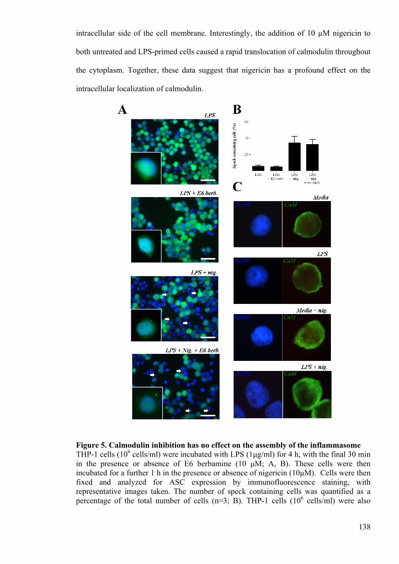
138
intracellular side of the cell membrane. Interestingly, the addition of 10 µM nigericin to
both untreated and LPS-primed cells caused a rapid translocation of calmodulin throughout
the cytoplasm. Together, these data suggest that nigericin has a profound effect on the
intracellular localization of calmodulin.
Figure 5. Calmodulin inhibition has no effect on the assembly of the inflammasome THP-1 cells (106 cells/ml) were incubated with LPS (1µg/ml) for 4 h, with the final 30 min in the presence or absence of E6 berbamine (10 µM; A, B). These cells were then incubated for a further 1 h in the presence or absence of nigericin (10µM). Cells were then fixed and analyzed for ASC expression by immunofluorescence staining, with representative images taken. The number of speck containing cells was quantified as a percentage of the total number of cells (n=3; B). THP-1 cells (106 cells/ml) were also
139
incubated with media or LPS (1µg/ml) for 4 h, with the final 30 min in the presence or absence of nigericin (10µM; D). Cells were fixed and analyzed for the expression of calmodulin by immunofluorescence staining and representative images taken (n=3 independent experiments). Scale bar = 50 µm.
4.8 Discussion
In general terms, the processing and release of IL-1β is fundamental to the initiation and
orchestration of inflammation. IL-1β is widely considered to be an essential effector in
host defense responses and its dysregulation is implicated in a multitude of pathologies
(Chen et al. 2006, Buchan et al. 1988, Apte et al. 2006, Cacabelos et al. 1994). The
mechanisms behind IL-1β secretion may represent significant therapeutic targets and are
thus of considerable academic interest. However, despite this interest, these processes
remain poorly understood. In the current study, we utilized a human proteome microarray
containing 19,951 unique proteins to identify globally proteins that bind human
recombinant pro-IL-1β. The rationale here was that the identification of such proteins may
help elucidate the mechanisms of IL-1β release.
From the microarray, a number of potentially important pro-IL-1β interacting proteins
were identified, including POGZ and IL-22Ra2. IL-22Ra2 is a secreted inhibitor of IL-22,
a cytokine involved in the initiation of innate immune responses (Weiss et al. 2004, Wolk
et al. 2004). Thus, it is tempting to speculate that pro-IL-1β may serve to enhance IL-22
induced innate immune responses by inhibiting IL-22Ra2. However, as IL-22Ra2 is a
secreted protein, it may be that the mature IL-1β plays more of a functional role here.
POGZ is a poorly characterized intracellular protein that has previously been shown to
interact with specificity protein 1 (SP1), a transcription factor involved in modulating gene
expression (Gunther et al. 2000). As SP1 has been shown to act at the same sites as NF-κB,
140
the interaction between POGZ and pro-IL-1β could have a role to play in modulating IL-1β
gene expression.
Of the 6 hits identified on the microarray, both calmodulin and PLCXD3 are associated
with intracellular calcium signalling. As discussed previously, the importance of calcium
in IL-1β release is well established. In Brough et al., ATP and nigericin induced IL-1β
secretion was associated with a marked elevation in intracellular calcium ion concentration
in murine macrophages (Brough et al. 2003). Importantly, the release of calcium from
intracellular stores was shown to be required for IL-1β release in this study. In more recent
investigations, inhibition of the pathways that lead to the release of intracellular Ca2+
stores, either by blocking IP3-gated Ca2+ release channels, store-operated Ca2+ entry or
phospholipase C, effectively inhibited IL-1β processing (Murakami et al. 2012). In the
current study, the addition of the intracellular calcium chelator BAPTA-AM inhibited
nigericin induced IL-1β processing and release in human monocytes. Together, these
studies demonstrate a significant role for calcium in the secretion of IL-1β.
Having established that calcium has a vital role to play in IL-1β release, the imperative
now is to decipher the mechanisms involved. Although there is evidence to suggest that
calcium is important for the assembly of the NLRP3 inflammasome (Murakami et al. 2012,
Lee et al. 2012), the current study provides data to suggest that calcium may also be
involved in mediating an intracellular interaction between pro-IL-1β and calmodulin. The
hypothesis here is that DAMP induced increases in intracellular Ca2+ cause a
conformational change in calmodulin that facilitates the interaction between calmodulin
and pro-IL-1β. In the current investigations, we provide evidence for the first time that pro-
IL-1β binds to calmodulin and that this interaction is dependent on increasing Ca2+
concentrations. Moreover, our data demonstrates that calmodulin is important for nigericin
141
induced IL-1β cleavage and secretion in both the THP-1 cell line and in primary human
monocytes. Thus, taken together, these data suggest that there is a calcium dependent
interaction between calmodulin and pro-IL-1β, and that this interaction is important for the
processing of the pro-IL-1β into its mature form.
To explore the potential roles of the Calmodulin-pro-IL-1β interaction in more detail, it is
necessary to consider current hypotheses regarding pro-IL-1β processing together with the
established roles of calmodulin. Intriguingly, calmodulin has been shown to bind to
numerous intracellular proteins associated with a variety of processes, including
inflammation, apoptosis, muscle contraction, intracellular movement, memory, nerve
growth and the immune response (Hoeflich and Ikura 2002). Thus, it is apparent that the
role of calmodulin is dependent upon the protein with which it interacts. It is well
established that calmodulin can function as a coenzyme for a variety of inactive enzymes,
including kinases and phosphatases (Hawley et al. 2005, Browne and Proud 2004). In these
cases, the interaction between calmodulin and its target results in the formation of an active
enzyme, or holoenzyme. As an example, calmodulin binds to inactive myosin light chain
kinase upon increasing calcium concentrations and forms an active complex capable of
phosphorylating myosin light chain during muscle contraction (Blumenthal and Stull 1980,
Kamm and Stull 1985). However, given that no evidence exists to suggest that pro-IL-1β
exhibits enzymatic activity, it is unlikely that the observed calmodulin-pro-IL-1β
interaction functions in this capacity.
More recently, evidence has suggested that calmodulin plays a role in the secretion of
small secretory proteins. Specifically, Shao et al. demonstrated that calmodulin acted as an
important chaperone in the translocation of these small proteins through the classic
secretory pathway (Shao and Hegde 2011). It was also shown that the interaction between
142
calmodulin and the small secretory proteins resulted in protection from protein aggregation
and degradation. It is therefore tempting to speculate that calmodulin has a role to play in
trafficking pro-IL-1β to caspase-1 for cleavage, especially given that the data presented
herein indicate that the calmodulin and pro-IL-1β interaction is required for IL-1β
processing. Specifically, we suggest that calmodulin may play a role either in transporting
the precursor towards the inflammasome for caspase-dependent processing or presenting
the precursor to the caspases for proteolytic cleavage. Although these hypotheses are
strengthened by the fact that calmodulin appears to translocate within the cytoplasm in
response to nigericin, further investigation is required to determine the role of calmodulin
in IL-1β release.
In addition to clarifying the role of the calmodulin-IL-1β interaction, the efforts to
investigate the pathways and mechanisms involved in IL-1β processing are ongoing
(Lopez-Castejon and Brough 2011). It is becoming increasingly apparent that these
pathways and mechanisms are remarkably complex, implicating a vast array of proteins
including messenger proteins, chaperones, proteases, ubiquitin ligases, cytosolic sensor
molecules and membrane bound proteins (Eder 2009). The current study highlights that by
utilizing a more global, proteomic approach to determine the interactome of pro-IL-1β, we
were able to elucidate a novel interaction that appears to be implicated in IL-1β secretion.
This suggests that the use of further proteomic analyses may be of great benefit in
determining the intricate processes that lead the maturation and release of IL-1β.
Specifically, analyzing the interactome of mature IL-1β, or other IL-1 cytokines such as
IL-1α, IL-18 or IL-33, could reveal numerous potentially important novel interactions and
mechanisms in this field.
143
To conclude, the current investigations highlight a number of pro-IL-1β interacting
proteins that may be relevant in elucidating the mechanisms of pro-IL-1β processing and
secretion. We demonstrate that calmodulin, a ubiquitously expressed calcium sensitive
protein, binds to pro-IL-1β in an interaction that is dependent on calcium. Moreover, we
show that calmodulin is also important in the processing and release of pro-IL-1β,
suggesting that this interaction is a key facet in the secretion of IL-1β.
4.9 Acknowledgements
JS Ainscough was funded by the Medical Research Council (MRC) and The Procter &
Gamble Company.
4.10 Conflicts of interest
The authors declare that they have no conflicts of interest with the contents of this article.
4.11 Author contributions
JSA conceived the study, coordinated the study and wrote the paper. GFG, IK and RJD
provided assistance with experimental design and interpretation, and contributed to the
preparation of the figures. All authors reviewed and approved the final version of the
manuscript.
4.12 References
Ainscough, J. S., Gerberick, G. F., Zahedi-Nejad, M., Lopez-Castejon, G., Brough, D., Kimber, I. and Dearman, R. J. (2014) 'Dendritic Cell IL-1 alpha and IL-1 beta Are Polyubiquitinated and Degraded by the Proteasome', Journal of Biological Chemistry, 289(51), 35582-35592.
144
Apte, R. N., Dotan, S., Elkabets, M., White, M. R., Reich, E., Carmi, Y., Song, X., Dvozkin, T., Krelin, Y. and Voronov, E. (2006) 'The involvement of IL-1 in tumorigenesis, tumor invasiveness, metastasis and tumor-host interactions', Cancer and Metastasis Reviews, 25(3), 387-408.
Benko, S., Philpott, D. J. and Girardin, S. E. (2008) 'The microbial and danger signals that
activate Nod-like receptors', Cytokine, 43(3), 368-373. Blumenthal, D. K. and Stull, J. T. (1980) 'Activation of skeletal-muscle myosin light chain
kinase by calcium(2+) and calmodulin', Biochemistry, 19(24), 5608-5614. Brough, D., Le Feuvre, R. A., Wheeler, R. D., Solovyova, N., Hilfiker, S., Rothwell, N. J.
and Verkhratsky, A. (2003) 'Ca2+ stores and Ca2+ entry differentially contribute to the release of IL-1 beta and IL-1 alpha from murine macrophages', Journal of Immunology, 170(6), 3029-3036.
Browne, G. J. and Proud, C. G. (2004) 'A novel mTOR-regulated phosphorylation site in
elongation factor 2 kinase modulates the activity of the kinase and its binding to calmodulin', Molecular and Cellular Biology, 24(7), 2986-2997.
Buchan, G., Barrett, K., Turner, M., Chantry, D., Maini, R. N. and Feldmann, M. (1988)
'Interleukin-1 and Tumor Necrosis Factor messenger-RNA expression in Rheumatoid-Arthritis - Prolonged production of IL-1α', Clinical and Experimental Immunology, 73(3), 449-455.
Cacabelos, R., Alvarez, X. A., Fernandeznovoa, L., Franco, A., Mangues, R., Pellicer, A.
and Nishimura, T. (1994) 'Brain Interleukin-1β in Alzheimers-Disease and vascular dementia', Methods and Findings in Experimental and Clinical Pharmacology, 16(2), 141-151.
Chen, C.-J., Shi, Y., Hearn, A., Fitzgerald, K., Golenbock, D., Reed, G., Akira, S. and
Rock, K. L. (2006) 'MyD88-dependent IL-1 receptor signaling is essential for gouty inflammation stimulated by monosodium urate crystals', Journal of Clinical Investigation, 116(8), 2262-2271.
Chin, D. and Means, A. R. (2000) 'Calmodulin: a prototypical calcium sensor', Trends in
Cell Biology, 10(8), 322-328. Cogswell, J. P., Godlevski, M. M., Wisely, G. B., Clay, W. C., Leesnitzer, L. M., Ways, J.
P. and Gray, J. G. (1994) 'NF-κB regulates IL-1β transcription through a consensus NF-κB binding-site and a nonconsensus Cre-like site', Journal of Immunology, 153(2), 712-723.
Dinarello, C. A. (2011) 'Interleukin-1 in the pathogenesis and treatment of inflammatory
diseases', Blood, 117(14), 3720-3732. Dostert, C., Petrilli, V., Van Bruggen, R., Steele, C., Mossman, B. T. and Tschopp, J.
(2008) 'Innate immune activation through Nalp3 inflammasome sensing of asbestos and silica', Science, 320(5876), 674-677.
Eder, C. (2009) 'Mechanisms of interieukin-1 beta release', Immunobiology, 214(7), 543-
553.
145
Ferrari, D., Chiozzi, P., Falzoni, S., DalSusino, M., Melchiorri, L., Baricordi, O. R. and
DiVirgilio, F. (1997) 'Extracellular ATP triggers IL-1β release by activating the purinergic P2Z receptor of human macrophages', Journal of Immunology, 159(3), 1451-1458.
Gabay, C., Lamacchia, C. and Palmer, G. (2010) 'IL-1 pathways in inflammation and
human diseases', Nature Reviews Rheumatology, 6(4), 232-241. Giles, N., Sierecki, E., Polinkovsky, M., Schroder, K., Alexandrov, K. and Gambin, Y.
(2014) 'A novel approach to investigate the assembly of the NOD-like receptor inflammasomes', Faseb Journal, 28(1).
Gunther, M., Laithier, M. and Brison, O. (2000) 'A set of proteins interacting with
transcription factor Sp1 identified in a two-hybrid screening', Molecular and Cellular Biochemistry, 210(1-2), 131-142.
Hawley, S. A., Pan, D. A., Mustard, K. J., Ross, L., Bain, J., Edelman, A. M., Frenguelli,
B. G. and Hardie, D. G. (2005) 'Calmodulin-dependent protein kinase kinase-beta is an alternative upstream kinase for AMP-activated protein kinase', Cell Metabolism, 2(1), 9-19.
Hoeflich, K. P. and Ikura, M. (2002) 'Calmodulin in action: Diversity in target recognition
and activation mechanisms', Cell, 108(6), 739-742. Hornung, V., Bauernfeind, F., Halle, A., Samstad, E. O., Kono, H., Rock, K. L., Fitzgerald,
K. A. and Latz, E. (2008) 'Silica crystals and aluminum salts activate the NALP3 inflammasome through phagosomal destabilization', Nature Immunology, 9(8), 847-856.
Ikura, M. (1996) 'Calcium binding and conformational response in EF-hand proteins',
Trends in Biochemical Sciences, 21(1), 14-17. Ikura, M., Hiraoki, T., Hikichi, K., Mikuni, T., Yazawa, M. and Yagi, K. (1996) 'Nuclear
Magnetic Resonance Studies on Calmodulin: Calcium-Induced Conformational Change', Biological Magnetic Resonance Data Bank.
Kamm, K. E. and Stull, J. T. (1985) 'The function of myosin and myosin light chain kinase
phosphorylation in smooth-muscle', Annual Review of Pharmacology and Toxicology, 25, 593-620.
Kawai, T. and Akira, S. (2010) 'The role of pattern-recognition receptors in innate
immunity: update on Toll-like receptors', Nature Immunology, 11(5), 373-384. Latz, E. (2010) 'The inflammasomes: mechanisms of activation and function', Current
Opinion in Immunology, 22(1), 28-33. Lee, G.-S., Subramanian, N., Kim, A. I., Aksentijevich, I., Goldbach-Mansky, R., Sacks,
D. B., Germain, R. N., Kastner, D. L. and Chae, J. J. (2012) 'The calcium-sensing receptor regulates the NLRP3 inflammasome through Ca2+ and cAMP', Nature, 492(7427), 123-+.
146
Lopez-Castejon, G. and Brough, D. (2011) 'Understanding the mechanism of IL-1β secretion', Cytokine & Growth Factor Reviews, 22(4), 189-195.
Lopez-Castejon, G., Luheshi, N. M., Compan, V., High, S., Whitehead, R. C., Flitsch, S.,
Kirov, A., Prudovsky, I., Swanton, E. and Brough, D. (2013) 'Deubiquitinases Regulate the activity of Caspase-1 and Interleukin-1β secretion via assembly of the inflammasome', Journal of Biological Chemistry, 288(4), 2721-2733.
Mariathasan, S., Weiss, D. S., Newton, K., McBride, J., O'Rourke, K., Roose-Girma, M.,
Lee, W. P., Weinrauch, Y., Monack, D. M. and Dixit, V. M. (2006) 'Cryopyrin activates the inflammasome in response to toxins and ATP', Nature, 440(7081), 228-232.
Martinon, F., Petrilli, V., Mayor, A., Tardivel, A. and Tschopp, J. (2007) 'Gout-associated
uric acid crystals activate the NALP3 inflammasome', Swiss Medical Weekly, 137, 26S-26S.
Meixenberger, K., Pache, F., Eitel, J., Schmeck, B., Hippenstiel, S., Slevogt, H.,
N'Guessan, P., Witzenrath, M., Netea, M. G., Chakraborty, T., Suttorp, N. and Opitz, B. (2010) 'Listeria monocytogenes-Infected Human Peripheral Blood Mononuclear Cells Produce IL-1 beta, Depending on Listeriolysin O and NLRP3', Journal of Immunology, 184(2), 922-930.
Moynagh, P. N. (2005) 'TLR signalling and activation of IRFs: revisiting old friends from
the NF-kappa B pathway', Trends in Immunology, 26(9), 469-476. Munoz-Planillo, R., Kuffa, P., Martinez-Colon, G., Smith, B. L., Rajendiran, T. M. and
Nunez, G. (2013) 'K+ Efflux Is the Common Trigger of NLRP3 Inflammasome Activation by Bacterial Toxins and Particulate Matter', Immunity, 38(6), 1142-1153.
Murakami, T., Ockinger, J., Yu, J., Byles, V., McColl, A., Hofer, A. M. and Horng, T.
(2012) 'Critical role for calcium mobilization in activation of the NLRP3 inflammasome', Proceedings of the National Academy of Sciences of the United States of America, 109(28), 11282-11287.
Ogura, Y., Sutterwala, F. S. and Flavell, R. A. (2006) 'The inflammasome: First line of the
immune response to cell stress', Cell, 126(4), 659-662. Petrilli, V., Papin, S., Dostert, C., Mayor, A., Martinon, F. and Tschopp, J. (2007)
'Activation of the NALP3 inflammasome is triggered by low intracellular potassium concentration', Cell Death and Differentiation, 14(9), 1583-1589.
Rubartelli, A., Cozzolino, F., Talio, M. and Sitia, R. (1990) 'A novel secretory pathway for
Interleukin-1β, a protein lacking A signal sequence', Embo Journal, 9(5), 1503-1510.
Schroder, K. and Tschopp, J. (2010) 'The Inflammasomes', Cell, 140(6), 821-832. Shao, S. and Hegde, R. S. (2011) 'A Calmodulin-Dependent Translocation Pathway for
Small Secretory Proteins', Cell, 147(7), 1576-1588.
147
Takeuchi, O. and Akira, S. (2010) 'Pattern recognition receptors and inflammation', Cell, 140(6), 805-820.
Thornberry, N. A., Bull, H. G., Calaycay, J. R., Chapman, K. T., Howard, A. D., Kostura,
M. J., Miller, D. K., Molineaux, S. M., Weidner, J. R., Aunins, J., Elliston, K. O., Ayala, J. M., Casano, F. J., Chin, J., Ding, G. J. F., Egger, L. A., Gaffney, E. P., Limjuco, G., Palyha, O. C., Raju, S. M., Rolando, A. M., Salley, J. P., Yamin, T. T., Lee, T. D., Shively, J. E., Maccross, M., Mumford, R. A., Schmidt, J. A. and Tocci, M. J. (1992) 'A novel heterodimeric cysteine protease is required for interleukin-1-beta processing in monocytes', Nature, 356(6372), 768-774.
Toutenhoofd, S. L. and Strehler, E. E. (2000) 'The calmodulin multigene family as a
unique case of genetic redundancy: multiple levels of regulation to provide spatial and temporal control of calmodulin pools?', Cell Calcium, 28(2), 83-96.
Weiss, B., Wolk, K., Grunberg, B. H., Volk, H. D., Sterry, W., Asadullah, K. and Sabat, R.
(2004) 'Cloning of murine IL-22 receptor alpha 2 and comparison with its human counterpart', Genes and Immunity, 5(5), 330-336.
Wolk, K., Kunz, S., Witte, E., Friedrich, M., Asadullah, K. and Sabat, R. (2004) 'IL-22
increases the innate immunity of tissues', Immunity, 21(2), 241-254.
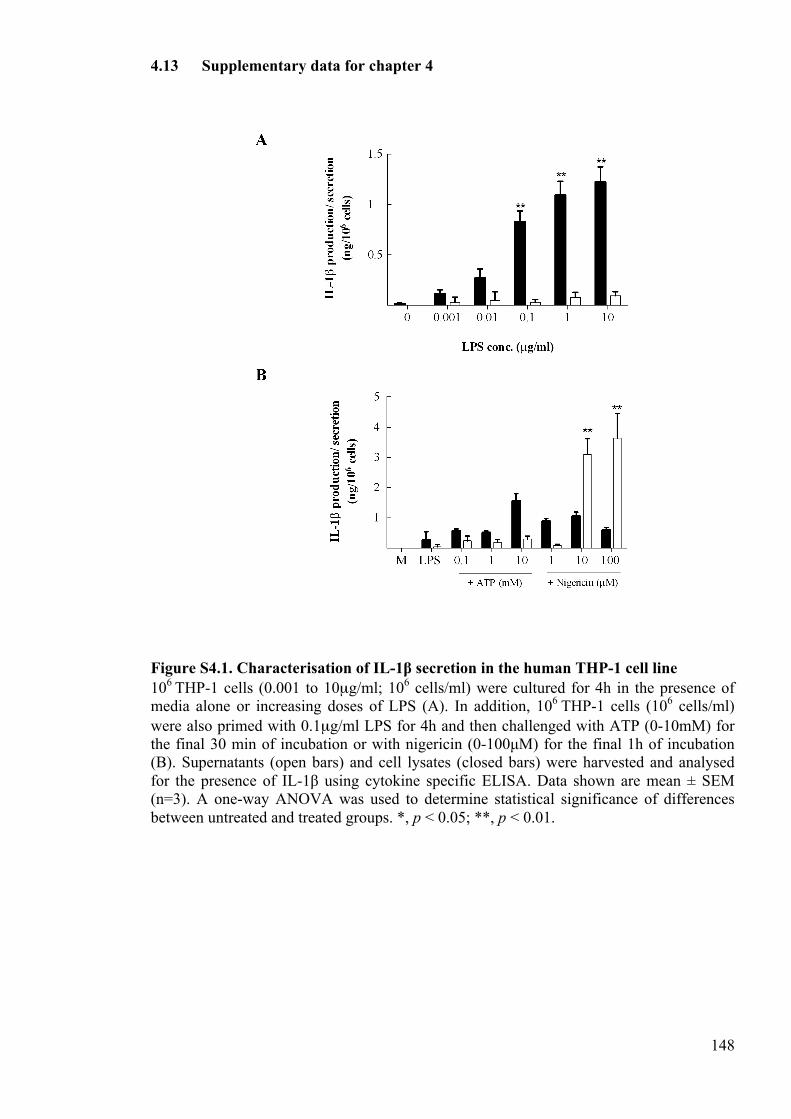
148
4.13 Supplementary data for chapter 4
Figure S4.1. Characterisation of IL-1β secretion in the human THP-1 cell line 106 THP-1 cells (0.001 to 10µg/ml; 106 cells/ml) were cultured for 4h in the presence of media alone or increasing doses of LPS (A). In addition, 106 THP-1 cells (106 cells/ml) were also primed with 0.1µg/ml LPS for 4h and then challenged with ATP (0-10mM) for the final 30 min of incubation or with nigericin (0-100µM) for the final 1h of incubation (B). Supernatants (open bars) and cell lysates (closed bars) were harvested and analysed for the presence of IL-1β using cytokine specific ELISA. Data shown are mean ± SEM (n=3). A one-way ANOVA was used to determine statistical significance of differences between untreated and treated groups. *, p < 0.05; **, p < 0.01.
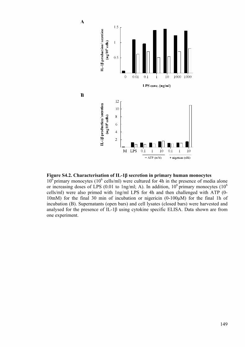
149
Figure S4.2. Characterisation of IL-1β secretion in primary human monocytes 106 primary monocytes (106 cells/ml) were cultured for 4h in the presence of media alone or increasing doses of LPS (0.01 to 1ng/ml; A). In addition, 106 primary monocytes (106 cells/ml) were also primed with 1ng/ml LPS for 4h and then challenged with ATP (0-10mM) for the final 30 min of incubation or nigericin (0-100µM) for the final 1h of incubation (B). Supernatants (open bars) and cell lysates (closed bars) were harvested and analysed for the presence of IL-1β using cytokine specific ELISA. Data shown are from one experiment.
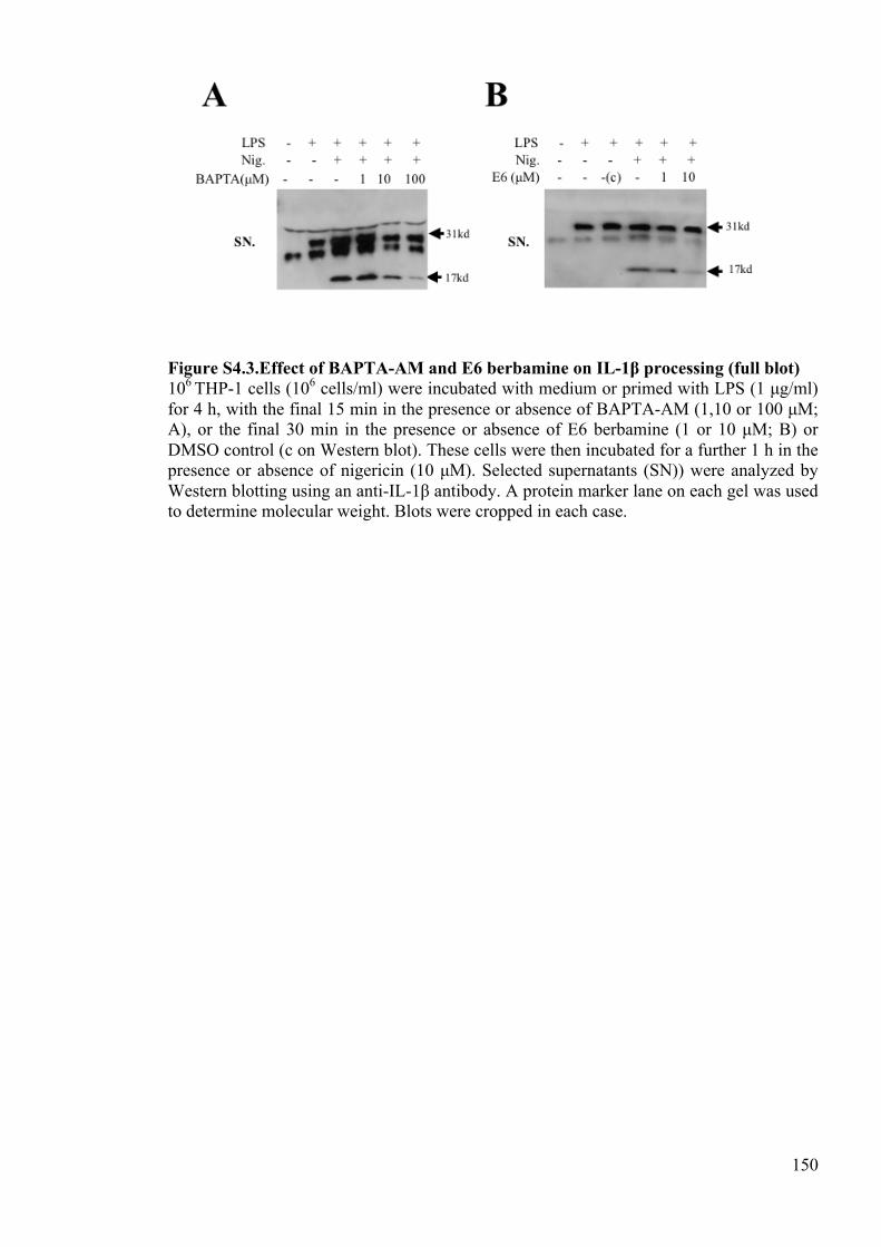
150
Figure S4.3.Effect of BAPTA-AM and E6 berbamine on IL-1β processing (full blot) 106 THP-1 cells (106 cells/ml) were incubated with medium or primed with LPS (1 µg/ml) for 4 h, with the final 15 min in the presence or absence of BAPTA-AM (1,10 or 100 µM; A), or the final 30 min in the presence or absence of E6 berbamine (1 or 10 µM; B) or DMSO control (c on Western blot). These cells were then incubated for a further 1 h in the presence or absence of nigericin (10 µM). Selected supernatants (SN)) were analyzed by Western blotting using an anti-IL-1β antibody. A protein marker lane on each gel was used to determine molecular weight. Blots were cropped in each case.

151
CHAPTER 5:
SUPPLEMENTARY CHAPTER
152
5 Supplementary chapter 5.1 Abbreviations
ATP Adenosine triphosphate BM Bone marrow BSA Bovine serum albumin DC Dendritic cell EDTA Ethylenediaminetetraacetic acid FCS Foetal calf serum FITC Fluorescein isothiocyanate GM-CSF Granulocyte macrophage-colony stimulating factor IL Interleukin LPS Lipopolysaccharide MFI Mean fluorescence intensity NLR NOD-like receptor PBMC Peripheral blood mononuclear cells PMSF Phenylmethanesulfonylfluoride PI Propidium iodide TLR TOLL-like receptor
5.2 Introduction
In the initial stages of the work described in this thesis (chapters 2 and 3), it was necessary
to establish an experimental model that could be used to investigate the regulation of
Interleukin (IL)-1. Specifically, a murine dendritic cell (DC)-like cell line or primary cell
that could up-regulate pro-IL-1 in response to TOLL-like receptor (TLR) stimulation and
secrete IL-1 in response to NOD-like receptor (NLR) stimulation was required for the
purpose of the studies herein. To find a suitable model, the expression and secretion of IL-
1β was characterised in a murine DC-like cell line called the XS106 cell line (Xu et al.
1995b). The expression and secretion of IL-1β was also characterised in murine bone
marrow-derived (BM)DC (Lutz et al. 1999). For the purpose of the work conducted in
chapter 4, it was also necessary to establish a human cell line or primary cell that could be
used to investigate IL-1 regulation. Again, this cell was required to up-regulate pro-IL-1 in
response to TLR stimulation, and to secrete IL-1 in response to NLR stimulation. To find a
suitable model for these experiments, the expression and secretion of IL-1β was
153
characterised in both the human monocytic cell line THP-1, and in primary monocytes
derived from peripheral blood mononuclear cells (PBMC).
5.3 Methods
Experimental animals
6-8 week old female BALB/c mice were used throughout these experiments (Harlan Olac,
Bicester, UK). The environmental conditions were maintained at an ambient temperature
of 21oC (+/- 2oC) and at a relative humidity of 55% (+/- 10%). Food and water was
available at all times. Animals were kept in a 12h light/dark cycle. All procedures were
carried out in accordance with the Animals (Scientific Procedures) Act 1986. Procedures
were performed as approved by the Home Office.
Reagents
Lipopolysaccharide (LPS) from Escherichia coli serotype 055:B5 (TLR2/4), adenosine
triphosphate (ATP) (the pH of the ATP solution was adjusted to 7.5 following
reconstitution), nigericin, caspase-1 inhibitor YVAD and the caspase-3 inhibitor DEVD
were all purchased from Sigma (St. Louis, MO, USA). The proteasome inhibitor MG132
was purchased from Merck Millipore (Billerica, MA, USA).Recombinant murine pro-IL-
1β was purchased from Affymetrix eBioscience (San Diego, CA).
Maintenance of the NS47 feeder cell line
154
The NS47 fibroblast cell line was maintained in RPMI-1640 growth medium supplemented
with 10% (v/v) heat inactivated foetal calf serum (FCS; Life Technologies, Carlsbad, CA,
USA), 0.05mM 2-mercaptoethanol, 1% (v/v) non-essential amino acids (both Sigma),
292µg/ml L-glutamine, 400µg/ml streptomycin/penicillin (both Life Technologies),
0.25µg/ml amphotericin B (Sigma) and 1mM sodium pyruvate (Sigma). NS47 cells were
cultured at 3.5x10⁵ cells/ml in T75 0.2µm vented culture flasks (Corning, Lowell, MA,
USA). Once cells were confluent, supernatant was collected, filtered through 0.2 µm
filters, and stored frozen at -20 oC.
Maintenance and treatment of the XS106 cell line
XS106 cells were obtained from Dr A. Takashima (University of Toledo, Toledo, OH,
USA). Cells were cultured in RPMI-1640 growth medium supplemented with 1% (v/v)
non-essential amino acids, 292µg/ml L-glutamine, 400µg/ml penicillin/streptomycin,
0.25µg/ml amphotericin B, 1mM sodium pyruvate, 10% (v/v) heat inactivated FCS,
0.05mM 2-mercaptoethanol (Life Technologies), 4ng/ml recombinant murine granulocyte
macrophage-colony stimulating factor (GM-CSF; Miltenyi Biotech, Bergisch Gladbach,
Germany) and 5% (v/v) NS47 fibroblast culture supernatant. XS106 cells were cultured at
3.5x10⁵ cells/ml in T75 0.2µm vented culture flasks (Corning) and incubated until
confluent. The non-adherent cells present in the supernatant were used for cell line
maintenance and the adherent cells used for assays. Cells were removed from flasks using
a cell scraper and centrifuged at 168g for 5 min. Cells were plated into (24-well plates) at 1
x 106 cells per well and treated with various concentrations of LPS for various lengths of
time. In additional experiments, LPS-primed cells (10µg/ml) were activated with various
concentrations of ATP for 30 min at the end of the culture.
155
Cell counts
Viable cell counts were assessed using a 5X5 graduated haemocytometer. Non-viable cells
were excluded using a 0.5% (v/v) Trypan Blue solution (Sigma Aldrich).
Generation and Culture of Murine Bone Marrow-derived DC
Bone marrow was obtained from the femurs and tibias of 6-8 week old female BALB/c
mice (Harlan Olac). Bone marrow was extracted by flushing the bones with PBS. The
cell/PBS suspension was centrifuged at 300g for 5 min at room temperature. The
remaining pellet was resuspended in pre-warmed FCS supplemented media (RPMI 1640)
containing 25mM HEPES, 400µg/ml penicillin/ streptomycin, 292µg/ml L- glutamine,
0.05mM 2- mercaptoethanol, 4ng/ml GM-CSF and 10% (v/v) FCS. Cells were cultured at
approximately 2x10⁶ cells/ml (20ml) in petri dishes and incubated at 37oC. An additional
10ml of culture media were added after 3 days. 10ml exhausted medium was replaced on
day 6 with 10ml of fresh culture medium. Cells were plated on day 8 of culture for assay.
BMDC treatments
BMDC were plated at 106 cells/well (24-well plate) in BMDC culture medium without
GM-CSF. To induce IL-1 production, cells were primed using 0.1µg/ml LPS for various
periods. To induce IL-1 secretion, LPS-primed cells were stimulated with various
concentrations of ATP for various periods at the end of the culture. The involvement of
caspase-1 in ATP-dependent IL-1 secretion was measured by priming cells with LPS for
4h, adding 50µM of the caspase-1 inhibitor YVAD or 50µM DEVD control (both Sigma)
156
for the final 1h of incubation, with addition of 1 or 10mM ATP for the final 30 min of
incubation.
Maintenance of the THP-1 cell line
THP-1 cells were cultured in RPMI-1640 growth medium supplemented with 292µg/ml L-
glutamine, 400µg/ml penicillin/streptomycin, 10% (v/v) heat inactivated FCS, 0.05mM 2-
mercaptoethanol (THP-1 culture medium; Life Technologies). Cells were cultured at 2x10⁵
cells/ml in T75 0.2 µm vented culture flasks and incubated until confluent.
THP-1 cell treatments
THP-1 cells were plated at 106 cells/well (24-well plate) in THP-1 culture medium. To
investigate TLR-induced IL-1β expression, cells were incubated with various
concentrations of LPS for 4h and supernatants and lysates collected. To investigate IL-1β
secretion, cells were primed with 0.1µg/ml of LPS for 4h and activated with various
concentrations of ATP for the final 30 min of culture, or with various concentrations of
nigericin for the final 1h of culture.
Isolation of primary monocytes
Buffy coats from the peripheral venous blood of healthy donors were acquired from a
National Health Service Blood and Transplant Service (Manchester Donor
Centre, Plymouth Grove, Manchester). The collection and use of this tissue was approved
by a blanket material transfer agreement and research ethics. PBMC were isolated from
whole blood by density centrifugation using Histopaque (Sigma). Briefly, 20ml of whole
157
blood was layered onto 15ml Histopaque, samples centrifuged at 500g for 30 min with the
brake off and PBMC layers collected. Cells were then washed in PBS and centrifuged at
500g for 10 min to remove any remaining platelets. Monocytes were enriched from PBMC
using the negative magnetic selection monocyte isolation kit II (Miltenyi Biotec). Briefly,
cells were resuspended at 3x10⁸ cells/ml in monocyte buffer (0.5% (w/v) bovine serum
albumin (BSA), 2mM EDTA in PBS). 10µl FcR blocking reagent and 10µl of biotin
antibody cocktail was added per 107 cells and incubated at 4oC for 10 min. 30µl of
monocyte buffer and 20µl of anti-biotin beads was added per 107 cells and incubated at 4
oC for 15 min. Cells were then washed with monocyte buffer and centrifuged at 300g for
10 min. The remaining pellet was resuspended in 500µl of monocyte buffer. The MACS
column and separator was assembled and prepared by rinsing the column with monocyte
buffer. The cells suspension was passed through the column and monocytes collected in
the flow-through.
To evaluate the monocyte purity, cell pellets were resuspended in 1ml of cold FACS buffer
(5% (v/v) FCS/PBS). 100µl aliquots were transferred into the wells of round-bottomed 96-
well plates. Samples were stained with 100µl of an allophycocyanin labelled anti-human
CD14 antibody (10µg/ml; BD biosciences, San Jose, CA, USA) and incubated on ice for
45 min. Cells were washed with 200µl FACS buffer, centrifuged at 300g for 5 min at 4oC,
blotted and aspirated. This wash process was repeated for a total of 3 times. Cells were
resuspended in 200µl sodium azide buffer (0.05% (v/v) sodium azide in 1% (v/v) FCS in
PBS). Samples were analysed using a FACScalibur machine and CellQuest Pro software
(both BD biosciences). 10,000 cells were acquired from each sample.
Primary monocyte treatments
158
Monocytes were plated at 106 cells/well (24-well plate) in THP-1 culture medium. To
investigate TLR-induced IL-1β expression, cells were incubated with various
concentrations of LPS for 4h and supernatants and lysates collected. To investigate IL-1β
secretion, cells were primed with 1ng/ml of LPS for 4h, and activated with various
concentration of nigericin for the final 1h of culture or various concentrations of ATP for
the final 30 min of culture
Processing of cell lysates and supernatants
Following incubation, the media from the plates was collected and centrifuged at 300g for
5 min. The supernatants from these were then harvested and stored at -20°C. Cell lysates
were harvested in 200µl of lysis buffer (20mM Tris HCl, 137mM NaCl, 20mM
ethylenediaminetetraacetic acid (EDTA), 10% (v/v) glycerol, 0.5% (v/v) Ipegal, 1mM
phenylmethanesulfonylfluoride (PMSF), 1% (v/v) protease inhibitor cocktail) and frozen at
-80°C. For each sample, 100µl of lysis buffer was added to both the cell pellet retained
following isolation of the supernatant and the cells remaining in the well of the culture
plate. The plate was incubated with shaking for 5 min to lyse attached cells. The lysate
from the tissue culture plate was pooled with the corresponding lysate from the cell pellet
and centrifuged at 15000g for 5 min at 4°C. The cell lysates were harvested and stored at -
80°C.
IL-1β ELISA
Both murine and human cytokine (IL-1β) analyses were conducted according to the
manufacturer’s instructions (Duoset, R&D systems, Minneapolis, MN, USA). Plastic
maxisorb® plates (Nunc, Copenhagen, Denmark) were coated with 100µl/well rat anti-
159
mouse IL-1β capture antibody (4.0µg/ml) or mouse anti-human IL-1β capture antibody
(4.0µg/ml) diluted in PBS. These plates were incubated overnight at 4°C. The plates were
then washed for a total of three cycles in wash buffer (0.05% (v/v) Tween in PBS) each for
3min. The plates were blocked for 1h at room temperature using 300µl/well of 1% (w/v)
BSA (Sigma Aldrich) in PBS. Plates were washed as described previously. A 9 point
standard curve was prepared using recombinant homologous IL-1β protein diluted in
RPMI-1640 growth medium containing 10% (v/v) FCS (top standard 10ng/ml). 100µl of
standards were added to triplicate wells. Samples were also diluted in RPMI-1640 growth
medium containing 10% (v/v) FCS. 100µl/well of the samples were loaded onto the plate
in duplicate. 100µl biotinylated goat anti-mouse IL-1β detection antibody (50ng/ml) or
biotinylated goat anti-human IL-1β antibody (200ng/ml) diluted in reagent diluent (0.1%
(w/v) BSA, 0.05% (v/v) Tween 20 in Tris-buffered saline (20mM Trizma base, 150mM
NaCl)) was added to each well. The plates were incubated for 2h at room temperature.
Plates were washed as described previously. 100µl of streptavidin-horseradish peroxidase
diluted in reagent diluent was added to each well. Plates were incubated for 20 min at room
temperature and protected from direct light. Plates were washed and 100µ1 of
tetramethylbenzidine stabilised chromogen (Life technologies) was added to each well.
Samples were incubated at room temperature for 20 min and protected from direct light.
50µl of stop solution (2M H2SO4) was added. Optical density was measured at 450nm
using an automated plate reader (Multiscan, Flow laboratories, Irvine, Ayrshire, UK). A
standard curve obtained from the 9 point standard dilution was used to calculate cytokine
concentrations in the samples.
Analysis of BMDC membrane marker expression using flow cytometry

160
BMDC cell pellets were resuspended in 1ml of cold FACS buffer. 100µl aliquots were
transferred into the wells of a round-bottomed 96-well plate. Cells were stained with rat
anti-mouse I-A/ I-E antibody (2.5µg/ml), rat anti-mouse CD86 antibody, rat anti-mouse
CD11c antibody, rat anti-mouse CD80 antibody, rat anti-mouse CD40 antibody, or rat
IgG2a isotype control antibody (all 10µg/ml; all BD biosciences) and incubated on ice for
45 min. Cells were washed with 200µl FACS buffer, centrifuged at 300g for 5 min at 4oC,
blotted and aspirated. This wash process was repeated for a total of 3 times. Cells were
stained with 100µl/well STAR69, a goat anti- rat IgG fluorescein isothiocyanate (FITC)-
labelled polyclonal antibody (10µg/ml; AbD Serotec, Kidlington, UK) and incubated on
ice for 45 min. The plates were washed as described previously and pellets resuspended in
200µl sodium azide buffer. Samples were analysed using a FACScalibur machine and
CellQuest Pro software. 2µl of propidium iodide (PI) was added to each sample prior to
analysis to gate out non-viable cells. 10,000 cells were acquired from each sample.
Analysis of BMDC viability profile using flow cytometry
Analyses of the BMDC viability profile experiments were conducted according to
manufacturers instructions (Trevigen, Gaithersburg, MD, USA). Briefly, BMDC cell
pellets were resuspended in 1ml of cold FACS buffer. 100µl aliquots were transferred into
the wells of round-bottomed 96-well plates. Samples were then incubated with 1µl of
annexin V-FITC and PI for 15 min at room temperature (Trevigen). Samples were
analysed using a FACScalibur machine and CellQuest Pro software. 10,000 cells were
acquired from each sample.
5.4 Results
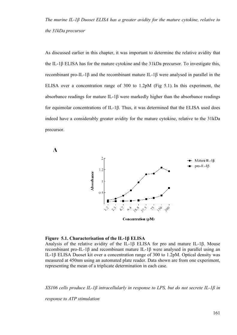
161
The murine IL-1β Duoset ELISA has a greater avidity for the mature cytokine, relative to
the 31kDa precursor
As discussed earlier in this chapter, it was important to determine the relative avidity that
the IL-1β ELISA has for the mature cytokine and the 31kDa precursor. To investigate this,
recombinant pro-IL-1β and the recombinant mature IL-1β were analysed in parallel in the
ELISA over a concentration range of 300 to 1.2pM (Fig 5.1). In this experiment, the
absorbance readings for mature IL-1β were markedly higher than the absorbance readings
for equimolar concentrations of IL-1β. Thus, it was determined that the ELISA used does
indeed have a considerably greater avidity for the mature cytokine, relative to the 31kDa
precursor.
Figure 5.1. Characterisation of the IL-1β ELISA Analysis of the relative avidity of the IL-1β ELISA for pro and mature IL-1β. Mouse recombinant pro-IL-1β and recombinant mature IL-1β were analysed in parallel using an IL-1β ELISA Duoset kit over a concentration range of 300 to 1.2pM. Optical density was measured at 450nm using an automated plate reader. Data shown are from one experiment, representing the mean of a triplicate determination in each case.
XS106 cells produce IL-1β intracellularly in response to LPS, but do not secrete IL-1β in
response to ATP stimulation
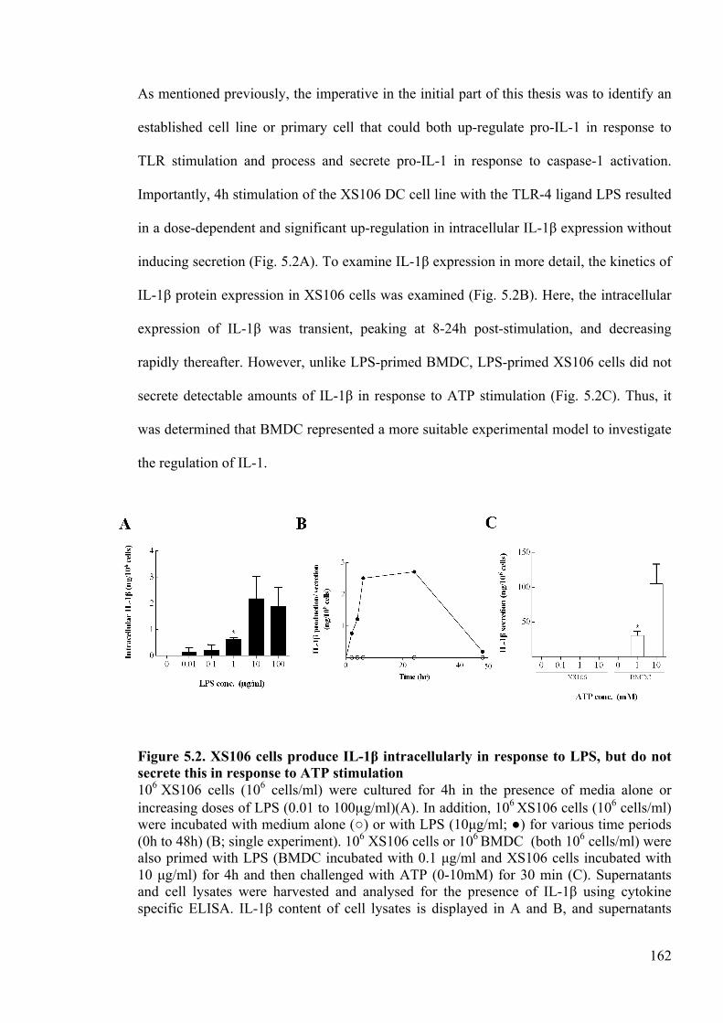
162
As mentioned previously, the imperative in the initial part of this thesis was to identify an
established cell line or primary cell that could both up-regulate pro-IL-1 in response to
TLR stimulation and process and secrete pro-IL-1 in response to caspase-1 activation.
Importantly, 4h stimulation of the XS106 DC cell line with the TLR-4 ligand LPS resulted
in a dose-dependent and significant up-regulation in intracellular IL-1β expression without
inducing secretion (Fig. 5.2A). To examine IL-1β expression in more detail, the kinetics of
IL-1β protein expression in XS106 cells was examined (Fig. 5.2B). Here, the intracellular
expression of IL-1β was transient, peaking at 8-24h post-stimulation, and decreasing
rapidly thereafter. However, unlike LPS-primed BMDC, LPS-primed XS106 cells did not
secrete detectable amounts of IL-1β in response to ATP stimulation (Fig. 5.2C). Thus, it
was determined that BMDC represented a more suitable experimental model to investigate
the regulation of IL-1.
Figure 5.2. XS106 cells produce IL-1β intracellularly in response to LPS, but do not secrete this in response to ATP stimulation 106 XS106 cells (106 cells/ml) were cultured for 4h in the presence of media alone or increasing doses of LPS (0.01 to 100µg/ml)(A). In addition, 106 XS106 cells (106 cells/ml) were incubated with medium alone (○) or with LPS (10µg/ml; ●) for various time periods (0h to 48h) (B; single experiment). 106 XS106 cells or 106 BMDC (both 106 cells/ml) were also primed with LPS (BMDC incubated with 0.1 µg/ml and XS106 cells incubated with 10 µg/ml) for 4h and then challenged with ATP (0-10mM) for 30 min (C). Supernatants and cell lysates were harvested and analysed for the presence of IL-1β using cytokine specific ELISA. IL-1β content of cell lysates is displayed in A and B, and supernatants
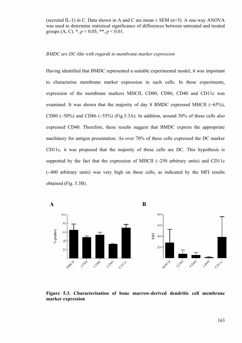
163
(secreted IL-1) in C. Data shown in A and C are mean ± SEM (n=3). A one-way ANOVA was used to determine statistical significance of differences between untreated and treated groups (A, C). *, p < 0.05; **, p < 0.01.
BMDC are DC-like with regards to membrane marker expression
Having identified that BMDC represented a suitable experimental model, it was important
to characterise membrane marker expression in such cells. In these experiments,
expression of the membrane markers MHCII, CD80, CD86, CD40 and CD11c was
examined. It was shown that the majority of day 8 BMDC expressed MHCII (~65%),
CD80 (~50%) and CD86 (~55%) (Fig.5.3A). In addition, around 30% of these cells also
expressed CD40. Therefore, these results suggest that BMDC express the appropriate
machinery for antigen presentation. As over 70% of these cells expressed the DC marker
CD11c, it was proposed that the majority of these cells are DC. This hypothesis is
supported by the fact that the expression of MHCII (~250 arbitrary units) and CD11c
(~400 arbitrary units) was very high on these cells, as indicated by the MFI results
obtained (Fig. 5.3B).
Figure 5.3. Characterisation of bone marrow-derived dendritic cell membrane marker expression
164
The expression of the membrane markers MHCII, CD86, CD80, CD40 and CD11c was analysed on unstimulated viable day 8 BMDC by flow cytometry. 10,000 cells were acquired for each sample. The percentage of positive cells (A) and the mean fluorescence intensity (MFI; arbitrary units) (B) for each marker was determined in 3 independent experiments. Data shown are mean ± SEM (n=3).
IL-1β secretion in BMDC is rapid and dependent on caspase-1
To characterise the nature of IL-1β secretion in greater detail, the kinetics of ATP-induced
IL-1β release was examined (Fig. 5.4A). As shown previously, no IL-1β was secreted from
LPS-primed BMDC without the addition of ATP. However, the addition of 1 or 10mM
ATP for as little as 10 min caused a marked up-regulation in the amount of secreted IL-1β.
Interestingly, the levels of extracellular IL-1β stabilized after 10 min of ATP stimulation;
thus these results indicate that the ATP-induced release of IL-1β is a rapid process. To
determine the optimum length of LPS priming for ATP-induced IL-1β secretion, BMDC
were incubated for various periods and stimulated with ATP for the final 30 min of the
incubation (Fig. 5.4B). In this experiment, maximal IL-1β secretion was observed
following 4h of LPS priming. As with intracellular expression, the ATP-induced IL-1β
secretion decreased rapidly, to the point where ATP did not induce any detectable IL-1β
secretion following priming for 24h. Finally, to determine whether ATP-induced secretion
was caspase-1 dependent in LPS-primed BMDC, cells were incubated with either the
caspase-1 inhibitor YVAD, or the control inhibitor DEVD, and the ATP-induced secretion
of IL-1β was examined (Fig. 5.4C). Importantly, stimulation with 1 or 10mM ATP induced
a potent and significant secretion of IL-1β in the control group, whereas ATP-induced
secretion was completely inhibited in the YVAD treated group. Therefore, these results
show that ATP-induced secretion is caspase-1 dependent in BMDC.
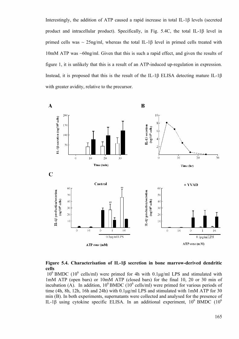
165
Interestingly, the addition of ATP caused a rapid increase in total IL-1β levels (secreted
product and intracellular product). Specifically, in Fig. 5.4C, the total IL-1β level in
primed cells was ~ 25ng/ml, whereas the total IL-1β level in primed cells treated with
10mM ATP was ~60ng/ml. Given that this is such a rapid effect, and given the results of
figure 1, it is unlikely that this is a result of an ATP-induced up-regulation in expression.
Instead, it is proposed that this is the result of the IL-1β ELISA detecting mature IL-1β
with greater avidity, relative to the precursor.
Figure 5.4. Characterisation of IL-1β secretion in bone marrow-derived dendritic cells 106 BMDC (106 cells/ml) were primed for 4h with 0.1µg/ml LPS and stimulated with 1mM ATP (open bars) or 10mM ATP (closed bars) for the final 10, 20 or 30 min of incubation (A). In addition, 106 BMDC (106 cells/ml) were primed for various periods of time (4h, 8h, 12h, 16h and 24h) with 0.1µg/ml LPS and stimulated with 1mM ATP for 30 min (B). In both experiments, supernatants were collected and analysed for the presence of IL-1β using cytokine specific ELISA. In an additional experiment, 106 BMDC (106
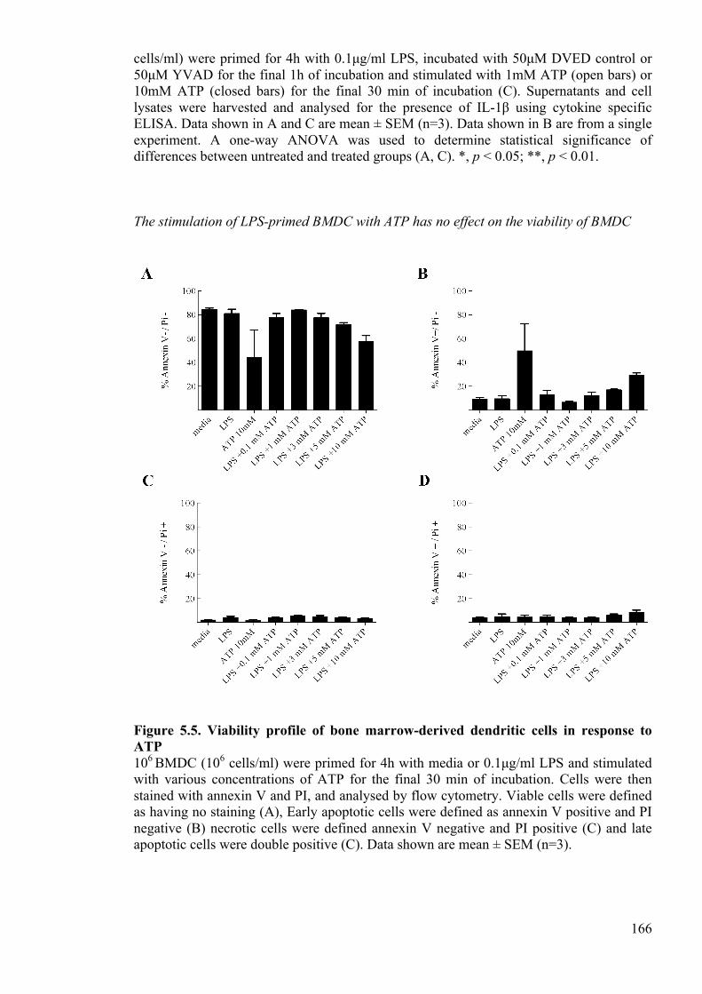
166
cells/ml) were primed for 4h with 0.1µg/ml LPS, incubated with 50µM DVED control or 50µM YVAD for the final 1h of incubation and stimulated with 1mM ATP (open bars) or 10mM ATP (closed bars) for the final 30 min of incubation (C). Supernatants and cell lysates were harvested and analysed for the presence of IL-1β using cytokine specific ELISA. Data shown in A and C are mean ± SEM (n=3). Data shown in B are from a single experiment. A one-way ANOVA was used to determine statistical significance of differences between untreated and treated groups (A, C). *, p < 0.05; **, p < 0.01.
The stimulation of LPS-primed BMDC with ATP has no effect on the viability of BMDC
Figure 5.5. Viability profile of bone marrow-derived dendritic cells in response to ATP 106 BMDC (106 cells/ml) were primed for 4h with media or 0.1µg/ml LPS and stimulated with various concentrations of ATP for the final 30 min of incubation. Cells were then stained with annexin V and PI, and analysed by flow cytometry. Viable cells were defined as having no staining (A), Early apoptotic cells were defined as annexin V positive and PI negative (B) necrotic cells were defined annexin V negative and PI positive (C) and late apoptotic cells were double positive (C). Data shown are mean ± SEM (n=3).
167
To explore the viability profile of BMDC in response to LPS and ATP, treated cells were
stained with both annexin V to detect apoptotic cells, and PI to detect dead cells. The
unstained (double negative) population was defined as viable (Fig. 5.5A). Early apoptotic
cells were defined as being annexin V positive but PI negative (Fig. 5.5B). Necrotic cells
were defined as being annexin V negative but PI positive (Fig, 5.5C) and late apoptotic
cells were defined as having both annexin V and PI-positive staining (Fig. 5.5D). Using
this assay, it was shown that the majority of untreated cells were viable (~85%).
Importantly, the addition of LPS had no impact on the viability of cells. The addition of
low concentrations of ATP (0.1-5mM ATP) to LPS-primed cells also had no impact upon
the viability of cells. However, the addition 10mM ATP to LPS-primed cells caused a
small reduction in the number of viable cells (~60% viable). Interestingly, the addition
10mM ATP to unprimed cells caused a larger reduction in cell viability (~ 40% viable).
Although no cells were found to be in necrotic or late apoptotic stages, the samples treated
with 10mM ATP (with or without LPS priming) caused a marked increase in the
percentage of early apoptotic cells. Thus, these results indicate that the addition of high
concentrations of ATP (>10mM) to either unprimed or primed cells results in the initiation
of apoptosis.
Human THP-1 cells and human monocytes produce IL-1β in response to LPS and secrete
IL-1β in response to nigericin
As discussed above, it was also important to establish a human cell line or primary cell that
could both express IL-1β, and secrete it in response to NLR stimulants. In both THP-1
cells (Fig. 5.6A) and primary monocytes (Fig. 5.7A), a 4h stimulation with the TLR-4
ligand LPS resulted in an up-regulation in intracellular IL-1β expression. In the THP-1
cells, there was a small amount of IL-1β secretion induced by LPS priming in the absence
168
of second signal, whereas in the primary monocytes, there was a marked secretion induced
by LPS alone, even at relatively low doses (0.01ng/ml). Interestingly, LPS induced IL-1β
expression was much more potent in the primary monocytes compared with the THP-1
cells. Although neither LPS-primed THP-1 cells (Fig. 5.6B), nor LPS-primed primary
monocytes (Fig. 5.7B), secreted IL-1β in response to ATP, both secreted large amounts of
IL-1β when stimulated with 10µM nigericin. Thus, it was ascertained that both THP-1
cells and primary monocytes were suitable to study the regulation of IL-1β production and
secretion. As previously observed with the murine cells, the addition of a second signal (in
this instance nigericin) caused a rapid increase in total IL-1β levels. In THP-1 cells
(Fig.5.6A) for example, the total IL-1β level in primed cells was ~0.5ng/ml, whereas the
total IL-1β level in primed cells treated with nigericin was ~5ng/ml. Again, as this is such
a rapid effect it is unlikely that it is a result of a nigericin-induced up-regulation in
expression. Instead, it is postulated that this is also an artefact of the ELISAs avidity for
mature and pro IL-1β.
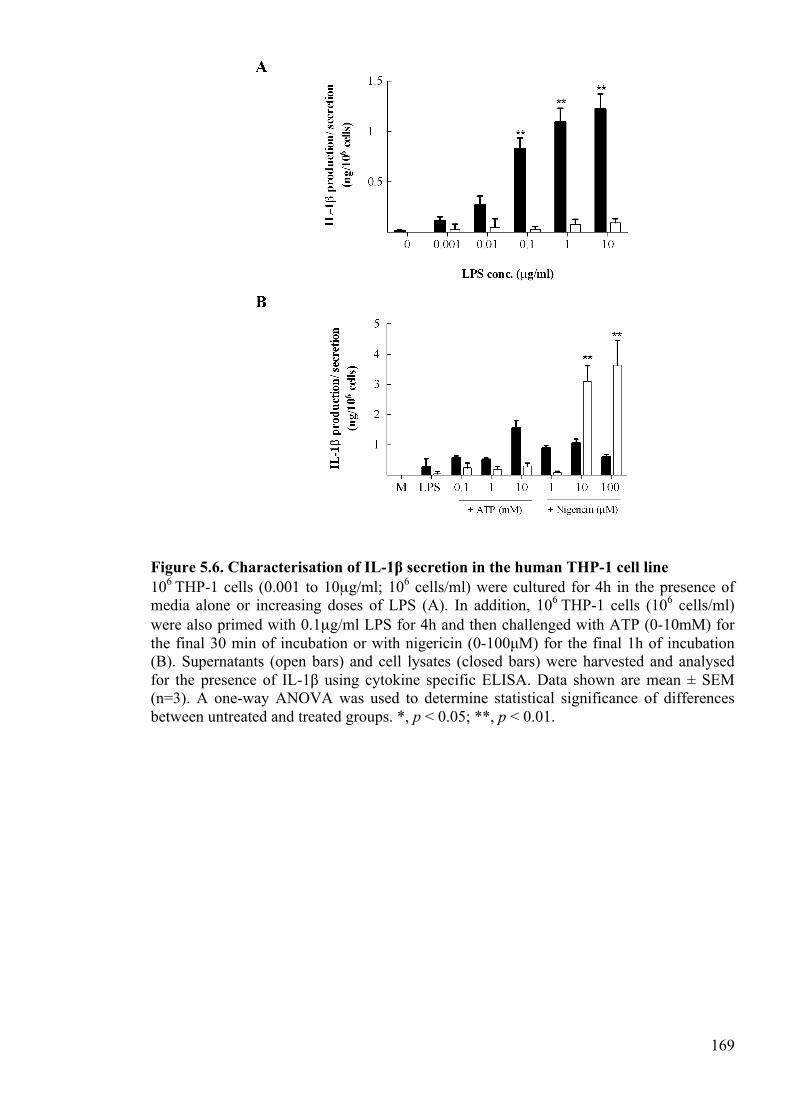
169
Figure 5.6. Characterisation of IL-1β secretion in the human THP-1 cell line 106 THP-1 cells (0.001 to 10µg/ml; 106 cells/ml) were cultured for 4h in the presence of media alone or increasing doses of LPS (A). In addition, 106 THP-1 cells (106 cells/ml) were also primed with 0.1µg/ml LPS for 4h and then challenged with ATP (0-10mM) for the final 30 min of incubation or with nigericin (0-100µM) for the final 1h of incubation (B). Supernatants (open bars) and cell lysates (closed bars) were harvested and analysed for the presence of IL-1β using cytokine specific ELISA. Data shown are mean ± SEM (n=3). A one-way ANOVA was used to determine statistical significance of differences between untreated and treated groups. *, p < 0.05; **, p < 0.01.
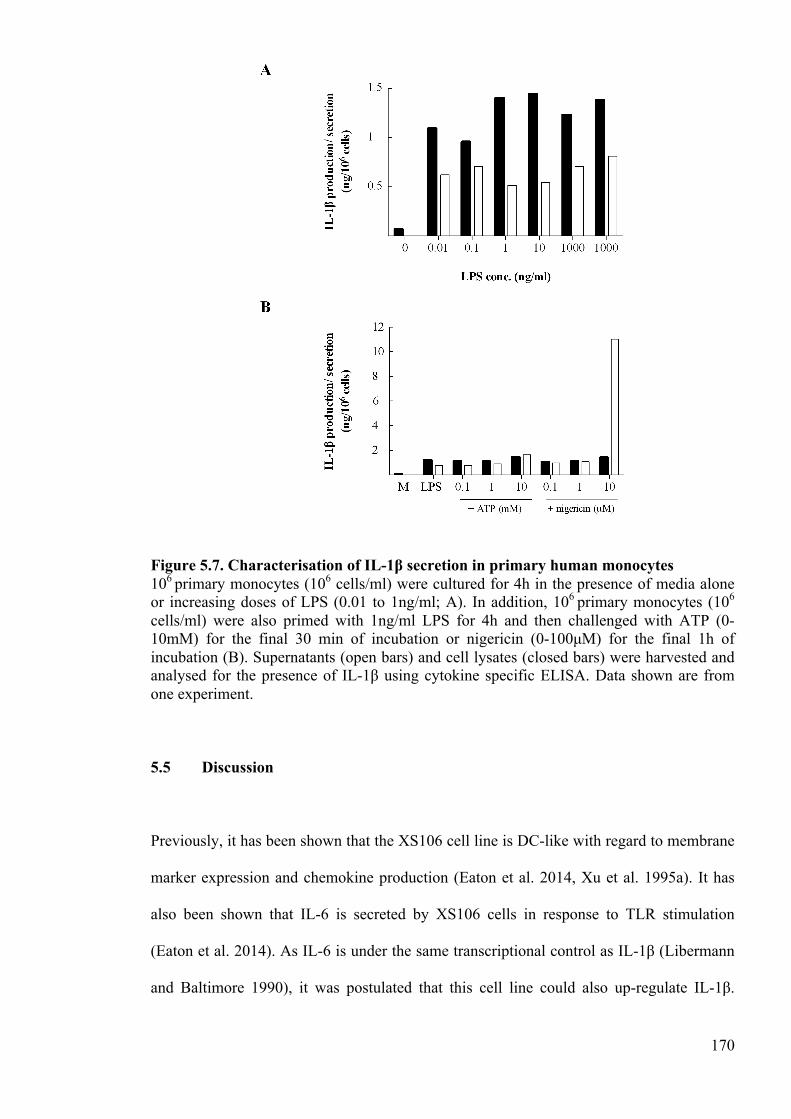
170
Figure 5.7. Characterisation of IL-1β secretion in primary human monocytes 106 primary monocytes (106 cells/ml) were cultured for 4h in the presence of media alone or increasing doses of LPS (0.01 to 1ng/ml; A). In addition, 106 primary monocytes (106 cells/ml) were also primed with 1ng/ml LPS for 4h and then challenged with ATP (0-10mM) for the final 30 min of incubation or nigericin (0-100µM) for the final 1h of incubation (B). Supernatants (open bars) and cell lysates (closed bars) were harvested and analysed for the presence of IL-1β using cytokine specific ELISA. Data shown are from one experiment.
5.5 Discussion
Previously, it has been shown that the XS106 cell line is DC-like with regard to membrane
marker expression and chemokine production (Eaton et al. 2014, Xu et al. 1995a). It has
also been shown that IL-6 is secreted by XS106 cells in response to TLR stimulation
(Eaton et al. 2014). As IL-6 is under the same transcriptional control as IL-1β (Libermann
and Baltimore 1990), it was postulated that this cell line could also up-regulate IL-1β.
171
Herein, it was shown that this cell line also produces IL-1β in response to TLR-
stimulation. However, it was also found that unlike DC in vitro, the XS106 cell line does
not process and secrete IL-1β in response to ATP stimulation. As ATP acts via the P2X7
receptor, it is postulated here that the XS106 cell line does not express the P2X7 receptor
(Ferrari et al. 1997). Alternatively, this cell line may not express components of the
NLRP3 inflammasome or caspase-1. In contrast to the XS106 cell line, it was shown that
LPS-primed BMDC produce and secrete IL-1β in response to ATP. Furthermore, it was
demonstrated that secretion is rapid, not associated with cell death, and dependent upon the
proteolytic enzyme caspase-1. This work is consistent with previous work (Fink and
Cookson 2006, Thornberry et al. 1992), and confirms that this cell represents a useful
model to study the regulation of IL-1β production and secretion. Although the viability of
untreated cells was relatively low (~85%), this is comparable with previous work using
BMDC (Englezou et al. 2015). Importantly, the current investigations also show that the
BMDC display a DC phenotype with regard to membrane marker expression. Specifically,
the majority of BMDC expressed relatively high levels of MHCII and the DC associated
marker CD11c. This is also consistent with previous investigations (Dearman et al. 2009,
Englezou et al. 2015) and highlights that the BMDC represent an important tool for
investigating the regulation of IL-1 in DC.
As previously discussed, it was also necessary to identify a suitable human cell line or
primary cell for the purpose of the investigations herein. It was shown that both THP-1
cells and primary human monocytes express large amounts of IL-1β intracellularly in
response to LPS stimulation. Interestingly, the primary monocytes appear to secrete IL-1β
in response to LPS stimulation also. This is consistent with a previous study, which
showed that monocytes can secrete IL-1β in response to TLR stimulation alone because
they have constitutively active caspase-1 (Netea et al. 2009). Although neither cell secreted
172
any extra IL-1β following ATP stimulation, secretion of this cytokine was markedly
increased following nigericin stimulation. This indicates that both cells have functioning
caspase-1 and NLRP3 inflammasome components, but probably do not express the P2X7
receptor. This is supported by previous investigations, which show that monocytes do not
express the P2X7 receptor unless differentiated into macrophages (Ferrari et al. 1997).
Thus, if we wanted to investigate the regulation of ATP-induced secretion in a human cell
specifically, we could differentiate the monocytes into macrophages using phorbol
myristate acetate- differentiation (Takashiba et al. 1999). Overall, these results suggest that
both the THP-1 cell line and primary human monocytes represent useful models to study
the regulation of human IL-1β production and secretion.
One interesting observation from this work was that total amount of IL-1β appeared to
increase dramatically following a second signal, both in murine cells and in human cells.
Importantly, it was also shown that the mouse IL-1β ELISA used does have a considerably
greater avidity for the mature cytokine, relative to the 31kDa precursor. Thus it was
postulated the signal 2 induced increase in total IL-1 levels was not the result of an
increase in IL-1β expression but was instead an artefact of the IL-1β ELISA. This finding
highlights that when studying IL-1β, it is important to determine both the level of
expression and nature of the cytokine. This approach has been taken in the previous
chapters of this thesis, where Western blotting was used in conjunction with the IL-1β
ELISA to ensure that any changes in expression were not the result of a change in form.
The work in this chapter demonstrates that the regulation of IL-1β is highly diverse across
different cell subsets and species. Some cells produce large amounts of IL-1β whereas
others do not, and some cells secrete IL-1β in response to TLR stimulation alone whereas
do not even secrete IL-1β in response to ATP stimulation. It is speculated here that the
173
differential regulation of IL-1β likely reflects the roles that different cell subsets play in the
regulation of inflammation. It is clear when comparing just a few cell subsets that the
spectrum of IL-1β production and secretion is broad. This observation is supported by a
number of previous studies (Guarda et al. 2011, Netea et al. 2009, Englezou et al. 2015,
Muzio et al. 2000), and highlights the importance of exploring IL-1β regulatory
mechanisms in a range of cell subsets.
5.6 References
Dearman, R. J., Cumberbatch, M., Maxwell, G., Basketter, D. A. and Kimber, I. (2009) 'Toll-like receptor ligand activation of murine bone marrow-derived dendritic cells', Immunology, 126(4), 475-484.
Eaton, L., Mellody, K. T., Kimber, I. and Dearman, R. J. (2014) 'The XS106 cell: a
Langerhans’ cell surrogate with a selective type 2 phenotype', 34( Englezou, P. C., Rothwell, S. W., Ainscough, J. S., Brough, D., Landsiedel, R.,
Verkhratsky, A., Kimber, I. and Dearman, R. J. (2015) 'P2X(7)R activation drives distinct IL-1 responses in dendritic cells compared to macrophages', Cytokine, 74(2), 293-304.
Ferrari, D., Chiozzi, P., Falzoni, S., DalSusino, M., Melchiorri, L., Baricordi, O. R. and
DiVirgilio, F. (1997) 'Extracellular ATP triggers IL-1β release by activating the purinergic P2Z receptor of human macrophages', Journal of Immunology, 159(3), 1451-1458.
Fink, S. L. and Cookson, B. T. (2006) 'Caspase-1-dependent pore formation during
pyroptosis leads to osmotic lysis of infected host macrophages', Cellular Microbiology, 8(11), 1812-1825.
Guarda, G., Zenger, M., Yazdi, A. S., Schroder, K., Ferrero, I., Menu, P., Tardivel, A.,
Mattmann, C. and Tschopp, J. (2011) 'Differential Expression of NLRP3 among Hematopoietic Cells', Journal of Immunology, 186(4), 2529-2534.
Libermann, T. A. and Baltimore, D. (1990) 'Activation of Interleukin-6 gene-expression
through the NF-kappa-B transcription factor', Molecular and Cellular Biology, 10(5), 2327-2334.
Lutz, M. B., Kukutsch, N., Ogilvie, A. L. J., Rossner, S., Koch, F., Romani, N. and
Schuler, G. (1999) 'An advanced culture method for generating large quantities of highly pure dendritic cells from mouse bone marrow', Journal of Immunological Methods, 223(1), 77-92.
174
Muzio, M., Bosisio, D., Polentarutti, N., D'Amico, G., Stoppacciaro, A., Mancinelli, R., van't Veer, C., Penton-Rol, G., Ruco, L. P., Allavena, P. and Mantovani, A. (2000) 'Differential expression and regulation of toll-like receptors (TLR) in human leukocytes: Selective expression of TLR3 in dendritic cells', Journal of Immunology, 164(11), 5998-6004.
Netea, M. G., Nold-Petry, C. A., Nold, M. F., Joosten, L. A. B., Opitz, B., van der Meer, J.
H. M., van de Veerdonk, F. L., Ferwerda, G., Heinhuis, B., Devesa, I., Funk, C. J., Mason, R. J., Kullberg, B. J., Rubartelli, A., van der Meer, J. W. M. and Dinarello, C. A. (2009) 'Differential requirement for the activation of the inflammasome for processing and release of IL-1β in monocytes and macrophages', Blood, 113(10), 2324-2335.
Takashiba, S., Van Dyke, T. E., Amar, S., Murayama, Y., Soskolne, A. W. and Shapira, L.
(1999) 'Differentiation of monocytes to macrophages primes cells for lipopolysaccharide stimulation via accumulation of cytoplasmic nuclear factor kappa B', Infection and Immunity, 67(11), 5573-5578.
Thornberry, N. A., Bull, H. G., Calaycay, J. R., Chapman, K. T., Howard, A. D., Kostura,
M. J., Miller, D. K., Molineaux, S. M., Weidner, J. R., Aunins, J., Elliston, K. O., Ayala, J. M., Casano, F. J., Chin, J., Ding, G. J. F., Egger, L. A., Gaffney, E. P., Limjuco, G., Palyha, O. C., Raju, S. M., Rolando, A. M., Salley, J. P., Yamin, T. T., Lee, T. D., Shively, J. E., Maccross, M., Mumford, R. A., Schmidt, J. A. and Tocci, M. J. (1992) 'A novel heterodimeric cysteine protease is required for interleukin-1-beta processing in monocytes', Nature, 356(6372), 768-774.
Xu, S., Ariizumi, K., Caceresdittmar, G., Edelbaum, D., Hashimoto, K., Bergstresser, P. R.
and Takashima, A. (1995a) 'Successive generation of antigen-presenting dendritic cell-lines from murine epidermis', Journal of Immunology, 154(6), 2697-2705.
Xu, S., Bergstresser, P. R. and Takashima, A. (1995b) 'Phenotypic and functional
heterogeneity among murine epidermal-derived dendritic cell clones', Journal of Investigative Dermatology, 105(6), 831-836.

175
CHAPTER 6:
DISCUSSION
176
6 Discussion
6.1 General discussion
6.1.1 Revisiting the initial aims
IL-1α and IL-1β are central to the initiation and orchestration of inflammation, and thus
there is an imperative to understand in detail the mechanisms of IL-1 regulation. Whilst
great progress has been made in determining the factors and processes that lead to pro-IL-1
up-regulation and secretion, the mechanisms that regulate the intracellular precursors of
IL-1α and IL-1β are relatively poorly understood (Garlanda et al. 2013). Post-translational
modifications were of particular interest in this thesis, as these modifications are becoming
increasingly diverse in scope and increasingly important in function (Perkins 2006).
Ubiquitination is rapidly emerging as a key player in mediating inflammatory responses,
and thus this modification was the focus of investigations in the initial phase of this thesis
(Hochrainer and Lipp 2007, Skaug et al. 2009). To complement this work, a broader
approach was taken, whereby the interactome of pro-IL-1β was assessed using a human
protein microarray. Following the initial assay, the roles of the hits identified were
investigated to determine novel mechanisms of intracellular pro-IL-1β regulation. The
overall hypothesis was that regulation of these precursors may serve to control the vigour
of IL-1 secretion and ultimately, may influence the vigour of pro-inflammatory responses.
6.1.2 IL-1: Regulation by degradation
In Chapter 2, evidence was provided that pro-IL-1α and pro-IL-1β are polyubiquitinated
and consequently degraded by the proteasome in DC and macrophages. Importantly, it was
shown that in the absence of inflammasome activation, these processes serve to rapidly

177
down-regulate intracellular IL-1 levels following LPS-priming. In addition, it was
demonstrated that proteasomal degradation regulates the basal turnover of pro-IL-1α and
pro-IL-1β. Collectively, these results highlight that the polyubiquitination and proteasomal
degradation is a critical process in the regulation of IL-1 and, therefore, should be
considered as an extra dimension to the current two-signal paradigm of IL-1 release (Fig.
7.1).
Figure 7.1. A new paradigm of IL-1 processing and release Following TLR-ligand and NFκB-induced pro-IL-1 expression, pro-IL-1α and pro-IL-1β are ubiquitinated and targeted for proteasomal degradation. IL-1 can be rescued from this pathway by inflammasome-activating signals. These signals induce inflammasome assembly, caspase-1 and calpain activation, pro-IL-1 processing and ultimately, IL-1 secretion. Given that IL-1α and IL-1β are such potent, multi-functional pro-inflammatory cytokines
(Dinarello 2009), and given that ubiquitination serves as a vital post-translational
178
modification in the regulation of inflammation (Wang and Maldonado 2006), the broader
implications of these findings are of great interest. As discussed previously, the process of
ubiquitination is mediated by a large family of enzymes called ubiquitin ligases, and these
serve to transfer ubiquitin onto target proteins (Pickart 2001). Importantly, ubiquitin
ligases are usually specific for a given substrate or family of substrates, and thus
ubiquitination can be used to mediate the degradation of specific proteins (Laney and
Hochstrasser 1999). Theoretically, this means that ubiquitination can function in concert
with mRNA expression to orchestrate the intracellular levels of specific proteins. In the
context of IL-1, the level of intracellular protein expression is of significant importance;
not least because the magnitude of intracellular pro-IL-1 protein expression has a major
impact on the vigour of IL-1 secretion and ultimately, the nature and severity of the pro-
inflammatory response.
Following on from the findings in chapter 2, and in light of the information presented
above, it was postulated that the mechanisms of IL-1 polyubiquitination and proteasomal
degradation serve as fundamental orchestrators of pro-IL-1α and pro-IL-1β protein up-
regulation. Specifically, it was hypothesised that the expression or activity of the IL-1
ubiquitin ligase in a given cell may have a marked impact on the capacity of that cell to up-
regulate intracellular pro-IL-1 protein levels. Likewise, it was also proposed that the
expression or activity of deubiquitinase enzymes could also have an effect on the capacity
of a cell to up-regulate pro-IL-1 protein. These hypotheses were addressed partly in
chapter 3, in studies that utilised 2 stably expressing fluorescent pro-IL-1β cell-lines
(venusIL-1βiBMDM and venusIL-1βTHP1 cells). These investigations revealed that IL-1β
ubiquitination occurs in unprimed cells and that TLR stimulation abrogates this
ubiquitination. Importantly, the inhibition of IL-1β ubiquitination resulted in an inhibition
of IL-1β degradation, thereby facilitating a more potent up-regulation of the IL-1β
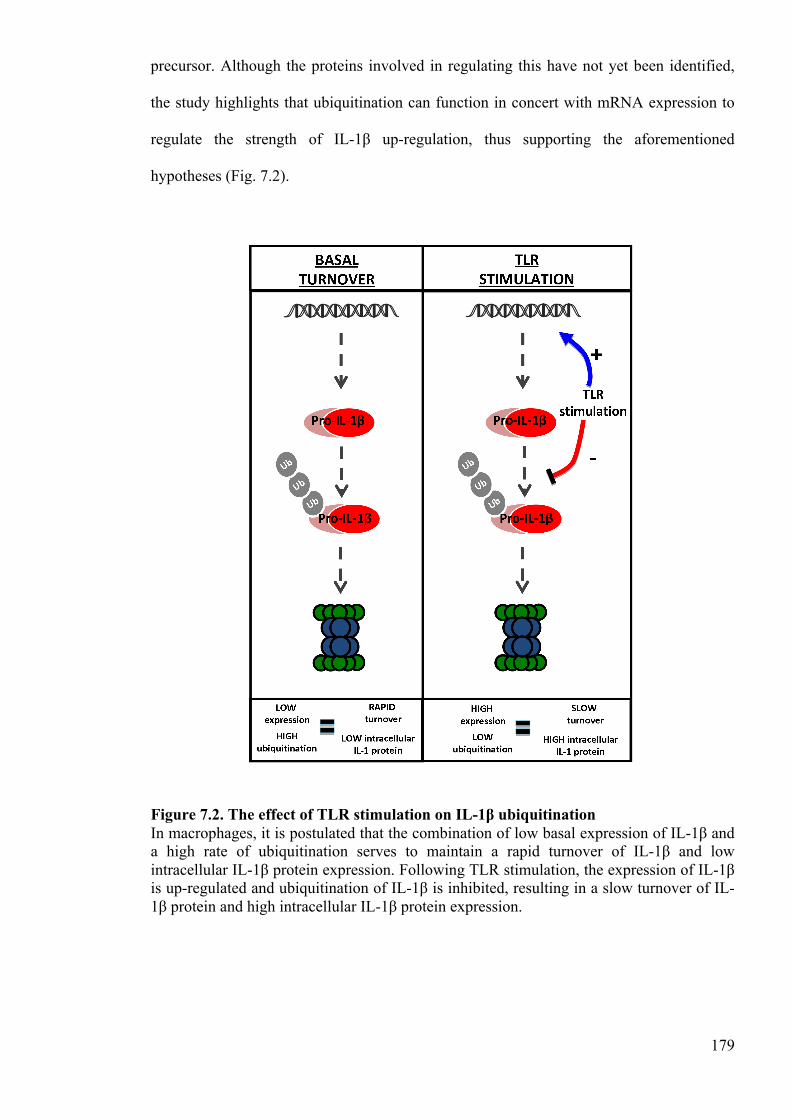
179
precursor. Although the proteins involved in regulating this have not yet been identified,
the study highlights that ubiquitination can function in concert with mRNA expression to
regulate the strength of IL-1β up-regulation, thus supporting the aforementioned
hypotheses (Fig. 7.2).
Figure 7.2. The effect of TLR stimulation on IL-1β ubiquitination In macrophages, it is postulated that the combination of low basal expression of IL-1β and a high rate of ubiquitination serves to maintain a rapid turnover of IL-1β and low intracellular IL-1β protein expression. Following TLR stimulation, the expression of IL-1β is up-regulated and ubiquitination of IL-1β is inhibited, resulting in a slow turnover of IL-1β protein and high intracellular IL-1β protein expression.
180
From the evidence presented, it is clear that ubiquitination has an important role to play in
regulating the vigour of IL-1β up-regulation. However, it is of interest to speculate further
about the role of ubiquitination in the regulation of IL-1 and inflammation. It well known
that some cell subsets, and some tissues are more capable of inducing a more vigourous
inflammatory response than others and thus it can be said that these cells and tissues have a
greater ‘inflammatory potential.’ It is imperative that different cells have different
inflammatory potentials because the inflammatory potential of a cell must reflect the
environment in which it resides, as well as its role within that environment. For instance,
cells that interface with the external microenvironment, such as skin or gut-resident cells,
are subject to a broad range of innocuous insults and thus need to have a low inflammatory
potential to tolerate these insults. In contrast, cells residing in protected sites such as blood-
resident cells are subject to very few, but typically very dangerous pathogenic threats and
so these must have a high inflammatory potential to fulfil their protective role.
The inflammatory potential of a cell is dictated in part by its capacity to up-regulate and
secrete IL-1. From the literature, it is already known that this potential can be modulated
by a range of factors including TLR expression (Muzio et al. 2000), NLR expression
(Guarda et al. 2011), basal caspase-1 activity (Netea et al. 2009) and post-transcriptional
modification (Kobayashi et al. 1988). Here, it is postulated that IL-1 ubiquitination may
also represent an important mediator of inflammatory potential. In this role, IL-1
ubiquitination could not only modulate the strength of TLR-induced IL-1 protein up-
regulation, but could also determine the speed of intracellular IL-1 clearance. In addition,
IL-1 ubiquitination could mediate the inflammatory potential of a cell or tissue by
regulating the basal turnover of IL-1 proteins. This is particularly intriguing in the context
of cutaneous immunobiology, as pro-IL-1α is constitutively expressed in the skin and thus
181
intracellular IL-1α levels must be modulated by the rate of IL-1α degradation (Ansel et al.
1988).
Interestingly, the inflammatory potential of cells and tissues can also be affected by
previous exposure to infection; therefore it is hypothesised that IL-1 ubiquitination may
also play an important role here. TLR tolerance is a well-studied phenomenon whereby
prolonged TLR stimulation leads to a transient state of TLR ligand hyporesponsiveness
(Broad et al. 2006). Ultimately, TLR tolerance results in a dramatic down-regulation in the
ability of TLR ligands to induce the expression of pro-inflammatory genes, including IL-1
(Ertel et al. 1995). This mechanism is important as it functions to limit excessive
inflammation in response to prolonged TLR ligand exposure. Although there are a number
of proposed mechanisms of TLR tolerance, such as decreased TLR expression (Nomura et
al. 2000) and decreased IRAK-1 activity (Medvedev et al. 2002), it is likely that there are
additional mechanisms that contribute (Medvedev et al. 2006). It is postulated here that an
increased rate of IL-1 ubiquitination may also serve as an important mechanism in TLR
tolerance.
Having speculated on the roles of ubiquitination in IL-1 regulation, it is important to
question why these additional mechanisms of regulation might be required. Whereas
properly controlled inflammation is a beneficial host response, the dysregulation of
inflammation can cause important adverse health effects (Dinarello 2011). As IL-1α and
IL-1β are such potent inducers of inflammation, it is vital that these cytokines are tightly
regulated to ensure the maintenance of health. Thus, it is likely that there exist numerous
mechanisms, including ubiquitination, that work in concert to ensure proper regulation of
IL-1. Moreover, many of these other mechanisms of IL-1 regulation, including the
modulation TLR expression or IRAK-1 activity, also serve to regulate the expression of
182
other pro-inflammatory mediators. As ubiquitin ligases are usually specific to the proteins
that they ubiquitinate, ubiquitination offers a mechanism whereby the expression of IL-1
can be controlled independently of other NFkB-dependent pro-inflammatory mediators
(Laney and Hochstrasser 1999). This could be of particular benefit in situations whereby a
pro-inflammatory response is beneficial, but IL-1 expression is not.
As discussed previously, the improper regulation of IL-1 leads to a number of important
inflammatory disorders and thus is of great interest therapeutically. The conclusions
reached in this thesis suggest that ubiquitin-dependent IL-1 degradation is a fundamental
regulator of IL-1 that is likely to serve in the control of inflammation per se. With that in
mind, it is possible that the improper regulation of IL-1 ubiquitination may be one cause of
certain inflammatory diseases. Of particular interest are inflammatory disorders of the skin,
such as psoriasis (Mee et al. 2006) and atopic dermatitis (Kezic et al. 2012), as these have
been frequently associated with IL-1 dysregulation. Given that there is a constitutive
turnover of IL-1α in the skin (Ansel et al. 1988), it is hypothesised that improper
degradation of this cytokine could lead to an accumulation of IL-1α protein, and
ultimately, the development of a number of inflammatory disorders. Elevated IL-1
production is also associated with a number of debilitating neurodegenerative diseases,
including Parkinson’s disease and Alzheimer’s disease (BlumDegen et al. 1995).
Curiously, there is strong evidence to suggest that the proteasome is impaired in both
Alzheimer’s and Parkinson’s disease (Riederer et al. 2011, Lim and Tan 2007). Therefore,
it is suggested that defects in proteasome functionality contribute to the elevated IL-1
levels observed in these neurodegenerative diseases. Furthermore, as inflammation is a
major component of both Alzheimer’s and Parkinson’s disease (Tuppo and Arias 2005,
Phani et al. 2012), it is postulated here that impaired degradation of IL-1 may even
contribute to the development of these disorders.
183
Although it is common for studies to examine IL-1 levels when investigating the cause of
inflammatory disorders, many of these investigations have looked only at the mRNA
expression of pro-inflammatory cytokines. Whilst mRNA expression represents a good
starting point, it is suggested that this should not be the only parameter considered when
investigating the role of IL-1 in these diseases. This is because, unlike most cytokines, an
increase in mRNA expression is not the only way in which the protein expression of IL-1α
or IL-1β can be up-regulated significantly. As we have observed in this thesis, a rapid
increase in intracellular IL-1 protein levels can also be driven by an inhibition of
degradation, especially in cells whereby the basal expression of IL-1α or IL-1β is already
high. Therefore, before a role for IL-1 in the development of a disease can be discounted, it
is important that the expression of IL-1 protein and the rate of IL-1 degradation are also
examined.
6.1.3 Analysing the interactome of pro-IL-1β
In chapter 4, the interactome of human pro-IL-1β was assessed using a human protein
microarray. Using this approach, a number of potentially important pro-IL-1β interacting
proteins were identified. From the initial microarray results, calmodulin was identified as
the most interesting hit. This is because calcium is strongly associated with pro-IL-1β
processing (Brough et al. 2003), and calmodulin is an important calcium-sensitive
messenger protein (Chin and Means 2000). Thus, interaction between calmodulin and pro-
IL-1β was examined further. However, before these investigations are discussed, it is
important to explore the potential importance of the other pro-IL-1β-interacting proteins
identified in the initial screen.
184
One of the most intriguing hits identified on the microarray was PLCXD3, a membrane-
bound phospholipase C enzyme (Kalujnaia et al. 2013). Phospholipase C enzymes are an
important group of enzymes that have been associated with cell signal transduction and
inflammation (Kadamur and Ross 2013). Phospholipases are most well known for their
role within the transduction of G protein coupled receptor (GPCR) signalling pathways
(Neer 1995). In this role, the binding of ligands to GPCR results in the activation of
phospholipase C. Active phospholipase C cleaves phosphatidylinositol bisphosphate,
releasing Inositol trisphosphate (IP3) into the cytosol (Smrcka et al. 1991). Cytosolic IP3
binds to IP3-gated Ca2+ channels on the endoplasmic reticulum, facilitating the release of
intracellular Ca2+ stores into the cytosol (Ehrlich and Watras 1988). As previously
discussed, and as demonstrated in chapter 4, the release of Ca2 from intracellular Ca2+
stores is central to pro-IL-1β processing, at least in the response to NLRP3 activators such
as ATP and nigericin. Interestingly, a recent paper has provided evidence that extracellular
Ca2+ can signal via a GPCR called GPRC6A, and this culminates in the release of
intracellular Ca2+ stores and the processing of pro-IL-1β (Rossol et al. 2012).
Given that the release of intracellular Ca2+ stores is associated with the response to other
DAMP/PAMP, such as ATP and nigericin (Brough et al. 2003), it could be that GPCR
pathways are implicated in the processing of pro-IL-1β induced by these other NLR
stimulants. Here, it is hypothesised that the interaction between pro-IL-1β and PLCXD3
may be important in facilitating the transduction of these signalling pathways when pro-IL-
1β is expressed. As previous studies have shown that that inflammasome is not usually
activated without TLR-priming (Compan et al. 2012), it is proposed that this mechanism
may serve to prevent GPCR-dependent inflammasome activation when IL-1β is not
expressed. This mechanism may serve as a necessary protective measure, preventing the
potentially detrimental effects of inflammasome activation when the cell is unprimed.
185
However, this hypothesis is only speculative at this stage and is apparently inconsistent
with studies that suggest that TLR-priming is required because it induces an up-regulation
in expression of inflammasome proteins (Schroder et al. 2012). As PLCXD3 is a
membrane bound protein, another possible role for the interaction is in the secretion of IL-
1β. However, this is unlikely given that there is no precedent for phospholipases to
function in this capacity.
Another particularly interesting hit was IL-22Ra2, a secreted inhibitor of IL-22 (Dumoutier
et al. 2001). IL-22 is part of the IL-10 family of cytokines and is a potent mediator of
inflammatory responses (Zheng et al. 2007). Like IL-1, IL-22 is secreted by activated DC
and plays an important role in mediating inflammation in tissue repair. IL-22Ra2 binds to
IL-22 and prevents the cytokine from interacting with the functional receptor on target
cells (Xu et al. 2001). Therefore, it is postulated here that tissue damage-induced IL-1β
binds to IL-22Ra2, facilitating IL-22-mediated tissue repair. When tissue damage has
resolved and IL-1β is no longer released, IL-22Ra2 can then bind IL-22, thereby inhibiting
the IL-22-induced reparatory mechanisms. Intriguingly, this hypothesis suggests, for the
first time, that released pro-IL-1β may have a functional role. Alternatively, it could be that
the mature IL-1β also interacts with IL-22Ra2, and that this interaction plays more of a
functional role here.
As mentioned previously, calmodulin was identified as the most relevant hit on the
microarray, and thus the interaction between this calcium-sensitive protein and IL-1β was
examined further. In this work, it was demonstrated that the interaction is specific for pro-
IL-1β, but not mature IL-1β, and is dependent on the presence of calcium ions.
Importantly, it was also shown that that calcium and calmodulin are required for IL-1β
processing by both the human THP-1 monocytic cell line, and by primary human
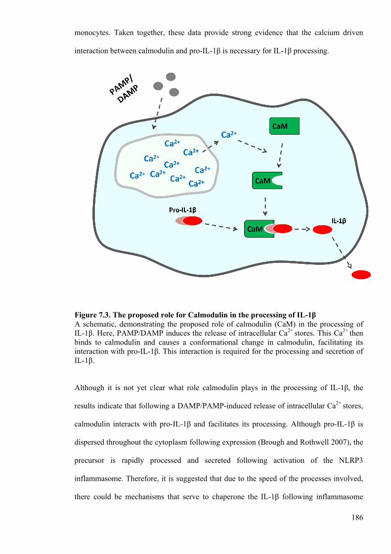
186
monocytes. Taken together, these data provide strong evidence that the calcium driven
interaction between calmodulin and pro-IL-1β is necessary for IL-1β processing.
Figure 7.3. The proposed role for Calmodulin in the processing of IL-1β A schematic, demonstrating the proposed role of calmodulin (CaM) in the processing of IL-1β. Here, PAMP/DAMP induces the release of intracellular Ca2+ stores. This Ca2+ then binds to calmodulin and causes a conformational change in calmodulin, facilitating its interaction with pro-IL-1β. This interaction is required for the processing and secretion of IL-1β.
Although it is not yet clear what role calmodulin plays in the processing of IL-1β, the
results indicate that following a DAMP/PAMP-induced release of intracellular Ca2+ stores,
calmodulin interacts with pro-IL-1β and facilitates its processing. Although pro-IL-1β is
dispersed throughout the cytoplasm following expression (Brough and Rothwell 2007), the
precursor is rapidly processed and secreted following activation of the NLRP3
inflammasome. Therefore, it is suggested that due to the speed of the processes involved,
there could be mechanisms that serve to chaperone the IL-1β following inflammasome

187
activation. In light of evidence suggesting that calmodulin can function as a chaperone
protein for certain target proteins (Shao and Hegde 2011), it was postulated in chapter 4
that calmodulin could play an important role in trafficking IL-1β.
As calmodulin binds the precursor of IL-1β only, and as calmodulin inhibition abrogates
nigericin induced pro-IL-1β processing, it is suggested that calmodulin must function prior
to the processing of IL-1β. Thus, if calmodulin is involved in IL-1β trafficking, it is likely
that it functions to chaperone the pro-protein towards the site of caspase-dependent
cleavage. In this role, calmodulin could present pro-IL-1β to caspase-1 in the cytoplasm,
and may even be required for efficient cleavage of the precursor. Alternatively, calmodulin
may play a role in packaging the IL-1β into caspase containing MVB or microvesicles. IL-
1β and caspase-containing microvesicles have been shown to be produced by THP-1 cells
in response to NLRP3 inflammasome activation (MacKenzie et al. 2001), and therefore
this hypothesis is in agreement with the evidence provided in chapter 4. Another
possibility is that the calmodulin is required to traffic the pro-IL-1β towards caspase-gated
membrane pores in the terminal release of IL-1β. Again, this pathway of IL-1β release has
been observed in human monocytes treated with NLRP3 stimulants (Singer et al. 1995),
and thus this hypothesis is also consistent with the evidence provided in chapter 4. As
discussed previously, the pathway of IL-1 release is very much dependent on factors such
as the type of cell, the type of stimulus and the strength of that stimulus (Lopez-Castejon
and Brough 2011). Therefore, if calmodulin is involved in IL-1β trafficking, it is likely that
it is only important in response to certain types of stimuli, and in certain cell types.
The potential to target calmodulin therapeutically has already been explored with some
success. Interestingly, the calmodulin inhibitor CV-159 exerts protective effects on smooth
muscle inflammatory responses (Usui et al. 2010), supporting the hypotheses presented
188
herein. However, in this study, it was suggested that calmodulin inhibition has an anti-
inflammatory effect because it inhibits tumour necrosis factor (TNF)-induced, Akt
phosphorylation-dependent expression of vascular cell adhesion molecule (VCAM)-1.
Whilst the conclusions in the Usia et al. study may be true, we propose that calmodulin
inhibition may also inhibit inflammation by abrogating the processing and release of IL-1β.
Although further investigation is required, it is believed that the findings of chapter 4 could
serve to inform the development of anti-calmodulin based anti-inflammatory therapeutics
in the future.
6.2 Future work
Although important progress has been made in elucidating the role of ubiquitination in the
regulation of IL-1, it is clear that there is still much work still to be done. One important
step is to determine the proteins involved in the ubiquitination of IL-1. Of particular
interest is the ubiquitin ligase of IL-1 as it is hypothesised that the expression or the
activity of this ligase will have a marked impact on the vigour of IL-1 up-regulation and
the potency of inflammation.
Identifying the ubiquitin ligase of IL-1 is a technically challenging objective, not least
because there are ~1000 different ubiquitin ligases expressed by humans (Hochstrasser
1995). One approach could be to use an siRNA library that targets the ubiquitin ligase
family on one of the stably expressing fluorescent IL-1β cell lines described previously (Li
et al. 2008). In such an experiment, cells would be incubated in plates, with each well
containing siRNA that targets a different ubiquitin ligase. These cells could then be
analysed for fluorescent IL-1β protein expression using flow cytometry. Wells containing
cells that expressed high levels of fluorescent IL-1β protein would be highlighted as hits,
189
and the ubiquitin ligases that are targeted in these wells identified as potential IL-1β
ubiquitinating ligases. From the list of hits, the ubiquitin ligase of IL-1β could be
determined by performing more comprehensive siRNA knockdown experiments. In these
experiments, the potential ubiquitin ligases of IL-1β would be knocked down in
appropriate cell lines and the level of IL-1β ubiquitination measured using co-
immunoprecipitation. Theoretically, the ubiquitination of IL-1 should be significantly
abrogated in the cell line where the IL-1β ubiquitin ligase is knocked down, and thus this
ubiquitin ligase could be identified using the co-immunoprecipitation experiment
described.
Another approach that could be used to identify the ubiquitin ligase of IL-1 is mass
spectrometry (Kalinichenko et al. 2012). In this approach, LPS-primed cells could be
treated with a cross-linker to covalently bind IL-1 to any proteins with which it interacts.
Following treatment, these cells could be lysed and IL-1 immunoprecipitated from the
lysates using an anti-IL-1 antibody. The immunoprecipitated proteins could then analysed
by mass spectrometry to identify IL-1-interacting proteins. This approach should not only
identify the IL-1 ubiquitin ligase of IL-1, but also should complement the results obtained
in chapter 4 of this thesis. Moreover, these techniques could also be used to identify other
proteins that are important in regulating the ubiquitination of IL-1, such as E1 ubiquitin-
activating enzymes, E2 ubiquitin-conjugating enzymes and deubiquitinase enzymes.
Once the proteins involved in the ubiquitination of IL-1 have been identified, there are a
number of important questions that could be addressed. In this thesis, it is proposed that
that the process of IL-1 ubiquitination serves as a fundamental regulator of inflammation in
vivo. To investigate the importance of this mechanism, IL-1 ubiquitination could be
ablated by blocking the expression of the IL-1 ubiquitin ligase in mice. Initially, the
190
importance of IL-1 ubiquitination could be assessed by exploring the phenotype of these
mice. In addition, the inflammatory response could also be investigated in these KO mice
by using a variety of inflammation models such as endotoxin induced sepsis, collagen
induced arthritis and EAE (Webb 2014). If IL-1 ubiquitination is an important regulator of
the inflammatory response, it is hypothesised that these mice would not only exhibit a
general inflammatory phenotype, but would also present with an excessive and prolonged
inflammatory response in the models aforementioned.
It was also hypothesised in this thesis that the expression of proteins involved in IL-1
ubiquitination are differentially regulated in different cells and tissues, and that this serves
to modulate the inflammatory potential of these cells and tissues. To address this
hypothesis, the mRNA expression of proteins involved in IL-1 ubiquitination could be
assessed in different cells and tissues, and these data used to determine whether the
expression of these proteins reflects the inflammatory potential of the cells and tissues
examined. The expression of proteins involved in IL-1 ubiquitination could also be
investigated in IL-1-mediated inflammatory diseases to determine whether these proteins
are implicated here. Finally, the expression of these proteins could be explored in TLR
tolerance to address the hypothesis that the phenomenon is mediated partly by an up-
regulation in the rate of IL-1 ubiquitination.
As mentioned previously, the results of the microarray experiment detailed in chapter 4
highlight a number of potentially important hits. Thus, it may be of interest to explore
these interactions in greater detail. Besides calmodulin, the most intriguing hits on the
microarray were PLCXD3 and IL-22Ra2. Therefore, the interaction between these proteins
and IL-1β should be investigated further. As with calmodulin, it is important to confirm the
interactions using a protein-binding assay before the implications of these interactions are
191
studied in greater detail. If the interactions can be confirmed, the functional implications
should then be examined based on the hypotheses described above. Therefore, the
implication of the interaction between pro-IL-1β and PLCXD3 should be explored by
inhibiting PLCXD3 expression in monocytes and investigating Ca2+-dependent IL-1β
processing in these cells. Likewise, the implication of the interaction between pro-IL-1β
and IL-22Ra2 could be explored by inducing tissue damage in IL-1β-deficient mice and
investigating whether the administration of recombinant pro-IL-1β has any impact upon
the resolution of injury.
In addition, there are still questions to be addressed regarding the interaction between pro-
IL-1β and calmodulin. In order to fully understand this interaction, it is important to
develop an experimental system that allows the investigator to observe the interaction
intracellularly. To do this, a transgenic cell line that stably expresses fluorescent pro-IL-1β
and fluorescent calmodulin could be developed. Using this cell line, a number of
approaches could be used. One approach could be to use fluorescence cross correlation
spectroscopy and examine when the proteins interact in response to a range of danger
signals (Ma et al. 2014). Another approach could be to use fluorescence resonance emitted
tomography to explore the interaction (Hoppe et al. 2002). These approaches could serve
to both strengthen the conclusions drawn in chapter 4, and elucidate the functional role of
the interaction.
6.3 Concluding remarks
The broad aim of this thesis was to explore the intracellular mechanisms that regulate pro-
IL-1. From the findings presented, it is clear that there are a number of intracellular
mechanisms that function in this capacity. In chapter 2, it was shown that pro-IL-1α and
192
pro-IL-1β are ubiquitinated and degraded by the proteasome, and that these mechanisms
serve as fundamental regulators of intracellular pro-IL-1 protein expression. In chapter 3,
evidence is presented to suggest that these mechanisms function to both clear away-
unprocessed IL-1, and to modulate actively the vigour of IL-1 up-regulation. Therefore, it
is speculated that the ubiquitination of IL-1 serves to regulate the vigour of the
inflammatory response. Although further work is required to address this hypothesis, it is
postulated that the mechanisms described herein are pivotal to the maintenance of tissue
homeostasis, and are thus of great academic and therapeutic interest. The work conducted
in this thesis also identifies calmodulin as a potentially essential mediator of IL-1β
secretion. Specifically, the data presented indicate that pro-IL-1β interacts with calmodulin
in a calcium-dependent manner, and that this interaction is required for the processing and
release of the precursor. Given that the mechanisms controlling IL-1 processing and
release are so poorly understood, this finding may represent an important breakthrough in
the field of IL-1 biology.

193
CHAPTER 7:
BIBLIOGRAPHY
194
7 Bibliography
Adams, J. (2003) 'The proteasome: structure, function, and role in the cell', Cancer Treatment Reviews, 29, 3-9.
Afonina, I. S., Tynan, G. A., Logue, S. E., Cullen, S. P., Bots, M., Luethi, A. U., Reeves,
E. P., McElvaney, N. G., Medema, J. P., Lavelle, E. C. and Martin, S. J. (2011) 'Granzyme B-Dependent Proteolysis Acts as a Switch to Enhance the Proinflammatory Activity of IL-1 alpha', Molecular Cell, 44(2), 265-278.
Agostini, L., Martinon, F., Burns, K., McDermott, M. F., Hawkins, P. N. and Tschopp, J.
(2004) 'NALP3 forms an IL-l beta-Processing inflammasome with increased activity in Muckle-Wells autoinflammatory disorder', Immunity, 20(3), 319-325.
Ainscough, J. S., Gerberick, G. F., Dearman, R. J. and Kimber, I. (2013) 'Danger,
intracellular signaling, and the orchestration of dendritic cell function in skin sensitization', Journal of Immunotoxicology, 10(3), 223-234.
Ainscough, J. S., Gerberick, G. F., Zahedi-Nejad, M., Lopez-Castejon, G., Brough, D.,
Kimber, I. and Dearman, R. J. (2014) 'Dendritic Cell IL-1 alpha and IL-1 beta Are Polyubiquitinated and Degraded by the Proteasome', Journal of Biological Chemistry, 289(51), 35582-35592.
Alexopoulou, L., Holt, A. C., Medzhitov, R. and Flavell, R. A. (2001) 'Recognition of
double-stranded RNA and activation of NF-kappa B by Toll-like receptor 3', Nature, 413(6857), 732-738.
Andrei, C., Dazzi, C., Lotti, L., Torrisi, M. R., Chimini, G. and Rubartelli, A. (1999) 'The
secretory route of the leaderless protein interleukin 1 beta involves exocytosis of endolysosome-related vesicles', Molecular Biology of the Cell, 10(5), 1463-1475.
Ansel, J. C., Luger, T. A., Lowry, D., Perry, P., Roop, D. R. and Mountz, J. D. (1988) 'The
expression and modulation of IL-1-alpha in murine keratincytes', Journal of Immunology, 140(7), 2274-2278.
Apte, R. N., Dotan, S., Elkabets, M., White, M. R., Reich, E., Carmi, Y., Song, X.,
Dvozkin, T., Krelin, Y. and Voronov, E. (2006) 'The involvement of IL-1 in tumorigenesis, tumor invasiveness, metastasis and tumor-host interactions', Cancer and Metastasis Reviews, 25(3), 387-408.
Baeuerle, P. A. and Baltimore, D. (1988a) 'Activation of dna-binding activity in an
apparently cytoplasmic precursor of the nf-kappa-b transcription factor', Cell, 53(2), 211-217.
Baeuerle, P. A. and Baltimore, D. (1988b) 'I-kappa-B- a specific inhibitor of the NF-
kappa-B', Science, 242(4878), 540-546. Bagnall, J., Boddington, C., Boyd, J., Brignall, R., Rowe, W., Jones, N. A., Schmidt, L.,
Spiller, D. G., White, M. R. H. and Paszek, P. (2015) 'Quantitative dynamic
195
imaging of immune cell signalling using lentiviral gene transfer', Integrative Biology, 7(6), 713-725.
Bailly, S., Fay, M., Ferrua, B. and Gougerotpocidalo, M. A. (1994) 'Comparative
production of Il-1-beta and Il-1-alpha LPS-stimulated human monocytes- ELISAs measurement revisited', Cytokine, 6(2), 111-115.
Banchereau, J. and Steinman, R. M. (1998) 'Dendritic cells and the control of immunity',
Nature, 392(6673), 245-252. Benko, S., Philpott, D. J. and Girardin, S. E. (2008) 'The microbial and danger signals that
activate Nod-like receptors', Cytokine, 43(3), 368-373. Bergsbaken, T., Fink, S. L. and Cookson, B. T. (2009) 'Pyroptosis: host cell death and
inflammation', Nature Reviews Microbiology, 7(2), 99-109. Bieber, T. (2008) 'Mechanisms of disease: Atopic dermatitis', New England Journal of
Medicine, 358(14), 1483-1494. BlumDegen, D., Muller, T., Kuhn, W., Gerlach, M., Przuntek, H. and Riederer, P. (1995)
'Interleukin-1 beta and interleukin-6 are elevated in the cerebrospinal fluid of Alzheimer's and de novo Parkinson's disease patients', Neuroscience Letters, 202(1-2), 17-20.
Blumenthal, D. K. and Stull, J. T. (1980) 'Activation of skeletal-muscle myosin light chain
kinase by calcium(2+) and calmodulin', Biochemistry, 19(24), 5608-5614. Brewer, G. J., Kanzer, S. H., Zimmerman, E. A., Molho, E. S., Celmins, D. F., Heckman,
S. M. and Dick, R. (2010) 'Subclinical Zinc Deficiency in Alzheimer's Disease and Parkinson's Disease', American Journal of Alzheimers Disease and Other Dementias, 25(7), 572-575.
Broad, A., Jones, D. E. J. and Kirby, J. A. (2006) 'Toll-Like Receptor (TLR) Response
Tolerance: A Key Physiological "Damage Limitation" Effect and an Important Potential Opportunity for Therapy', Current Medicinal Chemistry, 13(21), 2487-2502.
Brough, D., Le Feuvre, R. A., Wheeler, R. D., Solovyova, N., Hilfiker, S., Rothwell, N. J.
and Verkhratsky, A. (2003) 'Ca2+ stores and Ca2+ entry differentially contribute to the release of IL-1 beta and IL-1 alpha from murine macrophages', Journal of Immunology, 170(6), 3029-3036.
Brough, D. and Rothwell, N. J. (2007) 'Caspase-1-dependent processing of pro-interleukin-
1 beta is cytosolic and precedes cell death', Journal of Cell Science, 120(5), 772-781.
Brough, D., Tyrrell, P. J. and Allan, S. M. (2011) 'Regulation of interleukin-1 in acute
brain injury', Trends in Pharmacological Sciences, 32(10), 617-622. Browne, G. J. and Proud, C. G. (2004) 'A novel mTOR-regulated phosphorylation site in
elongation factor 2 kinase modulates the activity of the kinase and its binding to calmodulin', Molecular and Cellular Biology, 24(7), 2986-2997.
196
Bryant, C. and Fitzgerald, K. A. (2009) 'Molecular mechanisms involved in inflammasome
activation', Trends in Cell Biology, 19(9), 455-464. Buchan, G., Barrett, K., Turner, M., Chantry, D., Maini, R. N. and Feldmann, M. (1988)
'Interleukin-1 and Tumor Necrosis Factor messenger-RNA expression in Rheumatoid-Arthritis - Prolonged production of IL-1α', Clinical and Experimental Immunology, 73(3), 449-455.
Cacabelos, R., Alvarez, X. A., Fernandeznovoa, L., Franco, A., Mangues, R., Pellicer, A.
and Nishimura, T. (1994) 'Brain Interleukin-1β in Alzheimers-Disease and vascular dementia', Methods and Findings in Experimental and Clinical Pharmacology, 16(2), 141-151.
Cacabelos, R., Francomaside, A. and Alvarez, X. A. (1991) 'Interleukin-1 in Alzheimers-
Disease and Multiinfarct Dementia - neuropsychological correlations', Methods and Findings in Experimental and Clinical Pharmacology, 13(10), 703-708.
Cameron, P., Limjuco, G., Rodkey, J., Bennett, C. and Schmidt, J. A. (1985) 'Amino-acid
sequence-analysis of human interleukin-1(IL-1)- evidence for biochemically distinct forms of IL-1', Journal of Experimental Medicine, 162(3), 790-801.
Casiniraggi, V., Kam, L., Chong, Y. J. T., Fiocchi, C., Pizarro, T. T. and Cominelli, F.
(1995) 'Mucosal imbalance of Il-1 and Il-1 receptor antagonist in inflammatory bowel-disease- a novel mechanism of chronic intestinal inflammation', Journal of Immunology, 154(5), 2434-2440.
Chang, M., Jin, W. and Sun, S.-C. (2009) 'Peli1 facilitates TRIF-dependent Toll-like
receptor signaling and proinflammatory cytokine production', Nature Immunology, 10(10), 1089-U66.
Chen, C.-J., Shi, Y., Hearn, A., Fitzgerald, K., Golenbock, D., Reed, G., Akira, S. and
Rock, K. L. (2006) 'MyD88-dependent IL-1 receptor signaling is essential for gouty inflammation stimulated by monosodium urate crystals', Journal of Clinical Investigation, 116(8), 2262-2271.
Chen, F.-T., Liu, Y.-C., Yang, C.-M. and Yang, C.-H. (2012) 'Anti-inflammatory effect of
the proteasome inhibitor bortezomib on endotoxin-induced uveitis in rats', Investigative Ophthalmology & Visual Science, 53(7), 3682-3694.
Chin, D. and Means, A. R. (2000) 'Calmodulin: a prototypical calcium sensor', Trends in
Cell Biology, 10(8), 322-328. Chung, Y., Chang, S. H., Martinez, G. J., Yang, X. O., Nurieva, R., Kang, H. S., Ma, L.,
Watowich, S. S., Jetten, A. M., Tian, Q. and Dong, C. (2009) 'Critical Regulation of Early Th17 Cell Differentiation by Interleukin-1 Signaling', Immunity, 30(4), 576-587.
Church, L. D. and McDermott, M. F. (2009) 'Canakinumab, a fully human mAb against
IL-1 beta for the potential treatment of inflammatory disorders', Current Opinion in Molecular Therapeutics, 11(1), 81-89.
197
Cogswell, J. P., Godlevski, M. M., Wisely, G. B., Clay, W. C., Leesnitzer, L. M., Ways, J. P. and Gray, J. G. (1994) 'NF-κB regulates IL-1β transcription through a consensus NF-κB binding-site and a nonconsensus Cre-like site', Journal of Immunology, 153(2), 712-723.
Cohen, P. (2000) 'The regulation of protein function by multisite phosphorylation - a 25
year update', Trends in Biochemical Sciences, 25(12), 596-601. Colotta, F., Re, F., Muzio, M., Bertini, R., Polentarutti, N., Sironi, M., Giri, J. G., Dower,
S. K., Sims, J. E. and Mantovani, A. (1993) 'Interleukin-1 type-ii receptor - a decoy target for IL-1 that is regulated by IL-4', Science, 261(5120), 472-475.
Compan, V., Baroja-Mazo, A., Bragg, L., Verkhratsky, A., Perroy, J. and Pelegrin, P.
(2012) 'A Genetically Encoded IL-1 beta Bioluminescence Resonance Energy Transfer Sensor To Monitor Inflammasome Activity', Journal of Immunology, 189(5), 2131-2137.
Corish, P. and Tyler-Smith, C. (1999) 'Attenuation of green fluorescent protein half-life in
mammalian cells', Protein Engineering, 12(12), 1035-1040. Cubitt, A. B., Heim, R., Adams, S. R., Boyd, A. E., Gross, L. A. and Tsien, R. Y. (1995)
'Understanding, improving and using green fluorescent proteins', Trends in Biochemical Sciences, 20(11), 448-455.
Cumberbatch, M., Dearman, R. J., Groves, R. W., Antonopoulos, C. and Kimber, I. (2002)
'Differential regulation of epidermal Langerhans cell migration by interleukins (IL)-1α and IL-1β during irritant- and allergen-induced cutaneous immune responses', Toxicology and Applied Pharmacology, 182(2), 126-135.
Cumberbatch, M., Dearman, R. J. and Kimber, I. (1997) 'Stimulation of Langerhans cell
migration in mice by tumour necrosis factor alpha and interleukin 1 beta', Dendritic Cells in Fundamental and Clinical Immunology, Vol 3, 417, 121-124.
Dalbeth, N. and Haskard, D. O. (2005) 'Mechanisms of inflammation in gout',
Rheumatology, 44(9), 1090-1096. Dandona, P., Aljada, A. and Bandyopadhyay, A. (2004) 'Inflammation: the link between
insulin resistance, obesity and diabetes', Trends in Immunology, 25(1), 4-7. Daniele, R. P., Holian, S. K. and Nowell, P. C. (1978) 'Potassium Ionophore (nigericin)
inhibits stimulation of human lymphocytes by mitogens', Journal of Experimental Medicine, 147(2), 571-581.
Dearman, R. J., Cumberbatch, M., Maxwell, G., Basketter, D. A. and Kimber, I. (2009)
'Toll-like receptor ligand activation of murine bone marrow-derived dendritic cells', Immunology, 126(4), 475-484.
Dearman, R. J., Hope, J. C., Hopkins, S. J. and Kimber, I. (1996) 'Antigen-induced
unresponsiveness in contact sensitivity: Association of depressed T lymphocyte proliferative responses with decreased interleukin 6 secretion', Immunology Letters, 50(1-2), 29-34.
198
Deng, L., Wang, C., Spencer, E., Yang, L. Y., Braun, A., You, J. X., Slaughter, C., Pickart, C. and Chen, Z. J. (2000) 'Activation of the I kappa B kinase complex by TRAF6 requires a dimeric ubiquitin-conjugating enzyme complex and a unique polyubiquitin chain', Cell, 103(2), 351-361.
Devaraj, S., Dasu, M. R., Rockwood, J., Winter, W., Griffen, S. C. and Jialal, I. (2008)
'Increased toll-like receptor (TLR)2 and TLR4 expression in monocytes from patients with type 1 diabetes: Further evidence of a proinflammatory state', Journal of Clinical Endocrinology & Metabolism, 93(2), 578-583.
Dikic, I., Wakatsuki, S. and Walters, K. J. (2009) 'Ubiquitin-binding domains - from
structures to functions', Nature Reviews Molecular Cell Biology, 10(10), 659-671. Dinarello, C. A. (2009) 'Immunological and inflammatory functions of the Interleukin-1
family', Annual Review of Immunology, 27, 519-550. Dinarello, C. A. (2011) 'Interleukin-1 in the pathogenesis and treatment of inflammatory
diseases', Blood, 117(14), 3720-3732. Dinarello, C. A., Simon, A. and van der Meer, J. W. M. (2012) 'Treating inflammation by
blocking interleukin-1 in a broad spectrum of diseases', Nature Reviews Drug Discovery, 11(8), 633-652.
Dostert, C., Petrilli, V., Van Bruggen, R., Steele, C., Mossman, B. T. and Tschopp, J.
(2008) 'Innate immune activation through Nalp3 inflammasome sensing of asbestos and silica', Science, 320(5876), 674-677.
Dripps, D. J., Brandhuber, B. J., Thompson, R. C. and Eisenberg, S. P. (1991a)
'Interleukin-1 (IL-1) receptor antagonists binds to the 80-kDa IL-1 receptor but does not initiate IL-1 signal transduction', Journal of Biological Chemistry, 266(16), 10331-10336.
Dripps, D. J., Verderber, E., Ng, R. K., Thompson, R. C. and Eisenberg, S. P. (1991b)
'Interleukin-1 receptor antagonist binds to the type-II interleukin-1 receptor on B-cells and neutrophils', Journal of Biological Chemistry, 266(30), 20311-20315.
Dumoutier, L., Lejeune, D., Colau, D. and Renauld, J. C. (2001) 'Cloning and
characterization of IL-22 binding protein, a natural antagonist of IL-10-related T cell-derived inducible factor/IL-22', Journal of Immunology, 166(12), 7090-7095.
Eaton, L., Mellody, K. T., Kimber, I. and Dearman, R. J. (2014) 'The XS106 cell: a
Langerhans’ cell surrogate with a selective type 2 phenotype', 34( Eder, C. (2009) 'Mechanisms of interieukin-1 beta release', Immunobiology, 214(7), 543-
553. Ehrlich, B. E. and Watras, J. (1988) 'Inositol 1,4,5-triphosphate activates a channel from
smooth-muscle sarcoplasmic-reticulum', Nature, 336(6199), 583-586. El-Salhy, M. (2012) 'Irritable bowel syndrome: Diagnosis and pathogenesis', World
Journal of Gastroenterology, 18(37), 5151-5163.
199
Emsley, H. C. A., Smith, C. J., Georgiou, R. F., Vail, A., Hopkins, S. J., Rothwell, N. J., Tyrrell, P. J. and Investigators, I. L.-r. A. S. (2005) 'A randomised phase II study of interleukin-1 receptor antagonist in acute stroke patients', Journal of Neurology Neurosurgery and Psychiatry, 76(10), 1366-1372.
Englezou, P. C., Rothwell, S. W., Ainscough, J. S., Brough, D., Landsiedel, R.,
Verkhratsky, A., Kimber, I. and Dearman, R. J. (2015) 'P2X(7)R activation drives distinct IL-1 responses in dendritic cells compared to macrophages', Cytokine, 74(2), 293-304.
Enk, A. H., Angeloni, V. L., Udey, M. C. and Katz, S. I. (1993) 'An essential role for
Langerhans cell-derived IL-1-beta in the initiation of primary immune responses in the skin', Journal of Immunology, 150(9), 3698-3704.
Enk, A. H. and Katz, S. I. (1992) 'Early molecular events in the induction-phase of contact
sensitivity', Proceedings of the National Academy of Sciences of the United States of America, 89(4), 1398-1402.
Ertel, W., Kremer, J. P., Kenney, J., Steckholzer, U., Jarrar, D., Trentz, O. and Schildberg,
F. W. (1995) 'Down-regulation of proinflammatory cytokine release in whole blood from septic patients', Blood, 85(5), 1341-1347.
Fantuzzi, G. and Dinarello, C. A. (1996) 'The inflammatory response in interleukin-1 beta-
deficient mice: Comparison with other cytokine-related knock-out mice', Journal of Leukocyte Biology, 59(4), 489-493.
Ferrari, D., Chiozzi, P., Falzoni, S., DalSusino, M., Melchiorri, L., Baricordi, O. R. and
DiVirgilio, F. (1997) 'Extracellular ATP triggers IL-1β release by activating the purinergic P2Z receptor of human macrophages', Journal of Immunology, 159(3), 1451-1458.
Fink, S. L. and Cookson, B. T. (2006) 'Caspase-1-dependent pore formation during
pyroptosis leads to osmotic lysis of infected host macrophages', Cellular Microbiology, 8(11), 1812-1825.
Franchi, L., Eigenbrod, T., Munoz-Planillo, R. and Nunez, G. (2009) 'The inflammasome:
a caspase-1-activation platform that regulates immune responses and disease pathogenesis', Nature Immunology, 10(3), 241-247.
Frantz, S., Bauersachs, J. and Ertl, G. (2009) 'Post-infarct remodelling: contribution of
wound healing and inflammation', Cardiovascular Research, 81(3), 474-481. Gabay, C., Lamacchia, C. and Palmer, G. (2010) 'IL-1 pathways in inflammation and
human diseases', Nature Reviews Rheumatology, 6(4), 232-241. Gallucci, S. and Matzinger, P. (2001) 'Danger signals: SOS to the immune system',
Current Opinion in Immunology, 13(1), 114-119. Garlanda, C., Dinarello, C. A. and Mantovani, A. (2013) 'The Interleukin-1 Family: Back
to the Future', Immunity, 39(6), 1003-1018.
200
Gattorno, M., Piccini, A., Lasiglie, D., Tassi, S., Brisca, G., Carta, S., Delfino, L., Ferlito, F., Pelagatti, M. A., Caroli, F., Buoncompagni, A., Viola, S., Loy, A., Sironi, M., Vecchi, A., Ravelli, A., Martini, A. and Rubartelli, A. (2008) 'The pattern of response to anti-interleukin-1 treatment distinguishes two subsets of patients with systemic-onset juvenile idiopathic arthritis', Arthritis and Rheumatism, 58(5), 1505-1515.
Geiss-Friedlander, R. and Melchior, F. (2007) 'Concepts in sumoylation: a decade on',
Nature Reviews Molecular Cell Biology, 8(12), 947-956. Ghiringhelli, F., Apetoh, L., Tesniere, A., Aymeric, L., Ma, Y., Ortiz, C., Vermaelen, K.,
Panaretakis, T., Mignot, G., Ullrich, E., Perfettini, J.-L., Schlemmer, F., Tasdemir, E., Uhl, M., Genin, P., Civas, A., Ryffel, B., Kanellopoulos, J., Tschopp, J., Andre, F., Lidereau, R., McLaughlin, N. M., Haynes, N. M., Smyth, M. J., Kroemer, G. and Zitvogel, L. (2009) 'Activation of the NLRP3 inflammasome in dendritic cells induces IL-1β-dependent adaptive immunity against tumors', Nature Medicine, 15(10), 1170-U99.
Ghonime, M. G., Shamaa, O. R., Das, S., Eldomany, R. A., Fernandes-Alnemri, T.,
Alnemri, E. S., Gavrilin, M. A. and Wewers, M. D. (2014) 'Inflammasome priming by lipopolysaccharide is dependent upon ERK signaling and proteasome function', Journal of Immunology, 192(8), 3881-3888.
Giles, N., Sierecki, E., Polinkovsky, M., Schroder, K., Alexandrov, K. and Gambin, Y.
(2014) 'A novel approach to investigate the assembly of the NOD-like receptor inflammasomes', Faseb Journal, 28(1).
Goldstein, G., Scheid, M., Hammerling, U., Boyse, E. A., Schlesinger, D. H. and Niall, H.
D. (1975) 'Isolation of a polypeptide that has lymphocyte-differentiating properties and is probably represented universally in living cells', Proceedings of the National Academy of Sciences of the United States of America, 72(1), 11-15.
Greenfeder, S. A., Nunes, P., Kwee, L., Labow, M., Chizzonite, P. A. and Ju, G. (1995)
'Molecular-cloning and characterization of a 2nd subunit of interleukin-1 receptor complex', Journal of Biological Chemistry, 270(23), 13757-13765.
Griffin, W. S. T., Sheng, J. G., Gentleman, S. M., Graham, D. I., Mrak, R. E. and Roberts,
G. W. (1994) 'Microglial interleukin-1-alpha expression in human head injury: Correlations with neuronal and neuritic beta-amyloid precursor protein expression', Neuroscience Letters, 176(2), 133-136.
Griffin, W. S. T., Stanley, L. C., Ling, C., White, L., Macleod, V., Perrot, L. J., White, C.
L. and Araoz, C. (1989) 'Brain Interleukin-1 and s-100 immunoreactivity are elevated in Down Syndrome and Alzheimer-Disease', Proceedings of the National Academy of Sciences of the United States of America, 86(19), 7611-7615.
Guarda, G., Zenger, M., Yazdi, A. S., Schroder, K., Ferrero, I., Menu, P., Tardivel, A.,
Mattmann, C. and Tschopp, J. (2011) 'Differential Expression of NLRP3 among Hematopoietic Cells', Journal of Immunology, 186(4), 2529-2534.
Guillen, I., Blanes, M., Gomezlechon, M. J. and Castell, J. V. (1995) 'Cytokine signaling
during myocardial infarction- sequential appearance of IL-1beta and IL-6 ',
201
American Journal of Physiology-Regulatory Integrative and Comparative Physiology, 269(2), R229-R235.
Gunther, M., Laithier, M. and Brison, O. (2000) 'A set of proteins interacting with
transcription factor Sp1 identified in a two-hybrid screening', Molecular and Cellular Biochemistry, 210(1-2), 131-142.
Hacker, H. and Karin, M. (2006) 'Regulation and function of IKK and IKK-related
kinases', Science's STKE : signal transduction knowledge environment, 2006(357), re13.
Harris, J., Hartman, M., Roche, C., Zeng, S. G., O'Shea, A., Sharp, F. A., Lambe, E. M.,
Creagh, E. M., Golenbock, D. T., Tschopp, J., Kornfeld, H., Fitzgerald, K. A. and Lavelle, E. C. (2011) 'Autophagy controls IL-1β secretion by targeting pro-IL-1β for degradation', Journal of Biological Chemistry, 286(11), 9587-9597.
Hawkins, P. N., Lachmann, H. J. and McDermott, M. F. (2003) 'Interleukin-1-receptor
antagonist in the Muckle-Wells syndrome', New England Journal of Medicine, 348(25), 2583-2584.
Hawley, S. A., Pan, D. A., Mustard, K. J., Ross, L., Bain, J., Edelman, A. M., Frenguelli,
B. G. and Hardie, D. G. (2005) 'Calmodulin-dependent protein kinase kinase-beta is an alternative upstream kinase for AMP-activated protein kinase', Cell Metabolism, 2(1), 9-19.
Hayashi, F., Smith, K. D., Ozinsky, A., Hawn, T. R., Yi, E. C., Goodlett, D. R., Eng, J. K.,
Akira, S., Underhill, D. M. and Aderem, A. (2001) 'The innate immune response to bacterial flagellin is mediated by Toll-like receptor 5', Nature, 410(6832), 1099-1103.
Heneka, M. T., Kummer, M. P., Stutz, A., Delekate, A., Schwartz, S., Vieira-Saecker, A.,
Griep, A., Axt, D., Remus, A., Tzeng, T.-C., Gelpi, E., Halle, A., Korte, M., Latz, E. and Golenbock, D. T. (2013) 'NLRP3 is activated in Alzheimer's disease and contributes to pathology in APP/PS1 mice', Nature, 493(7434), 674-+.
Hess, D. T., Matsumoto, A., Kim, S. O., Marshall, H. E. and Stamler, J. S. (2005) 'Protein
S-nitrosylation: Purview and parameters', Nature Reviews Molecular Cell Biology, 6(2), 150-166.
Hochrainer, K. and Lipp, J. (2007) 'Ubiquitylation within signaling pathways in- and
outside of inflammation', Thrombosis and Haemostasis, 97(3), 370-377. Hochstrasser, M. (1995) 'Ubiquitin, proteasomes, and the regulation of intracellular protein
degradation', Current Opinion in Cell Biology, 7(2), 215-223. Hoeflich, K. P. and Ikura, M. (2002) 'Calmodulin in action: Diversity in target recognition
and activation mechanisms', Cell, 108(6), 739-742. Hoffman, H. M., Rosengren, S., Boyle, D. L., Cho, J. Y., Nayar, J., Mueller, J. L.,
Anderson, J. P., Wanderer, A. A. and Firestein, G. S. (2004) 'Prevention of cold-associated acute inflammation in familial cold autoinflammatory syndrome by interleukin-1 receptor antagonist', Lancet, 364(9447), 1779-1785.
202
Hogquist, K. A., Nett, M. A., Unanue, E. R. and Chaplin, D. D. (1991) 'Interleukin-1 is
processed and released during apoptosis', Proceedings of the National Academy of Sciences of the United States of America, 88(19), 8485-8489.
Holgate, S. T. (2011) 'Pathophysiology of asthma: What has our current understanding
taught us about new therapeutic approaches?', Journal of Allergy and Clinical Immunology, 128(3), 495-505.
Hoppe, A., Christensen, K. and Swanson, J. A. (2002) 'Fluorescence resonance energy
transfer-based stoichiometry in living cells', Biophysical Journal, 83(6), 3652-3664.
Hornung, V., Bauernfeind, F., Halle, A., Samstad, E. O., Kono, H., Rock, K. L., Fitzgerald,
K. A. and Latz, E. (2008) 'Silica crystals and aluminum salts activate the NALP3 inflammasome through phagosomal destabilization', Nature Immunology, 9(8), 847-856.
Ikeda, F., Deribe, Y. L., Skanland, S. S., Stieglitz, B., Grabbe, C., Franz-Wachtel, M., van
Wijk, S. J. L., Goswami, P., Nagy, V., Terzic, J., Tokunaga, F., Androulidaki, A., Nakagawa, T., Pasparakis, M., Iwai, K., Sundberg, J. P., Schaefer, L., Rittinger, K., Macek, B. and Dikic, I. (2011) 'SHARPIN forms a linear ubiquitin ligase complex regulating NF-kappa B activity and apoptosis', Nature, 471(7340), 637-U120.
Ikeda, F. and Dikic, I. (2008) 'Atypical ubiquitin chains: new molecular signals - 'Protein
modifications: Beyond the usual suspects' review series', Embo Reports, 9(6), 536-542.
Ikura, M. (1996) 'Calcium binding and conformational response in EF-hand proteins',
Trends in Biochemical Sciences, 21(1), 14-17. Ikura, M., Hiraoki, T., Hikichi, K., Mikuni, T., Yazawa, M. and Yagi, K. (1996) 'Nuclear
Magnetic Resonance Studies on Calmodulin: Calcium-Induced Conformational Change', Biological Magnetic Resonance Data Bank.
Iwai, K. and Tokunaga, F. (2009) 'Linear polyubiquitination: a new regulator of NF-kappa
B activation', Embo Reports, 10(7), 706-713. Jin, T., Perry, A., Smith, P., Jiang, J. and Xiao, T. S. (2013) 'Structure of the Absent in
Melanoma 2 (AIM2) Pyrin Domain Provides Insights into the Mechanisms of AIM2 Autoinhibition and Inflammasome Assembly', Journal of Biological Chemistry, 288(19), 13225-13235.
Joosten, L. A. B., Helsen, M. M. A., Saxne, T., van de Loo, F. A. J., Heinegard, D. and van
den Berg, W. B. (1999) 'IL-1 alpha beta blockade prevents cartilage and bone destruction in murine type II collagen-induced arthritis, whereas TNF-alpha blockade only ameliorates joint inflammation', Journal of Immunology, 163(9), 5049-5055.
Juliana, C., Fernandes-Alnemri, T., Kang, S., Farias, A., Qin, F. and Alnemri, E. S. (2012)
'Non-transcriptional priming and deubiquitination regulate NLRP3 inflammasome activation', Journal of Biological Chemistry, 287(43), 36617-36622.
203
Kadamur, G. and Ross, E. M. (2013) 'Mammalian Phospholipase C', Annual Review of
Physiology, Vol 75, 75, 127-154. Kalinichenko, S. V., Itoh, K., Korobko, E. V., Sokol, S. Y., Buchman, V. L. and Korobko,
I. V. (2012) 'Identification of Nedd4 E3 Ubiquitin Ligase as a Binding Partner and Regulator of MAK-V Protein Kinase', Plos One, 7(6).
Kalujnaia, S., Gellatly, S. and Cramb, G. (2013) 'Expression profiles of three isoforms of
phospholipase C Xdomain containing protein (PLCXD) in human and mouse', Faseb Journal, 27.
Kamm, K. E. and Stull, J. T. (1985) 'The function of myosin and myosin light chain kinase
phosphorylation in smooth-muscle', Annual Review of Pharmacology and Toxicology, 25, 593-620.
Kanayama, A., Seth, R. B., Sun, L. J., Ea, C. K., Hong, M., Shaito, A., Chiu, Y. H., Deng,
L. and Chen, Z. J. (2004a) 'TAB2 and TAB3 activate the NF-κBpathway through binding to polyubiquitin chains', Molecular Cell, 15(4), 535-548.
Kanayama, A., Seth, R. B., Sun, L. J., Ea, C. K., Hong, M., Shaito, A., Chiu, Y. H., Deng,
L. and Chen, Z. J. (2004b) 'TAB2 and TAB3 activate the NF-κB pathway through binding to polyubiquitin chains', Molecular Cell, 15(4), 535-548.
Katzen, F. (2007) 'Gateway (R) recombinational cloning: a biological operating system',
Expert Opinion on Drug Discovery, 2(4), 571-589. Kawai, T. and Akira, S. (2010) 'The role of pattern-recognition receptors in innate
immunity: update on Toll-like receptors', Nature Immunology, 11(5), 373-384. Kay, J. and Calabrese, L. (2004) 'The role of interleukin-1 in the pathogenesis of
rheumatoid arthritis', Rheumatology, 43, 2-9. Kezic, S., O'Regan, G. M., Lutter, R., Jakasa, I., Koster, E. S., Saunders, S., Caspers, P.,
Kemperman, P. M. J. H., Puppels, G. J., Sandilands, A., Chen, H., Campbell, L. E., Kroboth, K., Watson, R., Fallon, P. G., McLean, W. H. I. and Irvine, A. D. (2012) 'Filaggrin loss-of-function mutations are associated with enhanced expression of IL-1 cytokines in the stratum corneum of patients with atopic dermatitis and in a murine model of filaggrin deficiency', Journal of Allergy and Clinical Immunology, 129(4), 1031-U542.
Kimber, I., Basketter, D. A., Gerberick, G. F., Ryan, C. A. and Dearman, R. J. (2011)
'Chemical Allergy: Translating Biology into Hazard Characterization', Toxicological Sciences, 120, S238-S268.
Kisselev, A. F. and Goldberg, A. L. (2001) 'Proteasome inhibitors: from research tools to
drug candidates', Chemistry & Biology, 8(8), 739-758. Kobayashi, Y., Appella, E., Yamada, M., Copeland, T. D., Oppenheim, J. J. and
Matsushima, K. (1988) 'Phosphorylation of intracellular precursors of IL-1', Journal of Immunology, 140(7), 2279-2287.
204
Komander, D. (2009) 'The emerging complexity of protein ubiquitination', Biochemical Society Transactions, 37, 937-953.
Konishi, H., Tsutsui, H., Murakami, T., Yumikura-Futatsugi, S., Yamanaka, K., Tanaka,
M., Iwakura, Y., Suzuki, N., Takeda, K., Akira, S., Nakanishi, K. and Mizutani, H. (2002) 'IL-18 contributes to the spontaneous development of atopic dermatitis-like inflammatory skin lesion independently of IgE/stat6 under specific pathogen-free conditions', Proceedings of the National Academy of Sciences of the United States of America, 99(17), 11340-11345.
Krebs, E. G. and Beavo, J. A. (1979) 'Phosphorylation-dephosphorylation of enzymes',
Annual Review of Biochemistry, 48, 923-959. Kuida, K., Lippke, J. A., Ku, G., Harding, M. W., Livingston, D. J., Su, M. S. S. and
Flavell, R. A. (1995) 'Altered cytokine export and apoptosis in mice deficient in Interleukin-1beta converting-enzyme', Science, 267(5206), 2000-2003.
Kupper, T. S. (1990) 'Immune and inflammatory processes in cutaneous tissues -
mechanisms and speculations', Journal of Clinical Investigation, 86(6), 1783-1789. Labbe, K., McIntire, C. R., Doiron, K., Leblanc, P. M. and Saleh, M. (2011) 'Cellular
Inhibitors of Apoptosis Proteins cIAP1 and cIAP2 Are Required for Efficient Caspase-1 Activation by the Inflammasome', Immunity, 35(6), 897-907.
Labow, M., Shuster, D., Zetterstrom, M., Nunes, P., Terry, R., Cullinan, E. B., Bartfai, T.,
Solorzano, C., Moldawer, L. L., Chizzonite, R. and McIntyre, K. W. (1997) 'Absence of IL-1 signaling and reduced inflammatory response in IL-1 type I receptor-deficient mice', Journal of Immunology, 159(5), 2452-2461.
Laney, J. D. and Hochstrasser, M. (1999) 'Substrate targeting in the ubiquitin system', Cell,
97(4), 427-430. Latz, E. (2010) 'The inflammasomes: mechanisms of activation and function', Current
Opinion in Immunology, 22(1), 28-33. Latz, E., Xiao, T. S. and Stutz, A. (2013) 'Activation and regulation of the
inflammasomes', Nature Reviews Immunology, 13(6), 397-411. Lecker, S. H., Goldberg, A. L. and Mitch, W. E. (2006) 'Protein degradation by the
ubiquitin-proteasome pathway in normal and disease states', Journal of the American Society of Nephrology, 17(7), 1807-1819.
Lee, G.-S., Subramanian, N., Kim, A. I., Aksentijevich, I., Goldbach-Mansky, R., Sacks,
D. B., Germain, R. N., Kastner, D. L. and Chae, J. J. (2012) 'The calcium-sensing receptor regulates the NLRP3 inflammasome through Ca2+ and cAMP', Nature, 492(7427), 123-+.
Li, W., Bengtson, M. H., Ulbrich, A., Matsuda, A., Reddy, V. A., Orth, A., Chanda, S. K.,
Batalov, S. and Joazeiro, C. A. P. (2008) 'Genome-Wide and Functional Annotation of Human E3 Ubiquitin Ligases Identifies MULAN, a Mitochondrial E3 that Regulates the Organelle's Dynamics and Signaling', Plos One, 3(1).
205
Libermann, T. A. and Baltimore, D. (1990) 'Activation of Interleukin-6 gene-expression through the NF-kappa-B transcription factor', Molecular and Cellular Biology, 10(5), 2327-2334.
Lim, K.-L. and Tan, J. M. M. (2007) 'Role of the ubiquitin proteasome system in
Parkinson's disease', Bmc Biochemistry, 8. Lopez-Castejon, G. and Brough, D. (2011) 'Understanding the mechanism of IL-
1β secretion', Cytokine & Growth Factor Reviews, 22(4), 189-195. Lopez-Castejon, G., Luheshi, N. M., Compan, V., High, S., Whitehead, R. C., Flitsch, S.,
Kirov, A., Prudovsky, I., Swanton, E. and Brough, D. (2013) 'Deubiquitinases Regulate the activity of Caspase-1 and Interleukin-1β secretion via assembly of the inflammasome', Journal of Biological Chemistry, 288(4), 2721-2733.
Love, K. R., Catic, A., Schlieker, C. and Ploegh, H. L. (2007) 'Mechanisms, biology and
inhibitors of deubiquitinating enzymes', Nature Chemical Biology, 3(11), 697-705. Lu, F. and HogenEsch, H. (2013) 'Kinetics of the inflammatory response following
intramuscular injection of aluminum adjuvant', Vaccine, 31(37), 3979-3986. Lund, J. M., Alexopoulou, L., Sato, A., Karow, M., Adams, N. C., Gale, N. W., Iwasaki,
A. and Flavell, R. A. (2004) 'Recognition of single-stranded RNA viruses by Toll-like receptor 7', Proceedings of the National Academy of Sciences of the United States of America, 101(15), 5598-5603.
Lundberg, I. E. (2000) 'The role of cytokines, chemokines, and adhesion molecules in the
pathogenesis of idiopathic inflammatory myopathies', Current rheumatology reports, 2(3), 216-24.
Lutz, M. B., Kukutsch, N., Ogilvie, A. L. J., Rossner, S., Koch, F., Romani, N. and
Schuler, G. (1999) 'An advanced culture method for generating large quantities of highly pure dendritic cells from mouse bone marrow', Journal of Immunological Methods, 223(1), 77-92.
Ma, X., Foo, Y. H. and Wohland, T. (2014) 'Fluorescence Cross-Correlation Spectroscopy
(FCCS) in Living Cells', Fluorescence Spectroscopy and Microscopy: Methods and Protocols, 1076, 557-573.
MacKenzie, A., Wilson, H. L., Kiss-Toth, E., Dower, S. K., North, R. A. and Surprenant,
A. (2001) 'Rapid secretion of interleukin-1 beta by microvesicle shedding', Immunity, 15(5), 825-835.
Maedler, K., Sergeev, P., Ris, F., Oberholzer, J., Joller-Jemelka, H. I., Spinas, G. A.,
Kaiser, N., Halban, P. A. and Donath, M. Y. (2002) 'Glucose-induced beta cell production of IL-1 beta contributes to glucotoxicity in human pancreatic islets', Journal of Clinical Investigation, 110(6), 851-860.
Malynn, B. A. and Ma, A. (2010) 'Ubiquitin makes its mark on immune regulation',
Immunity, 33(6), 843-852.
206
Mancilla, J., Ikejima, T. and Dinarello, C. A. (1992) 'Glycosylation of the interleukin-1 receptor type-1 is required for optimal binding of interleukin-1', Lymphokine and Cytokine Research, 11(4), 197-205.
Mandrup-Poulsen, T., Pickersgill, L. and Donath, M. Y. (2010) 'Blockade of interleukin 1
in type 1 diabetes mellitus', Nature Reviews Endocrinology, 6(3), 158-166. Mao, X. Q., Kawai, M., Yamashita, T., Enomoto, T., Dake, Y., Sasaki, S., Kataoka, Y.,
Fukuzumi, T., Endo, K., Sano, H., Aoki, T., Kurimoto, F., Adra, C. N., Shirakawa, T. and Hopkin, J. M. (2000) 'Imbalance production between interleukin-1 beta (IL-1 beta) and IL-1 receptor antagonist (IL-1Ra) in bronchial asthma', Biochemical and Biophysical Research Communications, 276(2), 607-612.
March, C. J., Mosley, B., Larsen, A., Cerretti, D. P., Braedt, G., Price, V., Gillis, S.,
Henney, C. S., Kronheim, S. R., Grabstein, K., Conlon, P. J., Hopp, T. P. and Cosman, D. (1985) 'Cloning, sequence and expression of 2 distinct human interleukin-1 complementary DNAs', Nature, 315(6021), 641-647.
Mariathasan, S., Weiss, D. S., Newton, K., McBride, J., O'Rourke, K., Roose-Girma, M.,
Lee, W. P., Weinrauch, Y., Monack, D. M. and Dixit, V. M. (2006) 'Cryopyrin activates the inflammasome in response to toxins and ATP', Nature, 440(7081), 228-232.
Martinon, F., Burns, K. and Tschopp, J. (2002) 'The inflammasome: A molecular platform
triggering activation of inflammatory caspases and processing of proIL-beta', Molecular Cell, 10(2), 417-426.
Martinon, F., Petrilli, V., Mayor, A., Tardivel, A. and Tschopp, J. (2007) 'Gout-associated
uric acid crystals activate the NALP3 inflammasome', Swiss Medical Weekly, 137, 26S-26S.
Masters, S. L., Dunne, A., Subramanian, S. L., Hull, R. L., Tannahill, G. M., Sharp, F. A.,
Becker, C., Franchi, L., Yoshihara, E., Chen, Z., Mullooly, N., Mielke, L. A., Harris, J., Coll, R. C., Mills, K. H. G., Mok, K. H., Newsholme, P., Nunez, G., Yodoi, J., Kahn, S. E., Lavelle, E. C. and O'Neill, L. A. J. (2010) 'Activation of the NLRP3 inflammasome by islet amyloid polypeptide provides a mechanism for enhanced IL-1β in type 2 diabetes', Nature Immunology, 11(10), 897-U1501.
Matsuki, T., Nakae, S., Sudo, K., Horai, R. and Iwakura, Y. (2006) 'Abnormal T cell
activation caused by the imbalance of the IL-1/IL-1R antagonist system is responsible for the development of experimental autoimmune encephalomyelitis', International Immunology, 18(2), 399-407.
Matzinger, P. (1994) 'Tolerance, danger, and the extended family', Annual Review of
Immunology, 12, 991-1045. McGeer, P. L. and McGeer, E. G. (2004) 'Inflammation and neurodegeneration in
Parkinson's disease', Parkinsonism & Related Disorders, 10, S3-S7. Medvedev, A. E., Lentschat, A., Wahl, L. M., Golenbock, D. T. and Vogel, S. N. (2002)
'Dysregulation of LPS-induced Toll-like receptor 4-MyD88 complex formation and
207
IL-1 receptor-associated kinase 1 activation in endotoxin-tolerant cells', Journal of Immunology, 169(9), 5209-5216.
Medvedev, A. E., Sabroe, I., Hasday, J. D. and Vogel, S. N. (2006) 'Tolerance to microbial
TLR ligands: molecular mechanisms and relevance to disease', Journal of Endotoxin Research, 12(3), 133-150.
Medzhitov, R. (2008) 'Origin and physiological roles of inflammation', Nature, 454(7203),
428-435. Medzhitov, R. (2010) 'Inflammation 2010: New adventures of an old flame', Cell, 140(6),
771-776. Medzhitov, R., PrestonHurlburt, P. and Janeway, C. A. (1997) 'A human homologue of the
Drosophila Toll protein signals activation of adaptive immunity', Nature, 388(6640), 394-397.
Mee, J. B., Antonopoulos, C., Poole, S., Kupper, T. S. and Groves, R. W. (2005) 'Counter-
regulation of interleukin-1α (IL-1α) and IL-1 receptor antagonist in murine keratinocytes', Journal of Investigative Dermatology, 124(6), 1267-1274.
Mee, J. B., Cork, M. J., di Giovine, F. S., Duff, G. W. and Groves, R. W. (2006)
'Interleukin-1: A key inflammatory mediator in psoriasis?', Cytokine, 33(2), 72-78. Meixenberger, K., Pache, F., Eitel, J., Schmeck, B., Hippenstiel, S., Slevogt, H.,
N'Guessan, P., Witzenrath, M., Netea, M. G., Chakraborty, T., Suttorp, N. and Opitz, B. (2010) 'Listeria monocytogenes-Infected Human Peripheral Blood Mononuclear Cells Produce IL-1 beta, Depending on Listeriolysin O and NLRP3', Journal of Immunology, 184(2), 922-930.
Mellman, I. and Steinman, R. M. (2001) 'Dendritic cells: Specialized and regulated antigen
processing machines', Cell, 106(3), 255-258. Miao, E. A., Leaf, I. A., Treuting, P. M., Mao, D. P., Dors, M., Sarkar, A., Warren, S. E.,
Wewers, M. D. and Aderem, A. (2010) 'Caspase-1-induced pyroptosis is an innate immune effector mechanism against intracellular bacteria', Nature Immunology, 11(12), 1136-U94.
Mikami, N., Matsushita, H., Kato, T., Kawasaki, R., Sawazaki, T., Kishimoto, T., Ogitani,
Y., Watanabe, K., Miyagi, Y., Sueda, K., Fukada, S.-i., Yamamoto, H. and Tsujikawa, K. (2011) 'Calcitonin gene-related peptide is an important regulator of cutaneous immunity: effect on dendritic cell and T cell functions', Journal of Immunology, 186(12), 6886-6893.
Mogi, M., Harada, M., Narabayashi, H., Inagaki, H., Minami, M. and Nagatsu, T. (1996)
'Interleukin (IL)-1β, IL-2, IL-4, IL-6 and transforming growth factor-α levels are elevated in ventricular cerebrospinal fluid in juvenile parkinsonism and Parkinson's disease', Neuroscience Letters, 211(1), 13-16.
Molto, A. and Olive, A. (2010) 'Anti-IL-1 molecules: New comers and new indications',
Joint Bone Spine, 77(2), 102-107.
208
Monack, D. M., Detweiler, C. S. and Falkow, S. (2001) 'Salmonella pathogenicity island 2-dependent macrophage death is mediated in part by the host cysteine protease caspase-1', Cellular Microbiology, 3(12), 825-837.
Moors, M. A. and Mizel, S. B. (2000) 'Proteasome-mediated regulation of interleukin-
1β turnover and export in human monocytes', Journal of Leukocyte Biology, 68(1), 131-136.
Mosley, B., Urdal, D. L., Prickett, K. S., Larsen, A., Cosman, D., Conlon, P. J., Gillis, S.
and Dower, S. K. (1987) 'The interleukin-1 receptor binds the human interleukin-1-alpha precursor but not the interleukin-1 beta precursor', Journal of Biological Chemistry, 262(7), 2941-2944.
Moynagh, P. N. (2005) 'TLR signalling and activation of IRFs: revisiting old friends from
the NF-kappa B pathway', Trends in Immunology, 26(9), 469-476. Munoz-Planillo, R., Kuffa, P., Martinez-Colon, G., Smith, B. L., Rajendiran, T. M. and
Nunez, G. (2013) 'K+ Efflux Is the Common Trigger of NLRP3 Inflammasome Activation by Bacterial Toxins and Particulate Matter', Immunity, 38(6), 1142-1153.
Murakami, T., Ockinger, J., Yu, J., Byles, V., McColl, A., Hofer, A. M. and Horng, T.
(2012) 'Critical role for calcium mobilization in activation of the NLRP3 inflammasome', Proceedings of the National Academy of Sciences of the United States of America, 109(28), 11282-11287.
Muzio, M., Bosisio, D., Polentarutti, N., D'Amico, G., Stoppacciaro, A., Mancinelli, R.,
van't Veer, C., Penton-Rol, G., Ruco, L. P., Allavena, P. and Mantovani, A. (2000) 'Differential expression and regulation of toll-like receptors (TLR) in human leukocytes: Selective expression of TLR3 in dendritic cells', Journal of Immunology, 164(11), 5998-6004.
Nagai, T., Ibata, K., Park, E. S., Kubota, M., Mikoshiba, K. and Miyawaki, A. (2002) 'A
variant of yellow fluorescent protein with fast and efficient maturation for cell-biological applications', Nature Biotechnology, 20(1), 87-90.
Neer, E. J. (1995) 'Heterotrimeric G-proteins- Organizers of transmembrane signals', Cell,
80(2), 249-257. Netea, M. G., Nold-Petry, C. A., Nold, M. F., Joosten, L. A. B., Opitz, B., van der Meer, J.
H. M., van de Veerdonk, F. L., Ferwerda, G., Heinhuis, B., Devesa, I., Funk, C. J., Mason, R. J., Kullberg, B. J., Rubartelli, A., van der Meer, J. W. M. and Dinarello, C. A. (2009) 'Differential requirement for the activation of the inflammasome for processing and release of IL-1β in monocytes and macrophages', Blood, 113(10), 2324-2335.
Nicoll, J. A. R., Mrak, R. E., Graham, D. I., Stewart, J., Wilcock, G., MacGowan, S., Esiri,
M. M., Murray, L. S., Dewar, D., Love, S., Moss, T. and Griffin, W. S. T. (2000) 'Association of interleukin-1 gene polymorphisms with Alzheimer's disease', Annals of Neurology, 47(3), 365-368.
209
Nomura, F., Akashi, S., Sakao, Y., Sato, S., Kawai, T., Matsumoto, M., Nakanishi, K., Kimoto, M., Miyake, K., Takeda, K. and Akira, S. (2000) 'Cutting edge: Endotoxin tolerance in mouse peritoneal macrophages correlates with down-regulation of surface Toll-like receptor 4 expression', Journal of Immunology, 164(7), 3476-3479.
O'Neill, L. A. J. and Bowie, A. G. (2007) 'The family of five: TIR-domain-containing
adaptors in Toll-like receptor signalling', Nature Reviews Immunology, 7(5), 353-364.
Ogura, Y., Sutterwala, F. S. and Flavell, R. A. (2006) 'The inflammasome: First line of the
immune response to cell stress', Cell, 126(4), 659-662. Ohtsubo, K. and Marth, J. D. (2006) 'Glycosylation in cellular mechanisms of health and
disease', Cell, 126(5), 855-867. Ozinsky, A., Underhill, D. M., Fontenot, J. D., Hajjar, A. M., Smith, K. D., Wilson, C. B.,
Schroeder, L. and Aderem, A. (2000) 'The repertoire for pattern recognition of pathogens by the innate immune system is defined by cooperation between Toll-like receptors', Proceedings of the National Academy of Sciences of the United States of America, 97(25), 13766-13771.
Paik, W. K., Paik, D. C. and Kim, S. (2007) 'Historical review: the field of protein
methylation', Trends in Biochemical Sciences, 32(3), 146-152. Palombella, V. J., Rando, O. J., Goldberg, A. L. and Maniatis, T. (1994) 'The ubiquitin-
proteasome pathway is required for Processing the NF-κB1 precursor protein and the activation of NF-κB', Cell, 78(5), 773-785.
Papini, M. and Bruni, P. L. (1996) 'Cutaneous reactions to recombinant cytokine therapy',
Journal of the American Academy of Dermatology, 35(6), 1021-1021. Pascual, V., Allantaz, F., Arce, E., Punaro, M. and Banchereau, J. (2005) 'Role of
interleukin-1 (IL-1) in the pathogenesis of systemic onset juvenile idiopathic arthritis and clinical response to IL-1 blockade', Journal of Experimental Medicine, 201(9), 1479-1486.
Perkins, N. D. (2006) 'Post-translational modifications regulating the activity and function
of the NF-κB pathway', Oncogene, 25(51), 6717-6730. Perregaux, D. and Gabel, C. A. (1994) 'Interleukin-1-beta maturation and release in
response to ATP and nigericin- evidence that potassium-depletion mediated by these agents is a necessary and common feature of their activity', Journal of Biological Chemistry, 269(21), 15195-15203.
Petrilli, V., Papin, S., Dostert, C., Mayor, A., Martinon, F. and Tschopp, J. (2007)
'Activation of the NALP3 inflammasome is triggered by low intracellular potassium concentration', Cell Death and Differentiation, 14(9), 1583-1589.
Phani, S., Loike, J. D. and Przedborski, S. (2012) 'Neurodegeneration and inflammation in
Parkinson's disease', Parkinsonism & related disorders, 18 Suppl 1, S207-9.
210
Pickart, C. M. (2001) 'Mechanisms underlying ubiquitination', Annual Review of Biochemistry, 70, 503-533.
Pickart, C. M. and Eddins, M. J. (2004) 'Ubiquitin: structures, functions, mechanisms',
Biochimica Et Biophysica Acta-Molecular Cell Research, 1695(1-3), 55-72. Pickart, C. M. and Fushman, D. (2004) 'Polyubiquitin chains: polymeric protein signals',
Current Opinion in Chemical Biology, 8(6), 610-616. Pizzirani, C., Ferrari, D., Chiozzi, P., Adinolfi, E., Sandona, D., Savaglio, E. and Di
Virgilio, F. (2007) 'Stimulation of P2 receptors causes release of IL-1 beta-loaded microvesicles from human dendritic cells', Blood, 109(9), 3856-3864.
Py, B. F., Kim, M.-S., Vakifahmetoglu-Norberg, H. and Yuan, J. (2013) 'Deubiquitination
of NLRP3 by BRCC3 Critically Regulates Inflammasome Activity', Molecular Cell, 49(2), 331-338.
Qian, Y. C., Commane, M., Ninomiya-Tsuji, J., Matsumoto, K. and Li, X. X. (2001)
'IRAK-mediated translocation of TRAF6 and TAB2 in the interleukin-1-induced activation of N-F kappa B', Journal of Biological Chemistry, 276(45), 41661-41667.
Qu, Y., Franchi, L., Nunez, G. and Dubyak, G. R. (2007) 'Nonclassical IL-1 beta secretion
stimulated by P2X7 receptors is dependent on inflammasome activation and correlated with exosome release in murine macrophages', Journal of Immunology, 179(3), 1913-1925.
Rathinam, V. A. K., Jiang, Z., Waggoner, S. N., Sharma, S., Cole, L. E., Waggoner, L.,
Vanaja, S. K., Monks, B. G., Ganesan, S., Latz, E., Hornung, V., Vogel, S. N., Szomolanyi-Tsuda, E. and Fitzgerald, K. A. (2010) 'The AIM2 inflammasome is essential for host defense against cytosolic bacteria and DNA viruses', Nature Immunology, 11(5), 395-403.
Ratner, M. (2008) 'IL-1 trap go-ahead', Nature biotechnology, 26(5), 485-485. Record, M., Subra, C., Silvente-Poirot, S. and Poirot, M. (2011) 'Exosomes as intercellular
signalosomes and pharmacological effectors', Biochemical Pharmacology, 81(10), 1171-1182.
Rider, P., Carmi, Y., Voronov, E. and Apte, R. N. (2013) 'Interleukin-1 alpha', Seminars in
Immunology, 25(6), 430-438. Riederer, B. M., Leuba, G., Vernay, A. and Riederer, I. M. (2011) 'The role of the
ubiquitin proteasome system in Alzheimer's disease', Experimental Biology and Medicine, 236(3), 268-276.
Rossi, S., Studer, V., Motta, C., Germani, G., Macchiarulo, G., Buttari, F., Mancino, R.,
Castelli, M., De Chiara, V., Weiss, S., Martino, G., Furlan, R. and Centonze, D. (2014) 'Cerebrospinal fluid detection of interleukin-1 beta in phase of remission predicts disease progression in multiple sclerosis', Journal of Neuroinflammation, 11.
211
Rossol, M., Pierer, M., Raulien, N., Quandt, D., Meusch, U., Rothe, K., Schubert, K., Schoeneberg, T., Schaefer, M., Kruegel, U., Smajilovic, S., Brauner-Osborne, H., Baerwald, C. and Wagner, U. (2012) 'Extracellular Ca2+ is a danger signal activating the NLRP3 inflammasome through G protein-coupled calcium sensing receptors', Nature Communications, 3.
Rothwell, N. (2003) 'Interleukin-1 and neuronal injury: mechanisms, modification, and
therapeutic potential', Brain Behavior and Immunity, 17(3), 152-157. Rothwell, N. J. and Luheshi, G. N. (2000) 'Interleukin I in the brain: biology, pathology
and therapeutic target', Trends in Neurosciences, 23(12), 618-625. Rubartelli, A., Cozzolino, F., Talio, M. and Sitia, R. (1990) 'A novel secretory pathway for
Interleukin-1β, a protein lacking A signal sequence', Embo Journal, 9(5), 1503-1510.
Sasaki, Y., Sano, S., Nakahara, M., Murata, S., Kometani, K., Aiba, Y., Sakamoto, S.,
Watanabe, Y., Tanaka, K., Kurosaki, T. and Iwai, K. (2013) 'Defective immune responses in mice lacking LUBAC-mediated linear ubiquitination in B cells', Embo Journal, 32(18), 2463-2476.
Sato, S., Sugiyama, M., Yamamoto, M., Watanabe, Y., Kawai, T., Takeda, K. and Akira,
S. (2003) 'Toll/IL-1 receptor domain-containing adaptor inducing IFN-beta (TRIF) associates with TNF receptor-associated factor 6 and TANK-Binding kinase 1, and activates two distinct transcription factors, NF-kappa B and IFN-regulatory factor-3, in the toll-like receptor signaling', Journal of Immunology, 171(8), 4304-4310.
Scherer, D. C., Brockman, J. A., Chen, Z. J., Maniatis, T. and Ballard, D. W. (1995)
'Signal induced degradation of I-kappa-B-alpha requires site specific ubiquitination', Proceedings of the National Academy of Sciences of the United States of America, 92(24), 11259-11263.
Schroder, K., Sagulenko, V., Zamoshnikova, A., Richards, A. A., Cridland, J. A., Irvine,
K. M., Stacey, K. J. and Sweet, M. J. (2012) 'Acute lipopolysaccharide priming boosts inflammasome activation independently of inflammasome sensor induction', Immunobiology, 217(12), 1325-1329.
Schroder, K. and Tschopp, J. (2010) 'The Inflammasomes', Cell, 140(6), 821-832. Schumacher, H. R., Jr. (2008) 'The pathogenesis of gout', Cleveland Clinic journal of
medicine, 75 Suppl 5, S2-4. Schwager, I. and Jungi, T. W. (1993) 'The effect of human recombinant cytokines on the
induction of human macrophage procoagulant activity', European Journal of Haematology, 51(5), 337-337.
Schwandner, R., Dziarski, R., Wesche, H., Rothe, M. and Kirschning, C. J. (1999)
'Peptidoglycan- and lipoteichoic acid-induced cell activation is mediated by toll-like receptor 2', Journal of Biological Chemistry, 274(25), 17406-17409.
Schwarz, F. and Aebi, M. (2011) 'Mechanisms and principles of N-linked protein
glycosylation', Current Opinion in Structural Biology, 21(5), 576-582.
212
Serhan, C. N. and Savill, J. (2005) 'Resolution of inflammation: The beginning programs
the end', Nature Immunology, 6(12), 1191-1197. Shao, S. and Hegde, R. S. (2011) 'A Calmodulin-Dependent Translocation Pathway for
Small Secretory Proteins', Cell, 147(7), 1576-1588. Shaw, P. J., Lamkanfi, M. and Kanneganti, T.-D. (2010) 'NOD-like receptor (NLR)
signaling beyond the inflammasome', European Journal of Immunology, 40(3), 624-627.
Shimizu, M., Matsuda, A., Yanagisawa, K., Hirota, T., Akahoshi, M., Inomata, N., Ebe,
K., Tanaka, K., Sugiura, H., Nakashima, K., Tamari, M., Takahashi, N., Obara, K., Enomoto, T., Okayama, Y., Gao, P. S., Huang, S. K., Tominaga, S., Ikezawa, Z. and Shirakawa, T. (2005) 'Functional SNPs in the distal promoter of the ST2 gene are associated with atopic dermatitis', Human Molecular Genetics, 14(19), 2919-2927.
Sims, J. E., Gayle, M. A., Slack, J. L., Alderson, M. R., Bird, T. A., Giri, J. G., Colotta, F.,
Re, F., Mantovani, A., Shanebeck, K., Grabstein, K. H. and Dower, S. K. (1993) 'Interleukin-1 signalling occurs exclusively via the type-I receptor', Proceedings of the National Academy of Sciences of the United States of America, 90(13), 6155-6159.
Sims, J. E. and Smith, D. E. (2010) 'The IL-1 family: regulators of immunity', Nature
Reviews Immunology, 10(2), 89-102. Singer, II, Scott, S., Chin, J., Bayne, E. K., Limjuco, G., Weidner, J., Miller, D. K.,
Chapman, K. and Kostura, M. J. (1995) 'The interleukin-1-beta-converting enzyme (ICE) is localized on the external cell-surface membranes and in the cytoplasmic ground substance of human monocytes by immuno-electron microscopy', Journal of Experimental Medicine, 182(5), 1447-1459.
Skaug, B., Jiang, X. and Chen, Z. J. (2009) 'The Role of Ubiquitin in NF-kappa B
Regulatory Pathways', Annual Review of Biochemistry, 78, 769-796. Smrcka, A. V., Hepler, J. R., Brown, K. O. and Sternweis, P. C. (1991) 'Regulation of
polyphosphoinsitide-specific phospholipase-C activity by purified GQ EGULATION OF POLYPHOSPHOINOSITIDE-SPECIFIC PHOSPHOLIPASE-C ACTIVITY BY PURIFIED GQ', Science, 251(4995), 804-807. So, A., De Smedt, T., Revaz, S. and Tschopp, J. (2007) 'A pilot study of IL-1 inhibition by
anakinra in acute gout', Arthritis Research & Therapy, 9(2). Steinman, R. M. and Banchereau, J. (2007) 'Taking dendritic cells into medicine', Nature,
449(7161), 419-426. Stevenson, F. T., Torrano, F., Locksley, R. M. and Lovett, D. H. (1992) 'Interleukin-1- the
patterns of translation and intracellular distribution support alternative secretory mechanisms', Journal of Cellular Physiology, 152(2), 223-231.

213
Stieglitz, B., Morris-Davies, A. C., Koliopoulos, M. G., Christodoulou, E. and Rittinger, K. (2012) 'LUBAC synthesizes linear ubiquitin chains via a thioester intermediate', Embo Reports, 13(9), 840-846.
Summersgill, H., England, H., Lopez-Castejon, G., Lawrence, C. B., Luheshi, N. M.,
Pahle, J., Mendes, P. and Brough, D. (2014) 'Zinc depletion regulates the processing and secretion of IL-1 beta', Cell Death & Disease, 5.
Sun, L. J. and Chen, Z. J. (2004) 'The novel functions of ubiquitination in signaling',
Current Opinion in Cell Biology, 16(2), 119-126. Sutton, C., Brereton, C., Keogh, B., Mills, K. H. G. and Lavelle, E. C. (2006) 'A crucial
role for interleukin (IL)-1 in the induction of IL-17-producing T cells that mediate autoimmune encephalomyelitis', Journal of Experimental Medicine, 203(7), 1685-1691.
Takashiba, S., Van Dyke, T. E., Amar, S., Murayama, Y., Soskolne, A. W. and Shapira, L.
(1999) 'Differentiation of monocytes to macrophages primes cells for lipopolysaccharide stimulation via accumulation of cytoplasmic nuclear factor kappa B', Infection and Immunity, 67(11), 5573-5578.
Takeuchi, O. and Akira, S. (2010) 'Pattern recognition receptors and inflammation', Cell,
140(6), 805-820. Terpilowska, S. and Siwicki, A. K. (2011) 'The role of selected microelements: selenium,
zinc, chromium and iron in immune system', Central European Journal of Immunology, 36(4), 303-307.
Thornberry, N. A., Bull, H. G., Calaycay, J. R., Chapman, K. T., Howard, A. D., Kostura,
M. J., Miller, D. K., Molineaux, S. M., Weidner, J. R., Aunins, J., Elliston, K. O., Ayala, J. M., Casano, F. J., Chin, J., Ding, G. J. F., Egger, L. A., Gaffney, E. P., Limjuco, G., Palyha, O. C., Raju, S. M., Rolando, A. M., Salley, J. P., Yamin, T. T., Lee, T. D., Shively, J. E., Maccross, M., Mumford, R. A., Schmidt, J. A. and Tocci, M. J. (1992) 'A novel heterodimeric cysteine protease is required for interleukin-1-beta processing in monocytes', Nature, 356(6372), 768-774.
Thrower, J. S., Hoffman, L., Rechsteiner, M. and Pickart, C. M. (2000) 'Recognition of the
polyubiquitin proteolytic signal', Embo Journal, 19(1), 94-102. Tipping, P. G. and Hancock, W. W. (1993) 'Production of Tumor Necrosis Factor and
Interleukin-1 by macrophages from human atheromatous plaques', American Journal of Pathology, 142(6), 1721-1728.
Toutenhoofd, S. L. and Strehler, E. E. (2000) 'The calmodulin multigene family as a
unique case of genetic redundancy: multiple levels of regulation to provide spatial and temporal control of calmodulin pools?', Cell Calcium, 28(2), 83-96.
Tracy, R. P. (2006) 'The five cardinal signs of inflammation: Calor, dolor, rubor, tumor...
and penuria (apologies to Aulus Cornelius Celsus, De medicina, c. AD 25)', Journals of Gerontology Series a-Biological Sciences and Medical Sciences, 61(10), 1051-1052.
214
Traenckner, E. B. M., Wilk, S. and Baeuerle, P. A. (1994) 'A proteasome inhibitor prevents activation of NF-κB and stabilizes a newly phosphorylated form of I-κB that is still bound to NF-κB', Embo Journal, 13(22), 5433-5441.
Tuppo, E. E. and Arias, H. R. (2005) 'The role of inflammation in Alzheimer's disease',
International Journal of Biochemistry & Cell Biology, 37(2), 289-305. Usui, T., Yamawaki, H., Kamibayashi, M., Okada, M. and Hata, Y. (2010) 'Mechanisms
Underlying the Anti-inflammatory Effects of the Ca2+/Calmodulin Antagonist CV-159 in Cultured Vascular Smooth Muscle Cells', Journal of Pharmacological Sciences, 113(3), 214-223.
Verbsky, J. W. and White, A. J. (2004) 'Effective use of the recombinant interleukin 1
receptor antagonist anakinra in therapy resistant systemic onset juvenile rheumatoid arthritis', Journal of Rheumatology, 31(10), 2071-2075.
Verhelst, K., Verstrepen, L., Carpentier, I. and Beyaert, R. (2011) 'Linear ubiquitination in
NF-kappa B signaling and inflammation: What we do understand and what we do not', Biochemical Pharmacology, 82(9), 1057-1065.
Viatour, P., Merville, M. P., Bours, V. and Chariot, A. (2005) 'Phosphorylation of NF-
kappa B and I kappa B proteins: implications in cancer and inflammation', Trends in Biochemical Sciences, 30(1), 43-52.
Vicenova, B., Vopalensky, V., Burysek, L. and Pospisek, M. (2009) 'Emerging Role of
Interleukin-1 in Cardiovascular Diseases', Physiological Research, 58(4), 481-498. Voges, D., Zwickl, P. and Baumeister, W. (1999) 'The 26S proteasome: A molecular
machine designed for controlled proteolysis', Annual Review of Biochemistry, 68, 1015-1068.
Walz, J., Erdmann, A., Kania, M., Typke, D., Koster, A. J. and Baumeister, W. (1998) '26S
proteasome structure revealed by three-dimensional electron microscopy', Journal of Structural Biology, 121(1), 19-29.
Wang, C. C., Fu, C. L., Yang, Y. H., Lo, Y. C., Wang, L. C., Chuang, Y. H., Chang, D. M.
and Chiang, B. L. (2006) 'Adenovirus expressing interleukin-1 receptor antagonist alleviates allergic airway inflammation in a murine model of asthma', Gene Therapy, 13(19), 1414-1421.
Wang, J. and Maldonado, M. A. (2006) 'The Ubiquitin-Proteasome System and Its Role in
Inflammatory and Autoimmune Diseases', Cellular & Molecular Immunology, 3(4), 255-261.
Watanabe, H., Gaide, O., Petrilli, V., Martinon, F., Contassot, E., Roques, S., Kummer, J.
A., Tschopp, J. and French, L. E. (2007) 'Activation of the IL-1β processing inflammasome is involved in contact hypersensitivity', Journal of Investigative Dermatology, 127(8), 1956-1963.
Watanabe, N. and Kobayashi, Y. (1994) 'Selective release of a processed form of
Interleukin-1α', Cytokine, 6(6), 597-601.
215
Webb, D. R. (2014) 'Animal models of human disease: Inflammation', Biochemical Pharmacology, 87(1), 121-130.
Weiss, B., Wolk, K., Grunberg, B. H., Volk, H. D., Sterry, W., Asadullah, K. and Sabat, R.
(2004) 'Cloning of murine IL-22 receptor alpha 2 and comparison with its human counterpart', Genes and Immunity, 5(5), 330-336.
Welchman, R. L., Gordon, C. and Mayer, R. J. (2005) 'Ubiquitin and ubiquitin-like
proteins as multifunctional signals', Nature Reviews Molecular Cell Biology, 6(8), 599-609.
Wesche, H., Henzel, W. J., Shillinglaw, W., Li, S. and Cao, Z. D. (1997) 'MyD88: An
adapter that recruits IRAK to the IL-1 receptor complex', Immunity, 7(6), 837-847. Wiley, J. S., Sluyter, R., Gu, B. J., Stokes, L. and Fuller, S. J. (2011) 'The human P2X7
receptor and its role in innate immunity', Tissue Antigens, 78(5), 321-332. Wilkinson, K. D. (2000) 'Ubiquitination and deubiquitination: Targeting of proteins for
degradation by the proteasome', Seminars in Cell & Developmental Biology, 11(3), 141-148.
Wilson, H. L., Francis, S. E., Dower, S. K. and Crossman, D. C. (2004) 'Secretion, of
intracellular IL-1 receptor antagonist (type 1) is dependent on P2X(7) receptor activation', Journal of Immunology, 173(2), 1202-1208.
Wilson, K. P., Black, J. A. F., Thomson, J. A., Kim, E. E., Griffith, J. P., Navia, M. A.,
Murcko, M. A., Chambers, S. P., Aldape, R. A., Raybuck, S. A. and Livingston, D. J. (1994) 'Structure and mechanism of interleukin-1-beta converting-enzyme', Nature, 370(6487), 270-275.
Wilson, V. G. and Rangasamy, D. (2001) 'Intracellular targeting of proteins by
sumoylation', Experimental Cell Research, 271(1), 57-65. Wolf, J. S., Chen, Z., Dong, G., Sunwoo, J. B., Bancroft, C. C., Capo, D. E., Yeh, N. T. Y.,
Mukaida, N. and Van Waes, C. (2001) 'IL (interleukin)-1 alpha promotes nuclear factor-kappa B and AP-1-induced IL-8 expression, cell survival, and proliferation in head and neck squamous cell carcinomas', Clinical Cancer Research, 7(6), 1812-1820.
Wolk, K., Kunz, S., Witte, E., Friedrich, M., Asadullah, K. and Sabat, R. (2004) 'IL-22
increases the innate immunity of tissues', Immunity, 21(2), 241-254. Wu, C. J., Conze, D. B., Li, T., Srinivasula, S. M. and Ashwell, J. D. (2006) 'NEMO is a
sensor of Lys 63-linked polyubiquitination and functions in NF-κB activation', Nature Cell Biology, 8(4), 398-U58.
Wu, G. F. and Alvarez, E. (2011) 'The Immunopathophysiology of Multiple Sclerosis',
Neurologic Clinics, 29(2), 257-+. Wu, Y.-T., Tan, H.-L., Shui, G., Bauvy, C., Huang, Q., Wenk, M. R., Ong, C.-N.,
Codogno, P. and Shen, H.-M. (2010) 'Dual Role of 3-Methyladenine in modulation of autophagy via different temporal patterns of inhibition on Class I and III
216
Phosphoinositide 3-Kinase', Journal of Biological Chemistry, 285(14), 10850-10861.
Xu, S., Ariizumi, K., Caceresdittmar, G., Edelbaum, D., Hashimoto, K., Bergstresser, P. R.
and Takashima, A. (1995a) 'Successive generation of antigen-presenting dendritic cell-lines from murine epidermis', Journal of Immunology, 154(6), 2697-2705.
Xu, S., Bergstresser, P. R. and Takashima, A. (1995b) 'Phenotypic and functional
heterogeneity among murine epidermal-derived dendritic cell clones', Journal of Investigative Dermatology, 105(6), 831-836.
Xu, W. F., Presnell, S. R., Parrish-Novak, J., Kindsvogel, W., Jaspers, S., Chen, Z., Dillon,
S. R., Gao, Z., Gilbert, T., Madden, K., Schlutsmeyer, S., Yao, L., Whitmore, T. E., Chandrasekher, Y., Grant, F. J., Maurer, M., Jelinek, L., Storey, H., Brender, T., Hammond, A., Topouzis, S., Clegg, C. H. and Foster, D. C. (2001) 'A soluble class II cytokine receptor, IL-22RA2, is a naturally occurring IL-22 antagonist', Proceedings of the National Academy of Sciences of the United States of America, 98(17), 9511-9516.
Yamamoto, M., Takeda, K. and Akira, S. (2004) 'TIR domain-containing adaptors define
the specificity of TLR signaling', Molecular Immunology, 40(12), 861-868. Yang, X. L., Chang, H. Y. and Baltimore, D. (1998) 'Autoproteolytic activation of pro-
caspases by oligomerization', Molecular Cell, 1(2), 319-325. Zhao, Y., Yang, J., Shi, J., Gong, Y.-N., Lu, Q., Xu, H., Liu, L. and Shao, F. (2011) 'The
NLRC4 inflammasome receptors for bacterial flagellin and type III secretion apparatus', Nature, 477(7366), 596-U257.
Zheng, Y., Danilenko, D. M., Valdez, P., Kasman, I., Eastham-Anderson, J., Wu, J. and
Ouyang, W. (2007) 'Interleukin-22, a T(H)17 cytokine, mediates IL-23-induced dermal inflammation and acanthosis', Nature, 445(7128), 648-651.
Zheng, Y., Humphry, M., Maguire, J. J., Bennett, M. R. and Clarke, M. C. H. (2013)
'Intracellular Interleukin-1 Receptor 2 Binding Prevents Cleavage and Activity of Interleukin-1 alpha, Controlling Necrosis-Induced Sterile Inflammation', Immunity, 38(2), 285-295.
Zinngrebe, J., Montinaro, A., Peltzer, N. and Walczak, H. (2014) 'Ubiquitin in the immune
system', Embo Reports, 15(1), 28-45.
217
8 Appendices
8.1 Danger, Intracellular Signaling, and the Orchestration of Dendritic Cell
Function in Skin Sensitization
Paper published in the Journal of Immunotoxicology (2013)
8.2 Dendritic Cell IL-1α and IL-1β are Polyubiquitinated and Degraded by the
Proteasome
Paper published in the Journal of Biological Chemistry (2014)
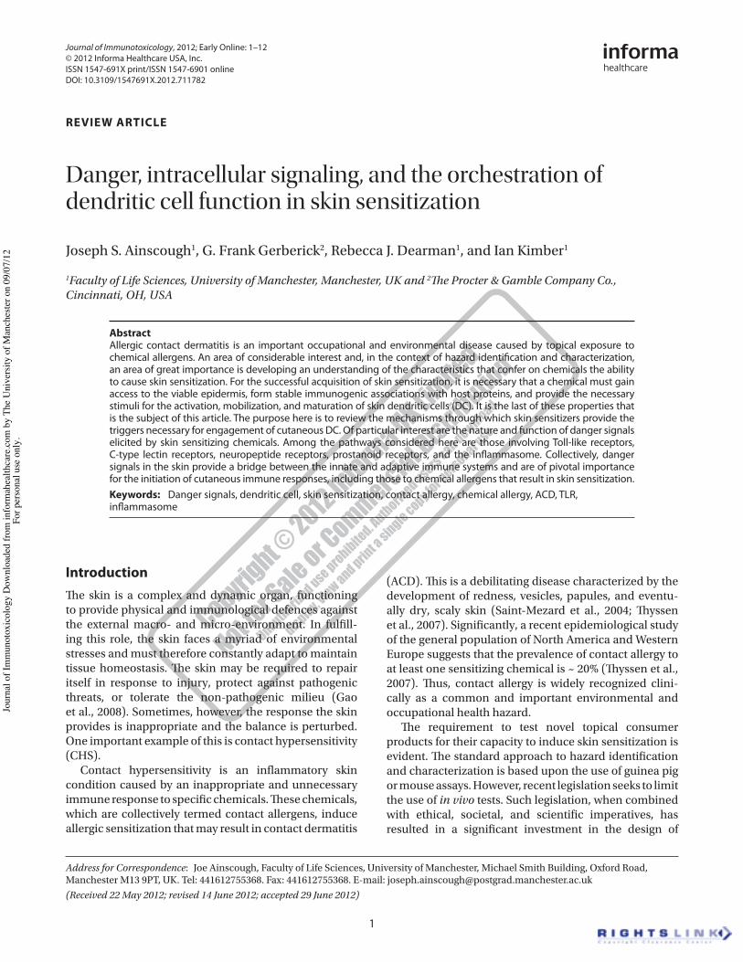
1
IntroductionThe skin is a complex and dynamic organ, functioning to provide physical and immunological defences against the external macro- and micro-environment. In fulfill-ing this role, the skin faces a myriad of environmental stresses and must therefore constantly adapt to maintain tissue homeostasis. The skin may be required to repair itself in response to injury, protect against pathogenic threats, or tolerate the non-pathogenic milieu (Gao et al., 2008). Sometimes, however, the response the skin provides is inappropriate and the balance is perturbed. One important example of this is contact hypersensitivity (CHS).
Contact hypersensitivity is an inflammatory skin condition caused by an inappropriate and unnecessary immune response to specific chemicals. These chemicals, which are collectively termed contact allergens, induce allergic sensitization that may result in contact dermatitis
(ACD). This is a debilitating disease characterized by the development of redness, vesicles, papules, and eventu-ally dry, scaly skin (Saint-Mezard et al., 2004; Thyssen et al., 2007). Significantly, a recent epidemiological study of the general population of North America and Western Europe suggests that the prevalence of contact allergy to at least one sensitizing chemical is ~ 20% (Thyssen et al., 2007). Thus, contact allergy is widely recognized clini-cally as a common and important environmental and occupational health hazard.
The requirement to test novel topical consumer products for their capacity to induce skin sensitization is evident. The standard approach to hazard identification and characterization is based upon the use of guinea pig or mouse assays. However, recent legislation seeks to limit the use of in vivo tests. Such legislation, when combined with ethical, societal, and scientific imperatives, has resulted in a significant investment in the design of
REVIEW ARTICLE
Danger, intracellular signaling, and the orchestration of dendritic cell function in skin sensitization
Joseph S. Ainscough1, G. Frank Gerberick2, Rebecca J. Dearman1, and Ian Kimber1
1Faculty of Life Sciences, University of Manchester, Manchester, UK and 2The Procter & Gamble Company Co., Cincinnati, OH, USA
AbstractAllergic contact dermatitis is an important occupational and environmental disease caused by topical exposure to chemical allergens. An area of considerable interest and, in the context of hazard identification and characterization, an area of great importance is developing an understanding of the characteristics that confer on chemicals the ability to cause skin sensitization. For the successful acquisition of skin sensitization, it is necessary that a chemical must gain access to the viable epidermis, form stable immunogenic associations with host proteins, and provide the necessary stimuli for the activation, mobilization, and maturation of skin dendritic cells (DC). It is the last of these properties that is the subject of this article. The purpose here is to review the mechanisms through which skin sensitizers provide the triggers necessary for engagement of cutaneous DC. Of particular interest are the nature and function of danger signals elicited by skin sensitizing chemicals. Among the pathways considered here are those involving Toll-like receptors, C-type lectin receptors, neuropeptide receptors, prostanoid receptors, and the inflammasome. Collectively, danger signals in the skin provide a bridge between the innate and adaptive immune systems and are of pivotal importance for the initiation of cutaneous immune responses, including those to chemical allergens that result in skin sensitization.Keywords: Danger signals, dendritic cell, skin sensitization, contact allergy, chemical allergy, ACD, TLR, inflammasome
Address for Correspondence: Joe Ainscough, Faculty of Life Sciences, University of Manchester, Michael Smith Building, Oxford Road, Manchester M13 9PT, UK. Tel: 441612755368. Fax: 441612755368. E-mail: [email protected](Received 22 May 2012; revised 14 June 2012; accepted 29 June 2012)
Journal of Immunotoxicology, 2012; Early Online: 1–12© 2012 Informa Healthcare USA, Inc.ISSN 1547-691X print/ISSN 1547-6901 onlineDOI: 10.3109/1547691X.2012.711782
Journal of Immunotoxicology
00
00
1
12
22May2012
14June2012
29June2012
1547-691X
1547-6901
© 2012 Informa Healthcare USA, Inc.
10.3109/1547691X.2012.711782
2012
Danger signals, dendritic cell function and skin sensitization
J. S. Ainscough et al.
Jour
nal o
f Im
mun
otox
icol
ogy
Dow
nloa
ded
from
info
rmah
ealth
care
.com
by
The
Uni
vers
ity o
f Man
ches
ter o
n 09
/07/
12Fo
r per
sona
l use
onl
y.
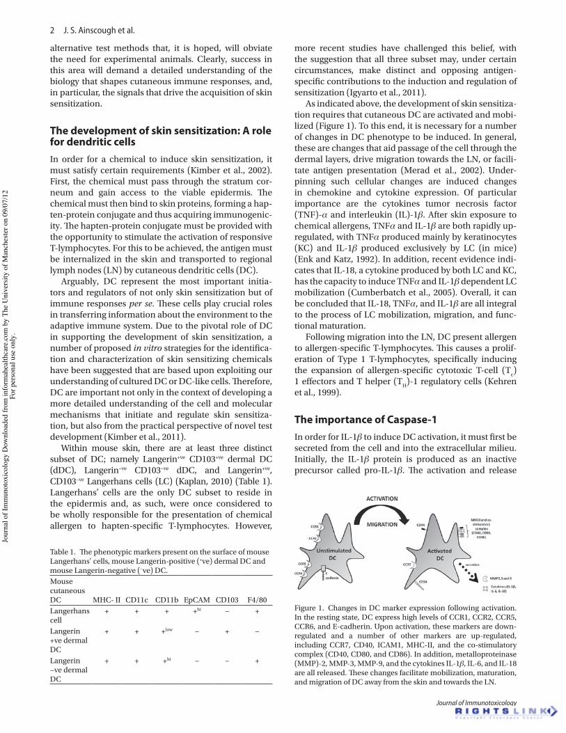
2 J. S. Ainscough et al.
Journal of Immunotoxicology
alternative test methods that, it is hoped, will obviate the need for experimental animals. Clearly, success in this area will demand a detailed understanding of the biology that shapes cutaneous immune responses, and, in particular, the signals that drive the acquisition of skin sensitization.
The development of skin sensitization: A role for dendritic cellsIn order for a chemical to induce skin sensitization, it must satisfy certain requirements (Kimber et al., 2002). First, the chemical must pass through the stratum cor-neum and gain access to the viable epidermis. The chemical must then bind to skin proteins, forming a hap-ten-protein conjugate and thus acquiring immunogenic-ity. The hapten-protein conjugate must be provided with the opportunity to stimulate the activation of responsive T-lymphocytes. For this to be achieved, the antigen must be internalized in the skin and transported to regional lymph nodes (LN) by cutaneous dendritic cells (DC).
Arguably, DC represent the most important initia-tors and regulators of not only skin sensitization but of immune responses per se. These cells play crucial roles in transferring information about the environment to the adaptive immune system. Due to the pivotal role of DC in supporting the development of skin sensitization, a number of proposed in vitro strategies for the identifica-tion and characterization of skin sensitizing chemicals have been suggested that are based upon exploiting our understanding of cultured DC or DC-like cells. Therefore, DC are important not only in the context of developing a more detailed understanding of the cell and molecular mechanisms that initiate and regulate skin sensitiza-tion, but also from the practical perspective of novel test development (Kimber et al., 2011).
Within mouse skin, there are at least three distinct subset of DC; namely Langerin+ve CD103+ve dermal DC (dDC), Langerin−ve CD103−ve dDC, and Langerin+ve, CD103−ve Langerhans cells (LC) (Kaplan, 2010) (Table 1). Langerhans’ cells are the only DC subset to reside in the epidermis and, as such, were once considered to be wholly responsible for the presentation of chemical allergen to hapten-specific T-lymphocytes. However,
more recent studies have challenged this belief, with the suggestion that all three subset may, under certain circumstances, make distinct and opposing antigen-specific contributions to the induction and regulation of sensitization (Igyarto et al., 2011).
As indicated above, the development of skin sensitiza-tion requires that cutaneous DC are activated and mobi-lized (Figure 1). To this end, it is necessary for a number of changes in DC phenotype to be induced. In general, these are changes that aid passage of the cell through the dermal layers, drive migration towards the LN, or facili-tate antigen presentation (Merad et al., 2002). Under-pinning such cellular changes are induced changes in chemokine and cytokine expression. Of particular importance are the cytokines tumor necrosis factor (TNF)-α and interleukin (IL)-1β. After skin exposure to chemical allergens, TNFα and IL-1β are both rapidly up-regulated, with TNFα produced mainly by keratinocytes (KC) and IL-1β produced exclusively by LC (in mice) (Enk and Katz, 1992). In addition, recent evidence indi-cates that IL-18, a cytokine produced by both LC and KC, has the capacity to induce TNFα and IL-1β dependent LC mobilization (Cumberbatch et al., 2005). Overall, it can be concluded that IL-18, TNFα, and IL-1β are all integral to the process of LC mobilization, migration, and func-tional maturation.
Following migration into the LN, DC present allergen to allergen-specific T-lymphocytes. This causes a prolif-eration of Type 1 T-lymphocytes, specifically inducing the expansion of allergen-specific cytotoxic T-cell (Tc) 1 effectors and T helper (TH)-1 regulatory cells (Kehren et al., 1999).
The importance of Caspase-1In order for IL-1β to induce DC activation, it must first be secreted from the cell and into the extracellular milieu. Initially, the IL-1β protein is produced as an inactive precursor called pro-IL-1β. The activation and release
Table 1. The phenotypic markers present on the surface of mouse Langerhans’ cells, mouse Langerin-positive (+ve) dermal DC and mouse Langerin-negative (−ve) DC.Mouse cutaneous DC MHC- II CD11c CD11b EpCAM CD103 F4/80Langerhans cell
+ + + +hi − +
Langerin +ve dermal DC
+ + +low − + −
Langerin −ve dermal DC
+ + +hi − − +
Figure 1. Changes in DC marker expression following activation. In the resting state, DC express high levels of CCR1, CCR2, CCR5, CCR6, and E-cadherin. Upon activation, these markers are down-regulated and a number of other markers are up-regulated, including CCR7, CD40, ICAM1, MHC-II, and the co-stimulatory complex (CD40, CD80, and CD86). In addition, metalloproteinase (MMP)-2, MMP-3, MMP-9, and the cytokines IL-1β, IL-6, and IL-18 are all released. These changes facilitate mobilization, maturation, and migration of DC away from the skin and towards the LN.
Jour
nal o
f Im
mun
otox
icol
ogy
Dow
nloa
ded
from
info
rmah
ealth
care
.com
by
The
Uni
vers
ity o
f Man
ches
ter o
n 09
/07/
12Fo
r per
sona
l use
onl
y.
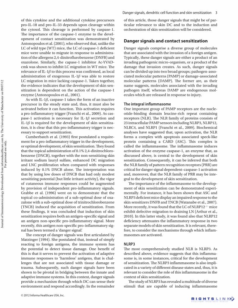
Danger signals, dendritic cell function and skin sensitization 3
© 2012 Informa Healthcare USA, Inc.
of this cytokine and the additional cytokine precursors pro-IL-18 and pro-IL-33 depends upon cleavage within the cytosol. This cleavage is performed by caspase-1. The importance of the caspase-1 enzyme to the devel-opment of contact sensitization was demonstrated by Antonopoulos et al. (2001), who observed that, unlike the LC of wild type (WT) mice, the LC of caspase-1 deficient mice were unable to migrate in response to administra-tion of the allergens 2,4-dinitroflurobenzene (DNFB) and oxazolone. Similarly, the capase-1 inhibitor Ac-YVAD-cmk was shown to inhibit LC migration in WT mice. The relevance of IL-1β to this process was confirmed, as local administration of exogenous IL-1β was able to restore LC migration in mice lacking caspase-1. Taken together, the evidence indicates that the development of skin sen-sitization is dependent on the action of the caspase-1 enzyme (Antonopoulos et al., 2001).
As with IL-1β, caspase-1 takes the form of an inactive precursor in the steady state and, thus, it must also be activated before it can function. This activation requires a pro-inflammatory trigger (Franchi et al., 2009). As cas-pase-1 activation is necessary for IL-1β secretion and IL-1β is required for the development of skin sensitiza-tion, it is clear that this pro-inflammatory trigger is nec-essary to support sensitization.
Cumberbatch et al. (1993) first postulated a require-ment for a pro-inflammatory trigger in the development, or optimal development, of skin sensitization. They found that the topical administration of 0.1% 2,4-dinitrochloro-benzene (DNCB), together with the non-sensitizing skin irritant sodium lauryl sulfate, enhanced DC migration and LNC proliferation when compared with responses induced by 0.1% DNCB alone. The interpretation was that by using low doses of DNCB that had only modest sensitizing potential (but little irritant activity), the vigor of cutaneous immune responses could be augmented by provision of independent pro-inflammatory signals. Grabbe et al. (1996) went on to demonstrate that the topical co-administration of a sub-optimal dose of oxa-zalone with a sub-optimal dose of trinitrochlorobenzene (TNCB) induced the acquisition of sensitization. From these findings, it was concluded that induction of skin sensitization requires both an antigen-specific signal and an antigen non-specific pro-inflammatory signal. More recently, this antigen non-specific pro-inflammatory sig-nal has been termed a ‘danger signal’.
The concept of danger signals was first articulated by Matzinger (1994). She postulated that, instead of simply reacting to foreign antigens, the immune system has the potential to detect tissue damage. One benefit of this is that it serves to prevent the activation of adaptive immune responses to ‘harmless’ antigens, that is chal-lenges that are not associated with tissue damage or trauma. Subsequently, such danger signals have been shown to be pivotal in bridging between the innate and adaptive immune systems. In this context, danger signals provide a mechanism through which DC can sense their environment and respond accordingly. In the remainder
of this article, those danger signals that might be of par-ticular relevance to skin DC and to the induction and orchestration of skin sensitization will be considered.
Danger signals and contact sensitizationDanger signals comprise a diverse group of molecules that are associated with the invasion of a foreign antigen. Typically, these danger signals are either a product of an invading pathogenic micro-organism, or a product of the damage that invasion creates. As such, danger signals can be divided up into two broad groups; pathogen-asso-ciated molecular patterns (PAMP) or damage-associated molecular patterns (DAMP). The former are, as their name suggests, molecules associated with the invading pathogen itself, whereas DAMP are endogenous mol-ecules which are released in response to injury.
The integral inflammasomeOne important group of PAMP receptors are the nucle-otide-binding domain leucine-rich repeat containing receptors (NLR). The NLR family of proteins consists of 23 intracellular PAMP sensors including NLRP3, NLRP12, NLRC4, and NLRP1 (Franchi et al., 2009). Biochemical analyses have suggested that, upon activation, the NLR forms a complex with apoptosis associated speck-like protein containing a CARD (ASC). This complex is called the inflammasome. The inflammasome induces activation of the enzyme caspase-1, an enzyme that, as discussed above, is central to the development of skin sensitization. Consequently, it can be inferred that both the NLR family of pattern recognition receptors (PRR) are critical for danger signal dependent–caspase-1 activation and, moreover, that the NLR family of PRR may be inte-gral to the development of skin sensitization.
The importance of the inflammasome to the develop-ment of skin sensitization can be demonstrated experi-mentally. For instance, it has been shown that ASC and NLRP3 deficient mice display an impaired response to the skin sensitizers DNFB and TNCB (Watanabe et al., 2007). More recently, it was found that the LC of NLRP12−/− mice exhibit defective migration to draining LN (Arthur et al., 2010). In this latter study, it was found also that NLRP12 deficiency attenuates the inflammatory response in two separate models of skin sensitization. It is relevant, there-fore, to consider the mechanisms through which inflam-masomes are activated.
NLRP3The most comprehensively studied NLR is NLRP3. As described above, evidence suggests that this inflamma-some is, in some instances, critical for the development of sensitization. The NLRP3 inflammasome is also impli-cated in a variety of different disease states and, thus, it is relevant to consider the role of this inflammasome in the context of skin sensitization.
The study of NLRP3 has revealed a multitude of diverse stimuli that are capable of inducing inflammasome
Jour
nal o
f Im
mun
otox
icol
ogy
Dow
nloa
ded
from
info
rmah
ealth
care
.com
by
The
Uni
vers
ity o
f Man
ches
ter o
n 09
/07/
12Fo
r per
sona
l use
onl
y.

4 J. S. Ainscough et al.
Journal of Immunotoxicology
activation. Included in this list are the crystalline com-pounds adenosine triphosphate (ATP), silica, asbestos and uric acid, the bacterial products listeriolysin O, potassium ionophore nigericin, aerolysin, and hemoly-sins, as well as the marine toxin maitotoxin (Pedra et al., 2009). Although many different substances have been shown to activate NLRP3, a direct ligand for this inflam-masome has yet to be identified. However, a number of NLRP3 stimuli-induced cellular changes that may induce NLRP3 activation have been identified.
One theory, based upon evidence that many NLRP3 stimulants induce pore formation, suggests that pore formation itself can cause inflammasome activation. Building upon this, two additional theories have emerged. One proposes that membrane damage leads to microbes entering the cell and activating NLRP3 (Kanneganti et al., 2007), whereas the other suggests that membrane dam-age leads to the release of endogenous molecules that can then directly activate the NLRP3 (Ogura et al., 2006). In support of the latter theory, studies have demonstrated that endosomal membrane damage by the crystalline molecules alum, silica, and amyloid-β causes the release of cathepsin B. As cathepsin B activates the inflamma-some, crystalline molecules may indeed activate NLRP3 by inducing endosomal membrane damage (Hornung et al., 2008). Nevertheless, other investigations suggest that this mechanism of NLRP3 activation is not common to all activating molecules. Indicative of this is evidence suggesting that ATP, unlike the crystalline compounds, induces pore formation by binding to the plasma mem-brane P2X7 receptor. It may be, therefore, that a single, unifying mechanism of NLRP3 inflammasome activation does not exist.
Interestingly, one study postulates that potassium efflux may be associated with toxin and crystal medi-ated NLRP3 activation (Petrilli et al., 2007). In that investigation, it was shown that inhibition of the efflux of potassium effectively inhibited NLRP3 activation. Furthermore, in vitro studies have demonstrated that inflammasome assembly and recruitment of caspase-1 occurs spontaneously at low intracellular potassium con-centrations. Taken together, it was concluded that potas-sium efflux is a necessary mechanism for the assembly of NLRP3 (Petrilli et al., 2007). However, characterization of the precise mechanism of potassium efflux-induced NLRP3 assembly requires further investigation. Equally intriguing is evidence that postulates that stimulation of the P2X7 receptors by ATP may lead to the opening of a potassium-selective pore. As this will cause potassium efflux, ATP may activate the NLRP3 inflammasome via this process (Khakh and North, 2006). In support of this hypothesis, mice deficient in P2X7 receptor fail to mount a skin sensitization response to TNCB. However, the skin sensitization was restored when the P2X7 receptor-deficient mice were injected intra-dermally with IL-1β (Weber et al., 2010). Taken together, these data suggest that it is likely that P2X7-dependent IL-1β production is an important aspect of skin sensitization.
Another process commonly associated with toxin and crystal mediated NLRP3 activation is the production of reactive oxygen species (ROS) (Dostert et al., 2008). Importantly, many in vitro studies have revealed that ROS production is induced by contact allergens (Bruchhausen et al., 2003; Mehrotra et al., 2005; Martin et al., 2011). In human monocytes, the inhibition of ROS production, as achieved by administration of the NADPH oxidase inhib-itor diphenyleneiodonium (DPI), inhibits ATP-mediated inflammasome activation (Hewinson et al., 2008). In addition, a variety of studies have reported increases in cellular ROS production after stimulation with both toxin and crystalline NLRP3 activators (Martinon, 2010). As mitochondria are believed to be the main source of cellu-lar ROS, it is proposed that mitochondria may have a role to play in NLRP3 activation. Zhou et al. (2011) showed in support of this that damaged, ROS-generating mito-chondria are capable of stimulating activation of NLRP3. The same authors also found that depletion of the mito-chondrial membrane protein voltage-dependent anion channel, a protein required for mitochondrial ROS pro-duction, impaired the activation of the NLRP3 inflamma-some. Collectively, the available evidence suggests that activation of the NLRP3 inflammasome may be depen-dent upon ROS production, which, may in turn be driven by mitochondrial function. However, as with potassium efflux induction, the molecular mechanisms that cause ROS production are not thoroughly understood.
NLRP12The NLRP12 inflammasome is less well-characterized. As described above, this inflammasome has also been shown to be implicated in the development of skin sen-sitization. However, in contrast to other NLR, NLRP12 appears to negatively regulate the production of IL-1β (Shaw et al., 2010). It has been shown that this inflam-masome prevents phosphorylation of IL-1 receptor-associated kinase 1 (IRAK-1) and enhances degradation of the transcription factor nuclear factor !-light-chain-enhancer of activated B-cell (NF-κB) inducing kinase (NIK). As the negative regulation of IL-1β by NLRP12 would effectively inhibit LC migration and DC accumu-lation, the presumption is that this mechanism would prevent the acquisition of skin sensitization. However, as NLRP12 is known to be required for the development of skin sensitization to certain allergens, it is postulated that there must be some, as yet unknown mechanism for the observed dependence upon NLRP12 (Arthur et al., 2010).
NLRC4 and NLRP1Other NLR implicated in the activation of pro-inflam-matory cytokines are the NLRC4 and NLRP1 inflamma-somes. Although these inflammasomes have yet to be directly implicated in the development of skin sensitiza-tion, their mechanisms of action suggest that they may also have a role to play. NLRC4 activation is induced by flagellin and the inner rod component of Type III bacterial secretion systems (Zhao et al., 2011). A recent
Jour
nal o
f Im
mun
otox
icol
ogy
Dow
nloa
ded
from
info
rmah
ealth
care
.com
by
The
Uni
vers
ity o
f Man
ches
ter o
n 09
/07/
12Fo
r per
sona
l use
onl
y.
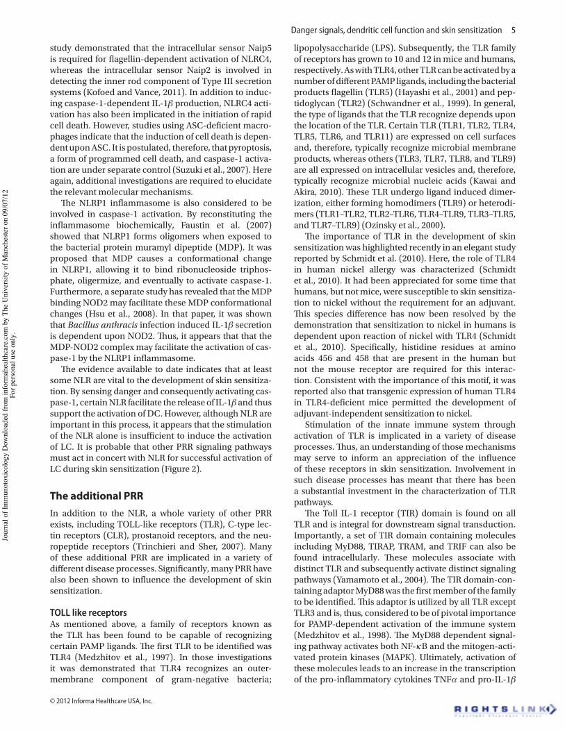
Danger signals, dendritic cell function and skin sensitization 5
© 2012 Informa Healthcare USA, Inc.
study demonstrated that the intracellular sensor Naip5 is required for flagellin-dependent activation of NLRC4, whereas the intracellular sensor Naip2 is involved in detecting the inner rod component of Type III secretion systems (Kofoed and Vance, 2011). In addition to induc-ing caspase-1-dependent IL-1β production, NLRC4 acti-vation has also been implicated in the initiation of rapid cell death. However, studies using ASC-deficient macro-phages indicate that the induction of cell death is depen-dent upon ASC. It is postulated, therefore, that pyroptosis, a form of programmed cell death, and caspase-1 activa-tion are under separate control (Suzuki et al., 2007). Here again, additional investigations are required to elucidate the relevant molecular mechanisms.
The NLRP1 inflammasome is also considered to be involved in caspase-1 activation. By reconstituting the inflammasome biochemically, Faustin et al. (2007) showed that NLRP1 forms oligomers when exposed to the bacterial protein muramyl dipeptide (MDP). It was proposed that MDP causes a conformational change in NLRP1, allowing it to bind ribonucleoside triphos-phate, oligermize, and eventually to activate caspase-1. Furthermore, a separate study has revealed that the MDP binding NOD2 may facilitate these MDP conformational changes (Hsu et al., 2008). In that paper, it was shown that Bacillus anthracis infection induced IL-1β secretion is dependent upon NOD2. Thus, it appears that that the MDP-NOD2 complex may facilitate the activation of cas-pase-1 by the NLRP1 inflammasome.
The evidence available to date indicates that at least some NLR are vital to the development of skin sensitiza-tion. By sensing danger and consequently activating cas-pase-1, certain NLR facilitate the release of IL-1β and thus support the activation of DC. However, although NLR are important in this process, it appears that the stimulation of the NLR alone is insufficient to induce the activation of LC. It is probable that other PRR signaling pathways must act in concert with NLR for successful activation of LC during skin sensitization (Figure 2).
The additional PRRIn addition to the NLR, a whole variety of other PRR exists, including TOLL-like receptors (TLR), C-type lec-tin receptors (CLR), prostanoid receptors, and the neu-ropeptide receptors (Trinchieri and Sher, 2007). Many of these additional PRR are implicated in a variety of different disease processes. Significantly, many PRR have also been shown to influence the development of skin sensitization.
TOLL like receptorsAs mentioned above, a family of receptors known as the TLR has been found to be capable of recognizing certain PAMP ligands. The first TLR to be identified was TLR4 (Medzhitov et al., 1997). In those investigations it was demonstrated that TLR4 recognizes an outer-membrane component of gram-negative bacteria;
lipopolysaccharide (LPS). Subsequently, the TLR family of receptors has grown to 10 and 12 in mice and humans, respectively. As with TLR4, other TLR can be activated by a number of different PAMP ligands, including the bacterial products flagellin (TLR5) (Hayashi et al., 2001) and pep-tidoglycan (TLR2) (Schwandner et al., 1999). In general, the type of ligands that the TLR recognize depends upon the location of the TLR. Certain TLR (TLR1, TLR2, TLR4, TLR5, TLR6, and TLR11) are expressed on cell surfaces and, therefore, typically recognize microbial membrane products, whereas others (TLR3, TLR7, TLR8, and TLR9) are all expressed on intracellular vesicles and, therefore, typically recognize microbial nucleic acids (Kawai and Akira, 2010). These TLR undergo ligand induced dimer-ization, either forming homodimers (TLR9) or heterodi-mers (TLR1–TLR2, TLR2–TLR6, TLR4–TLR9, TLR3–TLR5, and TLR7–TLR9) (Ozinsky et al., 2000).
The importance of TLR in the development of skin sensitization was highlighted recently in an elegant study reported by Schmidt et al. (2010). Here, the role of TLR4 in human nickel allergy was characterized (Schmidt et al., 2010). It had been appreciated for some time that humans, but not mice, were susceptible to skin sensitiza-tion to nickel without the requirement for an adjuvant. This species difference has now been resolved by the demonstration that sensitization to nickel in humans is dependent upon reaction of nickel with TLR4 (Schmidt et al., 2010). Specifically, histidine residues at amino acids 456 and 458 that are present in the human but not the mouse receptor are required for this interac-tion. Consistent with the importance of this motif, it was reported also that transgenic expression of human TLR4 in TLR4-deficient mice permitted the development of adjuvant-independent sensitization to nickel.
Stimulation of the innate immune system through activation of TLR is implicated in a variety of disease processes. Thus, an understanding of those mechanisms may serve to inform an appreciation of the influence of these receptors in skin sensitization. Involvement in such disease processes has meant that there has been a substantial investment in the characterization of TLR pathways.
The Toll IL-1 receptor (TIR) domain is found on all TLR and is integral for downstream signal transduction. Importantly, a set of TIR domain containing molecules including MyD88, TIRAP, TRAM, and TRIF can also be found intracellularly. These molecules associate with distinct TLR and subsequently activate distinct signaling pathways (Yamamoto et al., 2004). The TIR domain-con-taining adaptor MyD88 was the first member of the family to be identified. This adaptor is utilized by all TLR except TLR3 and is, thus, considered to be of pivotal importance for PAMP-dependent activation of the immune system (Medzhitov et al., 1998). The MyD88 dependent signal-ing pathway activates both NF-κB and the mitogen-acti-vated protein kinases (MAPK). Ultimately, activation of these molecules leads to an increase in the transcription of the pro-inflammatory cytokines TNFα and pro-IL-1β
Jour
nal o
f Im
mun
otox
icol
ogy
Dow
nloa
ded
from
info
rmah
ealth
care
.com
by
The
Uni
vers
ity o
f Man
ches
ter o
n 09
/07/
12Fo
r per
sona
l use
onl
y.
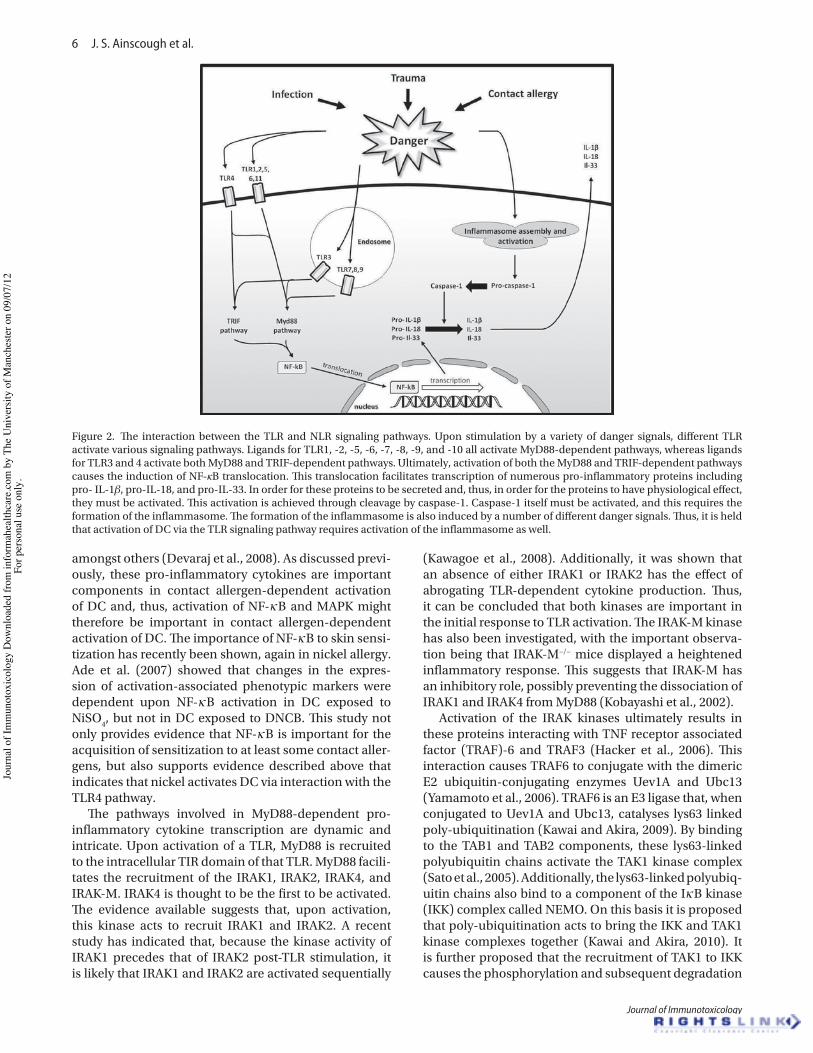
6 J. S. Ainscough et al.
Journal of Immunotoxicology
amongst others (Devaraj et al., 2008). As discussed previ-ously, these pro-inflammatory cytokines are important components in contact allergen-dependent activation of DC and, thus, activation of NF-κB and MAPK might therefore be important in contact allergen-dependent activation of DC. The importance of NF-κB to skin sensi-tization has recently been shown, again in nickel allergy. Ade et al. (2007) showed that changes in the expres-sion of activation-associated phenotypic markers were dependent upon NF-κB activation in DC exposed to NiSO4, but not in DC exposed to DNCB. This study not only provides evidence that NF-κB is important for the acquisition of sensitization to at least some contact aller-gens, but also supports evidence described above that indicates that nickel activates DC via interaction with the TLR4 pathway.
The pathways involved in MyD88-dependent pro-inflammatory cytokine transcription are dynamic and intricate. Upon activation of a TLR, MyD88 is recruited to the intracellular TIR domain of that TLR. MyD88 facili-tates the recruitment of the IRAK1, IRAK2, IRAK4, and IRAK-M. IRAK4 is thought to be the first to be activated. The evidence available suggests that, upon activation, this kinase acts to recruit IRAK1 and IRAK2. A recent study has indicated that, because the kinase activity of IRAK1 precedes that of IRAK2 post-TLR stimulation, it is likely that IRAK1 and IRAK2 are activated sequentially
(Kawagoe et al., 2008). Additionally, it was shown that an absence of either IRAK1 or IRAK2 has the effect of abrogating TLR-dependent cytokine production. Thus, it can be concluded that both kinases are important in the initial response to TLR activation. The IRAK-M kinase has also been investigated, with the important observa-tion being that IRAK-M−/− mice displayed a heightened inflammatory response. This suggests that IRAK-M has an inhibitory role, possibly preventing the dissociation of IRAK1 and IRAK4 from MyD88 (Kobayashi et al., 2002).
Activation of the IRAK kinases ultimately results in these proteins interacting with TNF receptor associated factor (TRAF)-6 and TRAF3 (Hacker et al., 2006). This interaction causes TRAF6 to conjugate with the dimeric E2 ubiquitin-conjugating enzymes Uev1A and Ubc13 (Yamamoto et al., 2006). TRAF6 is an E3 ligase that, when conjugated to Uev1A and Ubc13, catalyses lys63 linked poly-ubiquitination (Kawai and Akira, 2009). By binding to the TAB1 and TAB2 components, these lys63-linked polyubiquitin chains activate the TAK1 kinase complex (Sato et al., 2005). Additionally, the lys63-linked polyubiq-uitin chains also bind to a component of the IκB kinase (IKK) complex called NEMO. On this basis it is proposed that poly-ubiquitination acts to bring the IKK and TAK1 kinase complexes together (Kawai and Akira, 2010). It is further proposed that the recruitment of TAK1 to IKK causes the phosphorylation and subsequent degradation
Figure 2. The interaction between the TLR and NLR signaling pathways. Upon stimulation by a variety of danger signals, different TLR activate various signaling pathways. Ligands for TLR1, -2, -5, -6, -7, -8, -9, and -10 all activate MyD88-dependent pathways, whereas ligands for TLR3 and 4 activate both MyD88 and TRIF-dependent pathways. Ultimately, activation of both the MyD88 and TRIF-dependent pathways causes the induction of NF-κB translocation. This translocation facilitates transcription of numerous pro-inflammatory proteins including pro- IL-1β, pro-IL-18, and pro-IL-33. In order for these proteins to be secreted and, thus, in order for the proteins to have physiological effect, they must be activated. This activation is achieved through cleavage by caspase-1. Caspase-1 itself must be activated, and this requires the formation of the inflammasome. The formation of the inflammasome is also induced by a number of different danger signals. Thus, it is held that activation of DC via the TLR signaling pathway requires activation of the inflammasome as well.
Jour
nal o
f Im
mun
otox
icol
ogy
Dow
nloa
ded
from
info
rmah
ealth
care
.com
by
The
Uni
vers
ity o
f Man
ches
ter o
n 09
/07/
12Fo
r per
sona
l use
onl
y.
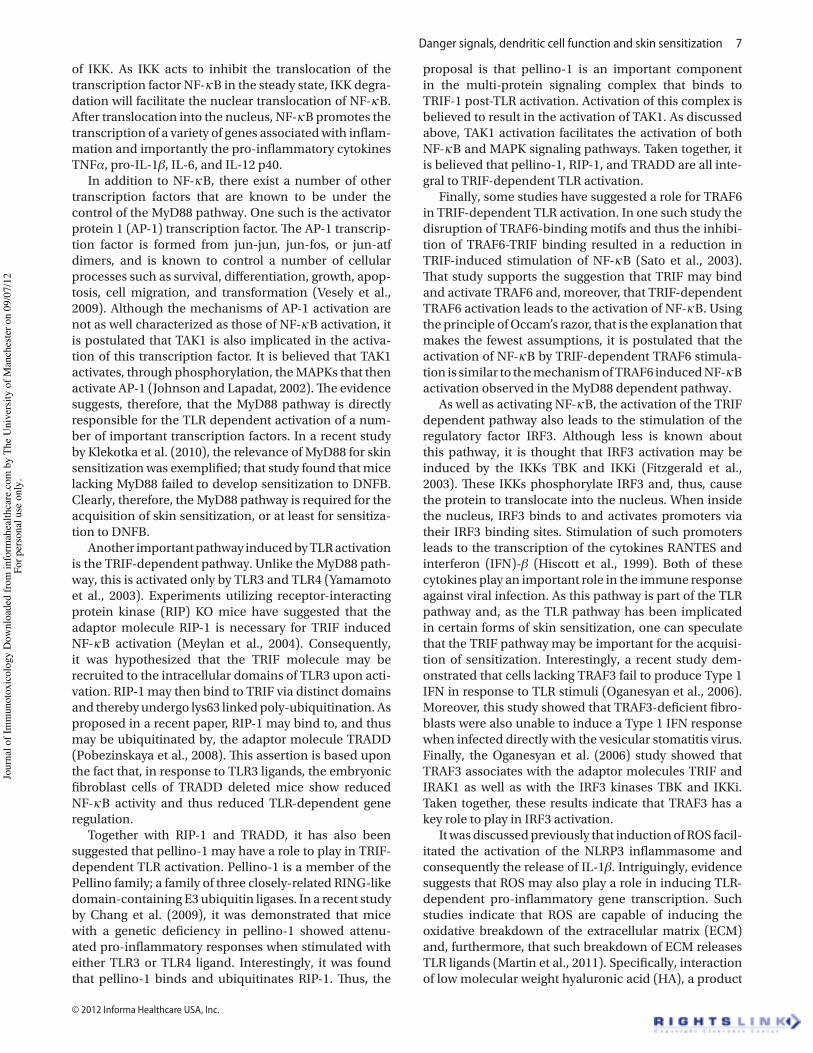
Danger signals, dendritic cell function and skin sensitization 7
© 2012 Informa Healthcare USA, Inc.
of IKK. As IKK acts to inhibit the translocation of the transcription factor NF-κB in the steady state, IKK degra-dation will facilitate the nuclear translocation of NF-κB. After translocation into the nucleus, NF-κB promotes the transcription of a variety of genes associated with inflam-mation and importantly the pro-inflammatory cytokines TNFα, pro-IL-1β, IL-6, and IL-12 p40.
In addition to NF-κB, there exist a number of other transcription factors that are known to be under the control of the MyD88 pathway. One such is the activator protein 1 (AP-1) transcription factor. The AP-1 transcrip-tion factor is formed from jun-jun, jun-fos, or jun-atf dimers, and is known to control a number of cellular processes such as survival, differentiation, growth, apop-tosis, cell migration, and transformation (Vesely et al., 2009). Although the mechanisms of AP-1 activation are not as well characterized as those of NF-κB activation, it is postulated that TAK1 is also implicated in the activa-tion of this transcription factor. It is believed that TAK1 activates, through phosphorylation, the MAPKs that then activate AP-1 (Johnson and Lapadat, 2002). The evidence suggests, therefore, that the MyD88 pathway is directly responsible for the TLR dependent activation of a num-ber of important transcription factors. In a recent study by Klekotka et al. (2010), the relevance of MyD88 for skin sensitization was exemplified; that study found that mice lacking MyD88 failed to develop sensitization to DNFB. Clearly, therefore, the MyD88 pathway is required for the acquisition of skin sensitization, or at least for sensitiza-tion to DNFB.
Another important pathway induced by TLR activation is the TRIF-dependent pathway. Unlike the MyD88 path-way, this is activated only by TLR3 and TLR4 (Yamamoto et al., 2003). Experiments utilizing receptor-interacting protein kinase (RIP) KO mice have suggested that the adaptor molecule RIP-1 is necessary for TRIF induced NF-κB activation (Meylan et al., 2004). Consequently, it was hypothesized that the TRIF molecule may be recruited to the intracellular domains of TLR3 upon acti-vation. RIP-1 may then bind to TRIF via distinct domains and thereby undergo lys63 linked poly-ubiquitination. As proposed in a recent paper, RIP-1 may bind to, and thus may be ubiquitinated by, the adaptor molecule TRADD (Pobezinskaya et al., 2008). This assertion is based upon the fact that, in response to TLR3 ligands, the embryonic fibroblast cells of TRADD deleted mice show reduced NF-κB activity and thus reduced TLR-dependent gene regulation.
Together with RIP-1 and TRADD, it has also been suggested that pellino-1 may have a role to play in TRIF-dependent TLR activation. Pellino-1 is a member of the Pellino family; a family of three closely-related RING-like domain-containing E3 ubiquitin ligases. In a recent study by Chang et al. (2009), it was demonstrated that mice with a genetic deficiency in pellino-1 showed attenu-ated pro-inflammatory responses when stimulated with either TLR3 or TLR4 ligand. Interestingly, it was found that pellino-1 binds and ubiquitinates RIP-1. Thus, the
proposal is that pellino-1 is an important component in the multi-protein signaling complex that binds to TRIF-1 post-TLR activation. Activation of this complex is believed to result in the activation of TAK1. As discussed above, TAK1 activation facilitates the activation of both NF-κB and MAPK signaling pathways. Taken together, it is believed that pellino-1, RIP-1, and TRADD are all inte-gral to TRIF-dependent TLR activation.
Finally, some studies have suggested a role for TRAF6 in TRIF-dependent TLR activation. In one such study the disruption of TRAF6-binding motifs and thus the inhibi-tion of TRAF6-TRIF binding resulted in a reduction in TRIF-induced stimulation of NF-κB (Sato et al., 2003). That study supports the suggestion that TRIF may bind and activate TRAF6 and, moreover, that TRIF-dependent TRAF6 activation leads to the activation of NF-κB. Using the principle of Occam’s razor, that is the explanation that makes the fewest assumptions, it is postulated that the activation of NF-κB by TRIF-dependent TRAF6 stimula-tion is similar to the mechanism of TRAF6 induced NF-κB activation observed in the MyD88 dependent pathway.
As well as activating NF-κB, the activation of the TRIF dependent pathway also leads to the stimulation of the regulatory factor IRF3. Although less is known about this pathway, it is thought that IRF3 activation may be induced by the IKKs TBK and IKKi (Fitzgerald et al., 2003). These IKKs phosphorylate IRF3 and, thus, cause the protein to translocate into the nucleus. When inside the nucleus, IRF3 binds to and activates promoters via their IRF3 binding sites. Stimulation of such promoters leads to the transcription of the cytokines RANTES and interferon (IFN)-β (Hiscott et al., 1999). Both of these cytokines play an important role in the immune response against viral infection. As this pathway is part of the TLR pathway and, as the TLR pathway has been implicated in certain forms of skin sensitization, one can speculate that the TRIF pathway may be important for the acquisi-tion of sensitization. Interestingly, a recent study dem-onstrated that cells lacking TRAF3 fail to produce Type 1 IFN in response to TLR stimuli (Oganesyan et al., 2006). Moreover, this study showed that TRAF3-deficient fibro-blasts were also unable to induce a Type 1 IFN response when infected directly with the vesicular stomatitis virus. Finally, the Oganesyan et al. (2006) study showed that TRAF3 associates with the adaptor molecules TRIF and IRAK1 as well as with the IRF3 kinases TBK and IKKi. Taken together, these results indicate that TRAF3 has a key role to play in IRF3 activation.
It was discussed previously that induction of ROS facil-itated the activation of the NLRP3 inflammasome and consequently the release of IL-1β. Intriguingly, evidence suggests that ROS may also play a role in inducing TLR-dependent pro-inflammatory gene transcription. Such studies indicate that ROS are capable of inducing the oxidative breakdown of the extracellular matrix (ECM) and, furthermore, that such breakdown of ECM releases TLR ligands (Martin et al., 2011). Specifically, interaction of low molecular weight hyaluronic acid (HA), a product
Jour
nal o
f Im
mun
otox
icol
ogy
Dow
nloa
ded
from
info
rmah
ealth
care
.com
by
The
Uni
vers
ity o
f Man
ches
ter o
n 09
/07/
12Fo
r per
sona
l use
onl
y.
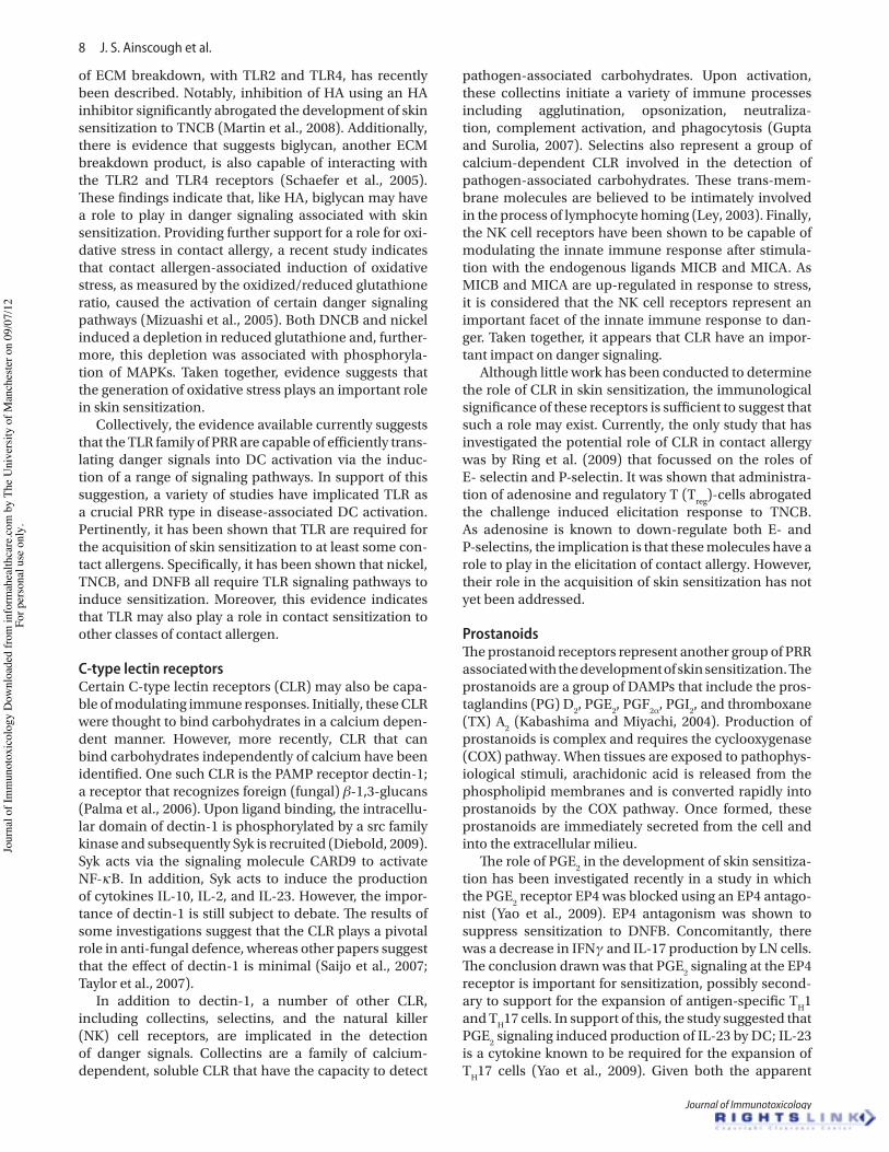
8 J. S. Ainscough et al.
Journal of Immunotoxicology
of ECM breakdown, with TLR2 and TLR4, has recently been described. Notably, inhibition of HA using an HA inhibitor significantly abrogated the development of skin sensitization to TNCB (Martin et al., 2008). Additionally, there is evidence that suggests biglycan, another ECM breakdown product, is also capable of interacting with the TLR2 and TLR4 receptors (Schaefer et al., 2005). These findings indicate that, like HA, biglycan may have a role to play in danger signaling associated with skin sensitization. Providing further support for a role for oxi-dative stress in contact allergy, a recent study indicates that contact allergen-associated induction of oxidative stress, as measured by the oxidized/reduced glutathione ratio, caused the activation of certain danger signaling pathways (Mizuashi et al., 2005). Both DNCB and nickel induced a depletion in reduced glutathione and, further-more, this depletion was associated with phosphoryla-tion of MAPKs. Taken together, evidence suggests that the generation of oxidative stress plays an important role in skin sensitization.
Collectively, the evidence available currently suggests that the TLR family of PRR are capable of efficiently trans-lating danger signals into DC activation via the induc-tion of a range of signaling pathways. In support of this suggestion, a variety of studies have implicated TLR as a crucial PRR type in disease-associated DC activation. Pertinently, it has been shown that TLR are required for the acquisition of skin sensitization to at least some con-tact allergens. Specifically, it has been shown that nickel, TNCB, and DNFB all require TLR signaling pathways to induce sensitization. Moreover, this evidence indicates that TLR may also play a role in contact sensitization to other classes of contact allergen.
C-type lectin receptorsCertain C-type lectin receptors (CLR) may also be capa-ble of modulating immune responses. Initially, these CLR were thought to bind carbohydrates in a calcium depen-dent manner. However, more recently, CLR that can bind carbohydrates independently of calcium have been identified. One such CLR is the PAMP receptor dectin-1; a receptor that recognizes foreign (fungal) β-1,3-glucans (Palma et al., 2006). Upon ligand binding, the intracellu-lar domain of dectin-1 is phosphorylated by a src family kinase and subsequently Syk is recruited (Diebold, 2009). Syk acts via the signaling molecule CARD9 to activate NF-κB. In addition, Syk acts to induce the production of cytokines IL-10, IL-2, and IL-23. However, the impor-tance of dectin-1 is still subject to debate. The results of some investigations suggest that the CLR plays a pivotal role in anti-fungal defence, whereas other papers suggest that the effect of dectin-1 is minimal (Saijo et al., 2007; Taylor et al., 2007).
In addition to dectin-1, a number of other CLR, including collectins, selectins, and the natural killer (NK) cell receptors, are implicated in the detection of danger signals. Collectins are a family of calcium-dependent, soluble CLR that have the capacity to detect
pathogen-associated carbohydrates. Upon activation, these collectins initiate a variety of immune processes including agglutination, opsonization, neutraliza-tion, complement activation, and phagocytosis (Gupta and Surolia, 2007). Selectins also represent a group of calcium-dependent CLR involved in the detection of pathogen-associated carbohydrates. These trans-mem-brane molecules are believed to be intimately involved in the process of lymphocyte homing (Ley, 2003). Finally, the NK cell receptors have been shown to be capable of modulating the innate immune response after stimula-tion with the endogenous ligands MICB and MICA. As MICB and MICA are up-regulated in response to stress, it is considered that the NK cell receptors represent an important facet of the innate immune response to dan-ger. Taken together, it appears that CLR have an impor-tant impact on danger signaling.
Although little work has been conducted to determine the role of CLR in skin sensitization, the immunological significance of these receptors is sufficient to suggest that such a role may exist. Currently, the only study that has investigated the potential role of CLR in contact allergy was by Ring et al. (2009) that focussed on the roles of E- selectin and P-selectin. It was shown that administra-tion of adenosine and regulatory T (Treg)-cells abrogated the challenge induced elicitation response to TNCB. As adenosine is known to down-regulate both E- and P-selectins, the implication is that these molecules have a role to play in the elicitation of contact allergy. However, their role in the acquisition of skin sensitization has not yet been addressed.
ProstanoidsThe prostanoid receptors represent another group of PRR associated with the development of skin sensitization. The prostanoids are a group of DAMPs that include the pros-taglandins (PG) D2, PGE2, PGF2α, PGI2, and thromboxane (TX) A2 (Kabashima and Miyachi, 2004). Production of prostanoids is complex and requires the cyclooxygenase (COX) pathway. When tissues are exposed to pathophys-iological stimuli, arachidonic acid is released from the phospholipid membranes and is converted rapidly into prostanoids by the COX pathway. Once formed, these prostanoids are immediately secreted from the cell and into the extracellular milieu.
The role of PGE2 in the development of skin sensitiza-tion has been investigated recently in a study in which the PGE2 receptor EP4 was blocked using an EP4 antago-nist (Yao et al., 2009). EP4 antagonism was shown to suppress sensitization to DNFB. Concomitantly, there was a decrease in IFNγ and IL-17 production by LN cells. The conclusion drawn was that PGE2 signaling at the EP4 receptor is important for sensitization, possibly second-ary to support for the expansion of antigen-specific TH1 and TH17 cells. In support of this, the study suggested that PGE2 signaling induced production of IL-23 by DC; IL-23 is a cytokine known to be required for the expansion of TH17 cells (Yao et al., 2009). Given both the apparent
Jour
nal o
f Im
mun
otox
icol
ogy
Dow
nloa
ded
from
info
rmah
ealth
care
.com
by
The
Uni
vers
ity o
f Man
ches
ter o
n 09
/07/
12Fo
r per
sona
l use
onl
y.
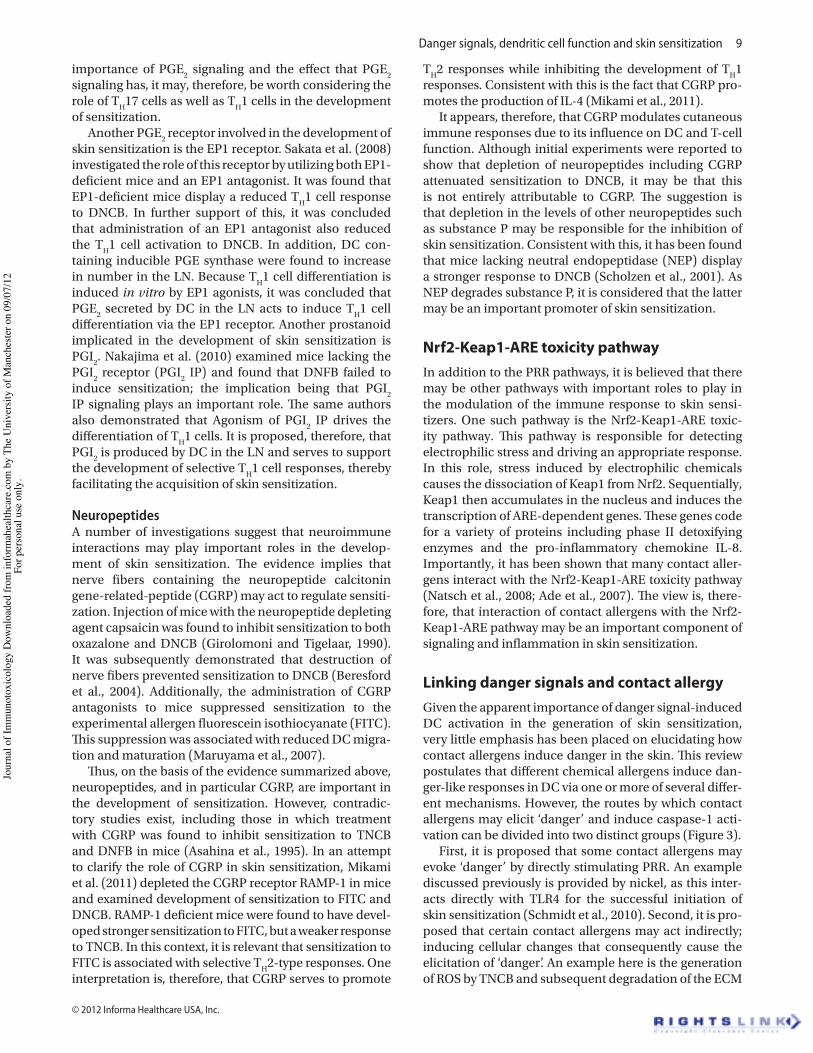
Danger signals, dendritic cell function and skin sensitization 9
© 2012 Informa Healthcare USA, Inc.
importance of PGE2 signaling and the effect that PGE2 signaling has, it may, therefore, be worth considering the role of TH17 cells as well as TH1 cells in the development of sensitization.
Another PGE2 receptor involved in the development of skin sensitization is the EP1 receptor. Sakata et al. (2008) investigated the role of this receptor by utilizing both EP1-deficient mice and an EP1 antagonist. It was found that EP1-deficient mice display a reduced TH1 cell response to DNCB. In further support of this, it was concluded that administration of an EP1 antagonist also reduced the TH1 cell activation to DNCB. In addition, DC con-taining inducible PGE synthase were found to increase in number in the LN. Because TH1 cell differentiation is induced in vitro by EP1 agonists, it was concluded that PGE2 secreted by DC in the LN acts to induce TH1 cell differentiation via the EP1 receptor. Another prostanoid implicated in the development of skin sensitization is PGI2. Nakajima et al. (2010) examined mice lacking the PGI2 receptor (PGI2 IP) and found that DNFB failed to induce sensitization; the implication being that PGI2 IP signaling plays an important role. The same authors also demonstrated that Agonism of PGI2 IP drives the differentiation of TH1 cells. It is proposed, therefore, that PGI2 is produced by DC in the LN and serves to support the development of selective TH1 cell responses, thereby facilitating the acquisition of skin sensitization.
NeuropeptidesA number of investigations suggest that neuroimmune interactions may play important roles in the develop-ment of skin sensitization. The evidence implies that nerve fibers containing the neuropeptide calcitonin gene-related-peptide (CGRP) may act to regulate sensiti-zation. Injection of mice with the neuropeptide depleting agent capsaicin was found to inhibit sensitization to both oxazalone and DNCB (Girolomoni and Tigelaar, 1990). It was subsequently demonstrated that destruction of nerve fibers prevented sensitization to DNCB (Beresford et al., 2004). Additionally, the administration of CGRP antagonists to mice suppressed sensitization to the experimental allergen fluorescein isothiocyanate (FITC). This suppression was associated with reduced DC migra-tion and maturation (Maruyama et al., 2007).
Thus, on the basis of the evidence summarized above, neuropeptides, and in particular CGRP, are important in the development of sensitization. However, contradic-tory studies exist, including those in which treatment with CGRP was found to inhibit sensitization to TNCB and DNFB in mice (Asahina et al., 1995). In an attempt to clarify the role of CGRP in skin sensitization, Mikami et al. (2011) depleted the CGRP receptor RAMP-1 in mice and examined development of sensitization to FITC and DNCB. RAMP-1 deficient mice were found to have devel-oped stronger sensitization to FITC, but a weaker response to TNCB. In this context, it is relevant that sensitization to FITC is associated with selective TH2-type responses. One interpretation is, therefore, that CGRP serves to promote
TH2 responses while inhibiting the development of TH1 responses. Consistent with this is the fact that CGRP pro-motes the production of IL-4 (Mikami et al., 2011).
It appears, therefore, that CGRP modulates cutaneous immune responses due to its influence on DC and T-cell function. Although initial experiments were reported to show that depletion of neuropeptides including CGRP attenuated sensitization to DNCB, it may be that this is not entirely attributable to CGRP. The suggestion is that depletion in the levels of other neuropeptides such as substance P may be responsible for the inhibition of skin sensitization. Consistent with this, it has been found that mice lacking neutral endopeptidase (NEP) display a stronger response to DNCB (Scholzen et al., 2001). As NEP degrades substance P, it is considered that the latter may be an important promoter of skin sensitization.
Nrf2-Keap1-ARE toxicity pathwayIn addition to the PRR pathways, it is believed that there may be other pathways with important roles to play in the modulation of the immune response to skin sensi-tizers. One such pathway is the Nrf2-Keap1-ARE toxic-ity pathway. This pathway is responsible for detecting electrophilic stress and driving an appropriate response. In this role, stress induced by electrophilic chemicals causes the dissociation of Keap1 from Nrf2. Sequentially, Keap1 then accumulates in the nucleus and induces the transcription of ARE-dependent genes. These genes code for a variety of proteins including phase II detoxifying enzymes and the pro-inflammatory chemokine IL-8. Importantly, it has been shown that many contact aller-gens interact with the Nrf2-Keap1-ARE toxicity pathway (Natsch et al., 2008; Ade et al., 2007). The view is, there-fore, that interaction of contact allergens with the Nrf2-Keap1-ARE pathway may be an important component of signaling and inflammation in skin sensitization.
Linking danger signals and contact allergyGiven the apparent importance of danger signal-induced DC activation in the generation of skin sensitization, very little emphasis has been placed on elucidating how contact allergens induce danger in the skin. This review postulates that different chemical allergens induce dan-ger-like responses in DC via one or more of several differ-ent mechanisms. However, the routes by which contact allergens may elicit ‘danger’ and induce caspase-1 acti-vation can be divided into two distinct groups (Figure 3).
First, it is proposed that some contact allergens may evoke ‘danger’ by directly stimulating PRR. An example discussed previously is provided by nickel, as this inter-acts directly with TLR4 for the successful initiation of skin sensitization (Schmidt et al., 2010). Second, it is pro-posed that certain contact allergens may act indirectly; inducing cellular changes that consequently cause the elicitation of ‘danger’. An example here is the generation of ROS by TNCB and subsequent degradation of the ECM
Jour
nal o
f Im
mun
otox
icol
ogy
Dow
nloa
ded
from
info
rmah
ealth
care
.com
by
The
Uni
vers
ity o
f Man
ches
ter o
n 09
/07/
12Fo
r per
sona
l use
onl
y.
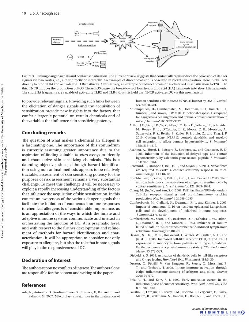
10 J. S. Ainscough et al.
Journal of Immunotoxicology
to provide relevant signals. Providing such links between the elicitation of danger signals and the acquisition of sensitization provide new insights into the factors that confer allergenic potential on certain chemicals and of the variables that influence skin sensitizing potency.
Concluding remarksThe question of what makes a chemical an allergen is a fascinating one. The importance of this conundrum is currently assuming greater importance due to the aspiration of finding suitable in vitro assays to identify and characterize skin-sensitizing chemicals. This is a daunting objective, since, although hazard identifica-tion using non-animal methods appears to be relatively tractable, assessment of skin sensitizing potency for the purposes of risk assessment remains a very substantial challenge. To meet this challenge it will be necessary to exploit a rapidly increasing understanding of the factors that influence the acquisition of skin sensitization. In this context an awareness of the various danger signals that facilitate the initiation of cutaneous immune responses to chemical allergens is of considerable importance, as is an appreciation of the ways in which the innate and adaptive immune systems communicate and interact in orchestrating the function of DC. At a pragmatic level, and with respect to the further development and refine-ment of methods for hazard identification and char-acterization, it will be appropriate to consider not only exposure to allergens, but also the role that innate signals will play in the responsiveness of DC.
Declaration of Interest The authors report no conflicts of interest. The authors alone are responsible for the content and writing of the paper.
ReferencesAde, N., Antonios, D., Kerdine-Romer, S., Boisleve, F., Rousset, F., and
Pallardy, M. 2007. NF-κB plays a major role in the maturation of
human dendritic cells induced by NiSO4 but not by DNCB. Toxicol. Sci.99:488–501.
Antonopoulos, H., Cumberbatch, M., Dearman, R. J., Daniel, R. J., Kimber, I., and Groves, R. W. 2001. Functional caspase-1 is required for Langerhans cell migration and optimal contact sensitization in mice. J. Immunol.166:3672–3677.
Arthur, J. C., Lich, J. D., Ye, Z., Allen, I. C., Gris, D., Wilson, J. E., Schneider, M., Roney, K. E., O’Connor, B. P., Moore, C. B., Morrison, A., Sutterwala, F. S., Bertin, J., Koller, B. H., Liu, Z., and Ting, J. P. 2010. Cutting Edge: NLRP12 controls dendritic and myeloid cell migration to affect contact hypersensitivity. J. Immunol. 185:4515–4519.
Asahina, A., Hosoi, J., Beissert, S., Stratigos, A., and Granstein, R. D. 1995. Inhibition of the induction of delayed-type and contact hypersensitivity by calcitonin-gene-related peptide. J. Immunol. 154:3056–3061.
Beresford, L., Orange, O., Bell, E. B., and Miyan, J. A. 2004. Nerve fibres are required to evoke a contact sensitivity response in mice. Immunology 111:118–125.
Bruchhausen, S., Zahn, S., Valk, E., Knop, J., and Becker, D. 2003. Thiol anti-oxidants block the activation of antigen-presenting cells by contact sensitizers. J. Invest. Dermatol. 121:1039–1044.
Chang, M., Jin, W., and Sun, S. C. 2009. Peli1 facilitates TRIF-dependent Toll-like receptor signaling and pro-inflammatory cytokine production. Nat. Immunol. 10:1089–1095.
Cumberbatch, M., Clelland, K., Dearman, R. J., and Kimber, I. 2005. Impact of cutaneous IL-10 on resident epidermal Langerhans’ cells and the development of polarized immune responses. J. Immunol.175:43–50.
Cumberbatch, M., Scott, R. C., Basketter, D. A., Scholes, E. W., Hilton, J., Dearman, R. J., and Kimber, I. 1993. Influence of sodium lauryl sulfate on 2,4-dinitrochlorobenzene-induced lymph-node activation. Toxicology 77:181–191.
Devaraj, S., Dau, M. R., Rockwood, J., Winter, W., Griffen, S. C., and Jialal, I. 2008. Increased toll-like receptor (TLR)-2 and TLR-4 expression in monocytes from patients with Type 1 diabetes: Further evidence of a pro-inflammatory state. J. Clin. Endocrinol. Metab. 93:578–583.
Diebold, S. S. 2009. Activation of dendritic cells by toll-like receptors and C-type lectins. Handbook Exp. Pharmacol. 188:3–30.
Dostert, C., Petrilli, V., van Bruggen, R., Steele, C., Mossman, B. T., and Tschopp, J. 2008. Innate immune activation through Nalp3 inflammasome sensing of asbestos and silica. Science 320:674–677.
Enk, A. H., and Katz, S. I. 1992. Early molecular events in the induction phase of contact sensitivity. Proc. Natl. Acad. Sci. USA 89:1398–1402.
Faustin, B., Lartigue, L., Bruey, J. M., Luciano, F., Sergienko, E., Bailly-Maitre, B., Volkmann, N., Hanein, D., Rouiller, I., and Reed, J. C.
Figure 3. Linking danger signals and contact sensitization. The current review suggests that contact allergens induce the provision of danger signals via two routes, i.e., either directly or indirectly. An example of direct provision is observed in nickel sensitization. Here, nickel acts directly to bind TLR4 and activate the TLR4 pathway. Alternatively, an example of indirect provision is observed in sensitization to TNCB. In this, TNCB induces the production of ROS. These ROS cause the breakdown of long hyaluronic acid (HA) fragments into short HA fragments. The short HA fragments are capable of activating TLR2 and TLR4, thus it is held that TNCB activates DC via this mechanism.
Jour
nal o
f Im
mun
otox
icol
ogy
Dow
nloa
ded
from
info
rmah
ealth
care
.com
by
The
Uni
vers
ity o
f Man
ches
ter o
n 09
/07/
12Fo
r per
sona
l use
onl
y.
Danger signals, dendritic cell function and skin sensitization 11
© 2012 Informa Healthcare USA, Inc.
2007. Reconstituted NALP1 inflammasome reveals two-step mechanism of caspase-1 activation. Mol. Cell 25:713–724.
Fitzgerald, K. A., McWhirter, S. M., Faia, K. L., Rowe, D. C., Latz, E., Golenbock, D. T., Coyle, A. J., Liao, S. M., and Maniatis, T. 2003. IKKε and TBK1 are essential components of the IRF3 signaling pathway. Nat. Immunol. 4:491–496.
Franchi, L., Eigenbrod, T., Munoz-Planillo, R., and Nunez, G. 2009. The inflammasome: A caspase-1-activation platform that regulates immune responses and disease pathogenesis. Nat. Immunol. 10:241–247.
Gao, Z., Tseng, C. H., Strober, B. E., Pei, Z., and Blaser, M. J. 2008. Substantial alterations of the cutaneous bacterial biota in psoriatic lesions. Plos One 3(7): e2719.
Girolomoni, G., and Tigelaar, R. E. 1990. Capsaicin-sensitive primary sensory neurons are potent modulators of murine delayed-type hypersentitivity reactions. J. Immunol.145:1105–1112.
Grabbe, S., Steinert, M., Mahnke, K., Schwarz, A., Luger, T. A., and Schwarz, T. 1996. Dissection of antigenic and irritative effects of epicutaneously applied haptens in mice. Evidence that not the antigenic component but non-specific proinflammatory effects of haptens determine the concentration-dependent elicitation of allergic contact dermatitis. J. Clin. Invest. 98:1158–1164.
Gupta, G., and Surolia, A. 2007. Collectins: Sentinels of innate immunity. Bioessays 29:452–464.
Hacker, H., Redecke, V., Blagoev, B., Kratchmarova, I., Hsu, L. C., Wang, G. G., Kamps, M. P., Raz, E., Wagner, H., Hacker, G., Mann, M., and Karin, M. 2006. Specificity in Toll-like receptor signalling through distinct effector functions of TRAF3 and TRAF6. Nature 439:204–207.
Hayashi, F., Smith, K. D., Ozinsky, A., Hawn, T. R., Yi, E. C., Goodlett, D. R., Eng, J. K., Akira, S., Underhill, D. M., and Aderem, A. 2001. The innate immune response to bacterial flagellin is mediated by Toll-like receptor 5. Nature 410:1099–1103.
Hewinson, J., Moore, S. F., Glover, C., Watts, A. G., and Mackenzie, A. B. 2008. A key role for redox signaling in rapid P2X7 receptor-induced IL-1# processing in human monocytes. J. Immunol. 180:8410–8420.
Hiscott, J., Pitha, P., Genin, P., Nguyen, H., Heylbroeck, C., Mamane, Y., Algarte, M., and Lin, R. T. 1999. Triggering the interferon response: The role of IRF-3 transcription factor. J. Interferon Cytokine Res. 19:1–13.
Hornung, V., Bauernfeind, F., Halle, A., Samstad, E. O., Kono, H., Rock, K. L., Fitzgerald, K. A., and Latz, E. 2008. Silica crystals and aluminum salts activate the NALP3 inflammasome through phagosomal destabilization. Nat. Immunol. 9:847–856.
Hsu, L. C., Ali, S. R., McGillivray, S., Tseng, P. H., Mariathasan, S., Humke, E. W., Eckmann, L., Powell, J. J., Nizet, V., Dixit, V. M., and Karin, M. 2008. A NOD2-NALP1 complex mediates caspase-1-dependent IL-1# beta secretion in response to Bacillus anthracis infection and muramyl dipeptide. Proc. Natl. Acad. Sci. USA 105:7803–7808.
Igyarto, B. Z., Haley, K., Ortner, D., Bobr, A., Gerami-Nejad, M., Edelson, B. T., Zurawski, S. M., Malissen, B., Zurawski, G., Berman, J., and Kaplan, D. H. 2011. Skin-resident murine dendritic cell subsets promote distinct and opposing antigen-specific T-helper cell responses. Immunity 35:260–272.
Johnson, G. L., and Lapadat, R. 2002. Mitogen-activated protein kinase pathways mediated by ERK, JNK, and p38 protein kinases. Science 298:1911–1912.
Kabashima, K., and Miyachi, Y. 2004. Prostanoids in the cutaneous immune response. J. Dermatol. Sci. 34:177–184.
Kanneganti, T. -D., Lamkanfi, M., Kim, Y. G., Chen, G., Park, J. H., Franchi, L., Vandenabeele, P., and Nunez, G. 2007. Pannexin-1-mediated recognition of bacterial molecules activates the cryopyrin inflammasome independent of Toll-like receptor signaling. Immunity 26:433–443.
Kaplan, D. H. 2010. In vivo function of Langerhans cells and dermal dendritic cells. Trends Immunol. 31:446–451.
Kawagoe, T., Sato, S., Matsushita, K., Kato, H., Matsui, K., Kumagai, Y., Saitoh, T., Kawai, T., Takeuchi, O., and Akira, S. 2008. Sequential
control of Toll-like receptor-dependent responses by IRAK1 and IRAK2. Nat. Immunol. 9:684–691.
Kawai, T., and Akira, S. 2009. The roles of TLRs, RLRs and NLRs in pathogen recognition. Int. Immunol. 21:317–337.
Kawai, T., and Akira, S. 2010. The role of pattern-recognition receptors in innate immunity: Update on Toll-like receptors. Nat. Immunol. 11:373–384.
Kehren, J., Desvignes, C., Krasteva, M., Ducluzeau, M. T., Assossou, O., Horand, F., Hahne, M., Kagi, D., Kaiserlian, D., and Nicolas, J. F. 1999. Cytotoxicity is mandatory for CD8+ T-cell-mediated contact hypersensitivity. J. Exp. Med.189:779–786.
Khakh, B. S., and North, R. A. 2006. P2X receptors as cell-surface ATP sensors in health and disease. Nature 442:527–532.
Kimber, I., Basketter, D. A., Gerberick, G. F., and Dearman, R. J. 2002. Allergic contact dermatitis. Int. Immunopharmacol. 2:201–211.
Kimber, I., Basketter, D. A., Gerberick, G. F., Ryan, C. A., and Dearman, R. J. 2011. Chemical allergy: Translating biology into hazard characterization. Toxicol. Sci. 120(Suppl 1):S238–S268.
Klekotka, P. A., Yang, L., and Yokoyama, W. M. 2010. Contrasting roles of the IL-1 and IL-18 receptors in MyD88-dependent contact hypersensitivity. J. Invest. Dermatol. 130:184–191.
Kobayashi, K., Hernandez, L. D., Galan, J. E., Janeway, C. A., Medzhitov, R., and Flavell, R. A. 2002. IRAK-M is a negative regulator of toll-like receptor signaling. Cell 110:191–202.
Kofoed, E. M., and Vance, R. E. 2011. Innate immune recognition of bacterial ligands by NAIPs determines inflammasome specificity. Nature 477:592–595.
Ley, K. 2003. The role of selectins in inflammation and disease. Trends Mol. Med. 9:263–268.
Martin, S. F., Dudda, J. C., Bachtanian, E., Lembo, A., Liller, S., Duerr, C., Heimesaat, M. M., Bereswill, S., Fejer, G., Vassileva, R., Jakob, T., Freudenberg, N., Termeer, C. C., Johner, C., Galanos, C., and Freudenberg, M. A. 2008. Toll-like receptor and IL-12 signaling control susceptibility to contact hypersensitivity. J. Exp. Med. 205:2151–2162.
Martin, S. F., Esser, P. R., Weber, F. C., Jakob, T., Freudenberg, M. A., Schmidt, M., and Goebeler, M. 2011. Mechanisms of chemical-induced innate immunity in allergic contact dermatitis. Allergy 66:1152–1163.
Martinon, F. 2010. Signaling by ROS drives inflammasome activation. Eur. J. Immunol. 40:616–619
Maruyama, T., Iizuka, H., Tobisawa, Y., Shiba, T., Matsuda, T., Kurohane, K., and Imai, Y. 2007. Influence of local treatments with capsaicin or allyl isothiocyanate in the sensitization phase of a fluorescein-isothiocyanate-induced contact sensitivity model. Int. Arch. Allergy Immunol. 143:144–154.
Matzinger, P. 1994. Tolerance, danger, and the extended family. Ann. Rev. Immunol. 12:991–1045.
Medzhitov, R., Preston-Hurlburt, P., Kopp, E., Stadlen, A., Chen, C. Q., Ghosh, S., and Janeway, C. A. 1998. MyD88 is an adaptor protein in the hToll/IL-1 receptor family signaling pathways. Mol. Cell 2:253–258.
Medzhitov, R., Preston-Hurlburt, P., and Janeway, C. A. 1997. A human homologue of the Drosophila Toll protein signals activation of adaptive immunity. Nature 388:394–397.
Mehrotra, P., Mishra, K. P., Raman, G., and Banerjee, G. 2005. Differential regulation of free radicals (reactive oxygen and nitrogen species) by contact allergens and irritants in human keratinocyte cell line. Toxicol. Mech. Meth. 15:343–350.
Merad, M., Manz, M. G., Karsunky, H., Wagers, A., Peters, W., Charo, I., Weissman, I. L., Cyster, J. G., and Engleman, E. G. 2002. Langerhans cells renew in the skin throughout life under steady-state conditions. Nat. Immunol. 3:1135–1141.
Meylan, E., Burns, K., Hofmann, K., Blancheteau, V., Martinon, F., Kelliher, M., and Tschopp, J. 2004. RIP1 is an essential mediator of Toll-like receptor 3-induced NF-κB activation. Nat. Immunol. 5:503–507.
Mikami, N., Matsushita, H., Kato, T., Kawasaki, R., Sawazaki, T., Kishimoto, T., Ogitani, Y., Watanabe, K., Miyagi, Y., Sueda, K.,
Jour
nal o
f Im
mun
otox
icol
ogy
Dow
nloa
ded
from
info
rmah
ealth
care
.com
by
The
Uni
vers
ity o
f Man
ches
ter o
n 09
/07/
12Fo
r per
sona
l use
onl
y.
12 J. S. Ainscough et al.
Journal of Immunotoxicology
Fukada, S. I., Yamamoto, H., and Tsujikawa, K. 2011. Calcitonin gene-related peptide is an important regulator of cutaneous immunity: Effect on dendritic cell and T-cell functions. J. Immunol. 186:6886–6893.
Mizuashi, M., Ohtani, T., Nakagawa, S., and Aiba, S. 2005. Redox imbalance induced by contact sensitizers triggers the maturation of dendritic cells. J. Invest. Dermatol. 124:579–586.
Nakajima, S., Honda, T., Sakata, D., Egawa, G., Tanizaki, H., Otsuka, A., Moniaga, C. S., Watanabe, T., Miyachi, Y., Narumiya, S., and Kabashima, K. 2010. Prostaglandin I2-IP signaling promotes TH1 Differentiation in a mouse model of contact hypersensitivity. J. Immunol. 184:5595–5603.
Natsch, A., Emter, R., and Ellis, G. 2008. Skin sensitizers induce antioxidant response element dependent genes: Application to the in vitro testing of the sensitization potential of chemicals. Toxicol. Lett. 180:S101.
Oganesyan, G., Saha, S. K., Guo, B. C., He, J. Q., Shahangian, A., Zarnegar, B., Pery, A., and Cheng, G. H. 2006. Critical role of TRAF3 in the Toll-like receptor-dependent and -independent anti-viral response. Nature 439:208–211.
Ogura, Y., Sutterwala, F. S., and Flavell, R. A. 2006. The inflammasome: First line of the immune response to cell stress. Cell 126:659–662.
Ozinsky, A., Underhill, D. M., Fontenot, J. D., Hajjar, A. M., Smith, K. D., Wilson, C. B., Schroeder, L., and Aderem, A. 2000. The repertoire for pattern recognition of pathogens by the innate immune system is defined by cooperation between Toll-like receptors. Proc. Natl. Acad. Sci. USA 97:13766–13771.
Palma, A. S., Feizi, T., Zhang, Y. B., Stoll, M. S., Lawson, A. M., Diaz-Rodriguez, E., Campanero-Rhodes, M. A., Costa, J., Gordon, S., Brown, G. D., and Chai, W. G. 2006. Ligands for the β-glucan receptor, Dectin-1, assigned using “designer” microarrays of oligosaccharide probes (neoglycolipids) generated from glucan polysaccharides. J. Biol. Chem. 281:5771–5779.
Pedra, J. H. F., Cassel, S. L., and Sutterwala, F. S. 2009. Sensing pathogens and danger signals by the inflammasome. Curr. Opin. Immunol. 21:10–16.
Petrilli, V., Papin, S., Dostert, C., Mayor, A., Martinon, F., and Tschopp, J. 2007. Activation of the NALP3 inflammasome is triggered by low intracellular potassium concentration. Cell Death Differen. 14:1583–1589.
Pobezinskaya, Y. L., Kim, Y. S., Choksi, S., Morgan, M. J., Li, T., Liu, C., and Liu, Z. 2008. The function of TRADD in signaling through tumor necrosis factor receptor 1 and TRIF-dependent Toll-like receptors. Nat. Immunol. 9:1047–1054.
Ring, S., Oliver, S. J., Cronstein, B. N., Enk, A. H., and Mahnke, K. 2009. CD4+CD25+ regulatory T-cells suppress contact hypersensitivity reactions through a CD39, adenosine-dependent mechanism. J. Allergy Clin. Immunol. 123:1287–1296.
Saijo, S., Fujikado, N., Furuta, T., Chung, S. H., Kotaki, H., Seki, K., Sudo, K., Akira, S., Adachi, Y., Ohno, N., Kinjo, T., Nakamura, K., Kawakami, K., and Iwakura, Y. 2007. Dectin-1 is required for host defense against Pneumocystis carinii but not against Candida albicans. Nat. Immunol. 8:39–46.
Saint-Mezard, P., Rosieres, A., Krasteva, M., Berard, F., Dubois, B., Kaiserlian, D., and Nicolas, J. F. 2004. Allergic contact dermatitis. Eur. J. Dermatol. 14:284–295.
Sakata, D., Nagamachi, M., Kabashima, K., Furuyashiki, T., Soontrapa, K., Matsuoka, T., Segi-Nishida, E., and Narumiya, S. 2008. Facilitation of TH1-mediated immune response by prostaglandin E receptor EP1. J Exp Med. 204(12):2865–2874.
Sato, S., Sanjo, H., Takeda, K., Ninomiya-Tsuji, J., Yamamoto, M., Kawai, T., matsumoto, K., Takeuchi, O., and Akira, S. 2005. Essential function for the kinase TAK1 in innate and adaptive immune responses. Nat. Immunol. 6:1087–1095.
Sato, S., Sugiyama, M., Yamamoto, M., Watanabe, Y., Kawai, T., Takeda, K., and Akira, S. 2003. Toll/IL-1 receptor domain-containing adaptor inducing IFNβ(TRIF) associates with TNF receptor-associated factor-6 and TANK-Binding kinase 1, and activates two distinct transcription factors, NF-κB and IFN-regulatory factor-3, in the toll-like receptor signaling. J. Immunol. 171:4304–4310.
Schaefer, L., Babelova, A., Kiss, E., Hausser, H. J., Baliova, M., Krzyzankova, M., Marsche, G., Young, M. F., Mihalik, D., Gotte, M., Malle, E., Schaefer, R. M., and Grone, H. J. 2005. The matrix component biglycan is proinflammatory and signals through Toll-like receptors 4 and 2 in macrophages. J. Clin. Invest. 115:2223–2233.
Schmidt, M., Raghavan, B., Mueller, V., Vogl, T., Fejer, G., Tchaptchet, S., Keck, S., Kalis, C., Nielsen, P. J., Galanos, C., Roth, J., Skerra, A., Martin, S. F., Freudenberg, M. A., and Goebller, M. 2010. Crucial role for human Toll-like receptor-4 in the development of contact allergy to nickel. Nat. Immunol. 11:814–819.
Scholzen, T. E., Steinhoff, M., Bonaccorsi, P., Klein, R., Amadesi, S., Geppetti, P., Lu, B., Gerard, N. P., Olerud, J. E., Luger, T. A., Bunnett, N. W., Grady, E. F., Armstrong, C. A., and Ansel, J. C. 2001. Neutral endopeptidase terminates substance P-induced inflammation in allergic contact dermatitis. J. Immunol.166:1285–1291.
Schwandner, R., Dziarski, R., Wesche, H., Rothe, M., and Kirschning, C. J. 1999. Peptidoglycan and lipoteichoic acid-induced cell activation is mediated by toll-like receptor-2. J. Biol. Chem. 274:17406–17409.
Shaw, P. J., Lamkanfi, M., and Kanneganti, T. D. 2010. NOD-like receptor (NLR) signaling beyond the inflammasome. Eur. J. Immunol. 40:624–627.
Suzuki, T., Franchi, L., Toma, C., Ashida, H., Ogawa, M., Yoshikawa, Y., Mimuro, H., Inohara, N., Sasakawa, C., and Nunez, G. 2007. Differential regulation of caspase-1 activation, pyroptosis, and autophagy via Ipaf and ASC in Shigella-infected macrophages. Plos Pathogens 3:1082–1091.
Taylor, P. R., Tsoni, S. V., Willment, J. A., Dennehy, K. M., Rosas, M., Findon, H., Haynes, K., Steele, C., Botto, M., Gordon, S., and Brown, G. D. 2007. Dectin-1 is required for #glucan recognition and control of fungal infection. Nat. Immunol. 8:31–38.
Thyssen, J. P., Linneberg, A., Menne, T., and Johansen, J. D. 2007. The epidemiology of contact allergy in the general population – prevalence and main findings. Contact Dermatitis 57:287–299.
Trinchieri, G., and Sher, A. 2007. Cooperation of Toll-like receptor signals in innate immune defense. Nat. Rev. Immunol. 7:179–190.
Vesely, P. W., Staber, P. B., Hoefler, G., and Kenner, L. 2009. Translational regulation mechanisms of AP-1 proteins. Mutat. Res. 682:7–12.
Watanabe, H., Gaide, O., Petrilli, V., Martinon, F., Contassot, E., Roques, S., Kummer, J. A., Tschopp, J., and French, L. E. 2007. Activation of the IL-1β-processing inflammasome is involved in contact hypersensitivity. J. Invest. Dermatol. 127:1956–1963.
Weber, F. C., Esser, P. R., Mueller, T., Ganesan, J., Pellegatti, P., Simon, M. M., Zeiser, R., Idzko, M., Jakob, T., and Martin, S. F. 2010. Lack of the purinergic receptor P2X(7) results in resistance to contact hypersensitivity. J. Exp. Med. 207:2609–2619.
Yamamoto, M., Okamoto, T., Takeda, K., Sato, S., Sanjo, H., Uematsu, S., Saitoh, T., Yamamoto, N., Sakurai, H., Ishii, K. J., Yamaoka, S., Kawai, T., Matsuura, Y., Takeuchi, O., and Akira, S. 2006. Key function for the Ubc13 E2 ubiquitin-conjugating enzyme in immune receptor signaling. Nat. Immunol. 7:962–970.
Yamamoto, M., Sato, S., Hemmi, H., Hoshino, K., Kaisho, T., Sanjo, H., Takeuchi, O., Sugiyama, M., Okabe, M., Takeda, K., and Akira, S. 2003. Role of adaptor TRIF in the MyD88-independent toll-like receptor signaling pathway. Science 301:640–643.
Yamamoto, M., Takeda, K., and Akira, S. 2004. TIR domain-containing adaptors define the specificity of TLR signaling. Mol. Immunol. 40:861–868.
Yao, C., Sakata, D., Esaki, Y., Li, Y., Matsuoka, T., Kuroiwa, K., Sugimoto, Y., and Narumiya, S. 2009. Prostaglandin E2-EP4 signaling promotes immune inflammation through TH1 cell differentiation and TH17 cell expansion. Nat. Med. 15:633–640.
Zhao, Y., Yang, J., Shi, J., Gong, Y. N., Lu, Q., Xu, H., Liu, L., and Shao, F. 2011. The NLRC4 inflammasome receptors for bacterial flagellin and Type III secretion apparatus. Nature 477:596–600.
Zhou, R., Yazdi, A. S., Menu, P., and Tschopp, J. 2011. A role for mitochondria in NLRP3 inflammasome activation. Nature 469:221.
Jour
nal o
f Im
mun
otox
icol
ogy
Dow
nloa
ded
from
info
rmah
ealth
care
.com
by
The
Uni
vers
ity o
f Man
ches
ter o
n 09
/07/
12Fo
r per
sona
l use
onl
y.
Dendritic Cell IL-1! and IL-1" Are Polyubiquitinated andDegraded by the Proteasome*
Received for publication, July 18, 2014, and in revised form, November 2, 2014 Published, JBC Papers in Press, November 4, 2014, DOI 10.1074/jbc.M114.595686
Joseph S. Ainscough‡1, G. Frank Gerberick§, Maryam Zahedi-Nejad‡, Gloria Lopez-Castejon‡, David Brough‡,Ian Kimber‡, and Rebecca J. Dearman‡
From the ‡Faculty of Life Sciences, University of Manchester, Manchester M13 9PT, United Kingdom and the §Procter & GambleCompany Co., Cincinnati, Ohio 45253
Background: Interleukin-1 secretion is an important process in inflammation and thus, the intracellular regulation of thesecytokines is of interest.Results: Inhibition of the proteasome in dendritic cells inhibits interleukin-1 degradation and leads to an accumulation ofpolyubiquitinated interleukin-1.Conclusion: Interleukin-1 cytokines are regulated by polyubiquitination and proteasomal degradation.Significance: Polyubiquitination and degradation are important processes in the intracellular regulation of interleukin-1.
IL-1! and " are key players in the innate immune system. Thesecretion of these cytokines by dendritic cells (DC) is integral tothe development of proinflammatory responses. These cyto-kines are not secreted via the classical secretory pathway.Instead, 2 independent processes are required; an initial signalto induce up-regulation of the precursor pro-IL-1! and -", anda second signal to drive cleavage and consequent secretion. Pro-IL-1! and -" are both cytosolic and thus, are potentially subjectto post-translational modifications. These modifications may,in turn, have a functional outcome in the context of IL-1! and -"secretion and hence inflammation. We report here that IL-1!and -" were degraded intracellularly in murine bone marrow-derived DC and that this degradation was dependent on activecellular processes. In addition, we demonstrate that degradationwas ablated when the proteasome was inhibited, whereasautophagy did not appear to play a major role. Furthermore,inhibition of the proteasome led to an accumulation of poly-ubiquitinated IL-1! and -", indicating that IL-1! and -" werepolyubiquitinated prior to proteasomal degradation. Finally,our investigations suggest that polyubiquitination and protea-somal degradation are not continuous processes but instead areup-regulated following DC activation. Overall, these data high-light that IL-1! and -" polyubiquitination and proteasomal deg-radation are central mechanisms in the regulation of intracellu-lar IL-1 levels in DC.
Dendritic cells (DC)2 are of fundamental importance to theimmune system, playing pivotal roles in the initiation andorchestration of immune responses (1, 2). They serve as
dynamic antigen presenting cells that bridge the innate andadaptive immune systems. Thus, DC survey the local microen-vironment, discriminating between a broad range of patho-genic and non-pathogenic cues and initiating immuneresponses, including inflammation (3). Inflammation is a com-plex response of the innate immune system that is associatedwith 5 characteristic features: erythema, edema, heat, pain, andloss of function (4, 5). These symptoms, which are crucial forthe resolution of infection and injury, occur as a result of a seriesof changes driven by the production of proinflammatory cyto-kines, including members of the interleukin-1 (IL-1) family.IL-1! and IL-1" are closely related potent proinflammatorymembers of the IL-1 family and are produced by DC (6). Thesecretion of these cytokines is an integral component of the roleof DC in orchestrating immune and inflammatory responses.Therefore, the transcriptional and post-transcriptional regula-tion of IL-1 by DC is of considerable importance, not only in thecontext of the resolution of infection and injury, but also in thecontext of preventing inappropriate or excessive inflammatoryreactions. This is evident in pathologies such as gout (7), rheu-matoid arthritis (8), cancer (9), and dementia (10), where thedysregulation of IL-1 is implicated strongly.
Unlike most cytokines, IL-1! and IL-1" are not secreted viathe classical secretory pathway. Instead, IL-1 is secreted via anon-conventional pathway and its release requires two inde-pendent signals. The first signal is typically provided by patho-gen-associated molecular patterns that act via pattern recogni-tion receptors, such as members of the Toll-like receptor (TLR)family, to stimulate complex signaling pathways (11, 12). Ulti-mately, the activation of these pathways results in the translo-cation of the transcription factor, nuclear factor #-light chainenhancer of activated B cells (NF-#B), to the nucleus. Thisdrives the transcription of a variety of pro-inflammatory pro-teins, including IL-1!, IL-1", and IL-6 (13), with IL-1! andIL-1" being transcribed as 31-kDa precursors (pro-IL-1). Thesecretion of these cytokines requires a second signal, which isprovided usually by additional pathogen-associated molecularpatterns or molecules associated with tissue trauma or damage(damage-associated molecular patterns). These damage-asso-
* This work was supported by grants from the Medical Research Council(MRC) and The Procter & Gamble Company (to J. S. A.).Author’s Choice—Final version full access.
1 To whom correspondence should be addressed: Michael Smith Building,Oxford Rd., Manchester M13 9PT, United Kingdom. Tel.: !44161 275 5368;E-mail: [email protected].
2 The abbreviations used are: DC, dendritic cells; TLR, Toll-like receptor; NF-#B,nuclear factor # light chain enhancer of activated B cells; CHX, cyclohexi-mide; BM, bone marrow; WCL, whole cell lysate; DMSO, dimethyl sulfoxide;ANOVA, analysis of variance.
THE JOURNAL OF BIOLOGICAL CHEMISTRY VOL. 289, NO. 51, pp. 35582–35592, December 19, 2014Author’s Choice © 2014 by The American Society for Biochemistry and Molecular Biology, Inc. Published in the U.S.A.
35582 JOURNAL OF BIOLOGICAL CHEMISTRY VOLUME 289 • NUMBER 51 • DECEMBER 19, 2014
by guest on Novem
ber 30, 2015http://w
ww
.jbc.org/D
ownloaded from
ciated molecular patterns signal via cytosolic pattern recogni-tion receptors, typically of the NOD-like receptor family, tostimulate assembly of inflammasome complexes (14). Forma-tion of the inflammasome complex induces activation of theenzyme caspase-1 (15). Active caspase-1 cleaves pro-IL-1! tothe 17-kDa bioactive form, facilitating its secretion (16). Theinflammasome is also thought to be involved in IL-1" secretion,although this process is less well characterized and is alsodependent on the calcium-dependent protease calpain. Simi-larly, the processing and secretion of pro-IL-1" involves cleav-age into its 17-kDa form (17, 18).
Although many studies have focused on the regulation ofIL-1 secretion, the intracellular control of pro-IL-1 remainspoorly understood. Both pro-IL-1" and pro-IL-1! are cytosolicand may therefore be subject to a number of post-translationalmodifications. Such modifications represent an importantmechanism by which cells can regulate the characteristics andfunction of proteins. For example, post-translational modifica-tions have been shown to act as regulators at various stages ofthe NF-#B signaling pathway (19). Ubiquitination involves theaddition of ubiquitin, an 8.5-kDa protein, to a given substrate(20). This process is mediated by a series of enzymes: E1, E2 andE3, that act sequentially to bind ubiquitin covalently to a Lysresidue on the substrate protein. It is the final E3 ubiquitinligase that confers the substrate specificity for ubiquitination.Substrate proteins may remain monoubiquitinated or may havefurther ubiquitin molecules added (polyubiquitination). In theformation of polyubiquitin chains, the carboxyl group on ubiq-uitin can bind to a number of different residues on the followingubiquitin, thus giving rise to various forms of polyubiquitina-tion. Ultimately, the type of ubiquitin chain bound has a funda-mental impact on the functional outcome of the modification(21). As an example, Lys48-linked polyubiquitin chains serve totarget proteins for proteasomal degradation (22), whereasLys63-linked polyubiquitin chains function in the regulation ofthe NF-#B pathway (23).
Here, we provide evidence that in murine DC, IL-1", andIL-1! are polyubiquitinated and that, in both DC and macro-phages, this polyubiquitination drives the proteasomal degra-dation of IL-1. Furthermore, these data demonstrate that in thepresence of a second signal, polyubiquitinated IL-1 is still avail-able for secretion. Collectively, our results demonstrate that inDC, the polyubiquitination and proteasomal degradation ofIL-1 serves as an essential process in the regulation of IL-1 and,therefore, should be considered as an extra dimension to thecurrent two-signal paradigm of IL-1 release.
EXPERIMENTAL PROCEDURES
Animals—Female BALB/c mice (6 – 8 weeks old) were usedthroughout these experiments (Harlan Olac, Bicester, UK).Mice were provided with environmental stimuli (bedding andnesting materials), food (SDS PCD pelleted diet; Special DietsServices Ltd, Witham, UK) and water were available ad libitum.Relative humidity was 55 ! 10% with a 12-h light/dark cycleand ambient temperature was maintained at 21 ! 2 °C. Main-tenance and treatment of animals were conducted as specifiedby the United Kingdom Animals (Scientific Procedures) Act1986. Mice were sacrificed by exposure to CO2 gas in rising
concentrations followed by dislocation of the neck in concor-dance with schedule 1 (U.K. Animals (Scientific Procedures)Act 1986).
Antibodies and Reagents—LPS from Escherichia coli serotype055:B5 (TLR2/4), poly(I:C), ATP, the autophagy inhibitor wort-manin and the translation inhibitor cycloheximide (CHX) werepurchased from Sigma. The proteasome inhibitor MG132 wasobtained from Merck Millipore (Billerica, MA). Recombinantmurine pro-IL-1! was purchased from Affymetrix eBioscience(San Diego, CA). For Western blot analysis, the primary anti-bodies were goat anti-mouse IL-1" antibody, goat anti-mouseIL-1! antibody (both R&D Systems; Minneapolis, MN), ormouse anti-ubiquitin antibody (Santa Cruz Biotechnology,Santa Cruz, CA). The HRP-conjugated secondary antibodieswere rabbit anti-goat IgG antibody (DAKO, Copenhagen, Den-mark) and goat anti-mouse light chain antibody (Millipore).
Generation and Culture of Murine Bone Marrow-derivedDC—Murine bone marrow-derived (BM) DC were generatedfollowing a previously described method (24). Briefly, bonemarrow was extracted by flushing the tibias and femurs withPBS. The cell suspension was centrifuged at 200 " g for 5 min atroom temperature. The remaining pellet was resuspended inpre-warmed, FCS-supplemented culture medium (RPMI 1640;Invitrogen), containing 400 $g/ml of penicillin/streptomycin,292 $g/ml of L-glutamine, 0.05 mM 2-mercaptoethanol, 4 ng/mlof GM-CSF (Miltenyi Biotech, Bisley, UK), and 10% FCS (Invit-rogen). A viable cell count was performed by trypan blue exclu-sion (0.5%; Sigma). Cells were cultured at #2 " 106 cells/ml inPetri dishes and incubated at 37 °C. The cultures were fed onday 3 by addition of 10 ml of fresh culture medium, and again onday 6 by gentle aspiration of 10 ml of medium followed by theaddition of 10 ml of fresh culture medium.
BMDC Treatments—BMDC were plated on day 8, in culturemedium without GM-CSF, at 106 cells/well (24-well plate) or107 cells/well (6-well plate; 106 cells/ml). Following an initial24-h dose-response experiment to determine the optimumdose of LPS to induce IL-1 production, cells were primed using0.1 $g/ml of LPS. BMDC were primed with LPS as indicated inthe text, and were activated with various concentrations of ATPfor 30 min at the end of the culture. MG132, wortmanin, or aDMSO control were added for the final 4 h of incubation. CHXwas added for the final 1 h of incubation. After incubation,supernatants were harvested and frozen at $80 °C. Cell lysateswere harvested in 200 $l of lysis buffer (20 mM Tris-HCl, 137mM NaCl, 20 mM EDTA, 10% glycerol, 0.5% Ipegal, 1 mM PMSF,protease inhibitor mixture (1:100)) and frozen at $80 °C. ForPCR analysis, lysates were prepared for RNA extraction follow-ing the manufacturer’s instructions (Purelink RNA mini kit;Invitrogen).
Immunoprecipitation of IL-1—To prepare lysates for immu-noprecipitation, supernatants were removed and cells werewashed twice with PBS. Cells were incubated on ice with washbuffer (20 mM N-ethylmaleimide in PBS). After a final washwith PBS, cell lysates were prepared in 500 $l of a specializedlysis buffer, formulated to prevent deubiquitination (25 mMTris, pH 7.4, 150 mM NaCl, 0.5% sodium deoxycholate, 1% Tri-ton X-100, 0.1% sodium dodecyl sulfate, 5 mM N-ethylmaleim-ide, 1 mM PMSF). An aliquot of lysate (50 $l) was retained as
Ubiquitination and Proteasomal Degradation of IL-1 in DC
DECEMBER 19, 2014 • VOLUME 289 • NUMBER 51 JOURNAL OF BIOLOGICAL CHEMISTRY 35583
by guest on Novem
ber 30, 2015http://w
ww
.jbc.org/D
ownloaded from
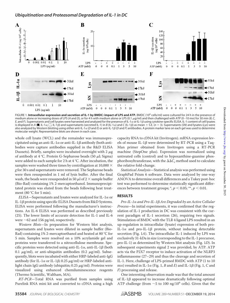
whole cell lysate (WCL) and the remainder was immunopre-cipitated using an anti-IL-1! or anti-IL-1" antibody (both anti-bodies were capture antibodies supplied in the R&D ELISADuosets). Briefly, samples were incubated overnight with 2 #gof antibody at 4 °C. Protein G-Sepharose beads (50 #l; Sigma)were added to each sample for 2 h at 4 °C. After incubation, thesamples were washed three times by centrifugation at 10,000 !g for 30 s and supernatants were removed. The Sepharose beadswere then resuspended in 1 ml of lysis buffer. After the finalwash, the beads were resuspended in 50 #l of 2 ! sample buffer(Bio-Rad) containing 1% 2-mercaptoethanol. Immunoprecipi-tated protein was eluted from the beads following heat treat-ment (80 °C for 5 min).
ELISA—Supernatants and lysates were analyzed for IL-1! orIL-1" protein using specific ELISA Duosets from R&D Systems.ELISA were performed following the manufacturer’s instruc-tions. An IL-6 ELISA was performed as described previously(25). The lower limits of accurate detection for IL-1 and IL-6were "62 and 156 pg/ml, respectively.
Western Blots—In preparation for Western blot analysis,supernatants and lysates were diluted in sample buffer (Bio-Rad) containing 1% 2-mercaptoethanol and heated at 80 °C for5 min. Samples were resolved on a 10% acrylamide gel andproteins were transferred to a nitrocellulose membrane. Spe-cific proteins were detected using anti-IL-1!, anti-IL-1" (both0.1 #g/ml), or anti-ubiquitin antibodies (0.2 #g/ml). Subse-quently, blots were incubated with either HRP-labeled anti-IgGantibody (for IL-1! or IL-1"; 0.25 #g/ml) or HRP-labeled anti-light chain IgG antibody (ubiquitin; 0.25 #g/ml). Proteins werevisualized using enhanced chemiluminescence reagents(Thermo Scientific, Waltham, MA).
RT-PCR—Total RNA was purified from samples usingPurelink RNA mini kit and converted to cDNA using a high
capacity RNA to cDNA kit (Invitrogen). mRNA expression lev-els of mouse IL-1" were determined by RT-PCR using a Taq-Man primer obtained from Invitrogen using a RT-PCRmachine (StepOne plus). Expression was normalized usinguntreated cells (control) and to hypoxanthine-guanine phos-phoribosyltransferase, with the ##Ct method used to calculatethe relative fold-change.
Statistical Analysis—Statistical analysis was performed usingGraphPad Prism 6 software. Data were analyzed by one-wayANOVA to determine overall differences and a Tukey post-hoctest was performed to determine statistically significant differ-ences between treatment groups; *, p $ 0.05; **, p $ 0.01.
RESULTS
Pro-IL-1! and Pro-IL-1" Are Degraded by an Active CellularProcess—In initial experiments, it was confirmed that the reg-ulation of IL-1 production in DC was consistent with the cur-rent paradigm of IL-1 secretion (26), requiring two signals.Stimulation of BMDC with the TLR 4 ligand LPS resulted in anup-regulation in intracellular (lysate) expression of both pro-IL-1! and pro-IL-1" protein, without inducing detectablesecretion (Fig. 1A). The intracellular IL-1 induced by LPS wasexclusively 31-kDa in size (corresponding to the IL-1 precursor,pro-IL-1) as determined by Western blot analysis (Fig. 1D). Insubsequent experiments signal 2 was provided, by ATP. ATPacts via the P2X7 receptor to induce activation of the NLRP3inflammasome (27–29) and thus the cleavage and secretion ofIL-1. Here, challenge of LPS-primed BMDC with ATP (1 to 10mM) resulted in IL-1! (Fig. 1, B and E) and IL-1" (Fig. 1, C andF) processing and release.
One interesting observation made was that the total amountof IL-1" appeared to increase dramatically following optimalATP challenge (from "5 to 100 ng/106 cells). Given that the
FIGURE 1. Intracellular expression and secretion of IL-1 by BMDC: impact of LPS and ATP. BMDC (106 cells/ml) were cultured for 24 h in the presence ofmedium alone or increasing doses of LPS (A and D), or for 4 h with medium alone or LPS (0.1 #g/ml) and then challenged with ATP (0 –10 mM) for 30 min (B, C,E, and F). Supernatants and cell lysates were harvested and analyzed for the presence of IL-1! or IL-1" using cytokine-specific ELISA. IL-1 content of cell lysatesis displayed in A (f, IL-1!; !, IL-1") and supernatants (secreted IL-1) in B (IL-1!) and C (IL-1") as mean % S.E. (n & 3). Supernatants (SN) and lysates (Lys) werealso analyzed by Western blotting using either anti-IL-1! (D and E) or anti-IL-1" (D and F) antibodies. A protein marker lane on each gel was used to determinemolecular weight. Representative blots are shown in each case.
Ubiquitination and Proteasomal Degradation of IL-1 in DC
35584 JOURNAL OF BIOLOGICAL CHEMISTRY VOLUME 289 • NUMBER 51 • DECEMBER 19, 2014
by guest on Novem
ber 30, 2015http://w
ww
.jbc.org/D
ownloaded from
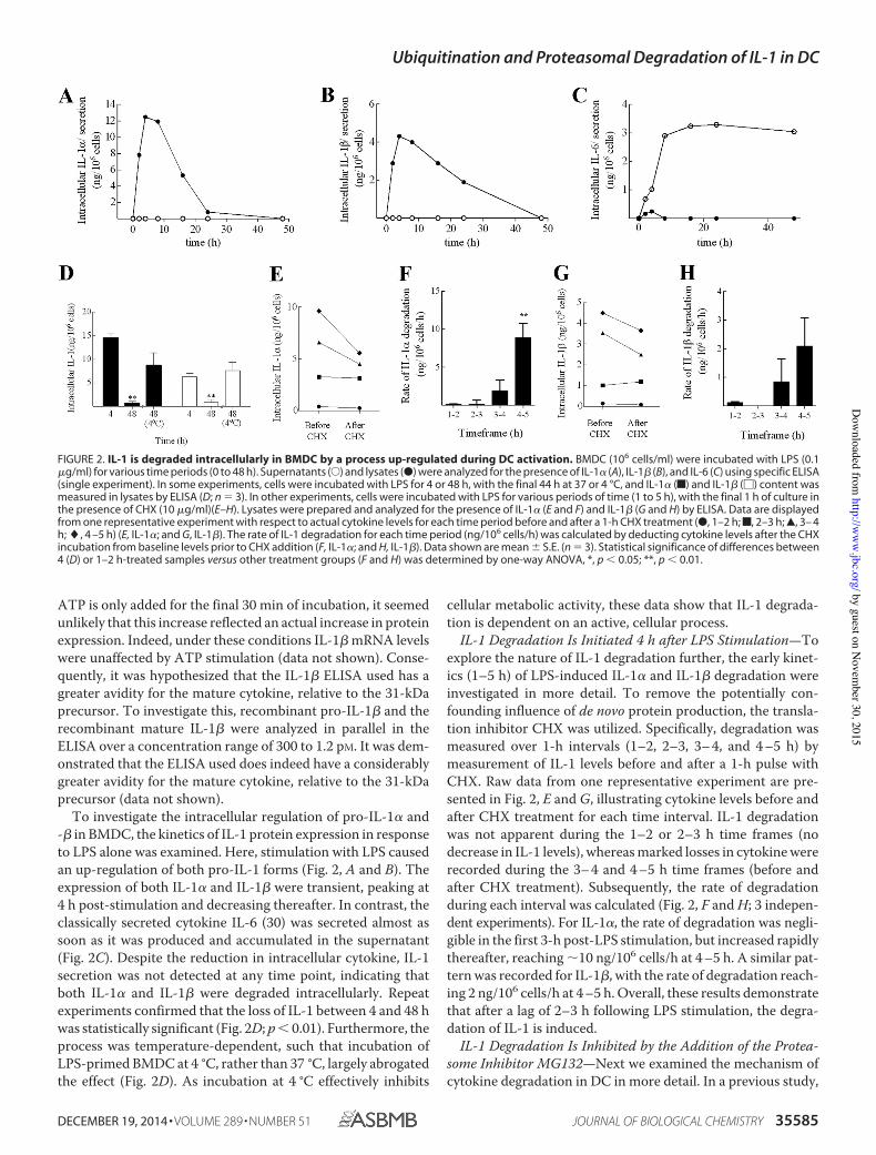
ATP is only added for the final 30 min of incubation, it seemedunlikely that this increase reflected an actual increase in proteinexpression. Indeed, under these conditions IL-1! mRNA levelswere unaffected by ATP stimulation (data not shown). Conse-quently, it was hypothesized that the IL-1! ELISA used has agreater avidity for the mature cytokine, relative to the 31-kDaprecursor. To investigate this, recombinant pro-IL-1! and therecombinant mature IL-1! were analyzed in parallel in theELISA over a concentration range of 300 to 1.2 pM. It was dem-onstrated that the ELISA used does indeed have a considerablygreater avidity for the mature cytokine, relative to the 31-kDaprecursor (data not shown).
To investigate the intracellular regulation of pro-IL-1" and-! in BMDC, the kinetics of IL-1 protein expression in responseto LPS alone was examined. Here, stimulation with LPS causedan up-regulation of both pro-IL-1 forms (Fig. 2, A and B). Theexpression of both IL-1" and IL-1! were transient, peaking at4 h post-stimulation and decreasing thereafter. In contrast, theclassically secreted cytokine IL-6 (30) was secreted almost assoon as it was produced and accumulated in the supernatant(Fig. 2C). Despite the reduction in intracellular cytokine, IL-1secretion was not detected at any time point, indicating thatboth IL-1" and IL-1! were degraded intracellularly. Repeatexperiments confirmed that the loss of IL-1 between 4 and 48 hwas statistically significant (Fig. 2D; p ! 0.01). Furthermore, theprocess was temperature-dependent, such that incubation ofLPS-primed BMDC at 4 °C, rather than 37 °C, largely abrogatedthe effect (Fig. 2D). As incubation at 4 °C effectively inhibits
cellular metabolic activity, these data show that IL-1 degrada-tion is dependent on an active, cellular process.
IL-1 Degradation Is Initiated 4 h after LPS Stimulation—Toexplore the nature of IL-1 degradation further, the early kinet-ics (1–5 h) of LPS-induced IL-1" and IL-1! degradation wereinvestigated in more detail. To remove the potentially con-founding influence of de novo protein production, the transla-tion inhibitor CHX was utilized. Specifically, degradation wasmeasured over 1-h intervals (1–2, 2–3, 3– 4, and 4 –5 h) bymeasurement of IL-1 levels before and after a 1-h pulse withCHX. Raw data from one representative experiment are pre-sented in Fig. 2, E and G, illustrating cytokine levels before andafter CHX treatment for each time interval. IL-1 degradationwas not apparent during the 1–2 or 2–3 h time frames (nodecrease in IL-1 levels), whereas marked losses in cytokine wererecorded during the 3– 4 and 4 –5 h time frames (before andafter CHX treatment). Subsequently, the rate of degradationduring each interval was calculated (Fig. 2, F and H; 3 indepen-dent experiments). For IL-1", the rate of degradation was negli-gible in the first 3-h post-LPS stimulation, but increased rapidlythereafter, reaching "10 ng/106 cells/h at 4 –5 h. A similar pat-tern was recorded for IL-1!, with the rate of degradation reach-ing 2 ng/106 cells/h at 4 –5 h. Overall, these results demonstratethat after a lag of 2–3 h following LPS stimulation, the degra-dation of IL-1 is induced.
IL-1 Degradation Is Inhibited by the Addition of the Protea-some Inhibitor MG132—Next we examined the mechanism ofcytokine degradation in DC in more detail. In a previous study,
FIGURE 2. IL-1 is degraded intracellularly in BMDC by a process up-regulated during DC activation. BMDC (106 cells/ml) were incubated with LPS (0.1#g/ml) for various time periods (0 to 48 h). Supernatants (E) and lysates (●) were analyzed for the presence of IL-1" (A), IL-1! (B), and IL-6 (C) using specific ELISA(single experiment). In some experiments, cells were incubated with LPS for 4 or 48 h, with the final 44 h at 37 or 4 °C, and IL-1" (f) and IL-1! (!) content wasmeasured in lysates by ELISA (D; n # 3). In other experiments, cells were incubated with LPS for various periods of time (1 to 5 h), with the final 1 h of culture inthe presence of CHX (10 #g/ml)(E–H). Lysates were prepared and analyzed for the presence of IL-1" (E and F) and IL-1! (G and H) by ELISA. Data are displayedfrom one representative experiment with respect to actual cytokine levels for each time period before and after a 1-h CHX treatment (●, 1–2 h;f, 2–3 h;Œ, 3– 4h; ", 4 –5 h) (E, IL-1"; and G, IL-1!). The rate of IL-1 degradation for each time period (ng/106 cells/h) was calculated by deducting cytokine levels after the CHXincubation from baseline levels prior to CHX addition (F, IL-1"; and H, IL-1!). Data shown are mean $ S.E. (n # 3). Statistical significance of differences between4 (D) or 1–2 h-treated samples versus other treatment groups (F and H) was determined by one-way ANOVA, *, p ! 0.05; **, p ! 0.01.
Ubiquitination and Proteasomal Degradation of IL-1 in DC
DECEMBER 19, 2014 • VOLUME 289 • NUMBER 51 JOURNAL OF BIOLOGICAL CHEMISTRY 35585
by guest on Novem
ber 30, 2015http://w
ww
.jbc.org/D
ownloaded from
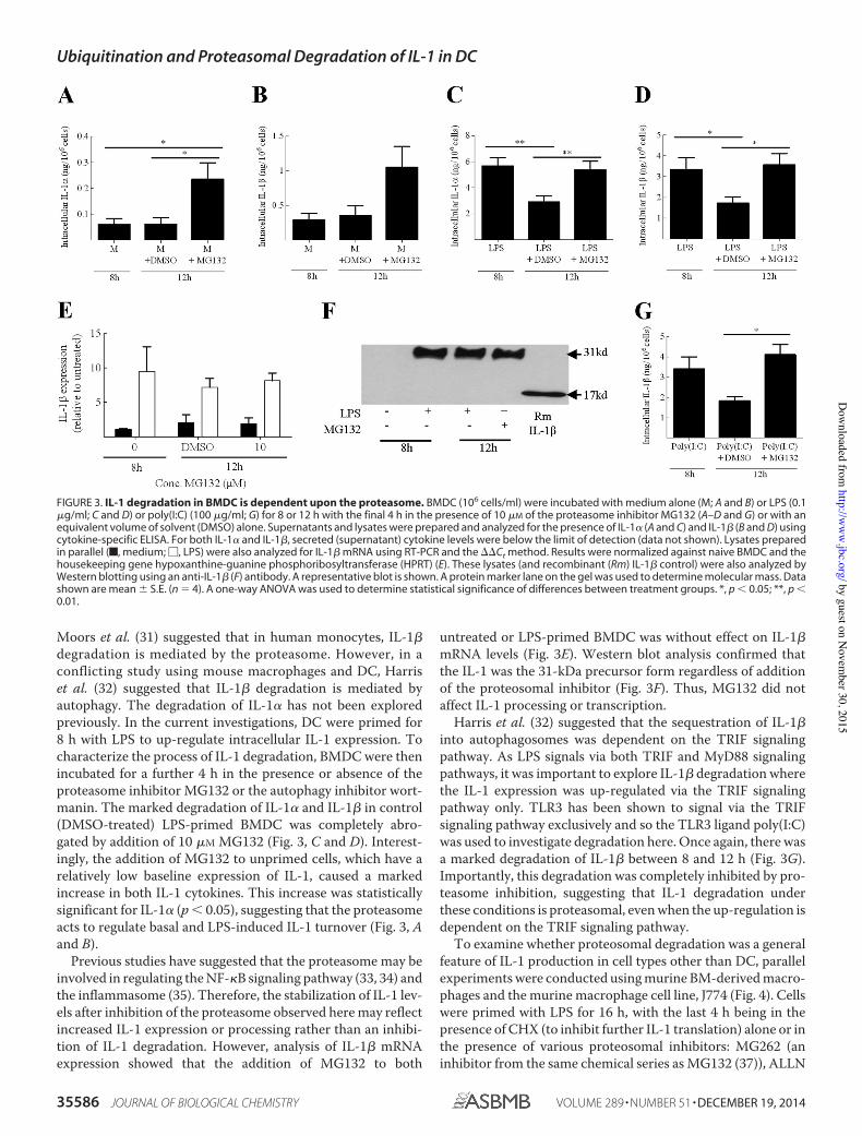
Moors et al. (31) suggested that in human monocytes, IL-1!degradation is mediated by the proteasome. However, in aconflicting study using mouse macrophages and DC, Harriset al. (32) suggested that IL-1! degradation is mediated byautophagy. The degradation of IL-1" has not been exploredpreviously. In the current investigations, DC were primed for8 h with LPS to up-regulate intracellular IL-1 expression. Tocharacterize the process of IL-1 degradation, BMDC were thenincubated for a further 4 h in the presence or absence of theproteasome inhibitor MG132 or the autophagy inhibitor wort-manin. The marked degradation of IL-1" and IL-1! in control(DMSO-treated) LPS-primed BMDC was completely abro-gated by addition of 10 #M MG132 (Fig. 3, C and D). Interest-ingly, the addition of MG132 to unprimed cells, which have arelatively low baseline expression of IL-1, caused a markedincrease in both IL-1 cytokines. This increase was statisticallysignificant for IL-1" (p ! 0.05), suggesting that the proteasomeacts to regulate basal and LPS-induced IL-1 turnover (Fig. 3, Aand B).
Previous studies have suggested that the proteasome may beinvolved in regulating the NF-$B signaling pathway (33, 34) andthe inflammasome (35). Therefore, the stabilization of IL-1 lev-els after inhibition of the proteasome observed here may reflectincreased IL-1 expression or processing rather than an inhibi-tion of IL-1 degradation. However, analysis of IL-1! mRNAexpression showed that the addition of MG132 to both
untreated or LPS-primed BMDC was without effect on IL-1!mRNA levels (Fig. 3E). Western blot analysis confirmed thatthe IL-1 was the 31-kDa precursor form regardless of additionof the proteosomal inhibitor (Fig. 3F). Thus, MG132 did notaffect IL-1 processing or transcription.
Harris et al. (32) suggested that the sequestration of IL-1!into autophagosomes was dependent on the TRIF signalingpathway. As LPS signals via both TRIF and MyD88 signalingpathways, it was important to explore IL-1! degradation wherethe IL-1 expression was up-regulated via the TRIF signalingpathway only. TLR3 has been shown to signal via the TRIFsignaling pathway exclusively and so the TLR3 ligand poly(I:C)was used to investigate degradation here. Once again, there wasa marked degradation of IL-1! between 8 and 12 h (Fig. 3G).Importantly, this degradation was completely inhibited by pro-teasome inhibition, suggesting that IL-1 degradation underthese conditions is proteasomal, even when the up-regulation isdependent on the TRIF signaling pathway.
To examine whether proteosomal degradation was a generalfeature of IL-1 production in cell types other than DC, parallelexperiments were conducted using murine BM-derived macro-phages and the murine macrophage cell line, J774 (Fig. 4). Cellswere primed with LPS for 16 h, with the last 4 h being in thepresence of CHX (to inhibit further IL-1 translation) alone or inthe presence of various proteosomal inhibitors: MG262 (aninhibitor from the same chemical series as MG132 (37)), ALLN
FIGURE 3. IL-1 degradation in BMDC is dependent upon the proteasome. BMDC (106 cells/ml) were incubated with medium alone (M; A and B) or LPS (0.1#g/ml; C and D) or poly(I:C) (100 #g/ml; G) for 8 or 12 h with the final 4 h in the presence of 10 #M of the proteasome inhibitor MG132 (A–D and G) or with anequivalent volume of solvent (DMSO) alone. Supernatants and lysates were prepared and analyzed for the presence of IL-1" (A and C) and IL-1! (B and D) usingcytokine-specific ELISA. For both IL-1" and IL-1!, secreted (supernatant) cytokine levels were below the limit of detection (data not shown). Lysates preparedin parallel (f, medium; !, LPS) were also analyzed for IL-1! mRNA using RT-PCR and the ""Ct method. Results were normalized against naive BMDC and thehousekeeping gene hypoxanthine-guanine phosphoribosyltransferase (HPRT) (E). These lysates (and recombinant (Rm) IL-1! control) were also analyzed byWestern blotting using an anti-IL-1! (F) antibody. A representative blot is shown. A protein marker lane on the gel was used to determine molecular mass. Datashown are mean # S.E. (n $ 4). A one-way ANOVA was used to determine statistical significance of differences between treatment groups. *, p ! 0.05; **, p !0.01.
Ubiquitination and Proteasomal Degradation of IL-1 in DC
35586 JOURNAL OF BIOLOGICAL CHEMISTRY VOLUME 289 • NUMBER 51 • DECEMBER 19, 2014
by guest on Novem
ber 30, 2015http://w
ww
.jbc.org/D
ownloaded from
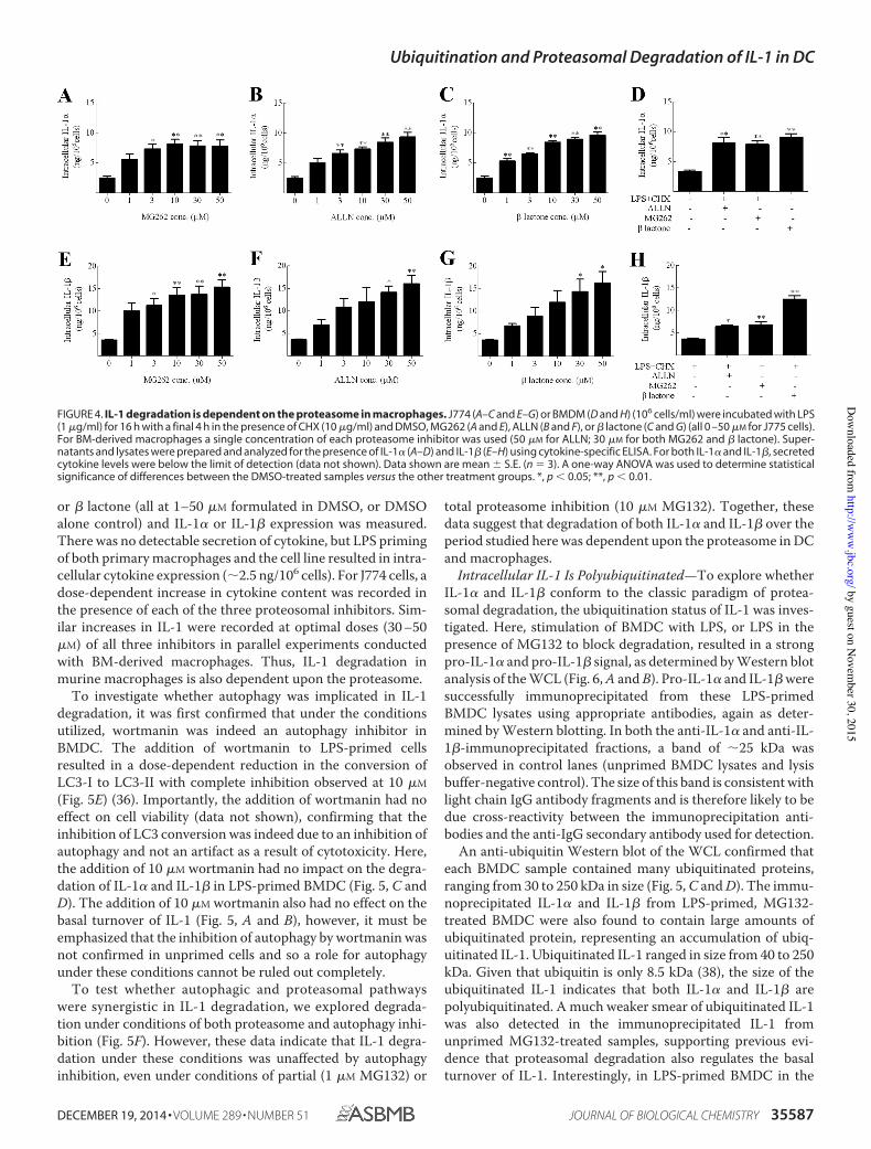
or ! lactone (all at 1–50 "M formulated in DMSO, or DMSOalone control) and IL-1# or IL-1! expression was measured.There was no detectable secretion of cytokine, but LPS primingof both primary macrophages and the cell line resulted in intra-cellular cytokine expression (!2.5 ng/106 cells). For J774 cells, adose-dependent increase in cytokine content was recorded inthe presence of each of the three proteosomal inhibitors. Sim-ilar increases in IL-1 were recorded at optimal doses (30 –50"M) of all three inhibitors in parallel experiments conductedwith BM-derived macrophages. Thus, IL-1 degradation inmurine macrophages is also dependent upon the proteasome.
To investigate whether autophagy was implicated in IL-1degradation, it was first confirmed that under the conditionsutilized, wortmanin was indeed an autophagy inhibitor inBMDC. The addition of wortmanin to LPS-primed cellsresulted in a dose-dependent reduction in the conversion ofLC3-I to LC3-II with complete inhibition observed at 10 "M(Fig. 5E) (36). Importantly, the addition of wortmanin had noeffect on cell viability (data not shown), confirming that theinhibition of LC3 conversion was indeed due to an inhibition ofautophagy and not an artifact as a result of cytotoxicity. Here,the addition of 10 "M wortmanin had no impact on the degra-dation of IL-1# and IL-1! in LPS-primed BMDC (Fig. 5, C andD). The addition of 10 "M wortmanin also had no effect on thebasal turnover of IL-1 (Fig. 5, A and B), however, it must beemphasized that the inhibition of autophagy by wortmanin wasnot confirmed in unprimed cells and so a role for autophagyunder these conditions cannot be ruled out completely.
To test whether autophagic and proteasomal pathwayswere synergistic in IL-1 degradation, we explored degrada-tion under conditions of both proteasome and autophagy inhi-bition (Fig. 5F). However, these data indicate that IL-1 degra-dation under these conditions was unaffected by autophagyinhibition, even under conditions of partial (1 "M MG132) or
total proteasome inhibition (10 "M MG132). Together, thesedata suggest that degradation of both IL-1# and IL-1! over theperiod studied here was dependent upon the proteasome in DCand macrophages.
Intracellular IL-1 Is Polyubiquitinated—To explore whetherIL-1# and IL-1! conform to the classic paradigm of protea-somal degradation, the ubiquitination status of IL-1 was inves-tigated. Here, stimulation of BMDC with LPS, or LPS in thepresence of MG132 to block degradation, resulted in a strongpro-IL-1# and pro-IL-1! signal, as determined by Western blotanalysis of the WCL (Fig. 6, A and B). Pro-IL-1# and IL-1! weresuccessfully immunoprecipitated from these LPS-primedBMDC lysates using appropriate antibodies, again as deter-mined by Western blotting. In both the anti-IL-1# and anti-IL-1!-immunoprecipitated fractions, a band of !25 kDa wasobserved in control lanes (unprimed BMDC lysates and lysisbuffer-negative control). The size of this band is consistent withlight chain IgG antibody fragments and is therefore likely to bedue cross-reactivity between the immunoprecipitation anti-bodies and the anti-IgG secondary antibody used for detection.
An anti-ubiquitin Western blot of the WCL confirmed thateach BMDC sample contained many ubiquitinated proteins,ranging from 30 to 250 kDa in size (Fig. 5, C and D). The immu-noprecipitated IL-1# and IL-1! from LPS-primed, MG132-treated BMDC were also found to contain large amounts ofubiquitinated protein, representing an accumulation of ubiq-uitinated IL-1. Ubiquitinated IL-1 ranged in size from 40 to 250kDa. Given that ubiquitin is only 8.5 kDa (38), the size of theubiquitinated IL-1 indicates that both IL-1# and IL-1! arepolyubiquitinated. A much weaker smear of ubiquitinated IL-1was also detected in the immunoprecipitated IL-1 fromunprimed MG132-treated samples, supporting previous evi-dence that proteasomal degradation also regulates the basalturnover of IL-1. Interestingly, in LPS-primed BMDC in the
FIGURE 4. IL-1 degradation is dependent on the proteasome in macrophages. J774 (A–C and E–G) or BMDM (D and H) (106 cells/ml) were incubated with LPS(1 "g/ml) for 16 h with a final 4 h in the presence of CHX (10 "g/ml) and DMSO, MG262 (A and E), ALLN (B and F), or ! lactone (C and G) (all 0 –50 "M for J775 cells).For BM-derived macrophages a single concentration of each proteasome inhibitor was used (50 "M for ALLN; 30 "M for both MG262 and ! lactone). Super-natants and lysates were prepared and analyzed for the presence of IL-1# (A–D) and IL-1! (E–H) using cytokine-specific ELISA. For both IL-1# and IL-1!, secretedcytokine levels were below the limit of detection (data not shown). Data shown are mean " S.E. (n # 3). A one-way ANOVA was used to determine statisticalsignificance of differences between the DMSO-treated samples versus the other treatment groups. *, p $ 0.05; **, p $ 0.01.
Ubiquitination and Proteasomal Degradation of IL-1 in DC
DECEMBER 19, 2014 • VOLUME 289 • NUMBER 51 JOURNAL OF BIOLOGICAL CHEMISTRY 35587
by guest on Novem
ber 30, 2015http://w
ww
.jbc.org/D
ownloaded from
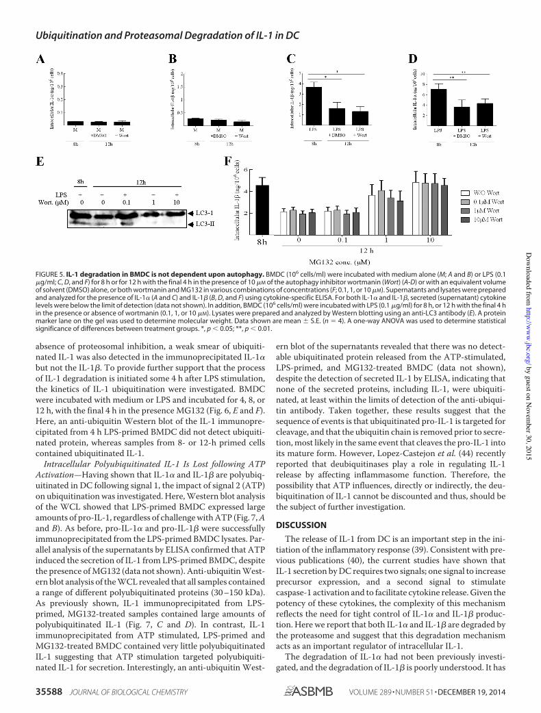
absence of proteosomal inhibition, a weak smear of ubiquiti-nated IL-1 was also detected in the immunoprecipitated IL-1!but not the IL-1". To provide further support that the processof IL-1 degradation is initiated some 4 h after LPS stimulation,the kinetics of IL-1 ubiquitination were investigated. BMDCwere incubated with medium or LPS and incubated for 4, 8, or12 h, with the final 4 h in the presence MG132 (Fig. 6, E and F).Here, an anti-ubiquitin Western blot of the IL-1 immunopre-cipitated from 4 h LPS-primed BMDC did not detect ubiquiti-nated protein, whereas samples from 8- or 12-h primed cellscontained ubiquitinated IL-1.
Intracellular Polyubiquitinated IL-1 Is Lost following ATPActivation—Having shown that IL-1! and IL-1" are polyubiq-uitinated in DC following signal 1, the impact of signal 2 (ATP)on ubiquitination was investigated. Here, Western blot analysisof the WCL showed that LPS-primed BMDC expressed largeamounts of pro-IL-1, regardless of challenge with ATP (Fig. 7, Aand B). As before, pro-IL-1! and pro-IL-1" were successfullyimmunoprecipitated from the LPS-primed BMDC lysates. Par-allel analysis of the supernatants by ELISA confirmed that ATPinduced the secretion of IL-1 from LPS-primed BMDC, despitethe presence of MG132 (data not shown). Anti-ubiquitin West-ern blot analysis of the WCL revealed that all samples containeda range of different polyubiquitinated proteins (30 –150 kDa).As previously shown, IL-1 immunoprecipitated from LPS-primed, MG132-treated samples contained large amounts ofpolyubiquitinated IL-1 (Fig. 7, C and D). In contrast, IL-1immunoprecipitated from ATP stimulated, LPS-primed andMG132-treated BMDC contained very little polyubiquitinatedIL-1 suggesting that ATP stimulation targeted polyubiquiti-nated IL-1 for secretion. Interestingly, an anti-ubiquitin West-
ern blot of the supernatants revealed that there was no detect-able ubiquitinated protein released from the ATP-stimulated,LPS-primed, and MG132-treated BMDC (data not shown),despite the detection of secreted IL-1 by ELISA, indicating thatnone of the secreted proteins, including IL-1, were ubiquiti-nated, at least within the limits of detection of the anti-ubiqui-tin antibody. Taken together, these results suggest that thesequence of events is that ubiquitinated pro-IL-1 is targeted forcleavage, and that the ubiquitin chain is removed prior to secre-tion, most likely in the same event that cleaves the pro-IL-1 intoits mature form. However, Lopez-Castejon et al. (44) recentlyreported that deubiquitinases play a role in regulating IL-1release by affecting inflammasome function. Therefore, thepossibility that ATP influences, directly or indirectly, the deu-biquitination of IL-1 cannot be discounted and thus, should bethe subject of further investigation.
DISCUSSION
The release of IL-1 from DC is an important step in the ini-tiation of the inflammatory response (39). Consistent with pre-vious publications (40), the current studies have shown thatIL-1 secretion by DC requires two signals; one signal to increaseprecursor expression, and a second signal to stimulatecaspase-1 activation and to facilitate cytokine release. Given thepotency of these cytokines, the complexity of this mechanismreflects the need for tight control of IL-1! and IL-1" produc-tion. Here we report that both IL-1! and IL-1" are degraded bythe proteasome and suggest that this degradation mechanismacts as an important regulator of intracellular IL-1.
The degradation of IL-1! had not been previously investi-gated, and the degradation of IL-1" is poorly understood. It has
FIGURE 5. IL-1 degradation in BMDC is not dependent upon autophagy. BMDC (106 cells/ml) were incubated with medium alone (M; A and B) or LPS (0.1#g/ml; C, D, and F) for 8 h or for 12 h with the final 4 h in the presence of 10 #M of the autophagy inhibitor wortmanin (Wort) (A-D) or with an equivalent volumeof solvent (DMSO) alone, or both wortmanin and MG132 in various combinations of concentrations (F; 0.1, 1, or 10 #M). Supernatants and lysates were preparedand analyzed for the presence of IL-1! (A and C) and IL-1" (B, D, and F) using cytokine-specific ELISA. For both IL-1! and IL-1", secreted (supernatant) cytokinelevels were below the limit of detection (data not shown). In addition, BMDC (106 cells/ml) were incubated with LPS (0.1 #g/ml) for 8 h, or 12 h with the final 4 hin the presence or absence of wortmanin (0.1, 1, or 10 #M). Lysates were prepared and analyzed by Western blotting using an anti-LC3 antibody (E). A proteinmarker lane on the gel was used to determine molecular weight. Data shown are mean ! S.E. (n " 4). A one-way ANOVA was used to determine statisticalsignificance of differences between treatment groups. *, p # 0.05; **, p # 0.01.
Ubiquitination and Proteasomal Degradation of IL-1 in DC
35588 JOURNAL OF BIOLOGICAL CHEMISTRY VOLUME 289 • NUMBER 51 • DECEMBER 19, 2014
by guest on Novem
ber 30, 2015http://w
ww
.jbc.org/D
ownloaded from
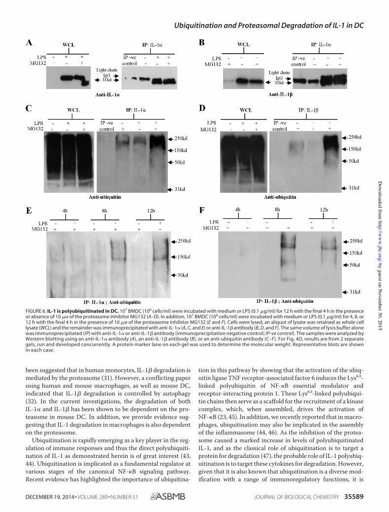
been suggested that in human monocytes, IL-1! degradation ismediated by the proteasome (31). However, a conflicting paperusing human and mouse macrophages, as well as mouse DC,indicated that IL-1! degradation is controlled by autophagy(32). In the current investigations, the degradation of bothIL-1" and IL-1! has been shown to be dependent on the pro-teasome in mouse DC. In addition, we provide evidence sug-gesting that IL-1 degradation in macrophages is also dependenton the proteasome.
Ubiquitination is rapidly emerging as a key player in the reg-ulation of immune responses and thus the direct polyubiquiti-nation of IL-1 as demonstrated herein is of great interest (43,44). Ubiquitination is implicated as a fundamental regulator atvarious stages of the canonical NF-#B signaling pathway.Recent evidence has highlighted the importance of ubiquitina-
tion in this pathway by showing that the activation of the ubiq-uitin ligase TNF receptor-associated factor 6 induces the Lys63-linked polyubiquitin of NF-#B essential modulator andreceptor-interacting protein 1. These Lys63-linked polyubiqui-tin chains then serve as a scaffold for the recruitment of a kinasecomplex, which, when assembled, drives the activation ofNF-#B (23, 45). In addition, we recently reported that in macro-phages, ubiquitination may also be implicated in the assemblyof the inflammasome (44, 46). As the inhibition of the protea-some caused a marked increase in levels of polyubiquitinatedIL-1, and as the classical role of ubiquitination is to target aprotein for degradation (47), the probable role of IL-1 polyubiq-uitination is to target these cytokines for degradation. However,given that it is also known that ubiquitination is a diverse mod-ification with a range of immunoregulatory functions, it is
FIGURE 6. IL-1 is polyubiquitinated in DC. 107 BMDC (106 cells/ml) were incubated with medium or LPS (0.1 $g/ml) for 12 h with the final 4 h in the presenceor absence of 10 $M of the proteasome inhibitor MG132 (A–D). In addition, 107 BMDC (106 cells/ml) were incubated with medium or LPS (0.1 $g/ml) for 4, 8, or12 h with the final 4 h in the presence of 10 $M of the proteasome inhibitor MG132 (E and F). Cells were lysed, an aliquot of lysate was retained as whole celllysate (WCL) and the remainder was immunoprecipitated with anti-IL-1" (A, C, and E) or anti-IL-1! antibody (B, D, and F). The same volume of lysis buffer alonewas immunoprecipitated (IP) with anti-IL-1" or anti-IL-1! antibody (immunoprecipitation negative control; IP-ve control). The samples were analyzed byWestern blotting using an anti-IL-1" antibody (A), an anti-IL-1! antibody (B), or an anti-ubiquitin antibody (C–F). For Fig. 4D, results are from 2 separategels, run and developed concurrently. A protein marker lane on each gel was used to determine the molecular weight. Representative blots are shownin each case.
Ubiquitination and Proteasomal Degradation of IL-1 in DC
DECEMBER 19, 2014 • VOLUME 289 • NUMBER 51 JOURNAL OF BIOLOGICAL CHEMISTRY 35589
by guest on Novem
ber 30, 2015http://w
ww
.jbc.org/D
ownloaded from
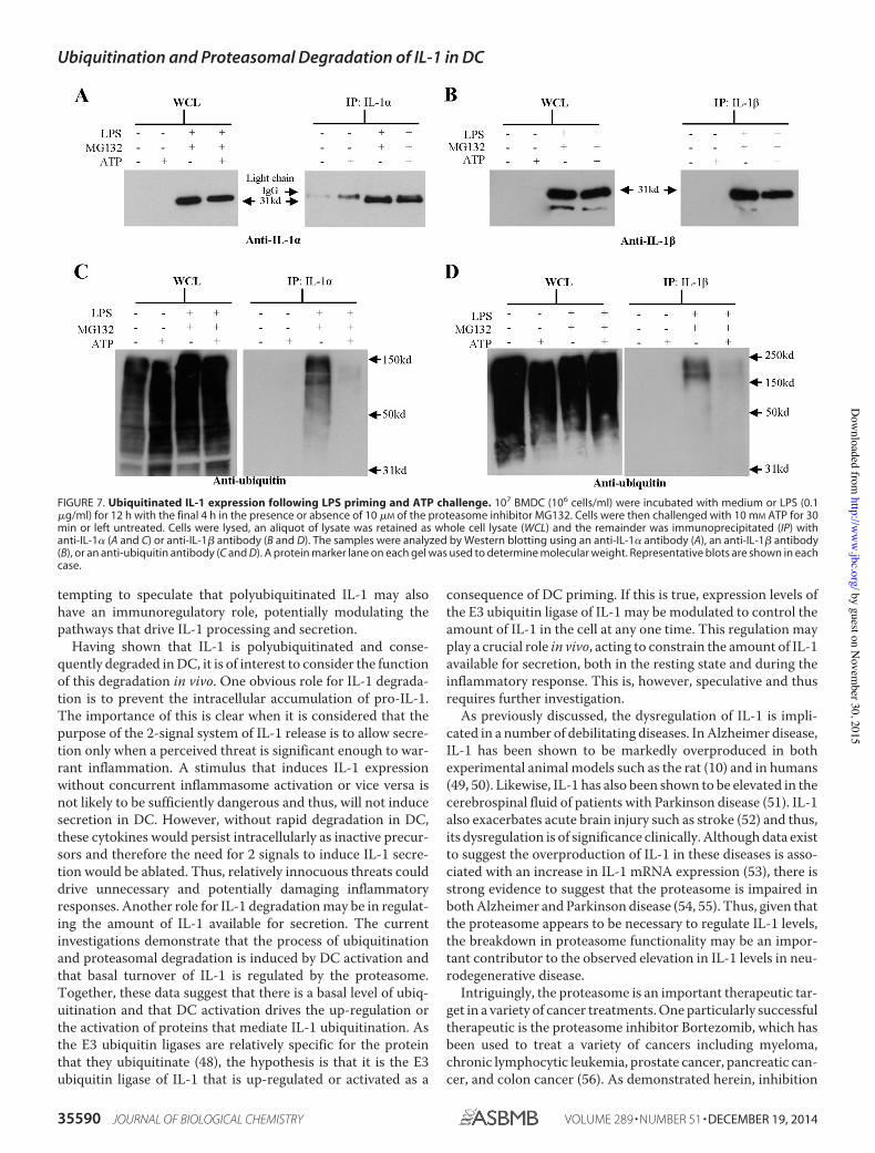
tempting to speculate that polyubiquitinated IL-1 may alsohave an immunoregulatory role, potentially modulating thepathways that drive IL-1 processing and secretion.
Having shown that IL-1 is polyubiquitinated and conse-quently degraded in DC, it is of interest to consider the functionof this degradation in vivo. One obvious role for IL-1 degrada-tion is to prevent the intracellular accumulation of pro-IL-1.The importance of this is clear when it is considered that thepurpose of the 2-signal system of IL-1 release is to allow secre-tion only when a perceived threat is significant enough to war-rant inflammation. A stimulus that induces IL-1 expressionwithout concurrent inflammasome activation or vice versa isnot likely to be sufficiently dangerous and thus, will not inducesecretion in DC. However, without rapid degradation in DC,these cytokines would persist intracellularly as inactive precur-sors and therefore the need for 2 signals to induce IL-1 secre-tion would be ablated. Thus, relatively innocuous threats coulddrive unnecessary and potentially damaging inflammatoryresponses. Another role for IL-1 degradation may be in regulat-ing the amount of IL-1 available for secretion. The currentinvestigations demonstrate that the process of ubiquitinationand proteasomal degradation is induced by DC activation andthat basal turnover of IL-1 is regulated by the proteasome.Together, these data suggest that there is a basal level of ubiq-uitination and that DC activation drives the up-regulation orthe activation of proteins that mediate IL-1 ubiquitination. Asthe E3 ubiquitin ligases are relatively specific for the proteinthat they ubiquitinate (48), the hypothesis is that it is the E3ubiquitin ligase of IL-1 that is up-regulated or activated as a
consequence of DC priming. If this is true, expression levels ofthe E3 ubiquitin ligase of IL-1 may be modulated to control theamount of IL-1 in the cell at any one time. This regulation mayplay a crucial role in vivo, acting to constrain the amount of IL-1available for secretion, both in the resting state and during theinflammatory response. This is, however, speculative and thusrequires further investigation.
As previously discussed, the dysregulation of IL-1 is impli-cated in a number of debilitating diseases. In Alzheimer disease,IL-1 has been shown to be markedly overproduced in bothexperimental animal models such as the rat (10) and in humans(49, 50). Likewise, IL-1 has also been shown to be elevated in thecerebrospinal fluid of patients with Parkinson disease (51). IL-1also exacerbates acute brain injury such as stroke (52) and thus,its dysregulation is of significance clinically. Although data existto suggest the overproduction of IL-1 in these diseases is asso-ciated with an increase in IL-1 mRNA expression (53), there isstrong evidence to suggest that the proteasome is impaired inboth Alzheimer and Parkinson disease (54, 55). Thus, given thatthe proteasome appears to be necessary to regulate IL-1 levels,the breakdown in proteasome functionality may be an impor-tant contributor to the observed elevation in IL-1 levels in neu-rodegenerative disease.
Intriguingly, the proteasome is an important therapeutic tar-get in a variety of cancer treatments. One particularly successfultherapeutic is the proteasome inhibitor Bortezomib, which hasbeen used to treat a variety of cancers including myeloma,chronic lymphocytic leukemia, prostate cancer, pancreatic can-cer, and colon cancer (56). As demonstrated herein, inhibition
FIGURE 7. Ubiquitinated IL-1 expression following LPS priming and ATP challenge. 107 BMDC (106 cells/ml) were incubated with medium or LPS (0.1!g/ml) for 12 h with the final 4 h in the presence or absence of 10 !M of the proteasome inhibitor MG132. Cells were then challenged with 10 mM ATP for 30min or left untreated. Cells were lysed, an aliquot of lysate was retained as whole cell lysate (WCL) and the remainder was immunoprecipitated (IP) withanti-IL-1" (A and C) or anti-IL-1# antibody (B and D). The samples were analyzed by Western blotting using an anti-IL-1" antibody (A), an anti-IL-1# antibody(B), or an anti-ubiquitin antibody (C and D). A protein marker lane on each gel was used to determine molecular weight. Representative blots are shown in eachcase.
Ubiquitination and Proteasomal Degradation of IL-1 in DC
35590 JOURNAL OF BIOLOGICAL CHEMISTRY VOLUME 289 • NUMBER 51 • DECEMBER 19, 2014
by guest on Novem
ber 30, 2015http://w
ww
.jbc.org/D
ownloaded from
of the proteasome causes a significant spike in IL-1 levels invitro. If the effect of proteasomal inhibition is mirrored in vivo,Bortezomib may also cause an elevation in the IL-1 available forsecretion and therefore an increase in inflammation. Althoughprevious evidence appears to negate this hypothesis, suggestingthat Bortezomib has an anti-inflammatory effect due to inhibi-tion of the NF-!B pathway (57), there may still be an impactfrom increased polyubiquitinated IL-1. Thus, the spike in IL-1observed here in vitro may still be of relevance in vivo and thus,may still have a considerable impact upon the efficacy of Bort-ezomib, and potentially any other proteasome inhibitors, asanti-cancer treatments.
In the current investigations, the regulation of IL-1" andIL-1# in DC appear to mirror each other. Although both cyto-kines serve as proinflammatory cytokines, the exact roles thatIL-1" and IL-1# play in vivo are distinct. In the initiation ofallergic contact dermatitis, for example, IL-1# but not IL-1"appears to be important, whereas in irritant contact dermatitisIL-1" appears to play a more important role (58). Thus, thesimilarities in IL-1" and IL-1# observed here may not be par-alleled in other cell types, other tissue types, or in response toother stimuli. Indeed, in human and murine skin, preformedIL-1" is detected in large amounts, whereas IL-1# is not detect-able (41, 42). Although this could be due to differences in tran-scription, it is plausible that the mechanism of ubiquitinationand degradation described herein could be a factor. Specifically,IL-1" degradation may be inhibited in keratinocytes, whereasIL-1# degradation may be functional, effectively resulting in anaccumulation of IL-1".
To conclude, the current investigations show that both IL-1"and IL-1# are tightly regulated by polyubiquitination and pro-teasomal degradation, adding to the growing body of work thatdemonstrates that ubiquitination is a key regulator of inflam-mation and immunity. These findings not only suggest that themechanism of IL-1 degradation could represent an importanttherapeutic target, but also highlight that degradation may bepivotal to the maintenance of tissue homeostasis.
REFERENCES1. Steinman, R. M. (2003) The control of immunity and tolerance by den-
dritic cells. Pathol. Biol. 51, 59 – 602. Banchereau, J., and Steinman, R. M. (1998) Dendritic cells and the control
of immunity. Nature 392, 245–2523. Mellman, I., and Steinman, R. M. (2001) Dendritic cells: specialized and
regulated antigen processing machines. Cell 106, 255–2584. Medzhitov, R. (2010) Inflammation 2010: New adventures of an old flame.
Cell 140, 771–7765. Netea, M. G., Nold-Petry, C. A., Nold, M. F., Joosten, L. A., Opitz, B., van
der Meer, J. H., van de Veerdonk, F. L., Ferwerda, G., Heinhuis, B., Devesa,I., Funk, C. J., Mason, R. J., Kullberg, B. J., Rubartelli, A., van der Meer,J. W., and Dinarello, C. A. (2009) Differential requirement for the activa-tion of the inflammasome for processing and release of IL-1# in mono-cytes and macrophages. Blood 113, 2324 –2335
6. Dinarello, C. A. (2009) Immunological and inflammatory functions of theinterleukin-1 family. Annu. Rev. Immunol. 27, 519 –550
7. Chen, C.-J., Shi, Y., Hearn, A., Fitzgerald, K., Golenbock, D., Reed, G.,Akira, S., and Rock, K. L. (2006) MyD88-dependent IL-1 receptor signal-ing is essential for gouty inflammation stimulated by monosodium uratecrystals. J. Clin. Investig. 116, 2262–2271
8. Buchan, G., Barrett, K., Turner, M., Chantry, D., Maini, R. N., and Feld-mann, M. (1988) Interleukin-1 and tumor necrosis factor messenger-RNA
expression in rheumatoid-arthritis: prolonged production of IL-1". Clin.Exp. Immunol. 73, 449 – 455
9. Apte, R. N., Dotan, S., Elkabets, M., White, M. R., Reich, E., Carmi, Y.,Song, X., Dvozkin, T., Krelin, Y., and Voronov, E. (2006) The involvementof IL-1 in tumorigenesis, tumor invasiveness, metastasis and tumor-hostinteractions. Cancer Metastasis Rev. 25, 387– 408
10. Cacabelos, R., Alvarez, X. A., Fernández-Novoa, L., Franco, A., Mangues,R., Pellicer, A., and Nishimura, T. (1994) Brain interleukin-1# in alzheim-ers disease and vascular dementia. Methods Find. Exp. Clin. Pharmacol.16, 141–151
11. Takeuchi, O., and Akira, S. (2010) Pattern recognition receptors and in-flammation. Cell 140, 805– 820
12. Devaraj, S., Dasu, M. R., Rockwood, J., Winter, W., Griffen, S. C., and Jialal,I. (2008) Increased toll-like receptor (TLR)2 and TLR4 expression inmonocytes from patients with type 1 diabetes: further evidence of a pro-inflammatory state. J. Clin. Endocrinol. Metab. 93, 578 –583
13. Cogswell, J. P., Godlevski, M. M., Wisely, G. B., Clay, W. C., Leesnitzer,L. M., Ways, J. P., and Gray, J. G. (1994) NF-!B regulates IL-1# transcrip-tion through a consensus NF-!B binding-site and a nonconsensus Cre-like site. J. Immunol. 153, 712–723
14. Franchi, L., Eigenbrod, T., Muñoz-Planillo, R., and Nuñez, G. (2009) Theinflammasome: a caspase-1-activation platform that regulates immuneresponses and disease pathogenesis. Nat. Immunol. 10, 241–247
15. Latz, E., Xiao, T. S., and Stutz, A. (2013) Activation and regulation of theinflammasomes. Nat. Rev. Immunol. 13, 397– 411
16. Watanabe, H., Gaide, O., Pétrilli, V., Martinon, F., Contassot, E., Roques,S., Kummer, J. A., Tschopp, J., and French, L. E. (2007) Activation of theIL-1# processing inflammasome is involved in contact hypersensitivity.J. Investig. Dermatol. 127, 1956 –1963
17. Mikami, N., Matsushita, H., Kato, T., Kawasaki, R., Sawazaki, T.,Kishimoto, T., Ogitani, Y., Watanabe, K., Miyagi, Y., Sueda, K., Fukada, S.,Yamamoto, H., and Tsujikawa, K. (2011) Calcitonin gene-related peptideis an important regulator of cutaneous immunity: effect on dendritic celland T cell functions. J. Immunol. 186, 6886 – 6893
18. Watanabe, N., and Kobayashi, Y. (1994) Selective release of a processedform of interleukin-1". Cytokine 6, 597– 601
19. Perkins, N. D. (2006) Post-translational modifications regulating the ac-tivity and function of the NF-!B pathway. Oncogene 25, 6717– 6730
20. Komander, D. (2009) The emerging complexity of protein ubiquitination.Biochem. Soc. Trans. 37, 937–953
21. Welchman, R. L., Gordon, C., and Mayer, R. J. (2005) Ubiquitin and ubiq-uitin-like proteins as multifunctional signals. Nat. Rev. Mol. Cell Biol. 6,599 – 609
22. Thrower, J. S., Hoffman, L., Rechsteiner, M., and Pickart, C. M. (2000)Recognition of the polyubiquitin proteolytic signal. EMBO J. 19, 94 –102
23. Wu, C. J., Conze, D. B., Li, T., Srinivasula, S. M., and Ashwell, J. D. (2006)NEMO is a sensor of Lys63-linked polyubiquitination and functions inNF-!B activation. Nat. Cell Biol. 8, 398 – 406
24. Lutz, M. B., Kukutsch, N., Ogilvie, A. L., Rössner, S., Koch, F., Romani, N.,and Schuler, G. (1999) An advanced culture method for generating largequantities of highly pure dendritic cells from mouse bone marrow. J. Im-munol. Methods 223, 77–92
25. Dearman, R. J., Hope, J. C., Hopkins, S. J., and Kimber, I. (1996) Antigen-induced unresponsiveness in contact sensitivity: association of depressedT lymphocyte proliferative responses with decreased interleukin 6 secre-tion. Immunol. Lett. 50, 29 –34
26. Lopez-Castejon, G., and Brough, D. (2011) Understanding the mechanismof IL-1# secretion. Cytokine Growth Factor Rev. 22, 189 –195
27. Ghiringhelli, F., Apetoh, L., Tesniere, A., Aymeric, L., Ma, Y., Ortiz, C.,Vermaelen, K., Panaretakis, T., Mignot, G., Ullrich, E., Perfettini, J.-L.,Schlemmer, F., Tasdemir, E., Uhl, M., Génin, P., Civas, A., Ryffel, B., Kanel-lopoulos, J., Tschopp, J., André, F., Lidereau, R., McLaughlin, N. M.,Haynes, N. M., Smyth, M. J., Kroemer, G., and Zitvogel, L. (2009) Activa-tion of the NLRP3 inflammasome in dendritic cells induces IL-1#-depen-dent adaptive immunity against tumors. Nat. Med. 15, 1170 –1178
28. Ferrari, D., Chiozzi, P., Falzoni, S., Dal Susino, M., Melchiorri, L., Bari-cordi, O. R., and Di Virgilio, F. (1997) Extracellular ATP triggers IL-1#release by activating the purinergic P2Z receptor of human macrophages.
Ubiquitination and Proteasomal Degradation of IL-1 in DC
DECEMBER 19, 2014 • VOLUME 289 • NUMBER 51 JOURNAL OF BIOLOGICAL CHEMISTRY 35591
by guest on Novem
ber 30, 2015http://w
ww
.jbc.org/D
ownloaded from
J. Immunol. 159, 1451–145829. Mariathasan, S., Weiss, D. S., Newton, K., McBride, J., O’Rourke, K.,
Roose-Girma, M., Lee, W. P., Weinrauch, Y., Monack, D. M., and Dixit,V. M. (2006) Cryopyrin activates the inflammasome in response to toxinsand ATP. Nature 440, 228 –232
30. Rubartelli, A., Cozzolino, F., Talio, M., and Sitia, R. (1990) A novel secre-tory pathway for Interleukin-1!, a protein lacking A signal sequence.EMBO J. 9, 1503–1510
31. Moors, M. A., and Mizel, S. B. (2000) Proteasome-mediated regulation ofinterleukin-1! turnover and export in human monocytes. J. LeukocyteBiol. 68, 131–136
32. Harris, J., Hartman, M., Roche, C., Zeng, S. G., O’Shea, A., Sharp, F. A.,Lambe, E. M., Creagh, E. M., Golenbock, D. T., Tschopp, J., Kornfeld, H.,Fitzgerald, K. A., and Lavelle, E. C. (2011) Autophagy controls IL-1! se-cretion by targeting pro-IL-1! for degradation. J. Biol. Chem. 286,9587–9597
33. Palombella, V. J., Rando, O. J., Goldberg, A. L., and Maniatis, T. (1994) Theubiquitin-proteasome pathway is required for processing the NF-"B1 pre-cursor protein and the activation of NF-"B. Cell 78, 773–785
34. Traenckner, E. B., Wilk, S., and Baeuerle, P. A. (1994) A proteasome in-hibitor prevents activation of NF-"B and stabilizes a newly phosphory-lated form of I-"B that is still bound to NF-"B. EMBO J. 13, 5433–5441
35. Ghonime, M. G., Shamaa, O. R., Das, S., Eldomany, R. A., Fernandes-Alnemri, T., Alnemri, E. S., Gavrilin, M. A., and Wewers, M. D. (2014)Inflammasome priming by lipopolysaccharide is dependent upon ERKsignaling and proteasome function. J. Immunol. 192, 3881–3888
36. Wu, Y.-T., Tan, H.-L., Shui, G., Bauvy, C., Huang, Q., Wenk, M. R., Ong,C.-N., Codogno, P., and Shen, H.-M. (2010) Dual role of 3-methyladeninein modulation of autophagy via different temporal patterns of inhibitionon Class I and III phosphoinositide 3-kinase. J. Biol. Chem. 285,10850 –10861
37. Kisselev, A. F., and Goldberg, A. L. (2001) Proteasome inhibitors: fromresearch tools to drug candidates. Chem. Biol. 8, 739 –758
38. Goldstein, G., Scheid, M., Hammerling, U., Schlesinger, D. H., Niall, H. D.,and Boyse, E. A. (1975) Isolation of a polypeptide that has lymphocyte-differentiating properties and is probably represented universally in livingcells. Proc. Natl. Acad. Sci. U.S.A. 72, 11–15
39. Gabay, C., Lamacchia, C., and Palmer, G. (2010) IL-1 pathways in inflam-mation and human diseases. Nat. Rev. Rheumatol. 6, 232–241
40. Masters, S. L., Dunne, A., Subramanian, S. L., Hull, R. L., Tannahill, G. M.,Sharp, F. A., Becker, C., Franchi, L., Yoshihara, E., Chen, Z., Mullooly, N.,Mielke, L. A., Harris, J., Coll, R. C., Mills, K. H., Mok, K. H., Newsholme, P.,Nuñez, G., Yodoi, J., Kahn, S. E., Lavelle, E. C., and O’Neill, L. A. (2010)Activation of the NLRP3 inflammasome by islet amyloid polypeptide pro-vides a mechanism for enhanced IL-1! in type 2 diabetes. Nat. Immunol.11, 897–904
41. Mee, J. B., Antonopoulos, C., Poole, S., Kupper, T. S., and Groves, R. W.(2005) Counter-regulation of interleukin-1# (IL-1#) and IL-1 receptorantagonist in murine keratinocytes. J. Investig. Dermatol. 124, 1267–1274
42. Kupper, T. S. (1990) Immune and inflammatory processes in cutaneoustissues: mechanisms and speculations. J. Clin. Investig. 86, 1783–1789
43. Malynn, B. A., and Ma, A. (2010) Ubiquitin makes its mark on immuneregulation. Immunity 33, 843– 852
44. Lopez-Castejon, G., Luheshi, N. M., Compan, V., High, S., Whitehead,R. C., Flitsch, S., Kirov, A., Prudovsky, I., Swanton, E., and Brough, D.(2013) Deubiquitinases regulate the activity of caspase-1 and interleu-kin-1! secretion via assembly of the inflammasome. J. Biol. Chem. 288,2721–2733
45. Kanayama, A., Seth, R. B., Sun, L., Ea, C. K., Hong, M., Shaito, A., Chiu,Y. H., Deng, L., and Chen, Z. J. (2004) TAB2 and TAB3 activate the NF-"Bpathway through binding to polyubiquitin chains. Mol. Cell 15, 535–548
46. Juliana, C., Fernandes-Alnemri, T., Kang, S., Farias, A., Qin, F., and Al-nemri, E. S. (2012) Non-transcriptional priming and deubiquitination reg-ulate NLRP3 inflammasome activation. J. Biol. Chem. 287, 36617–36622
47. Wilkinson, K. D. (2000) Ubiquitination and deubiquitination: targeting ofproteins for degradation by the proteasome. Semin. Cell Dev. Biol. 11,141–148
48. Pickart, C. M. (2001) Mechanisms underlying ubiquitination. Annu. Rev.Biochem. 70, 503–533
49. Cacabelos, R., Franco-Maside, A., and Alvarez, X. A. (1991) Interleukin-1in Alzheimers disease and multiinfarct dementia: neuropsychological cor-relations. Methods Find. Exp. Clin. Pharmacol. 13, 703–708
50. Griffin, W. S., Stanley, L. C., Ling, C., White, L., MacLeod, V., Perrot, L. J.,White, C. L., 3rd, and Araoz, C. (1989) Brain interleukin-1 and s-100immunoreactivity are elevated in Down syndrome and Alzheimer disease.Proc. Natl. Acad. Sci. U.S.A. 86, 7611–7615
51. Mogi, M., Harada, M., Narabayashi, H., Inagaki, H., Minami, M., and Na-gatsu, T. (1996) Interleukin (IL)-1!, IL-2, IL-4, IL-6 and transforminggrowth factor-# levels are elevated in ventricular cerebrospinal fluid injuvenile parkinsonism and Parkinson’s disease. Neurosci. Lett. 211, 13–16
52. Brough, D., Tyrrell, P. J., and Allan, S. M. (2011) Regulation of interleu-kin-1 in acute brain injury. Trends Pharmacol. Sci. 32, 617– 622
53. Nicoll, J. A., Mrak, R. E., Graham, D. I., Stewart, J., Wilcock, G.,MacGowan, S., Esiri, M. M., Murray, L. S., Dewar, D., Love, S., Moss, T.,and Griffin, W. S. (2000) Association of interleukin-1 gene polymor-phisms with Alzheimer’s disease. Ann. Neurol. 47, 365–368
54. Riederer, B. M., Leuba, G., Vernay, A., and Riederer, I. M. (2011) The roleof the ubiquitin proteasome system in Alzheimer’s disease. Exp. Biol. Med.236, 268 –276
55. Lim, K.-L., and Tan, J. M. M. (2007) Role of the ubiquitin proteasomesystem in Parkinson’s disease. BMC Biochem. 8, Suppl 1S13
56. Adams, J., and Kauffman, M. (2004) Development of the proteasome in-hihitor VeleadeTM (Bortezomib). Cancer Investig. 22, 304 –311
57. Chen, F.-T., Liu, Y.-C., Yang, C.-M., and Yang, C.-H. (2012) Anti-inflam-matory effect of the proteasome inhibitor bortezomib on endotoxin-in-duced uveitis in rats. Investig. Ophthalmol. Vis. Sci. 53, 3682–3694
58. Cumberbatch, M., Dearman, R. J., Groves, R. W., Antonopoulos, C., andKimber, I. (2002) Differential regulation of epidermal Langerhans cell mi-gration by interleukins (IL)-1# and IL-1! during irritant- and allergen-induced cutaneous immune responses. Toxicol. Appl. Pharmacol. 182,126 –135
Ubiquitination and Proteasomal Degradation of IL-1 in DC
35592 JOURNAL OF BIOLOGICAL CHEMISTRY VOLUME 289 • NUMBER 51 • DECEMBER 19, 2014
by guest on Novem
ber 30, 2015http://w
ww
.jbc.org/D
ownloaded from
and Rebecca J. DearmanLopez-Castejon, David Brough, Ian KimberMaryam Zahedi-Nejad, Gloria Joseph S. Ainscough, G. Frank Gerberick, ProteasomePolyubiquitinated and Degraded by the
Areβ and IL-1αDendritic Cell IL-1Immunology:
doi: 10.1074/jbc.M114.595686 originally published online November 4, 20142014, 289:35582-35592.J. Biol. Chem.
10.1074/jbc.M114.595686Access the most updated version of this article at doi:
.JBC Affinity SitesFind articles, minireviews, Reflections and Classics on similar topics on the
Alerts:
When a correction for this article is posted•
When this article is cited•
to choose from all of JBC's e-mail alertsClick here
http://www.jbc.org/content/289/51/35582.full.html#ref-list-1
This article cites 57 references, 15 of which can be accessed free at
by guest on Novem
ber 30, 2015http://w
ww
.jbc.org/D
ownloaded from


