Embed Size (px)
Citation preview
The ISME Journal (2019) 13:2068–2081https://doi.org/10.1038/s41396-019-0419-7
ARTICLE
Intracellular Burkholderia Symbionts induce extracellular secondaryinfections; driving diverse host outcomes that vary by genotype andenvironment
Niloufar Khojandi1,3 ● Tamara S. Haselkorn2 ● Madison N. Eschbach1● Rana A. Naser1 ● Susanne DiSalvo 1
Received: 19 October 2018 / Revised: 6 January 2019 / Accepted: 10 April 2019 / Published online: 24 April 2019© The Author(s) 2019. This article is published with open access
AbstractSymbiotic associations impact and are impacted by their surrounding ecosystem. The association between Burkholderiabacteria and the soil amoeba Dictyostelium discoideum is a tractable model to unravel the biology underlying symbiont-endowed phenotypes and their impacts. Several Burkholderia species stably associate with D. discoideum and typicallyreduce host fitness in food-rich environments while increasing fitness in food-scarce environments. Burkholderia symbiontsare themselves inedible to their hosts but induce co-infections with secondary bacteria that can serve as a food source. Thus,Burkholderia hosts are “farmers” that carry food bacteria to new environments, providing a benefit when food is scarce. Weexamined the ability of specific Burkholderia genotypes to induce secondary co-infections and assessed host fitness under arange of co-infection conditions and environmental contexts. Although all Burkholderia symbionts intracellularly infectedDictyostelium, we found that co-infections are predominantly extracellular, suggesting that farming benefits are derived fromextracellular infection of host structures. Furthermore, levels of secondary infection are linked to conditional host fitness; B.agricolaris infected hosts have the highest level of co-infection and have the highest fitness in food-scarce environments.This study illuminates the phenomenon of co-infection induction across Dictyostelium associated Burkholderia species andexemplifies the contextual complexity of these associations.
Introduction
Symbiotic interactions can alter the fitness and evolutionarytrajectory of both partners [1–4]. Clearly detrimental ormutualistic associations have been investigated for obviousreasons: to eliminate infectious disease, boost health, andrestore ecosystems. However, many symbiotic associationsevade simple characterization and related mechanisms can
underlie opposing outcomes [5, 6]. Invasion and replicationstrategies employed by mutualists and pathogens oftenresemble each other, while genotypes and external factorsmodify subsequent outcomes [7]. Genotype pairing deter-mines the outcome of plant-mycorrhizae interactions [8]and amplification of a genomic region in a normally bene-ficial Wolbachia symbiont leads to over-replication at thehosts expense [9]. Light mediates pathogenicity of a fungalplant endosymbiont [10], temperature affects reproductivefitness of aphids hosting Buchnera [11], and parasitoidpressure determines whether Hamiltonella defensa is ben-eficial to host aphids [12]. These examples demonstrate thateven canonically beneficial or detrimental associationsmay produce alternative effects in alternative contexts[4, 13–17].
Eukaryotic microbes, such as amoebae, are attractivemodels for exploring eukaryote-prokaryote interactions.Amoebae are ubiquitous and efficient phagocytic predatorsof bacterial prey, making them important shapers of themicrobial community [18]. This pressures prey microbes toevolve virulence strategies that enable evasion of phago-cytosis or subsequent digestion [19]. Amoebae are thereby
* Susanne [email protected]
1 Department of Biological Sciences, Southern Illinois UniversityEdwardsville, Edwardsville, IL 62026, USA
2 Department of Biology, University of Central Arkansas, 201Donaghey Avenue, Conway, AR 72035, USA
3 Present address: Department of Molecular Microbiology andImmunology, St. Louis University, St. Louis, MO 63104, USA
Supplementary information The online version of this article (https://doi.org/10.1038/s41396-019-0419-7) contains supplementarymaterial, which is available to authorized users.
1234
5678
90();,:
1234567890();,:
potential training grounds and environmental reservoirs forbacterial pathogens. Amoebae phagocytosis also enablesbacteria to gain easy access to an attractive intracellularniche, bypassing the requirement for evolving specializedcell-entry mechanisms. After invasion, bacteria can beretained in an environmentally resistant cyst or spore [20].A number of bacterial pathogens, such as Legionellaepneumophila and others [21, 22], are harbored in differentspecies of amoebae and there is a growing list of recentlyidentified amoebae symbionts [23, 24].
Dictyostelium discoideum has been appreciated as amodel host for studying bacterial pathogens for some time[25–27]. Recently, work with wild isolates has emphasizedits power for exploring naturally occurring microbial sym-bioses [28, 29]. As a social amoeba Dictyostelium exhibits aunique life cycle, transitioning between single- and multi-cellular forms. Under favorable conditions, it lives as aunicellular amoeba, consuming bacteria and dividing bybinary fission. When bacterial food is depleted, amoebaesecrete cAMP, which triggers the transition to multi-cellularity. During this phase, amoebae aggregate to forma multicellular slug that seeks out a location for fruitingbody formation (such as the soil surface). Fruiting bodiesare comprised of a spherical sorus containing hardy sporecells resting atop a long stalk of dead cells. This positioningof spore cells likely aids in their dispersal [30]. Once dis-persed, spores germinate, and the cycle continues.
D. discoideum grown with a variety of bacterial foodtraditionally form germ-free sori, clearing residual bacteriafrom the multicellular state during development. Microbialclearance is aided by immune-like sentinel cells, whichengulf debris and slough off the migrating slug [31, 32].However, approximately one third of wild D. discoideumisolates are naturally and stably colonized by Burkholderiabacteria [33, 34]. Burkholderia can be easily eliminatedfrom host populations with antibiotic treatment and newassociations can be readily initiated through co-culture.These Burkholderia symbionts establish intracellularinfections, which persist through host development, result-ing in sori containing both extracellular and intracellularbacteria [34]. Burkholderia symbionts thereby remainassociated with host populations during spore dispersal andcan be acquired through vertical and horizontal transmissionroutes. This mixed mode of transmission has interestingimplications for the fitness consequences and evolutionarytrajectory of the symbiosis.
Burkholderia symbionts of D. discoideum are membersof the plant beneficial environmental group within theBurkholderia genus [35]. Symbiont strains are geneticallydiverse, belonging to three species arising from two inde-pendent lineages: B. agricolaris, B. hayleyella, and B.bonniea [36]. Burkholderia differentially impacts host fit-ness according to host-symbiont genotype combinations
and environmental context [33, 34, 37]. Symbionts gen-erally reduce host fitness in food-rich conditions butenhance fitness in food-scarce environments [33, 34, 37].Fitness benefits are attributed to retention of bacteria withinhost spores, allowing them to reseed new environments withbacterial food. This trait is called farming and Burkholderiainfected hosts are thus referred to as “farmers”. Bur-kholderia symbionts themselves are poor food sources fortheir hosts [33, 34]. However, Burkholderia infectionappears to increase host susceptibility to secondary bacterialinfection, promoting the formation of a mini-microbiome. Itis these secondary bacteria that can serve as an amoebaefood source and thereby provide the farming benefit.
Given the importance of secondary infection in farming,we sought to explore the underlying dynamics of this inter-action. While a commonly used lab food strain, K. pneumo-niae, can be identified as an occasional co-infecting partner,co-infection dynamics might vary depending on particularbacterial pairings [34]. Thus, food bacteria identity is animportant environmental context that may affect outcomes.Furthermore, the three different Burkholderia symbiont spe-cies have divergent evolutionary histories of association withD. discoideum. While they have converged on the farmingphenotype, the effects and underlying mechanisms of infec-tion may differ across Burkholderia species [38].
Here, we reveal the density and location of secondary co-infections induced by each Burkholderia species with acollection of secondary bacteria. Next, we clarify thedownstream benefits of Burkholderia infection in variedfood availability contexts and link these to symbiont gen-otype and co-infection induction. Specifically, we analyzedco-infection patterns and host outcomes with a variety ofsecondary bacteria including: laboratory food Klebsiellapneumoniae, Rhizobium, and Serratia isolates that naturallyco-occur with D. discoideum, and Agrobacterium tumefa-ciens and Pseudomonas aeruginosa as pathogens that D.discoideum may encounter in nature. We found that allBurkholderia symbionts induce some degree of secondaryinfection in host sori but the density and location of sec-ondary infections is dependent on Burkholderia genotypeand secondary bacterial identity. Contrary to previousinference, secondary infections are predominantly extra-cellular with intracellular co-infections only readily visua-lized in B. agricolaris infected spores. Overall, B.agricolaris induces the highest density of combined co-infection resulting in a higher fitness benefits in food-scarceenvironments. B. bonniea and B. hayleyella induce lowerlevels of secondary co-infection but only B. bonniea pro-vides significant host benefits under specific dispersal con-ditions. This work illuminates the interplay betweensymbiont genotypes and environmental context in mediat-ing the expression and consequences of novel symbiont-endowed phenotypes.
Intracellular Burkholderia Symbionts induce extracellular secondary infections; driving diverse host. . . 2069

Materials and methods
Bacterial strains and culturing
All bacterial strains are described in Supplementary table 1.Briefly, Burkholderia were isolated from D. discoideumstocks. Rhizobium and Serratia were isolated from Dic-tyostelium grown directly from soil. Pseudomonas aerugi-nosa PAO1-GFP was provided by R. Fred Inglis [39].Agrobacterium tumefaciens was provided by Daniel Gage.Klebsiella pneumoniae is the Dictyostelium food strain. Weused GFP labeled secondary bacteria for all experiments,with the exception of K. pneumoniae, which is unlabeledwhen mixed with other bacteria. We grew all bacteria onSM/5 medium (Formedium: 2 g Peptone, 0.2 g yeastextract, 2 g glucose, 1.9 g KH2PO4, 1.3 g K2HPO4.3H20,0.49 g MgO4.anhydrous, 17 g agar/l) at room temperature.To prepare bacteria for culturing Dictyostelium, we sus-pended bacterial colonies from SM/5 medium into KK2(2.2 g KH2PO4 monobasic and 0.7 g K2HPO4 dibasic/l)and set to an OD600nm of 1.5. For K. pneumoniae/secondarybacterial mixtures, we combined bacterial suspensionsequally by volume. For Burkholderia infections, we added5% by volume Burkholderia-RFP to bacterial mixtures.
Construction of fluorescent bacterial strains
We generated RFP labeled Burkholderia by triparentalmating with E. coli helper E1354 (pTNS3-asdEc) and donorE2072 (pmini-Tn7-gat-P1-rfp) and confirmed identity ofRFP conjugants using a Burkholderia specific PCR aspreviously described [34, 40, 41]. We GFP labeled Rhizo-bium, Serratia, A. tumefaciens, and K. pneumoniae throughtriparental mating with E. coli donor WM3064 (pmini-Tn7-KS-GFP) and helper E1354 (pUXBF13) as previouslydescribed [42] and confirmed identity of GFP positiveconjugants through 16S rRNA gene sequencing.
Dictyostelium culture conditions
We used D. discoideum clone QS864 (naturally symbiontfree) for all experiments. Cultures were initiated by platingspores on SM/5 medium with K. pneumoniae and incu-bating under lights at room temperature until fruiting bodiesdeveloped (4–7 days). For experiments, 105 spores wereharvested from developed sori and plated with 200 μL of theappropriate bacterial mixtures. For all experiments (unlessotherwise indicated) we analyzed sori 5 days after plating.
For co-infection assays we plated uninfected spores onbacterial mixtures with Burkholderia, uninfected controlswere plated without Burkholderia. To compare spore pro-ductivity under food variable conditions, we harvested sori
from indicated co-infection conditions and plated 105 sporesonto SM/5 with K. pneumoniae at an OD600 of 1.5 for food-rich conditions or with heat-killed (30 min at 80 °C) K.pneumoniae at an OD600 of 6 for food-scarce conditions.
Spore production assays
To harvest total spores, we flooded each plate with 5–10 mLKK2+ 0.1% Nonidet P-40 alternative and collected theentire surface contents into 15-mL Falcon tubes. We thendiluted samples in KK2 and counted spores on a hemo-cytometer. At least five replicates were analyzed for eachtreatment.
Confocal microscopy
We imaged spores by staining with 1% calcofluor in KK2,placing on glass bottom culture dishes (Electron Micro-scopy Sciences) and overlaying with 2% agarose. Weimaged samples on an Olympus Fluoview FV1000 confocalmicroscope using Plan Apo Oil 1.4NA 60X objective.Z-sections were taken every 0.5 microns at 1024 resolution.Calcofluor was visualized with DAPI, GFP with FITC, andRFP with Cy3 then pseudocolored grey, green, and red,respectively. We imaged at least three individual replicatesand counted more than 30 spores for each.
Colony-forming unit quantification
To quantify secondary bacteria, we harvested sori grownfrom the indicated co-culture conditions from 6- or 14-dayincubations. We suspended individual sori in KK2+ 0.05%Nonidet P-40 alternative, counted spores on a hemocyt-ometer, plated serial dilutions on SM/5 medium and incu-bated plates at room temperature until colony formation(~2 days), and counted GFP colonies using a safe-lightimaging system. We performed three or more independentreplicates for each treatment.
Streak test
Our streak test assay was initiated from the indicated co-culture conditions by touching individual sori with sterilepipette tips and transferring them to SM/5 plates along a ~1inch streak. We incubated plates face up under lights atroom temperature and examined them 5 days (or 2 weeks)after streaking. We determined the percentage of streakswith bacterial growth, percentage of bacterial positivestreaks with fruiting bodies, and number of fruiting bodiesin positive streaks. Streaks were photographed on a CannonEos7D with a macro-lens. Six sori were streaked for eachreplicate for at least four individual replicates per condition.
2070 N. Khojandi et al.
Statistical analysis
We analyzed all data using R (version 3.3.1). For normallydistributed data we determined significance using a standardone-way analysis of variance (ANOVA) and a post hocTukey HSD test. For non-normally distributed data weperformed a Kruskall-wallis test and post hoc analysis witha Dunn test using the dunnTest function in the FSA package[43]. We used Burkholderia status as fixed effects for allconditions.
Results
Burkholderia and secondary bacterial combinations
To investigate the induction of secondary infection byBurkholderia symbionts, we cultured an uninfected naturalisolate of D. discoideum with different Burkholderia-RFPand secondary bacteria-GFP combinations. We began withthree Burkholderia strains: Ba.70, Bh.11, and Bb.859, eachrepresenting one D. discoideum symbiont species B. agri-colaris, B. hayleyella, and B. bonniea (SupplementaryFig. 1) [36]. Secondary bacteria consisted of a Klebsiellapneumoniae strain, soil isolated Rhizobium and Serratia,and lab Agrobacterium tumefaciens and Pseudomonasaeruginosa strains. We chose these representatives because:(1) K. pneumoniae is a widely used lab food source for D.discoideum and serves as a starting point for experimentalconditions while providing context to other Dictyosteliumresearch. (2) The Rhizobium and Serratia strains wherecultured from D. discoideum fruiting bodies that had beenplated directly from soil and are thereby ecologically rele-vant potential co-associates [44]. (3) A. tumefaciens, inaddition to its use in plant molecular biology, is animportant soil dwelling plant pathogen. As such, amoebae
may interact with A. tumefaciens in the environment andthis could subsequently impact the surrounding ecosystem.(4) P. aeruginosa is an important opportunistic humanpathogen whose association with other bacterial species inbiofilms (such as pathogenic Burkholderia cenocepacia)influences infection outcomes [45, 46]. Adding Pseudo-monas to the Burkholderia–Dictyostelium system providesa novel approach to explore microbial interactions andvirulence.
Host outcomes differ according to Burkholderia andsecondary bacteria conditions
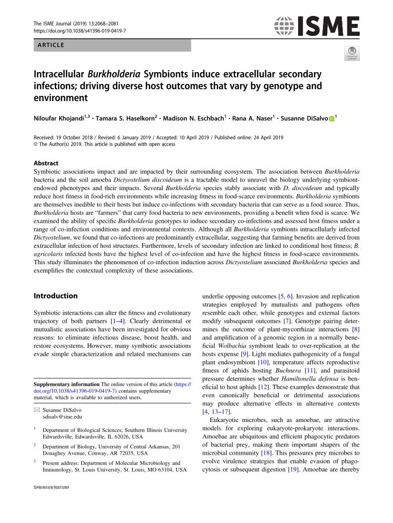
First, we examined host fitness when amoebae were co-cultured with Burkholderia and secondary bacteria. Wedetermined total spore productivity of host amoebae afterone social cycle on each labeled secondary bacterium,either alone or in a 50% mixture with K. pneumoniae.Five percent by volume of Burkholderia-RFP was inclu-ded to establish infections (Fig. 1). D. discoideum wasunable to develop on any conditions where P. aeruginosawas the only food source suggesting that this strain wastoxic and/or inedible for amoebae. All other conditionssupported fruiting body development, but spore pro-ductivity varied across conditions (Fig. 1). In line withprevious studies, Burkholderia species differentiallyimpact spore productivity on K. pneumoniae [34, 37].Typically, B. hayleyella was the most detrimental for hostfitness with B. agricolaris and B. bonniea being neutral ormoderately detrimental. However, these patterns and thedegree by which symbiont altered host fitness variedacross culture conditions (Fig. 1 and Table 1). Theseresults highlight the variability of fitness outcomes causedby distinct Burkholderia symbionts and suggest that sur-rounding bacterial communities also impact fitnessoutcomes.
Fig. 1 Culture Conditions Modify the Impact of Burkholderia Infec-tions on Host Fitness. Total spore productivity for D. discoideum afterone round of development on the indicated GFP labeled bacterialspecies (Kleb=K. pneumoniae, Rhiz= Rhizobium, Serr= Serratia,Agro= A. tumefaciens, and Pseu= P. aeruginosa. Fifty percentagecultures are mixed with 50% unlabeled K. pneumoniae by volume).
D. discoideum failed to develop on 100% Pseudomonas conditionsand is thereby not plotted. Fuschia, orange, and red boxes indicatecultures wherein Burkholderia infections are initiated by inclusion of5% B. agricolaris.70-RFP, B. hayleyella.11-RFP, and B. bonniea.859-RFP, respectively. Points within boxes indicate individual replicates.Letters indicate post hoc significance within panels
Intracellular Burkholderia Symbionts induce extracellular secondary infections; driving diverse host. . . 2071
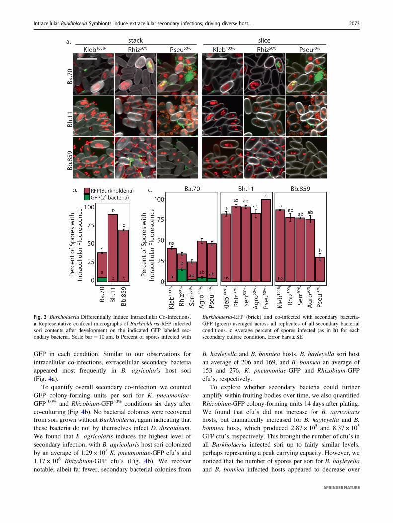
Intracellular co-infection is rare and depends onBurkholderia and secondary bacterial combinations
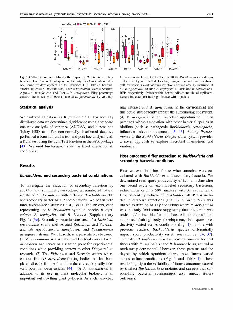
To investigate induction of secondary infection we imagedD. discoideum sori after development on Burkholderia andsecondary bacteria. We used 50/50 K. pneumoniae/second-ary bacteria-GFP conditions as they resulted in betteramoebae development than secondary bacteria-only condi-tions. We also imaged sori grown from K. pneumoniae-GFP. Importantly, we do not detect any secondary bacteriain sori in the absence of Burkholderia (Fig. 2). Thus, thesebacteria are not capable of infecting D. discoideum on theirown. In contrast, we can detect secondary-GFP cells in sorifrom amoebae co-exposed to Burkholderia (Fig. 3). Todetermine their prevalence in host spore populations, wequantified the percent of spores intracellularly infected withBurkholderia-RFP and with secondary bacteria-GFP. First,the percent of spores infected with each Burkholderia spe-cies significantly differs (χ2= 44.02, df= 2, p < 0.001). Inaggregate, B. hayleyella infects the most (89.2%), B. bon-niea infects an intermediate (68.6%), and B. agricolarisinfects the fewest (33%) percent of spores. However, weonly readily observe intracellular secondary co-infections inB. agricolaris host spores (average of 5.5% across condi-tions). We very rarely observe intracellular secondary
bacteria-GFP in B. hayleyella and B. bonniea hosts (0.01and 0.05%, respectively) (Fig. 3). We did not observeintracellular secondary bacteria-GFP in the absence ofintracellular Burkholderia-RFP, suggesting that secondarybacteria are only retained in Burkholderia co-infectedspores.
The identity of the secondary bacterium also plays a rolein the prevalence of intracellular infections for both Bur-kholderia and secondary bacteria. For instance, slightlyfewer spores are infected with B. bonniea when culturedwith P. aeruginosa-GFP (29%) than with all other bacteria(85.9–75.6%) (χ2= 10.843, df= 4, p= 0.028). In contrast,significantly more spores are infected with B. hayleyellawhen cultured with P. aeruginosa-GFP (98.9%) than withall other bacteria (81.2–91%) (χ2= 10.217, df= 4, p=0.036). For B. agricolaris hosts, the degree of secondary co-infections significantly varied by secondary bacteria (χ2=15.019, df= 4, p= 0.004). K. pneumoniae-GFP is localizedin only 0.2% of total spores while Rhizobium-GFP waslocalized in 14.9%. We observe similar co-infection pat-terns for each Burkholderia species when Rhizobium is usedas the sole food source (Supplementary Fig. 2). Whensecondary bacterial infections are considered as a percen-tage of spores co-infected with B. agricolaris, Rhizobium-GFP is co-localized in almost half of total infected spores.This suggests that should B. agricolaris infection levelsincrease in conditions that promote higher infection titers,secondary infections may correspondingly increase.
Burkholderia symbionts induce extracellularsecondary infections
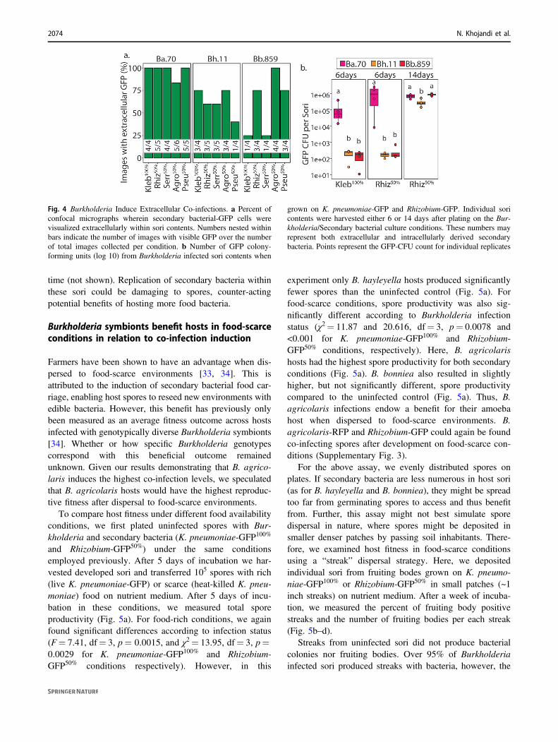
Although we found minimal intracellular co-infections inmost conditions, the farming phenotype may instead beexplained by extracellular secondary infections. To get aninitial indication of extracellular co-infections, we deter-mined the percent of confocal images in which any extra-cellular GFP could be visualized (Fig. 4a). We found that allBurkholderia symbionts induced at least some level ofextracellular co-infections, as we could visualize external
Fig. 2 Secondary bacteria do notinfect symbiont free spores.Representative confocalmicrographs of sori contentsafter growth on the indicatedGFP labeled bacterial cultures.Scale bar= 10 μm
Table 1 Burkholderia infections significantly alter spore productivityin most (but not all) bacterial culture conditions. Statistical analysis ofspore fitness from Fig. 1
Condition χ2 p Value
K. pneumoniae-GFP100% 23.537 >0.001
Rhizobium-GFP100% 20.81 >0.001
Rhizobium-GFP50% 18.759 >0.001
Serratia-GFP100% 6.411 0.09
Serratia-GFP50% 11.292 0.01
Agrobacterium-GFP100% 18.76 >0.001
Agrobacterium-GFP50% 16.267 >0.001
Pseudomonas-GFP50% 23.36 >0.001
All df values = 3
2072 N. Khojandi et al.
GFP in each condition. Similar to our observations forintracellular co-infections, extracellular secondary bacteriaappeared most frequently in B. agricolaris host sori(Fig. 4a).
To quantify overall secondary co-infection, we countedGFP colony-forming units per sori for K. pneumoniae-GFP100% and Rhizobium-GFP50% conditions six days afterco-culturing (Fig. 4b). No bacterial colonies were recoveredfrom sori grown without Burkholderia, again indicating thatthese bacteria do not by themselves infect D. discoideum.We found that B. agricolaris induces the highest level ofsecondary infection, with B. agricolaris host sori colonizedby an average of 1.29 × 10⁵ K. pneumoniae-GFP cfu’s and1.17 × 106 Rhizobium-GFP cfu’s (Fig. 4b). We recovernotable, albeit far fewer, secondary bacterial colonies from
B. hayleyella and B. bonniea hosts. B. hayleyella sori hostan average of 206 and 169, and B. bonniea an average of153 and 276, K. pneumoniae-GFP and Rhizobium-GFPcfu’s, respectively.
To explore whether secondary bacteria could furtheramplify within fruiting bodies over time, we also quantifiedRhizobium-GFP colony-forming units 14 days after plating.We found that cfu’s did not increase for B. agricolarishosts, but dramatically increased for B. hayleyella and B.bonniea hosts, which produced 2.87 × 10⁵ and 8.37 × 10⁵
GFP cfu’s, respectively. This brought the number of cfu’s inall Burkholderia infected sori up to fairly similar levels,perhaps representing a peak carrying capacity. However, wenoticed that the number of spores per sori for B. hayleyellaand B. bonniea infected hosts appeared to decrease over
Fig. 3 Burkholderia Differentially Induce Intracellular Co-Infections.a Representative confocal micrographs of Burkholderia-RFP infectedsori contents after development on the indicated GFP labeled sec-ondary bacteria. Scale bar= 10 μm. b Percent of spores infected with
Burkholderia-RFP (brick) and co-infected with secondary bacteria-GFP (green) averaged across all replicates of all secondary bacterialconditions. c Average percent of spores infected (as in b) for eachsecondary culture condition. Error bars ± SE
Intracellular Burkholderia Symbionts induce extracellular secondary infections; driving diverse host. . . 2073
time (not shown). Replication of secondary bacteria withinthese sori could be damaging to spores, counter-actingpotential benefits of hosting more food bacteria.
Burkholderia symbionts benefit hosts in food-scarceconditions in relation to co-infection induction
Farmers have been shown to have an advantage when dis-persed to food-scarce environments [33, 34]. This isattributed to the induction of secondary bacterial food car-riage, enabling host spores to reseed new environments withedible bacteria. However, this benefit has previously onlybeen measured as an average fitness outcome across hostsinfected with genotypically diverse Burkholderia symbionts[34]. Whether or how specific Burkholderia genotypescorrespond with this beneficial outcome remainedunknown. Given our results demonstrating that B. agrico-laris induces the highest co-infection levels, we speculatedthat B. agricolaris hosts would have the highest reproduc-tive fitness after dispersal to food-scarce environments.
To compare host fitness under different food availabilityconditions, we first plated uninfected spores with Bur-kholderia and secondary bacteria (K. pneumoniae-GFP100%
and Rhizobium-GFP50%) under the same conditionsemployed previously. After 5 days of incubation we har-vested developed sori and transferred 105 spores with rich(live K. pneumoniae-GFP) or scarce (heat-killed K. pneu-moniae) food on nutrient medium. After 5 days of incu-bation in these conditions, we measured total sporeproductivity (Fig. 5a). For food-rich conditions, we againfound significant differences according to infection status(F= 7.41, df= 3, p= 0.0015, and χ2= 13.95, df= 3, p=0.0029 for K. pneumoniae-GFP100% and Rhizobium-GFP50% conditions respectively). However, in this
experiment only B. hayleyella hosts produced significantlyfewer spores than the uninfected control (Fig. 5a). Forfood-scarce conditions, spore productivity was also sig-nificantly different according to Burkholderia infectionstatus (χ2= 11.87 and 20.616, df= 3, p= 0.0078 and<0.001 for K. pneumoniae-GFP100% and Rhizobium-GFP50% conditions, respectively). Here, B. agricolarishosts had the highest spore productivity for both secondaryconditions (Fig. 5a). B. bonniea also resulted in slightlyhigher, but not significantly different, spore productivitycompared to the uninfected control (Fig. 5a). Thus, B.agricolaris infections endow a benefit for their amoebahost when dispersed to food-scarce environments. B.agricolaris-RFP and Rhizobium-GFP could again be foundco-infecting spores after development on food-scarce con-ditions (Supplementary Fig. 3).
For the above assay, we evenly distributed spores onplates. If secondary bacteria are less numerous in host sori(as for B. hayleyella and B. bonniea), they might be spreadtoo far from germinating spores to access and thus benefitfrom. Further, this assay might not best simulate sporedispersal in nature, where spores might be deposited insmaller denser patches by passing soil inhabitants. There-fore, we examined host fitness in food-scarce conditionsusing a “streak” dispersal strategy. Here, we depositedindividual sori from fruiting bodes grown on K. pneumo-niae-GFP100% or Rhizobium-GFP50% in small patches (~1inch streaks) on nutrient medium. After a week of incuba-tion, we measured the percent of fruiting body positivestreaks and the number of fruiting bodies per each streak(Fig. 5b–d).
Streaks from uninfected sori did not produce bacterialcolonies nor fruiting bodies. Over 95% of Burkholderiainfected sori produced streaks with bacteria, however, the
Fig. 4 Burkholderia Induce Extracellular Co-infections. a Percent ofconfocal micrographs wherein secondary bacterial-GFP cells werevisualized extracellularly within sori contents. Numbers nested withinbars indicate the number of images with visible GFP over the numberof total images collected per condition. b Number of GFP colony-forming units (log 10) from Burkholderia infected sori contents when
grown on K. pneumoniae-GFP and Rhizobium-GFP. Individual soricontents were harvested either 6 or 14 days after plating on the Bur-kholderia/Secondary bacterial culture conditions. These numbers mayrepresent both extracellular and intracellularly derived secondarybacteria. Points represent the GFP-CFU count for individual replicates
2074 N. Khojandi et al.
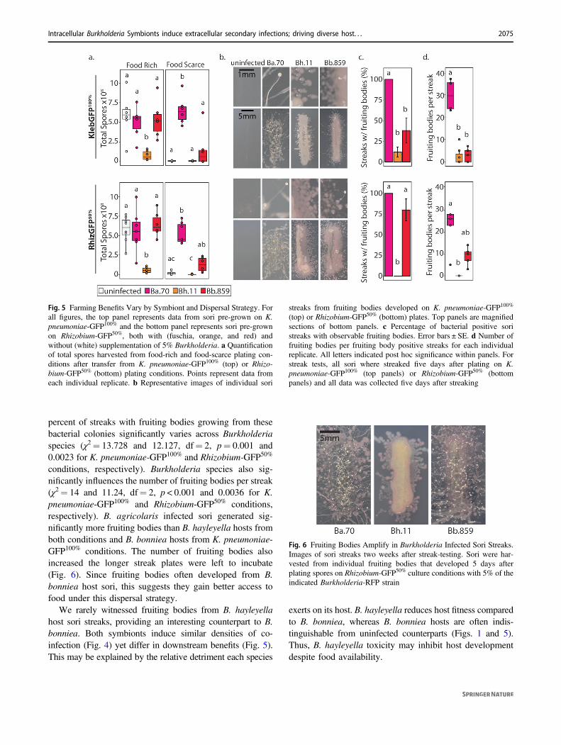
percent of streaks with fruiting bodies growing from thesebacterial colonies significantly varies across Burkholderiaspecies (χ2= 13.728 and 12.127, df= 2, p= 0.001 and0.0023 for K. pneumoniae-GFP100% and Rhizobium-GFP50%
conditions, respectively). Burkholderia species also sig-nificantly influences the number of fruiting bodies per streak(χ2= 14 and 11.24, df= 2, p < 0.001 and 0.0036 for K.pneumoniae-GFP100% and Rhizobium-GFP50% conditions,respectively). B. agricolaris infected sori generated sig-nificantly more fruiting bodies than B. hayleyella hosts fromboth conditions and B. bonniea hosts from K. pneumoniae-GFP100% conditions. The number of fruiting bodies alsoincreased the longer streak plates were left to incubate(Fig. 6). Since fruiting bodies often developed from B.bonniea host sori, this suggests they gain better access tofood under this dispersal strategy.
We rarely witnessed fruiting bodies from B. hayleyellahost sori streaks, providing an interesting counterpart to B.bonniea. Both symbionts induce similar densities of co-infection (Fig. 4) yet differ in downstream benefits (Fig. 5).This may be explained by the relative detriment each species
exerts on its host. B. hayleyella reduces host fitness comparedto B. bonniea, whereas B. bonniea hosts are often indis-tinguishable from uninfected counterparts (Figs. 1 and 5).Thus, B. hayleyella toxicity may inhibit host developmentdespite food availability.
Fig. 6 Fruiting Bodies Amplify in Burkholderia Infected Sori Streaks.Images of sori streaks two weeks after streak-testing. Sori were har-vested from individual fruiting bodies that developed 5 days afterplating spores on Rhizobium-GFP50% culture conditions with 5% of theindicated Burkholderia-RFP strain
Fig. 5 Farming Benefits Vary by Symbiont and Dispersal Strategy. Forall figures, the top panel represents data from sori pre-grown on K.pneumoniae-GFP100% and the bottom panel represents sori pre-grownon Rhizobium-GFP50%, both with (fuschia, orange, and red) andwithout (white) supplementation of 5% Burkholderia. a Quantificationof total spores harvested from food-rich and food-scarce plating con-ditions after transfer from K. pneumoniae-GFP100% (top) or Rhizo-bium-GFP50% (bottom) plating conditions. Points represent data fromeach individual replicate. b Representative images of individual sori
streaks from fruiting bodies developed on K. pneumoniae-GFP100%
(top) or Rhizobium-GFP50% (bottom) plates. Top panels are magnifiedsections of bottom panels. c Percentage of bacterial positive soristreaks with observable fruiting bodies. Error bars ± SE. d Number offruiting bodies per fruiting body positive streaks for each individualreplicate. All letters indicated post hoc significance within panels. Forstreak tests, all sori where streaked five days after plating on K.pneumoniae-GFP100% (top panels) or Rhizobium-GFP50% (bottompanels) and all data was collected five days after streaking
Intracellular Burkholderia Symbionts induce extracellular secondary infections; driving diverse host. . . 2075
Co-infections and conditional benefits areconsistent across Burkholderia species members
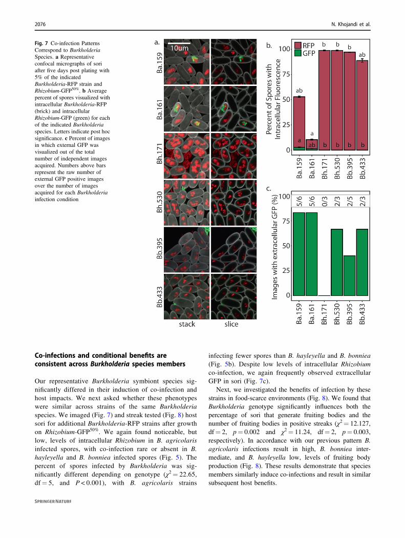
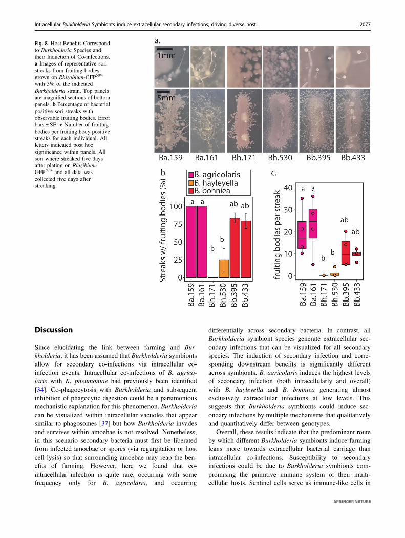
Our representative Burkholderia symbiont species sig-nificantly differed in their induction of co-infection andhost impacts. We next asked whether these phenotypeswere similar across strains of the same Burkholderiaspecies. We imaged (Fig. 7) and streak tested (Fig. 8) hostsori for additional Burkholderia-RFP strains after growthon Rhizobium-GFP50%. We again found noticeable, butlow, levels of intracellular Rhizobium in B. agricolarisinfected spores, with co-infection rare or absent in B.hayleyella and B. bonniea infected spores (Fig. 5). Thepercent of spores infected by Burkholderia was sig-nificantly different depending on genotype (χ2= 22.65,df= 5, and P < 0.001), with B. agricolaris strains
infecting fewer spores than B. hayleyella and B. bonniea(Fig. 5b). Despite low levels of intracellular Rhizobiumco-infection, we again frequently observed extracellularGFP in sori (Fig. 7c).
Next, we investigated the benefits of infection by thesestrains in food-scarce environments (Fig. 8). We found thatBurkholderia genotype significantly influences both thepercentage of sori that generate fruiting bodies and thenumber of fruiting bodies in positive streaks (χ2= 12.127,df= 2, p= 0.002 and χ2= 11.24, df= 2, p= 0.003,respectively). In accordance with our previous pattern B.agricolaris infections result in high, B. bonniea inter-mediate, and B. hayleyella low, levels of fruiting bodyproduction (Fig. 8). These results demonstrate that speciesmembers similarly induce co-infections and result in similarsubsequent host benefits.
Fig. 7 Co-infection PatternsCorrespond to BurkholderiaSpecies. a Representativeconfocal micrographs of soriafter five days post plating with5% of the indicatedBurkholderia-RFP strain andRhizobium-GFP50%. b Averagepercent of spores visualized withintracellular Burkholderia-RFP(brick) and intracellularRhizobium-GFP (green) for eachof the indicated Burkholderiaspecies. Letters indicate post hocsignificance. c Percent of imagesin which external GFP wasvisualized out of the totalnumber of independent imagesacquired. Numbers above barsrepresent the raw number ofexternal GFP positive imagesover the number of imagesacquired for each Burkholderiainfection condition
2076 N. Khojandi et al.
Discussion
Since elucidating the link between farming and Bur-kholderia, it has been assumed that Burkholderia symbiontsallow for secondary co-infections via intracellular co-infection events. Intracellular co-infections of B. agrico-laris with K. pneumoniae had previously been identified[34]. Co-phagocytosis with Burkholderia and subsequentinhibition of phagocytic digestion could be a parsimoniousmechanistic explanation for this phenomenon. Burkholderiacan be visualized within intracellular vacuoles that appearsimilar to phagosomes [37] but how Burkholderia invadesand survives within amoebae is not resolved. Nonetheless,in this scenario secondary bacteria must first be liberatedfrom infected amoebae or spores (via regurgitation or hostcell lysis) so that surrounding amoebae may reap the ben-efits of farming. However, here we found that co-intracellular infection is quite rare, occurring with somefrequency only for B. agricolaris, and occurring
differentially across secondary bacteria. In contrast, allBurkholderia symbiont species generate extracellular sec-ondary infections that can be visualized for all secondaryspecies. The induction of secondary infection and corre-sponding downstream benefits is significantly differentacross symbionts. B. agricolaris induces the highest levelsof secondary infection (both intracellularly and overall)with B. hayleyella and B. bonniea generating almostexclusively extracellular infections at low levels. Thissuggests that Burkholderia symbionts could induce sec-ondary infections by multiple mechanisms that qualitativelyand quantitatively differ between genotypes.
Overall, these results indicate that the predominant routeby which different Burkholderia symbionts induce farmingleans more towards extracellular bacterial carriage thanintracellular co-infections. Susceptibility to secondaryinfections could be due to Burkholderia symbionts com-promising the primitive immune system of their multi-cellular hosts. Sentinel cells serve as immune-like cells in
Fig. 8 Host Benefits Correspondto Burkholderia Species andtheir Induction of Co-infections.a Images of representative soristreaks from fruiting bodiesgrown on Rhizobium-GFP50%
with 5% of the indicatedBurkholderia strain. Top panelsare magnified sections of bottompanels. b Percentage of bacterialpositive sori streaks withobservable fruiting bodies. Errorbars ± SE. c Number of fruitingbodies per fruiting body positivestreaks for each individual. Allletters indicated post hocsignificance within panels. Allsori where streaked five daysafter plating on Rhizibium-GFP50% and all data wascollected five days afterstreaking
Intracellular Burkholderia Symbionts induce extracellular secondary infections; driving diverse host. . . 2077
multicellular slugs by trapping unwanted cargo throughphagocytosis and/or neutralization by DNA nets [31, 32].When sentinel cells have accumulated cargo, they drop outthe slug, thereby cleansing it of potential toxic entities [31].A gene deletion that reduces sentinel cells leads to retentionof secondary bacteria through the slug stage and into thesorus [31]. Burkholderia host slugs have fewer sentinel cellsthan uninfected counterparts and this defect goes awaywhen hosts are cured of their Burkholderia symbiont viaantibiotic treatment [47]. Thus, the induction of secondaryinfections could be an indirect consequence of Burkholderiasymbiosis resulting in sentinel cell reduction. This scenariomay be comparable to the phenomenon of secondaryinfections in mammalian systems whereby primary infec-tious agents compromise the immune system of their hosts.Despite this, we cannot rule out the possibility that extra-cellular secondary infection originates from intracellular co-infection. It is possible that co-infected cells are more sus-ceptible to lysis, rupturing and spewing their secondarybacterial passengers into the extracellular matrix. In eithersituation, secondary bacteria might then amplify within sori.
Intracellular co-infections are most frequent with B.agricolaris and the soil Rhizobium strain. This might reflectan ecologically relevant association between these species innature. Burkholderia and Rhizobium are both ubiquitous insoil and contain several important symbiont species whichhave been found co-colonizing the same hosts [48–51].Predation by amoebae in soil and aquatic systems shapesmicrobial community assembly and overall food webs [52].Given the likely co-occurrence of soil amoebae with Bur-kholderia and other soil microbes it’s tempting to speculateon how these multipartite interactions influence overallmicrobial communities and higher trophic levels. Here, weshow that amoebae co-disperse Burkholderia symbionts andsecondary bacterial hitchhikers to new environments. Thus,the impact of amoebae on their surrounding microbial net-work can go well beyond predator-prey dynamics. Finally,our observation of Burkholderia and P. aeruginosa co-infection amplifies the concern that soil amoebae can serveas reservoirs for bacterial pathogens. These results suggestthat Burkholderia symbionts can increase the suite ofpotential pathogenic partners hosted by amoebae.
Burkholderia–fungal associations have been wellrecognized for their importance in the soil ecosystem andfor their bio-restoration potential [53–55]. There are com-pelling parallels between Burkholderia–Dictyostelium andBurkholderia–fungal associations. Some Burkholderia(notably B. terrae) are capable of adhering to and migratingwith growing fungal hyphae through soil [56]. Similar toour system, some of these fungal associates assist in the co-migration of other (non-migrating) bacteria [57]. Severalmechanisms have been proposed to underlie these interac-tions, such as direct receptor binding and indirect biofilm
co-aggregation [58]. B. terrae extracellularly colonizesfungal hyphae but many other Burkholderia symbionts ofdiverse hosts persist intracellularly [13, 59]. An interestingexample is the Rhizopus microsporus endosymbiont B.rhizoxinica fungi, which produces the rice seedling blighttoxin [60]. Recently, secretion systems have been shown tobe important for the active invasion of B. rhizoxinica acrossthe fungal cell wall and into the host cytoplasm [59].Secretion systems have also been implicated in B. psuedo-mallei infections [61, 62]. However many plant mutualisticBurkholderia species, which are closer relatives to Bur-kholderia symbionts of Dictyostelium, appear to lack someof these systems [61–63] The hypothesized portal of entryinto Dictyostelium is via phagocytosis, which could cir-cumvent the need for invasion specific mechanisms. Over-all, Burkholderia symbionts of other hosts can help informour understanding of the Burkholderia–amoebae symbiosisand vice versa.
Biofilm formation is intriguing to consider as a mechan-istic explanation of secondary infection. Burkholderiaadherence to secondary bacteria would increase the like-lihood of co-phagocytosis or extracellular co-colonization.Different adhesive capacities of Burkholderia and secondaryspecies could explain differences in the extent of secondaryinfections across bacterial combinations. Interestingly,recent work implicates Dictyostelium lectins in the farmingphenomenon, higher lectin expression was detected infarmer D. discoideum clones and addition of endogenouslectins induced bacterial carriage [64]. Although this workdid not consider the presence or impact of Burkholderia, wethink Burkholderia symbionts play a key role. Burkholderiacould induce farming via induction of lectin expression inamoebae or more simplistically, Burkholderia lectins maymediate co-adherence of secondary bacteria. Indeed, lectinexpression by B. cenocepacia is an important component ofbiofilm formation and lectin aids in adherence of B. cepaciato host tissues [45, 65]. Future exploration into lectinexpression and adhesion mechanisms will be helpful forclarifying these themes.
In addition to elucidating the phenomenon of second-ary infections our results exemplify the context depen-dency of symbiotic outcomes in this system. We foundthat the costs and benefits of this symbiosis can be mod-ified by different bacterial conditions and spore dispersalprocesses. The nature and extent of farming induction byBurkholderia symbionts differs across symbiont speciesand so do their corresponding contextual fitness out-comes. Previously, all Burkholderia symbionts werethought to benefit their hosts under food-scarce condi-tions. Here, B. hayleyella strains, though similar in sec-ondary infection patterns to B. bonniea, are moredetrimental in food-rich, and not beneficial in food-scarce,conditions. Perhaps B. hayleyella strains encode virulence
2078 N. Khojandi et al.
genes that B. bonniea strains lack. We also find thatBurkholderia symbionts display different population wideinfectivity patterns that appear roughly similar acrossstrains of the same species. How symbiont density isregulated and how it influences the parasitism to mutu-alism continuum are compelling questions in infectiousbiology [66, 67]. The variation of infection metrics andoutcomes in this system, and the ease in which they can beexplored, makes it well poised for investigating thesequestions. Ultimately, further research into the mechan-isms, consequences, and ecological framework of theBurkholderia–Dictyostelium symbiosis will help illumi-nate microbial interaction dynamics relevant to infectionbiology and microbial ecology.
Data availability
All raw data supporting the conclusions of this manuscriptis available at https://doi.org/10.6084/m9.figshare.7547834.
Acknowledgements We thank Kyle Skottke for fruitful discussionsand manuscript review, Joan Strassmann and David Queller for initialguidance in the system, and all members of the DiSalvo lab at SIUE,particularly Jacob W. Miller for general laboratory support.
Funding This study was supported by SIUE start-up funds from theDiSalvo lab.
Author contributions NK and SD designed the study. NK and SDperformed experiments with assistance from MNE, RAN and TSH. SDand TSH wrote the manuscript.
Compliance with ethical standards
Conflict of interest The authors declare that they have no conflict ofinterest.
Publisher’s note: Springer Nature remains neutral with regard tojurisdictional claims in published maps and institutional affiliations.
Open Access This article is licensed under a Creative CommonsAttribution 4.0 International License, which permits use, sharing,adaptation, distribution and reproduction in any medium or format, aslong as you give appropriate credit to the original author(s) and thesource, provide a link to the Creative Commons license, and indicate ifchanges were made. The images or other third party material in thisarticle are included in the article’s Creative Commons license, unlessindicated otherwise in a credit line to the material. If material is notincluded in the article’s Creative Commons license and your intendeduse is not permitted by statutory regulation or exceeds the permitteduse, you will need to obtain permission directly from the copyrightholder. To view a copy of this license, visit http://creativecommons.org/licenses/by/4.0/.
References
1. Kiers ET, West SA. Evolving new organisms via symbiosis.Science. 2015;348:392–4.
2. Margulis L, Fester R. Bellagio conference and book. Symbiosis assource of evolutionary innovation: speciation and morphogenesis.Conference—June 25–30, 1989, Bellagio Conference Center,Italy. Symbiosis. 1991;11:93–101.
3. Douglas AE. Symbiosis as a general principle in eukaryoticevolution. Cold Spring Harb Perspect Biol. 2014;6:a016113.
4. Douglas AE. Symbiotic interactions. Oxford: Oxford UniversityPress, Oxford science publications Press; 2002. p. 148.
5. Ochman H, Moran NA. Genes lost and genes found: evolution ofbacterial pathogenesis and symbiosis. Science. 2001;292:1096–9.
6. Dale C, Moran NA. Molecular interactions between bacterialsymbionts and their hosts. Cell. 2006;126:453–65.
7. Wang D, Yang S, Tang F, Zhu H. Symbiosis specificity in thelegume: rhizobial mutualism. Cell Microbio. 2012;14:334–42.
8. Klironomos JN. Variation in plant response to native and exoticarbuscular mycorrhizal fungi. Ecology. 2003;84:2292–301.
9. Chrostek E, Teixeira L. Mutualism breakdown by amplification ofwolbachia genes. PLOS Biol. 2015;13:e1002065.
10. Álvarez-Loayza P Jr, Torres JFW, Balslev MS, Kristiansen H,Svenning J-C T, et al. Light converts endosymbiotic fungus topathogen, influencing seedling survival and niche-space filling ofa common tropical tree, iriartea deltoidea. PLOS ONE. 2011;6:e16386.
11. Dunbar HE, Wilson ACC, Ferguson NR, Moran NA. Aphidthermal tolerance is governed by a point mutation in bacterialsymbionts. PLOS Biol. 2007;5:e96.
12. Oliver KM, Campos J, Moran NA, Hunter MS. Populationdynamics of defensive symbionts in aphids. Proc R Soc B BiolSci. 2008;275:293–9.
13. Bonfante P, Anca I-A. Plants, mycorrhizal fungi, and bacteria: anetwork of interactions. Annu Rev Microbiol. 2009;63:363–83.
14. Bronstein JL. Conditional outcomes in mutualistic interactions.Trends Ecol Evol. 1994;9:214–7.
15. Hussa EA, Goodrich-Blair H. It takes a village: ecological andfitness impacts of multipartite mutualism. Annu Rev Microbiol.2013;67:161–78.
16. Newton AC, Fitt BDL, Atkins SD, Walters DR, Daniell TJPa-thogenesis. parasitism and mutualism in the trophic space ofmicrobe-plant interactions. Trends Microbiol. 2010;18:365–73.
17. Pérez-Brocal V, Latorre A, Moya A. Symbionts and pathogens:what is the difference? In: Dobrindt U, Hacker JH, Svanborg C,editors. Between pathogenicity and commensalism [Internet].Berlin, Heidelberg: Springer Berlin Heidelberg; 2013. p. 215–43.https://doi.org/10.1007/82_2011_190
18. Rønn R, McCaig AE, Griffiths BS, Prosser JI. Impact of proto-zoan grazing on bacterial community structure in soil microcosms.Appl Environ Microbiol. 2002;68:6094.
19. Paquet VE, Charette SJ. Amoeba-resisting bacteria found inmultilamellar bodies secreted by Dictyostelium discoideum: socialamoebae can also package bacteria. FEMS Microbiol Ecol.2016;92:fiw025.
20. Molmeret M, Horn M, Wagner M, Santic M, Abu Kwaik Y.Amoebae as training grounds for intracellular bacterial pathogens.Appl Environ Microbiol. 2005;71:20–8.
21. Taylor-Mulneix DL, Bendor L, Linz B, Rivera I, Ryman VE,Dewan KK, et al. Bordetella bronchiseptica exploits the complexlife cycle of Dictyostelium discoideum as an amplifying trans-mission vector. PLOS Biol. 2017;15:e2000420.
22. Greub G, Raoult D. Microorganisms resistant to free-livingamoebae. Clin Microbiol Rev. 2004;17:413–33.
23. Horn M, Wagner M. Bacterial endosymbionts of free-livingamoebae. J Eukaryot Microbiol. 2004;51:509–14.
24. Rubeniņa I, Kirjušina M, Bērziņš A, Valciņa O, Jahundoviča I.Relationships between free-living amoeba and their intracellularbacteria. Proc Latv Acad Sci Sect B Nat Exact Appl Sci. 2017;71:259–65.
Intracellular Burkholderia Symbionts induce extracellular secondary infections; driving diverse host. . . 2079
25. Bozzaro S, Eichinger L. The professional phagocyte Dictyoste-lium discoideum as a model host for bacterial pathogens. CurrDrug Targets. 2011;12:942–54.
26. Bozzaro S, Bucci C, Steinert M. Phagocytosis and host-pathogeninteractions in Dictyostelium with a look at macrophages. Int RevCell Mol Biol. 2008;271:253–300.
27. Cosson P, Soldati T. Eat, kill or die: when amoeba meets bacteria.Curr Opin Microbiol. 2008;11:271–6.
28. Nasser W, Santhanam B, Miranda ER, Parikh A, Juneja K, Rot G,et al. Bacterial discrimination by dictyostelid amoebae reveals thecomplexity of ancient interspecies interactions. Curr Biol.2013;23:862–72.
29. Skriwan C, Fajardo M, Hägele S, Horn M, Wagner M, Michel R,et al. Various bacterial pathogens and symbionts infect theamoeba Dictyostelium discoideum. Int J Med Microbiol.2002;291:615–24.
30. Smith jeff, Queller DC, Strassmann JE. Fruiting bodies of thesocial amoeba Dictyostelium discoideum increase spore transportby Drosophila. BMC Evol Biol. 2014;14:105.
31. Chen G, Zhuchenko O, Kuspa A. Immune-like phagocyte activityin the social amoeba. Science. 2007;317:678–81.
32. Zhang X, Zhuchenko O, Kuspa A, Soldati T. Social amoebae trapand kill bacteria by casting DNA nets. Nat Commun. 2016;7:10938.
33. Brock DA, Douglas TE, Queller DC, Strassmann JE. Primitiveagriculture in a social amoeba. Nature. 2011;469:393–6.
34. DiSalvo S, Haselkorn TS, Bashir U, Jimenez D, Brock DA,Queller DC, et al. Burkholderia bacteria infectiously induce theproto-farming symbiosis of Dictyostelium amoebae and foodbacteria. Proc Natl Acad Sci USA. 2015;112:E5029–37.
35. Suárez-Moreno ZR, Caballero-Mellado J, Coutinho BG,Mendonça-Previato L, James EK, Venturi V. Common features ofenvironmental and potentially beneficial plant-associated Bur-kholderia. Microb Ecol. 2012;63:249–66.
36. Brock DA, Hubert AM, Noh S, DiSalvo S, Geist KS, Haselkorn TS,et al. Endosymbiotic adaptations in three new bacterial speciesassociated with Dictyostelium discoideum: Burkholderia agricolarissp. nov., Burkholderia hayleyella sp. nov., and Burkholderia bon-niea sp. nov. 2018. http://biorxiv.org/lookup/doi/10.1101/304352.
37. Shu L, Brock DA, Geist KS, Miller JW, Queller DC, StrassmannJE, et al. Symbiont location, host fitness, and possible coadapta-tion in a symbiosis between social amoebae and bacteria. eLife.2018. https://elifesciences.org/articles/42660.
38. Haselkorn TS, DiSalvo S, Miller JW, Bashir U, Brock DA,Queller DC, et al. The specificity of Burkholderia symbionts in thesocial amoeba farming symbiosis: prevalence, species, genetic andphenotypic diversity. Mol Ecol. 2018;0(ja). https://doi.org/10.1111/mec.14982.
39. Inglis RF, Biernaskie JM, Gardner A, Kümmerli R. Presence of aloner strain maintains cooperation and diversity in well-mixedbacterial communities. Proc R Soc B Biol Sci. 2016;283:20152682.
40. Norris MH, Kang Y, Lu D, Wilcox BA, Hoang TT. Glyphosateresistance as a novel select-agent-compliant, non-antibiotic-selectable marker in chromosomal mutagenesis of the essentialgenes asd and dapb of Burkholderia pseudomallei. Appl EnvironMicrobiol. 2009;75:6062–75.
41. Su S, Bangar H, Saldanha R, Pemberton A, Aronow B, Dean GE,et al. Construction and characterization of stable, constitutivelyexpressed, chromosomal green and red fluorescent transcriptionalfusions in the select agents, Bacillus anthracis, Yersinia pestis,Burkholderia mallei, and Burkholderia pseudomallei. Micro-biologyOpen. 2014;3:610–29.
42. Kikuchi Y, Fukatsu T. Live imaging of symbiosis: spatiotemporalinfection dynamics of a GFP-labelled Burkholderia symbiont inthe bean bug Riptortus pedestris. Mol Ecol. 2014;23:1445–56.
43. Ogle DH, Wheeler P, Dinno A. Fsa: fisheries stock analysis[Internet]. 2018. https://github.com/droglenc/FSA.
44. Brock DA, Haselkorn TS, Garcia JR, Bashir U, Douglas TE,Galloway J, et al. Diversity of free-living environmental bacteriaand their interactions with a bactivorous amoeba. Front Cell InfectMicrobiol. 2018;8:411–411.
45. Fazli M, Almblad H, Rybtke ML, Givskov M, Eberl L, Tolker-Nielsen T. Regulation of biofilm formation in Pseudomonas andBurkholderia species: regulation of biofilm formation. EnvironMicrobiol. 2014;16:1961–81.
46. Tomlin KL, Coll OP, Ceri H. Interspecies biofilms of Pseudo-monas aeruginosa and Burkholderia cepacia. Can J Microbiol.2001;47:949–54.
47. Brock DA, Callison WÉ, Strassmann JE, Queller DC. Sentinelcells, symbiotic bacteria and toxin resistance in the socialamoeba Dictyostelium discoideum. Proc Biol Sci. 2016;283:2015–2727.
48. Compant S, Nowak J, Coenye T, Clément C, Ait Barka E.Diversity and occurrence of Burkholderia spp. in the naturalenvironment. FEMS Microbiol Rev. 2008;32:607–26.
49. Estrada-De Los Santos P, Bustillos-Cristales R, Caballero-Mellado J. Burkholderia, a genus rich in plant-associated nitro-gen fixers with wide environmental and geographic distribution.Appl Environ Microbiol. 2001;67:2790–8.
50. Gage DJ. Infection and invasion of roots by symbiotic, nitrogen-fixing rhizobia during nodulation of temperate legumes. MicrobiolMol Biol Rev. 2004;68:280–300.
51. Nguyen NH, Bruns TD. The microbiome of Pinus muricataectomycorrhizae: community assemblages, fungal species effects,and Burkholderia as important bacteria in multipartnered sym-bioses. Microb Ecol. 2015;69:914–21.
52. Edgcomb V. Marine protist associations and environmentalimpacts across trophic levels in the twilight zone and below. CurrOpin Microbiol. 2016;31:169–75.
53. Banitz T, Wick LY, Fetzer I, Frank K, Harms H, Johst K. Dis-persal networks for enhancing bacterial degradation in hetero-geneous environments. Environ Pollut. 2011;159:2781–8.
54. Ellegaard-Jensen L, Knudsen BE, Johansen A, Albers CN,Aamand J, Rosendahl S. Fungal–bacterial consortia increasediuron degradation in water-unsaturated systems. Sci TotalEnviron. 2014;466–467:699–705.
55. Rashid MI, Mujawar LH, Shahzad T, Almeelbi T, Ismail IMI,Oves M. Bacteria and fungi can contribute to nutrients bioavail-ability and aggregate formation in degraded soils. Microbiol Res.2016;183:26–41.
56. Nazir R, Zhang M, de Boer W, van Elsas JD. The capacity tocomigrate with Lyophyllum sp. strain Karsten through differentsoils is spread among several phylogenetic groups within thegenus Burkholderia. Soil Biol Biochem. 2012;50:221–33.
57. Warmink JA, Nazir R, Corten B, van Elsas JD. Hitchhikers on thefungal highway: The helper effect for bacterial migration viafungal hyphae. Soil Biol Biochem. 2011;43:760–5.
58. Vila T, Nazir R, Rozental S, dos Santos GMP, Calixto ROR,Barreto-Bergter E, et al. The role of hydrophobicity and surfacereceptors at hyphae of Lyophyllum sp. strain karsten in theinteraction with Burkholderia terrae BS001—implications forinteractions in soil. Front Microbiol. 2016. https://doi.org/10.3389/fmicb.2016.01689/full.
59. Moebius N, Üzüm Z, Dijksterhuis J, Lackner G, Hertweck C.Active invasion of bacteria into living fungal cells. eLife. 2014.https://elifesciences.org/articles/03007.
60. Partida-Martinez LP, Monajembashi S, Greulich K-O, HertweckC. Endosymbiont-dependent host reproduction maintainsbacterial-fungal mutualism. Curr Biol. 2007;17:773–7.
61. Gong L, Lai S-C, Treerat P, Prescott M, Adler B, Boyce JD, et al.Burkholderia pseudomallei type III secretion system cluster 3atpase bsas, a chemotherapeutic target for small-molecule atpaseinhibitors. Infect Immun. 2015;83:1276–85.
2080 N. Khojandi et al.
62. Stone JK, DeShazer D, Brett PJ, Burtnick MN. Melioidosis:molecular aspects of pathogenesis. Expert Rev Anti Infect Ther.2014;12:1487–99.
63. Angus AA, Agapakis CM, Fong S, Yerrapragada S, Estrada-de losSantos P, Yang P, et al. Plant-associated symbiotic burkholderiaspecies lack hallmark strategies required in mammalian patho-genesis. PLoS ONE. 2014;9:e83779.
64. Dinh C, Farinholt T, Hirose S, Zhuchenko O, Kuspa A. Lectinsmodulate the microbiota of social amoebae. Science. 2018;361:402–6.
65. Šulák O, Cioci G, Lameignère E, Balloy V, Round A, Gutsche I,et al. Burkholderia cenocepacia bc2l-c is a super lectin with dualspecificity and proinflammatory activity. PLoS Pathog. 2011;7:e1002238.
66. Schneider DS, Ayres JS. Two ways to survive infection: whatresistance and tolerance can teach us about treating infectiousdiseases. Nat Rev Immunol. 2008;8:889–95.
67. Cunnington AJ. The importance of pathogen load. PLoS Pathog.2015;11:e1004563.
Intracellular Burkholderia Symbionts induce extracellular secondary infections; driving diverse host. . . 2081


