Embed Size (px)
Citation preview
Journal of Medicine and Life Volume 4, Issue 3, July‐September 2011, pp.234‐243
© 2011, Carol Davila University Foundation
Traumatic brain injury in infants and toddlers, 0-3 years old
Ciurea AV, Gorgan MR, Tascu A, Sandu AM, Rizea RE
“Bagdasar-Arseni” Clinical Emergency Hospital, Department of Neurosurgery, Bucharest, Romania
Correspondence to: A. V. Ciurea, MD PhD, First Department of Neurosurgery
“Bagdasar-Arseni” Clinical Emergency Hospital 10-12, Berceni Street, District 4, 041915, Bucharest, Romania
Phone: 0040.21.3343025, E-mail: [email protected]
Received: December 14th, 2010 – Accepted: June 20th, 2011
Abstract Object: Children 0-3 years old present a completely different neurotraumatic pathology. The growing and the development
processes in this age group imply specific anatomical and pathophysiological features of the skull, subarachnoid space, CSF flow, and brain.
Most common specific neurotraumatic entities in children 0-3 years old are cephalhematoma, subaponeurotic (subgaleal) hematoma, diastatic skull fracture, grow skull fracture, depressed (“ping-pong”) skull fracture, and extradural hematoma.
Methods: We present our 10 years experience in neuropediatric traumatic brain injuries, between 1999 and 2009, in the First Department of Neurosurgery and Pediatric Intensive Care Unit. Including criteria were children, 0-3 years old, presenting only traumatic brain injury. We excluded patients with politrauma, who require a different management.
Results: We present the incidence of these specific head injuries, clinical and imagistic features, treatment, and outcome. We found 72 children with diastatic skull fracture, 61 cases with depressed (“ping-pong”) skull fracture, 22 cases with grow skull fracture, 11 children harboring intrusive skull fracture, 58 cephalhematomas, 26 extradural hematomas, and 7 children with severe brain injury and major posttraumatic diffuse ischemia (“black-brain”). Usually, infants and toddlers present with seizures, pallor, and rapid loss of consciousness. First choice examination, in all children was cerebral CT-scan, and for follow-up, we performed cerebral MRI. We emphasize on the importance of seizure prevention in this age group. Children presenting with extensive diffuse ischemia (“black-brain”) had a poor outcome, death occurring in all 7 cases.
Conclusion: Children 0-3 years old, present with a total distinctive pathology than adults. Children with head injury must be addressed to a pediatric department of neurosurgery and pediatric intensive care unit. Prophylaxis pays the most important role in improving the outcome.
K e y w o r d s : c e p h a l h e m a t o m a , c h i l d r e n , d e p r e s s e d ( “ p i n g - p o n g ” ) s k u l l f r a c t u r e , d i a s t a t i c s k u l l f r a c t u r e , e x t r a d u r a l h e m a t o m a
Abbreviations: CPH cephalhematoma; DBS diffuse brain swelling; EDH extradural hematoma; GCS Glasgow Coma Scale; PHI penetrating head injury; TBI traumatic brain injury
Introduction
Traumatic brain injury (TBI) is the leading cause of death and disability in children. Statistical analyses shows that almost half of patients with a TBI each year in the United Kingdom are children under 16 years, and approximately one third of the patients with cranial trauma per year in the United States are children aged between 0 and 14 years old [1,2].
The most common causes of TBI in children: falls, child abuse, motor vehicle accidents sport accidents, assaults, and instrumental delivery. Regarding age distribution of TBI, there are two risk groups: the first group aged between 0 and 4 years old, and the second 15-19 years old. Boys seem to be affected twice the rate of girls [1].
Traumatic pathology during the first 3 years of life is completely different when compared with adults. Raimondi emphasized the differentials between children and adults’ pathology: “children are not young adults” [3].
Specific pediatric scales, adapted according to age, must be used to correctly grade the severity of TBI in children. The most common neurotrauma pediatric scales are: Pediatric Coma Scale/Children Coma Scale (PCS)[4], Children’s Coma Score (CCS)[5], Trauma Infant Neurological Score (TINS)[6], and Glasgow Coma Scale (GCS) [7]. The outcome is graded by using neurotrauma pediatric outcome scales, such as: KOSCHI (King’s Outcome Scale for Childhood Head Injury) score [8],
Journal of Medicine and Life Volume 4, Issue 3, July‐September 2011
235 © 2011, Carol Davila University Foundation
Glasgow Outcome Scale (GOS)[9], and modified Rankin score [10].
Material and methods
We analyzed in a retrospective manner all the consecutive cases with TBI, aged between 0 and 3 years old, admitted into the Department of Pediatric Neurosurgery from “Bagdasar-Arseni” Clinical Hospital, in Bucharest, between 1st of January 1999 and 31st of December 2008 (10 years).
Inclusion criteria were age 0-3 years, TBI, no history of previous head injury, no multiple trauma and no birth trauma. Infants with birth trauma were excluded.
Results
312 consecutive cases of children 0-3 years old were admitted. Most children presented with minor head injuries, 283 cases (90.70%). Etiology
The most common causes of TBI in children 0-3 years old were falls, in 173 cases (55.45%), falls from the same level 102 cases (32.70%), and falls from other level 71 cases (22.75%). Motor vehicle accidents were the second cause of TBI in infants and toddlers, in 74 cases (23.72%), pedestrians 56 cases (17.95%), and passengers 18 cases (5.77%). Other cases were accidental struck of the head in 39 cases (12.50%), assaults/child abuse in 26 cases (8.33%). There were no bicycle or sport related TBI. Table 1. Causes of TBI in children.
Causes No. cases
% cases
Falls Falls from the same level Falls from other level
173 102 71
55.45% 32.70% 22.75%
Motor vehicle accidents Pedestrians Passengers
74 56 18
23.72% 17.95% 5.77%
Accidental struck of the head 39 12.50% Assaults/child abuse 26 8.33%
TOTAL 312 100%
Traumatic brain injuries There were 411 posttraumatic lesions in 312
children. Table 2. Posttraumatic lesions in the study group.
Injury No. patients
% patients
Cephalhematoma 58 14.11% Linear skull fracture 124 30.17% Diastatic skull fracture 72 17.52% Depressed skull fracture (ping-pong) 61 14.84% Depressed skull fracture (cominutive)
19 4.62%
Grow skull fracture 22 5.35% Penetrating head injury 11 2.68% Extradural hematoma 26 6.33% Subdural hematoma - - Diffuse brain swelling 18 4.38%
TOTAL 411 100%
Cephalhematoma (CPH)
In our study group, we had 58 children (18.59%) with CPH.
CPH, which does not spontaneously withdraw under conservatory treatment, requires surgical treatment. In persistent fluid, the hemorrhagic collection is evacuated by tapping (punctioning with a thick needle). In calcified CHP, surgery is performed for cosmetic reasons. A wide, arcuate skin incision in “horse shoe”, into the parietal area, on the border of CPH is performed. After a large scalp dissection, the calcified CHP is exposed. We perform a systematic resection of the calcified collection, with continuous wax hemostasis. For good cosmetic effects, level-adjustment of the parietal region is mandatory. No external drainage is needed. Skull fractures
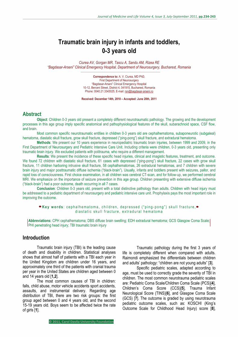
Linear skull fractures were found in 124 children (39.74%). All children presenting in the emergency room with head trauma, and linear skull fracture on CT-scan or Fig. 1. Causes of TBI in children.
Fig. 2. Posttraumatic lesions in the study group.
236
on skull XNeurosurgicadeveloping elinear skull intracranial p
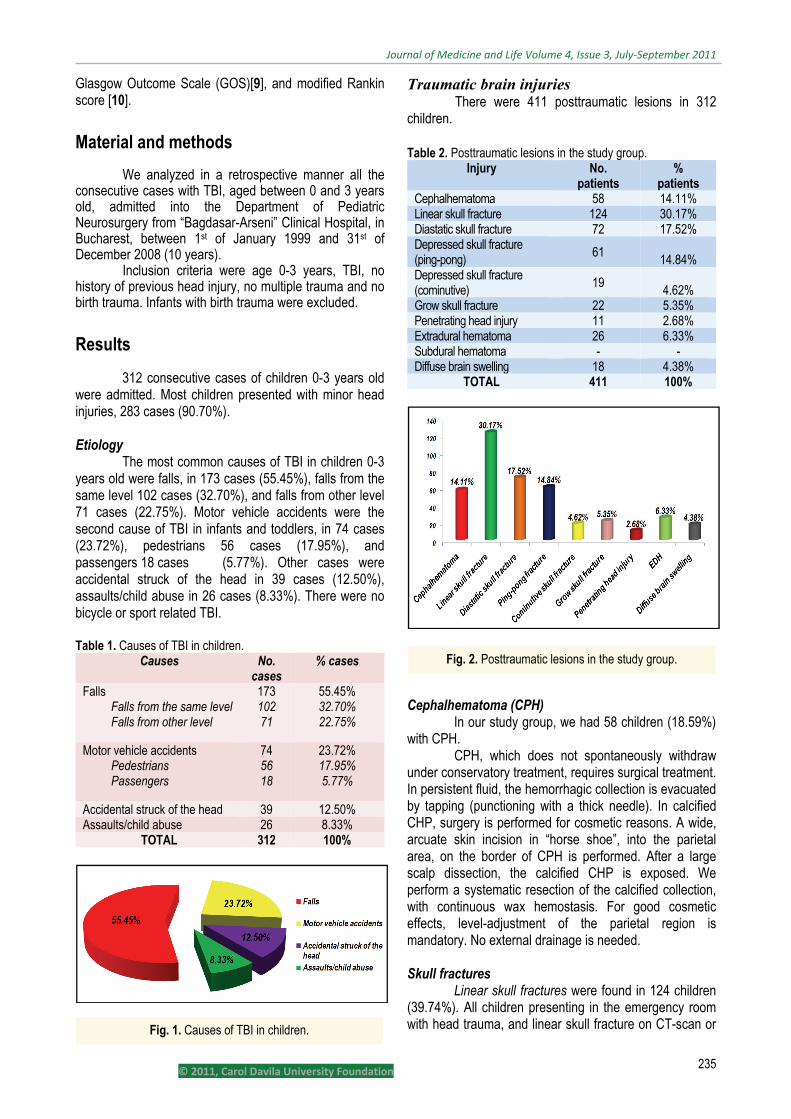
Dias(23.08%). Indiastatic skuinto a growdiastatic skobservation f
Depfound in 61 cfractures, a lthe depresseof the incisioelevator, int
Fi
© 2011
X-ray, must al Departmeextradural hem
fractures wpathology did n
static skull frac children agell fractures ca
wing skull frackull fracture for this reasonpressed skullchildren (19.55linear skin inced skull fractuon, were matroduced thro
ig. 3. Linear rig
Fig. 4. Diast
1, Carol Davila U
be admittednt because matomas. Ch
without any not require su
ctures were foed between 0arry a high riscture (GSF). were kepn. l fractures 5%). In patientcision of 4 cmre and a burr
ade. By meanough the bu
ht parietal skull
tatic skull fractu
Fig. 5. Disju
University Foun
d in a Pedof the risk
ildren with siother assocrgery.
ound in 72 chi and 3 yearssk of transfor All children t under ca
(ping-pong) ts with “ping-p
m long, posteri hole in the mns of a periorr hole, into
l fracture.
ure. Leptomenin
unction of the c
Journal o
ndation
diatric k of imple
ciated
ldren s old, rming with areful
were pong” ior to
middle osteal o the
epidwas
founcaredurafragmbonerecoold.
with childsinupatedeprrectaperfofragmTachstripthe sduracloscan manshoc
(7.05
Cas
histoThe frontdefediashern
ngeal herniation
coronal suture, r
of Medicine and
ural space be elevated.
Depressend in 19 chiefully elevatedal lacerations.ments were fie fragments
ommend crani
Angiogra coronal and
dren with deprs (SSS), in o
ency of the sressed fractangular free ormed, the boments are rehocomb to co
p is packed ovsinus. Dural laaplasty is donure. Bone flapalso be perfo
ndatory, becauck is high in sm
Grow sk5%).
se report
We repoory of minor h child presental cystic, non
ect. CT-scan atatic skull fra
niation protrud
n through diasta
reaching up to t
d Life Volume 4,
eneath the fra
ed skull fracildren (6.09%
d, and we perf. Brain laceraxed in place w could not oplasty in chil
aphy with vensagittal recon
ressed fracturorder to evalsinus beneatures acrossbone flap, ceone flap is camoved. The
over the rent wver, and a peracerations wee, in order to p was fixed wiormed. Rapid use the risk omall children.
kull fractures w
ort a case of head trauma, 2nted with a ntender massand 3D reconsacture, leptoming through th
atic fracture. Br
the anterior fon
, Issue 3, July‐Se
acture, the dep
ctures (comi%). Bone fragformed durapation was reswith wires. In be replaceldren younger
nous phase nstruction werre over the suuate the size
th the impacs the SSS,entered over arefully elevatSSS is repaiwithin the sinriosteal graft ire sewed in n perform a waith wires. Exte repair of lesiof developing
were found in
a 5 weeks o2 weeks befoprogressive , underlying p
struction CT-scmeningeal cyshe bone defec
ain contusion.
ntanelle.
eptember 2011
pressed bone
inutive) weregments werelasty to coversected. Bone some cases,ed. We dor than 3 years
and CT-scanre done in allperior sagittale, shape andted bone. In a medial, the SSS, isted and boneired by usingus, a surgicels sewed over
narrow step oratertight duralernal drainageion of SSS is hemorrhagic
n 22 children
ld girl, with are admission.growing rightpalpable bonycan showed ast, and brain
ct.
1
e
e e r e , o s
n l l
d n , s e g l r r l
e s c
n
a . t y a n
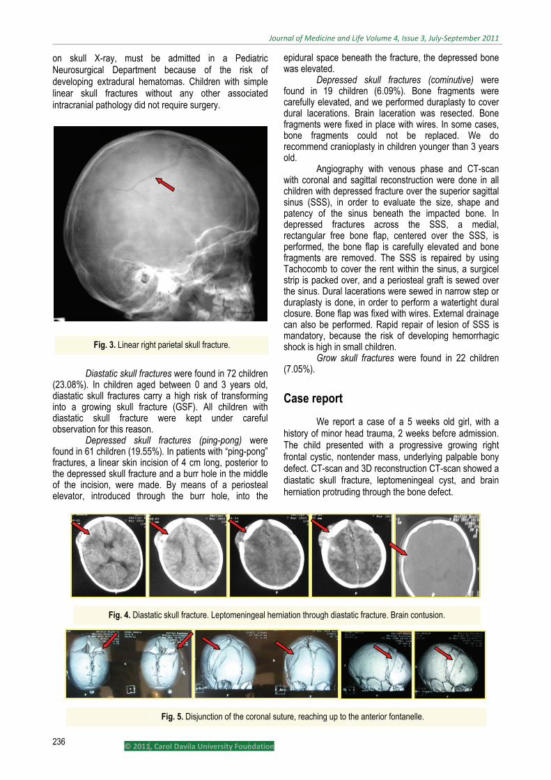
Theprogressive examination diagnostic ofafter admissi
Aftethe fracture, the bone de
The
Craniocerebinjury
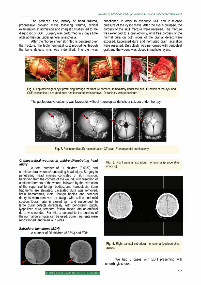
A tcraniocerebrapenetrating beginning frocontused borof the superfragments abrain hematdecrypts wersuction. Durlarge dural lyophilized ddura, was nethe normal drepositioned, Extradural h
A nu
Fig. 6. LCSF ev
© 2011
patient’s agrowing ma
at admission f GSF. Surgeron, under gen
er the “horse s the leptomenefects rims w
postoperative
bral wounds
otal number al wounds/penhead injuries
om the cornersrders of the wrficial foreign re elevated. tomas, clots, re removed ba mater is cdefects dura
dura, temporaeeded. For thura mater can, and fixed wit
hematoma (EDumber of 26 ch
Leptomeningeavacuation. Lace
1, Carol Davila U
ge, history ass following and imagisticry was performneral anesthesshoe” skin flaningeal cyst pwas indentifie
e outcome wa
in children/P
of 11 childnetrating heads consisted s of the wound
wound, followebodies, and Lacerated du foreign bodby lavage witlosed tight aplasty, with pl fascia, fasc
his, a suturedn be used. Bonh wires.
DH) hildren (8.33%
al cyst protrudinrated dura and
Fig. 7. Postope
University Foun
of head tra trauma, cl
c studies led tmed in 2 dayssia. p is centered protruding thred. The cyst
as favorable, w
/Penetrating h
dren (3.53%) d injury. Surgeof skin inci
d, with resectid by the extrahemostasis.
ura was remodies and certh saline and nd suspendeperiosteum p
cia lata or art to the bordene fragments
%) had EDH.
ng through the f lacerated brain
erative 3D reco
Journal o
ndation
uma, inical o the
s time
over rough was
puncpresbordwas normexpoweregraft
without neurolo
head
had ery in ision, on of
action Bone oved; rebral mild d. In
patch, tificial ers of were
hem
fracture bordersn removal. Dura
onstruction CT-s
Figim
Figas
of Medicine and
ctioned, in orssure of the cyders of the sk extended to
mal dura on osed. Lacerate resected. Dt and the wou
ogical deficits
We hadmorrhagic shoc
s, immediately aplasty with per
scan. Frontopar
g. 8. Right pamaging).
g. 9. Right parspect).
d Life Volume 4,
rder to evacuystic mass. Af
kull fracture wa craniectomboth sides o
ted dura and uraplasty wasnd was closed
or seizure und
3 cases wck.
under the skin. riosteum.
rietal craniectom
arietal extradura
rietal extradura
, Issue 3, July‐Se
uate CSF anfter the cyst’s
were revealed.y, until free b
of the cranial herniated bras performed wd in multiple la
der therapy.
with EDH pre
Punction of the
my.
al hematoma (
al hematoma (p
eptember 2011
237
nd to release collapse, the. The fractureborders of the defect wereain laceration
with periostealayers.
esenting with
e cyst and
(preoperative
postoperative
1
7
e e e e e n l
h
238
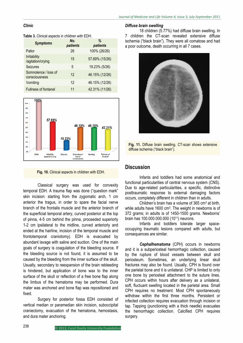
Clinic Table 3. Clinic
Symp
Pallor Irritability /agitation/crySeizures SomnolenceconsciousneVomiting
Fullness of f
Clastemporal EDskin incisionanterior the branch of thethe superficiaof pinna, 4-51-2 cm ipsilaended at thefrontotemporabundant lavgoals of surgthe bleedingcaused by thUsually, secois hindered, surface of ththe limbus omater was afixed.
Surgvertical medicraniectomy,and dura mat
Fig.
© 2011
cal aspects in c
toms
ying
e / loss of ess
fontanel
ssical surgerH. A trauma
n: starting frotragus, in or
e frontalis mual temporal ar5 cm behind tateral to the hairline, incis
ral craniotomvage with salingery is coagug source is ne bleeding froondary to reex but applicate skull or reflof the hematonchored and
gery for postian or parame evacuation ter anchoring.
10. Clinical asp
1, Carol Davila U
children with EDNo.
patients 26
15
5
12
12
11
ry was useflap was done
om the zygomrder to spare
uscle and the rtery, curved pthe pinna, promidline, curv
sion of the temmy). EDH isne and suctionlation of the bot found, it i
om the inner sxpansion of thion of bone ection of a freoma may bebone flap was
terior fossa Eedian skin incof the hemat.
pects in children
University Foun
DH. %
patients 100% (26/26)
57.69% (15/26
19.23% (5/26)
46.15% (12/26
46.15% (12/26
42.31% (11/26
ed for conve (“question mmatic arch, 1e the facial nanterior bran
posterior at thoceeded supeved anteriorlymporal muscles evacuatedn. One of the bleeding sours assumed t
surface of the he brain reblee
wax to the ee bone flap ae performed. s repositioned
EDH consistecision, subocctoma, hemost
n with EDH.
Journal o
ndation
6)
)
6)
6)
6)
vexity mark” 1 cm nerve ch of e top
eriorly y and e and d by main ce. If to be skull. eding inner along Dura
d and
ed of cipital tasis,
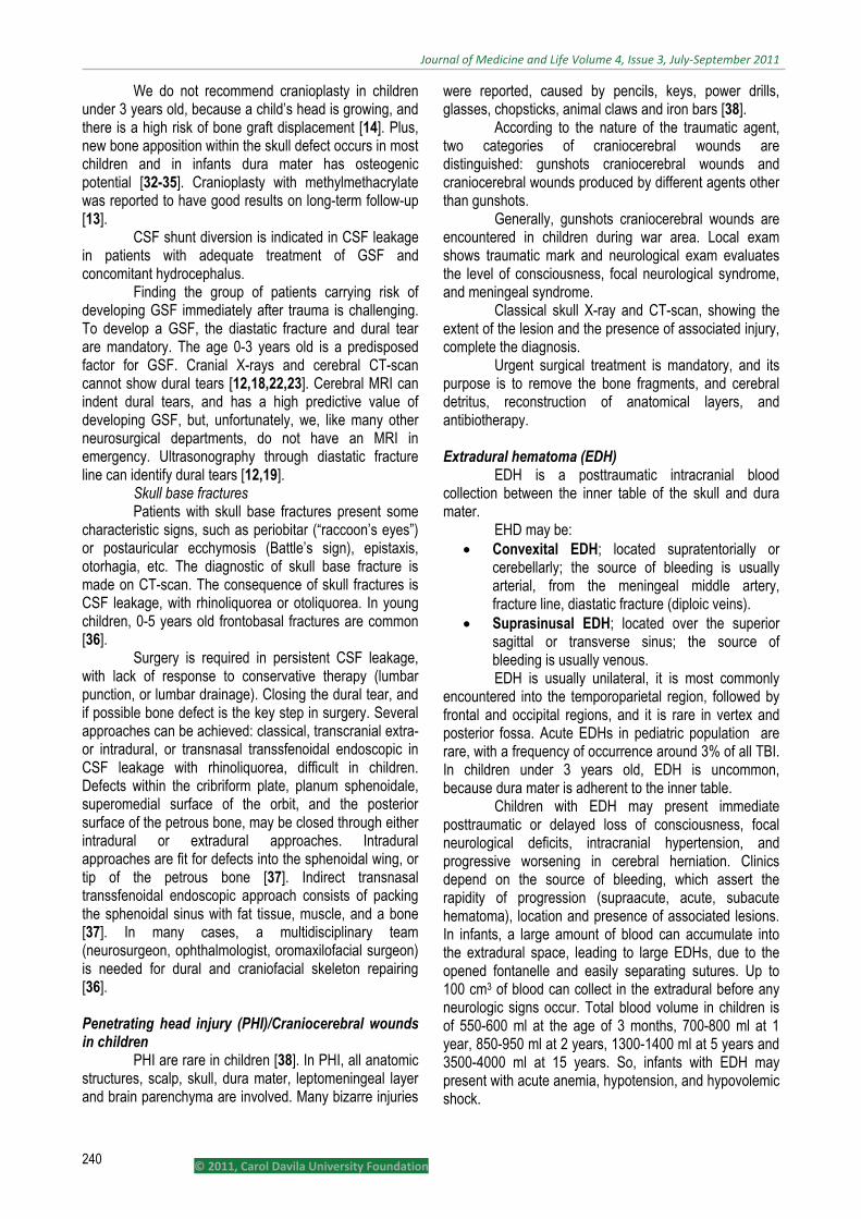
Diffu
7 cischea po
Dis
funcDuepostoccu
while372 brain
occucons
and by periofractthe pone CPHsoft,CPHwithdinfectap. the surg
Figdif
of Medicine and
use brain swe18 childre
children the emia (“black b
oor outcome, d
scussion
Infants actional particul to age-relatettraumatic resurs, completel
Children’e adults have grams; in adn has 100.000
Infants upying traumasequences are
Cephalh it is a subpethe rupture osteum. Somtures may alsparietal bone bone by per
H occurs with fluctuant swe
H requires nodraw within cted collection Tapping (pun
hemorrhagicgery.
g. 11. Diffuse ffuse ischemia (
d Life Volume 4,
welling en (5.77%) haCT-scan re
brain”). They wdeath occurrin
and toddlers harities of cented particularitsponse to ey different in cs brain has a 1600 cm3. Th
dults is of 1450.000.000 (101
and toddlersatic lesions ce similar.
hematoma (Ceriosteal hemoof blood ve
metimes, an o be found. Uand it is unilatriosteal attach
hin hours afteelling located o treatment. the first thre
n requires evanctioning with c collection.
brain swelling.(“black brain”).
, Issue 3, July‐Se
ad diffuse braevealed extenwere all coma
ng in all 7 case
had some antral nervous sties, a specif
external damachildren than i volume of 365he weight in n50-1500 gram11) neurons. s tolerate lacompared wit
CPH) occurs orrhagic collessels betwee underlying Usually, CPH teral. CHP is lhment to the er delivery as in the parietaMost CPH see months. acuation throu a thick need
Calcified C
. CT-scan show
eptember 2011
in swelling. Innsive diffuseatose and hades.
natomical andystem (CNS).fic, distinctiveaging factorsn adults. 5 cm3 at birth,
newborns is ofms. Newborns’
arger space-th adults, but
in newbornsction, causeden skull and
linear skullis found overlimited to only suture lines.
s a unilateral,al area. SmallspontaneouslyPersistent orgh incision orle) evacuates
CPH requires
ws extensive
1
n e d
d . e s
, f ’
-t
s d d l r y . , l
y r r s s
Journal of Medicine and Life Volume 4, Issue 3, July‐September 2011
239 © 2011, Carol Davila University Foundation
Skull fractures Depressed skull fracture may occur following
difficult labor, but these patients were not included in our study.
Linear skull fractures in children 0-3 years old are common, but usually have no clinical significance and require no specific treatment. Children with linear skull fracture must be admitted and followed up in a Department of Pediatric Neurosurgery for extradural hematoma occurrence. Diastatic skull fractures carry a high risk of transforming into a growing skull fracture (GSF) in this age group.
Depressed skull fractures may be closed (simple fracture) or open (compound fracture). Depressed skull fractures, with or without focal neurological deficits or seizures, require surgical elevation, according to the depth of deformity. The “Ping-pong” fracture is a particular type of depressed fracture found in newborns. It is usually encountered in parietal bone, and requires surgery.
The depressed fracture, closed or compound, situated over the superior sagittal sinus (SSS) requires an angiography with venous phase or a CT-scan with coronal and sagittal reconstruction, in order to evaluate the size, shape and patency of the sinus beneath the impacted bone. Repairing of SSS must be performed rapidly, in order not to lose blood (in children there is a bigger risk of developing hemorrhagic shock).
A compound fracture, with depressed bony fragments and dural laceration needs emergency surgery. CT-scan can reveal associated hematomas, which require surgical evacuation. Brain laceration carries the risk of subsequent epilepsy, and must be resected. If bone fragments could not be replaced, cranioplasty with methylmethacrylate can be performed within 6 months after initial surgery. We recommend cranioplasty only in children older than 3 years.
Growing skull fracture The grow skull fracture (GSF), is a rare
complication of the skull fracture, mainly encountered during infancy and early childhood, in children under three years old [11-14]. The incidence of GCS is of 0.05 to 1.6% of all skull fractures during childhood [12,13,15,16].
The most common causes of GSF are head trauma secondary to falls, road traffic accidents and instrumental delivery [17-20]. Grow skull fractures are usually located at the cranial vault, into parietal or frontoparietal areas, but skull base [15,16,21,22] and posterior fossa [21,23,24] can also be involved.
It is a progressive enlargement of diastatic fracture, along with leptomeningeal cyst and brain hernia.
The period of time between head injury and GSF diagnosis can go up to many years [25,26]. Usually, GSF develop within 3-4 months following TBI. Positive diagnosis is made on history of head trauma, followed by progressive enlargement of skull fracture and leptomeningeal cyst protruding through the bone defect.
Factors contributing to GSF occurrence are rapid brain growth and brain pulsation, found in young infants and children [27].
Pathogenesis consists of three phases. Within the first phase, following TBI, a linear skull fracture with periosteum tear and dural laceration occurs. Then, in time, fracture healing is hindered by intracranial hypertension and constant pulsation of the CSF, which favors invagination and entrapment of arachnoids into the diastatic fracture. During the third phase, patients present important bone diastasis, dural defect, progressive leptomeningeal and brain herniation through the diastatic fracture preventing apposition of bone, and elevated intracranial pressure [28]. In time, cerebroventricular changes occur, such as reactive gliosis into the underlying brain, with a subsequent development of porencephalic cavity [13,18,25,29].
During the first phase, patients have bone diastasis and seizures, the second phase presents with bone diastasis, seizures and motor deficits and the last phase with large bone diastasis, focal neurological signs and intracranial hyperpressure.
Skull X-ray, cerebral CT-scan, with bone window and 3D reconstruction reveal the diastatic fracture and leptomeningeal and brain herniation.
Surgery is always required in patients with GCS to prevent seizures and neurological deficits occurrence. The key step is tight closure of dura mater. When dura mater is lacerated and cannot be sutured, the lacerated dura is resected, and duraplasty with periosteum, free flap or pediculated or artificial dura is performed. We recommend dural closure with free flap periosteum, sutured watertight to the normal dura mater, because it is an autologous tissue and it is the most convenient dural substitute. Watertight dural closure is mandatory to avoid GSF recurrence or CSF leakage occurrence [14].
Usually, dural tear extends beyond the fracture borders, that is way the fracture should be extended to craniectomy, until normal dura is reveled [15,27,30,31]. In many cases, dural tears may involve transverse or sagittal sinus [23]. In such cases bleeding from the dural sinus can occur, that is life threatening in these age groups. Even if the sinus is not torn, its vicinity may pose problems regarding dural repair. If the fracture is parallel to the sinus, dural repair is difficult, because of the narrowness of the dural edge to the venous sinus. If proximity of the sinus does not allow graft suture with borders of normal dura mater, the graft can be sutured with dura across the sinus after extending the craniectomy or directly to the skull’s edge above the sinus. If the fracture is perpendicularly to the sinus, the end closest to the venous sinus does not need repairing. We extended the fracture to a right frontoparietal craniectomy over the coronal suture, reaching up to the anterior fontanelle and suturing a patch of periosteum to free dural borders.
Journal of Medicine and Life Volume 4, Issue 3, July‐September 2011
240 © 2011, Carol Davila University Foundation
We do not recommend cranioplasty in children under 3 years old, because a child’s head is growing, and there is a high risk of bone graft displacement [14]. Plus, new bone apposition within the skull defect occurs in most children and in infants dura mater has osteogenic potential [32-35]. Cranioplasty with methylmethacrylate was reported to have good results on long-term follow-up [13].
CSF shunt diversion is indicated in CSF leakage in patients with adequate treatment of GSF and concomitant hydrocephalus.
Finding the group of patients carrying risk of developing GSF immediately after trauma is challenging. To develop a GSF, the diastatic fracture and dural tear are mandatory. The age 0-3 years old is a predisposed factor for GSF. Cranial X-rays and cerebral CT-scan cannot show dural tears [12,18,22,23]. Cerebral MRI can indent dural tears, and has a high predictive value of developing GSF, but, unfortunately, we, like many other neurosurgical departments, do not have an MRI in emergency. Ultrasonography through diastatic fracture line can identify dural tears [12,19].
Skull base fractures Patients with skull base fractures present some
characteristic signs, such as periobitar (“raccoon’s eyes”) or postauricular ecchymosis (Battle’s sign), epistaxis, otorhagia, etc. The diagnostic of skull base fracture is made on CT-scan. The consequence of skull fractures is CSF leakage, with rhinoliquorea or otoliquorea. In young children, 0-5 years old frontobasal fractures are common [36].
Surgery is required in persistent CSF leakage, with lack of response to conservative therapy (lumbar punction, or lumbar drainage). Closing the dural tear, and if possible bone defect is the key step in surgery. Several approaches can be achieved: classical, transcranial extra- or intradural, or transnasal transsfenoidal endoscopic in CSF leakage with rhinoliquorea, difficult in children. Defects within the cribriform plate, planum sphenoidale, superomedial surface of the orbit, and the posterior surface of the petrous bone, may be closed through either intradural or extradural approaches. Intradural approaches are fit for defects into the sphenoidal wing, or tip of the petrous bone [37]. Indirect transnasal transsfenoidal endoscopic approach consists of packing the sphenoidal sinus with fat tissue, muscle, and a bone [37]. In many cases, a multidisciplinary team (neurosurgeon, ophthalmologist, oromaxilofacial surgeon) is needed for dural and craniofacial skeleton repairing [36].
Penetrating head injury (PHI)/Craniocerebral wounds in children
PHI are rare in children [38]. In PHI, all anatomic structures, scalp, skull, dura mater, leptomeningeal layer and brain parenchyma are involved. Many bizarre injuries
were reported, caused by pencils, keys, power drills, glasses, chopsticks, animal claws and iron bars [38].
According to the nature of the traumatic agent, two categories of craniocerebral wounds are distinguished: gunshots craniocerebral wounds and craniocerebral wounds produced by different agents other than gunshots.
Generally, gunshots craniocerebral wounds are encountered in children during war area. Local exam shows traumatic mark and neurological exam evaluates the level of consciousness, focal neurological syndrome, and meningeal syndrome.
Classical skull X-ray and CT-scan, showing the extent of the lesion and the presence of associated injury, complete the diagnosis.
Urgent surgical treatment is mandatory, and its purpose is to remove the bone fragments, and cerebral detritus, reconstruction of anatomical layers, and antibiotherapy.
Extradural hematoma (EDH)
EDH is a posttraumatic intracranial blood collection between the inner table of the skull and dura mater.
EHD may be: Convexital EDH; located supratentorially or
cerebellarly; the source of bleeding is usually arterial, from the meningeal middle artery, fracture line, diastatic fracture (diploic veins).
Suprasinusal EDH; located over the superior sagittal or transverse sinus; the source of bleeding is usually venous. EDH is usually unilateral, it is most commonly
encountered into the temporoparietal region, followed by frontal and occipital regions, and it is rare in vertex and posterior fossa. Acute EDHs in pediatric population are rare, with a frequency of occurrence around 3% of all TBI. In children under 3 years old, EDH is uncommon, because dura mater is adherent to the inner table.
Children with EDH may present immediate posttraumatic or delayed loss of consciousness, focal neurological deficits, intracranial hypertension, and progressive worsening in cerebral herniation. Clinics depend on the source of bleeding, which assert the rapidity of progression (supraacute, acute, subacute hematoma), location and presence of associated lesions. In infants, a large amount of blood can accumulate into the extradural space, leading to large EDHs, due to the opened fontanelle and easily separating sutures. Up to 100 cm3 of blood can collect in the extradural before any neurologic signs occur. Total blood volume in children is of 550-600 ml at the age of 3 months, 700-800 ml at 1 year, 850-950 ml at 2 years, 1300-1400 ml at 5 years and 3500-4000 ml at 15 years. So, infants with EDH may present with acute anemia, hypotension, and hypovolemic shock.
Journal of Medicine and Life Volume 4, Issue 3, July‐September 2011
241 © 2011, Carol Davila University Foundation
Clinical features may be: brief posttraumatic LOC, followed by lucid
interval for several hours, and then progressive alteration of the level of consciousness
persistent posttraumatic coma posttraumatic coma, followed by the regaining of
consciousness posttraumatic conservation of consciousness
without coma posttraumatic conservation of consciousness
with delayed coma Clinics in newborns and infants are vague
consisting of hypothonia, seizures, and tense fontanelle. EDH in children with hydrocephalus and
ventricular shunt is a serious and urgent chapter of children pathology. In such children, CT is mandatory immediately. Even after a mild TBI usually initially asymptomatic, coma may rapidly onset. CT-scan must be performed without delay [39]. Usually, there is no lucid period, rapid alteration of level of consciousness immediately after trauma and severe impairment of vegetative functions. The supratentorial EDH patients present with motor deficits, jacksonian seizures, anisocoria, and comatose state [40].
Posterior fossa EDH occurs less frequently than supratentorial ones, but it is the most common posttraumatic space-occupying lesion of the posterior fossa in children [41]. Posterior fossa EDH blocks cistern magna, causing brainstem compression and obstructive hydrocephalus with acute intracranial hypertension. Children with EDH posterior fossa may have a rapid deterioration without significant warning symptoms and may result in death.
The positive diagnosis, location and size of EDH are made on CT-scan. EDHs are extraaxial lenticular-shaped masses situated between the dura and the inner table of the skull, with density varying according to the age of the lesion. A normal CT-scan immediately after trauma, does not exclude the possibility of further development of an EDH.
Children with skull fractures, presenting with vomiting or alteration of the level of consciousness, must be clinically observed together with a CT-scan for the development of an EDH.
Surgery is indicated according to the clinical status and CT-scan:
Comatose patient with anisocoria, and CT-scan showing EDH require urgent surgery
Coma and worsening of neurological state in case of EDH’s volume > 25 ml
EDH’s volume > 30 ml, even in the absence of clinical signs
EDH’s volume > 25 ml, if EDH is located within the posterior fossa or temporal region
Midline shift > 4 mm, with worsening of neurological status
EDH volume increase
Surgery consists of the hematoma evacuation. One of the most important therapeutic targets is blood replacement in the shocked child, which should be done within 20-30 minutes, before starting the operation.
Conservative treatment can be attempted in an alert child, with no focal neurological deficits, in which CT-scan showed an EDH having a volume < 25 ml, with a thickness < 10 mm and midline shift < 4 mm. The child must be held in a neurosurgical center, where surgery can be performed if needed, under attentive clinical observation and CT-scan must be repeated [42]. Diffuse brain swelling (DBS)
Diffuse brain swelling in newborns is due to birth asphyxia and secondary reperfusion. DBS is mixed by vasogenic and cytotoxic edema.
The diagnosis is established by CT-scan and the treatment is specific, according to the clinical status. The particularity of DBS in children is represented by its early onset. It is very severe and it is often followed by diffuse cerebral ischemia (“black brain”).
Posttraumatic DBS can accompany any cerebral lesion in children and it requires intracranial pressure (ICP) monitoring. After failure of conservative therapy, bilateral decompressive craniectomy may be needed as the last resort treatment strategy. In children, decompressive craniectomy improves the neurological outcome and diminishes death risk [43,44]. The necessity of decompressive craniectomy and duraplasty is especially important in children, due to the lack of response to usual therapies, in the treatment of intracranial hypertension [45,46].
The surgical technique for diffuse brain swelling: wide bilateral hemicraniectomy, dural opening in a stellate fashion, dural graft to increase the available volume before closure, and finally wound closure.
Conclusions
Neurotrauma pathology is very different in infants and toddlers compared to different age patients. Accurate and rapid clinical and neuroimagistic diagnosis is the key of success. The Pediatric Neurosurgical Department and Pediatric Intensive Care Unit represent a vital necessity. Long time follow-up is mandatory.
CT-scan is the main investigation tool and must be performed in all children with TBI, in the first three hours.
Hemorrhagic shock may rapidly occur in infants and young children.
Grow skull fracture is a specific posttraumatic lesion in infants and young children. Surgery is always required to prevent neurological deficits and/or seizures occurrence.
Tight dural closure is the key step in surgical management. Duraplasty can be done with a patch of pericranium / periosteum. Cranioplasty is not indicated in infants.
Journal of Medicine and Life Volume 4, Issue 3, July‐September 2011
242 © 2011, Carol Davila University Foundation
References
1. Langlois J, Rutland-Brown W, Wald M. The epidemiology and impact of traumatic brain injury: a brief overview. J Head Trauma Rehabil 2006; 21: 375-378.
2. Williamson LM, Morrison A, Stone DH. Trends in head injury mortalityamong 0-14 years old in Scotland (1986-95). J Epidemiol Comm Health 2002; 56: 285-288.
3. Raimondi A. Pediatric neurosurgery. 1987.
4. Simpson D, Reilly P. Paediatric Coma Scale. Lancet 1982; 2: 450.
5. Raimondi AJ, Hirschauer J. Head injury in the infant and toddler. Childs Brain 1984; 11: 12-35.
6. Beni-Adani L, Flores I, Spektor S, Umansky F, Constantini S. Epidural hematoma in infants: a different entity? J Trauma 1999; 46: 306-311.
7. Reilly P, Simpson D. Assessing the conscious level in infants and young children: a paediatric version of the Glasgow Coma Scale. Childs Nerv Syst 1998; 4: 30-33.
8. Crouchman M, Rossiter L, Colaco T, Forsyth R. A practical outcome scale for pediatric head injury. Arch Dis Child 2001; 84: 120-124.
9. Jennett B, Bond M. Assessment of outcome after severe brain damage. Lancet 1975; 1: 480-484.
10. Rankin J. Cerebral vascular accidents in patients over the age of 60. Scott Med J 1957; 2: 200-215.
11. Arseni C, Ciurea AV. Clinicotheraputic aspects in the growing skull fracture. A review of the literature. Childs Brain 1981; 8: 161-172.
12. Djientcheu VP, Njamnshi AK, Ongolo-Zolo P, Kobela M, Rilliet B, Essomba A, Sosso MA. Grow skull fractures. Childs Nerv Syst 2006; 22: 721-725.
13. Hayashi Y, Yamaki T, Odake G, Hashimoto Y, Ueda S. Long-term follow up of a growing skull fracture treated by dura and cranioplasty with articial dura mater and methylmethacrylate. Childs Nerv Syst 1997; 13: 349-351.
14. Ciurea AV, Iliescu A, Sandu AM, Gheorghita A. Grow skull fracture – special consideration on a 5 weeks old case. J Ped Surg Special 2008; 3: 52-58.
15. Ershain Y, Gulmen V, Palali I, Mutluer S. Growing skull fractures
(craniocerebral erosion). Neurosurg Rev 2000; 23: 139-144.
16. Gupta V, Sinha S, Singh AK, Kumar S, Singh D. Growing skull fracture of ethmoid: a report of two cases. J Craniomaxillofac Surg 2000; 28: 224-228.
17. Moss SD, Walker Ml, Ostergagng S, Golembeski D. Intra-uterine growing skull fracture. Childs Nerv Syst 1990; 6: 468-470.
18. Djientcheu VP, Rilliet B, Delavelle J, Argyropoulo M, Gudinchet F, de Tribolet N. Leptomeningeal cyst in newborns due to vacuum extraction: report of two cases. Childs Nerv Syst 1996; 12: 399-403.
19. Voet D, Govaert P, Caemaert J, de Lille L, D`herde K, Afsshrift M. Leptomeningeal cysts and vacuum extraction (short communication). Acta Pediatr Scand 1992; 79: 232-233.
20. Papaefthymiou G, Oberbauer R, Pendl G. Craniocerebral birth trauma cased by vaccum extraction: a case of growing skull fracture as a perinatal complication. Childs Nerv Syst 1996; 12: 117-120.
21. Caffo M, Germano A, Caruso G, Meli F, Calisto A, Tomasello F. Growing skull fracture of the posterior cranial fossa and the orbital roof. Acta Neurochir 2003; 145: 201-208.
22. Menku A, Kemal KR, Tucer B, Kurtsoy A, Akdemir H. Growing skull fracture of the orbital roof. Neurosurg Rev 2004; 27: 133-136.
23. Lenthal R, Penney C. Growing skull fracture extending posteriorly to the superior saggital sinus with intradiploic extension. Br J Radiol 1999; 72: 714-716.
24. Palaoglu S, Beskonakli E, Senel K. Osseous location of posterior fossa post-traumatic leptomeningeal cyst. Demonstration by computerized tomography. Neuroradiology 1990; 32: 78.
25. Zegers B, Jira P, Willemsen M, Grotenhuis J. The growing skull fracture, a rare complication of pediatric head injury. Eur J Pediatr 2003; 162: 556-557.
26. Sharma RR, Chandy MJ. Shunt surgery in growing skull fracture: a report of two cases. Br J Neurosurg 1991; 5: 93-98.
27. Muhonen MG, Piper JG, Menezes AH. Pathogenesis and treatment of
growing skull fractures. Surg Neurol 1995; 43: 367-372.
28. Drapkin AJ. Growing skull fracture: a posttraumatic neosuture. Childs Nerv Syst 2006; 22: 394-397.
29. Ziyal IM, Aydin Y, Turkmen CS, Salas E, Kaya AR, Ozveren F. The natural history of a late diagnosed or untreated growing skull fractures: report on 2 cases. Acta Neurochir (Wien) 1998; 140: 651-654.
30. Singhal A, Steinbok P. Operative management of growing skull fracture: a technical note. Childs Nerv Syst 2008; 24: 605-607.
31. Gupta SK, Reddy NM, Khosla VK, Mathuriya SN, Sharma BS, Pathak A, Tewari MK, Kak VK. Growing skull fractures: a clinical study of 41 patients. Acta Neurochir (Wien) 1997; 139: 928-932.
32. Greenwald JA, Mehrara B, Spector JA, Chin CS, Steinbrech DS, Saadeh PB, Luchs JS, Paccione MF, Gittes GK, Longaker MT. Biomolecular mechanisms of calvarial bone induction: immature versus mature dura mater. Plast Reconstr Surg 2000; 105: 1382-1392.
33. Guzel MZ, Yildirim AM, Yucel A, Seradjmir M, Desvisoglu S. Osteogenic potential of infant dural grafts in different recipient beds. J Craniofac Surg 1995; 6: 489-493.
34. Oppermann LA, PAssarelli RW, Morgan EP, Reintjes M, Ogle RC. Cranial sutures require tissue interactions with dura mater to resist osseous obliteration in vitro. J Bone Miner Res 1995; 10: 1978-1987.
35. Yu JC, McClintock JS, Gannon F, Gao XX, Mobasser JP, Sharawy M. Regional differences of dura osteoinduction: squamous dura induces osteogeneris, sutural dura induces chondrogenesis and osteogenesis. Plast Reconstr Surg 1997; 100: 23-31.
36. Clauser L, Dallera V, Sarti E, Tieghi R. Frontobasilar fractures in children. Childs Nerv Syst 2004; 20: 168-175.
37. Raimondi AJ. Trauma. In: Raimondi AJ, editor. Pediatric neurosurgery. Springler-Verlag, 1998: 343-377.
38. Gonzales AL, Marin AG, Garijo JAA, Menguel MV. Penetrating head injury in a paediatric patient
Journal of Medicine and Life Volume 4, Issue 3, July‐September 2011
243 © 2011, Carol Davila University Foundation
caused by an electric plug. Childs Nerv Syst 2006; 22: 197-200.
39. Shu EBS, Leme RJA, Aguilar PH, Andrade AF, Teixeira MJ, Plese JPP. Traumatic acute giant epidural hematoma in a hydrocephalic shunted child. Pediatr Neurosurg 2000; 32: 176-179.
40. Ciurea AV, Kapsalaki EZ, Coman T, Roberts JL, Robinson JS 3rd, Tascu A, Brehar F, Fountas KN. Supratentorial epidural hematoma of traumatic etiology in infants. Childs Nerv Syst 2007; 23: 335-341.
41. Becker M, Cataltepe O, Ozcan OE. Traumatic epidural haematoma of the posterior fossa in childhood:
16 new cases and a review of the literature. Br J Neurosurg 2003; 17: 226-229.
42. Balmer B, Boltshauser E, Altermatt S, Gobet R. Conservative management of significant epidural haematomas in children. Childs Nerv Syst 2006; 22: 363-367.
43. Bissonnette B. Odeme cerebral chez l`enfant par rapport a l`adulte: quelles sont les differences? Ann Fr Anesth Reanim 2003; 22: 331-335.
44. Figaji AA, Fieggen AG, Peter JC. Early decompresive craniotomy in children with severe traumatic brain
injury. Childs Nerv Syst 2003; 19: 666-673.
45. Kan P, Amini A, Hansen K, White GL, Brockmeyer DL, Walker ML, Kestle JRW. Outcomes after decompressive craniectomy for severe traumatic brain injury in children. J Neurosurg (5 Suppl Pediatrics) 2006; 105: 337-342.
46. Ruf B, Heckmann M, Schoroth I, Hugens-Penzel M, Reiss I, Borkhardt A, Gortner L, Jodicke A. Early decompressive craniectomy and duraplasty for refractory intracranial hypertension in children: results of a pilot study. Critical Care 2003; 7: 133-138.


