Embed Size (px)
Citation preview
Tissue Microenvironment Modulates CXCR4 Expressionand Tumor Metastasis in Neuroblastoma1
Libo Zhang*,y, Herman Yeger y, Bikul Das*,y, Meredith S. Irwinz,§ and Sylvain Baruchel*,z,§
*New Agent and Innovative Therapy Program, The Hospital for Sick Children, Toronto, Canada; yDepartmentof Pediatric Laboratory Medicine, The Hospital for Sick Children, Toronto, Canada; zDivision of Hematologyand Oncology, Department of Pediatrics, The Hospital for Sick Children, Toronto, Canada; §Institute of MedicalSciences, University of Toronto, Toronto, Canada
Abstract
Neuroblastoma (NB) is derived from intrinsic migratory
neural crest cells and has a high potential for distant
metastasis. Growing evidence has implicated chemo-
kine receptors, especially CXCR4, which normally
control immune and inflammatory cell migration, as
having important roles in tumor progression. In this
study, we investigated the expression of CXCR4 in
eight different NB cell lines and found that CXCR4 ex-
pression is dynamically regulated in NB and can be
modulated by different tissue stromata. In addition,
we demonstrate that IL-5 and IFN-; are released
from stromal cells and act as differential mediators for
CXCR4 expression. We also overexpressed CXCR4 in
two NB cell lines, NUB-7 and SK-N-BE(2), and studied
the role of CXCR4 in NB metastasis both in vitro and
in vivo. In vitro transwell invasion assay showed
that CXCR4 overexpression promoted NB cell migra-
tion preferentially toward a bone marrow stromal
cell–conditioned medium. Using an in vivo xenograft
model, CXCR4-overexpressing cells showed an in-
creased incidence of metastasis, most notably bone
marrow metastasis. Our studies reveal critical roles for
CXCR4 in NB metastasis and provide insights into the
regulatory mechanism of chemokine receptors in NB
and the importance of the tissue microenvironment
in modulating tumor cell behavior.
Neoplasia (2007) 9, 36–46
Keywords: Neuroblastoma, metastasis, chemokine receptor, tissue micro-environment, CXCR4.
Introduction
Chemokines represent a large superfamily of small peptides
that currently comprise 42 members in humans, which
recruit different cell populations by interacting with seven
transmembrane domain G-protein–coupled receptors. At
present, 18 chemokine receptors have been identified.
The primary role of chemokines/receptors is to regulate
the recruitment and trafficking of leukocyte subsets to
inflammatory sites through chemoattraction by activating
leukocyte integrins that bind to their adhesion receptors
on endothelial cells [1,2]. Chemokines are also involved in
neuronal cell migration and patterning [3]. Recent studies
suggest that many cancers express an extensive network of
chemokines and chemokine receptors [4,5]. These tumors are
characterized by dysregulated production of chemokines and
abnormal chemokine receptor expression. It has become evi-
dent that chemokines are able to couple to distinct signal-
ing pathways. Most chemokines share the ability to activate
G-protein–sensitive phospholipase C isoforms, resulting in
inositol-3,4,5-trisphosphate generation. Some of the chemo-
kines can also inhibit adenylate cyclase, activate MEK-1 and
ERK-1/2, and stimulate the tyrosine phosphorylation of focal
adhesion complex components [6–8].
Among chemokine receptors, CXCR4 is frequently studied
because it is expressed by most cancer types, including can-
cers of epithelial, mesenchymal, and hematopoietic origin [9].
For example, tumor cells from breast, prostate, pancreatic,
lung, and ovarian carcinomas, as well as glioblastomas, all ex-
press CXCR4 [10–15]. CXCL12 is the only known ligand for
CXCR4. It is found at sites of metastasis in breast and thyroid
cancers [16]. Considering their original role in the trafficking
of hematopoietic stem cells to the bone marrow [17], CXCR4
and CXCL12 (also known as SDF-1) are believed to be in-
volved in tumor growth and metastasis to the bone marrow.
Neuroblastoma (NB) is derived from embryonic neural crest
cells that form the peripheral sympathetic nervous system and
have a high potential to migrate. Metastatic NB has a high
mortality rate; thus, understanding the mechanism of how NB
tumor cells invade and metastasize will help in designing more
effective therapies to control the development and metastasis
of NB, and will further help to develop an animal model that
more faithfully resembles tumor metastasis in patients. So far,
Address all correspondence to: Sylvain Baruchel, New Agent and Innovative Therapy
Program, The Hospital for Sick Children, 555 University Avenue, Toronto, Ontario, Canada
M5G 1X8. E-mail: [email protected] work was supported by the James Birrell Neuroblastoma Research Fund and the James
Fund-CIHR Neuroblastoma Fellowship. www.jamesfund.ca
Received 12 October 2006; Revised 27 November 2006; Accepted 28 November 2006.
Copyright D 2007 Neoplasia Press, Inc. All rights reserved 1522-8002/07/$25.00
DOI 10.1593/neo.06670
Neoplasia . Vol. 9, No. 1, January 2007, pp. 36 – 46 36
www.neoplasia.com
RESEARCH ARTICLE
most studies on NB chemokine system have been performed
in vitro with the SK-N-SH cell line and its subclone SK-SY5Y.
Using flow cytometry, Geminder et al. [18] demonstrated the
expression of CXCR4 in eight different NB cell lines (SH-
SY5Y, CHP126, NHB, LAI55N, KELLY, SK-N-MC, NBL-WN,
and SK-N-SH). A recent report demonstrated that a higher
expression of CXCR4 was found in primary NB from pa-
tients with high-stage disease and in patients with bone and
bone marrow metastases. Clinical outcome in patients with
tumors expressing high levels of CXCR4 is significantly
worse than in patients with low CXCR4 tumor expression
[19]. All these studies support the hypothesis that specific
chemokines/receptors may play important roles in NB tumor
cell behavior, but more direct evidence is required to estab-
lish the role of chemokines/receptors in NB cell invasion and
metastasis. In this study, we screened chemokine/receptor
profiles in eight different NB cell lines and investigated the
roles of CXCR4 in NB tumor growth and progression using
mouse xenograft models. We also demonstrate the key role
of stromal cells in NB metastasis and a potential regulatory
mechanism for CXCR4 in NB.
Materials and Methods
Cells and Antibodies
Eight human NB cell lines were used in this study. SK-N-
BE(2), IMR-32, and SK-N-SH cells were obtained from
ATCC (Manassas, VA). LAN-5 cells were kindly provided
by Dr. Robert Seeger (Children’s Hospital Los Angeles, Los
Angeles, CA), NBL-S cells were from Dr. Susan Cohn (Chil-
dren’s Memorial Hospital, Chicago, IL), and SK-N-BE(1) cells
were from Dr. R. A. Ross (Fordham University, Bronx, NY).
The GOTO NB cell line was purchased from Riken Gene
Bank (Riken, Tsukuba, Ibaraki, Japan). All NB cell lines
were cultured at 37jC in 5%CO2 ina-MEM (Multicell;Wisent,
Inc., St. Bruno, Quebec, Canada) with 10% fetal bovine
serum (FBS; Life Technologies, Grand Island, NY), 50 U/ml
penicillin, and 50 mg/ml streptomycin (Gibco, Carlsbad, CA).
Antibodies to CXCR4 were purchased from Abcam (Cam-
bridge, MA) (ab2090) and R&D Systems (Minneapolis, MN;
clone 44708). The CXCR4 inhibitor AMD3100 was obtained
from Sigma (St. Louis, MO). CXCR4–pBABE was kindly
provided by Dr. Nathaniel Landau (The Salk Institute for
Biological Studies, La Jolla, CA). HFK293TV cells and retro-
viral plasmid 10A1 were generously provided by Dr. H. Vaziri
(Ontario Cancer Institute, Ontario, Canada).
Primary Culture of Stromal Cells
Stromal cells were established as primary cultures from
different organs of NOD/SCID mice, including the lung,
liver, bone, bone marrow, and adrenal gland. Briefly, after
sacrificing the mice, organs were dissected, and fat and
connective tissues were removed. Tissues were minced
with scissors into 1-mm cubes and digested in 15 ml of
phosphate-buffered saline (PBS) containing 75 mg of colla-
genase type II at 37jC, with rapid shaking. After digestion,
cells were washed once with 10 ml of a-MEM supplemented
with 10% FBS. The cells were plated and allowed to adhere
to flasks overnight. The medium was changed on the follow-
ing day and thereafter at 3- to 5-day intervals. The cultures
were continuously observed for growth of cells with mesen-
chymal morphology and were passaged thrice. Bone mar-
row stromal cells were isolated from bone marrow aspirates
from the femurs of NOD/SCID mice. To obtain conditioned
media, confluent stromal cells were cultured in a-MEM with
10% FBS for 48 hours, and the conditioned medium was
harvested, filtered, and used for the induction of tumor cells.
Enzyme-Linked Immunosorbent Assay (ELISA)
Themeasurement of SDF-1awas carried out using ELISA
kit (R&D Systems) according to the manufacturer’s instruc-
tions. Units of fluorescence activity were converted to actual
concentrations by a standard curve.
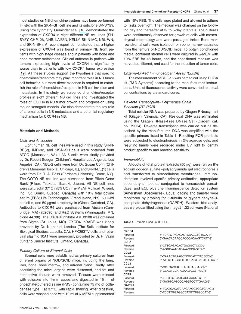
Reverse Transcription–Polymerase Chain
Reaction (RT-PCR)
Total cellular RNA was prepared by Qiagen RNeasy mini
kit (Qiagen, Valencia, CA). Residual DNA was eliminated
using the Qiagen RNase-Free DNase Set (Qiagen; cat.
no. 79254). Reverse transcription was carried out as de-
scribed by the manufacturer. DNA was amplified with the
specific primers listed in Table 1. Resulting PCR products
were subjected to electrophoresis in 2% agarose gels, and
resulting bands were recorded under UV light to identify
product specificity and reaction sensitivity.
Immunoblots
Aliquots of total protein extracts (50 mg) were run on 8%
sodium dodecyl sulfate–polyacrylamide gel electrophoresis
and transferred to nitrocellulose membranes. Immuno-
detection involved specific primary antibodies, appropriate
secondary antibodies conjugated to horseradish peroxi-
dase, and ECL plus chemiluminescence detection system
(Amersham Biosciences). Equal loading and transfer were
monitored by probing for a-tubulin or glyceraldehyde-3-
phosphate dehydrogenase (GAPDH). Western blot analy-
ses were quantified using the ImageJ 1.32 software (National
Table 1. Primers Used By RT-PCR.
CXCR4
Forward 5V-TCATCTACACAGTCAACCTCTACA-3V
Reverse 5V-GAACACAACCACCCACAAGTCATT-3V
SDF-1
Forward 5V-CTTCAGACACTGAGGCTCCC-3V
Reverse 5V-AGGCAATCACAAACCCAGTC-3V
CCL2
Forward 5V-CAAACTGAAGCTCGCACTCTCGCC-3V
Reverse 5V-ATTCTTGGGTTGTGGAGTGAGTGTTCA-3V
CCL3
Forward 5V-GCTGACTACTTTGAGACGAGC-3V
Reverse 5V-CCAGTCCATAGAAGAGGTAGC-3V
CCR7
Forward 5V-TCCTTCTCATCAGCAAGCTGT-3V
Reverse 5V-GAGGCAGCCCAGGTCCTTGAAG-3V
GAPDH
Forward 5V-TGATGACATCAAGAAGGTGGTGAAG-3V
Reverse 5V-TCCTTGGAGGCCATGTGGGCCAT-3V
Neuroblastoma and Chemokine Receptor CXCR4 Zhang et al. 37
Neoplasia . Vol. 9, No. 1, 2007
Institutes of Health, Bethesda, MD) after densitometric scan-
ning of films.
Immunocytochemistry
NUB-7 cells were seeded on coverslips and incubated for
24 hours at 37jC. The coverslips with cells were then fixed
with 4% paraformaldehyde in PBS for 10 minutes, washed
with PBS, permeabilized in 1% Triton X-100 in PBS for 5 min-
utes, washed, and blocked with PBS with 1% bovine serum
albumin (BSA). For CXCR4 staining, cells were incu-
bated with mouse anti-human CXCR4 monoclonal antibody
diluted in 1% BSA–PBS (0.01 mg/ml) for 1 hour at room tem-
perature, rinsed thrice with PBS, and then incubated for
30 minutes with secondary antibody (cy3-conjugated
goat anti-mouse IgG; Molecular Probes, Eugene, OR) di-
luted in 1% BSA–PBS (1:100). After washing, coverslips
were mounted and examined under a Zeiss Axioskop fluo-
rescence microscope (Zeiss, Standort Gottingen, Germany).
Overexpression of CXCR4
For CXCR4 overexpression, HFK293TV cells were
infected with the combination of pBABE–CXCR4 and retro-
viral plasmid 10A1 using SAINT-MIX (Synvolux Therapeutics,
Amsterdam, The Netherlands), as described by the manufac-
turer. Empty pBABE vector was also used in HAFK293TV
cell infection as control. The medium was changed after
48 hours, and the culture supernatant containing the CXCR4
virus was collected on days 3 and 4. The supernatant was
filtered through a 0.45-mm Millex HV filter (Millipore, Billerica,
MA), mixed with 8 mg/ml polybrene, and added directly to
NB cell culture. Stable transfectants of CXCR4 were estab-
lished by puromycin (1.0 mg/ml) screening. CXCR4 expres-
sion was assessed by RT-PCR and Western blot analysis.
Polyclonal populations form both CXCR4 transfectants were
used for further in vitro and in vivo experiments.
Cell Proliferation Assay
Cell proliferation was measured with Alamar Blue assay
according to manufacturer’s protocol (Trek Diagnostics Sys-
tems, Inc., Cleveland, OH). Briefly, Alamar Blue was diluted
1:10 in the cell culture medium, and color change was
monitored after 3 hours. Colorimetric evaluation of cell pro-
liferation was performed using a SPECTRAmax Gemini
spectrophotometer (Molecular Devices Corporation, Sunny-
vale, CA), with 540 nm as excitation wavelength and 590 nm
as emission wavelength, and values were expressed as
relative fluorescence units.
Cell Adhesion Assay
Briefly, 24-well plates (Costar; Corning, Acton, MA) were
coated with rat tail collagen at 37jC for 1 hour, with some
wells left uncoated as negative controls. After washing the
plates with PBS, blocking buffer (0.5% BSA in DMEM) was
added at 37jC for 1 hour. A total of 1 � 105 cells/well was
plated in 250 ml of a-MEM plus 1mg/ml BSA in a 24-well plate
and incubated in 5% CO2 at 37jC for 0.5 hour. The plate was
then washed thrice with PBS. The percentage of adherent
cells was determined by Alamar Blue assay. Three indepen-
dent experiments were carried out in triplicate.
In Vitro Cell Invasion Assays
Cell invasion was assessed with the BD BioCoat (BD
Biosciences, San Jose, CA) tumor invasion system based on
the Boyden chamber principle using 8-mm pore positron emis-
sion tomography membrane inserts coated with a layer of
BD Matrigel Matrix (BD Biosciences). After detaching cells
with nonenzymatic cell dissociation solution (Sigma), NB cells
were incubated in suspension in a-MEM with 10% FBS for
2 hours and then seeded onto the extracellular matrix layer,
which had been previously rehydrated at room tempera-
ture for 2 hours. For chemokine-dependent invasion assay,
stromal cell–conditioned medium (SCM) was added to the
lower chamber as chemoattractant. Cells were incubated
for 48 hours at 37 jC in an incubator (5% CO2). Invaded cells
on the bottom of the insert membrane were fixed with 70%
ethanol and stained with hematoxylin. For quantification, the
average number of migrating cells per field was assessed
by counting 10 random fields under a microscope (original
magnification, �250).
Cytokine Antibody Array
Conditioned media for the liver, lung, and adrenal gland
were obtained as described above. a-MEM supplemented
with 10% FBS was used as negative control. Cytokine anti-
body array was performed with ChemiArray system (Mouse
Cytokine Antibody Array I; Chemicon International, Temec-
ula, CA) according to the manufacturer’s protocol. Briefly,
array membranes were blocked with 1� blocking buffer at
room temperature for 30 minutes and then incubated with
conditioned media at room temperature for 2 hours. After
washing thrice with 2 ml of wash buffer at room temperature,
biotin-conjugated anti-cytokine primary antibody was added
to each membrane and incubated at room temperature for
2 hours. Subsequently, membranes were washed thrice and
incubated with diluted HRP-conjugated streptavidin at room
temperature for 2 hours. Cytokines were detected by chemo-
luminescence reaction with exposure to Kodak X-mat film
(Estman Kodak Company, Rochester, NY) at room tem-
perature for 1 minute. Relative expression levels among dif-
ferent samples were compared to positive controls.
Mouse Xenograft Model
Both CXCR4 transfectants and control NUB-7 cells were
injected intravenously into NOD/SCID mice at 1 � 106 cells/
mouse. Mice were sacrificed after 3 or 4 weeks. For sub-
cutaneousgrowth, 1�106CXCR4–pBABE–transfectedSK-
N-BE(2) cells were injected subcutaneously, and mice were
sacrificed after 3 weeks. Studies of tumorigenicity, growth
rate, patterns of metastasis, tumor histology, immunocyto-
chemistry, and molecular characterizations were conducted.
Statistical Analysis
Data represent the mean ± standard error of the indi-
cated number of independent experiments. Differences be-
tween groups were assessed using a two-way analysis of
38 Neuroblastoma and Chemokine Receptor CXCR4 Zhang et al.
Neoplasia . Vol. 9, No. 1, 2007
variance, followed by two-tailed Student’s t test. P < .05 was
considered significant.
Results
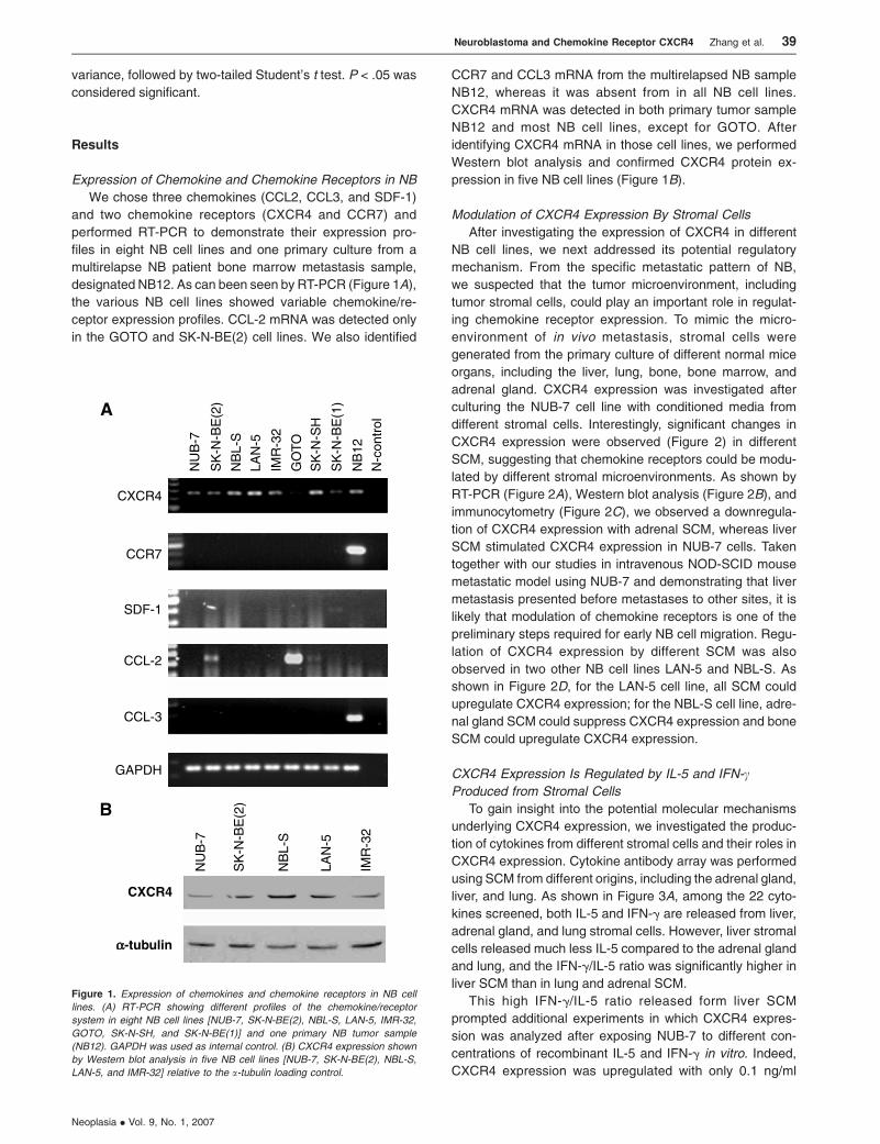
Expression of Chemokine and Chemokine Receptors in NB
We chose three chemokines (CCL2, CCL3, and SDF-1)
and two chemokine receptors (CXCR4 and CCR7) and
performed RT-PCR to demonstrate their expression pro-
files in eight NB cell lines and one primary culture from a
multirelapse NB patient bone marrow metastasis sample,
designated NB12. As can been seen by RT-PCR (Figure 1A),
the various NB cell lines showed variable chemokine/re-
ceptor expression profiles. CCL-2 mRNA was detected only
in the GOTO and SK-N-BE(2) cell lines. We also identified
CCR7 and CCL3 mRNA from the multirelapsed NB sample
NB12, whereas it was absent from in all NB cell lines.
CXCR4 mRNA was detected in both primary tumor sample
NB12 and most NB cell lines, except for GOTO. After
identifying CXCR4 mRNA in those cell lines, we performed
Western blot analysis and confirmed CXCR4 protein ex-
pression in five NB cell lines (Figure 1B).
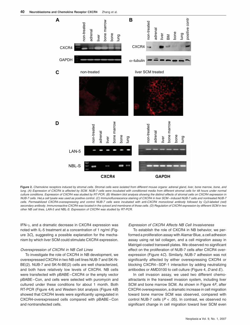
Modulation of CXCR4 Expression By Stromal Cells
After investigating the expression of CXCR4 in different
NB cell lines, we next addressed its potential regulatory
mechanism. From the specific metastatic pattern of NB,
we suspected that the tumor microenvironment, including
tumor stromal cells, could play an important role in regulat-
ing chemokine receptor expression. To mimic the micro-
environment of in vivo metastasis, stromal cells were
generated from the primary culture of different normal mice
organs, including the liver, lung, bone, bone marrow, and
adrenal gland. CXCR4 expression was investigated after
culturing the NUB-7 cell line with conditioned media from
different stromal cells. Interestingly, significant changes in
CXCR4 expression were observed (Figure 2) in different
SCM, suggesting that chemokine receptors could be modu-
lated by different stromal microenvironments. As shown by
RT-PCR (Figure 2A), Western blot analysis (Figure 2B), and
immunocytometry (Figure 2C), we observed a downregula-
tion of CXCR4 expression with adrenal SCM, whereas liver
SCM stimulated CXCR4 expression in NUB-7 cells. Taken
together with our studies in intravenous NOD-SCID mouse
metastatic model using NUB-7 and demonstrating that liver
metastasis presented before metastases to other sites, it is
likely that modulation of chemokine receptors is one of the
preliminary steps required for early NB cell migration. Regu-
lation of CXCR4 expression by different SCM was also
observed in two other NB cell lines LAN-5 and NBL-S. As
shown in Figure 2D, for the LAN-5 cell line, all SCM could
upregulate CXCR4 expression; for the NBL-S cell line, adre-
nal gland SCM could suppress CXCR4 expression and bone
SCM could upregulate CXCR4 expression.
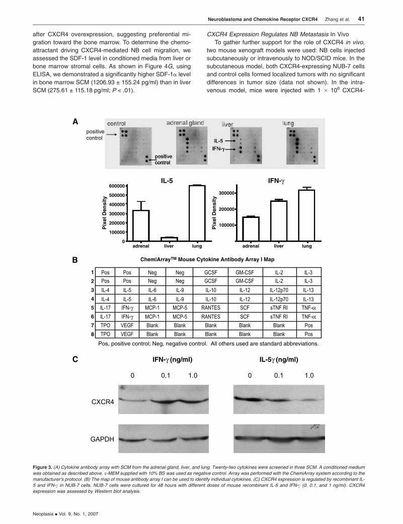
CXCR4 Expression Is Regulated by IL-5 and IFN-cProduced from Stromal Cells
To gain insight into the potential molecular mechanisms
underlying CXCR4 expression, we investigated the produc-
tion of cytokines from different stromal cells and their roles in
CXCR4 expression. Cytokine antibody array was performed
using SCM from different origins, including the adrenal gland,
liver, and lung. As shown in Figure 3A, among the 22 cyto-
kines screened, both IL-5 and IFN-g are released from liver,
adrenal gland, and lung stromal cells. However, liver stromal
cells released much less IL-5 compared to the adrenal gland
and lung, and the IFN-g/IL-5 ratio was significantly higher in
liver SCM than in lung and adrenal SCM.
This high IFN-g/IL-5 ratio released form liver SCM
prompted additional experiments in which CXCR4 expres-
sion was analyzed after exposing NUB-7 to different con-
centrations of recombinant IL-5 and IFN-g in vitro. Indeed,
CXCR4 expression was upregulated with only 0.1 ng/ml
Figure 1. Expression of chemokines and chemokine receptors in NB cell
lines. (A) RT-PCR showing different profiles of the chemokine/receptor
system in eight NB cell lines [NUB-7, SK-N-BE(2), NBL-S, LAN-5, IMR-32,
GOTO, SK-N-SH, and SK-N-BE(1)] and one primary NB tumor sample
(NB12). GAPDH was used as internal control. (B) CXCR4 expression shown
by Western blot analysis in five NB cell lines [NUB-7, SK-N-BE(2), NBL-S,
LAN-5, and IMR-32] relative to the a-tubulin loading control.
Neuroblastoma and Chemokine Receptor CXCR4 Zhang et al. 39
Neoplasia . Vol. 9, No. 1, 2007
IFN-g, and a dramatic decrease in CXCR4 expression was
noted with IL-5 treatment at a concentration of 1 ng/ml (Fig-
ure 3C), suggesting a possible explanation for the mecha-
nism by which liver SCM could stimulate CXCR4 expression.
Overexpression of CXCR4 in NB Cell Lines
To investigate the role of CXCR4 in NB development, we
overexpressed CXCR4 in two NB cell lines NUB-7 and SK-N-
BE(2). NUB-7 and SK-N-BE(2) cells are well characterized,
and both have relatively low levels of CXCR4. NB cells
were transfected with pBABE–CXCR4 or the empty vector
pBABE–Con, and cells were selected with puromycin and
cultured under these conditions for about 1 month. Both
RT-PCR (Figure 4A) and Western blot analysis (Figure 4B)
showed that CXCR4 levels were significantly upregulated in
CXCR4-overexpressed cells compared with pBABE–Con
and nontransfected cells.
Expression of CXCR4 Affects NB Cell Invasiveness
To establish the role of CXCR4 in NB behavior, we per-
formed a proliferation assaywith Alamar Blue, a cell adhesion
assay using rat tail collagen, and a cell migration assay in
Matrigel-coated transwell plates. We observed no significant
effect on the proliferation of NUB-7 cells after CXCR4 over-
expression (Figure 4C). Similarly, NUB-7 adhesion was not
significantly affected by either overexpressing CXCR4 or
blocking CXCR4–SDF-1 interaction by adding neutralizing
antibodies or AMD3100 to cell culture (Figure 4, D and E ).
In cell invasion assay, we used two different chemo-
attractants in the transwell invasion system, including liver
SCM and bone marrow SCM. As shown in Figure 4F, after
CXCR4 overexpression, a dramatic increase in cell migration
toward bone marrow SCM was observed, compared with
control NUB-7 cells (P < .05). In contrast, we observed no
significant change in cell migration toward liver SCM even
Figure 2. Chemokine receptors induced by stromal cells. Stromal cells were isolated from different mouse organs: adrenal gland, liver, bone marrow, bone, and
lung. (A) Expression of CXCR4 is affected by SCM. NUB-7 cells were incubated with conditioned media from different stromal cells for 48 hours under normal
culture conditions. Expression of CXCR4 was studied by RT-PCR. (B) Western blot analysis showing the distinct effects of stromal cells on CXCR4 expression in
NUB-7 cells. HeLa cell lysate was used as positive control. (C) Immunofluorescence staining of CXCR4 in liver SCM– induced NUB-7 cells and nontreated NUB-7
cells. Permeabilized CXCR4-overexpressing and control NUB-7 cells were incubated with anti-CXCR4 monoclonal antibody followed by Cy3-labeled (red)
secondary antibody. Immunoreactive CXCR4 was located in the cytosol and membrane of those cells. (D) Regulation of CXCR4 expression by different SCM in two
other NB cell lines, LAN-5 and NBL-S. Expression of CXCR4 was studied by RT-PCR.
40 Neuroblastoma and Chemokine Receptor CXCR4 Zhang et al.
Neoplasia . Vol. 9, No. 1, 2007
after CXCR4 overexpression, suggesting preferential mi-
gration toward the bone marrow. To determine the chemo-
attractant driving CXCR4-mediated NB cell migration, we
assessed the SDF-1 level in conditioned media from liver or
bone marrow stromal cells. As shown in Figure 4G, using
ELISA, we demonstrated a significantly higher SDF-1a level
in bone marrow SCM (1206.93 ± 155.24 pg/ml) than in liver
SCM (275.61 ± 115.18 pg/ml; P < .01).
CXCR4 Expression Regulates NB Metastasis In Vivo
To gather further support for the role of CXCR4 in vivo,
two mouse xenograft models were used: NB cells injected
subcutaneously or intravenously to NOD/SCID mice. In the
subcutaneous model, both CXCR4-expressing NUB-7 cells
and control cells formed localized tumors with no significant
differences in tumor size (data not shown). In the intra-
venous model, mice were injected with 1 � 106 CXCR4-
Figure 3. (A) Cytokine antibody array with SCM from the adrenal gland, liver, and lung. Twenty-two cytokines were screened in three SCM. A conditioned medium
was obtained as described above. a-MEM supplied with 10% BS was used as negative control. Array was performed with the ChemiArray system according to the
manufacturer’s protocol. (B) The map of mouse antibody array I can be used to identify individual cytokines. (C) CXCR4 expression is regulated by recombinant IL-
5 and IFN-c in NUB-7 cells. NUB-7 cells were cultured for 48 hours with different doses of mouse recombinant IL-5 and IFN-c (0, 0.1, and 1 ng/ml). CXCR4
expression was assessed by Western blot analysis.
Neuroblastoma and Chemokine Receptor CXCR4 Zhang et al. 41
Neoplasia . Vol. 9, No. 1, 2007
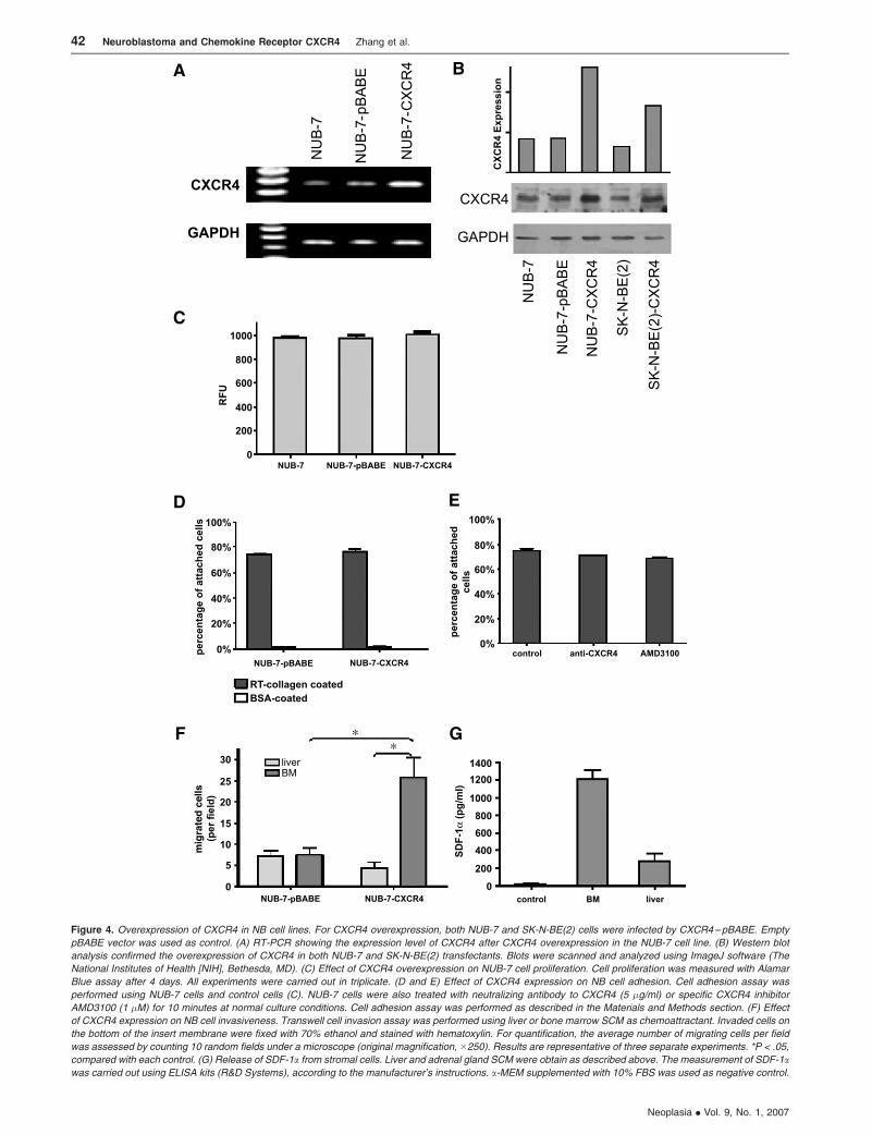
Figure 4. Overexpression of CXCR4 in NB cell lines. For CXCR4 overexpression, both NUB-7 and SK-N-BE(2) cells were infected by CXCR4–pBABE. Empty
pBABE vector was used as control. (A) RT-PCR showing the expression level of CXCR4 after CXCR4 overexpression in the NUB-7 cell line. (B) Western blot
analysis confirmed the overexpression of CXCR4 in both NUB-7 and SK-N-BE(2) transfectants. Blots were scanned and analyzed using ImageJ software (The
National Institutes of Health [NIH], Bethesda, MD). (C) Effect of CXCR4 overexpression on NUB-7 cell proliferation. Cell proliferation was measured with Alamar
Blue assay after 4 days. All experiments were carried out in triplicate. (D and E) Effect of CXCR4 expression on NB cell adhesion. Cell adhesion assay was
performed using NUB-7 cells and control cells (C). NUB-7 cells were also treated with neutralizing antibody to CXCR4 (5 �g/ml) or specific CXCR4 inhibitor
AMD3100 (1 �M) for 10 minutes at normal culture conditions. Cell adhesion assay was performed as described in the Materials and Methods section. (F) Effect
of CXCR4 expression on NB cell invasiveness. Transwell cell invasion assay was performed using liver or bone marrow SCM as chemoattractant. Invaded cells on
the bottom of the insert membrane were fixed with 70% ethanol and stained with hematoxylin. For quantification, the average number of migrating cells per field
was assessed by counting 10 random fields under a microscope (original magnification, �250). Results are representative of three separate experiments. *P < .05,
compared with each control. (G) Release of SDF-1a from stromal cells. Liver and adrenal gland SCM were obtain as described above. The measurement of SDF-1awas carried out using ELISA kits (R&D Systems), according to the manufacturer’s instructions. a-MEM supplemented with 10% FBS was used as negative control.
42 Neuroblastoma and Chemokine Receptor CXCR4 Zhang et al.
Neoplasia . Vol. 9, No. 1, 2007
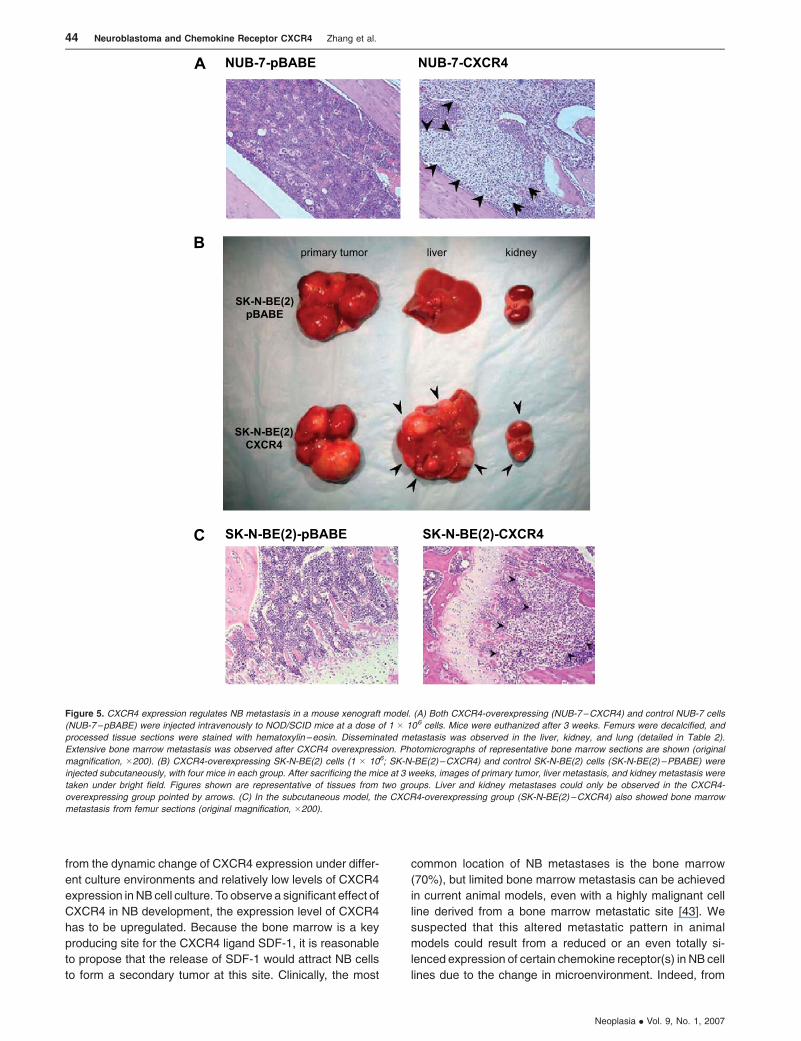
overexpressing or control NUB-7 cells. After 3 weeks, liver,
kidney, and lung metastases were observed in both groups.
However, only one of seven mice in the control group devel-
oped bone marrow metastasis, whereas six of seven mice in
the NUB-7–CXCR4 group had bone marrow metastasis
(Table 2 and Figure 5A). Thus, CXCR4 overexpression en-
hanced NB cell migration toward the bone marrow. Strik-
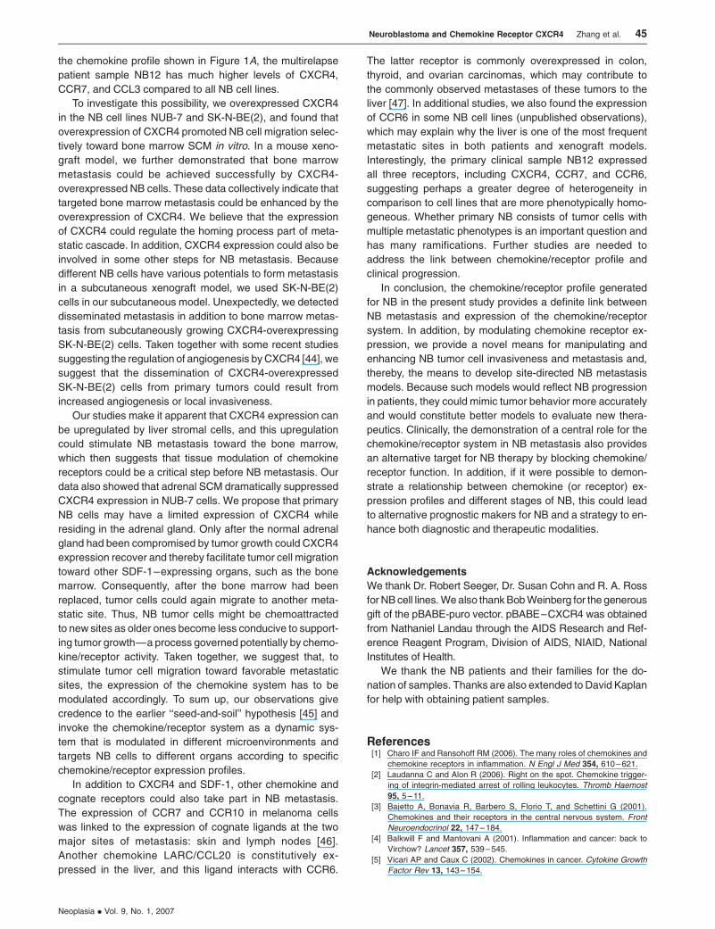
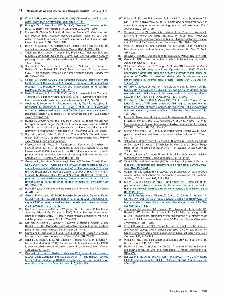
ingly, CXCR4-overexpressing SK-N-BE(2) cells and control
cells also produced similar size subcutaneous tumors;
however, extensive liver, kidney, and bone marrow metas-
tases developed only in the CXCR4 overexpression group
(Figure 5, B and C). All metastases were confirmed by
histologic analysis (data not shown). These results suggest
that the expression of CXCR4 could also stimulate NB
metastasis from a primary tumor site.
Discussion
It is believed that chemokines and chemokine receptors
form a complicated network in tumor progression [20]. Both
their expression profiles and regulatory mechanisms have
yet to be completely elucidated. To establish the role of the
chemokine system in NB metastasis, we first profiled five
chemokines and receptors (CCL2, CCL3, SDF-1, CXCR4,
and CCR7) and demonstrated differential expression in eight
NB cell lines and one primary culture from an NB patient
sample, NB12. These five chemokines and receptors have
been reported frequently for their potential role in the metas-
tasis of other tumor types to the bone marrow, liver, lymph
node, bone, or skin [21–25]—all common metastatic sites
in NB patients. Our studies demonstrated that CXCR4 is
expressed in most NB cell lines and in the primary tumor
sample NB12, which points to its potential involvement in NB
metastasis. Consistent with our finding, CXCR4 expression
was reported in other NB cell lines (including SH-SY5Y and
SK-N-MC), and CXCR4 was proven to be functional be-
cause it could bind its ligand SDF-1 on the cell surface, in-
ducing a rapid and transient intracellular calcium increase in
SK-N-SH cells [26].
Our key findings showed that in NB cells, the chemokine/
receptor system is a dynamic system that can be dynamically
modulated. A similar phenomenon was observed in dendritic
cell (DC) maturation where circulating DC precursors typi-
cally express just two chemokine receptors, CCR2 and
CXCR4.On entering tissues and differentiating into immature
DC, they express CCR1, CCR2, CCR5, CCR6, CXCR1,
CXCR2, and CXCR4. As these cells mature, expression of
these receptors is gradually lost, whereas expression of a
single lymphoid chemokine receptor CCR7 is rapidly induced
[27,28]. Studies in NB and other cancer types indicate that
different cell lines express different levels of chemokine
receptors. Even for the same cell line, different independent
studies demonstrated different levels [9,18]. All these find-
ings and our results suggest that the expression of chemo-
kine receptors could change in response to changes in their
microenvironments, especially during different stages in
NB metastasis.
Although it seems clear that some chemokine receptors,
especially CXCR4, are often highly expressed in malignant
tumors [9], the mechanism responsible for its upregulation
has not been completely elucidated. Tumor–stromal inter-
action could be a potential mechanism. Tumor–stromal
interaction is a very complicated process in tumor progres-
sion. Most studies on stromal cells have focused on integrins,
growth factors, and cytokines [29–31]. In this study, we ex-
plored tumor–stromal interaction through the chemokine/
receptor system and demonstrated for the first time that, in
NB, different stromata have divergent effects on the expres-
sion of chemokine receptors. Another important finding in
this study is that, in NB, the divergent effects of different stro-
mata on chemokine receptor expression are achieved by the
release of IL-5 and IFN-g at differential levels by stromal cells,
which indicates that tumor–stromal interaction could regu-
late the movement of NB cells to distant sites by modulating
the chemokine/receptor system. It has been demonstrated
that CXCR4 expression can be regulated positively by cyto-
kines such as TGF-b1, VEGF, and bFGF, and can be regu-
lated negatively by cytokines such as IL-5 and IFN-a in
leukocytes, endothelial cells, and neural cells [32–36]. It
was also demonstrated that hypoxia strongly affects the
chemokine system in macrophages [37]. Human macro-
phages exposed to low-oxygen conditions do not migrate in
response to CCL12 in hypoxic conditions [38]. By contrast,
CXCR4 expression and function in myeloid populations are
strongly increased through the activation of hypoxia-inducible
factor-1a [39]. There are very limited published data on cyto-
kine concentration in tissue microenvironments. Hogan and
Landreth [40] detected the production of IL-5 in bone marrow
stromal cells. It was also demonstrated that local overexpres-
sion of IFN-g in stromal cells was much more potent than the
exogenous addition of soluble cytokine to culture media [41].
Our in vitro studies demonstrated the regulation of CXCR4
expression by IL-5 and IFN-g at the nanomolar level. It is
therefore conceivable that physiological significant effects
could be achieved in in vivo microenvironments.
Chemokines and their receptors constitute a key chemo-
attractant mechanism used by both normal and tumor cells
[1–5]. So far, limited studies have been carried out on the
role of CXCR4 in NB metastasis, and unexplained inconsis-
tency was observed among results from different studies
[18,19,42]. We suspected that this discrepancy could result
Table 2. Dissemination of NUB-7 in NOD/SCID Mice after Intravenous
Injection of CXCR4-Overexpressing Cells.
Group n Lung
[n (%)]
Liver
[n (%)]
Kidney
[n (%)]
Bone Marrow
[n (%)]
NUB-7–pBABE 7 2 (28.6) 7 (100) 6 (85.7) 1 (14.3)
NUB-7–CXCR4 7 3 (42.9) 7 (100) 6 (85.7) 6 (85.7)
CXCR4 was overexpressed in NUB-7 cells. CXCR4-overexpressing NUB-7
cells (1 � 106; NUB-7–CXCR4) and vector control cells (NUB-7–pBABE)
were injected intravenously to NOD/SCID mice. Three weeks after injection,
the mice were sacrificed. Metastatic site formation was determined histo-
logically after hematoxylin –eosin staining. For each organ site, the number of
animals with disseminated NB is indicated, and the dissemination frequency
is shown in parentheses.
Neuroblastoma and Chemokine Receptor CXCR4 Zhang et al. 43
Neoplasia . Vol. 9, No. 1, 2007
from the dynamic change of CXCR4 expression under differ-
ent culture environments and relatively low levels of CXCR4
expression in NB cell culture. To observe a significant effect of
CXCR4 in NB development, the expression level of CXCR4
has to be upregulated. Because the bone marrow is a key
producing site for the CXCR4 ligand SDF-1, it is reasonable
to propose that the release of SDF-1 would attract NB cells
to form a secondary tumor at this site. Clinically, the most
common location of NB metastases is the bone marrow
(70%), but limited bone marrow metastasis can be achieved
in current animal models, even with a highly malignant cell
line derived from a bone marrow metastatic site [43]. We
suspected that this altered metastatic pattern in animal
models could result from a reduced or an even totally si-
lenced expression of certain chemokine receptor(s) in NB cell
lines due to the change in microenvironment. Indeed, from
Figure 5. CXCR4 expression regulates NB metastasis in a mouse xenograft model. (A) Both CXCR4-overexpressing (NUB-7–CXCR4) and control NUB-7 cells
(NUB-7–pBABE) were injected intravenously to NOD/SCID mice at a dose of 1 � 106 cells. Mice were euthanized after 3 weeks. Femurs were decalcified, and
processed tissue sections were stained with hematoxylin –eosin. Disseminated metastasis was observed in the liver, kidney, and lung (detailed in Table 2).
Extensive bone marrow metastasis was observed after CXCR4 overexpression. Photomicrographs of representative bone marrow sections are shown (original
magnification, �200). (B) CXCR4-overexpressing SK-N-BE(2) cells (1 � 106; SK-N-BE(2)–CXCR4) and control SK-N-BE(2) cells (SK-N-BE(2) –PBABE) were
injected subcutaneously, with four mice in each group. After sacrificing the mice at 3 weeks, images of primary tumor, liver metastasis, and kidney metastasis were
taken under bright field. Figures shown are representative of tissues from two groups. Liver and kidney metastases could only be observed in the CXCR4-
overexpressing group pointed by arrows. (C) In the subcutaneous model, the CXCR4-overexpressing group (SK-N-BE(2) –CXCR4) also showed bone marrow
metastasis from femur sections (original magnification, �200).
44 Neuroblastoma and Chemokine Receptor CXCR4 Zhang et al.
Neoplasia . Vol. 9, No. 1, 2007
the chemokine profile shown in Figure 1A, the multirelapse
patient sample NB12 has much higher levels of CXCR4,
CCR7, and CCL3 compared to all NB cell lines.
To investigate this possibility, we overexpressed CXCR4
in the NB cell lines NUB-7 and SK-N-BE(2), and found that
overexpression of CXCR4 promoted NB cell migration selec-
tively toward bone marrow SCM in vitro. In a mouse xeno-
graft model, we further demonstrated that bone marrow
metastasis could be achieved successfully by CXCR4-
overexpressed NB cells. These data collectively indicate that
targeted bone marrow metastasis could be enhanced by the
overexpression of CXCR4. We believe that the expression
of CXCR4 could regulate the homing process part of meta-
static cascade. In addition, CXCR4 expression could also be
involved in some other steps for NB metastasis. Because
different NB cells have various potentials to form metastasis
in a subcutaneous xenograft model, we used SK-N-BE(2)
cells in our subcutaneous model. Unexpectedly, we detected
disseminated metastasis in addition to bone marrow metas-
tasis from subcutaneously growing CXCR4-overexpressing
SK-N-BE(2) cells. Taken together with some recent studies
suggesting the regulation of angiogenesis byCXCR4 [44], we
suggest that the dissemination of CXCR4-overexpressed
SK-N-BE(2) cells from primary tumors could result from
increased angiogenesis or local invasiveness.
Our studies make it apparent that CXCR4 expression can
be upregulated by liver stromal cells, and this upregulation
could stimulate NB metastasis toward the bone marrow,
which then suggests that tissue modulation of chemokine
receptors could be a critical step before NB metastasis. Our
data also showed that adrenal SCM dramatically suppressed
CXCR4 expression in NUB-7 cells. We propose that primary
NB cells may have a limited expression of CXCR4 while
residing in the adrenal gland. Only after the normal adrenal
gland had been compromised by tumor growth could CXCR4
expression recover and thereby facilitate tumor cell migration
toward other SDF-1–expressing organs, such as the bone
marrow. Consequently, after the bone marrow had been
replaced, tumor cells could again migrate to another meta-
static site. Thus, NB tumor cells might be chemoattracted
to new sites as older ones become less conducive to support-
ing tumor growth—a process governed potentially by chemo-
kine/receptor activity. Taken together, we suggest that, to
stimulate tumor cell migration toward favorable metastatic
sites, the expression of the chemokine system has to be
modulated accordingly. To sum up, our observations give
credence to the earlier ‘‘seed-and-soil’’ hypothesis [45] and
invoke the chemokine/receptor system as a dynamic sys-
tem that is modulated in different microenvironments and
targets NB cells to different organs according to specific
chemokine/receptor expression profiles.
In addition to CXCR4 and SDF-1, other chemokine and
cognate receptors could also take part in NB metastasis.
The expression of CCR7 and CCR10 in melanoma cells
was linked to the expression of cognate ligands at the two
major sites of metastasis: skin and lymph nodes [46].
Another chemokine LARC/CCL20 is constitutively ex-
pressed in the liver, and this ligand interacts with CCR6.
The latter receptor is commonly overexpressed in colon,
thyroid, and ovarian carcinomas, which may contribute to
the commonly observed metastases of these tumors to the
liver [47]. In additional studies, we also found the expression
of CCR6 in some NB cell lines (unpublished observations),
which may explain why the liver is one of the most frequent
metastatic sites in both patients and xenograft models.
Interestingly, the primary clinical sample NB12 expressed
all three receptors, including CXCR4, CCR7, and CCR6,
suggesting perhaps a greater degree of heterogeneity in
comparison to cell lines that are more phenotypically homo-
geneous. Whether primary NB consists of tumor cells with
multiple metastatic phenotypes is an important question and
has many ramifications. Further studies are needed to
address the link between chemokine/receptor profile and
clinical progression.
In conclusion, the chemokine/receptor profile generated
for NB in the present study provides a definite link between
NB metastasis and expression of the chemokine/receptor
system. In addition, by modulating chemokine receptor ex-
pression, we provide a novel means for manipulating and
enhancing NB tumor cell invasiveness and metastasis and,
thereby, the means to develop site-directed NB metastasis
models. Because such models would reflect NB progression
in patients, they could mimic tumor behavior more accurately
and would constitute better models to evaluate new thera-
peutics. Clinically, the demonstration of a central role for the
chemokine/receptor system in NB metastasis also provides
an alternative target for NB therapy by blocking chemokine/
receptor function. In addition, if it were possible to demon-
strate a relationship between chemokine (or receptor) ex-
pression profiles and different stages of NB, this could lead
to alternative prognostic makers for NB and a strategy to en-
hance both diagnostic and therapeutic modalities.
Acknowledgements
We thank Dr. Robert Seeger, Dr. Susan Cohn and R. A. Ross
for NBcell lines.Wealso thankBobWeinberg for the generous
gift of the pBABE-puro vector. pBABE–CXCR4 was obtained
from Nathaniel Landau through the AIDS Research and Ref-
erence Reagent Program, Division of AIDS, NIAID, National
Institutes of Health.
We thank the NB patients and their families for the do-
nation of samples. Thanks are also extended to David Kaplan
for help with obtaining patient samples.
References[1] Charo IF and Ransohoff RM (2006). The many roles of chemokines and
chemokine receptors in inflammation. N Engl J Med 354, 610–621.
[2] Laudanna C and Alon R (2006). Right on the spot. Chemokine trigger-
ing of integrin-mediated arrest of rolling leukocytes. Thromb Haemost
95, 5–11.
[3] Bajetto A, Bonavia R, Barbero S, Florio T, and Schettini G (2001).
Chemokines and their receptors in the central nervous system. Front
Neuroendocrinol 22, 147–184.
[4] Balkwill F and Mantovani A (2001). Inflammation and cancer: back to
Virchow? Lancet 357, 539–545.
[5] Vicari AP and Caux C (2002). Chemokines in cancer. Cytokine Growth
Factor Rev 13, 143–154.
Neuroblastoma and Chemokine Receptor CXCR4 Zhang et al. 45
Neoplasia . Vol. 9, No. 1, 2007
[6] Ward SG, Bacon K, and Westwick J (1998). Chemokines and T lympho-
cytes: more than an attraction. Immunity 9, 1–11.
[7] Kuang Y, Wu Y, Jiang H, and Wu D (1996). Selective G protein coupling
by C–C chemokine receptors. J Biol Chem 271, 3975–3978.
[8] Sozzani S, Molino M, Locati M, Luini W, Cerletti C, Vecchi A, and
Mantovani A (1993). Receptor-activated calcium influx in human mono-
cytes exposed to monocyte chemotactic protein-1 and related cyto-
kines. J Immunol 150, 1544–1553.
[9] Balkwill F (2004). The significance of cancer cell expression of the
chemokine receptor CXCR4. Semin Cancer Biol 14, 171–179.
[10] Taichman RS, Cooper C, Keller ET, Pienta KJ, Taichman NS, and
McCauley LK (2002). Use of the stromal cell – derived factor-1/CXCR4
pathway in prostate cancer metastasis to bone. Cancer Res 62,
1832–1837.
[11] Scotton CJ, Wilson JL, Scott K, Stamp G, Wilbanks GD, Fricker S,
Bridger G, and Balkwill FR (2002). Multiple actions of the chemokine
CXCL12 on epithelial tumor cells in human ovarian cancer. Cancer Res
62, 5930–5938.
[12] Rempel SA, Dudas S, Ge S, and Gutierrez JA (2000). Identification and
localization of the cytokine SDF1 and its receptor, CXC chemokine
receptor 4, to regions of necrosis and angiogenesis in human glio-
blastoma. Clin Cancer Res 6, 102–111.
[13] Muller A, Homey B, Soto H, Ge N, Catron D, BuchananME, McClanahan
T, Murphy E, Yuan W, Wagner SN, et al. (2001). Involvement of chemo-
kine receptors in breast cancer metastasis. Nature 410, 50–56.
[14] Koshiba T, Hosotani R, Miyamoto Y, Ida J, Tsuji S, Nakajima S,
Kawaguchi M, Kobayashi H, Doi R, Hori T, et al. (2000). Expression
of stromal cell – derived factor 1 and CXCR4 ligand receptor system in
pancreatic cancer: a possible role for tumor progression. Clin Cancer
Res 6, 3530–3535.
[15] Burger M, Glodek A, Hartmann T, Schmitt-Graff A, Silberstein LE, Fujii
N, Kipps TJ, and Burger JA (2003). Functional expression of CXCR4
(CD184) on small-cell lung cancer cells mediates migration, integrin
activation, and adhesion to stromal cells. Oncogene 22, 8093–8101.
[16] Kryczek I, Wei S, Keller E, Liu R, and Zou W (2006). Stromal derived
factor (SDF-1/CXCL12) and human tumor pathogenesis. Am J Physiol
Cell Physiol (Epub ahead of print).
[17] Wysoczynski M, Reca R, Ratajczak J, Kucia M, Shirvaikar N,
Honczarenko M, Mills M, Wanzeck J, Janowska-Wieczorek A, and
Ratajczak MZ (2005). Incorporation of CXCR4 into membrane lipid rafts
primes homing-related responses of hematopoietic stem/progenitor
cells to an SDF-1 gradient. Blood 105, 40–48.
[18] Geminder H, Sagi-Assif O,Goldberg L,Meshel T, Rechavi G,Witz IP, and
Ben Baruch A (2001). A possible role for CXCR4 and its ligand, the CXC
chemokine stromal cell –derived factor-1, in the development of bone
marrow metastases in neuroblastoma. J Immunol 167, 4747–4757.
[19] Russell HV, Hicks J, Okcu MF, and Nuchtern JG (2004). CXCR4 ex-
pression in neuroblastoma primary tumors is associated with clinical
presentation of bone and bone marrow metastases. J Pediatr Surg
39, 1506–1511.
[20] Balkwill F (2004). Cancer and the chemokine network. Nat Rev Cancer
4, 540–550.
[21] Ghadjar P, Coupland SE, Na IK, Noutsias M, Letsch A, Stroux A, Bauer
S, Buhr HJ, Thiel E, Scheibenbogen C, et al. (2006). Chemokine re-
ceptor CCR6 expression level and liver metastases in colorectal cancer.
J Clin Oncol 24, 1910–1916.
[22] Menten P, Saccani A, Dillen C, Wuyts A, Struyf S, Proost P, Mantovani
A, Wang JM, and Van Damme J (2002). Role of the autocrine chemo-
kines MIP-1alpha and MIP-1beta in the metastatic behavior of murine T
cell lymphoma. J Leukoc Biol 72, 780–789.
[23] Lebrecht A, Grimm C, Lantzsch T, Ludwig E, Hefler L, Ulbrich E, and
Koelbl H (2004). Monocyte chemoattractant protein-1 serum levels in
patients with breast cancer. Tumour Biol 25, 14–17.
[24] Murakami T, Cardones AR, and Hwang ST (2004). Chemokine recep-
tors and melanoma metastasis. J Dermatol Sci 36, 71–78.
[25] Mashino K, Sadanaga N, Yamaguchi H, Tanaka F, Ohta M, Shibuta K,
Inoue H, and Mori M (2002). Expression of chemokine receptor CCR7
is associated with lymph node metastasis of gastric carcinoma. Cancer
Res 62, 2937–2941.
[26] Banisadr G, Dicou E, Berbar T, Rostene W, Lombet A, and Haour F
(2000). Characterization and visualization of [125I] stromal cell –derived
factor-1alpha binding to CXCR4 receptors in rat brain and human
neuroblastoma cells. J Neuroimmunol 110, 151–160.
[27] Sallusto F, Schaerli P, Loetscher P, Schaniel C, Lenig D, Mackay CR,
Qin S, and Lanzavecchia A (1998). Rapid and coordinated switch in
chemokine receptor expression during dendritic cell maturation. Eur J
Immunol 28, 2760–2769.
[28] Sozzani S, Luini W, Borsatti A, Polentarutti N, Zhou D, Piemonti L,
D’Amico G, Power CA, Wells TN, Gobbi M, et al. (1997). Receptor
expression and responsiveness of human dendritic cells to a defined
set of CC and CXC chemokines. J Immunol 159, 1993–2000.
[29] Park CC, Bissell MJ, and Barcellos-Hoff MH (2000). The influence of
the microenvironment on the malignant phenotype. Mol Med Today 6,
324–329.
[30] Bernards R (2003). Cancer: cues for migration. Nature 425, 247–248.
[31] Varani J (1987). Interaction of tumor cells with the extracellular matrix.
Revis Biol Cell 12, 1–113.
[32] Salcedo R, Wasserman K, Young HA, Grimm MC, Howard OM, Anver
MR, Kleinman HK, Murphy WJ, and Oppenheim JJ (1999). Vascular
endothelial growth factor and basic fibroblast growth factor induce ex-
pression of CXCR4 on human endothelial cells: in vivo neovasculari-
zation induced by stromal-derived factor-1alpha. Am J Pathol 154,
1125–1135.
[33] Rostasy K, Gorgun G, Kleyner Y, Garcia A, Kramer M, Melanson SM,
Mathys JM, Yiannoutsos C, Skolnik PR, and Navia BA (2005). Tumor
necrosis factor alpha leads to increased cell surface expression of
CXCR4 in SK-N-MC cells. J Neurovirol 11, 247–255.
[34] Franitza S, Kollet O, Brill A, Vaday GG, Petit I, Lapidot T, Alon R, and
Lider O (2002). TGF-beta1 enhances SDF-1alpha– induced chemo-
taxis and homing of naive T cells by up-regulating CXCR4 expression
and downstream cytoskeletal effector molecules. Eur J Immunol 32,
193–202.
[35] Iikura M, Miyamasu M, Yamaguchi M, Kawasaki H, Matsushima K,
Kitaura M, Morita Y, Yoshie O, Yamamoto K, and Hirai K (2001). Chemo-
kine receptors in human basophils: inducible expression of functional
CXCR4. J Leukoc Biol 70, 113–120.
[36] Shirazi Y and Pitha PM (1998). Interferon downregulates CXCR4 (fusin)
gene expression in peripheral blood mononuclear cells. J Hum Virol 1,
69–76.
[37] Schioppa T, Uranchimeg B, Saccani A, Biswas SK, Doni A, Rapisarda
A, Bernasconi S, Saccani S, Nebuloni M, Vago L, et al. (2003). Regu-
lation of the chemokine receptor CXCR4 by hypoxia. J Exp Med 198,
1391–1402.
[38] Turner L, Scotton C, Negus R, and Balkwill F (1999). Hypoxia inhibits
macrophage migration. Eur J Immunol 29, 2280–2287.
[39] Ceradini DJ and Gurtner GC (2005). Homing to hypoxia: HIF-1 as a
mediator of progenitor cell recruitment to injured tissue. Trends Cardio-
vasc Med 15, 57–63.
[40] Hogan MB and Landreth KS (2000). IL-5 production by bone marrow
stromal cells: implications for eosinophilia associated with asthma.
J Allergy Clin Immunol 106, 329–336.
[41] Selleri C, Maciejewski JP, Sato T, and Young NS (1996). Interferon-
gamma constitutively expressed in the stromal microenvironment of
human marrow cultures mediates potent hematopoietic inhibition. Blood
87, 4149–4157.
[42] Airoldi I, Raffaghello L, Piovan E, Cocco C, Carlini B, Amadori A,
Corrias MV, and Pistoia V (2006). CXCL12 does not attract CXCR4+
human metastatic neuroblastoma cells: clinical implications. Clin Can-
cer Res 12, 77–82.
[43] Thompson J, Guichard SM, Cheshire PJ, Richmond LB, Poquette CA,
Ragsdale ST, Webber B, Lorsbach R, Danks MK, and Houghton PJ
(2001). Development, characterization and therapy of a disseminated
model of childhood neuroblastoma in SCID mice. Cancer Chemother
Pharmacol 47, 211–221.
[44] Chen GS, Yu HS, Lan CCE, Chow KC, Lin TY, Kok LF, Lu MP, Liu CH,
and Wu MT (2006). CXC chemokine receptor CXCR4 expression en-
hances tumorigenesis and angiogenesis of basal cell carcinoma. Br J
Dermatol 154, 910–918.
[45] Paget S (1889). The distribution of secondary growths in cancer of the
breast. Lancet 133, 571–573.
[46] Payne AS and Cornelius LA (2002). The role of chemokines in
melanoma tumor growth and metastasis. J Invest Dermatol 118,
915–922.
[47] Schutyser E, Struyf S, and Van Damme J (2003). The CC chemokine
CCL20 and its receptor CCR6. Cytokine Growth Factor Rev 14,
409–426.
46 Neuroblastoma and Chemokine Receptor CXCR4 Zhang et al.
Neoplasia . Vol. 9, No. 1, 2007


