Embed Size (px)
Citation preview

The in situ light microenvironment of corals
Daniel Wangpraseurt,1 Lubos Polerecky,2,3 Anthony W. D. Larkum,1 Peter J. Ralph,1
Daniel A. Nielsen,1 Mathieu Pernice,1,4 and Michael Kuhl 1,5,6,*
1 Plant Functional Biology and Climate Change Cluster, University of Technology, Sydney, Australia2 Max Planck Institute for Marine Microbiology, Bremen, Germany3 Universiteit Utrecht, Department of Earth Sciences, Utrecht, The Netherlands4 Laboratory for Biological Geochemistry, Ecole Polytechnique Federale de Lausanne (EPFL), Lausanne, Switzerland5 Marine Biological Section, Department of Biology, University of Copenhagen, Helsingør, Denmark6 Singapore Centre on Environmental Life Sciences Engineering, School of Biological Sciences, Nanyang Technological University,
Singapore
Abstract
We used a novel diver-operated microsensor system to collect in situ spectrally resolved light fields on coralswith a micrometer spatial resolution. The light microenvironment differed between polyp and coenosarc tissueswith scalar irradiance (400–700 nm) over polyp tissue, attenuating between 5.1- and 7.8-fold from top to base ofsmall hemispherical coral colonies, whereas attenuation was at most 1.5-fold for coenosarc tissue. Fluctuations inambient solar irradiance induced changes in light and oxygen microenvironments, which were more pronouncedand faster in coenosarc compared with polyp tissue. Backscattered light from the surrounding benthoscontributed . 20% of total scalar irradiance at the coral tissue surface and enhanced symbiont photosynthesisand the local O2 concentration, indicating an important role of benthos optics for coral ecophysiology. Lightfields on corals are species and tissue specific and exhibit pronounced variation on scales from micrometers todecimeters. Consequently, the distribution, genetic diversity, and physiology of coral symbionts must be coupledwith the measurements of their actual light microenvironment to achieve a more comprehensive understanding ofcoral ecophysiology.
The quantity and quality of light is one of the mostimportant environmental factors affecting the ecology ofreef-forming symbiont-bearing corals (Dubinsky et al.1984; Falkowski et al. 1990; Iglesias-Prieto et al. 2004).Light drives photosynthesis of the endosymbiotic dinofla-gellate microalgae of the genus Symbiodinium that areknown as zooxanthellae and are harbored within the tissueof the cnidarian animal host. The coral host provides aprotected environment for its symbionts with limited butconstant nutrient availability in oligotrophic marinewaters. Zooxanthellae photosynthesis generates O2 andphotosynthates that provide the coral host with organiccarbon that can support . 95% of its respiratory demand(Muscatine et al. 1981). Although zooxanthellate corals aredependent on sufficient light for photosynthesis, high solarradiation during summertime in shallow waters can bestressful and cause the breakdown of symbiosis throughsymbiont expulsion or degradation, leading to visiblepaling of the colony (i.e., coral bleaching; Glynn 1996;Hoegh-Guldberg 1999). Various physiological aspects oflight harvesting and light-related bleaching have beenintensively studied over the past decades. However, adetailed understanding of the actual light field experiencedby the photosymbionts in the coral tissue is limited,although such knowledge is a prerequisite for a betterunderstanding of coral photobiology (Falkowski et al.1990; Iglesias-Prieto and Trench 1994; Lesser and Farrell2004) and ecophysiology (Rowan et al. 1997).
Solar radiation takes many detours until it reaches thetissue surface of a coral on a natural reef (Kirk 1994). Theinitial interaction of sunlight that has passed through theatmosphere is largely determined by the refractive indexdifference between air and seawater, causing refraction andreflection of incident radiation at the air–water interface.Light that has entered the water column undergoesscattering and absorption, which is caused by the inherentoptical properties of the water and a major contribution ofdissolved substances and solid particles (e.g., dissolvedorganic matter, plankton, suspended sediment, etc.; Kirk1994). The quantity of downwelling irradiance reaching acoral reef at a certain depth could in principle be calculatedby the spectral attenuation coefficient of the givenoverlying water mass, which would give a macroscale(i.e., meters to kilometers) approximation of irradianceover the area of interest (Kirk 1994).
However, for a given coral reef, irradiance is highlyvariable in both space and time. On a spatial scale, strongmeso- (millimeter to meter) and microscale (micrometer tomillimeter) light–matter interactions alter the light avail-ability and quality for photosynthetic reef organisms(Anthony and Hoegh-Guldberg 2003). Over time, irradi-ance varies on scales ranging from yearly down to thesmallest scales of milliseconds (Kirk 1994; Darecki et al.2011). Optical phenomena such as wave focusing can be animportant source of variability in the underwater light fieldcausing light flashes of high amplitude and frequency(Stramski and Dera 1988). Especially in shallow-waterenvironments, such as on coral reefs, wave focusing caninduce light flashes at frequencies of . 100 times per* Corresponding author: [email protected]
Limnol. Oceanogr., 59(3), 2014, 917–926
E 2014, by the Association for the Sciences of Limnology and Oceanography, Inc.doi:10.4319/lo.2014.59.3.0917
917

minute with maximal amplitudes exceeding the meanirradiance by more than fivefold (Darecki et al. 2011).
Studies dealing with the mesoscale light distribution oncoral reefs show that reef structures such as crevices andtopographic elevations are important sources of variabilityin the diffuse light component present within the coral reefframework (Brakel 1979; Stimson 1985; Anthony andHoegh-Guldberg 2003). Also, characteristic features of thecolony morphology (e.g., colony shape, branch length,spacing, etc.) cause significant light attenuation andredistribution within a single coral colony (Helmuth et al.1997; Anthony et al. 2005; Kaniewska et al. 2011). Forinstance, Kaniewska et al. (2011) showed that the incidentdownwelling irradiance measured above the coral tissuesurface varies about one order of magnitude from the tiptoward the base of a branch in the coral Stylophorapistillata. Although these mesoscale studies have givenvaluable insights, there are two major shortcomings withregard to their relevance for microalgal physiology.
Previous in situ studies have mainly quantified availablelight in terms of the incident downwelling irradiance (Ed).This parameter quantifies the downwelling quantum fluxfrom the upper hemisphere through a horizontal surfacearea and does not take backscattered light into account.However, light from all directions can be used for mic-roalgal photosynthesis; thus, Ed measurements generallyunderestimate the light available for symbiont photosyn-thesis in hospite (Kuhl et al. 1995). A more appropriateparameter for quantifying light exposure relevant formicroalgal photosynthesis is scalar irradiance, which is ameasure of the total radiant flux from all directions arounda point (Kuhl et al. 1995).
All in situ light field studies on corals have used sensorsthat detect light variation only on a macro- or mesoscale.However, recent laboratory studies have revealed thattissue and skeleton optics strongly alter coral light fields ona microscale (Enriquez et al. 2005; Wangpraseurt et al.2012a; Marcelino et al. 2013).
Light is strongly scattered at the water–tissue interfaceand within the coral tissue, where photon trapping andredistribution leads to significant enhancement in the localscalar irradiance compared with the incident downwellingirradiance (Kuhl et al. 1995; Wangpraseurt et al. 2012a).Light that has entered the tissue can be transferredlaterally, most likely through anisotropic scattering (Wang-praseurt et al. 2014). Additionally, reflective, fluorescent, orboth host pigments are synthesized by many corals, whichfurther alters the intensity and spectral quality of light dueto, for example, intense scattering and red-shifted emission(Salih et al. 2000). Finally, photons that pass through thetissue are backscattered by the aragonite skeleton, furtherenhancing tissue scalar irradiance and thus photonavailability for zooxanthellae photosynthesis (Enriquez etal. 2005; Marcelino et al. 2013). On a microscale, light isthus strongly affected by the inherent optical properties ofcorals, which can vary between coral species depending ontheir skeletal microstructure, tissue types, and degree ofpolyp contraction and expansion (Wangpraseurt et al.2012a; Marcelino et al. 2013; Yost et al. 2013). Microscalelight–tissue interactions can thus not be neglected if one
aims at a detailed understanding of coral photobiology(Wangpraseurt et al. 2014).
The assessment of microscale optics in corals in theirnatural habitats has until now been limited by the lack ofsuitable technology, making it impossible to examine therelationships between the macro-, meso- and microscalelight distributions in coral reefs. To bridge this gap, wedeveloped here a submersible, fiber-optic–based spectrom-eter module that can be connected to a diver-operatedmicrosensor system (Weber et al. 2007) to measure the firstspectrally resolved in situ microscale light measurements incorals. We used this instrument to study in situ spectralscalar irradiance at the coral tissue surface of variousmassive faviid corals and one branching acroporid. Wecompared the attenuation of light in a coral colony fromtop to base, focusing on differences between coenosarc andpolyp tissues. Additionally, we quantified the contributionof the benthos surrounding the coral to the local scalarirradiance at the coral surface and assessed its role in coralphotosynthesis. We discuss our results in the context ofmicroenvironmental controls of coral function and Sym-biodinium ecophysiology.
Methods
Study site and coral species—In situ microsensormeasurements were taken in November 2012 on theshallow reef flat next to the Heron Island Research station(152u069E, 20u299S), Southern Great Barrier Reef, Aus-tralia. Measurements were performed between 09:00 h and17:00 h at water depths ranging from 0.5 to 2.5 m (asmeasured from the benthos to the water surface). Low andhigh tide measurements were taken by snorkeling andSCUBA diving, respectively.
Massive corals of the family Faviidae (Goniastrea aspera,Platygyra lamellina, Favites pentagona) were chosen be-cause of their microscale tissue optical properties, aspreviously measured with microsensors under laboratoryconditions (Wangpraseurt et al. 2012a). The branchingAcropora millepora specimens were additionally selected tocompare light attenuation over massive corals with themore pronounced light attenuation known to occur inbranching growth forms (Kaniewska et al. 2011).
Underwater microsensor system—Ambient scalar irradi-ance of photosynthetically active radiation (PAR, 400–700 nm) was measured with a miniature scalar irradiancesensor (3 mm diameter; Walz GmbH) connected to anunderwater microsensor meter (UnderWater Meter system,Unisense A/S). Spectrally resolved scalar irradiance wasmeasured with a fiber-optic scalar irradiance microsensorprepared as previously described (Lassen et al. 1992). Themicrosensor had a spherical light-collecting tip with adiameter of , 80 mm and an isotropic angular response.Both sensors were linearly calibrated against a calibratedspherical quantum sensor (US-SQS/L, Heinz Walz GmbH)connected to a PAR light meter (Li-250A, Li-COR);equipment calibration took place at midday in a whiteseawater-filled container. The sensors were aligned next toeach other (2–3 cm distance) and submersed in the
918 Wangpraseurt et al.
container (depth of , 15 cm) such that the angle betweenthe sun and the sensor axis was 45u. Subsequently, thesensor readings were taken at 50% and 100% solarradiation (blue sky), the former achieved by a neutraldensity filter with 50% transmittance.
Oxygen measurements were made with a Clark-type O2
microsensor (Revsbech 1989). The sensor had a tipdiameter of , 50 mm, response time of , 2 s, and stirringsensitivity of , 1.5% and was adapted for underwater useas previously described (Wangpraseurt et al. 2012b). Linearcalibrations before and after each dive were done usingreadings in air-saturated and anoxic seawater, the latterachieved by flushing with N2 gas.
In situ microsensor measurements were performed using adiver-operated motorized microsensor (DOMS) profileroperated as previously described (Weber et al. 2007). The O2
microsensor and the PAR minisensor meter were connected tothe analog inputs of the DOMS, whereas the fiber-optic–basedscalar irradiance microsensor was connected to a separatewater-proof module. This module contained a spectrometer(USB 4000, Ocean Optics) and a custom-made board thatallowed acquisition and storage of spectra at time intervals of1 s or more, as triggered by a digital signal provided externallyby the DOMS (Fig. 1). The integration time intervals of thespectral acquisition were adjusted interactively during themeasurements to optimize the dynamic range of the sensor.The spectral signal output was followed during the measure-ments via a custom-made underwater PC module (Fig. 1). Atthe end of each deployment, the raw spectral data were readout via a custom-built circuit connected to a computer andprocessed as described below.
In situ measurements of the scalar irradiance distribution—To identify differences between coral species and differenttissue types with respect to their light microenvironment,spectral scalar irradiance was first measured on the upperlight-exposed surface of the corals and compared with theincident downwelling spectral irradiance (Fig. 2a). Thiswas done for coenosarc and polyp tissue of each of thethree massive faviid corals (P. lamellina, F. pentagona, andG. aspera). For each measurement, the microsensor wascarefully positioned at the corresponding tissue surfacewith the aid of a magnifying glass. The angle between thesensor and the coral–sun line was 45u to avoid self-shading.Scalar irradiance spectra were recorded in 5 s intervals overa period of 0.5–1 min and averaged. The incident down-welling spectral irradiance (Ed) was determined by mea-suring the signal above a black nonreflective surface next tothe coral at approximately the same height as the coralmeasurement spots; this was done for each coral after themicroscale scalar irradiance mapping (every 20 min).
To quantify the distribution of light at the coral surfaceover a larger scale, spectral scalar irradiance was addition-ally mapped from top to bottom of the coral colonies atthree or four positions over the coral colony (Fig. 2b) andfor each position over one coenosarc and one polyp tissuearea. During all measurements, the ambient PAR photonscalar irradiance next to the coral was monitored using theminiature spherical PAR sensor, arranged in the samedirection and at about the same height as the scalarirradiance microsensor. These data were used to accountfor small variations (generally , 10%) in the ambient lightfield by multiplying the values measured with the light
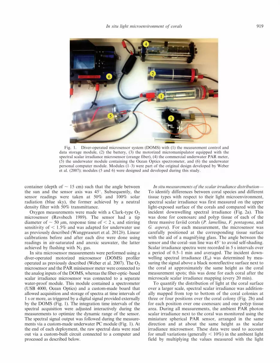
Fig. 1. Diver-operated microsensor system (DOMS) with (1) the measurement control anddata storage module, (2) the battery, (3) the motorized micromanipulator equipped with thespectral scalar irradiance microsensor (orange fiber), (4) the commercial underwater PAR meter,(5) the underwater module containing the Ocean Optics spectrometer, and (6) the underwaterpersonal computer module. Modules (1–3) were part of the original design developed by Weberet al. (2007); modules (5 and 6) were designed and developed during this study.
In situ light microenvironment of corals 919

microsensor on the coral with the factor by which theambient light field had changed.
Effect of backscattered light on coral light andO2 microenvironments—The relevance of diffuse light forscalar irradiance and O2 levels at the coral tissue surfacewas studied for G. aspera. The scalar irradiance and oxygenmicrosensors were positioned on the tissue surface close toeach other, both oriented at an angle of 45u relative to thecoral–sun line. The measured locations were on a coralsurface oriented at about 45u relative to the benthos surfaceand about 5 cm away from the benthos. Subsequently, athick black cloth (0.5 3 0.5 m) was placed above the coralor above the benthos next to the coral to block,respectively, the direct sunlight or backscattered light fromthe benthos (Fig. 2c,d) while measuring the scalar irradi-ance and oxygen concentrations. Measurements were doneat solar noon on both coenosarc and polyp tissues. Duringall measurements, the ambient PAR was recorded to ensurecomparable ambient irradiance regimes.
In situ dynamics of microscale scalar irradiance and O2—Using the same arrangement of microsensors as above,spectral scalar irradiance and O2 concentrations in coeno-sarc and polyp tissues of F. pentagona were monitoredcontinuously during early afternoon on a partially cloudyday. Ambient scalar irradiance was recorded during allmeasurements.
Data analysis—Data were analyzed with routines writtenin Matlab (MathWorks, version 2012a). Spectral data wereeither normalized to the incident downwelling irradiance orconverted to photon spectral scalar irradiance (mmolphotons m22 s21 nm21). The latter conversion involvedtwo steps. The raw USB4000 spectrometer data wascorrected for spectral sensitivity (mmol photons count21),based on sensitivity data acquired previously (Finke et al.2013) using a calibrated spectrometer (Jazz, Ocean Optics).The spectra acquired during the calibration experiment (seeabove) were then integrated over wavelengths in the PARregion and plotted against the corresponding output of thePAR sensor. This resulted in a calibration line whose slopewas subsequently used to convert all spectral sensitivity–corrected spectra to micromole photons per square meterper second per nanometer (mmol photons m22 s21 nm21).When relevant, spectra were also integrated over the400–700 nm wavelength range to quantify the total photonscalar irradiance of PAR.
Results
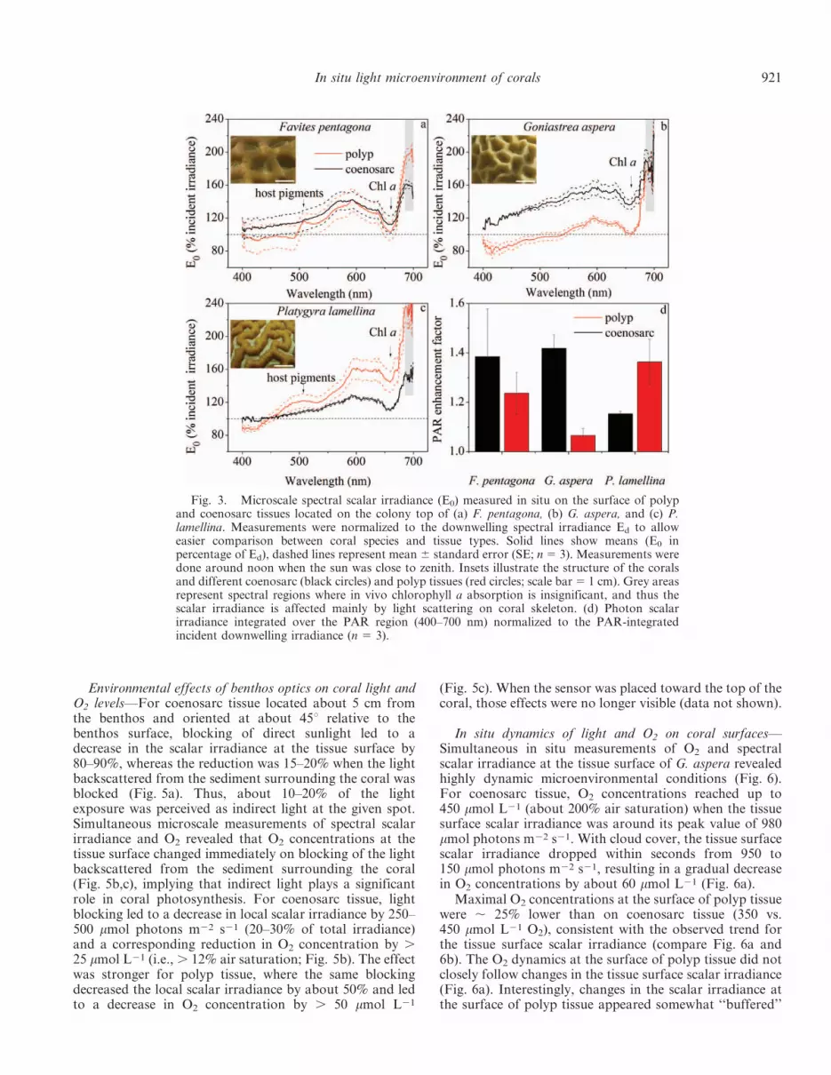
In situ spectral scalar irradiance at the upper surface offaviid corals—Spectral scalar irradiance at the uppersurfaces of faviid corals (E0) differed markedly from theincident downwelling irradiance (Ed; Fig. 3). Depending onthe wavelength in the PAR region, the E0 : Ed ratio variedbetween 0.8 and 2.4, with the most pronounced enhance-ment at wavelengths 500–640 nm and . 680 nm (Fig.3a–c). Coenosarc and polyp tissues had characteristicspectral signatures, which differed between the studiedcoral species (Fig. 3a–c). Contributions of fluorescent hostpigments could be clearly seen in the scalar irradiancespectra of the polyp tissue in P. lamellina and F. pentagona(arrows in Figs. 3a, 1c). Light in the far-red region (685–700 nm) was enhanced by about 40% and 80% in the polyptissue compared with coenosarc tissue in F. pentagona andP. lamellina, respectively, whereas such enhancement wasnot present in G. aspera.
The relative enhancement of integrated PAR (400–700 nm) differed at the tissue surface between coral speciesand tissue types (Fig. 3d). For instance, for P. lamellina,PAR was enhanced by about 36% in polyp tissue comparedwith 15% in coenosarc tissue, whereas this trend wasreversed for G. aspera (42% in coenosarc vs. 6% in polyp).
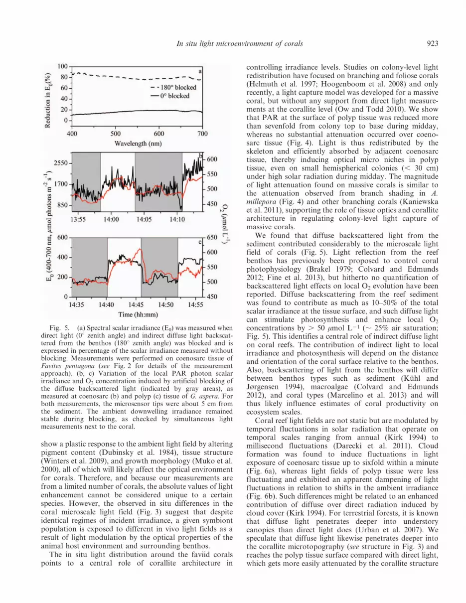
Light distribution along colony architecture—Variation ofscalar irradiance across massive corals differed stronglybetween polyp and coenosarc tissues (Fig. 4). While thedecrease in scalar irradiance from top to base of the coralcolonies was strong at the surface of polyp tissues (up to asevenfold decrease), for coenosarc tissues the scalarirradiance was fairly homogeneously distributed for F.pentagona, decreased up to 1.5-fold for P. lamellina, or evenincreased by about 10% toward the base for G. aspera. Forthe branching species A. millepora, scalar irradiance at thetissue surface decreased by about one order of magnitudefrom the apical tip toward the base of the branch (Fig.4m–o). For all studied coral species, these trends weresimilar for all wavelengths in the PAR region.
Fig. 2. Schematic representation of measurement geometryfor (a) upper surface mapping of different faviid coral species,where E0 was measured exclusively at the upper light-exposedsurfaces for coenosarc and polyp tissue (n 5 3). (b) Colony surfacemapping, where E0 was mapped from top to base around thecolony; one coenosarc and polyp tissue area were mapped each.(c) Contribution of direct (0u zenith angle) and indirect light (180uzenith angle) to E0 measured at , 45u from hemispherical colonycenter (around 5 cm from the benthos). We used a black cloth toblock out light from the different zenith angles. (d) Microscale O2
and E0 measurements following repeated darkening of thesediment benthos. (e) Temporal O2 and E0 dynamics on polypand coenosarc tissue measured on a cloudy day. The hemisphererepresents the idealized structure of the massive faviid corals. Thethick arrow represents the incident solar radiation (at 0u zenithangle, or varying angles over time if not specified), and the smallwhite arrows represent indirect, diffuse light. Black and white dotsshow relative measurement positions of tissue scalar irradiance E0
and O2 concentration, respectively.
920 Wangpraseurt et al.
Environmental effects of benthos optics on coral light andO2 levels—For coenosarc tissue located about 5 cm fromthe benthos and oriented at about 45u relative to thebenthos surface, blocking of direct sunlight led to adecrease in the scalar irradiance at the tissue surface by80–90%, whereas the reduction was 15–20% when the lightbackscattered from the sediment surrounding the coral wasblocked (Fig. 5a). Thus, about 10–20% of the lightexposure was perceived as indirect light at the given spot.Simultaneous microscale measurements of spectral scalarirradiance and O2 revealed that O2 concentrations at thetissue surface changed immediately on blocking of the lightbackscattered from the sediment surrounding the coral(Fig. 5b,c), implying that indirect light plays a significantrole in coral photosynthesis. For coenosarc tissue, lightblocking led to a decrease in local scalar irradiance by 250–500 mmol photons m22 s21 (20–30% of total irradiance)and a corresponding reduction in O2 concentration by .25 mmol L21 (i.e., . 12% air saturation; Fig. 5b). The effectwas stronger for polyp tissue, where the same blockingdecreased the local scalar irradiance by about 50% and ledto a decrease in O2 concentration by . 50 mmol L21
(Fig. 5c). When the sensor was placed toward the top of thecoral, those effects were no longer visible (data not shown).
In situ dynamics of light and O2 on coral surfaces—Simultaneous in situ measurements of O2 and spectralscalar irradiance at the tissue surface of G. aspera revealedhighly dynamic microenvironmental conditions (Fig. 6).For coenosarc tissue, O2 concentrations reached up to450 mmol L21 (about 200% air saturation) when the tissuesurface scalar irradiance was around its peak value of 980mmol photons m22 s21. With cloud cover, the tissue surfacescalar irradiance dropped within seconds from 950 to150 mmol photons m22 s21, resulting in a gradual decreasein O2 concentrations by about 60 mmol L21 (Fig. 6a).
Maximal O2 concentrations at the surface of polyp tissuewere , 25% lower than on coenosarc tissue (350 vs.450 mmol L21 O2), consistent with the observed trend forthe tissue surface scalar irradiance (compare Fig. 6a and6b). The O2 dynamics at the surface of polyp tissue did notclosely follow changes in the tissue surface scalar irradiance(Fig. 6a). Interestingly, changes in the scalar irradiance atthe surface of polyp tissue appeared somewhat ‘‘buffered’’
Fig. 3. Microscale spectral scalar irradiance (E0) measured in situ on the surface of polypand coenosarc tissues located on the colony top of (a) F. pentagona, (b) G. aspera, and (c) P.lamellina. Measurements were normalized to the downwelling spectral irradiance Ed to alloweasier comparison between coral species and tissue types. Solid lines show means (E0 inpercentage of Ed), dashed lines represent mean 6 standard error (SE; n 5 3). Measurements weredone around noon when the sun was close to zenith. Insets illustrate the structure of the coralsand different coenosarc (black circles) and polyp tissues (red circles; scale bar 5 1 cm). Grey areasrepresent spectral regions where in vivo chlorophyll a absorption is insignificant, and thus thescalar irradiance is affected mainly by light scattering on coral skeleton. (d) Photon scalarirradiance integrated over the PAR region (400–700 nm) normalized to the PAR-integratedincident downwelling irradiance (n 5 3).
In situ light microenvironment of corals 921

compared with the dynamic changes in the ambient scalarirradiance. For instance, a 4.4-fold decrease in ambientscalar irradiance (from 1750 to 400 mmol photons m22 s21)led to only a 2.4-fold decrease in scalar irradiance at thepolyp tissue (from 126 to 53 mmol photons m22 s21;Fig. 6b). In contrast, the relative changes in microscale andambient scalar irradiance were equal for coenosarc tissue.
Discussion
We used a novel diver-operated microsensor system forthe first in situ characterization of coral spectral light fieldswith micrometer spatial resolution. Our study providesevidence for the occurrence of different optical niches indifferent spatial compartments of corals under natural reef
conditions and highlights the importance of microscaleoptics in controlling coral light exposure.
Photon scalar irradiance of PAR was enhanced over theincident PAR, and the magnitude of light enhancementdiffered between the investigated coral colonies and theirtissue types (Fig. 3). Such modulation of microscaleirradiance with respect to incident irradiance is attributableto skeleton- and tissue-type–specific scattering and absorp-tion properties (Wangpraseurt et al. 2012a, 2014; Marce-lino et al. 2013). For instance, spectral signatures of hostpigments in polyp tissue of P. lamellina (Fig. 3a,c) likelyexplained the , 20% enhancement of PAR in polypcompared with coenosarc tissue, because fluorescent hostpigments around the polyp mouth can scatter light and leadto longer wavelength emission (Salih et al. 2000). Corals
Fig. 4. Macroscale in situ distributions of spectral scalar irradiance over coral colonies and branches measured separately on thesurface of polyp (dashed lines) and coenosarc (solid lines) tissues in locations marked by circles in the coral images. For A. millepora,polyp and coenosarc were not differentiated because of small polyp size. Also, because position 4 was deeper along the branch of thiscoral, it is not marked in the image. Bar graphs on the right show scalar irradiance integrated over three wavelength bands in the PARregion (see legend in panel O). Note the different y-axis scales. During the measurements, the PAR photon scalar irradiance above thesediment next to the coral was 2500 (F. pentagona), 2400 (G. aspera), 1700 (P. lamellina), and 1300 mmol photons m22 s21 (A. millepora).Scale bar 5 2 cm.
922 Wangpraseurt et al.
show a plastic response to the ambient light field by alteringpigment content (Dubinsky et al. 1984), tissue structure(Winters et al. 2009), and growth morphology (Muko et al.2000), all of which will likely affect the optical environmentfor corals. Therefore, and because our measurements arefrom a limited number of corals, the absolute values of lightenhancement cannot be considered unique to a certainspecies. However, the observed in situ differences in thecoral microscale light field (Fig. 3) suggest that despiteidentical regimes of incident irradiance, a given symbiontpopulation is exposed to different in vivo light fields as aresult of light modulation by the optical properties of theanimal host environment and surrounding benthos.
The in situ light distribution around the faviid coralspoints to a central role of corallite architecture in
controlling irradiance levels. Studies on colony-level lightredistribution have focused on branching and foliose corals(Helmuth et al. 1997; Hoogenboom et al. 2008) and onlyrecently, a light capture model was developed for a massivecoral, but without any support from direct light measure-ments at the corallite level (Ow and Todd 2010). We showthat PAR at the surface of polyp tissue was reduced morethan sevenfold from colony top to base during midday,whereas no substantial attenuation occurred over coeno-sarc tissue (Fig. 4). Light is thus redistributed by theskeleton and efficiently absorbed by adjacent coenosarctissue, thereby inducing optical micro niches in polyptissue, even on small hemispherical colonies (, 30 cm)under high solar radiation during midday. The magnitudeof light attenuation found on massive corals is similar tothe attenuation observed from branch shading in A.millepora (Fig. 4) and other branching corals (Kaniewskaet al. 2011), supporting the role of tissue optics and corallitearchitecture in regulating colony-level light capture ofmassive corals.
We found that diffuse backscattered light from thesediment contributed considerably to the microscale lightfield of corals (Fig. 5). Light reflection from the reefbenthos has previously been proposed to control coralphotophysiology (Brakel 1979; Colvard and Edmunds2012; Fine et al. 2013), but hitherto no quantification ofbackscattered light effects on local O2 evolution have beenreported. Diffuse backscattering from the reef sedimentwas found to contribute as much as 10–50% of the totalscalar irradiance at the tissue surface, and such diffuse lightcan stimulate photosynthesis and enhance local O2
concentrations by . 50 mmol L21 (, 25% air saturation;Fig. 5). This identifies a central role of indirect diffuse lighton coral reefs. The contribution of indirect light to localirradiance and photosynthesis will depend on the distanceand orientation of the coral surface relative to the benthos.Also, backscattering of light from the benthos will differbetween benthos types such as sediment (Kuhl andJørgensen 1994), macroalgae (Colvard and Edmunds2012), and coral types (Marcelino et al. 2013) and willthus likely influence estimates of coral productivity onecosystem scales.
Coral reef light fields are not static but are modulated bytemporal fluctuations in solar radiation that operate ontemporal scales ranging from annual (Kirk 1994) tomillisecond fluctuations (Darecki et al. 2011). Cloudformation was found to induce fluctuations in lightexposure of coenosarc tissue up to sixfold within a minute(Fig. 6a), whereas light fields of polyp tissue were lessfluctuating and exhibited an apparent dampening of lightfluctuations in relation to shifts in the ambient irradiance(Fig. 6b). Such differences might be related to an enhancedcontribution of diffuse over direct radiation induced bycloud cover (Kirk 1994). For terrestrial forests, it is knownthat diffuse light penetrates deeper into understorycanopies than direct light does (Urban et al. 2007). Wespeculate that diffuse light likewise penetrates deeper intothe corallite microtopography (see structure in Fig. 3) andreaches the polyp tissue surface compared with direct light,which gets more easily attenuated by the corallite structure
Fig. 5. (a) Spectral scalar irradiance (E0) was measured whendirect light (0u zenith angle) and indirect diffuse light backscat-tered from the benthos (180u zenith angle) was blocked and isexpressed in percentage of the scalar irradiance measured withoutblocking. Measurements were performed on coenosarc tissue ofFavites pentagona (see Fig. 2 for details of the measurementapproach). (b, c) Variation of the local PAR photon scalarirradiance and O2 concentration induced by artificial blocking ofthe diffuse backscattered light (indicated by gray areas), asmeasured at coenosarc (b) and polyp (c) tissue of G. aspera. Forboth measurements, the microsensor tips were about 5 cm fromthe sediment. The ambient downwelling irradiance remainedstable during blocking, as checked by simultaneous lightmeasurements next to the coral.
In situ light microenvironment of corals 923

(e.g., polyp walls; Figs. 3, 4). Thus, enhanced penetrationof diffuse light into the corallite matrix may counterbalancea decrease in the intensity of light during cloud cover andcould thus explain the observed dampening of temporalfluctuations in light capture present over polyp tissue.The dynamics reported are limited by the temporalresolution of our underwater meter, which operates onthe scale of seconds. High-amplitude, millisecond pulses oflight from wave focusing (Darecki et al. 2011) could thusnot be captured. Future in situ studies combining lightmicrosensors with systems capable of capturing high-frequency irradiance fluctuations are thus needed to resolvethe importance of high-frequency light pulses in coralphotophysiology.
Additionally, we found that the O2 microenvironmentwas highly dynamic in coenosarc tissue and fluctuatedclosely with changes in the ambient irradiance, whereas theO2 microenvironment of polyp tissue was less dynamic anddid not fluctuate simultaneously with changes to theambient irradiance (Fig. 6b). Such decoupling of O2 vs.irradiance fluctuations in polyp tissue is likely related to theintricate polyp topography and associated flow patternsforming complex patterns of O2 exchange with theenvironment (Wangpraseurt et al. 2012b). These observa-tions highlight that different spatial compartments within asingle coral colony also exhibit different temporal fluctu-ations of the local physicochemical microenvironment,
adding further complexity to the landscape of ecologicalmicroniches in corals.
Our results shed new light onto the control ofSymbiodinium ecophysiology. The distribution of Symbio-dinium geno- and phenotypes can be controlled byirradiance across water depth gradients (Rowan andKnowlton 1995) and within a single colony (Rowan et al.1997). However, often such spatial distribution patterns ofSymbiodinium in relation to irradiance are ambiguous(Warner et al. 2006; Ulstrup et al. 2007) and thus the role oflight compared with, for example, host specificity (Lajeu-nesse et al. 2004) in regulating Symbiodinium distributionwithin corals has remained disputed. If it is true thatirradiance controls Symbiodinium distribution (Rowanet al. 1997; Iglesias-Prieto et al. 2004), then any detailedpatterns will be masked by the spatial and temporalcomplexity of the light microenvironment reported here.Our results thus call for a reassessment of Symbiodiniumdistribution in relation to its actual light microenviron-ment. As a first step, it will be useful to compare differencesbetween coenosarc and polyp tissue because they differ intotal light exposure and spectral quality (Figs. 3, 4;Wangpraseurt et al. 2012a) and can exhibit differentpatterns of photoacclimation (Ralph et al. 2002).
The presence of different optical microniches in differentspatial compartments within corals supports the suggestionthat such niches can serve as refugia during light-related
Fig. 6. In situ dynamics of scalar irradiance (PAR) and O2 concentration at the surface of (a) coenosarc and (b) polyp tissue of theupper colony surface of P. lamellina during a sunny day with many intermittent clouds (onsets marked by arrows). Ambient scalarirradiance was measured next to the coral above strongly reflecting sediment.
924 Wangpraseurt et al.

bleaching conditions (Hoegh-Guldberg 1999; Loya et al.2001). For instance, polyp tissue at the sides of massivecorals will be effectively sheltered (Fig. 4), thereby allevi-ating local light stress during bleaching conditions. It isthus possible that minor symbiont populations are har-bored within those niches and can play an important rolefor the repopulation and redistribution of symbionts after ableaching event.
It has long been reported that an organism’s capacity toadapt to environmental change depends on its previousexposure to a given environmental parameter (e.g.,temperature or irradiance; Brown et al. 2002). Whereasinitially only the role of the organism’s exposure to theaverage of that parameter has been considered, morerecently, it has been proposed that adaptive capacity isdetermined by the degree of environmental variability(i.e., differences in the magnitude of fluctuation) theorganism has been exposed to (Deutsch et al. 2008). Thedifferences in fluctuation of the physicochemical micro-environment (i.e., light and O2) reported here thus suggestthat symbionts harbored within different spatial compart-ments (e.g., coenosarc vs. polyp; Fig. 6) have a differentexposure history of environmental variability. Suchdifferent exposure history could translate to and explaindifferential patterns of adaptation, acclimation capacity,or both characteristics observed in corals (Loya et al.2001). Although the detailed ecological implicationsremain to be investigated, we show here that coralsharbor complex light microenvironments that can now becharacterized at micrometer resolution under in situconditions. Such optical microniches show pronouncedspatiotemporal variation and differ strongly from theincident underwater irradiance regime, in terms of bothintensity and spectral quality. The optical properties ofthe surrounding benthos also affect local light fields andphotosynthesis in corals, and such interaction needsfurther attention in coral photobiology studies. A detailedunderstanding of the in situ microenvironmental ecologyof healthy corals will thus be a key to better interpret thespatiotemporal complexity of stress-related patternsobserved on reefs.
AcknowledgmentsWe thank Miriam Weber, Dirk De Beer, and Paul Faerber for
technical assistance and for providing the profiling underwatermicrosensor system, Ponchalart Chotikarn and Jim Franklin forlogistical and technical assistance, and L. F. Rickelt formanufacturing scalar irradiance microprobes. The technicians ofthe microsensor group at the Max Planck Institute in Bremen,Germany, are thanked for constructing O2 microsensors, as areKyra Hay and the staff at Heron Island Research Station, TheUniversity of Queensland, Australia, for their help during fieldwork. The research was conducted under research permits for fieldwork on the Great Barrier Reef (G12/35118.1).
This research was funded by grants from the AustralianResearch Council (A.W.D.L., P.J.R.), the Danish Council forIndependent Research | Natural Sciences (M.K.), the PlantFunctional Biology and Climate Change Cluster (D.W., M.K.,P.J.R.), the Max Planck Institute for Marine Microbiology (L.P.),and a postgraduate stipend from the University of Technology,Sydney (D.W.).
References
ANTHONY, K. R. N., AND O. HOEGH-GULDBERG. 2003. Variation incoral photosynthesis, respiration and growth characteristics incontrasting light microhabitats: an analogue to plants inforest gaps and understoreys? Funct. Ecol. 17: 246–259,doi:10.1046/j.1365-2435.2003.00731.x
———, M. O. HOOGENBOOM, AND S. R. CONNOLLY. 2005.Adaptive variation in coral geometry and the optimizationof internal colony light climates. Funct. Ecol. 19: 17–26,doi:10.1111/j.0269-8463.2005.00925.x
BRAKEL, W. H. 1979. Small-scale spatial variation in lightavailable to coral reef benthos: Quantum irradiance measure-ments from a Jamaican reef. Bull. Mar. Sci. 29: 406–413.
BROWN, B., R. DUNNE, M. GOODSON, AND A. DOUGLAS. 2002.Experience shapes the susceptibility of a reef coral tobleaching. Coral Reefs 21: 119–126.
COLVARD, N. B., AND P. J. EDMUNDS. 2012. Macroalgae on shallowtropical reefs reduce the availability of reflected light for usein coral photosynthesis. Bull. Mar. Sci. 88: 1019–1033,doi:10.5343/bms.2011.1084
DARECKI, M., D. STRAMSKI, AND M. SOKOLSKI. 2011. Measure-ments of high-frequency light fluctuations induced by seasurface waves with an Underwater Porcupine RadiometerSystem. J. Geophys. Res. Oceans 116: 1978–2012,doi:10.1029/2011JC007338
DEUTSCH, C. A., J. J. TEWKSBURY, R. B. HUEY, K. S. SHELDON,C. K. GHALAMBOR, D. C. HAAK, AND P. R. MARTIN. 2008.Impacts of climate warming on terrestrial ectotherms acrosslatitude. Proc. Natl. Acad. Sci. USA 105: 6668–6672,doi:10.1073/pnas.0709472105
DUBINSKY, Z., P. FALKOWSKI, J. PORTER, AND L. MUSCATINE. 1984.Absorption and utilization of radiant energy by light- andshade-adapted colonies of the hermatypic coral Stylophorapistillata. Proc. R. Soc. B 222: 203–214, doi:10.1098/rspb.1984.0059
ENRIQUEZ, S., E. R. MENDEZ, AND R. IGLESIAS-PRIETO. 2005.Multiple scattering on coral skeletons enhances light absorp-tion by symbiotic algae. Limnol. Oceanogr. 50: 1025–1032,doi:10.4319/lo.2005.50.4.1025
FALKOWSKI, P. G., P. L. JOKIEL, AND R. KINZIE. 1990. Irradianceand corals, p. 109–131. In Z. Dubisnky [ed.], Coral reefs.Ecosystems of the World. Elsevier.
FINE, M., S. SABBAH, N. SHASHAR, AND O. HOEGH-GULDBERG.2013. Light from down under. J. Exp. Bio. 216: 4341–4346,doi:10.1242/jeb.025106
FINKE, N., T. M. HOEHLER, L. POLERECKY, B. BUEHRING, AND B.THAMDRUP. 2013. Competition for inorganic carbon betweenoxygenic and anoxygenic phototrophs in a hypersalinemicrobial mat, Guerrero Negro, Mexico. Environ Microbiol.15: 1532–1550, doi:10.1111/1462-2920.12032
GLYNN, P. W. 1996. Coral reef bleaching: Facts, hypotheses andimplications. Glob. Change Biol. 2: 495–509, doi:10.1111/j.1365-2486.1996.tb00063.x
HELMUTH, B. S. T., B. E. H. TIMMERMAN, AND K. P. SEBENS. 1997.Interplay of host morphology and symbiont microhabitat incoral aggregations. Mar. Biol. 130: 1–10, doi:10.1007/s002270050219
HOEGH-GULDBERG, O. 1999. Climate change, coral bleaching andthe future of the world’s coral reefs. Mar. Freshw. Res. 50:839–866, doi:10.1071/MF99078
HOOGENBOOM, M. O., S. R. CONNOLLY, AND K. R. N. ANTHONY.2008. Interactions between morphological and physiologicalplasticity optimize energy acquisition in corals. Ecology 89:1144–1154, doi:10.1890/07-1272.1
In situ light microenvironment of corals 925

IGLESIAS-PRIETO, R., V. BELTRAN, T. LAJEUNESSE, H. REYES-BONILLA, AND P. THOME. 2004. Different algal symbiontsexplain the vertical distribution of dominant reef corals in theeastern Pacific. Proc. R. Soc. B 271: 1757–1763, doi:10.1098/rspb.2004.2757
———, AND R. K. TRENCH. 1994. Acclimation and adaptation toirradiance in symbiotic dinoflagellates. I. Responses of thephotosynthetic unit to changes in photon flux density. Mar.Ecol. Prog. Ser. 113: 163–175, doi:10.3354/meps113163
KANIEWSKA, P., S. H. MAGNUSSON, K. R. N. ANTHONY, R. REEF,M. KUHL, AND O. HOEGH-GULDBERG. 2011. Importance ofmacro- versus microstructure in modulating light levels insidecoral colonies. J. Phycol. 47: 846–860, doi:10.1111/j.1529-8817.2011.01021.x
KIRK, J. 1994. Light and photosynthesis in aquatic ecosystems.Cambridge Univ. Press.
KUHL, M., Y. COHEN, T. DALSGAARD, B. B. JØRGENSEN, AND N. P.REVSBECH. 1995. Microenvironment and photosynthesis ofzooxanthellae in scleractinian corals studied with microsen-sors for O2, pH and light. Mar. Ecol. Prog. Ser. 117: 159–172,doi:10.3354/meps117159
———, AND B. B. JØRGENSEN. 1994. The light-field of microbe-nthic communities—radiance distribution and microscaleoptics of sandy coastal sediments. Limnol. Oceanogr. 39:1368–1398, doi:10.4319/lo.1994.39.6.1368
LAJEUNESSE, T. C., D. J. THORNHILL, E. F. COX, F. G. STANTON,W. K. FITT, AND G. W. SCHMIDT. 2004. High diversity andhost specificity observed among symbiotic dinoflagellates inreef coral communities from Hawaii. Coral Reefs 23: 596–603.
LASSEN, C., H. PLOUG, AND B. B. JØRGENSEN. 1992. A fiberopticscalar irradiance microsensor—application for spectral lightmeasurements in sediments. FEMS Microbiol. Ecol. 86:247–254.
LESSER, M. P., AND J. H. FARRELL. 2004. Exposure to solarradiation increases damage to both host tissues and algalsymbionts of corals during thermal stress. Coral Reefs 23:367–377, doi:10.1007/s00338-004-0392-z
LOYA, Y., K. SAKAI, K. YAMAZATO, Y. NAKANO, H. SAMBALI, AND R.VAN WOESIK. 2001. Coral bleaching: The winners and the losers.Ecol. Lett. 4: 122–131, doi:10.1046/j.1461-0248.2001.00203.x
MARCELINO, L. A., AND OTHERS. 2013. Modulation of light-enhancement to symbiotic algae by light-scattering in coralsand evolutionary trends in bleaching. PLoS ONE 8: e61492,doi:10.1371/journal.pone.0061492
MUKO, S., K. KAWASAKI, K. SAKAI, F. TAKASU, AND N. SHIGESADA.2000. Morphological plasticity in the coral Porites sillimanianiand its adaptive significance. Bull. Mar. Sci. 66: 225–239.
MUSCATINE, L., L. R. MCCLOSKEY, AND R. E. MARIAN. 1981.Estimating the daily contribution of carbon from zooxan-thellae to coral animal respiration. Limnol. Oceanogr. 26:601–611, doi:10.4319/lo.1981.26.4.0601
OW, Y., AND P. TODD. 2010. Light-induced morphologicalplasticity in the scleractinian coral Goniastrea pectinata andits functional significance. Coral Reefs 29: 797–808,doi:10.1007/s00338-010-0631-4
RALPH, P., R. GADEMANN, A. LARKUM, AND M. KUHL. 2002.Spatial heterogeneity in active chlorophyll fluorescence andPSII activity of coral tissues. Mar. Biol. 141: 639–646,doi:10.1007/s00227-002-0866-x
REVSBECH, N. P. 1989. An oxygen microsensor with a guardcathode. Limnol. Oceanogr. 34: 474–478, doi:10.4319/lo.1989.34.2.0474
ROWAN, R., AND N. KNOWLTON. 1995. Intraspecific diversity andecological zonation in coral-algal symbiosis. Proc. Natl. Acad.Sci. USA 92: 2850–2853, doi:10.1073/pnas.92.7.2850
———, ———, A. BAKER, AND J. JARA. 1997. Landscape ecologyof algal symbionts creates variation in episodes of coralbleaching. Nature 388: 265–269, doi:10.1038/40843
SALIH, A., A. LARKUM, G. COX, M. KUHL, AND O. HOEGH-GULDBERG. 2000. Fluorescent pigments in corals are photo-protective. Nature 408: 850–853, doi:10.1038/35048564
STIMSON, J. 1985. The effect of shading by the table coral Acroporahyacinthus on understory corals. Ecology 66: 40–53,doi:10.2307/1941305
STRAMSKI, D., AND J. DERA. 1988. On the mechanism forproducing flashing light under a wind-disturbed watersurface. Oceanologia 25: 5–21.
ULSTRUP, K. E., M. J. H. VAN OPPEN, M. KUHL, AND P. J. RALPH.2007. Inter-polyp genetic and physiological characterisationof Symbiodinium in an Acropora valida colony. Mar. Biol. 153:225–234, doi:10.1007/s00227-007-0806-x
URBAN, O., AND OTHERS. 2007. Ecophysiological controls over thenet ecosystem exchange of mountain spruce stand. Compar-ison of the response in direct vs. diffuse solar radiation. Glob.Change Biol. 13: 157–168, doi:10.1111/j.1365-2486.2006.01265.x
WANGPRASEURT, D., A. W. D. LARKUM, P. J. RALPH, AND M.KUHL. 2012a. Light gradients and optical microniches in coraltissues. Front. Microbiol. 3: 316, doi:10.3389/fmicb.2012.00316
———, ———, J. FRANKLIN, M. SZABO, P. J. RALPH, AND M.KUHL. 2014. Lateral light transfer ensures efficient resourcedistribution in symbiont-bearing corals. J. Exp. Biol. 217:489–498, doi:10.1242/jeb.091116
———, M. WEBER, H. RØY, L. POLERECKY, D. DE BEER,SUHARSONO, AND M. M. NUGUES. 2012b. In situ oxygendynamics in coral–algal interactions. PLoS ONE 7: e31192,doi:10.1371/journal.pone.0031192
WARNER, M. E., T. C. LAJEUNESSE, J. D. ROBISON, AND R. M.THUR. 2006. The ecological distribution and comparativephotobiology of symbiotic dinoflagellates from reef corals inBelize: Potential implications for coral bleaching. Limnol.Oceanogr. 51: 1887–1897, doi:10.4319/lo.2006.51.4.1887
WEBER, M., P. FAERBER, V. MEYER, C. LOTT, G. EICKERT, K. E.FABRICIUS, AND D. DE BEER. 2007. In situ applications of anew diver-operated motorized microsensor profiler. Environ.Sci. Technol. 41: 6210–6215, doi:10.1021/es070200b
WINTERS, G., S. BEER, B. B. ZVI, I. BRICKNER, AND Y. LOYA. 2009.Spatial and temporal photoacclimation of Stylophora pistil-lata: Zooxanthella size, pigmentation, location and clade.Mar. Ecol. Prog. Ser. 384: 107–119, doi:10.3354/meps08036
YOST, D. M., L.-H. WANG, T.-Y. FAN, C.-S. CHEN, R. W. LEE, E.SOGIN, AND R. D. GATES. 2013. Diversity in skeletalarchitecture influences biological heterogeneity and Symbio-dinium habitat in corals. Zoology 116: 262–269, doi:10.1016/j.zool.2013.06.001
Associate editor: Dariusz Stramski
Received: 09 September 2013Accepted: 27 January 2014
Amended: 10 February 2014
926 Wangpraseurt et al.