Embed Size (px)
Citation preview
RESEARCH ARTICLE
Does Dark-Spot Syndrome ExperimentallyTransmit among Caribbean Corals?Carly J. Randall1*, Adán G. Jordán-Garza1¤, Erinn M. Muller2, Robert vanWoesik1
1 Department of Biological Sciences, Florida Institute of Technology, Melbourne, Florida, United States ofAmerica, 2 Mote Marine Laboratory, Sarasota, Florida, United States of America
¤ Current address: Universidad Veracruzana, Tuxpan, Veracruz, México* [email protected]
AbstractOver the last half-century, coral diseases have contributed to the rapid decline of coral pop-
ulations throughout the Caribbean region. Some coral diseases appear to be potentially
infectious, yet little is known about their modes of transmission. This study experimentally
tested whether dark-spot syndrome on Siderastrea siderea was directly or indirectly trans-missible to neighboring coral colonies. We also tested whether open wounds were neces-
sary to facilitate disease transmission. At the completion of the experiments, we sampled
bacterial communities on diseased, exposed, and healthy coral colonies to determine
whether bacterial pathogens had transmitted to the susceptible colonies. We saw no evi-
dence of either direct or waterborne transmission of dark-spot syndrome, and corals that
received lesions by direct contact with diseased tissue, healed and showed no signs of
infection. There were no significant differences among bacterial communities on healthy,
exposed, and diseased colonies, although nine individual ribotypes were significantly
higher in diseased corals compared with healthy and exposed corals, indicating a lack of
transmission. Although our experiments do not fully refute the possibility that dark-spot syn-
drome is infectious and transmissible, our results suggest that in situmacroscopic signs of
dark-spot syndrome are not always contagious.
IntroductionOver the last half-century, marine diseases have contributed to the wide-spread decline ofCaribbean corals [1–4]. Despite decades of research, the etiologies of most coral diseases arepoorly understood. Several coral diseases are known to be infectious, and are associated withpathogens such as bacteria, protists, fungi, or with a consortium of microbes [5–16]. Yet thedegree to which infectious diseases are contagious is highly variable [17], and few studies haveexperimentally tested the potential transmission of several common coral diseases in the Carib-bean. Understanding the dynamics of coral-disease transmission is necessary to accurately pre-dict future outbreaks of coral diseases, and to potentially mitigate the spread of those diseases.
Infectious diseases are caused by the invasion of a host by a pathogenic agent [17–18]. Inveterinary epidemiology, there are two types of infectious agents: exogenous pathogens andendogenous pathogens [17]. Exogenous pathogens are present in the external environment,
PLOSONE | DOI:10.1371/journal.pone.0147493 January 20, 2016 1 / 16
OPEN ACCESS
Citation: Randall CJ, Jordán-Garza AG, Muller EM,van Woesik R (2016) Does Dark-Spot SyndromeExperimentally Transmit among Caribbean Corals?PLoS ONE 11(1): e0147493. doi:10.1371/journal.pone.0147493
Editor: Christina A. Kellogg, U.S. Geological Survey,UNITED STATES
Received: October 29, 2015
Accepted: January 5, 2016
Published: January 20, 2016
Copyright: © 2016 Randall et al. This is an openaccess article distributed under the terms of theCreative Commons Attribution License, which permitsunrestricted use, distribution, and reproduction in anymedium, provided the original author and source arecredited.
Data Availability Statement: All data are containedwithin the paper or the supplementary figures andtables.
Funding: Funding for this study was provided by theNational Science Foundation award #OCE-1219804awarded to RvW.
Competing Interests: The authors have declaredthat no competing interests exist.
and are acquired by exposure to a carrier organism, to a non-biological vector, or to an infectedhost. These pathogens cause diseases that have clear clinical signs and have distinct pathologi-cal lesions [17]. By contrast, endogenous pathogens often are present in healthy hosts, butcause disease only when the host becomes stressed. For most coral diseases, it is unknownwhether the diseases are caused by exogenous or endogenous pathogens, or if instead, they arethe result of non-infectious diseases [19].
Addressing the question of coral-disease transmission has proved difficult for many reasons,including the following: firstly, pathogens for many diseases are unknown or are inconsistentlydiagnosed [20–22]. Secondly, the diagnosis of infection is based on the subjective identificationof macroscopic signs, potentially of different origins [21, 23]. Thirdly, complex interactionsamong the pathogens, the host corals, and the environment may exist, which could result insuccessful transmission of a disease under one set of environmental conditions, and in unsuc-cessful transmission of a disease under another set of environmental conditions [24]. Fourthly,unknown and potentially complex stages of a disease can exist whereby transmission succeedswhen the disease is in one stage, and fails when the disease is in another stage [25]. Fifthly,there is the potential existence of vectors, or carriers, in which disease signs do not manifest[26]. Despite these difficulties, examining coral-disease transmission is necessary for elucidat-ing disease etiology.
In this study we define contagious diseases as those which are transmissible through directphysical contact between corals, or which are transmissible indirectly through a vector [27].Given that corals are sessile and that Caribbean reefs currently support low coral cover [28],physical contact between corals is limited, and it is likely, therefore, that direct transmission ofcontemporary coral diseases is uncommon [29]. It is more likely that if coral diseases are beingtransmitted throughout the Caribbean region, the transmission would be indirect, either via awaterborne pathogen or through a reef-associated vector.
Dark-spot syndrome is one of the most prevalent and ubiquitous coral diseases in the Carib-bean. Although this disease is thought to be potentially infectious and transmissible, dark-spottransmission has yet to be experimentally demonstrated [30–37]. Dark-spot syndrome affectsprimarily Siderastrea siderea, O. faveolata, and Stephanocoenia intersepta, although it has beenobserved on at least seventeen species of scleractinian corals in the Caribbean [30, 34, 38, 39],and some researchers have argued that the extent of dark-spot syndrome is an important indica-tor of overall reef health [33, 40]. A causative agent of dark-spot syndrome has not been identi-fied, although several infectious agents have been proposed, including bacteria from the generaVibrio [32], Corynebacterium, Photobacterium, and Acinetobacter, [36], bacteria from the familyParvularculaceae [36], fungi resembling Aspergillus spp. [35] and Rhytisma spp. [36], and cyano-bacteria resemblingOscillatoria spp. [36]. A study recently published by Kellogg et al. [37] foundno significant distinctions between bacterial communities in healthy and dark-spot affected S.siderea from St. John, USVI and the Dry Tortugas, suggesting that bacterial pathogens were notthe causative agents of the disease. Some assessments suggest that dark-spot syndrome affectsprimarily the symbiotic zooxanthellae within the coral tissue, causing impairment of mitosis, anda reduction in the density of symbionts [31]. Yet, field studies have found that dark-spot syn-drome on corals can result from the physical abrasion of the coral tissue, and that disease signsdo not always cause tissue mortality [33, 41]. This has led some researchers to suggest that dark-spot syndrome is a stress response, rather than an infectious disease [19, 33].
The objectives of the present study were to: (1) test direct-contact transmission and water-borne transmission of dark-spot syndrome on Siderastrea siderea; and (2) compare the bacte-rial communities on diseased, disease-exposed, and healthy Siderastrea siderea colonies at thecompletion of the experiment to examine whether potential bacterial pathogens transmitted tothe exposed colonies.
Testing Transmission of Caribbean Dark-Spot Syndrome
PLOSONE | DOI:10.1371/journal.pone.0147493 January 20, 2016 2 / 16
Materials and Methods
Ethics statementCoral collections took place under a permit issued by the Florida Keys National Marine Sanctu-ary numbered FKNMS-2013-086. No ethical approval was required for the laboratory researchdescribed in this study.
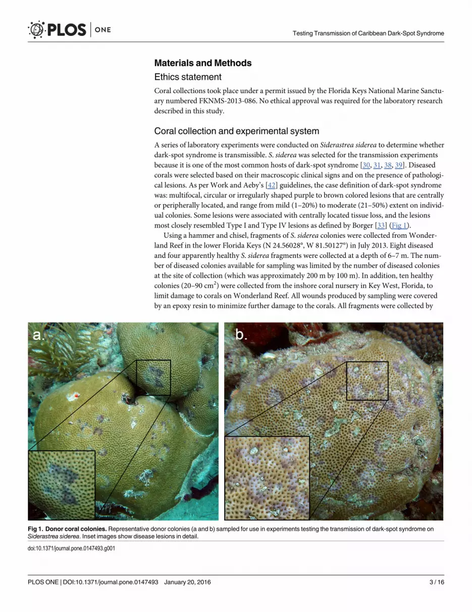
Coral collection and experimental systemA series of laboratory experiments were conducted on Siderastrea siderea to determine whetherdark-spot syndrome is transmissible. S. siderea was selected for the transmission experimentsbecause it is one of the most common hosts of dark-spot syndrome [30, 31, 38, 39]. Diseasedcorals were selected based on their macroscopic clinical signs and on the presence of pathologi-cal lesions. As per Work and Aeby’s [42] guidelines, the case definition of dark-spot syndromewas: multifocal, circular or irregularly shaped purple to brown colored lesions that are centrallyor peripherally located, and range from mild (1–20%) to moderate (21–50%) extent on individ-ual colonies. Some lesions were associated with centrally located tissue loss, and the lesionsmost closely resembled Type I and Type IV lesions as defined by Borger [33] (Fig 1).
Using a hammer and chisel, fragments of S. siderea colonies were collected fromWonder-land Reef in the lower Florida Keys (N 24.56028°, W 81.50127°) in July 2013. Eight diseasedand four apparently healthy S. siderea fragments were collected at a depth of 6–7 m. The num-ber of diseased colonies available for sampling was limited by the number of diseased coloniesat the site of collection (which was approximately 200 m by 100 m). In addition, ten healthycolonies (20–90 cm2) were collected from the inshore coral nursery in Key West, Florida, tolimit damage to corals on Wonderland Reef. All wounds produced by sampling were coveredby an epoxy resin to minimize further damage to the corals. All fragments were collected by
Fig 1. Donor coral colonies. Representative donor colonies (a and b) sampled for use in experiments testing the transmission of dark-spot syndrome onSiderastrea siderea. Inset images show disease lesions in detail.
doi:10.1371/journal.pone.0147493.g001
Testing Transmission of Caribbean Dark-Spot Syndrome
PLOSONE | DOI:10.1371/journal.pone.0147493 January 20, 2016 3 / 16
divers wearing surgical gloves, and healthy fragments were collected prior to diseased frag-ments, to prevent the potential exposure of healthy fragments to disease during the collectionprocess. All corals were individually packaged in Ziploc1 bags with seawater and transportedin separate coolers, for healthy and diseased corals, to Mote Marine Laboratory in SummerlandKey. Immediately following collection and transportation, corals were fragmented, with ableach-rinsed (25% bleach solution) wet-tile saw, into ~16 cm2 colonies, mounted on 2.5 cmwide by 2.0 cm tall polyvinyl chloride (PVC) bases with Plastilina1modeling clay, and placedinto randomly assigned treatment tanks. All corals were handled with surgical gloves, and allequipment was rinsed with a 25% bleach solution in-between the handling of each coralfragment.
The outdoor experimental system was supplied with flow-through seawater fromMoteMarine Laboratory’s well-water system [43]. Seawater was off-gassed in a head-tank to main-tain pH at 7.9–8.1, and was mechanically filtered with a sand-particle filter. Seawater enteringthe flow-through system was ultraviolet-light (UV) sterilized with an 18W Coralife1 Tur-botwist UV Sterilizer to ensure that the supply of water was free of pathogens. The system wascovered with shade cloth to match the irradiance levels in the aquaria as closely as possiblewith the irradiance levels that were measured at the site of coral collection (58 μmol quanta m-2
s-1 at approximately 10:50 am). All experiments were conducted between 10 July and 14 August2013 at Mote Marine Laboratory in Summerland Key, Florida.
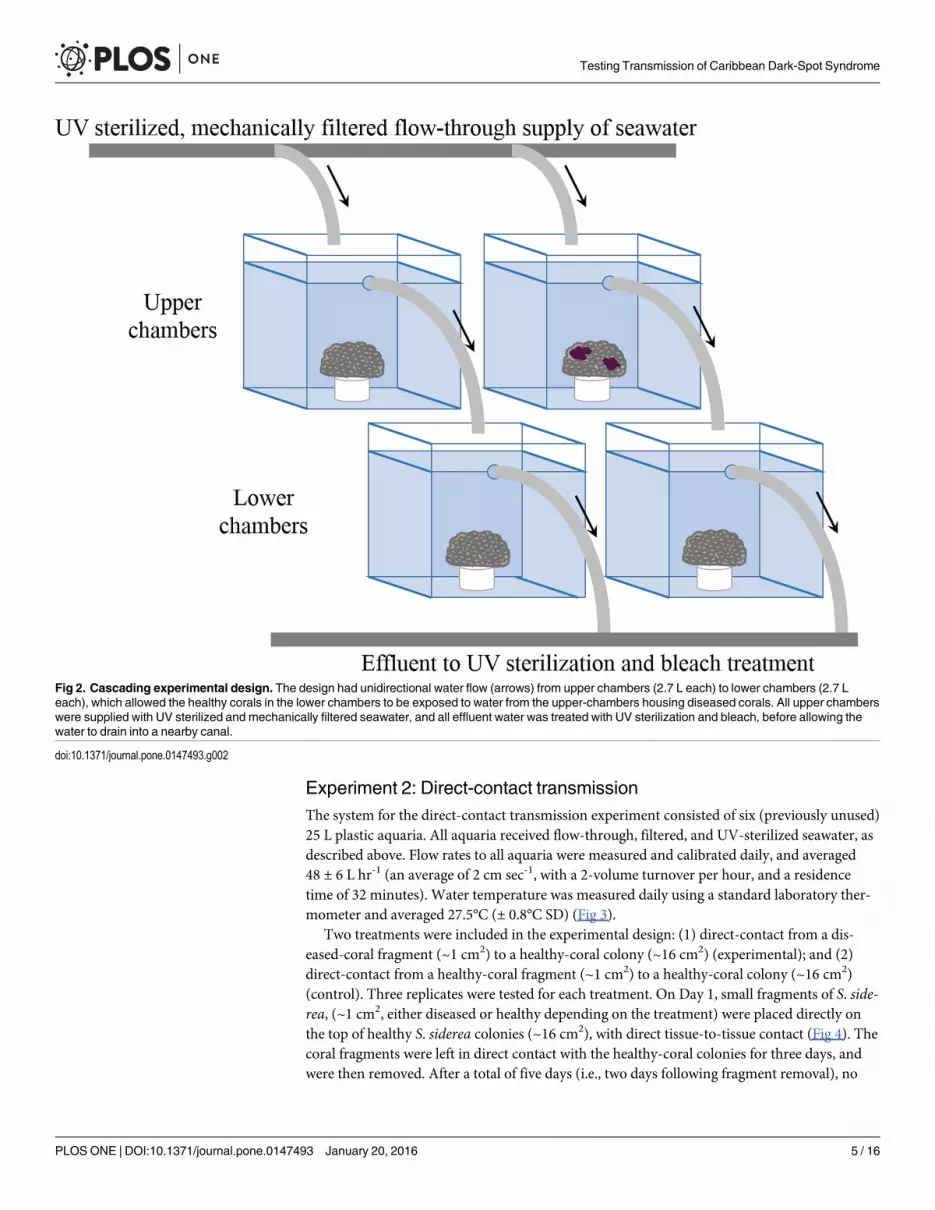
Experiment 1: Waterborne transmissionThe system for the waterborne-transmission experiments consisted of sixty (previouslyunused) 2.7 L acrylic aquaria, set-up in a cascading design (Fig 2). This design allowed for uni-directional flow to test waterborne-disease transmission by exposing corals in the ‘lower cham-bers’ to water that had been previously circulated in the ‘upper chambers’ housing diseasedcorals. Flow rates in all aquaria were measured and calibrated daily. The flow rates averaged28 ± 4 L hr-1 (an average of 2 cm sec-1 with 11 volume turnovers per hour, and a residence timeof 6 minutes). Water temperatures were measured daily with a standard laboratory thermome-ter and averaged 27.4°C (± 0.6°C SD; Fig 3).
Four treatments were included in the experimental design: (i) transmission from a diseased-coral colony to a healthy-coral colony (experimental); (ii) transmission from a healthy-coralcolony to a healthy-coral colony (control 1); (iii) transmission from a rubble fragment to ahealthy-coral colony (control 2); and (iv) transmission from an empty chamber to a healthy-coral colony (control 3). Seven replicates of each treatment pair were tested. The waterborne-transmission experiment was run for 11 days, after which the experimental corals were placedin a holding tank and monitored for an additional 17 days (for a total of 28 days). All coral col-onies were monitored for signs of disease and photographed daily.
To compare the photochemical yields of colonies within each treatment, pulse amplitudemodulated (PAM) fluorometry measurements were taken three hours after sunset (i.e., three-hour dark-acclimated) on Days 6 and 9 of the experiment. Two technical-replicate measurementswere taken on each colony on different locations within the same colony, and were averaged toobtain one measurement of photochemical yield per colony. PAMmeasurements of the diseasedcolony were taken directly on tissue with visible lesions. The PAM probe was rinsed with a 10%bleach solution, and then rinsed with sterilized seawater in-between each measurement.
A repeated measures analysis of variance (RM-ANOVA) was used to assess differences inphotochemical yields among treatments on the same corals through time. Photochemical-yielddata met the assumptions of RM-ANOVA, and statistical analyses were conducted using baseR, and the packages ‘car’ [45] and ‘userfriendlyscience’ [46] in the statistical program R [47].
Testing Transmission of Caribbean Dark-Spot Syndrome
PLOSONE | DOI:10.1371/journal.pone.0147493 January 20, 2016 4 / 16
Experiment 2: Direct-contact transmissionThe system for the direct-contact transmission experiment consisted of six (previously unused)25 L plastic aquaria. All aquaria received flow-through, filtered, and UV-sterilized seawater, asdescribed above. Flow rates to all aquaria were measured and calibrated daily, and averaged48 ± 6 L hr-1 (an average of 2 cm sec-1, with a 2-volume turnover per hour, and a residencetime of 32 minutes). Water temperature was measured daily using a standard laboratory ther-mometer and averaged 27.5°C (± 0.8°C SD) (Fig 3).
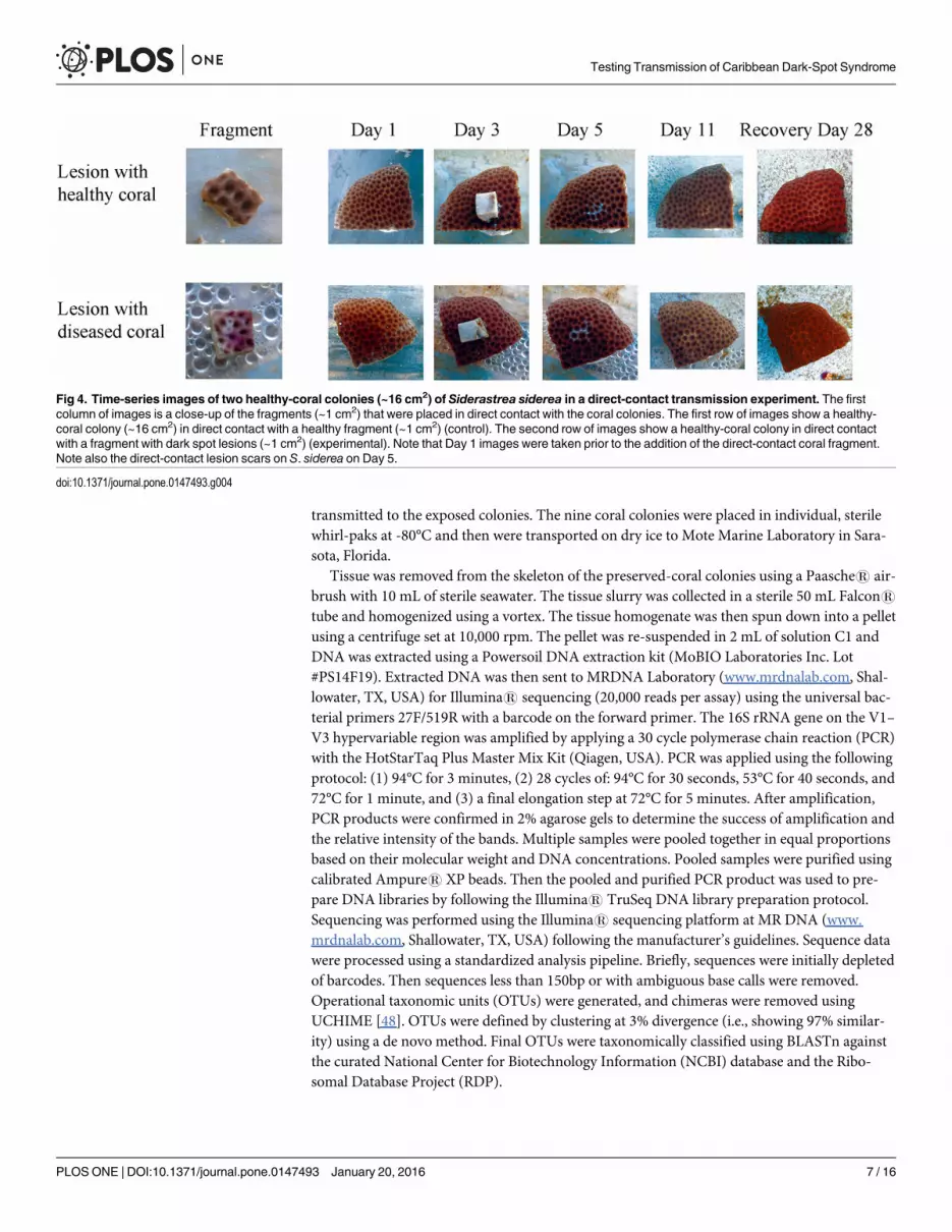
Two treatments were included in the experimental design: (1) direct-contact from a dis-eased-coral fragment (~1 cm2) to a healthy-coral colony (~16 cm2) (experimental); and (2)direct-contact from a healthy-coral fragment (~1 cm2) to a healthy-coral colony (~16 cm2)(control). Three replicates were tested for each treatment. On Day 1, small fragments of S. side-rea, (~1 cm2, either diseased or healthy depending on the treatment) were placed directly onthe top of healthy S. siderea colonies (~16 cm2), with direct tissue-to-tissue contact (Fig 4). Thecoral fragments were left in direct contact with the healthy-coral colonies for three days, andwere then removed. After a total of five days (i.e., two days following fragment removal), no
Fig 2. Cascading experimental design. The design had unidirectional water flow (arrows) from upper chambers (2.7 L each) to lower chambers (2.7 Leach), which allowed the healthy corals in the lower chambers to be exposed to water from the upper-chambers housing diseased corals. All upper chamberswere supplied with UV sterilized and mechanically filtered seawater, and all effluent water was treated with UV sterilization and bleach, before allowing thewater to drain into a nearby canal.
doi:10.1371/journal.pone.0147493.g002
Testing Transmission of Caribbean Dark-Spot Syndrome
PLOSONE | DOI:10.1371/journal.pone.0147493 January 20, 2016 5 / 16
dark-spot transmission was observed. Therefore, on Day 5, lesions were created on the healthyS. siderea colonies to test the hypothesis that direct-contact transmission only occurs when awound is present, allowing for pathogen entry. Lesions were created by forcibly grinding thecoral fragments into the healthy-coral colonies, thereby breaking the healthy tissue and damag-ing the coral skeleton, so that potential pathogens, wherever they may be located in the colony,would be exposed to the healthy coral colonies (Fig 4, Day 5). Coral fragments were left in con-tact with the injured tissue of the healthy-coral colonies for 13 hours to facilitate potential path-ogen transmission, and then removed. The S. siderea colonies were monitored for six moredays (for a total of 11 days) and were photographed daily for the duration of the experiment,after which all S. siderea colonies were placed in a holding tank and monitored for an addi-tional 17 days (for a total of 28 days).
Examination of the transmission of potential bacterial pathogensImmediately following completion of the waterborne-transmission experiments (Experiment1), three each, of diseased, exposed, and healthy colonies of S. siderea were randomly selectedfor bacterial-community analyses, to determine whether potential bacterial pathogens had
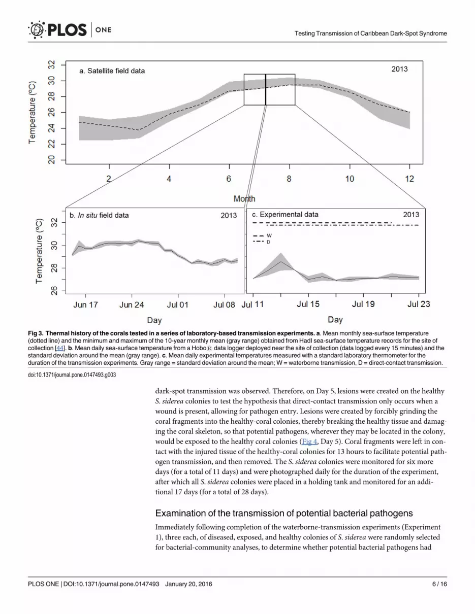
Fig 3. Thermal history of the corals tested in a series of laboratory-based transmission experiments. a. Mean monthly sea-surface temperature(dotted line) and the minimum and maximum of the 10-year monthly mean (gray range) obtained from HadI sea-surface temperature records for the site ofcollection [44]. b. Mean daily sea-surface temperature from a Hobo1 data logger deployed near the site of collection (data logged every 15 minutes) and thestandard deviation around the mean (gray range). c. Mean daily experimental temperatures measured with a standard laboratory thermometer for theduration of the transmission experiments. Gray range = standard deviation around the mean; W = waterborne transmission, D = direct-contact transmission.
doi:10.1371/journal.pone.0147493.g003
Testing Transmission of Caribbean Dark-Spot Syndrome
PLOSONE | DOI:10.1371/journal.pone.0147493 January 20, 2016 6 / 16
transmitted to the exposed colonies. The nine coral colonies were placed in individual, sterilewhirl-paks at -80°C and then were transported on dry ice to Mote Marine Laboratory in Sara-sota, Florida.
Tissue was removed from the skeleton of the preserved-coral colonies using a Paasche1 air-brush with 10 mL of sterile seawater. The tissue slurry was collected in a sterile 50 mL Falcon1tube and homogenized using a vortex. The tissue homogenate was then spun down into a pelletusing a centrifuge set at 10,000 rpm. The pellet was re-suspended in 2 mL of solution C1 andDNA was extracted using a Powersoil DNA extraction kit (MoBIO Laboratories Inc. Lot#PS14F19). Extracted DNA was then sent to MRDNA Laboratory (www.mrdnalab.com, Shal-lowater, TX, USA) for Illumina1 sequencing (20,000 reads per assay) using the universal bac-terial primers 27F/519R with a barcode on the forward primer. The 16S rRNA gene on the V1–V3 hypervariable region was amplified by applying a 30 cycle polymerase chain reaction (PCR)with the HotStarTaq Plus Master Mix Kit (Qiagen, USA). PCR was applied using the followingprotocol: (1) 94°C for 3 minutes, (2) 28 cycles of: 94°C for 30 seconds, 53°C for 40 seconds, and72°C for 1 minute, and (3) a final elongation step at 72°C for 5 minutes. After amplification,PCR products were confirmed in 2% agarose gels to determine the success of amplification andthe relative intensity of the bands. Multiple samples were pooled together in equal proportionsbased on their molecular weight and DNA concentrations. Pooled samples were purified usingcalibrated Ampure1 XP beads. Then the pooled and purified PCR product was used to pre-pare DNA libraries by following the Illumina1 TruSeq DNA library preparation protocol.Sequencing was performed using the Illumina1 sequencing platform at MR DNA (www.mrdnalab.com, Shallowater, TX, USA) following the manufacturer’s guidelines. Sequence datawere processed using a standardized analysis pipeline. Briefly, sequences were initially depletedof barcodes. Then sequences less than 150bp or with ambiguous base calls were removed.Operational taxonomic units (OTUs) were generated, and chimeras were removed usingUCHIME [48]. OTUs were defined by clustering at 3% divergence (i.e., showing 97% similar-ity) using a de novo method. Final OTUs were taxonomically classified using BLASTn againstthe curated National Center for Biotechnology Information (NCBI) database and the Ribo-somal Database Project (RDP).
Fig 4. Time-series images of two healthy-coral colonies (~16 cm2) of Siderastrea siderea in a direct-contact transmission experiment. The firstcolumn of images is a close-up of the fragments (~1 cm2) that were placed in direct contact with the coral colonies. The first row of images show a healthy-coral colony (~16 cm2) in direct contact with a healthy fragment (~1 cm2) (control). The second row of images show a healthy-coral colony in direct contactwith a fragment with dark spot lesions (~1 cm2) (experimental). Note that Day 1 images were taken prior to the addition of the direct-contact coral fragment.Note also the direct-contact lesion scars on S. siderea on Day 5.
doi:10.1371/journal.pone.0147493.g004
Testing Transmission of Caribbean Dark-Spot Syndrome
PLOSONE | DOI:10.1371/journal.pone.0147493 January 20, 2016 7 / 16
The relative-percentage contributions of each genus (and higher taxonomic levels) to thebacterial assemblages were calculated for each of the nine coral colonies. Differences in coral-bacterial communities among treatments were tested using a permutational multivariate analy-sis of variance (PERMANOVA). The results were visualized using non-metric multidimen-sional scaling plots. Differences in mean bacterial richness among treatments at eachtaxonomic level were tested using an analysis of variance (ANOVA), or using a Kruskal-Wallistest in cases where the assumptions of ANOVA could not be met through data transforma-tions. Shannon-Weiner diversity indices were calculated for all samples, at each taxonomiclevel, and differences in diversity among treatments were tested using ANOVA. Differences inthe relative abundance of each bacterial taxon among treatments were calculated using a Krus-kal-Wallis rank-sum test with a chi-square distribution. Analyses were conducted using thepackages ‘vegan’ [49] and ‘ggplot2’ [50] in R [47].
ResultsDark-spot syndrome did not visually transmit to healthy corals in any experiment in the pres-ent study (Figs 4 and 5). There was no apparent transmission to the lesions that were generatedby direct contact with diseased fragments, and lesions were completely healed by Day 28 (Fig4). Analysis of the Illumina1 sequencing data revealed no significant differences among thebacterial communities on healthy, exposed, and diseased corals at the completion of the water-borne-transmission experiment (Table 1, S1 and S2 Figs). Furthermore, no significant differ-ences in bacterial richness were detected among treatments (Table 2, S3 Fig), and nosignificant differences in the relative abundance of six purported pathogens were found amongtreatments (S1 Table). However, the relative abundance of nine bacterial taxa increased signifi-cantly in diseased corals compared with healthy and exposed corals, pointing to potential path-ogens, but indicating a lack of transmission (S4 Fig).
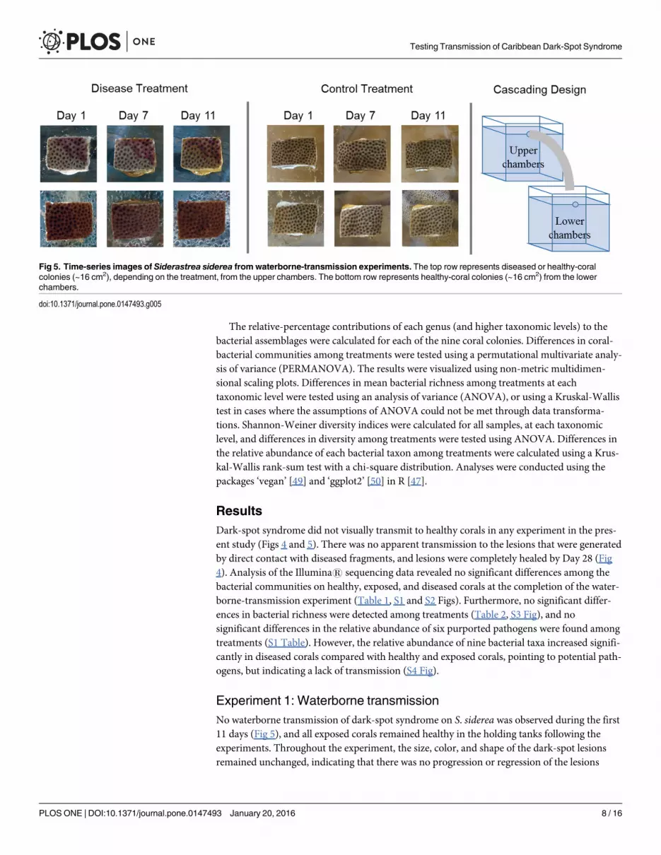
Experiment 1: Waterborne transmissionNo waterborne transmission of dark-spot syndrome on S. siderea was observed during the first11 days (Fig 5), and all exposed corals remained healthy in the holding tanks following theexperiments. Throughout the experiment, the size, color, and shape of the dark-spot lesionsremained unchanged, indicating that there was no progression or regression of the lesions
Fig 5. Time-series images of Siderastrea siderea from waterborne-transmission experiments. The top row represents diseased or healthy-coralcolonies (~16 cm2), depending on the treatment, from the upper chambers. The bottom row represents healthy-coral colonies (~16 cm2) from the lowerchambers.
doi:10.1371/journal.pone.0147493.g005
Testing Transmission of Caribbean Dark-Spot Syndrome
PLOSONE | DOI:10.1371/journal.pone.0147493 January 20, 2016 8 / 16
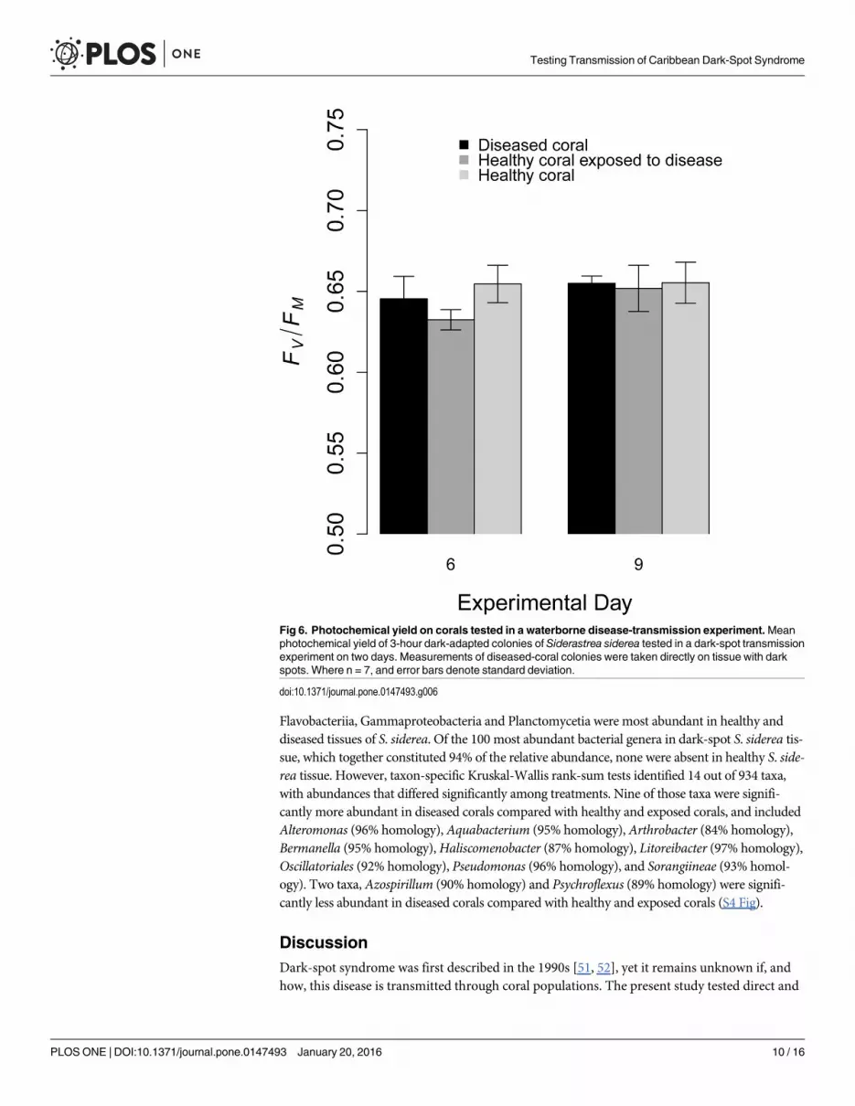
through time (Fig 5). Furthermore, there were no significant differences in the photochemicalyield among treatments or through time (F = 2.17, df = 2, p = 0.125 for treatment, and F = 1.46,df = 1, p = 0.233 for time; Fig 6).
Experiment 2: Direct-contact transmissionNo direct transmission of dark-spot syndrome on S. siderea, was observed during the first 11days (Fig 4), and all exposed corals remained healthy in the holding tanks following the experi-ments (Fig 4). All healthy S. siderea colonies showed partial recovery from their artificially-induced lesions by Day 11, and did not develop dark spots. Following the recovery period, allhealthy-coral colonies had completely healed from their artificially-induced lesions, and nodark-spot syndrome had developed after 28 days (Fig 4).
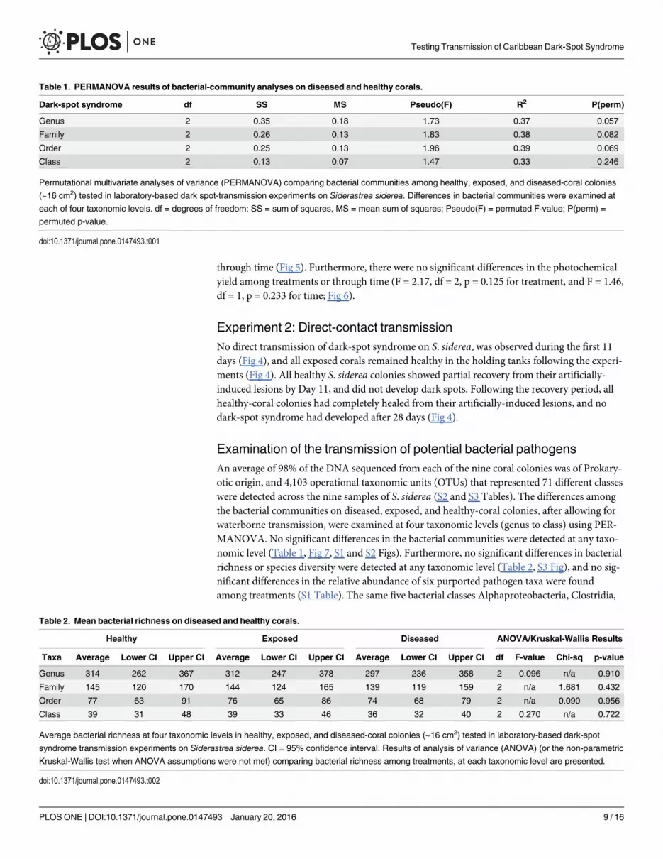
Examination of the transmission of potential bacterial pathogensAn average of 98% of the DNA sequenced from each of the nine coral colonies was of Prokary-otic origin, and 4,103 operational taxonomic units (OTUs) that represented 71 different classeswere detected across the nine samples of S. siderea (S2 and S3 Tables). The differences amongthe bacterial communities on diseased, exposed, and healthy-coral colonies, after allowing forwaterborne transmission, were examined at four taxonomic levels (genus to class) using PER-MANOVA. No significant differences in the bacterial communities were detected at any taxo-nomic level (Table 1, Fig 7, S1 and S2 Figs). Furthermore, no significant differences in bacterialrichness or species diversity were detected at any taxonomic level (Table 2, S3 Fig), and no sig-nificant differences in the relative abundance of six purported pathogen taxa were foundamong treatments (S1 Table). The same five bacterial classes Alphaproteobacteria, Clostridia,
Table 1. PERMANOVA results of bacterial-community analyses on diseased and healthy corals.
Dark-spot syndrome df SS MS Pseudo(F) R2 P(perm)
Genus 2 0.35 0.18 1.73 0.37 0.057
Family 2 0.26 0.13 1.83 0.38 0.082
Order 2 0.25 0.13 1.96 0.39 0.069
Class 2 0.13 0.07 1.47 0.33 0.246
Permutational multivariate analyses of variance (PERMANOVA) comparing bacterial communities among healthy, exposed, and diseased-coral colonies
(~16 cm2) tested in laboratory-based dark spot-transmission experiments on Siderastrea siderea. Differences in bacterial communities were examined at
each of four taxonomic levels. df = degrees of freedom; SS = sum of squares, MS = mean sum of squares; Pseudo(F) = permuted F-value; P(perm) =
permuted p-value.
doi:10.1371/journal.pone.0147493.t001
Table 2. Mean bacterial richness on diseased and healthy corals.
Healthy Exposed Diseased ANOVA/Kruskal-Wallis Results
Taxa Average Lower CI Upper CI Average Lower CI Upper CI Average Lower CI Upper CI df F-value Chi-sq p-value
Genus 314 262 367 312 247 378 297 236 358 2 0.096 n/a 0.910
Family 145 120 170 144 124 165 139 119 159 2 n/a 1.681 0.432
Order 77 63 91 76 65 86 74 68 79 2 n/a 0.090 0.956
Class 39 31 48 39 33 46 36 32 40 2 0.270 n/a 0.722
Average bacterial richness at four taxonomic levels in healthy, exposed, and diseased-coral colonies (~16 cm2) tested in laboratory-based dark-spot
syndrome transmission experiments on Siderastrea siderea. CI = 95% confidence interval. Results of analysis of variance (ANOVA) (or the non-parametric
Kruskal-Wallis test when ANOVA assumptions were not met) comparing bacterial richness among treatments, at each taxonomic level are presented.
doi:10.1371/journal.pone.0147493.t002
Testing Transmission of Caribbean Dark-Spot Syndrome
PLOSONE | DOI:10.1371/journal.pone.0147493 January 20, 2016 9 / 16
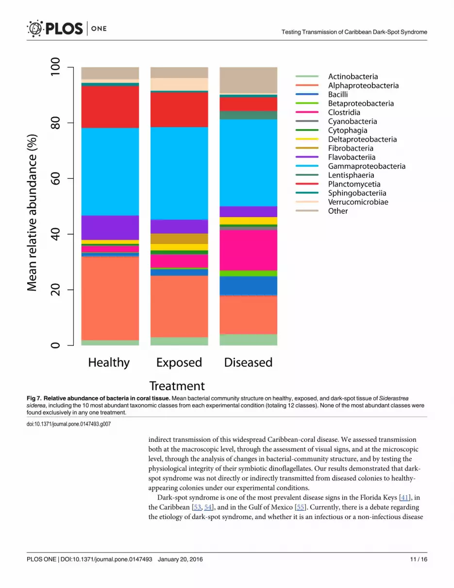
Flavobacteriia, Gammaproteobacteria and Planctomycetia were most abundant in healthy anddiseased tissues of S. siderea. Of the 100 most abundant bacterial genera in dark-spot S. siderea tis-sue, which together constituted 94% of the relative abundance, none were absent in healthy S. side-rea tissue. However, taxon-specific Kruskal-Wallis rank-sum tests identified 14 out of 934 taxa,with abundances that differed significantly among treatments. Nine of those taxa were signifi-cantly more abundant in diseased corals compared with healthy and exposed corals, and includedAlteromonas (96% homology), Aquabacterium (95% homology), Arthrobacter (84% homology),Bermanella (95% homology),Haliscomenobacter (87% homology), Litoreibacter (97% homology),Oscillatoriales (92% homology), Pseudomonas (96% homology), and Sorangiineae (93% homol-ogy). Two taxa, Azospirillum (90% homology) and Psychroflexus (89% homology) were signifi-cantly less abundant in diseased corals compared with healthy and exposed corals (S4 Fig).
DiscussionDark-spot syndrome was first described in the 1990s [51, 52], yet it remains unknown if, andhow, this disease is transmitted through coral populations. The present study tested direct and
Fig 6. Photochemical yield on corals tested in a waterborne disease-transmission experiment.Meanphotochemical yield of 3-hour dark-adapted colonies of Siderastrea siderea tested in a dark-spot transmissionexperiment on two days. Measurements of diseased-coral colonies were taken directly on tissue with darkspots. Where n = 7, and error bars denote standard deviation.
doi:10.1371/journal.pone.0147493.g006
Testing Transmission of Caribbean Dark-Spot Syndrome
PLOSONE | DOI:10.1371/journal.pone.0147493 January 20, 2016 10 / 16
indirect transmission of this widespread Caribbean-coral disease. We assessed transmissionboth at the macroscopic level, through the assessment of visual signs, and at the microscopiclevel, through the analysis of changes in bacterial-community structure, and by testing thephysiological integrity of their symbiotic dinoflagellates. Our results demonstrated that dark-spot syndrome was not directly or indirectly transmitted from diseased colonies to healthy-appearing colonies under our experimental conditions.
Dark-spot syndrome is one of the most prevalent disease signs in the Florida Keys [41], inthe Caribbean [53, 54], and in the Gulf of Mexico [55]. Currently, there is a debate regardingthe etiology of dark-spot syndrome, and whether it is an infectious or a non-infectious disease
Fig 7. Relative abundance of bacteria in coral tissue.Mean bacterial community structure on healthy, exposed, and dark-spot tissue of Siderastreasiderea, including the 10 most abundant taxonomic classes from each experimental condition (totaling 12 classes). None of the most abundant classes werefound exclusively in any one treatment.
doi:10.1371/journal.pone.0147493.g007
Testing Transmission of Caribbean Dark-Spot Syndrome
PLOSONE | DOI:10.1371/journal.pone.0147493 January 20, 2016 11 / 16
[19, 31–35]. Our results agree with the recent findings of Kellogg et al. [37], indicating no sig-nificant shifts in the overall bacterial communities between diseased and healthy corals. Yet, bycomparison, the bacterial communities on S. siderea that are reported here were strikingly dif-ferent from those on S. siderea reported by Kellogg et al. [37]. These results suggest thathealthy-coral bacterial communities vary across space and through time (S5 Fig). Alternatively,differences in the bacterial communities detected in the present study and those detected byKellogg et al. [37] could be the result of the ‘tank effect’ [56, 57], or a result of the use of differ-ent detection methods (Illumina1 sequencing in the present study versus microarrays in Kel-logg et al. [37]). Furthermore, significant increases in a few individual taxa, not detected by thecommunity-level analyses, point to nine potential pathogenic bacterial taxa that warrant fur-ther investigation (S4 Fig). Interestingly, none of these taxa, with the exception of Oscillatoria,have been previously proposed as pathogens of dark-spot syndrome (S1 Table). However, ifindeed any of these taxa are pathogenic, their abundances did not increase significantly inexposed corals, compared with healthy corals, suggesting that they did not transmit in ourstudy (S4 Fig).
It has been suggested that dark-spot syndrome is primarily a disease of the algal symbionts(Symbiodinium spp.) that reside within the host-coral tissue [31]. In a study of S. siderea tissuewith dark-spot syndrome, Cervino et al. [31] identified a reduction in the number of symbiontsby 56%, a reduction in the mitotic indices of the symbionts, and changes in symbiont physiol-ogy. Results from our experiments indicated that there were no significant differences in thephotochemical yields of the corals among treatments (Fig 6), suggesting that there were no dif-ferences in the ‘health’ of the Symbiodinium spp. in the diseased and healthy corals in theexperiment. Our results could indicate that the etiology of dark-spot syndrome in our experi-ments was different from the disease tested by Cervino et al. [31]. Alternatively, it could be thatthe diseases were in different stages of infectivity, or that a second affliction was causing thereduction in Symbiodinium spp. in the corals tested by Cervino et al. [31].
Previous laboratory and field studies of some coral diseases have observed rapid transmis-sion (i.e.< 10 days) from infected to healthy corals [14, 58–62]. By contrast, other coral dis-eases have taken longer to transmit, or altogether failed to transmit [63–64]. Our resultssuggest that dark-spot syndrome on S. siderea is comparably less transmissible than white-signdiseases, ciliate-associated diseases, and black-band disease. Yet, our experiments do not fullyrefute the possibility that dark-spot syndrome is infectious and transmissible. For example,dark-spot syndrome may need a biological vector that wasn’t present in the experiments, or thedisease may take more than 28 days to develop and manifest. It is possible, also, that the diseasemay be contagious under non-sterile conditions, only during certain stages of the pathogenlife-cycle, only under anomalously stressful conditions for the host, or under anomalous condi-tions that promote pathogen virulence. Additional research examining disease transmissionunder varying environmental conditions with different potential vectors are needed toaddresses these research questions. In conclusion, the results from these transmission experi-ments indicate that dark-spot syndrome is not a readily transmissible coral disease. While wecannot rule out the possibility of other mechanisms of transmission, these results are a positivestep toward understanding the dynamics of a prevalent Caribbean-coral disease.
Supporting InformationS1 Fig. Heatmaps of the relative abundance of bacteria.Heatmaps of the relative abundanceof bacteria identified on each sample of healthy, exposed, and diseased coral tissue, tested inwaterborne transmission experiments of dark-spot syndrome on Siderastrea siderea. Bacteriawere examined at the class (a), order (b), family (c), and genus (d) taxonomic levels. Cluster
Testing Transmission of Caribbean Dark-Spot Syndrome
PLOSONE | DOI:10.1371/journal.pone.0147493 January 20, 2016 12 / 16
dendrograms are indicated above each heatmap.(PDF)
S2 Fig. Heatmap of the relative abundance of OTUs.Heatmap of the relative abundance ofoperational taxonomic units (OTUs) identified on each sample of exposed, and diseased coraltissue, tested in waterborne transmission experiments of dark-spot syndrome on Siderastreasiderea. A cluster dendrogram is indicated above the heatmap.(PDF)
S3 Fig. Shannon-Weiner diversity indices. Shannon-Weiner diversity of microbes, at six tax-onomic levels, in three samples each of healthy (H), exposed (E) and diseased (D) coral tissue,tested in waterborne transmission experiments of dark-spot syndrome on Siderastrea siderea.(PDF)
S4 Fig. Mean relative abundance of individual bacterial taxa in diseased, exposed andhealthy corals. Only taxa whose relative abundances were significantly different among treat-ments, as determined by a Kruskal-Wallis rank-sum test using a chi-squared distribution arereported. Error bars denote standard deviation. n = 3.(PDF)
S5 Fig. Comparison of class-level bacterial data from the current study with Kellogg et al.[37]. Cluster dendrogram is based on the relative abundance of class-level bacterial data fromhealthy and dark-spot affected Siderastrea siderea from the Florida Keys in 2013 (presentstudy) compared with the Dry Tortugas and St. John, USVI in 2009 (Kellogg et al. [51]). Sam-ple numbers in black represent apparently-healthy corals, whereas sample numbers in red rep-resent corals with dark-spot syndrome.(PDF)
S1 Table. Results of non-parametric analyses of variance of five purported pathogen taxa.Results of Kruskal-Wallis rank-sum tests using a chi-squared distribution comparing relativeabundances of bacteria of six purported pathogenic taxa on diseased, exposed, and healthy cor-als colonies (~16 cm2) at the completion of laboratory-based dark-spot syndrome transmissionexperiments on Siderastrea siderea, where df = degrees of freedom, and n/a indicates that noOscillatoria were identified in any sample.(DOCX)
S2 Table. Absolute abundance of OTUs in each coral sample. Operational taxonomic units(OTUs) and their corresponding classifications and percentage homology for each of ninecoral samples of Siderastrea siderea tested in waterborne-transmission experiments of dark-spot syndrome. D4U, D13U and D27U were healthy corals; D1L, D3L and D22L were exposedcorals; D1U, D3U and D22U were dark-spot affected corals.(XLSX)
S3 Table. Nucleotide sequences for OTUs.Nucleotide sequences for all operational taxo-nomic units (OTUs) identified on Siderastrea siderea corals tested in waterborne transmissionexperiments of dark-spot syndrome.(XLSX)
AcknowledgmentsWe are grateful to the staff at Mote Marine Laboratory in Summerland Key, especially M.Knowles, E. Bartels, C. Walter, and D. Vaughan for assistance with field collections and system
Testing Transmission of Caribbean Dark-Spot Syndrome
PLOSONE | DOI:10.1371/journal.pone.0147493 January 20, 2016 13 / 16
set-up. We thank R. Iglesias-Prieto for valuable discussion of symbiont physiology. Our thanksextend also to S. J. van Woesik for editorial comments on the manuscript.
Author ContributionsConceived and designed the experiments: CJR AJG EMM RvW. Performed the experiments:CJR AJG. Analyzed the data: CJR. Contributed reagents/materials/analysis tools: CJR AJGEMM RvW.Wrote the paper: CJR AJG EMM RvW.
References1. Aronson RB, Precht WF. White-band disease and the changing face of Caribbean coral reefs. Hydro-
biologia. 2001; 460:25–38.
2. Porter JW, Dustan P, JaapWC, Patterson KL, Kosmynin V, Meier OW, et al. Patterns of spread of coraldisease in the Florida Keys. Hydrobiologia. 2001; 1:24.
3. Miller J, Muller E, Rogers C, Waara R, Atkinson A, Whelan KRT, et al. Coral disease following massivebleaching in 2005 causes 60% decline in coral cover on reefs in the US Virgin Islands. Coral Reefs.2009; 28:925–937.
4. Weil E, Cróquer A, Urreiztieta I, Irizarry E. Temporal variability and impact of coral diseases and bleach-ing in La Parguera, Puerto Rico from 2003–2007. Caribb J Sci. 2009; 45:221–246.
5. Smith GW, Harvel CD, Kim K. Response of sea fans to infection with Aspergillus sp. (Fungi). Rev BiolTrop. 1998; 46: 5:205–208.
6. Bruckner AW, Bruckner RJ, Williams EH Jr. Spread of a black-band disease epizootic through the coralreef system in St. Ann's Bay, Jamaica. Bull Mar Sci. 1997; 61:919–928.
7. Richardson LL, GoldbergWM, Kuta KG, Aronson RB, Smith GW, Ritchie KB, et al. Florida's mysterycoral-killer identified. Nature (Lond). 1998a; 392:557–558.
8. Antonius A. Halofolliculina corallasia, a new coral-killing ciliate on Indo-Pacific reefs. Coral Reefs. 1999;18:300–300.
9. Alker AP, Smith GW, Kim K. Characterization of Aspergillus sydowii (Thom et Church), a fungal patho-gen of Caribbean sea fan corals. Hydrobiologia. 2001; 460:105–111.
10. Ben-Haim Y, Rosenberg E. A novel Vibrio sp. pathogen of the coral Pocillopora damicornis. Mar Biol.2002; 142:47–55.
11. Rosenberg E, Ben‐Haim Y. Microbial diseases of corals and global warming. Environ Microbiol. 2002;4:318–326. PMID: 12071977
12. Cooney RP, Pantos O, Tissier MDL, Barer MR, Bythell JC. Characterization of the bacterial consortiumassociated with black band disease in coral using molecular microbiological techniques. Environ Micro-biol. 2002; 4:401–413. PMID: 12123476
13. Patterson KL, Porter JW, Ritchie KB, Polson SW, Mueller E, Peters EC, et al. The etiology of white pox,a lethal disease of the Caribbean elkhorn coral, Acropora palmata. Proc Natl Acad Sci USA. 2002;99:8725–8730. PMID: 12077296
14. Aeby GS, Santavy DL. Factors affecting susceptibility of the coralMontastraea faveolata to black-banddisease. Mar Ecol Prog Ser. 2006; 318:103–110.
15. Kline DI, Vollmer SV. White Band Disease (type I) of endangered Caribbean acroporid corals is causedby pathogenic bacteria. Sci Rep. 2011; 1:7. doi: 10.1038/srep00007 PMID: 22355526
16. Sutherland KP, Shaban S, Joyner JL, Porter JW, Lipp EK. Human pathogen shown to cause disease inthe threatened eklhorn coral Acropora palmata. PLoS ONE. 2011; 6:8:1–7.
17. Thrusfield M. Veterinary epidemiology. 3rd ed. United Kingdom: Blackwell Science; 2005.
18. Casadevall A, Pirofski LA. Host-pathogen interactions: basic concepts of microbial commensalism, col-onization, infection, and disease. Infect Immun. 2000; 68:6511–6518. PMID: 11083759
19. Lesser MP, Bythell JC, Gates RD, Johnstone RW, Hoegh-Guldberg O. Are infectious diseases reallykilling corals? Alternative interpretations of the experimental and ecological data. J Exp Mar Biol Ecol.2007; 346:36–44.
20. Sutherland KP, Porter JW, Torres C. Disease and immunity in Caribbean and Indo-Pacific zooxanthel-late corals. Mar Ecol Prog Ser. 2004; 266:265–272.
21. Weil E, Smith G, Gil-Agudelo DL. Status and progress in coral reef disease research. Dis Aquat Org.2006; 69:1–7. PMID: 16703761
Testing Transmission of Caribbean Dark-Spot Syndrome
PLOSONE | DOI:10.1371/journal.pone.0147493 January 20, 2016 14 / 16
22. Weil E, Rogers CS. Coral reef diseases in the Atlantic-Caribbean. In: Dubinsky Z, Stambler N (eds)Coral reefs: An ecosystem in transition. Springer Dordrecht Heidelberg London New York; 2011. p.465–492.
23. Bythell J, Pantos O, Richardson L. White plague, white band, and other “white” diseases. In: RosenbergE, Loya Y, editors. Coral Health and Disease. Springer-Verlag, Berlin, Germany; 2004. p. 351–365.
24. Harvell CD, Kim K, Burkholder JM, Colwell RR, Epstein PR, Grimes DJ, et al. Emerging marine dis-eases–Climate links and anthropogenic factors. Science. 1999; 285:1505–1510. PMID: 10498537
25. Riegl B. Effects of the 1996 and 1998 positive sea-surface temperature anomalies on corals, coral dis-eases and fish in the Arabian Gulf (Dubai, UAE). Mar Biol. 2002; 140:29–40.
26. SussmanM, Loya Y, Fine M, Rosenberg E. The marine fireworkHermodice carunculata is a winter res-ervoir and spring-summer vector for the coral-bleaching pathogen Vibrio shiloi. Environ Microbiol.2003; 5:250–255. PMID: 12662172
27. Stedman TL. Stedman's Medical Dictionary. Williams andWilkin Company, Baltimore, MD. 1976.
28. Gardner TA, Côté IM, Gill JA, Grant A, Watkinson AR. Long-term region wide declines in Caribbeancorals. Science. 2003; 301:958–960. PMID: 12869698
29. Muller E, vanWoesik R. Caribbean coral disease: primary transmission or secondary infection? GlobChange Biol. 2012; 18:3529–3535.
30. Gil-Agudelo DL, Garzon-Ferreira J. Spatial and seasonal variation of dark-spot syndrome disease incoral communities of the Santa Marta area (Colombian Caribbean). Bull Mar Sci. 2001; 69:619–629.
31. Cervino JM, Goreau TJ, Nagelkerken I, Smith GW, Hayes R. Yellow band and dark spot syndromes inCaribbean corals: distribution, rate of spread, cytology, and effects on abundance and division rate ofzooxanthellae. In: Porter JW, editor. The Ecology and Etiology of Newly Emerging Marie Diseases.Netherlands: Kluwer Academic Publishers; 2001. p. 53–63.
32. Gil-Agudelo DL, Fonseca DP, Weil E, Garzon-Ferreira J, Smith GW. Bacterial communities associatedwith the mucopolysaccharide layers on three coral species affected and unaffected with dark spots dis-ease. Canadian J of Microbiol. 2007:465–471.
33. Borger JL. Dark spot syndrome: a scleractinian coral disease or a general stress response? CoralReefs. 2005; 24:139–144.
34. Gochfeld DJ, Olson JB, Slattery M. Colony versus population variation in susceptibility and resistanceto dark spot syndrome in the Caribbean coral Siderastrea siderea. Dis Aquat Org. 2006; 69:53–65.PMID: 16703766
35. Renegar DA, Blackwelder PL, Miller D, Gochfeld DJ, Moulding AL. Ultrastructural and histological anal-ysis of Dark Spot Syndrome in Siderastrea siderea and Agaricia agaricites. In: Riegl BM, Dodge RE,editors. Proc 11th Int Coral Reef Symp. Ft. Lauderdale, FL. 2008. P. 185–189.
36. Sweet MJ, Burn D, Croquer A, Leary P. Characterisation of the bacterial and fungal communities asso-ciated with different lesion sizes of dark spot syndrome occurring in the coral Stephanocoenia inter-septa. PLoS ONE. 2013; 8:e62580. doi: 10.1371/journal.pone.0062580 PMID: 23630635
37. Kellogg CA, Piceno YM, Tom LM, DeSantis TZ, Gray MA, Andersen GL. Comparing bacterial commu-nity composition of healthy and dark spot-affected Siderastrea siderea in Florida and the Caribbean.PLoS ONE. 2014: 9:e108767. doi: 10.1371/journal.pone.0108767 PMID: 25289937
38. Weil E. Coral reef diseases in the wider Caribbean. In: Rosenberg E, Loya Y (eds) Coral health and dis-ease. Springer-Verlag, Berlin, Germany; 2004. p. 35–68.
39. Randall CJ, Jordán-Garza AG, Muller E, vanWoesik R. Relationships between the history of thermalstress and the relative risk of diseases of Caribbean corals. Ecology (NY). 2014; 95:1981–1994.
40. Brandt ME, McManus JW. Disease incidence is related to bleaching extent in reef-building corals. Ecol-ogy (NY). 2009: 90:2859–2867.
41. Porter JW, Torres C, Sutherland KP, Meyers MK, Callahan MK, Ruzicka R, et al. Prevalence, severity,lethality, and recovery of dark-spot syndrome among three Floridian reef-building corals. J Exp Mar BiolEcol. 2011; 408:79–87.
42. Work TM, Aeby GS. Systematically describing gross lesions in corals. Dis Aquat Org. 2006: 70:155–160. PMID: 16875402
43. Hall ER, Vaughan D, Crosby MP. Development of ocean acidification flow-thru experimental racewayunits (OAFTERU). In: Yellowless D, Hughes TP, editors. Proc 12th Int Coral Reef Symp. Cairns, Austra-lia; 2012. p. 1–5.
44. Rayner NA, Parker DE, Horton EB, Folland CK, Alexander LV, Rowell DP, et al. Global analyses of seasurface temperature, sea ice, and night marine air temperature since the late nineteenth century. JGeophys Res. 2003; 108:1984–2012.
Testing Transmission of Caribbean Dark-Spot Syndrome
PLOSONE | DOI:10.1371/journal.pone.0147493 January 20, 2016 15 / 16
45. Fox J, Weisberg S (2011). An {R} Companion to Applied Regression, Second Edition. Thousand OaksCA: Sage. Available: http://socserv.socsci.mcmaster.ca/jfox/Books/Companion
46. Peters G. userfriendlyscience: Quantitative analysis made accessible. R package version 0.3–0, 2011.Available: http://CRAN.R-project.org/package=userfriendlyscience
47. R Core Team. R: A language and environment for statistical computing. R Foundation for StatisticalComputing, Vienna, Austria. 2014.
48. Edgar RC, Haas BJ, Clemente JC, Quince C, Knight R. UCHIME improves sensitivity and speed of chi-mera detection. Bioinformatics. 2011: 27(16):2194–2200. doi: 10.1093/bioinformatics/btr381 PMID:21700674
49. Oksanen JFG, Blanchet FG, Kindt R, Legendre P, Minchin PR, O’Hara RB, et al. vegan: CommunityEcology Package. R package version 2.2–0. [cited 2015 June 1]. Available: http://CRAN.R-project.org/package=vegan
50. Wickham H. ggplot2: elegant graphics for data analysis. Springer, New York. 2009.
51. Garzón-Ferreira J, Gil DL. Another unknown Caribbean coral phenomenon. Reef Encounter. 1998;24:10.
52. Santavy DL, Peters EC. Microbial pests: coral disease in the Western Atlantic. In: Lessios HA, Macin-tyre IG, editors. Proc 8th Int Coral Reef Symp; Smithsonian Tropical Research Institute, Panamá; 1997.p. 607–612.
53. Ruiz-Moreno D, Willis BL, Page AC, Weil E, Cróquer A, Vargas-Angel B, et al. Global coral diseaseprevalence associated with sea temperature anomalies and local factors. Dis Aquat Org. 2012;100:249–261. doi: 10.3354/dao02488 PMID: 22968792
54. Gil-Agudelo DL, Smith GW, Garzón-Ferreira J, Weil E, Petersen D. Dark spots disease and yellowband disease, two poorly known coral diseases with high incidence in Caribbean reefs. In: RosenbergE, Loya Y, editors. Coral Health and Disease. Germany: Springer-Verlag, Berlin; 2004. p. 337–350.
55. Carricart-Ganivet JP, Beltrán-Torres AU, Horta-Puga G. Distribution and prevalence of coral diseasesin the Veracruz Reef System, Southern Gulf of Mexico. Dis Aquat Org. 2011; 95:181–187. doi: 10.3354/dao02359 PMID: 21932529
56. Kooperman N, Ben-Dov E, Kramarsky-Winter E, Barak Z, Kushmaro A. Coral mucus-associated bacte-rial communities from natural and aquarium environments. FEMSMicrobiol Lett. 2007;106–113.
57. Pratte Z, Richardson LL, Mils DK. Microbiota shifts in the surface mucopolysaccharide layer of coralstransferred from natural to aquaria settings. J Invert Path. 2015; 125:42–44.
58. Gignoux-Wolfsohn SA, Marks CJ, Vollmer SV. White Band Disease transmission in the threatenedcoral, Acropora cervicornis. Sci Rep. 2012; 2:1–3.
59. Williams DE, Miller MW. Coral disease outbreak: pattern, prevalence and transmission in Acropora cer-vicornis. Mar Ecol Prog Ser. 2005; 301:119–128.
60. Clemens E, Brandt ME. Multiple mechanisms of transmission of the Caribbean coral disease whiteplague. Coral Reefs. 2015;1–10.
61. Dalton SJ, Godwin S, Smith SDA, Pereg L. Australian subtropical white syndrome: a transmissible,temperature-dependent coral disease. Mar Fresh Res. 2010; 61(3):342–350.
62. Rodríguez S, Cróquer A, Guzman HM, Bastidas C. A mechanism of transmission and factors affectingcoral susceptibility to Halofolliculina sp. infection. Coral reefs. 2009; 28:67–77.
63. Jordán-Garza AG, Jordán-Dahlgren E. Caribbean yellow-band syndrome onMontastraea faveolata isnot transmitted mechanically under field conditions. Dis Aquat Org. 2011; 96:83–87. doi: 10.3354/dao02368 PMID: 21991668
64. Sudek M, Williams GJ, Runyon C, Aeby GS, Davy SK. Disease dynamics of Porites bleaching with tis-sue loss: prevalence, virulence, transmission, and environmental drivers. Dis Aquat Org. 2015; 113(1):59–68. doi: 10.3354/dao02828 PMID: 25667337
Testing Transmission of Caribbean Dark-Spot Syndrome
PLOSONE | DOI:10.1371/journal.pone.0147493 January 20, 2016 16 / 16


