Embed Size (px)
Citation preview
The synthesis of a physically crosslinked NVP based hydrogel
Declan M. Devine, Clement L. Higginbotham*
Department of Polymer, Athlone Institute of Technology, Dublin Road, Athlone, Co. Westmeath, Ireland
Received 12 June 2003; received in revised form 17 September 2003; accepted 8 October 2003
Abstract
Complexes of polyvinyl pyrrolidinone–polyacrylic acid (PVP–PAA) were prepared by photopolymerisation from a mixture of the
monomers NVP and AA. The complexes were characterised by means of differential scanning calorimetry, Fourier transform infrared
spectroscopy (Ftir), potentiometric titration, swelling studies and gel permeation chromatography. The Ftir spectra of PVP–PAA copolymer
complexes indicates hydrogen bonding between the carbonyl group in the PVP and the carboxylic acid group in the PAA moiety. As the
percentage of AA increases in the copolymer there is evidence of increased intermolecular hydrogen bonding between the carboxylic acid
groups of the AA segments. Swelling of the PVP–PAA complex in a higher pH medium is significantly different from results in low pH
solutions. The critical pH range was found to be between 4.07 and 4.49. Above a pH of 4.49, there is a progressive break up of the polymer
chain due to a reduction in the amount of intermolecular hydrogen bonding. There is also a significant increase in the solubility of the
copolymer complex at higher pHs. The low solubility of the copolymer at low pH may make the complex suitable for gastric drug delivery
systems.
q 2003 Elsevier Ltd. All rights reserved.
Keywords: Polymer; Polymer chemistry; Polymer synthesis
1. Introduction
The term hydrogel is used to describe materials that are
three-dimensional, hydrophilic, polymeric networks
capable of imbibing large amounts of water or biological
fluids. These hydrogels exhibit a thermodynamic compat-
ibility with water that allows them to swell in aqueous
media. Hydrogels are best considered as polymeric
materials, which are able to swell in water and retain a
significant fraction of water within their structure, but do not
completely dissolve in water [1–8]. Kunioka et al. [9],
however, describes the thermal hydrolytic degradability of
hydrogels prepared from microbial poly (g-glutamic acid)
(PGA) and poly (1-lysine) (PL) by g-irradiation. It was
found that PGA and PL hydrogels were not degraded near
ambient temperature but were hydro-degradable over 60 8C
on heating.
Hydrogels are becoming increasingly important
materials for pharmaceutical applications. They are used
in a variety of applications including diagnostic, thera-
peutic, and implantable devices, for example, controlled
release drug delivery systems which have being studied
extensively [2,3,5,8,10–20] contact lenses [2,8,21–23] and
tissue engineering [7]. Hydrogels have been widely used in
such applications because of their biocompatibility with the
human body and also because they resemble natural living
tissue more than any other class of synthetic biomaterials.
This is due to their high water content and soft consistency
that is similar to natural tissue [2–7,24–25].
Hydrogels are prepared by synthetic chemical reactions
from a range of speciality vinyl and acrylic monomers.
Nguyen et al. [7] presented a review on UV curable
hydrogels that may be used as biomaterials in medical
applications including tissue engineering. The use of
photopolymerisation of these hydrogels in situ is also
discussed, and the possibility of their use in a minimally
invasive manner where the liquid monomer can be injected
and applications for these hydrogels such as barriers,
localised drug delivery depots, cell encapsulation materials
and scaffold materials are also noted. Hydrogels may be
composed of homopolymers or copolymers, and are
insoluble due to the presence of chemical crosslinks
(covalent bonding) or physical crosslinks, such as entangle-
ments or crystallites [1,2,4–5,26]. Anseth et al. [6] explored
0032-3861/$ - see front matter q 2003 Elsevier Ltd. All rights reserved.
doi:10.1016/j.polymer.2003.10.017
Polymer 44 (2003) 7851–7860
www.elsevier.com/locate/polymer
* Corresponding author. Tel.: þ353-9064-24463; fax: þ353-9064-
24493.
E-mail address: [email protected] (C.L. Higginbotham).
methods for analysing mechanical properties of hydrogels
and their dependence on polymer structure, especially the
crosslinking density and the degree of swelling. Methods for
measuring the elastic and viscoelastic properties of hydro-
gels were examined along with mechanisms for controlling
the properties; examples of these variables are variations in
the polymer composition, the crosslinking density and the
polymerisation conditions.
Chemical gels are three-dimensional molecular networks
formed by the introduction of primary covalent crosslinks.
Unless the covalent bonds can be broken, these types of gels
do not dissolve in water or other organic solvents even upon
heating; rather, they are observed to swell. Two common
techniques used to produce chemically crosslinked hydro-
gels are the polymerisation of a water-soluble monomer in
the presence of an appropriate crosslinking agent and
crosslinking of an existing hydrophilic polymer, again with
the use of a crosslinking agent. Both general methods obey
common reaction mechanisms and can be implemented in a
variety of ways. It is essential that monomeric species have
functionality greater than two in order to form the
characteristic three-dimensional structure of a gel [8].
Physical gels are hydrophilic networks comprised of an
amorphous hydrophilic polymer phase held together by
highly ordered aggregates of polymer chain segments
arising from secondary molecular forces (van der Waals
forces) in conjunction with other types of molecular
interaction. Unlike chemical gels, these types of hydrogel
systems eventually will dissolve in water or solvents and
can be melted by applying heat to the systems. A novel
approach to preparing physical crosslinks is the free-
ze/thawing technique [27–29]. Stauffer et al. [27] described
a novel method of preparing a strong PVOH hydrogel
without utilisation of chemical crosslinking or other
reinforcing agents. This was achieved by freezing/thawing
cycles of an aqueous solution containing 10–15 wt%
PVOH. The process of reinforcement was a densification
of the macro molecular structure. It was noted that the
strength, stability and swelling ratio of the gels were a
function of the solution concentration, freezing time and
number of freezing/thawing cycles, and that freezing
15 wt% PVOH solution at 220 8C for 24 h followed by
thawing at 23 8C for 24 h produced the strongest gels.
Investigation of the swelling ratios indicated that denser
structures were observed after five freezing/thawing cycles.
Hassan et al. [29] further discussed the use of PVOH with
particular interest for physically crosslinking hydrogels that
were prepared by repeated cycles of freezing and thawing.
This method of preparing hydrogels addresses the toxicity
issues because it does not require the presence of a
crosslinking agent. It was also noted that such physically
crosslinked materials also exhibit improved properties, most
notably higher mechanical strength than PVOH gels
crosslinked by chemical or irradiative techniques because
the mechanical load can be distributed along the crystallites
of the three-dimensional structures. However, the long-term
stability of such gels still remains an important issue.
The configuration of the aforementioned ordered
domains varies with the type of secondary forces involved
in their formation, as well as the general molecular structure
and configuration of the hydrophilic polymers that comprise
them. Some of the intermolecular forces responsible for
giving rise to these physical bonds include London
dispersion forces, permanent dipoles, hydrogen bonding
and ionic attraction. These forces are cumulative in nature
and contribute greatly to the ordered structure exhibited by
the physical bonds. The so-called ‘bonds’ that hold these
types of gels together are actually sections of the polymer
chain, which are in the crystalline state. Segments of the
molecule arrange themselves into a tightly packed repeating
structure. To achieve this, the polymer must exhibit a certain
degree of structural regularity along the main chain [8]
(Fig. 1).
2. Experimental
2.1. Preparation of samples
The hydrogels investigated in this work were prepared by
free-radical polymerisation. The monomers used were 1-
vinyl-2-pyrrolidinone (NVP, Lancaster synthesis) and
acrylic acid (AA, Merck-Schuchardt, Germany). Both
monomers were used as received. To initiate the reactions,
1-hydroxycyclohexylphenylketone (Irgacurew 184, Ciba
speciality chemicals) was used as a UV-light sensitive
initiator at 3 wt% of the total monomer weight. This was
added to the NVP/AA monomeric mixture and stirred
continuously until completely dissolved. The solution was
then pipetted into a silicone mould (W.P. Notcutt,
Middlesex) that contained disk impressions and rectangular
impressions for use in Fourier transform infrared spec-
troscopy (Ftir). The mould was then positioned horizontally
to the gravity direction under two UVA 340 UV lamps (Q-
panel products) and the solution was cured for 1–2 h in an
enclosed environment. The films were then dried in a
vacuum oven at 40 8C, 500 mm Hg for 24 h prior to use.
Fig. 1. Schematic representation of general structure of (a) chemical and (b)
physical gels [8].
D.M. Devine, C.L. Higginbotham / Polymer 44 (2003) 7851–78607852
2.2. Differential scanning calorimetry
Differential scanning calorimetry (DSC) was carried out
using a Perkin–Elmer, Pyris 6 DSC. Firstly, a sample of
between 10–12.5 mg was weighed out using a Sartorius
scales capable of being read to five decimal places. The tests
were carried out in pierced lid pans by heating the samples
from 20 to 200 8C at 30 8C per minute, the samples were
then held isothermally at that temperature for 10 min, the
samples were then cooled back to 20 8C at 30 8C per minute
and were again held isothermally for 10 min. This was
carried out in order to remove any thermal history. The
samples were then reheated to 200 8C at 10 8C per minute.
All DSC tests were carried out under a 20 mL per minute
flow of nitrogen to prevent oxidation.
2.3. Fourier transform infrared spectroscopy
Fourier transform infrared spectroscopy was carried
out on the rectangular samples that had being exposed
to atmospheric conditions for a minimum of 7 days, using
a Nicolet Avator 360 Ftir, with a 32 scan per sample
cycle.
2.4. Potentiometric titration
The potentiometric titrations of the UV cured PVP–PAA
copolymers were executed by firstly grinding the sample
that had being vacuum dried as described previously. The
sample was placed into a 200 mL bottle, into which 25 mL
of 0.05N HCl containing 0.1 M NaCl. A lid was placed on
the bottle to prevent evaporation. The sample was stirred
continuously for 24 h prior to use. A burette that could be
read to ^0.1 mL was used to titrate 0.05N NaOH contain-
ing 0.05 M NaCl with the HCl solution. An Orion 420A þ
pH meter capable of reading pH changes of 0.01 was used to
monitor the progress of the titration.
2.5. Swelling studies
Swelling experiments were preformed in buffered
solutions (buffered tablets, BDH Ltd., Poole, England) in
triplicate at pH values of pH 4.0, pH 7.0 and pH 9.2 at
ambient temperature. Samples of the cured polymer with a
mass of 1.38 ^ 0.42 g were placed into a petri dish; the petri
dish was then filled with the appropriate buffered solution.
Periodically excess-buffered solution was removed by
pouring the solution through a Buchner funnel, the samples
were then pat-dried with filter paper and weighed. The
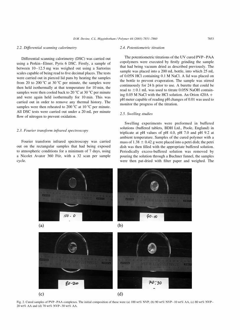
Fig. 2. Cured samples of PVP–PAA complexes. The initial composition of these were (a) 100 wt% NVP, (b) 90 wt% NVP–10 wt% AA, (c) 80 wt% NVP–
20 wt% AA and (d) 70 wt% NVP–30 wt% AA.
D.M. Devine, C.L. Higginbotham / Polymer 44 (2003) 7851–7860 7853
samples were re-submerged in fresh-buffered solution. The
percentage that the hydrogels swelled was calculated using
the formula:
Swelling ð%Þ ¼ Wt=W0 £ 100
Where Wt is the weight of the hydrogel at a predetermined
time, and W0 is the weight of the hydrogel before swelling
experiments took place. Pictures of the swollen samples
were also taken of the samples before the removal of the
buffered solution for comparative reasons. This process was
continued until the sample appeared to have dissolved.
2.6. Gel permeation chromatography
Gel permeation chromatography was carried out using a
polymer labs aquagel-OH 30 8 mm mixed column (using a
suitable guard column), with a mobile phase of water:
methanol in the ratio of 10:1. This solution was filtered and
degassed using a Millipore filtration system under vacuum
using 50 mm PTFE filter paper. The solution was then
further degassed by passing helium through it.
The detector used for these tests was a polymer labs
evaporative light scattering (PL ELS 1000) detector, which
had an evaporator temperature set to 100 8C, nebuliser
temperature set to 85 8C and a nitrogen flow rate set to
1.5 mL min21. Polyethylene glycol (PEG) calibration
standards were used as reference standards. However,
results dictated that a wider range was necessary, therefore,
high molecular weight polyethylene oxide’s (up to eight
million) were also used. These standards were prepared by
weighing 25–25.5 mg of PEG into 28 mL bottles, 25 mL of
high purity water was then pipetted into the bottle. The
samples were allowed to dissolve for 24 h before use. All
tests were carried out using a flow rate of 1 mL min21.
The samples tested were collected by swelling the
hydrogels in deionised water for defined periods of time,
removing a sample of water, and then replacing the water
with fresh water.
3. Results and discussion
3.1. Preparation of samples
Samples of both NVP and NVP/AA were photopoly-
merised using Irgacurew 184 as a photoinitiator. These
samples were cured on a silicone moulding, and prior to use
dried for 24 h in a vacuum oven. Cured samples are shown
in Fig. 2. The sample containing 100 wt% NVP cured with a
uniform smooth surface, which would indicate that it could
have potential in coating applications. With the addition of
AA, the uniformity of the surface of the samples
deteriorated. This deterioration increased with an increase
in AA concentration; in the samples whose initial
composition was 100 wt% NVP all the samples were
transparent in appearance and yellowish in colour after
photopolymerisation. However, the addition of AA caused
the formation of some white solids [30] being formed as is
shown in Fig. 2. These white solids would suggest a degree
of phase separation in the cured copolymer.
3.2. Differential scanning calorimetry
DSC were carried out by heating the sample to 200 8C,
and then cooling it back to 20 8C to remove all thermal
history. The samples were then reheated back to 200 8C at
10 8C per minute.
DSC scans were performed in order to determine the
effect that the addition of PAA had on the properties of PVP.
Yaung et al. [1] also performed DSC scans on PVP–PAA
complexes and found, that for the three formulations tested
the Tg of the samples were 134, 142 and 146 8C. In this work
the Tg of the samples tested were found to be 146.1, 146.5,
140.4 and 141.3 8C for samples whose initial composition
was 100 wt% NVP, 90 wt% NVP–10 wt% AA, 80 wt%
NVP–20 wt% AA and 70 wt% NVP–30 wt% AA, respect-
ively. These results correlate to those presented by Yaung
and would give evidence to a slight reduction in stiffness
with an increase in AA content.
3.3. Fourier transform infrared spectroscopy
The formation of hydrogen bonding is exhibited on the
IR spectrum as a negative shift of the stretching vibration of
the functional group involved in the hydrogen bond, which
is typically a carbonyl group. Bures et al. [31] states that the
carboxylic acid groups can exist in both free and dimeric
form depending upon their environment. Lee et al. [32,33]
describes how the stretching frequency of the carbonyl
moiety in the carboxylic acid group as is defined by
absorption at 1750 cm21, whereas the dimer stretching
frequency was reported as been 1700 cm21 by both Lee and
Bures et al. [31] this was also reported by Yaung et al. [1] as
1717 cm21. In this work, the dimer was identified by the
clear formation of a shoulder on the PVP carboxyl group
absorption peak. This shoulder is visible in both samples
90–10 and 80–20. This shoulder develops into a peak in the
70–30 sample, which appears at 1719 cm21 and corre-
sponds to values reported in literature.
Yaung et al. [1] describes how the frequency of the PVP
carbonyl group shifts from 1670 to 1680 cm21 to 1630 to
1640 cm21 when it forms hydrogen bonds to the carboxyl
group of the AA. In this work, the PVP carboxyl group for
PVP was found to be at 1650 cm21, and shifted to
1639 cm21 with the inclusion of AA thus giving evidence
of hydrogen bonding in the copolymer complex.
3.4. Potentiometric titration
Potentiometric titrations were carried out on pre-dried
samples that were firstly ground and then dissolved in a
0.05N HCl solution. Into this solution, a 0.05N solution of
D.M. Devine, C.L. Higginbotham / Polymer 44 (2003) 7851–78607854
NaOH was titrated using a burette capable of being read to
^0.1 mL. All titrations were repeated to confirm
reproducibility.
From the resultant graphs two neutralisation transitions
could clearly be seen. The latter transition is merely the
neutralisation of the HCl. The first neutralisation transition
can be attributed to the neutralisation of the carboxylic acid
group associated with PAA/AA. From this neutralisation
transition, the pKinitial can be defined. The pKinitial was taken
as the first point of pH drop from equilibrium. The range of
pKinitial values obtained in this work was 4.07–4.49. These
values compare favourably to values obtained by Yaung
et al. [1] who recorded a pKinitial range of 4.39–4.22.
Yaung also described pKinitial for a weak polyelectrolyte
such as PAA. The number of ionic charges on the backbone
depends upon the pH of the solution. When the pH value of
the solution is above pKinitial; which is the lowest pH at
which the carboxylic acid can be neutralised, a fraction of
the carboxylic acid groups dissociate to form carboxylate
ions. Therefore, under pH4 swelling conditions the
carboxylic acid groups will not be fully dissociated.
Therefore, the bonds between PVP and PAA are expected
to remain intact.
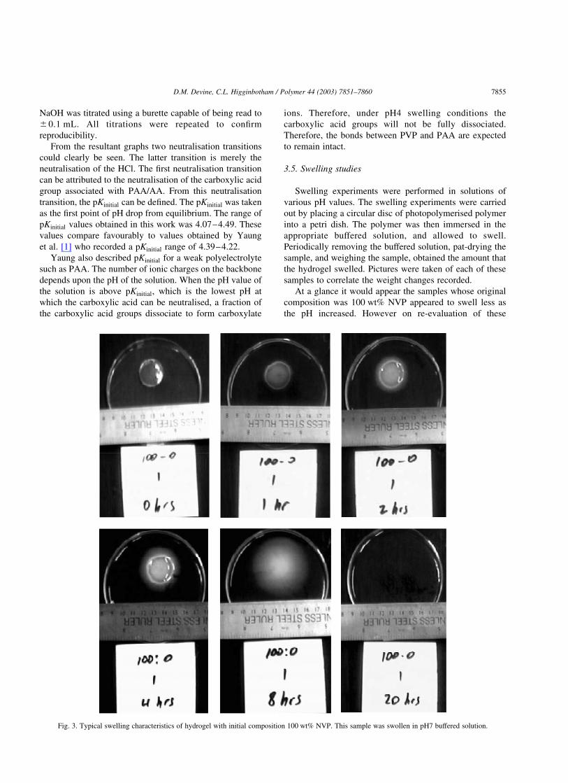
3.5. Swelling studies
Swelling experiments were performed in solutions of
various pH values. The swelling experiments were carried
out by placing a circular disc of photopolymerised polymer
into a petri dish. The polymer was then immersed in the
appropriate buffered solution, and allowed to swell.
Periodically removing the buffered solution, pat-drying the
sample, and weighing the sample, obtained the amount that
the hydrogel swelled. Pictures were taken of each of these
samples to correlate the weight changes recorded.
At a glance it would appear the samples whose original
composition was 100 wt% NVP appeared to swell less as
the pH increased. However on re-evaluation of these
Fig. 3. Typical swelling characteristics of hydrogel with initial composition 100 wt% NVP. This sample was swollen in pH7 buffered solution.
D.M. Devine, C.L. Higginbotham / Polymer 44 (2003) 7851–7860 7855

samples with reference to the pictures taken it would appear
that the samples swelled more as the pH increased. As no
crosslinking could occur the rapid swelling of this polymer led
to rapid dissolution of the samples in all pHs used (within
24 h). All samples reached their maximum swollen weight
from 0 to 2 h, which would also indicate rapid dissolution.
When the pH value of the solution is above pKinitial;
which is the lowest pH at which the carboxylic acid can be
neutralised, a fraction of the carboxylic acid groups
dissociate to form carboxylate ions [1]. From examination
of samples that originally contained 90 wt% NVP and
10 wt% AA, it can be seen that the samples swollen in pH4
Fig. 4. Typical swelling characteristics of hydrogel with initial composition 70 wt% NVP–30 wt% AA. This sample was swollen in pH7 buffered solution.
D.M. Devine, C.L. Higginbotham / Polymer 44 (2003) 7851–78607856

tended to swell less than samples swollen in a higher pH.
However, if all three swelling medias are compared at 30 h
it can be seen that samples swelled in higher pHs had
dissolved while the samples swollen at pH4 were still
approximately 20% of original weight. However, samples
that were swollen in pH9 did not appear to swell as much as
samples swollen in pH7. In pH7 and pH9 buffered solutions
the pKinitial values (between 4.07 and 4.49) are exceeded,
and the progressive break up of the polymer chains liberates
more and more free polymer. The number of carboxylate
ions also increases in the process. This leads to an increase
in solubility of the complex as the pH increases and this may
give rise to an apparent decrease in swelling at pH9. Similar
trends are seen for samples whose original composition was
80 wt% NVP–20 wt% AA and 70 wt% NVP–30 wt% AA.
As the AA content was increased the hydrogels were
capable of swelling more and did not dissolve as quickly.
This is due to the increase in intermolecular bonding caused
by the addition of AA. Figs. 3 and 4 show typical dissolution
processes observed.
3.6. Gel permeation chromatography
GPC experiments were carried out using a mobile—
phase of water/methanol in the ratio of 10:1 at a flow rate of
1 mL min21, using an aquagel-OH 30 8 mm mixed column.
The calibration used in this experiment had a range between
400 and 8,000,000. It was possible to calculate molecular
weights outside this range, however, as these results
would be estimates at best they will not be presented in
this work. The calibration curve used was a polynomial
of the 4th order. This curve was used as each calibration
point had a ‘fit ratio’ within ^0.029. The molecular weight
values quoted are Mp or peak average molecular weight,
where [34]:
Mp , ðMw £ MnÞ0:5
From the analysis of the samples whose original compo-
sition was 100 wt% NVP (Fig. 5) it can be seen that as the
sample dissolved an increase in Mp was recorded up to a
time of 8 h. The Mp value of the polymer eluted after 24 h
showed a reduction in molecular weight. However, the
molecular weight distribution was broader after 24 h as the
sample had completely dissolved. This could cause a
reduction in the Mp value as up to this point only surface
polymer was released, but at 24 h the bulk material had
completely dissolved, reducing the Mp value (Fig. 6).
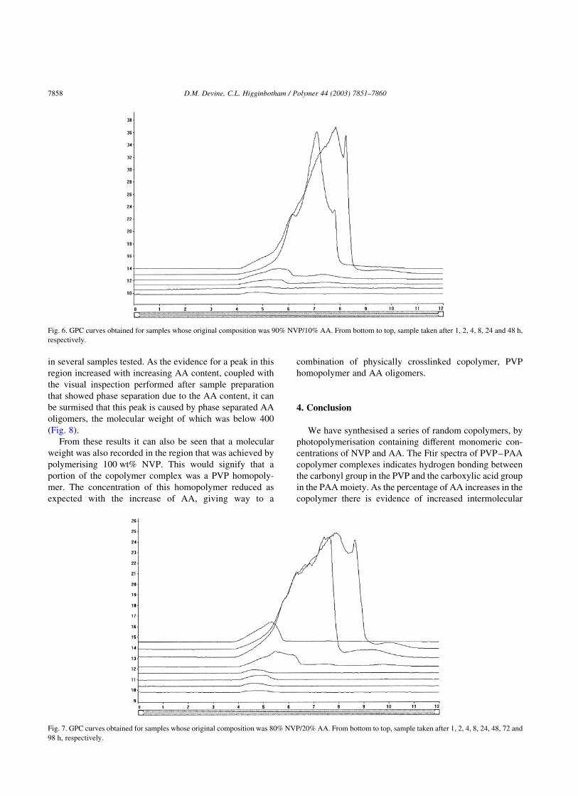
From analysis of the samples whose original composition
was 90 wt% NVP–10 wt% AA, 80 wt% NVP–20 wt% AA
and 70 wt% NVP–30 wt% AA, respectively, there is a large
number of molecular weight points outside of calibration.
The Mp values eluted prior to 6.25 min are all in excess of a
molecular weight of eight million. As the polymerisation
process was identical to that which provided a molecular
weight in the region of 290,000 for polymerised NVP it is
reasonable to assume that a polymer of this size should not
be possible using this polymerisation method. Thus, it must
be assumed that these ultra high molecular weights achieved
were caused by a physically crosslinked polymer rapidly
passing through the GPC column (Fig. 7).
On the other hand polymers eluted after 9.78 min had a
molecular weight of less than 400. From the sample whose
original composition was 90 wt% NVP–10 wt% AA, there
is evidence of a peak in this region after 24 h. From the
80 wt% NVP–20 wt% AA, there is even more evidence of
peaks in this region after 24 h, 48 and 72 h. However, the
sample containing 70 wt% NVP–30 wt% AA there is a
clear peak obtained after 24 h with further evidence of peaks
Fig. 5. GPC curves obtained for samples whose original composition was 100% NVP. From bottom to top, sample taken after 1, 2, 4, 8 and 24 h, respectively.
D.M. Devine, C.L. Higginbotham / Polymer 44 (2003) 7851–7860 7857
in several samples tested. As the evidence for a peak in this
region increased with increasing AA content, coupled with
the visual inspection performed after sample preparation
that showed phase separation due to the AA content, it can
be surmised that this peak is caused by phase separated AA
oligomers, the molecular weight of which was below 400
(Fig. 8).
From these results it can also be seen that a molecular
weight was also recorded in the region that was achieved by
polymerising 100 wt% NVP. This would signify that a
portion of the copolymer complex was a PVP homopoly-
mer. The concentration of this homopolymer reduced as
expected with the increase of AA, giving way to a
combination of physically crosslinked copolymer, PVP
homopolymer and AA oligomers.
4. Conclusion
We have synthesised a series of random copolymers, by
photopolymerisation containing different monomeric con-
centrations of NVP and AA. The Ftir spectra of PVP–PAA
copolymer complexes indicates hydrogen bonding between
the carbonyl group in the PVP and the carboxylic acid group
in the PAA moiety. As the percentage of AA increases in the
copolymer there is evidence of increased intermolecular
Fig. 6. GPC curves obtained for samples whose original composition was 90% NVP/10% AA. From bottom to top, sample taken after 1, 2, 4, 8, 24 and 48 h,
respectively.
Fig. 7. GPC curves obtained for samples whose original composition was 80% NVP/20% AA. From bottom to top, sample taken after 1, 2, 4, 8, 24, 48, 72 and
98 h, respectively.
D.M. Devine, C.L. Higginbotham / Polymer 44 (2003) 7851–78607858

hydrogen bonding between the carboxylic acid groups of the
AA segments. Swelling of the PVP–PAA complex in a
higher pH medium is significantly different from results in
low pH solutions. The critical pH range was found to be
between 4.07 and 4.49. Above a pH of 4.49 there is a
progressive break up of the polymer chain due to a reduction
in the amount of intermolecular hydrogen bonding. This is
caused by an increase in the amount of carboxylate ions as
the pH increases. There is also a significant increase in the
solubility of the copolymer complex at higher pHs. The low
solubility of the copolymer at low pH may make the
complex suitable for gastric drug delivery systems. Finally,
some of the copolymers produced seemed to have
extraordinary high molecular weights and this needs to be
investigated further.
Acknowledgements
This study was supported in parts by grants from both
Enterprise Ireland and the Athlone Institute of Technology
research and development fund.
References
[1] Yaung J-F, Kwei TK. pH-sensitive hydrogels based on polyvinylpyr-
rolidone–polyacrylic acid (PVP–PAA) semi-interpenetrating net-
works (semi-ipn): swelling and controlled release. J Applied Polym
Sci 1998;69:921–30.
[2] Kishida A, Ikada Y. Hydrogels for biomedical and pharmaceutical
applications. In: Dumitriu S, editor. Polymeric biomaterials, 2nd ed.;
2002. p. 133–45.
[3] Ravichandran P, Shantha KL, Panduranga Rao K. Preparation,
swelling characteristics and evaluation of hydrogels for stomach
specific drug delivery. Inter J Pharm 1997;154:89–94.
[4] Peppas NA, Bures P, Leobandung W, Ichikawa H. Hydrogels in
pharmaceutical formulations. Eur J Pharm Biopharm 2000;50:27–46.
[5] Risbud MV, Hardikar AA, Bhat SV, Bhonde RR. pH-sensitive freeze-
dried chitosan-polyvinyl pyrrolidone hydrogels as controlled release
system for antibiotic delivery. J Controlled Release 2000;68:23–30.
[6] Anseth KS, Bowman CN, Brannon-Peppas L. Mechanical properties
of hydrogels and their experimental determination. Biomaterials
1996;17:1647–57.
[7] Nguyen KT, West JL. Photopolymerizable hydrogels for tissue
engineering applications. Biomaterials 2002;23:4307–14.
[8] LaPorte RJ. Hydrophilic polymer coatings for medical devices.
Technomic Pub. Co. Inc; Lancaster, Basel; 1997.
[9] Kunioka M, Choi HJ. Hydrolytic degradation and mechanical
properties of hydrogels prepared from microbial poly amino acids.
Polym Degrad Stab 1998;59:33–7.
[10] Am Ende MT, Peppas NA. Transport of ionisable drugs and proteins
in crosslinked poly (acrylic acid) and poly (acrylic acid-co-2-
hydroxyethyl methacrylate) hydrogels. 1. Polymer characterization.
J Applied Polym Sci 1996;59:673–85.
[11] Varshosaz J, Koopaie N. Cross-linked poly (vinyl alcohol) hydrogel:
study of swelling and drug release behaviour. Iranian Polym J 2002;
11(2):123–31.
[12] Brazel CS, Peppas NA. On the mechanisms of water transport and
drug release from swellable hydrogels. ACS Polym Mater: Sci Eng
1996;70:370–1.
[13] Aikawa K, Mitsutake N, Uda H, Shimamura H, Aramaki Y, Tsuchiya
S. Drug release from pH-response polyvinylacetal diethylaminoace-
tate hydrogel, and application to nasal delivery. Inter J Pharm 1998;
168:181–8.
[14] Graham NB. Controlled drug delivery systems. Chem Ind 1990;
482–6.
[15] Grass M, Colombo I, Lapasin R. Drug release from an ensemble of
swellable crosslinked polymer particles. J Controlled Release 2000;
68:97–113.
[16] Murata Y, Sasaki N, Miyamoto E, Kawashima S. Use of floating
alginate gel beads for stomach-specific drug delivery. Eur J Pharm
Biopharm 2000;50:221–6.
[17] Florence AT. New drug delivery systems. Chem Ind 1993;1000–4.
[18] Ruel-Gariepy E, Chenite A, Chaput C, Guirguis S, Leroux J-C.
Fig. 8. GPC curves obtained for samples whose original composition was 70% NVP/30% AA. From bottom to top, sample taken after 1, 2, 4, 8, 24, 48, 72 and
98 h, respectively.
D.M. Devine, C.L. Higginbotham / Polymer 44 (2003) 7851–7860 7859
Characterization of thermosensitive chitosan gels for the sustained
delivery of drugs. Inter J Pharm 2000;203:89–98.
[19] Aikawa K, Matsumoto K, Uda H, Tanaka S, Shimamura H, Aramaki
Y, Tsuchiya S. Hydrogel formation of the pH response polymer
polyvinylacetal diethylaminoacetate (AEA). Inter J Pharm 1998;167:
97–104.
[20] McNair AM. Using hydrogel polymers for drug delivery. MDT 1996;
16–22.
[21] Perera DI, Shanks RA. Swelling and mechanical properties of
crosslinked hydrogels containing N-vinylpyrrolidinone. Polym Inter
1996;39:121–7.
[22] Pohlmann T, Muller A, Seiferling B. Polymeric networks from water-
soluble prepolymers; 1997; US Patent No. 5, 665, 840.
[23] Shoji N, Nomura M, Yokoyama Y. Methods for producing 2-hydroxy-
ethyl methacrylate polymer, hydrogel and water-containing soft
contact lenses; 1997; European Patent No. EP 0874 007 A1.
[24] Peppas NA, Wright SL. Drug diffusion and binding in ionizable
interpenetrating networks from poly (vinyl alcohol) and poly (acrylic
acid). J Pharm Biopharm 1998;46:15–29.
[25] Rathner BD, Hoffman AS. Hydrogels for medical and related
applications. ACS Symposium Series, Washington DC: American
Chemical Society; 1976. p. 1–36.
[26] Dyson RW, editor. Specially polymers. 1987.
[27] Stauffer SR, Peppas NA. Poly (vinyl alcohol) hydrogels prepared by
freezing-thawing cyclic processing. Polymer 1992;33:3932–6.
[28] Hassan CH, Stewart JE, Peppas NA. Diffusional characteristics of
freeze/thawed poly (vinyl alcohol) hydrogels: applications to protein
controlled release from multilaminate devices. Eur J Pharm Biopharm
2000;49:161–5.
[29] Hassan CM, Peppas NA. Structure and applications of poly (vinyl
alcohol) hydrogels produced by conventional crosslinking or by
freezing/thawing methods. Adv Polym Sci 2000;153:37–65.
[30] Lau C, Mi Y. A study of blending and complexation of poly(acrylic
acid)/poly(vinyl pyrrolidone). Polymer 2002;43:823–9.
[31] Bures P, Peppas NA. Molecular dynamics of pH-sensitive hydrogels
based on poly(acrylic acid). ACS Polym Mater: Sci Eng 2000;83:
506–7.
[32] Lee JY, Painter PC, Coleman MM. Hydrogen bonding in polymer
blends. 3. Blends involving polymers containing methacrylic acid and
ether groups. Macromolecules 1988;21:346–54.
[33] Lee JY, Painter PC, Coleman MM. Hydrogen bonding in polymer
blends. 4. Blends involving polymers containing methacrylic acid and
vinylpyridine groups. Macromolecules 1988;21:954–60.
[34] Senak L, Wu CS, Malawer EG. Size exclusion chromatography of
poly(vinylpyrrolidinone): II. Absolute molecular weight distribution
by SEC/LALLS and SEC with universal calibration. J Liq Chroma-
togr 1987;10:1127–50.
D.M. Devine, C.L. Higginbotham / Polymer 44 (2003) 7851–78607860


