Embed Size (px)
Citation preview
Developmental Immunology, 1998, Vol. 6, pp. 53-60Reprints available directly from the publisherPhotocopying permitted by license only
(C) 1998 OPA (Overseas Publishers Association)N.V. Published by license under the
Harwood Academic Publishers imprint,part of the Gordon and Breach Publishing Group
Printed in Malaysia
The Influence of Costimulation and Regulatory CD4+ TCells on Intestinal IgA Immune Responses
EVA GRDBY*, DUBRAV KAGRDIC, MARTIN KJERRULF, ANNAKARI BROMANDER, MICHAEL VAJDY,ELISABETH HRNQUIST and NILS LYCKE
Department of Medical Microbiology and Immunology, University of G6teborg, S-413 46 G6teborg, Sweden
(Received 11 August 1996; In final form 15 June 1997)
It is thought that IgA B-cell differentiation is highly dependent on activated CD4 T cells. Inparticular, cell-cell interactions in the Peyer’ patches involving CD40 and/or CD80/CD86 havebeen implicated in germinal-center formation and IgA B-cell development. Also soluble factors,such as IL-4, IL-5, IL-6, and TGF/3 may be critical for IgA B-cell differentiation in vivo. Herewe report on some paradoxical findings with regard to IgA B-cell differentiation and specificmucosal immune responses that we have recently made using gene knockout mice. Morespecifically, we have investigated to what extent absence of CD4 T cells, relevant cytokines, orT-cell-B-cell interactions would influence IgA B-cell differentiation in vivo. Using CD4- or IL-4-gene knockout mice or mice made transgenic for CTLA4Ig, we found that, although specificresponses were impaired, total IgA production and IgA B-cell differentiation appeared toproceed normally. However, a poor correlation was found between, on the one hand, GCformation and IgA differentiation and, on the other hand, the ability to respond to T-cell-dependent soluble protein antigens in these mice. Thus, despite the various deficiencies in CD4T-cell functions seemingly intact IgA B-cell development was observed.
INTRODUCTION
Mucosal surfaces are constantly exposed to a myriadof foreign antigens. To protect against pathogenicmicroorganisms or hostile compounds, such as toxins,the mucosa has developed effective barrier functions.For the maintenance of these barrier functions,secretory IgA (sIgA) is thought to play a particularlyimportant role (McGhee et al., 1992). In the intestinalimmune system, IgA B cells are derived from
precursor cells in the Peyer’s patches (PP) (Cebra and
*Corresponding author.
Shroff, 1994). After antigen stimulation, IgA lympho-blasts migrate from the PP via the mesenteric lymphnode (MLN) and the thoracic lymph duct to the bloodand eventually home back to the lamina propria of the
gut mucosa. The development of normal PP morphol-ogy appears to be dependent on the local exposure to
antigens in the intestine. Investigations of gnotobioticor neonatal mice have demonstrated only a few IgA Bcells in poorly developed PP (Cebra et al., 1980).After bacterial colonization of these mice, IgA B cellsappear in great numbers in the PP, which now have
53

54 E. GRDBY et al.
increased in size and numbers. The IgA B cells in thePP largely represent dividing cells and the PP make
up the largest pool of IgA-committed B cells in the
body. Most of these IgA B cells colocalize to
germinal centers (GC) in the B-cell follicles of the PP.It is important to note that even without specificimmunization, conventionally reared animals, in con-
trast to spleen and lymph nodes, exhibit largenumbers of GC in PP (Butcher et al., 1982). Oneexplanation for this may be that the IgA-committed Bcells from PP commonly have specificities againstphosphocholine,/32-1 fructosyl groups, or/3 galacto-syl groups, which are antigens that may be associated
with the normal bacterial flora of the gut.
GERMINAL CENTERS
Germinal centers are histologically defined areas
containing rapidly dividing B lymphoblasts enmeshedin a network of follicular dendritic cells (FDC) and
relatively few CD4 T cells (Liu and Banchereau,1996). The GC is formed after antigen stimulation andreaches a maximum size after 10 to 14 days. Ageneral view is that the GC reaction is dependent on
the presence of CD4 T cells. Also in the gut mucosalimmune system, CD4 T cells in PP are thought to
play a critical role for the generation of IgA immune
responses (Cebra et al., 1980). Studies in nude mice,
lacking cq3TCR cells but fully competent in B cells,have demonstrated impaired IgA B-cell differen-
tiation (Cebra et al., 1980; Gottstein et al., 1993). Inthe PP, the GC contains 75-80% IgA B cells,whereas in peripheral lymph nodes, very few IgA Bcells are seen (Butcher et al., 1982). Therefore, it is
thought that the GC in PP differ intrinsically fromthose of spleen and lymph nodes in their content of
particular kinds of antigen-presenting cells (APC),stromal cells, or regulatory T cells, and, hence, in the
repertoire of cytokines and cell-cell interactions
available for promoting isotype switching from IgMto IgA. Earlier experiments by Cebra et al. with localreovirus infection in germ-free mice demonstratedthat the microenvironment of the PP may be selectingfor direct isotype switching from IgM to IgA B-cell
differentiation (Weinstein and Cebra, 1991). The
prevailing theory on IgA B-cell differentiation arguesfor this process being a highly directed event stronglydependent on CD4 T cells (Strober and Ehrhandt,1994). Recently, it was reported that a majority of theGC T cells may be phenotypically distinct Thyl-CD4 cells (Harriman et al., 1990; Zheng et al., 1996).Also accessory cells can provide necessary signals to
drive IgA B-cell differentiation in PP (Schrader et al.,1990). Important accessory cells in the PP are
dendritic cells (DC), both the interdigitating DC andthe FDC in the GC. Whether there exists distinct
functional subsets of DCs in the PP is currentlyunclear (Kelsall and Strober, 1996; Ruedl et al.,1996). Recent preliminary reports have suggested thatdendritic cells isolated from PP or treated with IL-10
may favor Th2 differentiation and cytokine secretion
of CD4 T cells.
COSTIMULATION AND GC FORMATION
Earlier studies have clearly demonstrated that cell-cellinteractions via the CD40L and CD28 on the activated
T cell critically influence the formation of GC (Foy et
al., 1994; Bluestone, 1995; Sharpe 1995). Whereasboth CD40L and CD28 interactions appear to affectGC formation, isotype switching, and development of
memory, there may also be differential effects on
these events as well as on other events such as Ighypermutation (Han et al., 1995). Moreover, it is also
probable that these T-cell-B-cell interactions affectthe development of the CD4 T-helper functions in
vivo (Essen et al., 1995). We and others have found
that oral immunization preferentially generates CD4T cells of the Th2 type and experiments in IL-4-/-
mice have shown that this cytokine may be requiredfor intestinal immune responses against soluble
protein antigens (Xu-Amano et al., 1993; Vajdy et al.,1995). Recent studies have indicated that expressionof CD40L on activated T cells may be more stronglyassociated with Th2 rather than Thl development in
CD4 T cells (Freeman et al., 1995). Therefore, Tcells that are high in CD40L expression may be one ofthe defining features of the IgA-promoting environ-
ment in PP.

REGULATORY CD4 T CELLS ON INTESTINAL IGA IMMUNE RESPONSES 55
REQUIREMENT FOR CD4 T CELLS IN lgAB-CELL DIFFERENTIATION
For IgA B-cell differentiation, CD4 T cells are
thought to play a critical role as helper cells producingsoluble factors, such as TGF/3, IL-5, and IL-6, as wellas engaging in contact-dependent cell-cell inter-
actions (Strober and Ehrhardt, 1994). Whereas TGF/3acts as a possible switch factor early in IgA B-celldifferentiation, IL-5 and IL-6 act later, on alreadycommitted IgA B cells. Since most of the cytokinesthat have been implicated in IgA B-cell developmentare truely pleiotropic and produced by macrophages,fibroblasts, epithelial cells as well as B and Tlymphocytes in the mucosa, our understanding of theinteractions between these cells and the IgA B-cellprogeny is still poor. The introduction of transgene-and gene-targeting techniques has provided powerfulnew tools with which to address complex issues suchas the regulation of mucosal immune responses andIgA B-cell development (Lycke et al., 1995). In our
laboratory, we have taken advantage of these mice to
address some of the critical questions in mucosalimmunolgy.
PARADOXES IN IgA B-CELLDIFFERENTIATION IN GENE KNOCKOUTMICE
In this review, we will discuss some of the paradoxesof IgA B-cell immunity that we have observed in
mice made genetically deficient for critical T-cellfunctions. Our experimental model system uses
soluble protein antigens such as keyhole limpethemocyanin (KLH) or ovalbumin (OVA) given orallytogether with cholera toxin (CT) adjuvant for induc-
tion of intestinal immune responses. We have studied
IgA B-cell differentiation and the ability to respond to
oral immunizations in CD4 gene-targeted (CD4-/-)mice, IL-4-/-, and IL-6-/- mice or mice over-
expressing the CTLA4-Hyl protein, which blocks T-cell-B-cell interaction via the CD80 and CD86surface molecules. The latter CTLA4-Hyl transgenicmice (TG) have previously been reported to lack GC
formation and isotype switch differentiation in sys-temic tissues following specific immunizations with
T-cell-dependent antigens (Lane et al., 1994).Using normal and CD4-/- mice, we asked whether
mucosal immune responses and IgA B-cell differen-
tiation required the presence of CD4 T-helper cells(H6rnquist et al., 1995). Quite unexpectedly we foundthat CD4-/- mice had numerous GC in PP and other
gut-associated lymphoid tissues (GALT), whereasonly few GCs were evident in spleen and lymphnodes. Membrane IgA B cells were found to
colocalize to GC areas and CD4-CD8- double-
negative (DN) CD3 T cells had replaced CD4 Tcells in the follicular areas of the PP. A recent studyanalyzing TCRc-/- mice also reported on GCformation in PP despite the lack of classical cq?TCRcells, suggesting that the lymphoid tissue in PP maybe differently regulated from that in peripheral lymphnodes (Dianda et al., 1996). Alternatively, there mayexist microbial antigens in the gut microenvironment
that can drive GC formation with a limited require-ment for cq3TCR CD4 T-cell help. The latter notionis supported by the finding that germ-free TCRc-/-
mice had less GC in PP than did conventionallyreared mice. However, at variance with our findings,in CD4-/- mice, the latter study suggested that
expression of CD4 was important for GC formation in
PP (H6rnquist et al., 1995; Dianda et al., 1996).Nevertheless, at present, we must conclude that therelationship between CD4 T cells and the formationof GC in PP is poorly understood.The CD4-/- mice had normal levels of IgA-
producing cells in GALT and gut lavage containedunaltered levels of total IgA (H6rnquist et al., 1995).In spite of what appeared to be adequate T-cell helpfor IgA B-cell differentiation, CD4-/- mice did not
respond with Ag-specific intestinal or serum IgAfollowing oral immunization with CT. Moreover,perorally immunized CD4-/- mice were completelyunprotected against CT-induced diarrhea, whereasnormal mice were well-protected and demonstratedhigh levels of antitoxin IgA in gut lavage. Thus,paradoxically, whereas IgA B-cell differentiation
appeared to proceed normally in CD4-/- mice,
specific gut mucosal immune responses were
56 E. GRDBY et al.
impaired in the absence of CD4 T cells. A poorcorrelation between GC formation and presence ofserum antibody has previously been reported byStedra and Cerny (1994), and based on our findings,it seems reasonable to assume that the presence of GCin PP does not constitute proof of the ability to
respond to oral immunization with T-cell-dependentantigens.
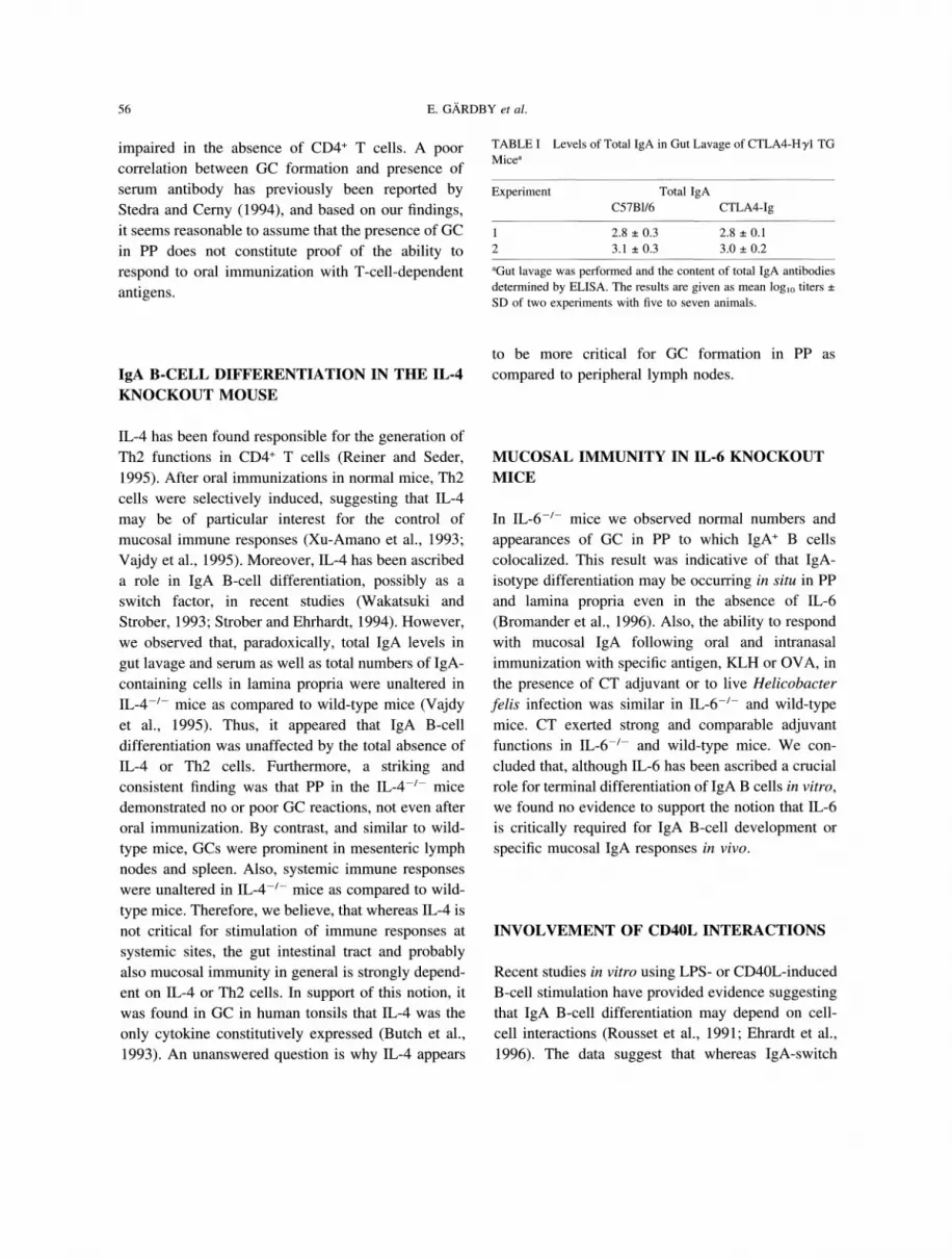
TABLE Levels of Total IgA in Gut Lavage of CTLA4-Hyl TGMice
Experiment Total IgAC57B1/6 CTLA4-Ig
2.8 0.3 2.8 0.12 3.1 0.3 3.0 0.2
aGut lavage was performed and the content of total IgA antibodiesdetermined by ELISA. The results are given as mean loglo titers _+
SD of two experiments with five to seven animals.
IgA B-CELL DIFFERENTIATION IN THE IL-4KNOCKOUT MOUSE
IL-4 has been found responsible for the generation ofTh2 functions in CD4 T cells (Reiner and Seder,1995). After oral immunizations in normal mice, Th2cells were selectively induced, suggesting that IL-4may be of particular interest for the control ofmucosal immune responses (Xu-Amano et al., 1993;Vajdy et al., 1995). Moreover, IL-4 has been ascribeda role in IgA B-cell differentiation, possibly as a
switch factor, in recent studies (Wakatsuki andStrober, 1993; Strober and Ehrhardt, 1994). However,we observed that, paradoxically, total IgA levels in
gut lavage and serum as well as total numbers of IgA-containing cells in lamina propria were unaltered inIL-4-/- mice as compared to wild-type mice (Vajdyet al., 1995). Thus, it appeared that IgA B-celldifferentiation was unaffected by the total absence ofIL-4 or Th2 cells. Furthermore, a striking andconsistent finding was that PP in the IL-4-/- micedemonstrated no or poor GC reactions, not even afteroral immunization. By contrast, and similar to wild-
type mice, GCs were prominent in mesenteric lymphnodes and spleen. Also, systemic immune responseswere unaltered in IL-4-/- mice as compared to wild-
type mice. Therefore, we believe, that whereas IL-4 isnot critical for stimulation of immune responses at
systemic sites, the gut intestinal tract and probablyalso mucosal immunity in general is strongly depend-ent on IL-4 or Th2 cells. In support of this notion, itwas found in GC in human tonsils that IL-4 was the
only cytokine constitutively expressed (Butch et al.,1993). An unanswered question is why IL-4 appears
to be more critical for GC formation in PP as
compared to peripheral lymph nodes.
MUCOSAL IMMUNITY IN IL-6 KNOCKOUTMICE
In IL-6-/- mice we observed normal numbers and
appearances of GC in PP to which IgA B cellscolocalized. This result was indicative of that IgA-isotype differentiation may be occurring in situ in PPand lamina propria even in the absence of IL-6(Bromander et al., 1996). Also, the ability to respondwith mucosal IgA following oral and intranasalimmunization with specific antigen, KLH or OVA, in
the presence of CT adjuvant or to live Helicobacter
felis infection was similar in IL-6-/- and wild-typemice. CT exerted strong and comparable adjuvantfunctions in IL-6-/- and wild-type mice. We con-
cluded that, although IL-6 has been ascribed a crucial
role for terminal differentiation of IgA B cells in vitro,
we found no evidence to support the notion that IL-6is critically required for IgA B-cell development or
specific mucosal IgA responses in vivo.
INVOLVEMENT OF CD40L INTERACTIONS
Recent studies in vitro using LPS- or CD40L-inducedB-cell stimulation have provided evidence suggestingthat IgA B-cell differentiation may depend on cell-cell interactions (Rousset et al., 1991; Ehrardt et al.,1996). The data suggest that whereas IgA-switch
REGULATORY CD4 T CELLS ON INTESTINAL IGA IMMUNE RESPONSES 57
differentiation may proceed in the presence of solubleT-cell factors, that is, cytokines, terminal differen-tiation of IgA B cells may critically depend on B-cellinteraction with activated CD40L-expressing T cells.IgA B cells, in contrast to IgA-/IgM B cells, were
strikingly more responsive to T-cell dependent activa-
tion, that is, CD40L interaction, than to T-cell-independent activation via membrane cross-linking ofreceptors. Thus, the close interaction with the acti-vated T cell most probably is a necessary preconditionfor terminal IgA B-cell differentiation in vivo. How-ever, there was no difference in responsivenessbetween PNA-high B cells isolated from GC of PPand PNA-low-lamina-propria IgA B cells, suggest-ing that CD40L expression may not be sufficient to
explain the regulatory role of CD4 T cells in thesetwo locations. Nevertheless, collectively, the dataprovide evidence that costimulation is playing a
pivotal role in IgA B-cell differentiation.
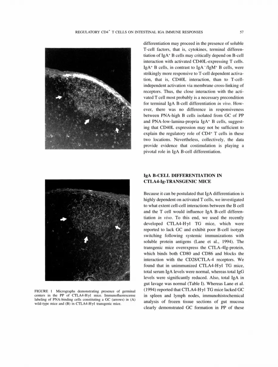
FIGURE Micrographs demonstrating presence of germinalcenters in the PP of CTLA4-Hyl mice. Immunofluorescenselabeling of PNA-binding cells constituting a GC (arrows) in (A)wild-type mice and (B) in CTLA4-Hyl transgenic mice.
IgA B-CELL DIFFERENTIATION INCTLA4-Ig-TRANSGENIC MICE
Because it can be postulated that IgA differentiation is
highly dependent on activated T cells, we investigatedto what extent cell-cell interactions between the B celland the T cell would influence IgA B-cell differen-tiation in vivo. To this end, we used the recentlydeveloped CTLA4-Hyl TG mice, which were
reported to lack GC and exhibit poor B-cell isotypeswitching following systemic immunizations withsoluble protein antigens (Lane et al., 1994). The
transgenic mice overexpress the CTLA-4Ig-protein,which binds both CD80 and CD86 and blocks theinteraction with the CD28/CTLA-4 receptors. Wefound that in unimmunized CTLA4-Hyl TG mice,total serum IgA levels were normal, whereas total IgGlevels were significantly reduced. Also, total IgA in
gut lavage was normal (Table I). Whereas Lane et al.(1994) reported that CTLA4-Hyl TG mice lacked GCin spleen and lymph nodes, immunohistochemical
analysis of frozen tissue sections of gut mucosa
clearly demonstrated GC formation in PP of these
58 E. GRDBY et al.
AImpaired mucosal IgA anti-KLH responsesdespite normal levels of total gut IgA.
A
60004000
2000--
0
1000-
g
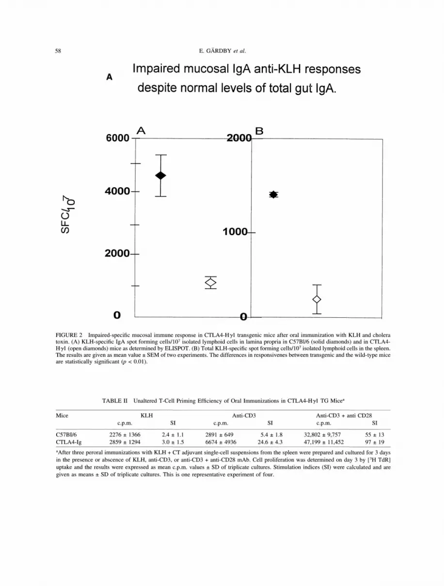
FIGURE 2 Impaired-specific mucosal immune response in CTLA4-Hyl transgenic mice after oral immunization with KLH and choleratoxin. (A) KLH-specific IgA spot forming cells/107 isolated lymphoid cells in lamina propria in C57B1/6 (solid diamonds) and in CTLA4-Hyl (open diamonds) mice as determined by ELISPOT. (B) Total KLH-specific spot forming cells/107 isolated lymphoid cells in the spleen.The results are given as mean value SEM of two experiments. The differences in responsivenes between transgenic and the wild-type miceare statistically significant (p < 0.01).
TABLE II Unaltered T-Cell Priming Efficiency of Oral Immunizations in CTLA4-HT1 TG Mice
Mice KLH Anti-CD3 Anti-CD3 + anti CD28c.p.m. SI c.p.m. SI c.p.m. SI
C57BI/6 2276 1366 2.4 1.1 2891 649 5.4 1.8 32,802 9,757 55 13CTLA4-Ig 2859 1294 3.0 _+ 1.5 6674 _+ 4936 24.6 4.3 47,199 11,452 97 19
aAfter three peroral immunizations with KLH + CT adjuvant single-cell suspensions from the spleen were prepared and cultured for 3 daysin the presence or abscence of KLH, anti-CD3, or anti-CD3 + anti-CD28 mAb. Cell proliferation was determined on day 3 by [3H TdR]uptake and the results were expressed as mean c.p.m, values SD of triplicate cultures. Stimulation indices (SI) were calculated and are
given as means SD of triplicate cultures. This is one representative experiment of four.

REGULATORY CD4 T CELLS ON INTESTINAL IGA IMMUNE RESPONSES 59
mice (Figure 1). In accordance with previous findingsin normal mice, the IgA B cells colocalized to theGC areas in PP. Thus, similar to the IL-4-/- mice, wefound unaltered total IgA concentrations in serum andintestinal lavage, but, in contrast to these mice, weobserved many GC in PP of CTLA4-HT1 TG mice.
Previously, it was reported that systemic antibodyresponses to T-cell-dependent antigens were signifi-cantly impaired in the CTLA4-H3/1 TG mice (Lane et
al., 1994). As the lack of an IgG response appeared to
be directly related to the absence of systemic GCformation, we tested whether mucosal IgA responsesrequired less T-cell-B-cell cooperation to generatemucosal IgA immunity. Following oral immunization
with KLH plus CT-adjuvant local IgA as well as
systemic anti-KLH responses in CTLA4-HT1, TGmice were strongly impaired (Figure 2). Thus,paradoxically, we observed unimpeded total IgAlevels in the gut lamina propria, and GC formation
and IgA B-cell isotype-switch differentiation in PP,and yet the response to both KLH and CT were
severely impaired in the CTLA4-Hyl TG mice.Moreover, in contrast to IL-4-/- mice (Vajdy et al.,
1995), we found that oral immunizations efficientlyprimed T cells in the CTLA4-Hyl TG mice. Asillustrated in Table II, we obtained comparableresponses to recall antigen in spleen T-cell culturesfrom CTLA4-HT1 TG and normal C57B1/6 mice
following oral immunizations with KLH plus CTadjuvant. Also, the ability of splenic T cells from
CTLA4-HT1 TG mice to respond to anti-CD3 andanti-CD28 costimulation was unaltered as comparedto normal mice (Table 2). This finding agrees wellwith previous reports demonstrating unaltered abilityto induce T-cell responses in the CTLA4-HT1 TGmice (Ronchese et al., 1994), whereas T-cell help forIgA B-cell differentiation after specific oral immuni-
zations appears to be inhibited.
CONCLUDING REMARKS
In conclusion, the CTLA4-Hyl TG mice, as well as
CD4-/- and IL-4-/- mice, have shown impaired
mucosal immune responses to oral immunizationswith the highly immunogenic combination of KLHand CT adjuvant. Although, specific responses were
impaired, total IgA production and IgA B-celldifferentiation appeared to proceed normally in thesemice, despite the various deficiencies in CD4 T-cellfunctions. Thus, paradoxically, we found poor corre-
lation between, on the one hand, GC formation andIgA differentiation and, on the other hand, the abilityto respond to T-cell-dependent soluble protein anti-
gens. Experiments are ongoing to resolve theseconflicting findings.
References
Bluestone J.A. (1995). New perspectives of CD28-B7-mediated T-cell costimulation. Immunity. 2: 555-559.
Bromander A., Ekman L., Kopf M., Nedrud J., and Lycke N.(1996). IL-6-deficient mice exhibit normal mucosal IgAresponses to local immunizations and Helicobacter felis infec-tion. J. Immunol. 156: 4290-4297.
Butch A.W., Chung G.H., Hoffmann J.W., and Nahm M.H. (1993).Cytokine expression by germinal center cells. J. Immunol. 150:39-47.
Butcher E.C., Rouse R.L., Nottenberg C.N., Hardy R.R., andWeissman I. (1982). Surface phenotype of Peyer’s patchgerminal centers: Implications for the role of germinal centers inB-cell differentiation. J. Immunol. 129: 2698.
Cebra J.J., Gearhart P.J., Halsey J.F., Hurwitz J.L., and Shahin R.D.(1980). Role of environmental antigens in the ontogeny of thesecretory immune response. J. Reticuloendothel. Soc. 28:61-71.
Cebra J.J., and Shroff K. (1994). Peyer’s patches as inductive sitesfor IgA comittment. In Handbook of Mucosal Immunology,Ogra P., Strober W., Mestecky J., McGhee J., Lamm M., andBienenstock J., Eds. (San Diego: Academic Press), pp.151-158.
Dianda L., Gulbranson-Judge A., Pao W., Hayday A., MacLennanI., and Owen M. (1996). Germinal center formation in micelacking ab T cells. Eur. J. Immunol. 26: 1603-1607.
Ehrardt R., Gray B., Duchmann R., Inman J., and Strober W.(1996). Reciprocal regulation of mucosal surface IgA+ B cellsby Ig receptor cross-linking and CD40 ligand. J. Immunol. 157:1397-1405.
Essen D., Kikutani H., and Gray D. (1995). CD40 ligand-transduced co-stimulation of T cells in the development ofhelper function. Nature 378: 620-623.
Foy T.M., Laman J.D., Ledbetter J.A., Aruffo A., Claassen E., andNoelle R. (1994). gp-39-CD40 interactions are essential forgerminal center formation and the development of B cellmemory. J. Exp. Med. 180: 157-163.
Freeman G.J., Boussiotis V.A., Anumanthan A., Bernstein G.M.,Ke X.Y., Rennert P.D., Gray G.S., Gribben J.G., and NadlerL.M. (1995). B7-1 and B7-2 do not deliver identical costimula-tory signals, since B7-2 but not B7-1 preferentially costimulatesthe initial production of IL-4. Immunity 2: 523-532.

60 E. GRDBY et al.
Gottstein B., Deplazes P., and Tanner I. (1993). In vitro synthezisedimmunoglobulin A from nu/+ and reconstituted nu/nu miceagainst a dominant surface antigen of Giardia lamblia. Parasitol.Res. 79: 644.
Han S., Hathcock K., Zheng B., Kepler T. Hodes R., and Kelsoe G.(1995). Cellular interaction in germinal centers. Roles of CD40ligand and B7-2 in established germinal centers. J. immunol.155: 556-567.
Harriman G.R., Lycke N.Y., Elwood L.J., and Strober W. (1990).T lymphocytes that express CD4 and the ab-T cell receptor butlack Thy-1. J. Immunol. 145: 2406-2414.
H6rnquist E., Ekman L., Grdic D., Sch6n K. and Lycke N. (1995).Paradoxical IgA immunity in CD4-deficient mice: Lack ofcholera toxin specific protective immunity despite normal gutmucosal IgA differentiation. J. Immunol. 155: 2877-2887.
Kelsall B.L., and Strober W. (1996). Distinct populations ofdendritic cells are present in the subepithelial dome and T cellregions of the murine Peyer’s patch. J Exp. Med. 183:237-247.
Lane P., Burdet C., Hubele S., Schedegger D., Muller U., McConellF., and Kosco-Vilbois M. (1994). B cell function in micetransgenic for mCTLA4-Hgl: Lack of germinal centers corre-lated with poor affinity maturation and class switching despitenormal priming of CD4+ T cells. J. Exp. Med. 179: 819-830.
Liu Y.J., and Banchereau J. (1996). The paths and molecularcontrols of peripheral B-cell development. Immunologist 4:55-66.
Lycke N., Bromander A.K., Ekman L., Grdic D., H6rnquist E.,Kjerrulf M., Kopf M., Kosco-Vilbois M., Sch6n K., and VajdyM. (1995). The use of knock-out mice in studies of induction andregulation of gut mucosal immunity. Mucosal Immunol. Update3, 000-000.
McGhee J.R., Mestecky J., Dertzbaugh M.T., Eldridge J.H.,Hirasawa M., and Kiyono H. (1992). The mucosal immunesystem: From fundamental concepts to vaccine development.Vaccine 10: 75.
Reiner S.L., and Seder R.A. (1995). T helper cell differentiation inimmune response. Curr. Opin. Immunol. 7: 360-366.
Ronchese F., Hausmann B., Hubele S., and Lane P. (1994). Micetransgenic for a soluble form of murine CTLA4 show enhanced
expansion of antigen-specific CD4+ T-cells and defectiveantibody production in vivo. J. Exp. Med. 179: 809-817.
Rousset F., Gracia E., and Banchereau J. (1991). Cytokine-inducedproliferation and immunoglobulin production of human Blymphocytes triggered through their CD40 antigen. J. Exp. Med.173: 705-710.
Ruedl C., Rieser C., B6ck G., Wick G. and Wolf H. (1996).Phenotypic and functional characterization of CD11c+ dendriticcell population in mouse Peyer’s patches. Eur. J. Immunol. 26:1801-1806.
Schrader C.E., George A., Kerlin R.L., and Cebra J.J. (1990).Dendritic cells support production of IgA and other non-IgMisotypes in clonal microculture. Int. Immunol. 2: 563-570.
Sharpe A. (1995). Analysis of costimulation in vivo usingtransgenic and knockout mice. C. O. Immunol. 7: 389-395.
Stedra J., and Cerny J. (1994). Distinct pathways of B celldifferentiation. J. Immunol. 152: 1718.
Strober W., and Ehrhardt R. (1994). Regulation of IgA B celldevelopment. In Handbook of Mucosal Immunology, Ogra P.,Strober W., Mestecky J., McGhee J., Lamm M., and Bienen-stock J., Eds. (San Diego: Academic Press), pp. 159-176.
Vajdy M., Kosco-Vilbois M., Kopf M., K6hler G., and Lycke N.(1995). Impaired mucosal immune responses in interleukin4-targeted mice. J. Exp. Med. 181: 41-53.
Wakatsuki Y., and Strober W. (1993). Effect of downregulation ofgermline transcripts on immunoglobulin A isotype differen-tiation. J. Exp. Med. 178: 129-138.
Weinstein P.D., and Cebra J.J. (1991). The preference for switchingto IgA expression by Peyer’s patch germinal center B cells islikely due to the intrinsic influence of their microenvironment. J.Immunol. 147: 4126-4135.
Xu-Amano J., Kiyono H., Jackson R., Stats H.F., Fujihashi K.,Burrows P.D., Elson C.O., Pilla S., and McGhee J.R. (1993).Helper T cell subsets for immunoglobulin A responses: Oralimmunization with tetanus toxoid and cholera toxin as adjuvantselectively induces Th2 cells in mucosa associated tissues. J.Exp. Med. 178: 1309.
Zheng B., Han S., and Kelsoe G. (1996). T helper cells in murinegerminal centers are antigen-specific emigrants that down-regulate Thy-1. J. Exp. Med. 184: 1083-1091.

Submit your manuscripts athttp://www.hindawi.com
Stem CellsInternational
Hindawi Publishing Corporationhttp://www.hindawi.com Volume 2014
Hindawi Publishing Corporationhttp://www.hindawi.com Volume 2014
MEDIATORSINFLAMMATION
of
Hindawi Publishing Corporationhttp://www.hindawi.com Volume 2014
Behavioural Neurology
EndocrinologyInternational Journal of
Hindawi Publishing Corporationhttp://www.hindawi.com Volume 2014
Hindawi Publishing Corporationhttp://www.hindawi.com Volume 2014
Disease Markers
Hindawi Publishing Corporationhttp://www.hindawi.com Volume 2014
BioMed Research International
OncologyJournal of
Hindawi Publishing Corporationhttp://www.hindawi.com Volume 2014
Hindawi Publishing Corporationhttp://www.hindawi.com Volume 2014
Oxidative Medicine and Cellular Longevity
Hindawi Publishing Corporationhttp://www.hindawi.com Volume 2014
PPAR Research
The Scientific World JournalHindawi Publishing Corporation http://www.hindawi.com Volume 2014
Immunology ResearchHindawi Publishing Corporationhttp://www.hindawi.com Volume 2014
Journal of
ObesityJournal of
Hindawi Publishing Corporationhttp://www.hindawi.com Volume 2014
Hindawi Publishing Corporationhttp://www.hindawi.com Volume 2014
Computational and Mathematical Methods in Medicine
OphthalmologyJournal of
Hindawi Publishing Corporationhttp://www.hindawi.com Volume 2014
Diabetes ResearchJournal of
Hindawi Publishing Corporationhttp://www.hindawi.com Volume 2014
Hindawi Publishing Corporationhttp://www.hindawi.com Volume 2014
Research and TreatmentAIDS
Hindawi Publishing Corporationhttp://www.hindawi.com Volume 2014
Gastroenterology Research and Practice
Hindawi Publishing Corporationhttp://www.hindawi.com Volume 2014
Parkinson’s Disease
Evidence-Based Complementary and Alternative Medicine
Volume 2014Hindawi Publishing Corporationhttp://www.hindawi.com