Embed Size (px)
Citation preview
A DNA Vaccine Encoding Multiple HIV CD4 EpitopesElicits Vigorous Polyfunctional, Long-Lived CD4+ andCD8+ T Cell ResponsesDaniela Santoro Rosa1,3,4., Susan Pereira Ribeiro1,3., Rafael Ribeiro Almeida1, Eliane Conti Mairena2,3,
Edilberto Postol2,3, Jorge Kalil1,2,3, Edecio Cunha-Neto1,2,3*
1 Laboratory of Clinical Immunology and Allergy-LIM60, Division of Clinical Immunology and Allergy, Department of Medicine, University of Sao Paulo School of Medicine,
Sao Paulo, Brazil, 2 Heart Institute (InCor), University of Sao Paulo School of Medicine, Sao Paulo, Brazil, 3 Institute for Investigation in Immunology-INCT, Sao Paulo, Brazil,
4 Division of Immunology-Federal University of Sao Paulo-UNIFESP, Sao Paulo, Brazil
Abstract
T-cell based vaccines against HIV have the goal of limiting both transmission and disease progression by inducing broadand functionally relevant T cell responses. Moreover, polyfunctional and long-lived specific memory T cells have beenassociated to vaccine-induced protection. CD4+ T cells are important for the generation and maintenance of functionalCD8+ cytotoxic T cells. We have recently developed a DNA vaccine encoding 18 conserved multiple HLA-DR-binding HIV-1CD4 epitopes (HIVBr18), capable of eliciting broad CD4+ T cell responses in multiple HLA class II transgenic mice. Here, weevaluated the breadth and functional profile of HIVBr18-induced immune responses in BALB/c mice. Immunized micedisplayed high-magnitude, broad CD4+/CD8+ T cell responses, and 8/18 vaccine-encoded peptides were recognized. Inaddition, HIVBr18 immunization was able to induce polyfunctional CD4+ and CD8+ T cells that proliferate and produce anytwo cytokines (IFNc/TNFa, IFNc/IL-2 or TNFa/IL-2) simultaneously in response to HIV-1 peptides. For CD4+ T cells exclusively,we also detected cells that proliferate and produce all three tested cytokines simultaneously (IFNc/TNFa/IL-2). The vaccinealso generated long-lived central and effector memory CD4+ T cells, a desirable feature for T-cell based vaccines. By virtue ofinducing broad, polyfunctional and long-lived T cell responses against conserved CD4+ T cell epitopes, combinedadministration of this vaccine concept may provide sustained help for CD8+ T cells and antibody responses- elicited byother HIV immunogens.
Citation: Rosa DS, Ribeiro SP, Almeida RR, Mairena EC, Postol E, et al. (2011) A DNA Vaccine Encoding Multiple HIV CD4 Epitopes Elicits Vigorous Polyfunctional,Long-Lived CD4+ and CD8+ T Cell Responses. PLoS ONE 6(2): e16921. doi:10.1371/journal.pone.0016921
Editor: Lishomwa Ndhlovu, University of California San Francisco, United States of America
Received October 1, 2010; Accepted January 5, 2011; Published February 11, 2011
Copyright: � 2011 Rosa et al. This is an open-access article distributed under the terms of the Creative Commons Attribution License, which permits unrestricteduse, distribution, and reproduction in any medium, provided the original author and source are credited.
Funding: This research was supported by the Brazilian National Research Council (CNPq), Sao Paulo State Research Funding Agency (FAPESP), InternationalCentre of Genetic Engineering and Biotechnology (ICGEB) and by the Brazilian Ministry of Health (Brazil). D. S. Rosa, S. P. Ribeiro and R. R. Almeida are recipients ofa Sao Paulo State Research Funding Agency (FAPESP) fellowship. The funders had no role in study design, data collection and analysis, decision to publish, orpreparation of the manuscript.
Competing Interests: The authors have declared that no competing interests exist. The International Centre of Genetic Engineering and Biotechnology (ICGEB)is a non-profit organization related to the UNESCO which funds basic research in developing and middle-income countries. There are no links (employment,consultancy, patents, products in development or marketed products) between the authors and the ICGEB that may alter the authors’ adherence to all the PLoSONE policies on sharing data and materials. The use of the peptide combination for vaccination purposes, among others, has been patented (internationalapplication number PCT/BR2006/000175).
* E-mail: [email protected]
. These authors contributed equally to this work.
Introduction
Despite the success of antiretroviral treatment, a safe and
effective HIV vaccine is the most promising strategy for controlling
the AIDS pandemic, especially in developing countries. HIV
vaccine strategies that focus on the generation of virus-specific T-
cell responses have the goal of limiting both transmission and
disease progression by controlling HIV viral loads [1].
To date, two efficacy trials assessed HIV-specific cellular
mediated immunity. The STEP vaccine trial developed by Merck
used a replication-defective Ad5 vector, expressing Gag, Pol and
Nef proteins from HIV-1 [2]. The results from this trial
demonstrated that it neither prevented HIV-1 infection nor reduced
viral load in subsequently infected subjects [3,4]. Immunological
analyses revealed that each vaccinated individual recognized an
average of only three epitopes [5]. The narrowness of the induced
immune responses may have been an important factor in the lack of
vaccine efficacy [3]. Indeed, non-human primate studies have
shown that vaccines that induced broad CD8+ and CD4+ T cell
responses can control peak SIV viremia [6]. In the recently reported
RV144 HIV-1 vaccine trial conducted in Thailand, an immuniza-
tion strategy based on recombinant canarypox priming followed by
a protein boosting generated modest protection against the
acquisition of HIV infection. Immunological analysis of samples
from the study showed that the vaccine-induced immune response
was essentially composed of CD4+ T cells and binding antibodies;
no IFNc or IL-2 secreting HIV-specific CD8+ T cells were detected
[7]. However, the same immunogens induced cytotoxic immune
responses in a minority of vaccinees in previous studies [8]. At any
event, data from the RV 144 trial supported the notion that CD4+T cells could play a protective role in HIV vaccine-induced
immunity.
PLoS ONE | www.plosone.org 1 February 2011 | Volume 6 | Issue 2 | e16921
CD4+ T cells can contribute to protection against viral infection
by both indirect and direct manners [9–11]. CD4+ T cell can help
induce and maintain CD8+ and B cell responses. The main
contribution of CD4+ T cells is to provide help to full
differentiation and maintenance of cytotoxic CD8+ T cells and B
cells. They promote the generation of CD8+ cytotoxic T cell
response (CTL) able to control viral replication [12–14] as well as
mobilization of CTLs to peripheral sites of infection [15].
Furthermore, CD4+ T cells can promote B cell differentiation
into plasma cells to produce neutralizing antibodies and assist
memory B cells responses to re-infection [16]. CD4+ T cells can
also exert direct and indirect antiviral effects in retroviral infection.
The outcome of retroviral infection depends on the magnitude and
duration of virus-specific CD4+T cell responses [17]. A direct
antiviral effect of CD4+ T cells was also observed in SIV infection.
CD4+ T cells induce apoptosis of SIV-infected macrophages [18].
The presence of SIV-specific CD4+ T cell responses with a
cytotoxic phenotype was associated with the control of rebounding
viremia in CD8+ depleted SIV-infected macaques [19]. Further in
support of a protective role for CD4+ T cell responses, it has been
shown that elite controller SIV-infected macaques mount broad
CD4+-specific T cell responses, and that certain macaque class II
alleles are associated with significantly decreased viral loads [20].
Vaccination strategies that induced broad, polyfunctional and
long-lasting SIV-specific CD4+ and CD8+ T cell responses were able
to lower viral load after repeated mucosal challenge in the absence of
antibodies [6,21]. Recently, the impact of CD4+ T cell help on the
generation of adaptive CD8+ T cell responses in SIV infection of
nonhuman primates was evaluated. Indeed, vaccination in the
absence of CD4+ T cells reduced protection mediated by CD8+ T
cells after SIV infection [22]. Moreover, passive immunization with a
SIV-specific neutralizing antibody led to a significant increase in
polyfunctional SIV-Gag specific CD4+ T cells, and the frequency of
these cells was inversely correlated with the plasma viral load during
chronic infection [23]. Although a major concern is that CD4+ T cells
induced by vaccination can serve as immediate HIV-1 targets, to date
no evidence exists that CD4+ T cell activation or vaccine-induced
CD4+ T cells results in heightened HIV-1 acquisition or viremia after
infection [5,7]. Indeed, in SIV-challenged rhesus macaques, data
suggest that CD8+ T cell function has a greater impact on viremia
than the activation status of CD4+ target cells [24]. In addition, Ad5-
based SIV vaccine regimens induce powerful CD4+ T cell responses
in animals that control viremia [6]. It could thus be hypothesized that
a vaccine inducing potent CD4+ T cell responses might have a
protective effect in HIV/SIV infection, possibly due to cognate help,
leading to induction, maintenance and differentiation of CD8+
cytotoxic and/or B cell responses. It is thus clear that a successful HIV
vaccine should also induce a strong CD4+ T cell response [25]. Our
group has recently identified conserved CD4 epitopes from HIV that
were able to bind to multiple HLA-DR molecules. Peptides encoding
such epitopes were recognized by PBMC from over 90% of HIV-1
infected individuals [26]. We recently reported that a DNA vaccine
encoding such promiscuous epitopes (HIVBr18) was able to induce
broad specific CD4+ and CD8+ T cell responses in mice transgenic to
common HLA class II alleles (HLA-DR2, -DR4, -DQ6, -DQ8) [27].
Significantly, 16 out of the 18 encoded epitopes were recognized.
However, the functional profile induced by the vaccine was not
evaluated in the HLA class II transgenic mice.
Several lines of evidence suggest an important role for specific
polyfunctional T cell responses in immune control of different
pathogens [28]. In HIV infection, the presence of polyfunctional T
cell responses has been associated with both the control of virus
replication [29] and protection from disease progression [14,30–
32]. Moreover, polyfunctional HIV-specific CD4+ T cells were
found in the rectal mucosa of infected individuals that spontane-
ously control HIV replication, named elite controllers. The
proportion of such cells directly correlated with the total
magnitude of the mucosal specific CD8+ T cell responses [14].
The maturation status of T cells is also an important issue. Central
memory T cells are thought to ensure the long-term maintenance
of antiviral responses due to their long half-life and self-renewal
capacity [33]. HIV controllers have a preserved central memory
CD4+ T cell compartment and sustain an effector memory CD4+
T cell population [34]. A vaccine able to induce SIV-specific
effector memory CD4+ and CD8+ T cell responses, in the absence
of neutralizing antibodies, was able to prevent establishment of
progressive systemic infection after mucosal challenge with a
highly pathogenic SIV [21]. Indeed, memory T cells have been
associated with long-term vaccine induced protection [35].
In the current study, we have evaluated the polyfunctionality,
longevity and memory phenotype of the HIV- specific T cell
responses induced by HIVBr18, a DNA vaccine encoding
promiscuous CD4 epitopes, in BALB/c mice. We found that
HIVBr18 was able to induce high magnitude, broad and
polyfunctional CD4+/CD8+ T cell responses, and 8/18 vaccine-
encoded peptides were recognized. Moreover, the vaccine also
generated long-lived central and effector memory CD4+ T cells, a
desirable feature for T cell-based vaccines.
Results
Broad specific T cell responses following immunizationwith a DNA vaccine encoding promiscuous HIV-1epitopes
To analyze whether the HIVBr18 vaccine could be immuno-
genic in BALB/c mice, we first performed an in silico analysis. For
this purpose, we evaluated the ability of the HIV peptides to bind
to BALB/c MHC molecules, using the PREDBALB/c algorithm, a
prediction algorithm specific for the H-2 Dd, Kd, I-Ad, and I-Ed
molecules [30]. All HIV-1 peptides encoded by the vaccine were
predicted to bind to BALB/c H-2d class II molecules; most
peptides were predicted to bind to at least one H-2d class I
molecule as well (Table S1).
To analyze whether immunization with HIVBr18 could induce
specific T cell immune responses, BALB/c mice were immunized
with HIVBr18 or the empty vector pVAX1. Fifteen days after the
last dose, splenocytes from immunized mice were incubated with
each of the 18 HIV-1 peptides encoded by the DNA vaccine, and
specific IFNc and IL-2 secretion was measured by ELISPOT
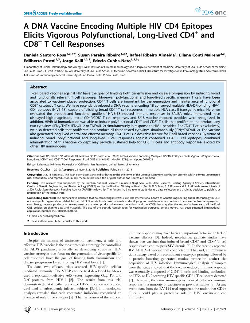
assay. We detected IFNc and IL-2 secreting cells against eight
(Figure 1A) HIVBr18- encoded peptides; all peptides that induced
IFNc secretion were also capable to elicit IL-2 secretion. The
recognized peptides p17 (73–89), p6 (32–46), pol (785–799), gp160
(188–201), rev (11027), vpr (58–65), vif (144–158) and nef (180–
194) consistently presented positive responses over multiple
independent immunization experiments. In contrast, T cells from
pVAX1 immunized mice presented negligible numbers of IFNcand IL-2 secreting cells after incubation with HIVBr18 peptides,
in all performed experiments.
To identify CD4+ and CD8+ T cell responses, we performed a
CFSE-based proliferation assay. Taking into account multiple
independent experiments, we detected consistent CD4+ T cell
proliferative responses (Figure 2B, upper panels) against five
peptides (p6 (32–46), pol (785–799), gp160 (188–201), vif (144–
158) and nef (180–194)) and CD8+ T cell proliferative responses
(Figure 2B, lower panels) against two peptides (gp160 (188–201)
and nef (180–194)). Interestingly, all peptides that elicited
proliferative CD4+ and/or CD8+ T cell responses were also able
CD4+ T Cell-Based HIV Vaccine
PLoS ONE | www.plosone.org 2 February 2011 | Volume 6 | Issue 2 | e16921
to elicit cytokine (IFNc and IL-2) secretion. Thus, vaccination of
BALB/c mice with HIVBr18 was able to induce broad specific
immune responses against eight epitopes encoded by the vaccine.
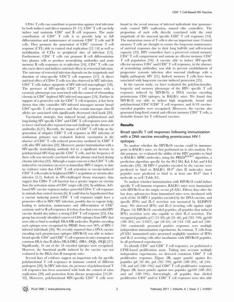
Magnitude of T cell responsesTo assess the magnitude of vaccine-induced T cell responses, we
evaluated the cellular immune responses of splenocytes from
HIVBr18 or pVAX1 immunized BALB/c mice against pooled
HIV-1 peptides. We observed that over 10% of both CD3+CD4+
and CD3+CD8+ splenic T cells from HIVBr18 immunized mice
displayed specific proliferation against the HIV-1 peptides
encoded by HIVBr18 (Figure 2A and B). In addition, BALB/c
mice immunized with HIVBr18 displayed a significant number of
peptide-specific IFNc and IL-2 secreting cells (Figure 2C). In
Figure 1. Immunization with HIVBr18 induces IFNc and IL-2 secretion and proliferation against multiple HIV-1 epitopes. Two weeksafter the last immunization with HIVBr18 or the empty pVAX1 vector, pooled spleen cells from 6 BALB/c mice were cultured in the presence of HIV-1peptides (5 mM) or medium only. (A) Frequencies of HIV peptide-specific IFNc (left pie chart) and IL-2 (right pie chart) secreting cells were measuredby ELISPOT assay. The responses are shown by displaying each the number of SFU/106 cells for each positive peptide as a proportion of the sum ofSFU/106 cells for all positive peptides. (B) Proliferative T cell responses were assessed by CFSE dilution assay. Splenocytes were labeled with CFSE(1.25 mM) and cultured for 5 days. After staining with fluorochrome-labeled anti-CD3, -CD4 and -CD8 monoclonal antibodies, cells were analyzed byflow cytometry. CFSE dilution on gated CD3+CD4+ or CD3+CD8+ cells was used as a readout for antigen-specific proliferation. Representative dot plotsof CD4+ (upper panels) and CD8+ (lower panels) T cell proliferation (values in boxes represent % CFSElow cells) of splenocytes from HIVBr18immunized mice; Data are representative of nine independent immunization experiments.doi:10.1371/journal.pone.0016921.g001
CD4+ T Cell-Based HIV Vaccine
PLoS ONE | www.plosone.org 3 February 2011 | Volume 6 | Issue 2 | e16921
CD4+ T Cell-Based HIV Vaccine
PLoS ONE | www.plosone.org 4 February 2011 | Volume 6 | Issue 2 | e16921
contrast, splenocytes from pVAX1 immunized mice presented
negligible levels of proliferation and cytokine secreting cells to the
same pooled HIV-1 peptides (Figures S1, 2B and C).
The results showed above indicate that immunization with
HIVBr18 is able to induce Th1 cytokines. In order to analyze the
vaccine induced cytokine secretion profile of BALB/c splenocytes
incubated with the pooled HIV-1 peptides, we used the cytometric
bead array (CBA) for assessment of Th1 and Th2 cytokine
secretion. After 48 hours of culture, we found that splenocytes
from HIVBr18 immunized mice produced higher levels of type I
cytokines like IFNc, IL-2 and TNFa and negligible levels of IL-5
and IL-4. After 120 hours of culture, the levels of IFNc and TNFaincreased substantially (Figure 2D and S2). Of note, IL-10
production was undetectable (data not shown). Splenocytes from
pVAX1 immunized mice failed to secrete cytokines above the
detection limit. Taken together, these results indicated that the
HIVBr18 DNA vaccine induced potent, specific type 1 cytokine T
cell responses.
Functional profile of cellular immune response afterHIVBr18 immunization
Since the quality of the immune responses has been associated
with vaccine-mediated protection against certain pathogens, we
subsequently characterized the phenotype and functional profile of
the induced T cells. Using multiparameter flow cytometry, we
sought to characterize antigen-specific T cells (CD4+ and CD8+)
based on their ability to proliferate (CFSE dilution assay) and
produce the effector cytokines IFNc, TNFa and IL-2 at a single cell
level. As shown in Figure 3A, immunization with HIVBr18 induced
HIV-1 peptide-specific production of IFNc, IL-2 and TNFa by
CD4+ and to a lesser extent by CD8+ T cells. We also observed that
both T cell subsets showed a higher proportion of of IFNc+ and
TNFa+ cells than IL-2+ cells. A simultaneous analysis of
proliferation and intracellular cytokine production demonstrated
that 2.3% of CD4+ and 1.0% of CD8+ T cells proliferated (CFSElow)
and produced any cytokine tested in response to pooled HIV-1
peptides (Figure 3B). Boolean combinations of proliferating
(CFSElow) and cytokine-positive populations indicated that
HIVBr18 immunization induced polyfunctional CD4+ and CD8+
T cells, i.e., that proliferate (CFSElow) and produce IFNc/TNFasimultaneously (Figure 3C). We also observed high proportions of
CFSElow/IFNc or CFSElow/TNFa in CD4+ and CD8+ T cells from
mice that received HIVBr18. For CD4+ T cells exclusively, we also
detected CFSElow cells that simultaneously produced all three tested
cytokines (IFNc/TNFa/IL-2). Splenocytes from the pVAX1
immunized group produced negligible levels of cytokines. Further-
more, triple-cytokine producing cells produced more IFNc and
TNFa than single-cytokine producing cells, as determined by
median fluorescent intensity (MFI) (Figure S3). In contrast, there
was no difference in the MFI for IL-2 in the triple-cytokine
producing cells when compared to cells producing IL-2 alone.
We next examined whether antigen-specific proliferating T cells
were the major cytokine producers. As shown in Figures 4A and B,
the vast majority (ca. 80%) of CD4+ and CD8+ T cells that
produced the effector cytokines IFNc and TNFa are within the
proliferating (CFSElow) population. In contrast, 50% of IL-2
producing T cells also proliferated (Figure 4B). These experiments
also showed that mice immunized with HIVBr18 displayed 2.31,
0.37 and 2.80% of HIV-specific CD4+ T cells that proliferated
(CFSElow) and produced either IFNc, IL-2 or TNFa, respectively.
A similar response was observed in CD8+ T cells, showing that
0.60, 0.22 and 0.64% of CD8+ T cells proliferated and produced
either IFNc, IL-2 or TNFa, respectively. In contrast, splenocytes
from mice immunized with the control pVAX1 displayed a
negligible percentage of specific proliferating/cytokine producing
T cells (Figure S4).
Proliferating (CFSElow) and non-proliferating (CFSEhi) CD4+
and CD8+ T cells were also evaluated by their ability to produce
cytokines. Figure 4C summarizes the percentage of proliferating T
cells that produced each of the tested cytokines. Significantly, 20%
of the specific proliferating CD4+ T cells produced IFNc, 5%
produced IL-2 and 30% produced TNFa. A similar profile, albeit
with lower values, was observed for proliferating CD8+ T cells. In
contrast, less than 0.5% of non-proliferating (CFSEhi) T cells
showed cytokine production, either on CD4+ or CD8+ compart-
ment. Overall, these data showed that immunization with the
DNA vaccine, HIVBr18, successfully induced polyfunctional
CD4+ and CD8+ T cells that proliferated and produced effector
cytokines to epitopes encoded by the vaccine.
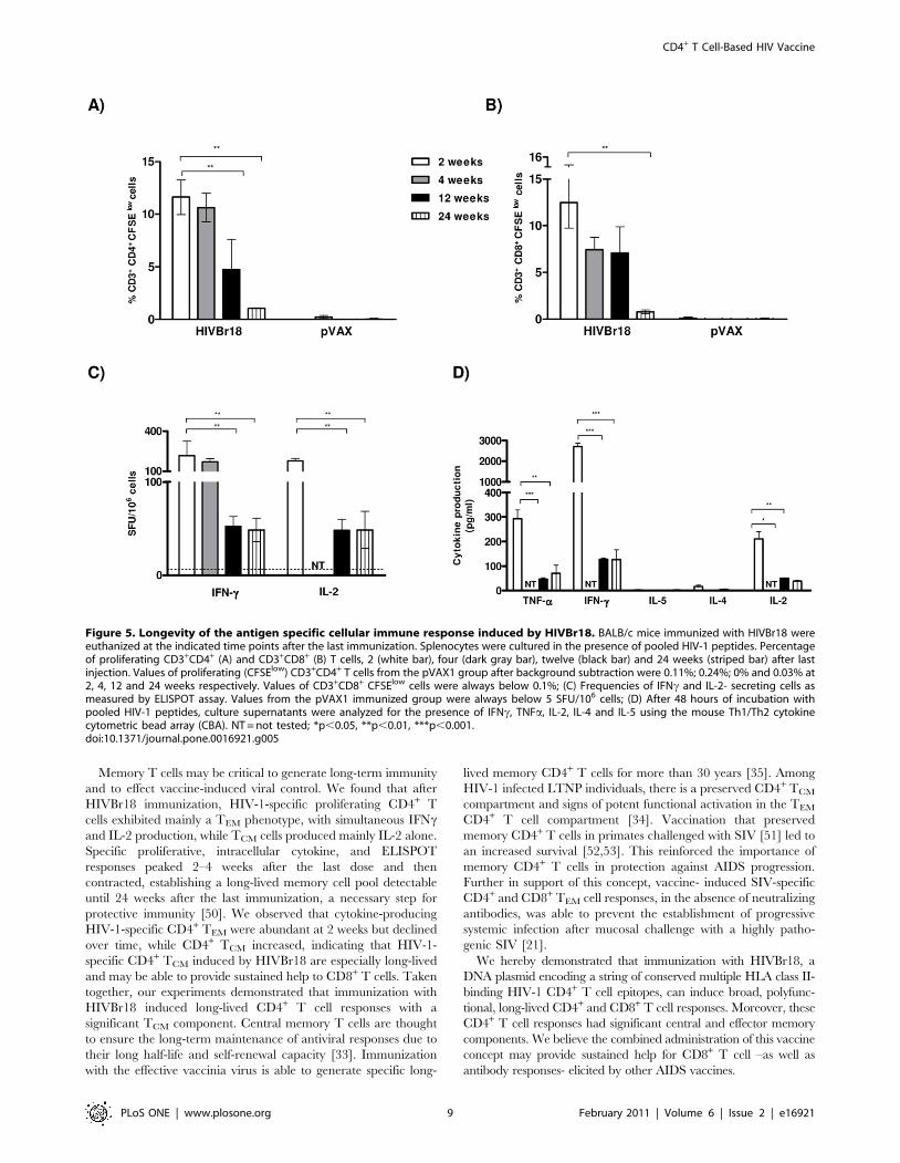
Induction of long-lasting HIV-1-specific T cells aftervaccination with HIVBr18
To assess whether immunization with HIVBr18 induced long-
lasting T cells in BALB/c mice, we measured vaccine-induced
CD4+ and CD8+ T cell proliferation and cytokine secretion 2, 4,
12 and 24 weeks after the last DNA immunization. A measurable
response was observed at all time points. At 2 and 4 weeks post-
immunization, a similar proportion of CD4+ T cells (11–12%)
proliferated against the pooled HIV-1 peptides (Figure 5A).
Twelve weeks after the last dose, a statistically significant decrease
in the magnitude of the CD4+ T cell proliferative response was
observed (4.76% CFSE low cells, versus 11.62% in early time
points, p,0.01). This response continued to decline down to 1%
of specific proliferating CD4+ T cells, 24 weeks after the last dose.
Of note, this response was several-fold higher than the values
measured in the pVAX1 immunized group. For CD8+ T cells
(Figure 5B), only the 24 week time point showed a statistically
significant decrease in proliferation, as compared to the 2 week
time point. Splenic T cells from mice immunized with pVAX1
showed negligible levels of proliferation at all time points for both
CD4+ and CD8+ populations (values in the legend of Figure 5A
and B). Of note, 24 weeks after the last immunization, total
Figure 2. Immunization with HIVBr18 induces CD4+ and CD8+ T cell proliferation and type 1 cytokine production in response topooled HIV-1 peptides. Two weeks after the last immunization with HIVBr18 or the empty pVAX1 vector, pooled spleen cells from 6 BALB/c micewere cultured in the presence of 5 mM of pooled HIV-1 peptides or medium only. (A and B) Splenocytes were labeled with CFSE (1.25 mM) andcultured for 5 days. After staining with fluorochrome-labeled anti-CD3, -CD4 and -CD8 monoclonal antibodies, cells were analyzed by flow cytometryand CFSE dilution on gated CD3+CD4+ or CD3+CD8+ cells was used as a readout for antigen-specific proliferation. (A) Representative dot plots of CD4+
(left) and CD8+ (right) T cell proliferation (% CFSElow cells) of splenocytes stimulated with medium only or pooled peptides from HIVBr18 immunizedmice. (B) Quantitative analysis of proliferating CD4+ and CD8+ CFSE low T cells (values in boxes represent % CFSElow cells). The percentage ofproliferating T cells was calculated subtracting the values of peptide-stimulated from non-stimulated cultures; (C) Frequencies of IFNc and IL-2secreting cells measured by ELISPOT; (D) Splenocytes from immunized BALB/c mice were cultured in the presence of pooled HIV-1 peptides. After 48and 120 hours, levels of IFNc, TNFa, IL-2, IL-4 and IL-5 in culture supernatants were measured using the mouse Th1/Th2 cytokine cytometric beadarray (CBA) by flow cytometry and analyzed using CBA software. Values of cytokine production by peptide-stimulated splenocytes from pVAX1immunized group were always below the detection limit. Bars represent the mean + 3 SD from nine independent immunization experiments.doi:10.1371/journal.pone.0016921.g002
CD4+ T Cell-Based HIV Vaccine
PLoS ONE | www.plosone.org 5 February 2011 | Volume 6 | Issue 2 | e16921
CD4+ T Cell-Based HIV Vaccine
PLoS ONE | www.plosone.org 6 February 2011 | Volume 6 | Issue 2 | e16921
frequencies of proliferating CD4+ and CD8+ T cells were ca. 1%
above baseline, as well as above the values from the pVAX1
immunized group. The ability of T cells from HIVBr18
immunized mice to secrete cytokines was also evaluated by
ELISPOT (Figure 5C) and CBA (Figure 5D) assays at different
time points. As observed for proliferative responses, there was no
significant difference in IFNc secretion to pooled HIV-1 peptides
at 2 or 4 weeks after the last HIVBr18 immunization. IFNcsecretion decreased significantly (p,0.01) 12 weeks after the last
dose and was sustained until the 24 week time point (Figure 5C).
Similar results were observed when we measured IL-2 secretion
(Figure 5C). Splenocytes from HIVBr18 immunized mice were
able to secrete type I cytokines like IFNc, IL-2 and TNFa, which
declined over time, as evaluated by the CBA assay, but were still
detectable and above pVAX1 immunized group, even 24 weeks
after the last dose (Figure 5D). Therefore, long-term proliferative
CD4+ and CD8+ T cell responses as well as cytokine-secreting
responses were detectable up to 24 weeks after the last
immunization with HIVBr18.
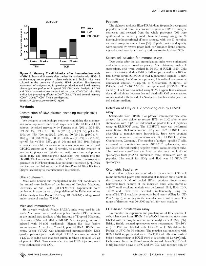
HIVBr18 immunization induces long-term memory T cellsIn order to characterize memory T cell responses induced by
immunization, we assessed the expression of memory markers
(CD44 and CD62L) among proliferating (CFSElow) CD4+ and
CD8+ T cell populations. We evaluated responses against pooled
HIV-1 peptides, 2 weeks after the last immunization with HIVBr18.
The candidate vaccine HIVBr18 elicited higher levels of specific
effector (TEM) than central (TCM) memory T cells. Among
proliferating CFSElow CD4+ T cells (8% of total CD4+ T cells),
ca. 70% and 24% had effector and central memory phenotypes,
respectively (data not shown). Similarly, 57% and 39% of
proliferating CD8+ T cells (7% of total CD8+ T cells) had effector
and central memory phenotypes, respectively (data not shown).
We also investigated the phenotype of specific cytokine-
producing CD4+ T cells at two time points. Two weeks after the
last immunization, the most abundant population was TEM cells in
which more than half produced IFNc/IL-2 simultaneously
(Figure 6A). CD4+ TCM predominantly produced IL-2 alone. The
kinetic of CD4+Tcells demonstrated that the frequency of cytokine-
producing CD4+ TEM declined over time (2.5% at 2 weeks versus
0.65% at 24 weeks, IFNc and/or IL-2 cells) (Figure 6A). On the
other hand, cytokine-producing CD4+ TCM, especially the IL-2
component, showed an increase over time (0.8% at 2 weeks versus
1.52% at 24 weeks, IFNc and/or IL-2 cells). Collectively, these data
indicated that the DNA vaccine HIVBr18 was able to induce
specific long-lasting effector and central memory, CD4+ and CD8+
T cells that produced type 1 cytokines.
Discussion
In this paper, we showed that immunization with HIVBr18
induced strong, broad, polyfunctional and long-lasting HIV-
specific CD4+ and CD8+ T cell responses in BALB/c mice.
Moreover, the vaccine induced central and effector memory CD4+
T cells.
In our study, all the 18 TEPITOPE-selected promiscuous
epitopes were also predicted by the algorithm PREDBALB/c [36] to
bind to at least one BALB/c (H-2d) mouse MHC class II molecule,
and 17 were predicted to bind to H-2d MHC class I molecules.
The cross-species recognition of epitopes selected to bind to
human MHC molecules is actually expected. The TEPITOPE
algorithm has previously identified peptides that can be recognized
across species barriers, including H-2d mice [37]. This may be due
to the fact that the identification of multiple HLA-DR binding
peptides by TEPITOPE may select for promiscuous peptides that
share MHC class II binding motifs similar to many other human
and non-human MHC class II molecules [38–40]. Indeed, we also
tested this concept in non-human primates. TEPITOPE selection
for promiscuous peptides allowed the identification of a similar set
of conserved SIV peptides that were frequently recognized by
PBMC from SIV-infected elite controller rhesus macaques
(unpublished data). Here, we have shown that BALB/c mice
immunized with HIVBr18 presented a broad T cell response,
directed to 8 out of the 18 epitopes encoded by the vaccine. The
induction of broad T cell responses towards conserved epitopes
appears to be an essential pre-requisite for protection, in light of
recent efficacy trials of T cell-based HIV vaccines [1,4,5]. Indeed,
an Adenovirus 5-based SIV vaccine encoding 8 SIV proteins was
able to elicit broad CD4+ and CD8+ T cell responses and reduced
viral load after heterologous challenge [6]. In addition, broad
vaccine-induced pre-challenge T cell responses were correlated
with lower viral loads and higher CD4+ T lymphocyte counts [41].
The breadth of HIV-specific responses in infected individuals has
also been associated with viral control [42].
We also evaluated the functional profile of the vaccine-induced
specific CD4+ and CD8+ T cells. The ability of the HIVBr18
vaccine to induce IFNc, TNFa and IL-2, with little or no IL-4, IL-
5 or IL-10, indicated a type 1 cytokine response. We extended the
functional analysis of splenocytes from HIVBr18 immunized mice
and observed that immunization induced CD4+ and CD8+ T cells
with a polyfunctional repertoire. It has become increasingly
evident that rather than the magnitude, the quality of the immune
response is a crucial factor in defining a protective response [28].
Recent data from immunization studies using the efficacious
smallpox and yellow fever vaccines have shown that induction of
specific polyfunctional CD4+ or CD8+ T cells may play a role in
protective immunity [43–45]. Furthermore, vaccine-induced
polyfunctional (IFNc+IL-2+TNFa+) CD4+ T cell populations were
shown to provide protection against Leishmania major [46] and M.
tuberculosis [47] challenge. Polyfunctional HIV-1-specific CD4+ T
cells were present among HIV-1 infected individuals with non-
progressive disease [24,25]. Long-term nonprogressor (LTNP)
HIV-1 infected patients displayed higher number of polyfunctional
CD8+ T cells when compared to progressor patients [48].
Figure 3. Immunization with HIVBr18 induces proliferating T cells with a polyfunctional type 1 cytokine profile. Two weeks after thelast immunization with HIVBr18 or the empty vector pVAX1, spleen cells from 6 BALB/c mice were collected, labeled with CFSE (1.25 mM) and culturedfor 4 days in the presence of pooled HIV-1 peptides or medium only. On day 4, cells were pulsed for 12 hours with pooled peptides in the presence ofcostimulatory antibody and Brefeldin A. Cells were then surface stained with antibodies to CD4 and CD8, permeabilized and stained for intracellularcytokines (IFNc, TNFa and IL-2) and CD3. (A) Multiparameter flow cytometry was used to determine the frequency of IFNc, IL-2 or TNFa CD4+ andCD8+ T cells. (B) Total frequencies of proliferating (CFSElow) and cytokine-producing CD4+ and CD8+ T cells; (C) After gating on proliferating (CFSElow)and cytokine-producing cells, Boolean combinations were then created using FlowJo software to determine the frequency of each response based onall possible combinations of cytokine expression. Background responses detected in negative control tubes were subtracted from those detected instimulated samples for every specific functional combination. Negative control tubes include cells stimulated with medium and cells from pVAX1immunized mice stimulated with pooled peptides. For each sample 500,000 events were collected in the live lymphocyte gate. Results arerepresentative of two to three independent experiments.doi:10.1371/journal.pone.0016921.g003
CD4+ T Cell-Based HIV Vaccine
PLoS ONE | www.plosone.org 7 February 2011 | Volume 6 | Issue 2 | e16921
Moreover, HIV elite controllers presented strong polyfunctional
CD4+ and CD8+ T cell responses in blood and rectal mucosa
[14,32]. CD4+ T cells from HIV-2 infected subjects, who display a
longer time of progression to AIDS than HIV-1 infected patients,
were more polyfunctional than those found in viral load and CD4+
T cell-matched HIV-1 infected subjects [49]. Our data thus
suggest that HIVBr18, like some validated efficacious vaccines,
can elicit polyfunctional T cells, a putative correlate of protection.
Figure 4. Immunization with HIVBr18 induces specific CD4+ and CD8+ T cells that proliferate and produce type 1 cytokinessimultaneously. Two weeks after the last immunization with HIVBr18 or the empty vector pVAX1, spleen cells from 6 BALB/c mice were labeledwith CFSE (1.25 mM) and cultured for 4 days in the presence of pooled HIV-1 peptides or medium only. On day 4, cells were pulsed for 12 hours withpooled peptides or medium, in the presence of costimulatory antibody and Brefeldin A. (A) CFSE and intracellular cytokine staining were used tosimultaneously assess proliferation and IFNc, TNFa or IL2 production. Frequencies of antigen-specific cytokine-producing T cells in proliferating(CFSElow) and non-proliferating (CFSEhi) gates are displayed; (B) Proportion of proliferating cells (CFSElow) in the total cytokine gate (sum of % ofcytokine producing cells in gated CFSEhi and CFSElow cells); (C) Percentage of cytokine producing CD4+ and CD8+ T cells in proliferating cells (CFSElow)(values of cytokine producing cells in CFSElow population 6100 divided by total CFSElow population).doi:10.1371/journal.pone.0016921.g004
CD4+ T Cell-Based HIV Vaccine
PLoS ONE | www.plosone.org 8 February 2011 | Volume 6 | Issue 2 | e16921
Memory T cells may be critical to generate long-term immunity
and to effect vaccine-induced viral control. We found that after
HIVBr18 immunization, HIV-1-specific proliferating CD4+ T
cells exhibited mainly a TEM phenotype, with simultaneous IFNcand IL-2 production, while TCM cells produced mainly IL-2 alone.
Specific proliferative, intracellular cytokine, and ELISPOT
responses peaked 2–4 weeks after the last dose and then
contracted, establishing a long-lived memory cell pool detectable
until 24 weeks after the last immunization, a necessary step for
protective immunity [50]. We observed that cytokine-producing
HIV-1-specific CD4+ TEM were abundant at 2 weeks but declined
over time, while CD4+ TCM increased, indicating that HIV-1-
specific CD4+ TCM induced by HIVBr18 are especially long-lived
and may be able to provide sustained help to CD8+ T cells. Taken
together, our experiments demonstrated that immunization with
HIVBr18 induced long-lived CD4+ T cell responses with a
significant TCM component. Central memory T cells are thought
to ensure the long-term maintenance of antiviral responses due to
their long half-life and self-renewal capacity [33]. Immunization
with the effective vaccinia virus is able to generate specific long-
lived memory CD4+ T cells for more than 30 years [35]. Among
HIV-1 infected LTNP individuals, there is a preserved CD4+ TCM
compartment and signs of potent functional activation in the TEM
CD4+ T cell compartment [34]. Vaccination that preserved
memory CD4+ T cells in primates challenged with SIV [51] led to
an increased survival [52,53]. This reinforced the importance of
memory CD4+ T cells in protection against AIDS progression.
Further in support of this concept, vaccine- induced SIV-specific
CD4+ and CD8+ TEM cell responses, in the absence of neutralizing
antibodies, was able to prevent the establishment of progressive
systemic infection after mucosal challenge with a highly patho-
genic SIV [21].
We hereby demonstrated that immunization with HIVBr18, a
DNA plasmid encoding a string of conserved multiple HLA class II-
binding HIV-1 CD4+ T cell epitopes, can induce broad, polyfunc-
tional, long-lived CD4+ and CD8+ T cell responses. Moreover, these
CD4+ T cell responses had significant central and effector memory
components. We believe the combined administration of this vaccine
concept may provide sustained help for CD8+ T cell –as well as
antibody responses- elicited by other AIDS vaccines.
Figure 5. Longevity of the antigen specific cellular immune response induced by HIVBr18. BALB/c mice immunized with HIVBr18 wereeuthanized at the indicated time points after the last immunization. Splenocytes were cultured in the presence of pooled HIV-1 peptides. Percentageof proliferating CD3+CD4+ (A) and CD3+CD8+ (B) T cells, 2 (white bar), four (dark gray bar), twelve (black bar) and 24 weeks (striped bar) after lastinjection. Values of proliferating (CFSElow) CD3+CD4+ T cells from the pVAX1 group after background subtraction were 0.11%; 0.24%; 0% and 0.03% at2, 4, 12 and 24 weeks respectively. Values of CD3+CD8+ CFSElow cells were always below 0.1%; (C) Frequencies of IFNc and IL-2- secreting cells asmeasured by ELISPOT assay. Values from the pVAX1 immunized group were always below 5 SFU/106 cells; (D) After 48 hours of incubation withpooled HIV-1 peptides, culture supernatants were analyzed for the presence of IFNc, TNFa, IL-2, IL-4 and IL-5 using the mouse Th1/Th2 cytokinecytometric bead array (CBA). NT = not tested; *p,0.05, **p,0.01, ***p,0.001.doi:10.1371/journal.pone.0016921.g005
CD4+ T Cell-Based HIV Vaccine
PLoS ONE | www.plosone.org 9 February 2011 | Volume 6 | Issue 2 | e16921
Methods
Construction of DNA plasmid encoding multiple HIV-1epitopes
We designed a multiepitope construct containing the mamma-
lian codon optimized nucleotide sequences of the 18 HIV-1 CD4
epitopes described previously by Fonseca et al. [26]: p17(73–89),
p24 (33–45), p24 (131–150), p6 (32–46), pol (63–77), pol (136–
150), pol (785–799), gp41(261–276), gp160 (19–31), gp160 (174–
185), gp160 (188–201), gp160 (481–498), rev (11–27), vpr (58–72),
vpr (65–82), vif (144–158), vpu (6–20) and nef (180–194). Epitope
sequences, assembled in tandem in the above mentioned order, had
GPGPG spacers at C and N termini, to avoid the creation of
junctional epitopes and interference with processing and presen-
tation [54]. The artificial gene (EZBiolab) was cloned into the
HindIII/XhoI restriction site of the pVAX1 vector (Invitrogen) to
generate the HIVBr18 plasmid, as previously described [27]. DNA
vaccine was purified using the Endofree Plasmid Giga Kit from
Qiagen according to manufacturer’s instructions.
Ethics StatementMice were housed and manipulated under SPF conditions in
the animal care facilities of the Institute of Tropical Medicine,
University of Sao Paulo (IMT/FMUSP). Experiments were
performed in accordance to the guidelines of the Ethics committee
of University of Sao Paulo (CAPPesq- HCFMUSP) and approved
under protocol number 775-06.
Mice and ImmunizationsSix to eight week-old female BALB/c mice were used in this
study. Mice were housed and manipulated under SPF conditions
in the animal care facilities of the Institute of Tropical Medicine,
University of Sao Paulo (IMT/FMUSP). Six mice per group were
injected with 10 mM cardiotoxin (Sigma) five days before
immunization. At weeks 0, 2 and 4, plasmid DNA HIVBr18 or
empty vector pVAX1 was administered intramuscularly. Each
quadriceps was injected with 50 mL of DNA at a concentration of
1 mg/mL in saline such that each animal received a total of 100 mg
of plasmid DNA. Two weeks after the last DNA injection, mice
were euthanized with CO2.
PeptidesThe eighteen multiple HLA-DR binding, frequently recognized
peptides, derived from the conserved regions of HIV-1 B subtype
consensus and selected from the whole proteome [26] were
synthesized in house by solid phase technology using the 9-
fluorenylmethoxycarbonyl (Fmoc) strategy, with the C- terminal
carboxyl group in amide form [55]. Peptide purity and quality
were assessed by reverse-phase high performance liquid chroma-
tography and mass spectrometry and was routinely above 90%.
Spleen cell isolation for immune assaysTwo weeks after the last immunization, mice were euthanized
and spleens were removed aseptically. After obtaining single cell
suspensions, cells were washed in 10 mL of RPMI 1640. Cells
were then resuspended in R-10 (RPMI supplemented with 10% of
fetal bovine serum (GIBCO), 2 mM L-glutamine (Sigma), 10 mM
Hepes (Sigma), 1 mM sodium piruvate, 1% vol/vol non-essential
aminoacid solution, 40 mg/mL of Gentamicin, 20 mg/mL of
Peflacin and 561025 M 2- mercaptoetanol (SIGMA). The
viability of cells was evaluated using 0.2% Trypan Blue exclusion
dye to discriminate between live and dead cells. Cell concentration
was estimated with the aid of a Neubauer chamber and adjusted in
cell culture medium.
Detection of IFNc or IL-2 producing cells by ELISPOTassay
Splenocytes from HIVBr18 or pVAX1 immunized mice were
assayed for their ability to secrete IFNc or IL-2 after in vitro
stimulation with 5 mM of individual or pooled HIV-1 peptides
using an ELISPOT assay. The ELISPOT assay was performed
using Becton Dickinson murine IFNc and IL-2 ELISPOT kits
according to manufacturer’s instructions. Spots were counted
using an automated stereomicroscope (KS ELISPOT, Zeiss,
Oberkochem, Germany). The number of antigen- specific T cells,
expressed as spot-forming units (SFU)/106 splenocytes, was
calculated after subtracting negative control values (medium only).
The positivity cutoff was calculated as the mean + 3 SD of
splenocytes from pVAX1 immunized mice, stimulated with all
peptides. The cutoff for IFNc and IL-2 was 15 SFU/106
splenocytes.
Cytometric Bead arrayOne million splenocytes were added to each well of 96 well
round-bottomed plates and incubated at indicated time points in
the presence 5 mM of pooled HIV-1 peptides. Supernatants
harvested from cultures at the indicated times were stored at
220uC until cytokine analysis was performed. IL-2, IL-4, IL-5,
TNFa and IFNc were detected simultaneously using the
mouseTh1/Th2 cytokine cytometric bead array (CBA) kit (BD
PharMingen), according to the manufacturer’s instructions. The
range of detection was 20–5000 pg/mL for each cytokine.
CFSE-based proliferation assayTo monitor the expansion and proliferation of HIV-specific T
cells, splenocytes from HIVBr18 or pVAX1 immunized mice were
labeled with carboxyfluorescein succinimidyl ester (CFSE) [56].
Briefly, freshly isolated splenocytes were resuspended (506106/
mL) in PBS and labeled with 1.25 mM of CFSE (Molecular
Probes) at 37uC for 10 minutes. The reaction was quenched with
RPMI 1640 supplemented with 10% FBS and cells were washed
before resuspending in RPMI 1640 at a density of 1.56106/mL.
Cells were cultured in 96 well round-bottomed plates (36105/well
in triplicate) for 5 days at 37uC and 5% CO2 with medium only or
Figure 6. Memory T cell kinetics after immunization withHIVBr18. Two and 24 weeks after the last immunization with HIVBr18or the empty vector pVAX1, spleen cells from 6 BALB/c mice werecultured in the presence of pooled HIV-1 peptides. Simultaneousassessment of antigen-specific cytokine production and T cell memoryphenotype was performed in gated CD3+CD4+ cells. Analysis of CD44and CD62L expression was determined on gated CD3+CD4+ cells. IFNcand/or IL-2 producing effector (CD44hi CD62Llow) and central memory(CD44hi CD62Lhi) CD4+ T cells are depicted.doi:10.1371/journal.pone.0016921.g006
CD4+ T Cell-Based HIV Vaccine
PLoS ONE | www.plosone.org 10 February 2011 | Volume 6 | Issue 2 | e16921
5 mM of HIV peptides. Positive controls were stimulated with
2.5 mg/mL of Concanavalin A (Sigma). Cells were then harvested,
washed with 100 mL of FACS buffer (PBS with 0.5% BSA and
2 mM EDTA) and stained with anti-mouse CD3 phycoerythrin
(PE), anti-mouse CD4 peridinin chlorophyll protein (PerCP) and
anti-mouse CD8 allophycocyanin (APC) monoclonal antibodies
(BD Pharmingen, San Jose, CA) for 45 minutes at 4uC. To analyze
the memory phenotype of proliferating cells we stained the cells
with anti-CD3 APCCy7, anti-CD4 PerCP, anti-CD8 PECy7, anti-
CD44 PE and anti-CD62L APC monoclonal antibodies (BD
Pharmingen). Cells were then washed twice with FACS buffer,
fixed with 4% paraformaldehyde, and resuspended in FACS
buffer. Samples were acquired on a FACSCanto flow cytometer
(BD Biosciences) and then analyzed using FlowJo software (version
9.0.2, Tree Star, San Carlo, CA). Fifty thousand events
(proliferation evaluation) and 100,000 events (memory phenotype)
were acquired in a live lymphocyte gate. The percent of
proliferating CD4 + and CD8+ CFSElow cells was determined in
the CD3+ cell population. The criteria for scoring as positive the
proliferating cell cultures included CFSElow cells . cutoff. The
cutoff of unspecific proliferative response was determined based on
the median percentage of proliferating cells (% of CD3+CD4+ or
CD3+CD8+ CFSElow cells) on splenocytes from pVAX1 immu-
nized groups after stimulating with individual peptides +3 standard
deviation (SD).
Analysis of polyfunctional HIV-specific T cell responsesSplenocytes from immunized mice were labeled with CFSE as
described above. CFSE-labeled cells were incubated at a density of
2.56106 cells/mL and cultured in 96 well round-bottomed plates
(56105/well in triplicate) for 4 days at 37uC and 5% CO2 with
medium only or pooled HIV peptides (5 mM). After 4 days of
incubation, cells were restimulated in the presence of 2 mg/mL
anti-CD28 (BD Pharmingen), 5 mM of pooled HIV peptides and
Brefeldin A- GolgiPlugTM (BD Pharmingen) for the last 12 hours.
After the incubation period, cells were washed with FACS buffer
and surface stained using monoclonal antibodies to CD8-Alexa700
and CD4-PerCP for 30 minutes at 4uC. Cells were fixed and
permeabilized using the Cytofix/CytopermTM kit (BD Pharmin-
gen). Permeabilized cells were washed with Perm/Wash buffer
(BD Biosciences) and stained with monoclonal antibodies to CD3-
APCCy7, IL2-PE, TNFa-PECY7 and IFNc-APC for 30 minutes
at 4uC. Following staining, cells were washed twice and
resuspended in FACS buffer. All antibodies were from BD
Pharmingen. Samples were acquired on a FACSCanto flow
cytometer (BD Biosciences) and then analyzed using FlowJo
software (version 9.0.2, Tree Star, San Carlo, CA). Each analysis
was gated on forward (FSC)/side scatter (SSC) lymphocytes
(500,000 events) and CD3+ T cells followed by a subsequent gate
on CD4+ or CD8+ populations. After identification of CD4+ and
CD8+ populations, a gate was done in each positive population for
IFNc, TNFa and IL-2 expression. In addition, we used the
Boolean gate (FlowJo software (version 9.0.2, Tree Star, San
Carlo, CA)) platform to create several combinations of the three
cytokine (IL-2, TNFa and IFNc) within CFSElow population
resulting in seven distinct patterns. The percentages of cytokine-
producing cells were calculated by subtracting background values.
For each flow cytometry experiment performed in this paper,
unstained and all single-color controls were processed to allow
proper compensation.
Data AnalysisStatistical significance (p-values) was calculated by using One-
way ANOVA and Tukey’s honestly significantly different (HSD).
Statistical analysis and graphical representation of data was
performed using GraphPad Prism version 5.0 software.
Supporting Information
Figure S1 Proliferative responses of CD4+ and CD8+ Tcells from pVAX1 immunized mice against pooled HIV-1peptides. BALB/c mice were immunized with the empty vector
pVAX1. Two weeks after the last dose, pooled spleen cells from 6
mice were labeled with CFSE (1.25 mM) and cultured for 5 days in
the presence of 5 mM of pooled HIV-1 peptides. Cells were
analyzed by flow cytometry and CFSE dilution on gated
CD3+CD4+ or CD3+CD8+cells was used as a readout for
antigen-specific proliferation. Representative dot plots of CD4+
(left) and CD8+ (right) T cell proliferation (% CFSElow cells) from
splenocytes stimulated with medium or pooled HIV-1peptides.
Data are representative of nine independent immunization
experiments.
(TIF)
Figure S2 Immunization with HIVBr18 induces a HIV-1peptide-specific type 1 cytokine response. Splenocytes
from immunized BALB/c mice were cultured in the presence of
pooled HIV-1 peptides. After 48 hours, levels of IFNc, TNFa, IL-
2, IL-4 and IL-5 in culture supernatants were measured using the
mouse Th1/Th2 cytokine cytometric bead array (CBA) by flow
cytometry. Representative dot plot profiles of the 6-plex Th1/Th2
cytokine CBA assay for culture supernatants from pVAX1 (left)
and HIVBr18 (right) immunized mice after stimulation with
pooled HIV-1 peptides.
(TIF)
Figure S3 Polyfunctional CD4+ T cells produce highercytokine levels on a per cell basis than single cytokine-producing CD4+ T cells. Two weeks after the last immuniza-
tion with HIVBr18, spleen cells from 6 BALB/c mice were labeled
with CFSE and cultured in the presence of pooled HIV-1 peptides
or medium only for 4 days. On day 4, cells were pulsed for
12 hours with pooled HIV-1 peptides or medium in the presence
of costimulatory antibody and Brefeldin. Multiparameter flow
cytometry was used to identify polyfunctional and single cytokine-
producing CD3+CD4+ T cells. Intracellular cytokine levels
expressed as MFI values are compared for CFSElow cells
producing all 3 tested cytokines (polyfunctional cells) and CFSElow
cells producing a single cytokine. MFI values for IFNc (left),TNFa(middle) and IL-2 (right).
(TIF)
Figure S4 Proliferative responses and cytokine produc-tion in splenocytes from pVAX1 immunized mice. Two
weeks after the last immunization with the empty vector pVAX1,
spleen cells from 6 BALB/c mice were labeled with CFSE and
cultured in the presence of pooled HIV-1 peptides or medium only
for 4 days. On day 4, cells were pulsed for 12 hours with pooled
HIV-1 peptides or medium in the presence of costimulatory
antibody and Brefeldin A. CFSE and intracellular cytokine
staining were used to simultaneously assess proliferation and
IFNc, TNFa or IL-2 production. Frequencies of antigen-specific
cytokine-producing T cells in proliferating (CFSElow) and non
proliferating (CFSEhi) gates are displayed.
(TIF)
Table S1 Peptide binding predictions for H-2d MHCclass I and class II.
(PDF)
CD4+ T Cell-Based HIV Vaccine
PLoS ONE | www.plosone.org 11 February 2011 | Volume 6 | Issue 2 | e16921
Acknowledgments
We thank Dr. Claudio Puschel and Mr. Washington Robert da Silva for
peptide synthesis; Mr. Luis Roberto Mundel for assistance at the animal
facility.
Author Contributions
Conceived and designed the experiments: DSR SPR EC-N. Performed the
experiments: DSR SPR RRA ECM EP. Analyzed the data: DSR SPR
RRA JK EC-N. Contributed reagents/materials/analysis tools: JK EC-N.
Wrote the paper: DSR SPR RRA EC-N.
References
1. Watkins DI, Burton DR, Kallas EG, Moore JP, Koff WC (2008) Nonhuman
primate models and the failure of the Merck HIV-1 vaccine in humans. Nat
Med 14: 617–621.
2. Buchbinder SP, Mehrotra DV, Duerr A, Fitzgerald DW, Mogg R, et al. (2008)
Efficacy assessment of a cell-mediated immunity HIV-1 vaccine (the Step Study):
a double-blind, randomised, placebo-controlled, test-of-concept trial. Lancet
372: 1881–1893.
3. Corey L, McElrath MJ, Kublin JG (2009) Post-step modifications for research on
HIV vaccines. AIDS 23: 3–8.
4. Sekaly RP (2008) The failed HIV Merck vaccine study: a step back or a
launching point for future vaccine development? J Exp Med 205: 7–12.
5. McElrath MJ, De Rosa SC, Moodie Z, Dubey S, Kierstead L, et al. (2008) HIV-
1 vaccine-induced immunity in the test-of-concept Step Study: a case-cohort
analysis. Lancet 372: 1894–1905.
6. Wilson NA, Keele BF, Reed JS, Piaskowski SM, MacNair CE, et al. (2009)
Vaccine-induced cellular responses control simian immunodeficiency virus
replication after heterologous challenge. J Virol 83: 6508–6521.
7. Rerks-Ngarm S, Pitisuttithum P, Nitayaphan S, Kaewkungwal J, Chiu J, et al.
(2009) Vaccination with ALVAC and AIDSVAX to prevent HIV-1 infection in
Thailand. N Engl J Med 361: 2209–2220.
8. Nitayaphan S, Pitisuttithum P, Karnasuta C, Eamsila C, de Souza M, et al.
(2004) Safety and immunogenicity of an HIV subtype B and E prime-boost
vaccine combination in HIV-negative Thai adults. J Infect Dis 190: 702–706.
9. Khanolkar A, Fuller MJ, Zajac AJ (2004) CD4 T cell-dependent CD8 T cell
maturation. J Immunol 172: 2834–2844.
10. Novy P, Quigley M, Huang X, Yang Y (2007) CD4 T cells are required for CD8
T cell survival during both primary and memory recall responses. J Immunol
179: 8243–8251.
11. Rajasagi NK, Kassim SH, Kollias CM, Zhao X, Chervenak R, et al. (2009)
CD4+ T cells are required for the priming of CD8+ T cells following infection
with herpes simplex virus type 1. J Virol 83: 5256–5268.
12. Martinez V, Costagliola D, Bonduelle O, N’go N, Schnuriger A, et al. (2005)
Combination of HIV-1-specific CD4 Th1 cell responses and IgG2 antibodies is
the best predictor for persistence of long-term nonprogression. J Infect Dis 191:
2053–2063.
13. Pancre V, Delhem N, Yazdanpanah Y, Delanoye A, Delacre M, et al. (2007)
Presence of HIV-1 Nef specific CD4 T cell response is associated with non-
progression in HIV-1 infection. Vaccine 25: 5927–5937.
14. Ferre AL, Hunt PW, McConnell DH, Morris MM, Garcia JC, et al. (2010) HIV
Controllers HLA-DRB1*13 and HLA-DQB1*06 Have Strong, Polyfunctional
Mucosal CD4+ T-cell Responses. J Virol.
15. Nakanishi Y, Lu B, Gerard C, Iwasaki A (2009) CD8(+) T lymphocyte
mobilization to virus-infected tissue requires CD4(+) T-cell help. Nature 462:
510–513.
16. Yang X, Yu X (2009) An introduction to epitope prediction methods and
software. Rev Med Virol 19: 77–96.
17. Pike R, Filby A, Ploquin MJ, Eksmond U, Marques R, et al. (2009) Race
between retroviral spread and CD4+ T-cell response determines the outcome of
acute Friend virus infection. J Virol 83: 11211–11222.
18. Sacha JB, Giraldo-Vela JP, Buechler MB, Martins MA, Maness NJ, et al. (2009)
Gag- and Nef-specific CD4+ T cells recognize and inhibit SIV replication in
infected macrophages early after infection. Proc Natl Acad Sci U S A 106:
9791–9796.
19. von Gegerfelt A, Valentin A, Alicea C, Van Rompay KK, Marthas ML, et al.
(2010) Emergence of simian immunodeficiency virus-specific cytotoxic CD4+ T
cells and increased humoral responses correlate with control of rebounding
viremia in CD8-depleted macaques infected with Rev-independent live-
attenuated Simian immunodeficiency virus. J Immunol 185: 3348–3358.
20. Giraldo-Vela JP, Rudersdorf R, Chung C, Qi Y, Wallace LT, et al. (2008) The
major histocompatibility complex class II alleles Mamu-DRB1*1003 and -
DRB1*0306 are enriched in a cohort of simian immunodeficiency virus-infected
rhesus macaque elite controllers. J Virol 82: 859–870.
21. Hansen SG, Vieville C, Whizin N, Coyne-Johnson L, Siess DC, et al. (2009)
Effector memory T cell responses are associated with protection of rhesus
monkeys from mucosal simian immunodeficiency virus challenge. Nat Med 15:
293–299.
22. Vaccari M, Mattapallil J, Song K, Tsai WP, Hryniewicz A, et al. (2008) Reduced
protection from simian immunodeficiency virus SIVmac251 infection afforded
by memory CD8+ T cells induced by vaccination during CD4+ T-cell
deficiency. J Virol 82: 9629–9638.
23. Yamamoto T, Iwamoto N, Yamamoto H, Tsukamoto T, Kuwano T, et al.
(2009) Polyfunctional CD4+ T-cell induction in neutralizing antibody-triggered
control of simian immunodeficiency virus infection. J Virol 83: 5514–5524.
24. Okoye A, Park H, Rohankhedkar M, Coyne-Johnson L, Lum R, et al. (2009)Profound CD4+/CCR5+ T cell expansion is induced by CD8+ lymphocyte
depletion but does not account for accelerated SIV pathogenesis. J Exp Med
206: 1575–1588.
25. Virgin HW, Walker BD (2010) Immunology and the elusive AIDS vaccine.
Nature 464: 224–231.
26. Fonseca SG, Coutinho-Silva A, Fonseca LA, Segurado AC, Moraes SL, et al.
(2006) Identification of novel consensus CD4 T-cell epitopes from clade B HIV-1 whole genome that are frequently recognized by HIV-1 infected patients.
AIDS 20: 2263–2273.
27. Ribeiro SP, Rosa DS, Fonseca SG, Mairena EC, Postol E, et al. (2010) A vaccine
encoding conserved promiscuous HIV CD4 epitopes induces broad T cellresponses in mice transgenic to multiple common HLA class II molecules. PLoS
One 5: e11072.
28. Seder RA, Darrah PA, Roederer M (2008) T-cell quality in memory and
protection: implications for vaccine design. Nat Rev Immunol 8: 247–258.
29. Kannanganat S, Kapogiannis BG, Ibegbu C, Chennareddi L, Goepfert P, et al.
(2007) Human immunodeficiency virus type 1 controllers but not noncontrollers
maintain CD4 T cells coexpressing three cytokines. J Virol 81: 12071–12076.
30. Harari A, Petitpierre S, Vallelian F, Pantaleo G (2004) Skewed representation of
functionally distinct populations of virus-specific CD4 T cells in HIV-1-infectedsubjects with progressive disease: changes after antiretroviral therapy. Blood 103:
966–972.
31. Emu B, Sinclair E, Hatano H, Ferre A, Shacklett B, et al. (2008) HLA class I-
restricted T-cell responses may contribute to the control of human immuno-deficiency virus infection, but such responses are not always necessary for long-
term virus control. J Virol 82: 5398–5407.
32. Ferre AL, Hunt PW, Critchfield JW, Young DH, Morris MM, et al. (2009)
Mucosal immune responses to HIV-1 in elite controllers: a potential correlate of
immune control. Blood 113: 3978–3989.
33. Lanzavecchia A, Sallusto F (2005) Understanding the generation and function of
memory T cell subsets. Curr Opin Immunol 17: 326–332.
34. Potter SJ, Lacabaratz C, Lambotte O, Perez-Patrigeon S, Vingert B, et al. (2007)
Preserved central memory and activated effector memory CD4+ T-cell subsetsin human immunodeficiency virus controllers: an ANRS EP36 study. J Virol 81:
13904–13915.
35. Wang M, Tang ST, Lund O, Dziegiel MH, Buus S, et al. (2009) High-affinity
human leucocyte antigen class I binding variola-derived peptides induce CD4+T cell responses more than 30 years post-vaccinia virus vaccination. Clin Exp
Immunol 155: 441–446.
36. Zhang GL, Srinivasan KN, Veeramani A, August JT, Brusic V (2005)
PREDBALB/c: a system for the prediction of peptide binding to H2d
molecules, a haplotype of the BALB/c mouse. Nucleic Acids Res 33: W180–183.
37. BenMohamed L, Bertrand G, McNamara CD, Gras-Masse H, Hammer J, et al.
(2003) Identification of novel immunodominant CD4+ Th1-type T-cell peptideepitopes from herpes simplex virus glycoprotein D that confer protective
immunity. J Virol 77: 9463–9473.
38. Iwai LK, Yoshida M, Sidney J, Shikanai-Yasuda MA, Goldberg AC, et al.
(2003) In silico prediction of peptides binding to multiple HLA-DR moleculesaccurately identifies immunodominant epitopes from gp43 of Paracoccidioides
brasiliensis frequently recognized in primary peripheral blood mononuclear cell
responses from sensitized individuals. Mol Med 9: 209–219.
39. Rosa DS, Iwai LK, Tzelepis F, Bargieri DY, Medeiros MA, et al. (2006)
Immunogenicity of a recombinant protein containing the Plasmodium vivaxvaccine candidate MSP1(19) and two human CD4+ T-cell epitopes administered
to non-human primates (Callithrix jacchus jacchus). Microbes Infect 8:2130–2137.
40. Rosa DS, Ribeiro SP, Cunha-Neto E (2010) CD4+ T cell epitope discovery andrational vaccine design. Arch Immunol Ther Exp (Warsz) 58: 121–130.
41. Martins MA, Wilson NA, Reed JS, Ahn CD, Klimentidis YC, et al. (2010) T-cellcorrelates of vaccine efficacy after a heterologous simian immunodeficiency virus
challenge. J Virol 84: 4352–4365.
42. Kiepiela P, Ngumbela K, Thobakgale C, Ramduth D, Honeyborne I, et al.
(2007) CD8+ T-cell responses to different HIV proteins have discordant
associations with viral load. Nat Med 13: 46–53.
43. Precopio ML, Betts MR, Parrino J, Price DA, Gostick E, et al. (2007)
Immunization with vaccinia virus induces polyfunctional and phenotypicallydistinctive CD8(+) T cell responses. J Exp Med 204: 1405–1416.
44. Gaucher D, Therrien R, Kettaf N, Angermann BR, Boucher G, et al. (2008)Yellow fever vaccine induces integrated multilineage and polyfunctional immune
responses. J Exp Med 205: 3119–3131.
45. Akondy RS, Monson ND, Miller JD, Edupuganti S, Teuwen D, et al. (2009) The
yellow fever virus vaccine induces a broad and polyfunctional human memoryCD8+ T cell response. J Immunol 183: 7919–7930.
CD4+ T Cell-Based HIV Vaccine
PLoS ONE | www.plosone.org 12 February 2011 | Volume 6 | Issue 2 | e16921
46. Darrah PA, Patel DT, De Luca PM, Lindsay RW, Davey DF, et al. (2007)
Multifunctional TH1 cells define a correlate of vaccine-mediated protectionagainst Leishmania major. Nat Med 13: 843–850.
47. Lindenstrom T, Agger EM, Korsholm KS, Darrah PA, Aagaard C, et al. (2009)
Tuberculosis subunit vaccination provides long-term protective immunitycharacterized by multifunctional CD4 memory T cells. J Immunol 182:
8047–8055.48. Betts MR, Nason MC, West SM, De Rosa SC, Migueles SA, et al. (2006) HIV
nonprogressors preferentially maintain highly functional HIV-specific CD8+ T
cells. Blood 107: 4781–4789.49. Duvall MG, Precopio ML, Ambrozak DA, Jaye A, McMichael AJ, et al. (2008)
Polyfunctional T cell responses are a hallmark of HIV-2 infection. Eur J Immunol38: 350–363.
50. Shedlock DJ, Shen H (2003) Requirement for CD4 T cell help in generatingfunctional CD8 T cell memory. Science 300: 337–339.
51. Mattapallil JJ, Douek DC, Buckler-White A, Montefiori D, Letvin NL, et al.
(2006) Vaccination preserves CD4 memory T cells during acute simianimmunodeficiency virus challenge. J Exp Med 203: 1533–1541.
52. Liu J, O’Brien KL, Lynch DM, Simmons NL, La Porte A, et al. (2009) Immune
control of an SIV challenge by a T-cell-based vaccine in rhesus monkeys. Nature
457: 87–91.
53. Letvin NL, Mascola JR, Sun Y, Gorgone DA, Buzby AP, et al. (2006) Preserved
CD4+ central memory T cells and survival in vaccinated SIV-challenged
monkeys. Science 312: 1530–1533.
54. Livingston B, Crimi C, Newman M, Higashimoto Y, Appella E, et al. (2002) A
rational strategy to design multiepitope immunogens based on multiple Th
lymphocyte epitopes. J Immunol 168: 5499–5506.
55. Atherton ND (1989) HPLC measurement of phenylalanine by direct injection of
plasma onto an internal-surface reversed-phase silica support. Clin Chem 35:
975–978.
56. Quah BJ, Warren HS, Parish CR (2007) Monitoring lymphocyte proliferation in
vitro and in vivo with the intracellular fluorescent dye carboxyfluorescein
diacetate succinimidyl ester. Nat Protoc 2: 2049–2056.
CD4+ T Cell-Based HIV Vaccine
PLoS ONE | www.plosone.org 13 February 2011 | Volume 6 | Issue 2 | e16921


