Embed Size (px)
Citation preview
STEM CELLS 2014;00:00‐00 www.StemCells.com ©AlphaMed Press 2014
EMBRYONIC STEM CELLS/INDUCED PLURIPOTENT STEM CELLS a Clinical and Translational Re‐search Center of Shanghai First Maternity & Infant Hospital, School of Life Sciences and Technology, Tongji University, Shanghai 200092, China; b National Institute of Biological Sciences, NIBS, Beijing 102206, China; c State Key Labora‐tory of Environment Chemistry and Ecotoxicology, Research Center for Eco‐Environmental Sciences, Chi‐nese Academy of Science, Beijing 100085, China Correspondence: Shaorong Gao, Ph.D., 1239 Siping Road, Shanghai 200092, China. Telephone: 86‐21‐65985182; Fax: 86‐21‐65985182; e‐mail: [email protected] or Jiayu Chen Ph.D., 1239 Siping Road, Shanghai 200092, China. Telephone: 86‐21‐65987363; Fax: 86‐21‐65987363; e‐mail: chen‐[email protected]; dThese au‐thors contributed equally to this work Received August 04, 2014; ac‐cepted for publication September 04, 2014 ©AlphaMed Press 1066‐5099/2014/$30.00/0 This article has been accepted for publication and undergone full peer review but has not been through the copyediting, typeset‐ting, pagination and proofreading process which may lead to differ‐ences between this version and the Version of Record. Please cite this article as doi: 10.1002/stem.1879
The Combination of Tet1 with Oct4 Generates High‐Quality Mouse Induced Pluripotent Stem Cells (iPSCs) JIAYU CHEN a,b,d, YAWEI GAO
a,b,d, HUA HUANG c, KAI XU a, XIA CHEN a,
YONGHUA JIANG b, HUI LI b, SHUAI GAO b, YU TAO b, HONG
WANG a, YONG ZHANG a, HAILIN WANG
c, TAO CAI b, SHAORONG GAO
a,b
Key words. Tet1 • iPSCs • DNA demethylation • pluripotency ABSTRACT The DNA dioxygenase Tet1 has recently been proposed to play an impor‐tant role in the reprogramming of somatic cells to pluripotency. Its oxidiza‐tion product 5‐hydroxymethylcytosine (5hmC), formerly considered an intermediate in the demethylation of 5‐methylcytosine (5mC), has recently been implicated as being important in epigenetic reprogramming. Here, we provide evidence that Tet1 (T) can replace multiple transcription factors during somatic cell reprogramming and can generate high‐quality mouse induced pluripotent stem cells (iPSCs) with Oct4 (O). The OT‐iPSCs can effi‐ciently produce viable mice derived entirely from iPSCs through tetraploid complementation; all 47 adult OT‐iPSC mice grew healthily, without tumo‐rigenesis, and had a normal life span. Furthermore, a new secondary re‐programming system was established using the OT all‐iPSC mice‐derived somatic cells. Our results provide the first evidence that the DNA dioxyge‐nase Tet1 can replace multiple pluripotency transcription factors and can generate high‐quality iPSCs with Oct4. STEM CELLS 2014; 00:000–000
INTRODUCTION
Direct reprogramming of differentiated somatic cells by defined transcription factors, such as Oct4 (O), Sox2 (S), Klf4 (K) and c‐Myc (M), can generate induced pluripo‐tent stem cells (iPSCs) exhibiting embryonic stem cell (ESC)‐like characteristics [1‐5]. Currently, different cell
types from mice and humans have been successfully converted to iPSCs via different reprogramming cock‐tails with improved culture medium and induction means [2, 6‐11]. Despite these successes, the quality of the derived iPSCs has been variable, requiring further testing by transcription profile analysis and develop‐
Tet1 and Oct4 generate high‐quality iPSCs
www.StemCells.com ©AlphaMed Press 2014
2
mental potential in vivo (e.g., teratoma formation) and in vitro (e.g., EB formation).
The gold standard for verifying full pluripotency of mouse iPSCs is to determine whether these cells can give rise to viable mice produced entirely from iPSCs by tetraploid complementation assay. In recent years, we and other labs have reported that only a few iPSC lines—called “tetra‐on” iPSCs—possess the most strin‐gent characteristics [12‐15]. However, the efficiency of generating full‐term live pups derived entirely from iPSCs is low, despite generating these iPSCs using dif‐ferent reprogramming factors, induction time points, culture medium and derivation approaches [8, 12‐16]. Moreover, the lack of proper defined ESC controls, technical limitations and the distinct genetic back‐ground of iPSCs make it almost impossible to compare the efficiency results generated by different labs. De‐spite the ES‐like expression and development potential of OSKM‐iPSCs, the risk of tumorigenicity is a major ob‐stacle in somatic cell reprogramming. Reactivation of c‐Myc during differentiation may result in the develop‐ment of a variety of somatic tumors, leading to the death of a great proportion of OSKM chimeric mice; moreover, tumors were even observed in their offspring [2, 17, 18]. Furthermore, few all‐iPSC pups generated by “tetra‐on” OSKM‐iPSCs can survive into adulthood, and the adult mice often die within a year because of aggra‐vated tumors [13, 14, 16].
Recent work has revealed that a considerable pro‐portion of carcinomas and cancers, including those in the human breast, colon, prostate, and liver, as well as melanoma, exhibit a marked reduction in 5hmC (5‐hydroxymethylcytosine) levels. It is believed that 5hmC loss may cause abnormal DNA methylation patterns and induce tumor development [19‐23]. Conversely, upre‐gulation of 5hmC can suppress melanoma growth [23]. Meanwhile, 5hmC exists at a high level in pluripotent stem cells and undergoes hydroxylation from 5mC (5‐methylcytosine) by the ten‐eleven translocation (TET) family proteins (Tet1‐3) [24, 25]. The reduction of all three TET genes has been associated with tu‐mor development in mouse models [20]. Tet1, specifi‐cally, is highly expressed in embryonic stem (ES) cells and plays important roles in ES cell pluripotency, self‐renewal and differentiation [26‐29]. Furthermore, re‐cent studies have indicated a functional role of Tet1 and 5hmC in epigenetic programming as well as in the es‐tablishment of pluripotency [30‐32].
Here, we report that Tet1 (T) can reprogram somatic cells to pluripotency with Oct4 (O) and generate high‐quality OT‐iPSCs with normal 5hmC levels. These OT‐iPSCs can efficiently generate mice produced entirely from iPSCs with a normal life span (more than 18 months), and, more notably, no obvious tumorigenicity was observed in all 47 adult OT‐iPSC mice within an 18‐month period of observation. Moreover, a new second‐ary (2°) reprogramming system that does not involve c‐Myc has also been established for the first time.
MATERIALS AND METHODS Mice and Cell Culture
Mouse embryonic fibroblasts (MEFs) were derived from 13.5‐dpc (days post coitum) embryos that were collected from female Oct4‐GFP transgenic mice mated with male Rosa26‐M2rtTA mice. The specific pathogen‐free (SPF) mice were housed in the animal facility of the National Institute of Biological Sciences. All of our study procedures were consistent with the National Institute of Biological Sciences Guide for the care and use of la‐boratory animals. T‐iPSCs, OSK/OSKM‐iPSCs, Tet1kd ESCs/iPSCs, Dnmt TKO ESCs and control ESCs were cul‐tured on mitomycin C‐treated MEFs, whereas E14 were feeder free. ES medium contains DMEM (Merk Milli‐pore) supplemented with 15% (v/v) fetal bovine serum (Hyclone), 1 mM L‐glutamine (Merk Millipore), 0.1 mM mercaptoethanol (Merk Millipore), 1% nonessential amino acid stock (Merk Millipore), penicil‐lin/streptomycin (100×, Merk Millipore), nucleosides (100×, Merk Millipore), 1000 U/ml LIF (Merk Millipore), 1�M PD0325901 (Selleck), and 3�M CHIR99021 (Sel‐leck). Plasmid Construction
The FUW‐TetO‐TET1 vector and the FUW‐TetO‐Tet1ΔCD vector were constructed as previously re‐ported [31] by using PrimeStar HS DNA Ploymerase (Ta‐kara, R010B) and Clontech In‐Fusion PCR Cloning Sys‐tem (638909). FUW‐TetO‐Oct4, Sox2, Klf4 and c‐Myc were generously provided by Dr. Rudolf Jaenisch’s la‐boratory at the Whitehead Institute for Biomedical Re‐search.
iPSCs Generation For primary iPSCs generation, all plasmids were ex‐tracted using an EndoFree Plasmid Kit (CoWin Biotech Co., Beijing, China). 293T cells were transfected by Vigo‐fect (Vigorous Biotechnology Beijing Co., Ltd.) with the FUW‐TetO vectors with the packaging plasmids psPAX2 and pMD2.G (5:3:2). The medium was replaced 12 hours after transfection, and virus‐containing superna‐tant was collected after 48 hours. MEFs at passage 3 were cultured in 35‐mm dishes at a density of 5×10⁴ cells/dish and were incubated with filtered viral super‐natants filtered through a 0.45‐mm filter (Merk Milli‐pore) containing 5 μg/ml polybrene (Merk Millipore). The infection medium was replaced after 12 hours with ES medium supplemented with 1 μg/ml doxycycline (Sigma). The medium was replaced every day until the Oct4‐GFP‐positive and ES‐like colonies appeared. The cells were cultured for another 4 days with ES medium without doxycycline, and then the colonies were picked and propagated. For 2° OT‐iPSCs generation, 1×10⁵ so‐matic fibroblasts from OT‐iPSC mice were plated on a 60‐mm dish and induced by a reported VC6T me‐dium[33] with doxycycline. For 2° OSKM‐iPSCs genera‐tion, somatic cells from different germ layers were de‐
Tet1 and Oct4 generate high‐quality iPSCs
www.StemCells.com ©AlphaMed Press 2014
3
rived from OSKM‐iPSCs mice and then induced by ESC culture medium with doxycycline. Reverse Transcription Polymerase Chain Reaction (RT‐PCR) and Quantitative RT‐PCR (qRT‐PCR) Analysis
Total RNA was purified using Trizol reagent (Invitro‐gen) and reverse transcribed using 5X All‐In‐One RT MasterMix (ABM, G492) according to the manufactur‐er’s recommendations. Quantitative RT‐PCR was per‐formed using a SYBR Premix Ex Taq II(Takara, RR820B), and signals were detected with an ABI7500 Real‐Time PCR System (Applied BioSystems). Glyceraldehyde 3‐phosphate dehydrogenase (Gapdh) was used as an en‐dogenous control. Primers for endogenous pluripotent genes’ and exogenous transgenes’ detection, as well as mesenchymal‐to‐epithelial transition (MET) analysis are listed in Table S1. Other primers have been previously described[31]. All the primers were synthetized in San‐gonBiotech (Shanghai) Co.,Ltd. Alkaline Phosphatase (AP) and Immunofluo‐rescence Staining AP staining was performed according to the manufac‐turer’s recommendations using the Leukocyte Alkaline Phosphatase Kit (Sigma). For the immunofluorescence staining, cells growing on gelatin‐coated slides were fixed with 4% paraformaldehyde for 15 min and then permeabilized for 15 min with 0.3% Triton X‐100. The slides were blocked in 2.5% BSA for 1 hour at room temperature and incubated with primary antibodies against Oct4 (1:500, Santa Cruz), Sox2 (1:500, Abcam), Nanog (1:500, Cosmo BioCo), and SSEA1 (1:50, ES Cell Characterization Kit, Merk Millipore) overnight at 4°C. For 5mC (1:500~1000, Calbiochem) and 5hmC (1:500~1000, Active Motif) staining, an additional 20 min 2 M HCl treatment before blocking was needed. The samples were washed three times and incubated with the appropriate secondary antibodies. DNA was stained with 1 µg/ml of DAPI (Merk Millipore) for 10 min, and the slides were mounted in antifade solution (Applygen Technologies Inc.). Stained cells were ob‐served using a LSM 510 META microscope (Zeiss) with a Plan Neofluar 40× DIC or 63× Oil DIC objective lenses. Bisulfite Sequencing Genomic DNA was isolated by TIANamp Genomic DNA kit (TIANGN BIOTECH) and then treated with the EpiTect Bisulfite kit (QIAGEN). Two‐round nested PCR was per‐formed to amplify the promoter region of Oct4 and Na‐nog by KAPA 2G Robust HotStart DNA ReadyMix (KK5702), and the PCR products were purified using a Gel and PCR clean up kit (Macherey‐Nagel). The ampli‐fied products were cloned into vectors by the pEASYTM‐T5 Zero cloning kit (TransGen Biotech), and ten to six‐teen randomly selected clones were sequenced in San‐gonBiotech (Shanghai) Co.,Ltd. Primers used in this
analysis have been described previously [31] and are listed in Table S1.
Teratoma Assay and Karyotype Analysis A total of 1×10⁶ iPS cells were suspended in 250 µl of PBS and subcutaneously injected into the groin of a SCID mouse. Three to four weeks post‐injection, tumors were dissected and processed for hematoxylin‐eosin (HE) staining, which was performed at the Shanghai Jeayea Biotech Co., Ltd. The G‐banded karyotype analy‐sis of all the tested iPSCs were also performed at the Shanghai Jeayea Biotech Co., Ltd.
Chimera Generation, Tetraploid Complemen‐tation and SSLP To produce chimeric mice, seven to ten iPSCs were mi‐croinjected into ICR 8‐cell embryos using a piezo‐actuated microinjection pipette. After being cultured for 1 day, the embryos were transplanted into the ute‐rus of pseudo‐pregnant mice. To perform the tetraploid complementation assay, the tetraploid embryos were first produced by the electrofusion of 2‐cell‐stage em‐bryos that were collected from BDF1 mice. For the mi‐croinjection approach, approximately 10‐15 iPSCs were subsequently injected into the cavity of the tetraploid blastocysts and transplanted into the oviducts of pseu‐do‐pregnant mice 2‐3 hours later. For the aggregation approach, approximately 10‐15 iPS cells were sand‐wiched between two tetraploid 8‐cell or morula em‐bryos. After being cultured for 1 day, the embryos were transplanted into the uterus of pseudo‐pregnant mice. Caesarean section was carried out at day 19.5, and the pups were fostered by lactating ICR mothers. Mean‐while, some pseudo‐pregnant mice were able to deliver all‐iPSC mice themselves. Simple sequence polymor‐phism (SSLP) analyses were performed for D2Mit102, D8Mit94 and D11Mit236. The PCR primers sequences for SSLP were obtained from the Mouse Genome In‐formatics website (The Jackson Laboratory, http://www.informatics.jax.org). Dot Blotting for 5hmC Genomic DNA was denatured in 0.4 M NaOH, 10 mM EDTA at 95°C for 10 min, and then neutralized by adding an equal volume of cold 2 M ammonium acetate (pH 7.0). Denatured DNA samples were serially diluted in TE buffer, and a total of 2 �l of the diluted samples were spotted onto a Hybond‐N1 nitrocellulose membrane (GE Healthcare). After ultraviolet cross‐linking, the membrane was baked at 65°C for 10 min and blocked for 2 hours with 10% non‐fat milk and 1% BSA (Sigma) in TBST at room temperature. The membrane was then incubated with a 1:500~1000 dilution of 5hmC antibody (Active Motif 39769) at 4°C overnight. After washing three times with TBST, the membrane was incubated with the HRP‐conjugated anti‐rabbit IgG secondary an‐tibody for 1 hour (GE Healthcare; 1:10,000). The mem‐
Tet1 and Oct4 generate high‐quality iPSCs
www.StemCells.com ©AlphaMed Press 2014
4
brane was then washed with TBST three times and treated with the ECL+ detection system. Microarray and Data Analysis
Total RNA was extracted using Trizol reagent (Invi‐trogen) from fibroblasts, intermediate‐stage OT induc‐tion cells and the fully reprogrammed iPSCs in three separate experiments. Analysis with the Affymetrix Mouse Gene 1.0 ST array (Affymetrix, Inc.) was per‐formed at the Beijing CapitalBio Corporation. The raw image files were processed by Bioconductor “Affy” package, the intensity of probes in arrays passed quality control were calculated and normalized for expression value. Then, Bioconductor “limma” package was applied for the statistical comparison. The multiple tests were adjusted with FDR methods (BH95, R p.adjust com‐mand) and the cutoff FDR<=0.05 was chosen as the sig‐nificance criteria. The highly changed genes were fur‐ther extracted (largest expression value minus smallest value was more than 2) and then K‐means algorithm was applied to examine the expression pattern[34].
hMeDIP‐PCR
Genomic DNA was prepared using a DNA extraction kit (Qiagen). More than 4 μg of genomic DNA was di‐gested with BfaI overnight and was used to prepare the sample for fragmentation. A total of 1 μg of the purified fragment was used for the hMeDIP reaction. The hMe‐DIP was performed as previously described [28] with antibody of 5hmC(Active Motif 39791), respectively. The enriched DNA was treated with protein K and fur‐ther purified (Macherey‐Nagel) for q‐PCR analysis (Ta‐kara). Rabbit IgG was used in place of 5hmC antibody for the control reaction. The 5hmC DNA of human APC cDNA, which was provided in the kit (Active Motif), was used at the ratio of 1:20,000 to examine the immuno‐precipitation efficiency. Primers used in this analysis have been described previously [31] and are listed in Table S1.
UHPLC‐MRM‐QQQ analysis Sample preparation prior to the UHPLC‐MRM‐QQQ (Ul‐tra High Performance Liquid Chromatography‐Multiple Reaction Monitoring‐Triple Quadrupole) analysis was operated as described before[35, 36]. In brief, Genomic DNA was extracted from the cultured cells using a Ge‐nomic DNA Purification Kit (Promega, Madison, WI, USA) according to the manufacturer’s instructions. In the digestion procedure, the extracted DNA (10 μg) was incubated with a mixture of 1.0 U DNaseI, 2.0 U calf intestinal phosphatase, and 0.005 U snake venom phosphodiesterase I at 37 °C for 24h. Subsequently, the digests were filtered by ultrafiltration tubes to remove the enzymes and then were subjected to UHPLC‐MRM‐QQQ analysis for detection of 5mC and 5hmC. A UHPLC‐MS/MS method has been developed for the detection of 5mC and 5hmC, as previously described for the mea‐surement of 5mC and its oxidation products[35, 36].
The Agilent 1290 Rapid Resolution LC system and a re‐verse‐phase Zorbax SB‐C18 2.1×100mm column (1.8 μm particles) were applied in the UHPLC analysis. The di‐gested DNA (5.0–10.0 μL) was injected onto the column and nucleosides separation was using the mobile phase of 95% water (containing 0.1% formic acid) and 5.0% methanol at a flow rate of 0.3 mL/min with isocratic elution. The stable isotope 5�‐(methyl‐d3) 2�‐deoxycytidine ([2D3] 5mC) was used as an internal stan‐dard for calibrating quantity of 5mC.
ACCESSION NUMBERS The microarray datasets have been deposited in Gene Expression Omnibus (GEO) and are accessible through the GEO accession number GSE60708.
Statistics Student’s t test was used for statistical comparisons. RESULTS
Derivation of T‐iPSCs with the Combination of Tet1 with Other Transcription Factors By using the TSKM system, we have recently demon‐strated the important role of Tet1 and 5hmC modifica‐tion during somatic reprogramming and noticed that several key pluripotent genes, such as Oct4, Nanog, Gdf3, Zfp42 and Dppa4, were quickly upregulated when Tet1 was ectopically expressed [31]. To further investi‐gate a potential role of Tet1 in the generation of fully pluripotent iPSCs, the previously established doxycyc‐line (Dox)‐inducible system was applied[37]. Mouse embryonic fibroblasts (MEFs), which were collected from mice that were produced by mating OG2 (Oct4‐GFP transgenic) and ROSA26‐M2rtTA transgenic mice, were transfected with Oct4(O), Sox2(S), Klf4(K) and C‐Myc(M). Oct4‐GFP‐positive colonies were greatly re‐duced after 14 days of induction when Tet1 was knocked down by specific shRNA as compared to the control‐treated cells (Figure 1A). However, the Oct4‐GFP‐positive colonies increased to relatively normal levels when Tet1 was rescued by the overexpression of wild‐type Tet1 but not by overexpression of the catalyt‐ic domain deleted Tet1 (Tet1ΔCD) (Figure 1A). Alkaline phosphatase (AP) staining further demonstrated that the catalytic activity of Tet1 in converting 5mC to 5hmC played a critical role in generating iPSCs (Figure 1B). Considering the recent findings that Tet1 is highly ex‐pressed in mouse ESCs and iPSCs and that 5hmC acts as an important epigenetic marker as well as a demethyla‐tion force during reprogramming [26‐29, 31, 32], we next explored whether Tet1 can replace some of the Yamanaka factors to generate fully reprogrammed T‐iPSCs .
Lentiviruses carrying wide‐type or truncated Tet1 (Tet1ΔCD) with Oct4(O), Sox2(S), Klf4(K) and c‐Myc (M) by different combinations were transduced into OG2*M2rtTA MEF cells; cells induced by classic OSK
Tet1 and Oct4 generate high‐quality iPSCs
www.StemCells.com ©AlphaMed Press 2014
5
combination were set as a positive control. After viral transduction, the somatic cells were cultured in ES cul‐ture medium and induced by doxycycline (Dox). The upregulation of exogenous factors was quickly detected in the early stage (Figure S1A). When the ES‐like and Oct4‐GFP‐positive colonies appeared, GFP‐positive co‐lonies were counted, and the reprogramming efficiency was analyzed after the withdrawal of Dox for about four days (Table S2). As expected, wide‐type Tet1 but not Tet1ΔCD facilitated OSK‐mediated reprogramming. More strikingly, we noticed that Tet1 had the ability to substitute for Sox2 and Klf4 separately. Thus, OTKM (without Sox2) and OSTM (without Klf4) combinations could successfully induce somatic cells to pluripotency, and the Dox‐independent Oct4‐GFP‐positive iPSC lines could be established. Additionally, c‐Myc was not es‐sential for these reprogramming processes (Figures 1C and S1B; Table S2), and a gradual decrease of repro‐gramming efficiency was noticed when less factors were used (Figure 1C). Furthermore, Oct4‐GFP‐positive colo‐nies were not observed when cells were induced by Tet1ΔCD together with OS(M) combinations, whereas OK(M)+Tet1ΔCD generated iPSCs with lower efficiency (Figure 1C).
Considering the successful OTK(M) and OST(M) re‐programming, we further investigated whether Tet1 could be a substitution for all of the three Yamanaka factors (Sox2, Klf4 and c‐Myc), such that iPSCs could be generated with just the combination of Oct4 and Tet1. Using more than three independent experiments, we demonstrated that Dox‐independent Oct4‐GFP‐positive colonies could be visualized under fluorescence micro‐scopy 4 weeks post virus infection by using the OT com‐bination (Figure 1C‐1E; Table S2). The efficiency of de‐riving OT‐iPSCs by counting Oct4‐GFP‐positive colonies was 0.013(±0.001)%, which was lower than the classic OSK combination, with an efficiency of 0.075(±0.006)% (Figure 1C). Meanwhile, this reprogramming efficiency was comparable to some of the reported generation of single Oct4 iPSCs with or without the treatment of small molecules but lower than that with others [6, 9, 10, 33, 38, 39] (Figure 1F). The low efficiency in generating OT reprogrammed iPSCs might be partially due to the re‐stricted transcription and/or translation of ectopic Tet1 because fewer of the Tet1‐overexpressed cells were observed in the whole reprogramming population when compared to the Oct4‐overexpressed cells (Figure S1C). Fluorescence‐activated cell sorting analysis (FACS) fur‐ther demonstrated no Oct4‐GFP‐positive cells could be detected when OG2*M2rtTA MEF cells were induced by Oct4+Control or Tet1ΔCD. Moreover, an increase of GFP‐positive cells by Oct4+Tet1 was noticed when in‐duction lasted for 40 days (Figures 1D and S1D). We finally successfully established eight OT iPS cell lines (#a‐#h), and all showed a high Oct4‐GFP signal (Figures 1E and S1E).
T‐iPSCs Are Pluripotent and Can Produce High‐grade Chimeric Mice
Because all these T‐iPSCs had typical ES‐like morpholo‐gies and presented an Oct4‐GFP signal, we next asked whether they showed pluripotency comparable to that of traditional OSK/OSKM‐iPSCs. We chose one repre‐sentative cell line from the OTK(M) and OST(M) combi‐nations as well as several OT‐iPSCs to characterize the pluripotency of these T‐iPSCs by transcription, DNA modification and in vivo/in vitro differentiation poten‐tial.
RT‐PCR and quantitative real‐time PCR (qRT‐PCR) analyses demonstrated that the expression of pluripo‐tency marker genes, including Oct4, Nanog, Sox2 and other pluripotency‐related genes, appeared comparable among all of the T‐iPSCs, OSK(3F)‐iPSCs, OSKM(4F)‐iPSCs and R1 ESCs (Figures 2A, 2B, S2A and S2B). Meanwhile, the expression of Tet1 was normal (Figure 2A and S2A), and all the exogenous lentiviral vectors were silenced (Figure S2C). Immunofluorescent staining results further confirmed the anticipated protein level of three master transcription factors (Oct4, Nanog and Sox2) as well as the ES‐specific surface marker SSEA‐1 in T‐iPSCs (Figure 2C). The karyotypes of these T‐iPSCs were normal, with 40 chromosomes (Figure S2D). We used microarray analysis to compare the global gene expression profiles of one representative OT‐iPS cell line (OT#b iPSC) with those of the R1 ESCs and the somatic fibroblast cells. The results demonstrated that the gene expression pro‐files of OT#b iPSCs were very similar to those of the R1 ESCs but showed a great difference to those of the fi‐broblasts (Figures 2D and S2E). Moreover, bisulfite se‐quencing analysis of the Oct4 and Nanog promoter in the OT#b iPSCs and other T‐iPSCs demonstrated suc‐cessful demethylation during reprogramming when indicated factors were substituted by Tet1 (Figures 2E and S2F).
To further validate the pluripotency of T‐iPSCs, in vi‐tro differentiation, in vivo teratoma formation and chi‐mera production were performed. All the T‐iPSCs could form embryoid bodies (EBs) in vitro, and hematoxylin and eosin (H&E) staining results confirmed the forma‐tion of the three germ layers in each teratoma generat‐ed by different T‐iPS cell lines (Figures 2F, 2G and S2G). Furthermore, chimeric mice with germline transmission could be generated from the T‐iPSCs (Figure S2G and S2H), and, most notably, high‐grade chimerism (>95%) was noticed in mice generated by several OT‐iPS cell lines (Figure 2H).
OT‐iPSCs Generate All‐iPSC Mice with High Efficiency
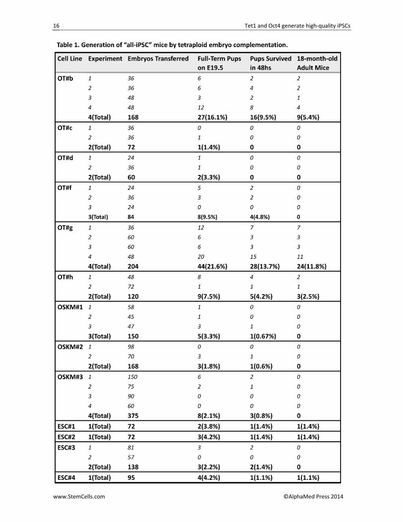
Because the OT‐iPSCs showed good pluripotency both in transcription and protein levels and contributed to high‐grade chimerism (>95%), we next performed tetraploid (4n) complementation experiments to test the stringent pluripotency of OT‐iPSCs. Using microin‐jection approaches, six of eight OT‐iPS cell lines (OT#b, OT#c, OT#d, OT#f, OT#g and OT#h) presented the ability to give birth to live breathing pups, termed “all‐iPSC” mice, when caesarean section was performed on em‐
Tet1 and Oct4 generate high‐quality iPSCs
www.StemCells.com ©AlphaMed Press 2014
6
bryonic day (E) 19.5 (Figures S3A and S3B). Although some pups died within several days because of weak suckling, cannibalization after fostering or other internal defects—features also common in tetraploid comple‐mentation experiments performed by OSK/OSKM‐iPSCs and ESCs [12‐14, 40, 41]—certain OT‐iPSCs produced live all‐iPSC mice approximately 7‐10 times more effi‐ciently than OSK or OSKM‐iPSCs did (Figure S3B). More important, 11 OT‐iPSC mice matured normally and sur‐vived more than 18 months (Figure S3A).
A major disadvantage in generating full‐term mice was that the birth rate was greatly influenced by pro‐longed culture, different culture conditions, state of donor 4n embryos and even the techniques applied [40]. These drawbacks often lead to the production of all‐iPSC mice by individual iPSC lines inconsistent. To further obtain evidence supporting the full pluripotency of these OT‐iPSCs, we extended the cell passages and performed tetraploid complementation using another approach, called aggregation [40‐42], with several expe‐rimental repeats. Although efficiency was lower in pro‐duction of OSKM‐iPSC mice by previously confirmed tetra‐on OSKM iPSCs and lower 1‐year survival rates were also observed, an increase in both birth rate and survival was apparent in certain OT‐iPSCs as assessed by aggregation means (Table 1; Figures 3A and 3B). Nota‐bly, two OT‐iPS cell lines (OT#b and OT#g) were 5‐times more efficient in producing full‐term pups compared with the control ESCs (Table 1 and Figure 3A), and some pseudo‐pregnant mice were able to deliver all OT‐iPSC mice by themselves. Thus, the reproducible generation of all‐iPSC mice by certain OT‐iPSCs was definitively con‐firmed. Moreover, all 47 adult OT‐iPSC mice (13 for OT#b, 3 for OT#f, 26 for OT#g and 5 for OT#h) produced using the two approaches of the tetraploid experiment could generate their germline offspring after mating with ICR mice (Figure 3B). Genotyping by simple se‐quence‐length polymorphism (SSLP) analysis demon‐strated that all these mice were entirely produced by the donor OT‐iPSCs (Figure S3C).
Even more noteworthy, a high proportion of the OT‐iPSCs (~75%, 6 of 8 OT‐iPS cell lines) fulfilled the gold standard of 4n competence, which was markedly higher than originally reported [13] (~8%, 3 of 37 OSKM‐iPS cell lines) (Figure 3C). Previous findings indicated that se‐rum‐replacement medium and supplementation with ascorbic acid (Vitamin C) in derivation conditions during reprogramming had a substantial effect on the epige‐netic and biological characteristics of OSKM‐iPSCs and could facilitate the generation of all‐iPSC mice [11]. Here, further analysis indicated an increase in the pro‐portion of all‐iPSC mice produced (~33.3%, 5 of 15 OSKM‐iPS cell lines) as compared to original reports [13, 14] (Figure 3C).
Moreover, we generated certain 2° OSKM‐iPSCs via the Dox‐inducible system [37]. Despite the reported “epigenetic memory” of tissue‐specific iPSCs [43, 44], iPSCs from different layers were capable of generating all‐iPSC mice, although some with greater potential.
However, of all these iPSCs, OT combinations seemed more efficient in obtaining good‐quality and 4n‐compentance iPSCs as compared to OSK‐iPSCs [15], OSKM‐iPSCs [13, 14], 2° OSKM‐iPSCs and even the ESCs, irrespective of the starting cell types and the repro‐gramming vectors used (Figure 3C).
All OT‐iPSC Mice Show Normal Life Span with No Obvious Tumorigenicity
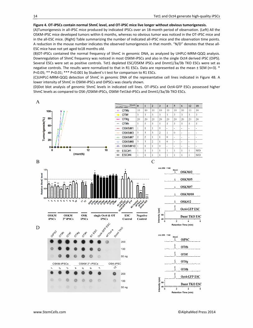
A more exciting finding here was that none of the 47 adult OT‐iPSC mice developed tumors in more than one year of observation; no obvious tumor was detected in even the oldest mice, which are now more than 18 months old (Figures 4A and S4A). Furthermore, all the OT‐iPSC mice behaved normally, and their germline offspring also showed no tumorigenicity (0/220 tested offspring in all) over the 6‐month period of observation (Table S3). However, a high risk of tumorigenesis was noticed in many of the OSKM chimeric mice and the OSKM‐iPSC mice as well as the germline offspring of both [2, 11, 17]. In this study, tumors were seen in OSKM‐iPSC mice as early as the age of 2 months, and all OSKM‐iPSC mice died with tumors within 6 months (Figures 4A and S4B). Moreover, even an F1 germline mouse, generated by one of the OSKM#3‐iPSC mice after mating with ICR, developed tumors within 3 months and died soon thereafter (Figure S4B; Table S3).
A potential correlation between aberrant Tet1/5hmC levels with several cancers was previously reported [19‐23]. We also recently demonstrated that TSKM‐iPSCs that contained normal levels of 5mC and 5hmC could produce all‐iPSC mice without obvious tu‐mors in 2 years [31]. To further investigate whether 5hmC levels in iPSCs may contribute to lower tumorige‐nicity, we performed UHPLC‐MRM‐QQQ (Ultra High Performance Liquid Chromatography‐Multiple Reaction Monitoring‐Triple Quadrupole) analysis [35, 36]on these OT‐iPSCs, OSKM‐iPSCs, OSK‐iPSCs and single Oct4‐iPSCs (OiPS)[10]; normal ESCs, Tet1‐depleted 4F (OSKM) iPS cells (4FTkd‐a and 4FTkd‐b), Tet1‐depleted R1 ESCs (R1‐Tet1kd) and Dnmt1/3a/3b triple knockout ESCs (Dnmt TKO) were set as controls (Figures 4B and 4C).
Interestingly, a level of 5hmC comparable to those in normal ESCs was noticed in almost all the OT‐iPSCs; however, most OSKM‐iPSCs had lower 5hmC levels, and the dot blot analysis for 5hmC further confirmed this result (Figure 4B‐4D). Although the 5mC modifications in these iPSCs seem more complicated (Figure S4C), the upregulated 5hmC levels in OT‐iPSCs may be one of the reasons for the lowest tumorigenicity observed in the OT‐iPSC mice when the cells were differentiated to pro‐duce a whole mouse in tetraploid complementary expe‐riments.

Tet1 and Oct4 generate high‐quality iPSCs
www.StemCells.com ©AlphaMed Press 2014
7
OT 2° system reveals rapid 5hmC upregula‐tion and MET in reprogramming
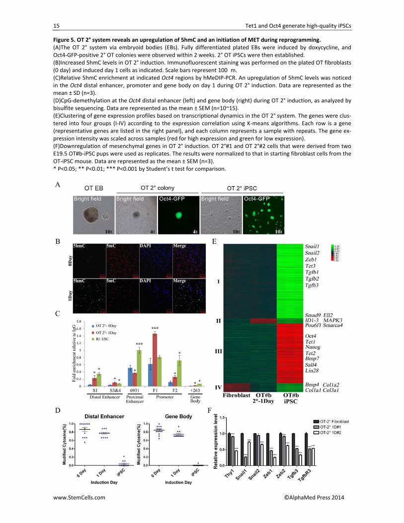
Because the OT‐iPSCs were derived by a Dox‐inducible system, we investigated whether a secondary (2°) induction system could be set up. Primary OT‐iPSCs were first differentiated into fibroblasts according to the standard protocols [45] and then induced by Dox. All four of the tested OT‐iPSCs (OT#b, OT#f, OT#g and OT#h) could form Oct4‐GFP‐positive 2° OT‐iPSCs after a 2‐week induction (Figure 5A). However, we failed to generate 2° OT‐iPSCs directly by inducing fibroblasts from E19.5 OT‐iPSC pups under traditional induction conditions, whereas the reported optimal VC6T medium successfully completed the reprogramming (Figure S5A) [33]. The restricted expression of exogenous Oct4 might be the major reason behind this observation (Figure S5B); the further expression of ectopic Oct4 in the in‐duced population finally generated Oct4‐GFP‐positive iPSCs (Figure S5C).
We then attempted to investigate how Tet1 and Oct4 could successfully reprogram somatic cells to plu‐ripotency. Because Tet1 is a DNA hydroxylase that can convert 5mC into 5hmC in DNA, one reason might be a rapid upregulation of 5hmC by wild‐type Tet1 in the reprogramming cell populations, which was noticed in both the primary and 2° OT systems (Figures 5B and S5D). Moreover, with the 2° system, hMeDIP‐PCR dem‐onstrated upregulation of 5hmC levels at key regulatory regions of Oct4 after Dox induction (Figure 5C). In addi‐tion, bisulfate sequencing indicated rapid demethyla‐tion (~10%) at the distal enhancer and gene body of Oct4 on the same day (Figure 5D).
The transcription profile on induction day 1 was fur‐ther examined by microarray analysis and compared with somatic fibroblasts and pluripotent OT#b iPSCs (Figure 5E). Although the induced cells on day 1 showed an expression pattern more similar to that of the start‐ing fibroblasts, the expression of a small subset of genes was greatly changed (Figure 5E). We clustered the sig‐nificantly changed genes into four groups (6353 genes in total) based on the expression level during the entire reprogramming process. Group IV are the genes that are highly expressed in fibroblasts but rapidly repressed within 24 h after induction, indicating a disruption of the somatic transcription landscape, as also noticed in the OSKM system [46]. The downregulation of collagen genes was marked, and we recently demonstrated that extracellular collagen is a barrier to reprogramming; decreasing collagen gene expression could further im‐prove the reprogramming [47]. Interestingly, genes in groups II were upregulated only at the early stage. Gene ontology (GO) analysis indicated that these genes were related with positive regulation of transcription and the biosynthetic process. qRT‐PCR further demonstrated rapid downregulation of mesenchymal genes in both the primary and 2° system, as well as an upregulation of epithelial genes (Figure 5F; Figure S5E and S5F), which indicated an initiation of mesenchymal‐to‐epithelial
transition (MET) [46, 47] during the OT‐mediated repro‐gramming process.
DISCUSSION
To our knowledge, this is the first study showing
that DNA hydroxylase Tet1 has the ability to generate high‐quality iPSCs with Oct4 and can efficiently produce tumor‐free all‐iPSC mice. The catalytic activity of Tet1 is essential for its function during reprogramming, as truncated Tet1 cannot generate Oct4‐GFP‐positive iPSCs with Oct4 addition (Figures 1D and S1D).
Until now, all the reported live all‐iPSC mice gener‐ated by OSKM iPSCs displayed a decrease in long‐term survival due to tumorigenesis [8, 12‐16], which was also observed in our experiments (Figures 4A and S4B). Here, we demonstrate that the OT combination can generate a high percentage (~75%) of iPSCs with full pluripotency (4n competence) as compared to primary OSK‐ and OSKM‐iPSCs (~8% to ~33%), which were es‐tablished by our labs and others (Figure 3C). These re‐sults were confirmed by two types of tetraploid com‐plementation approaches and several experimental repeats to rule out such influences as prolonged culture and state of donor 4n embryos (Table 1; Figures 3A, S3A and S3B). More importantly, all the OT‐iPSC mice grew to a normal life span (more than 18 months) without obvious tumors (Figures 4A and S4A). Considering the loss of 5hmC in certain tumors [19‐23], a more correctly rebuilt 5hmC landscape in OT‐iPSCs mediated by ectopic expression of wild‐type Tet1 (Figure 4B and 4C) as well as the omitted c‐Myc during reprogramming might par‐tially contribute to the zero tumorigenicity seen in these mice (Figure 4A).
Moreover, low efficiency and an extended repro‐gramming period were observed in other single‐Oct4 mediated reprogramming [6, 9, 10, 33, 38, 39] (Figure 1F), which might be due to a reduction in reprogram‐ming factors (Figure 1C). It is noteworthy that, despite the successful reprogramming by Oct4 alone, the endo‐genous expression of the remain‐ing reprogramming factors in certain cell types [6, 9, 10] and the additional use of small molecules [33, 38, 39] indicated that the iPSC reprogramming was not me‐diated solely by Oct4 (Figure 1F). In addition, relatively low chimerism was observed in some of these single‐Oct4‐iPSCs [6, 9, 39]. Although Tsai and colleagues gen‐erated several all‐iPSC mice from their single‐Oct4‐iPSCs, all the pups died within a few days, which further called into question the quality of these “tetra‐on” sin‐gle Oct4‐iPSCs [9]. In this study, a new 2° induction system was established by inducing either differentiated fibroblasts from OT‐EBs or somatic fibroblasts derived from OT‐iPSC mice (Figures 5A and S5A). Rapid upregulation of genome‐wide 5hmC and a specific 5hmC increase on the Oct4 locus might play important roles in this type of repro‐gramming (Figure 5B‐5D). In addition, the downregula‐tion of extracellular collagen genes and mesenchymal
Tet1 and Oct4 generate high‐quality iPSCs
www.StemCells.com ©AlphaMed Press 2014
8
genes, including Thy1, Snail1 and Snail2, as well as the upregulation of BMP signal and pluripotent genes dur‐ing OT‐mediated reprogramming might further facilitate mesenchymal‐to‐epithelial transition and finally com‐plete the reprogramming (Figures 5E, 5F, S5E and S5F) [46‐49]. CONCLUSION
In summary, our study demonstrates that the com‐bination of Oct4 with DNA dioxygenase Tet1 can gener‐ate fully pluripotent iPSCs with normal 5hmC levels, which provides a robust method to generate high‐quality “tetra‐on” iPSCs without tumorigenicity, as ob‐served in the all‐iPSC mice up to 18 months old. Our results suggest that the role of 5hmC levels in humans should be considered in efforts to generate fully pluri‐potent human iPSCs.
ACKNOWLEDGMENTS
We are grateful to our colleagues in the laboratory for their assistance with the experiments and in the prepa‐ration of this manuscript. This project was supported by the National Natural Science Foundation of China (31325019, 91319306, 31401247 and 31401266) and Ministry of Science and Technology of China (Grants 2010CB944900 and 2015CB964800). DISCLOSURE OF POTENTIAL CONFLICTS OF INTEREST The authors have no potential conflicts of interest. AUTHOR CONTRIBUTIONS J.C., Y.G.: conception and design, collection and/or as‐sembly of the data, data analysis and interpretation, and manuscript writing; H.H., K.X., X.C., Y.J., H.L., S.G., Y.T., H.W., Y.Z., H.W., T.C.: provision of study material and collection and/or assembly of the data; S.G.: con‐ception and design, financial support, data analysis and interpretation, manuscript writing, and final approval of the manuscript.
REFERENCES 1 Wernig M, Meissner A, Foreman R et al.
In vitro reprogramming of fibroblasts into a pluripotent ES‐cell‐like state. Nature. 2007;448:318‐324. 2 Okita K, Ichisaka T, Yamanaka S.
Generation of germline‐competent induced pluripotent stem cells. Nature. 2007;448:313‐317. 3 Maherali N, Sridharan R, Xie W et al.
Directly reprogrammed fibroblasts show global epigenetic remodeling and widespread tissue contribution. Cell stem cell. 2007;1:55‐70. 4 Takahashi K, Yamanaka S. Induction of
pluripotent stem cells from mouse embryonic and adult fibroblast cultures by defined factors. Cell. 2006;126:663‐676. 5 Takahashi K, Tanabe K, Ohnuki M et al.
Induction of pluripotent stem cells from adult human fibroblasts by defined factors. Cell. 2007;131:861‐872. 6 Kim JB, Sebastiano V, Wu G et al. Oct4‐
induced pluripotency in adult neural stem cells. Cell. 2009;136:411‐419. 7 Warren L, Manos PD, Ahfeldt T et al.
Highly efficient reprogramming to pluripotency and directed differentiation of human cells with synthetic modified mRNA. Cell stem cell. 2010;7:618‐630. 8 Carey BW, Markoulaki S, Hanna JH et al.
Reprogramming factor stoichiometry influences the epigenetic state and biological properties of induced pluripotent stem cells. Cell stem cell. 2011;9:588‐598. 9 Tsai SY, Bouwman BA, Ang YS et al. Single
transcription factor reprogramming of hair follicle dermal papilla cells to induced
pluripotent stem cells. Stem cells. 2011;29:964‐971. 10 Wu T, Wang H, He J et al.
Reprogramming of trophoblast stem cells into pluripotent stem cells by Oct4. Stem cells. 2011;29:755‐763. 11 Stadtfeld M, Apostolou E, Ferrari F et al.
Ascorbic acid prevents loss of Dlk1‐Dio3 imprinting and facilitates generation of all‐iPS cell mice from terminally differentiated B cells. Nat Genet. 2012;44:398‐405, S391‐392. 12 Boland MJ, Hazen JL, Nazor KL et al.
Adult mice generated from induced pluripotent stem cells. Nature. 2009;461:91‐94. 13 Zhao XY, Li W, Lv Z et al. iPS cells
produce viable mice through tetraploid complementation. Nature. 2009;461:86‐90. 14 Kang L, Wang J, Zhang Y et al. iPS cells
can support full‐term development of tetraploid blastocyst‐complemented embryos. Cell stem cell. 2009;5:135‐138. 15 Kang L, Wu T, Tao Y et al. Viable mice
produced from three‐factor induced pluripotent stem (iPS) cells through tetraploid complementation. Cell Res. 2011;21:546‐549. 16 Stadtfeld M, Apostolou E, Akutsu H et
al. Aberrant silencing of imprinted genes on chromosome 12qF1 in mouse induced pluripotent stem cells. Nature. 2010;465:175‐181. 17 Wernig M, Meissner A, Cassady JP et al.
c‐Myc is dispensable for direct reprogramming of mouse fibroblasts. Cell stem cell. 2008;2:10‐12. 18 Stadtfeld M, Maherali N, Borkent M et
al. A reprogrammable mouse strain from gene‐targeted embryonic stem cells. Nat Methods. 2010;7:53‐55.
19 Haffner MC, Chaux A, Meeker AK et al. Global 5‐hydroxymethylcytosine content is significantly reduced in tissue stem/progenitor cell compartments and in human cancers. Oncotarget. 2011;2:627‐637. 20 Yang H, Liu Y, Bai F et al. Tumor
development is associated with decrease of TET gene expression and 5‐methylcytosine hydroxylation. Oncogene. 2013;32:663‐669. 21 Ko M, Huang Y, Jankowska AM et al.
Impaired hydroxylation of 5‐methylcytosine in myeloid cancers with mutant TET2. Nature. 2010;468:839‐843. 22 Kudo Y, Tateishi K, Yamamoto K et al.
Loss of 5‐hydroxymethylcytosine is accompanied with malignant cellular transformation. Cancer Sci. 2012;103:670‐676. 23 Lian CG, Xu Y, Ceol C et al. Loss of 5‐
hydroxymethylcytosine is an epigenetic hallmark of melanoma. Cell. 2012;150:1135‐1146. 24 Tahiliani M, Koh KP, Shen Y et al.
Conversion of 5‐methylcytosine to 5‐hydroxymethylcytosine in mammalian DNA by MLL partner TET1. Science. 2009;324:930‐935. 25 Ito S, D'Alessio AC, Taranova OV et al.
Role of Tet proteins in 5mC to 5hmC conversion, ES‐cell self‐renewal and inner cell mass specification. Nature. 2010;466:1129‐1133. 26 Williams K, Christensen J, Pedersen MT
et al. TET1 and hydroxymethylcytosine in transcription and DNA methylation fidelity. Nature. 2011;473:343‐348. 27 Wu H, D'Alessio AC, Ito S et al. Dual
functions of Tet1 in transcriptional regulation in mouse embryonic stem cells. Nature. 2011;473:389‐393.
Tet1 and Oct4 generate high‐quality iPSCs
www.StemCells.com ©AlphaMed Press 2014
9
28 Ficz G, Branco MR, Seisenberger S et al. Dynamic regulation of 5‐hydroxymethylcytosine in mouse ES cells and during differentiation. Nature. 2011;473:398‐402. 29 Xu Y, Wu F, Tan L et al. Genome‐wide
regulation of 5hmC, 5mC, and gene expression by Tet1 hydroxylase in mouse embryonic stem cells. Mol Cell. 2011;42:451‐464. 30 Doege CA, Inoue K, Yamashita T et al.
Early‐stage epigenetic modification during somatic cell reprogramming by Parp1 and Tet2. Nature. 2012;488:652‐655. 31 Gao Y, Chen J, Li K et al. Replacement of
Oct4 by Tet1 during iPSC induction reveals an important role of DNA methylation and hydroxymethylation in reprogramming. Cell stem cell. 2013;12:453‐469. 32 Costa Y, Ding J, Theunissen TW et al.
NANOG‐dependent function of TET1 and TET2 in establishment of pluripotency. Nature. 2013;495:370‐374. 33 Li Y, Zhang Q, Yin X et al. Generation of
iPSCs from mouse fibroblasts with a single gene, Oct4, and small molecules. Cell Res. 2011;21:196‐204. 34 de Hoon MJ, Imoto S, Nolan J et al.
Open source clustering software. Bioinformatics. 2004;20:1453‐1454. 35 Yin R, Mao SQ, Zhao B et al. Ascorbic
acid enhances Tet‐mediated 5‐methylcytosine oxidation and promotes DNA demethylation
in mammals. Journal of the American Chemical Society. 2013;135:10396‐10403. 36 Zhao B, Yang Y, Wang X et al. Redox‐
active quinones induces genome‐wide DNA methylation changes by an iron‐mediated and Tet‐dependent mechanism. Nucleic acids research. 2014;42:1593‐1605. 37 Kou Z, Kang L, Yuan Y et al. Mice cloned
from induced pluripotent stem cells (iPSCs). Biol Reprod. 2010;83:238‐243. 38 Yuan X, Wan H, Zhao X et al. Brief
report: combined chemical treatment enables Oct4‐induced reprogramming from mouse embryonic fibroblasts. Stem cells. 2011;29:549‐553. 39 Chen J, Liu J, Yang J et al. BMPs
functionally replace Klf4 and support efficient reprogramming of mouse fibroblasts by Oct4 alone. Cell Res. 2011;21:205‐212. 40 Nagy A, Rossant J, Nagy R et al.
Derivation of completely cell culture‐derived mice from early‐passage embryonic stem cells. Proc Natl Acad Sci U S A. 1993;90:8424‐8428. 41 Nagy A, Gocza E, Diaz EM et al.
Embryonic stem cells alone are able to support fetal development in the mouse. Development. 1990;110:815‐821. 42 Wu G, Liu N, Rittelmeyer I et al.
Generation of healthy mice from gene‐corrected disease‐specific induced pluripotent stem cells. PLoS Biol. 2011;9:e1001099.
43 Kim K, Doi A, Wen B et al. Epigenetic memory in induced pluripotent stem cells. Nature. 2010;467:285‐290. 44 Polo JM, Liu S, Figueroa ME et al. Cell
type of origin influences the molecular and functional properties of mouse induced pluripotent stem cells. Nat Biotechnol. 2010;28:848‐855. 45 Hockemeyer D, Soldner F, Cook EG et al.
A drug‐inducible system for direct reprogramming of human somatic cells to pluripotency. Cell stem cell. 2008;3:346‐353. 46 Samavarchi‐Tehrani P, Golipour A, David
L et al. Functional genomics reveals a BMP‐driven mesenchymal‐to‐epithelial transition in the initiation of somatic cell reprogramming. Cell stem cell. 2010;7:64‐77. 47 Jiao J, Dang Y, Yang Y et al. Promoting
reprogramming by FGF2 reveals that the extracellular matrix is a barrier for reprogramming fibroblasts to pluripotency. Stem cells. 2013;31:729‐740. 48 Li R, Liang J, Ni S et al. A mesenchymal‐
to‐epithelial transition initiates and is required for the nuclear reprogramming of mouse fibroblasts. Cell stem cell. 2010;7:51‐63. 49 Lin Z, Perez P, Lei D et al. Two‐phase
analysis of molecular pathways underlying induced pluripotent stem cell induction. Stem cells. 2011;29:1963‐1974.
See www.StemCells.com for supporting information available online. STEM
CELLS ; 00:000–000
Tet1 and Oct4 generate high‐quality iPSCs
www.StemCells.com ©AlphaMed Press 2014
10
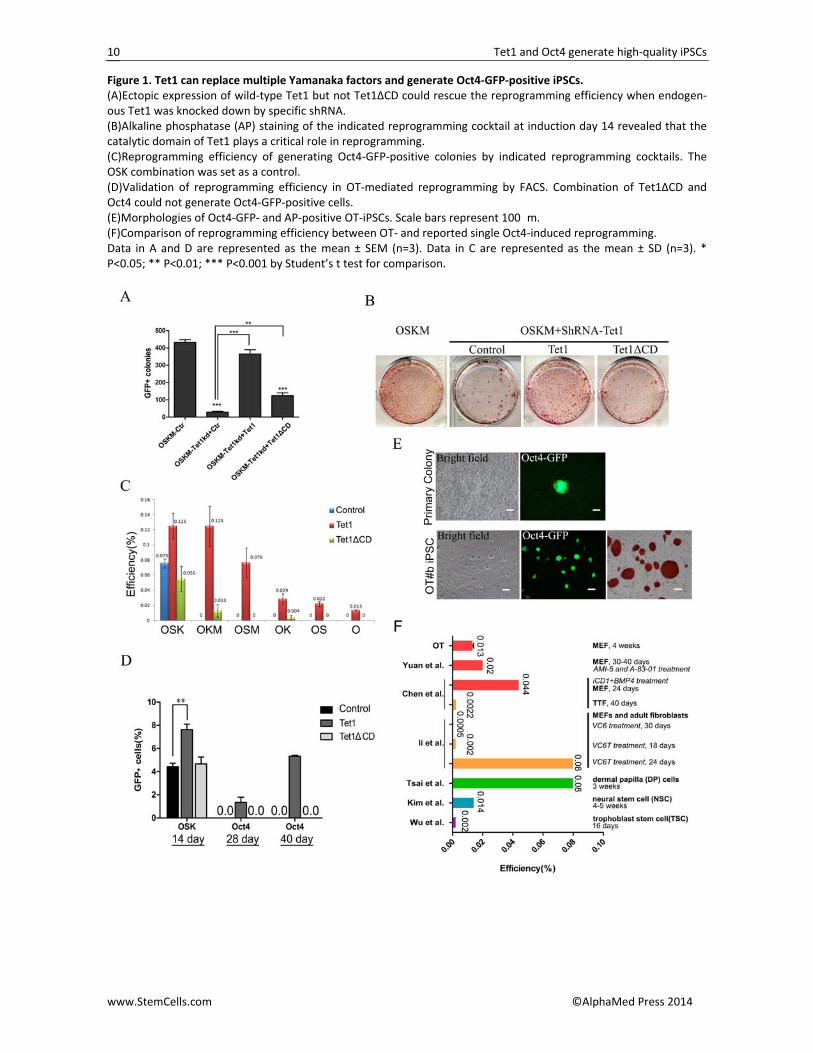
Figure 1. Tet1 can replace multiple Yamanaka factors and generate Oct4‐GFP‐positive iPSCs. (A)Ectopic expression of wild‐type Tet1 but not Tet1ΔCD could rescue the reprogramming efficiency when endogen‐ous Tet1 was knocked down by specific shRNA. (B)Alkaline phosphatase (AP) staining of the indicated reprogramming cocktail at induction day 14 revealed that the catalytic domain of Tet1 plays a critical role in reprogramming. (C)Reprogramming efficiency of generating Oct4‐GFP‐positive colonies by indicated reprogramming cocktails. The OSK combination was set as a control. (D)Validation of reprogramming efficiency in OT‐mediated reprogramming by FACS. Combination of Tet1ΔCD and Oct4 could not generate Oct4‐GFP‐positive cells. (E)Morphologies of Oct4‐GFP‐ and AP‐positive OT‐iPSCs. Scale bars represent 100�m. (F)Comparison of reprogramming efficiency between OT‐ and reported single Oct4‐induced reprogramming. Data in A and D are represented as the mean ± SEM (n=3). Data in C are represented as the mean ± SD (n=3). * P<0.05; ** P<0.01; *** P<0.001 by Student’s t test for comparison.
Tet1 and Oct4 generate high‐quality iPSCs
www.StemCells.com ©AlphaMed Press 2014
11
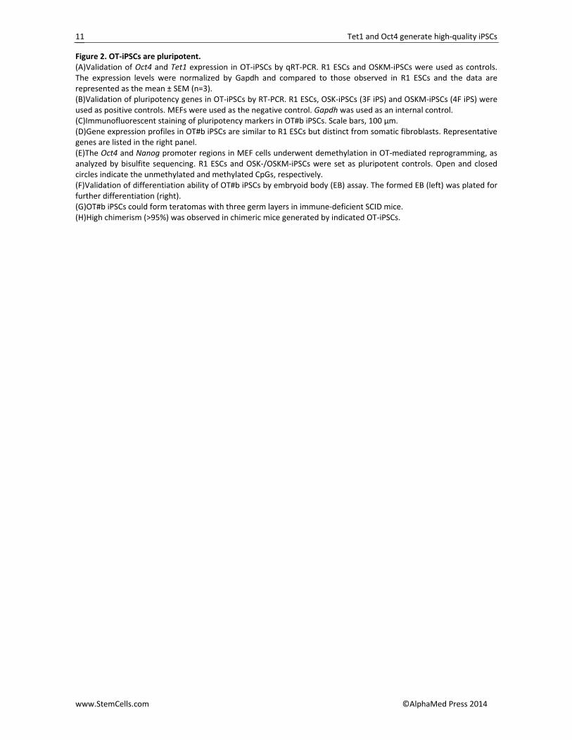
Figure 2. OT‐iPSCs are pluripotent. (A)Validation of Oct4 and Tet1 expression in OT‐iPSCs by qRT‐PCR. R1 ESCs and OSKM‐iPSCs were used as controls. The expression levels were normalized by Gapdh and compared to those observed in R1 ESCs and the data are represented as the mean ± SEM (n=3). (B)Validation of pluripotency genes in OT‐iPSCs by RT‐PCR. R1 ESCs, OSK‐iPSCs (3F iPS) and OSKM‐iPSCs (4F iPS) were used as positive controls. MEFs were used as the negative control. Gapdh was used as an internal control. (C)Immunofluorescent staining of pluripotency markers in OT#b iPSCs. Scale bars, 100 µm. (D)Gene expression profiles in OT#b iPSCs are similar to R1 ESCs but distinct from somatic fibroblasts. Representative genes are listed in the right panel. (E)The Oct4 and Nanog promoter regions in MEF cells underwent demethylation in OT‐mediated reprogramming, as analyzed by bisulfite sequencing. R1 ESCs and OSK‐/OSKM‐iPSCs were set as pluripotent controls. Open and closed circles indicate the unmethylated and methylated CpGs, respectively. (F)Validation of differentiation ability of OT#b iPSCs by embryoid body (EB) assay. The formed EB (left) was plated for further differentiation (right). (G)OT#b iPSCs could form teratomas with three germ layers in immune‐deficient SCID mice. (H)High chimerism (>95%) was observed in chimeric mice generated by indicated OT‐iPSCs.
Tet1 and Oct4 generate high‐quality iPSCs
www.StemCells.com ©AlphaMed Press 2014
12
Tet1 and Oct4 generate high‐quality iPSCs
www.StemCells.com ©AlphaMed Press 2014
13
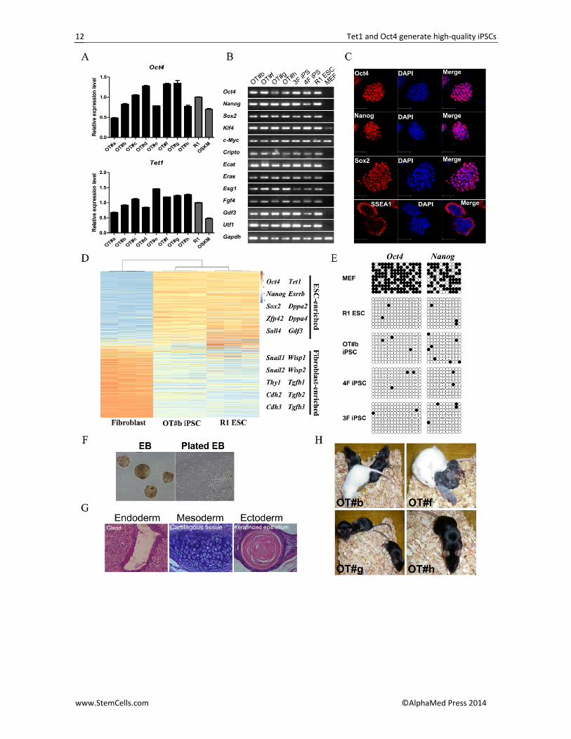
Figure 3. OT‐iPSCs can efficiently produce live all‐iPSC mice. (A)Percentage of transferred blastocysts surviving to birth are plotted for “tetra‐on” OT iPSCs, OSKM iPSCs and ESCs shown in Table 1.Only cell lines which were capable of producing all‐iPSC mouse were calculated. Data are represented as the mean ± SEM. The total number of blastocysts was 708 for OT iPSCs, 693 for OSKM iPSCs and 377 for ESCs. * P<0.05; ** P<0.01 by Student’s t test for comparison. (B)Representative images of full‐term (E19.5) litters (left) and adult OT‐iPSC mice with their F1 offspring (right). The adult OT#g‐iPSC mice were produced by microinjection. (C)The proportion of “tetra‐on” iPSCs to all the derived iPSCs was higher in OT‐iPSCs than that in primary OSK‐/OSKM‐iPSCs, 2° OSKM‐iPSCs and control ESCs.
Tet1 and Oct4 generate high‐quality iPSCs
www.StemCells.com ©AlphaMed Press 2014
14
Figure 4. OT‐iPSCs contain normal 5hmC level, and OT‐iPSC mice live longer without obvious tumorigenesis. (A)Tumorigenesis in all‐iPSC mice produced by indicated iPSCs over an 18‐month period of observation. (Left) All the OSKM‐iPSC mice developed tumors within 6 months, whereas no obvious tumor was noticed in the OT‐iPSC mice and in the all‐ESC mice. (Right) Table summarizing the number of indicated all‐iPSC mice and the observation time points. A reduction in the mouse number indicates the observed tumorigenesis in that month. “N/D” denotes that these all‐ESC mice have not yet aged to18 months old. (B)OT‐iPSCs contained the normal frequency of 5hmC in genomic DNA, as analyzed by UHPLC‐MRM‐QQQ analysis. Downregulation of 5hmC frequency was noticed in most OSKM‐iPSCs and also in the single Oct4 derived iPSC (OiPS). Several ESCs were set as positive controls. Tet1 depleted ESC/OSKM iPSCs and Dnmt1/3a/3b TKO ESCs were set as negative controls. The results were normalized to that in R1 ESCs. Data are represented as the mean ± SEM (n=3). * P<0.05; ** P<0.01; *** P<0.001 by Student’s t test for comparison to R1 ESCs. (C)UHPLC‐MRM‐QQQ detection of 5hmC in genomic DNA of the representative cell lines indicated in Figure 4B. A lower intensity of 5hmC in OSKM‐iPSCs and OiPSCs was clearly shown. (D)Dot blot analysis of genomic 5hmC levels in indicated cell lines. OT‐iPSCs and Oct4‐GFP ESCs possessed higher 5hmC levels as compared to OSK‐/OSKM‐iPSCs, OSKM‐Tet1kd‐iPSCs and Dnmt1/3a/3b TKO ESCs.
Tet1 and Oct4 generate high‐quality iPSCs
www.StemCells.com ©AlphaMed Press 2014
15
Figure 5. OT 2° system reveals an upregulation of 5hmC and an initiation of MET during reprogramming. (A)The OT 2° system via embryoid bodies (EBs). Fully differentiated plated EBs were induced by doxycycline, and Oct4‐GFP‐positive 2° OT colonies were observed within 2 weeks. 2° OT iPSCs were then established. (B)Increased 5hmC levels in OT 2° induction. Immunofluorescent staining was performed on the plated OT fibroblasts (0 day) and induced day 1 cells as indicated. Scale bars represent 100�m. (C)Relative 5hmC enrichment at indicated Oct4 regions by hMeDIP‐PCR. An upregulation of 5hmC levels was noticed in the Oct4 distal enhancer, promoter and gene body on day 1 during OT 2° induction. Data are represented as the mean ± SD (n=3). (D)CpG‐demethylation at the Oct4 distal enhancer (left) and gene body (right) during OT 2° induction, as analyzed by bisulfite sequencing. Data are represented as the mean ± SEM (n=10~15). (E)Clustering of gene expression profiles based on transcriptional dynamics in the OT 2° system. The genes were clus‐tered into four groups (I‐IV) according to the expression correlation using K‐means algorithms. Each row is a gene (representative genes are listed in the right panel), and each column represents a sample with repeats. The gene ex‐pression intensity was scaled across samples (red for high expression and green for low expression). (F)Downregulation of mesenchymal genes in OT 2° induction. OT 2°#1 and OT 2°#2 cells that were derived from two E19.5 OT#b‐iPSC pups were used as replicates. The results were normalized to that in starting fibroblast cells from the OT‐iPSC mouse. Data are represented as the mean ± SEM (n=3). * P<0.05; ** P<0.01; *** P<0.001 by Student’s t test for comparison.
Tet1 and Oct4 generate high‐quality iPSCs
www.StemCells.com ©AlphaMed Press 2014
16


