Embed Size (px)
Citation preview
Cell Reports
Report
The Distribution of Genomic Variationsin Human iPSCs Is Related to Replication-TimingReorganization during ReprogrammingJunjie Lu,1,2,8 Hu Li,3,8 Ming Hu,4,9 Takayo Sasaki,5 Anna Baccei,1,2 David M. Gilbert,5 Jun S. Liu,4 James J. Collins,6,7
and Paul H. Lerou1,2,*1Department of Pediatric NewbornMedicine and Division of Genetics, Department of Medicine, Brigham andWomen’s Hospital, Boston, MA
02115, USA2Harvard Medical School, Harvard Stem Cell Institute, Boston, MA 02115, USA3Center for Individualized Medicine, Department of Molecular Pharmacology & Experimental Therapeutics, Mayo Clinic, Rochester,MN 55905, USA4Department of Statistics, Harvard University, Cambridge, MA 02138, USA5Department of Biological Science, Florida State University, Tallahassee, FL 32306, USA6Howard Hughes Medical Institute, Department of Biomedical Engineering and Center of Synthetic Biology, Boston University, Boston,
MA 02215, USA7Wyss Institute for Biologically Inspired Engineering, Harvard University, Boston, MA 02115, USA8These authors contributed equally to this work9Present address: Division of Biostatistics, Department of Population Health, New York University School of Medicine, New York,
NY 10016, USA
*Correspondence: [email protected]
http://dx.doi.org/10.1016/j.celrep.2014.03.007This is an open access article under the CC BY license (http://creativecommons.org/licenses/by/3.0/).
SUMMARY
Cell-fate change involves significant genome reorga-nization, including changes in replication timing, buthow these changes are related to genetic variationhas not been examined. To study how a change inreplication timing that occurs during reprogrammingimpacts the copy-number variation (CNV) landscape,we generated genome-wide replication-timing pro-files of induced pluripotent stem cells (iPSCs) andtheir parental fibroblasts. A significant portion ofthe genome changes replication timing as a resultof reprogramming, indicative of overall genome reor-ganization. We found that early- and late-replicatingdomains in iPSCs are differentially affected bycopy-number gains and losses and that in particular,CNV gains accumulate in regions of the genome thatchange to earlier replication during the reprogram-ming process. This differential relationship was pre-sent irrespective of reprogramming method. Overall,our findings reveal a functional association betweenreorganization of replication timing and the CNVlandscape that emerges during reprogramming.
INTRODUCTION
The genome is topologically organized in three-dimensional
space within the nucleus and is highly dynamic as cell fate
changesduring normal development, aswell as indisease states,
such as cancer (Ryba et al., 2012). The significant cell-fate
70 Cell Reports 7, 70–78, April 10, 2014 ª2014 The Authors
changes that occur during embryonic stem cell (ESC) differentia-
tion and reprogramming of somatic cells to pluripotency also
involve genome-wide resetting of the higher-order chromatin
structure (Watanabe et al., 2013). In both scenarios, genome
reorganization precedes gene expression changes, suggesting
a potential causal relationship (Apostolou et al., 2013; Phillips-
Cremins et al., 2013; Wei et al., 2013; Zhang et al., 2013).
Higher-order genome organization has been shown to signifi-
cantly influence the distribution of genomic aberrations in both
immortalized somatic cells and cancer cells, but the underlying
mechanism remains elusive (De and Michor, 2011; Fudenberg
et al., 2011; Koren et al., 2012; Schuster-Bockler and Lehner,
2012). One method of mapping genomic organization is by
segmenting the genome into domains based on replication
timing, which occurs in a tightly regulated cell-type-specific
manner (Gilbert et al., 2010). Domains with similar replication
timing tend to colocalize within the nucleus, and as a result,
genome organization defined by replication timing overlaps
with other methods of defining chromatin topology, including
DNase I-hypersensitivity profiles, various epigenetic markers,
and genome-wide chromatin-interaction mapping (e.g., Hi-C)
(Dixon et al., 2012; Hu et al., 2013; Ryba et al., 2010). Several
studies have revealed that the cancer mutational landscape is
closely associated with replication timing by demonstrating that
copy-number variation (CNV) boundaries tend to have similar
replication timing, and gains and losses distribute differentially
with respect to replication timing (De and Michor, 2011; Liu
et al., 2013; Schuster-Bockler and Lehner, 2012). These studies
share one important limitation: the replication-timing maps
were not generated from the same cell types in which the CNVs
were analyzed. This limitation is significant because cellular iden-
tity is closely coupled to genome organization; in fact, only half of
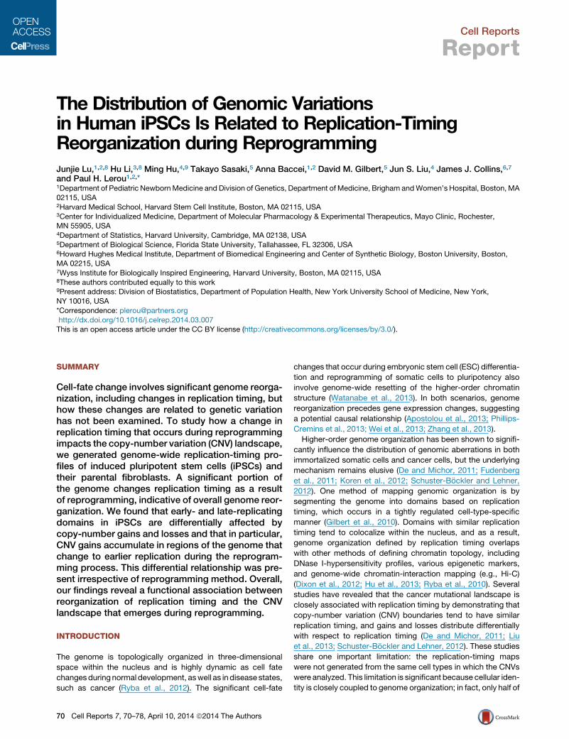
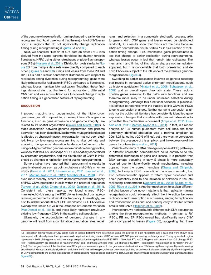
Figure 1. iPSC CNVs Reside within Replication-Timing Domains and Are Distributed Nonrandomly
(A)Newly replicatedDNA fromearly and lateSphase isdifferentially labeled andhybridized to awhole-genomeoligonucleotidemicroarray toproducea replication-
timing profile. Loess-smoothed replication-timing ratio (filled gray) of the first 50Mb of chromosome (Chr) 1 is shown as an example. Segmentation algorithms are
used to define replication domains (black segments) as regions with similar replication-timing ratio. TTRs are defined as genomic regions with a significant rate of
replication-timing ratio change (purple). TTRs cover approximately one-third of the genome. CNVs (green segments) are plotted above the replication profile, and
they can fall either entirely within individual domains or cross boundaries and involve more than one domain. A CNV may or may not involve a TTR.
(B) Permutation analysis demonstrates that CNVs are significantly more likely to reside within individual replication domains than expected by chance in both
iPSCs and parental fibroblasts (fib). Boxes display the percentages obtained from 1,000 simulations, and red diamonds represent the actual percentages.
(C) TTRs are not enriched within CNVs. The actual amount (black) of TTR and non-TTRs affected by CNVs, compared to expected numbers (gray) based on
random distribution, is shown with p values (Fisher’s exact test) above each plot.
See also Figure S1.
the genome has stable replication timing across all cell types,
with the remainder being organized in a highly cell-type-specific
manner (Hansen et al., 2010; Hiratani et al., 2008, 2010). Thus,
when an unmatched replication-timing profile is used, up to
50%of the genomemay not be accounted for during the analysis
due to the cell-type specificity of genome organization.
How cell fate change and its associated genome reorganiza-
tion are related to genome variation has not been examined.
We overcome the limitation of unmatched replication-timing
maps by generating replication-timing profiles of both induced
pluripotent stem cells (iPSCs) and their parental fibroblasts in
order to explore the linkbetween theCNV landscapeandgenome
reorganization due to cell fate change. We show that nuclear
reprogramming results in dramatic replication-timing reorganiza-
tion that influences the observed CNV landscape in iPSCs.
RESULTS
CNVs Reside within Replication-Timing DomainsPrimary dermal fibroblasts from one healthy volunteer (wild-type,
WT) were reprogrammed using retroviral transduction of the
standard Yamanaka factors to generate multiple iPSC lines (Ta-
kahashi et al., 2007) (Figure S1). Replication-timing profiles were
generated from an iPSC line and the parental fibroblasts. Newly
replicated DNA from early and late S phase was differentially
labeled and hybridized to a whole-genome oligonucleotide
microarray. The ratio of the abundance of each probe in the early
versus late fractions generates a replication profile, which
reveals clear demarcation between megabase-sized regions of
coordinated replication called replication domains (Figure 1A).
Computational segmentation algorithms can be used to define
the replication domains (Ryba et al., 2011). The iPSC profile we
generated is identical to previously published profiles of both
human ESCs and iPSCs, reflecting human pluripotent stem
cell-specific genome organization (Figure S1D) (Ryba et al.,
2010).
To map the corresponding genome-variation landscape of
iPSCs, we analyzed the fibroblast and low-passage (p6) iPSC
lines using high-resolution Affymetrix SNP Array 6.0 (Tables S1
and S2). CNV calls were validated using quantitative PCR with
an overall validation rate of 91% (Supplemental Experimental
Procedures). Raw data from the replication-timing profiles also
Cell Reports 7, 70–78, April 10, 2014 ª2014 The Authors 71
independently validated all the homozygous losses detected by
SNP arrays in the same cell lines (Figure S1E) (Ryba et al., 2012).
Most CNVs of iPSCs were not detected in the parental fibro-
blasts and are heretofore referred to as ‘‘iPSC manifested’’
(Tables S1 and S2). iPSC-manifested CNVs in low-passage
iPSCs could represent clonal expansion of low-frequency
genetic aberrations in the tissue of origin or de novo mutations
during the reprogramming process (Liang and Zhang, 2013).
We found that, whereas two-thirds of CNVs detected in iPSCs
involve genic regions (including exons and introns), the affected
genes do not fall into any clear functional groups by Gene
Ontology analysis (Table S3). The CNV-affected genes did not
show any particular relationship to pluripotency, nor did we
find any enrichment of tumor suppression genes or oncogenes.
Our data are similar to other published reports that failed
to demonstrate consistent functional consequences due to
genomic aberrations in iPSCs (Amps et al., 2011; Hussein
et al., 2011; Martins-Taylor et al., 2011).
We mapped the iPSC CNVs to the newly generated replica-
tion-timing profiles and found that more than 95% of CNVs are
contained within individual replication domains, irrespective of
whether the iPSC or the parental fibroblast profile was used.
Permutation analysis using size-controlled random CNV lists
demonstrates that the detected CNVs are significantly more
likely to reside completely within individual replication domains
and do not span domain boundaries than would be expected
to occur by chance (Figure 1B).
Timing transition regions (TTRs), defined by genomic regions
with a significant rate of replication-timing ratio change, have
different replication kinetics compared to the center of the repli-
cation domain. TTRs probably represent sequences vulnerable
to DNA damage (Labib and Hodgson, 2007; Watanabe et al.,
2002, 2004). However, we did not find an enrichment of TTR
sequence in CNV-affected genomic regions in iPSCs (Figure 1C).
Impact of Replication-Timing Dynamics on the CNVLandscapeReprogramming and ESC differentiation involve global changes
in chromosome structure that include temporal reorganization of
replication domains and spatial reorganization of the genome; in
fact, one-half of the genome changes organization at some stage
during development (Hiratani et al., 2008; Orkin and Hochedlin-
ger, 2011; Ryba et al., 2010). Comparison of fibroblast and
iPSC replication-timing profiles revealed that during reprogram-
ming, 40% of the genome changes replication timing, as defined
by a greater than 0.5 difference in the early/late ratio between
fibroblast and iPSC profiles (Figure 2A). This cutoff corresponds
to the amount of time (about 1 hr) required to complete a replica-
tion factory (Leonhardt et al., 2000) and is the same cutoff for
significance for transcription (D.M.G., unpublished data). This
change is nearly double in magnitude compared to the change
observed when ESCs are differentiated into neural precursor
cells (Hiratani et al., 2008), reflecting the extensive genome reor-
ganization during nuclear reprogramming.
To test how genome reorganization influences the genome-
variation landscape, we determined the fibroblast and iPSC
replication-timing values of genomic loci affected by iPSC-
manifested CNVs using cell-type-specific replication-timing
72 Cell Reports 7, 70–78, April 10, 2014 ª2014 The Authors
profiles. The distribution of replication-timing values of CNV
gain loci is statistically different in fibroblasts and iPSCs; they
are predominantly late replicating in fibroblasts but early repli-
cating in iPSCs. In contrast, loci affected by CNV losses are
generally late replicating in both cell types. (Figure 2B).
To better understand the dynamics of the CNV landscape, a
genome-wide representation of replication-timing dynamics
during reprogramming was generated by plotting fibroblast
versus iPSC replication-timing values for all 620,000 probes on
the array (Figure 2C). In this plot, the gray diagonal region repre-
sents genomic loci with no change in replication timing during
reprogramming, defined as a less than 0.5 difference in replica-
tion-timing ratio between fibroblasts and iPSCs. The portions of
the genome that replicate either earlier (Figure 2C, red) or later
(blue) in iPSCs than in fibroblasts fall outside the diagonal.
When the replication-timing values of CNVs were plotted on
this genome background, we show that CNV gains are enriched
in the genome compartment that changes to earlier replication
during reprogramming. This scatterplot presentation reveals
the replication-timing dynamics underlying the distribution shift
of gains seen in Figure 2B. In contrast, the majority of losses
reside in late replicating regions in both fibroblasts and iPSCs
(Figure 2C), consistent with the absence of a shift in the CNV
loss distribution (Figure 2B). Together, these findings further
support the strong relationship between replication timing
and genome variation by demonstrating that the distribution of
CNVs observed in low-passage iPSCs is influenced by replica-
tion-timing reorganization associated with cell fate change.
Human iPSC genomic variation has been studied using a vari-
ety of approaches. Hussein et al. and Laurent et al. generated
iPSC using the same retroviral factors as we did here, but
whereas Hussein et al. also used the Affymetrix 6.0 SNP array,
Laurent et al. used the IlluminaOmniquad v.1 SNP array (Hussein
et al., 2011; Laurent et al., 2011). More recently, Abyzov et al.
used whole-genome sequencing to identify CNVs in retroviral
reprogrammed iPSC lines (Abyzov et al., 2012). None of these
studies considered the role of higher-order chromatin organiza-
tion on genomic variation, and their data sets serve as de facto
controls to exclude potential lab-specific, CNV detection
platform-specific, or parental fibroblast-specific effects in our
experiments. Therefore, we analyzed these data sets relative
to our newly generated replication-timing profiles. To maintain
consistency with our experimental approach, we limited the
analysis to data sets of low-passage iPSCs generated from
dermal fibroblasts. An overall similar trend was seen using
data sets from different laboratories: CNV distribution is
nonrandom, with gains predominating in regions changing to
earlier replication in iPSCs (Figure S2B).
Nonrandom, Differential CNV Distribution Is aGeneralized Feature of ReprogrammingTo exclude the possibility that the nonrandom replication-timing
distribution of the CNVs is due to a direct effect of the retroviral
vectors (RVs) used for reprogramming, we applied our analysis
to iPSCs generated using other reprogramming methods. To
this end, we generated iPSCs using episomal (EP) factors that
do not integrate into the genome. In low-passage EP iPSCs,
like RV iPSCs, CNV gains are significantly enriched in regions
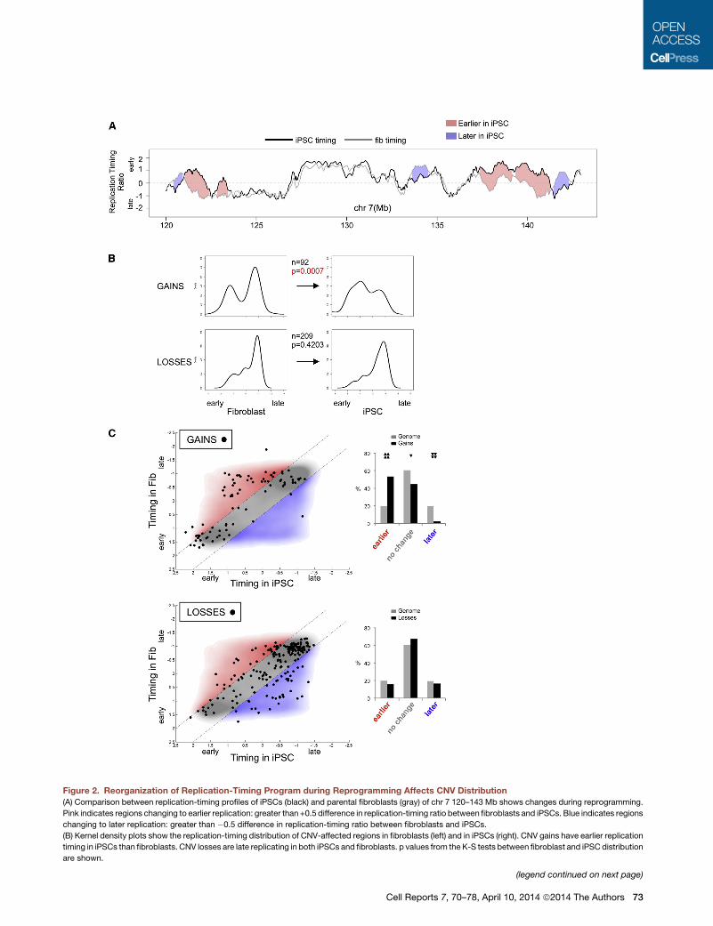
Figure 2. Reorganization of Replication-Timing Program during Reprogramming Affects CNV Distribution
(A) Comparison between replication-timing profiles of iPSCs (black) and parental fibroblasts (gray) of chr 7 120–143 Mb shows changes during reprogramming.
Pink indicates regions changing to earlier replication: greater than +0.5 difference in replication-timing ratio between fibroblasts and iPSCs. Blue indicates regions
changing to later replication: greater than �0.5 difference in replication-timing ratio between fibroblasts and iPSCs.
(B) Kernel density plots show the replication-timing distribution of CNV-affected regions in fibroblasts (left) and in iPSCs (right). CNV gains have earlier replication
timing in iPSCs than fibroblasts. CNV losses are late replicating in both iPSCs and fibroblasts. p values from the K-S tests between fibroblast and iPSC distribution
are shown.
(legend continued on next page)
Cell Reports 7, 70–78, April 10, 2014 ª2014 The Authors 73
of the genomewhose replication timing changed to earlier during
reprogramming. Again, we found that the majority of CNV losses
occur at regions that do not significantly change replication
timing during reprogramming (Figures 3A and S3).
Next, we analyzed Hussein et al.’s data on sister iPSC lines
derived from the same parental fibroblast line (human foreskin
fibroblasts, HFFs) using either retroviruses or piggyBac transpo-
sons (PBs) (Hussein et al., 2011). Distribution plots similar to Fig-
ure 2B from multiple data sets were directly compared to each
other (Figures 3B and S3). Gains and losses from these PB and
RV iPSCs had a similar nonrandom distribution with respect to
replication-timing dynamics during reprogramming: gains were
likely to have earlier replication in iPSCs compared to fibroblasts,
whereas losses maintain late replication. Together, these find-
ings demonstrate that the trend for nonrandom, differential
CNV gain and loss accumulation as a function of change in repli-
cation timing is a generalized feature of reprogramming.
DISCUSSION
Improved mapping and understanding of the higher-order
genomeorganization is providing a clearer picture of howgenome
functions, such as gene expression and genome integrity, are
related to its spatial organization (Cavalli and Misteli, 2013). A
static association between genome organization and genome
aberration has been described, but how themutagenic landscape
is affected by changes in genomeorganization has not been stud-
ied. Here, by leveraging reprogramming to alter cell fate and
analyzing the genome aberration landscape before and after
usingcell-type-matchedgenome-wide replication-timingprofiles,
we show that theCNV landscape of iPSCs is spatially constrained
by chromatin organization, and the distribution of CNVs is influ-
enced by changes in replication timing due to reprogramming.
Some studies have reported that reprogramming results in
genetic aberrations such as point mutations and CNVs in human
iPSCs (Gore et al., 2011; Hussein et al., 2011; Laurent et al.,
2011; Martins-Taylor et al., 2011; Mayshar et al., 2010). How-
ever, more recently, others have demonstrated that the majority
of iPSC CNVs preexisted in cells within the parental population
(Abyzov et al., 2012; Cheng et al., 2012; Quinlan et al., 2011).
Consistent with these reports, we found shared iPSC-
manifested CNVs among the iPSC clones in each of our reprog-
ramming experiments, albeit at low frequency. Interestingly, we
also found that about 50% of iPSC-manifested iPSC CNVs have
overlap with known CNVs in the Database of Genomic Variation
(MacDonald et al., 2014), and these likely represent previously
existing low-frequency CNVs in the starting cell population.
Ultimately, the accumulation of genomic changes in any
genome will result from a combination of mutation rates, repair
(C) Replication-timing values of CNV gains (top) or losses (bottom) were determ
scatterplot with density-smoothed genome-wide replication-timing values (RT
represents�60% of the genome with no change in replication timing defined as iP
RTV� fibroblast RTV) are classified as ‘‘earlier in iPSC’’ (red), and those with less t
(blue). The bar graphs depict the distribution of CNV gains or losses compared to
arrowheads indicate statistically significant enrichment of CNVs in the region, whe
of CNVs compared to the genome distribution in corresponding regions based on
Figure S2).
74 Cell Reports 7, 70–78, April 10, 2014 ª2014 The Authors
rates, and selection. In a completely stochastic process, akin
to genetic drift, CNV gains and losses would be distributed
randomly. Our results show that this is not the case because
CNVs are nonrandomly distributed in iPSCs as a function of repli-
cation-timing change. iPSC-manifested gains predominate in
loci that change to earlier replication during reprogramming,
whereas losses occur in loci that remain late replicating. The
mechanism and timing of this relationship are not immediately
apparent, but it is conceivable that both preexisting and de
novo CNVs are subject to the influence of the extensive genome
reorganization (Figure 4).
Switching to earlier replication involves epigenetic resetting
that results in increased active chromatin modifications, such
as histone acetylation (Hiratani et al., 2008; Schwaiger et al.,
2009) and an overall open chromatin state. These regions
contain genes essential to the cell’s new functions and are
therefore more likely to be under increased selection during
reprogramming. Although this functional selection is plausible,
it is difficult to reconcile with the inability to link CNVs in iPSCs
to gene expression changes. Neither our gene expression anal-
ysis nor the published literature has consistently identified gene
expression changes that correlate with genomic aberration to
prove that this mechanism is dominant (Amps et al., 2011; Hus-
sein et al., 2011; Martins-Taylor et al., 2011). In fact, in a recent
analysis of 125 human pluripotent stem cell lines, the most
commonly identified aberration was a minimal amplicon at
20q11.21 (affecting >20% of lines), yet there was no correlation
between the presence of this amplicon and the expression of the
genes it contains (Amps et al., 2011).
Variable efficiency of DNA damage response (DDR) pathways
in different chromatin compartments could contribute to the
differential distribution of gains and losses. It is possible that
DNA damage occurring in early S phase is more accurately
repaired due to higher-fidelity repair mechanisms, including
copying from the correct template (Chang and Cimprich,
2009). Not only is DDR more efficient in open chromatin, but
also heterochromatin appears to retard repair processes and
could potentially lead to accumulation of deletions in the late
replicating compartment (Goodarzi et al., 2008; Murga et al.,
2007; Rube et al., 2011). Another mechanism to explain differen-
tial distribution of de novo mutations is that replication-timing
reorganization could adversely affect the interaction between
replication and transcription machineries, leading to replication
and transcription collisions, and consequently to double-strand
breaks and CNVs (Helmrich et al., 2013).
Interestingly, we noticed subtle differences in CNV occurrence
among the three reprogramming methods. In contrast to RV
iPSCs, PB and EP iPSCs overall had significantly more CNV
gains compared to losses (Figure 3B), suggesting that there
ined using the profiles of both fibroblasts and iPSCs and were shown as a
V) of over 620,000 probes serving as background. The gray central region
SC RTV� fibroblast RTV <±0.5. Regions with greater than +0.5 change (iPSC
han�0.5 change (iPSC RTV� fibroblast RTV) are classified as ‘‘later in iPSCs’’
the genome-wide distribution of RTVs among these regions. Upward-pointing
reas downward-pointing arrowheads indicate statistically significant depletion
binomial test. Number of arrowheads correlates with p value significance (see
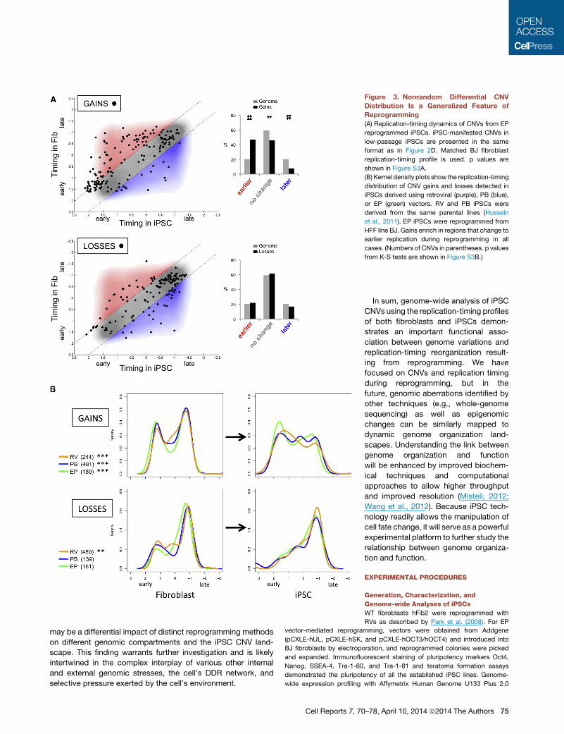
Figure 3. Nonrandom Differential CNV
Distribution Is a Generalized Feature of
Reprogramming
(A) Replication-timing dynamics of CNVs from EP
reprogrammed iPSCs. iPSC-manifested CNVs in
low-passage iPSCs are presented in the same
format as in Figure 2D. Matched BJ fibroblast
replication-timing profile is used. p values are
shown in Figure S3A.
(B) Kernel density plots show the replication-timing
distribution of CNV gains and losses detected in
iPSCs derived using retroviral (purple), PB (blue),
or EP (green) vectors. RV and PB iPSCs were
derived from the same parental lines (Hussein
et al., 2011). EP iPSCs were reprogrammed from
HFF line BJ. Gains enrich in regions that change to
earlier replication during reprogramming in all
cases. (Numbers of CNVs in parentheses. p values
from K-S tests are shown in Figure S3B.)
may be a differential impact of distinct reprogramming methods
on different genomic compartments and the iPSC CNV land-
scape. This finding warrants further investigation and is likely
intertwined in the complex interplay of various other internal
and external genomic stresses, the cell’s DDR network, and
selective pressure exerted by the cell’s environment.
Cell Reports 7, 70
In sum, genome-wide analysis of iPSC
CNVs using the replication-timing profiles
of both fibroblasts and iPSCs demon-
strates an important functional asso-
ciation between genome variations and
replication-timing reorganization result-
ing from reprogramming. We have
focused on CNVs and replication timing
during reprogramming, but in the
future, genomic aberrations identified by
other techniques (e.g., whole-genome
sequencing) as well as epigenomic
changes can be similarly mapped to
dynamic genome organization land-
scapes. Understanding the link between
genome organization and function
will be enhanced by improved biochem-
ical techniques and computational
approaches to allow higher throughput
and improved resolution (Misteli, 2012;
Wang et al., 2012). Because iPSC tech-
nology readily allows the manipulation of
cell fate change, it will serve as a powerful
experimental platform to further study the
relationship between genome organiza-
tion and function.
EXPERIMENTAL PROCEDURES
Generation, Characterization, and
Genome-wide Analyses of iPSCs
WT fibroblasts hFib2 were reprogrammed with
RVs as described by Park et al. (2008). For EP
vector-mediated reprogramming, vectors were obtained from Addgene
(pCXLE-hUL, pCXLE-hSK, and pCXLE-hOCT3/hOCT4) and introduced into
BJ fibroblasts by electroporation, and reprogrammed colonies were picked
and expanded. Immunofluorescent staining of pluripotency markers Oct4,
Nanog, SSEA-4, Tra-1-60, and Tra-1-81 and teratoma formation assays
demonstrated the pluripotency of all the established iPSC lines. Genome-
wide expression profiling with Affymetrix Human Genome U133 Plus 2.0
–78, April 10, 2014 ª2014 The Authors 75
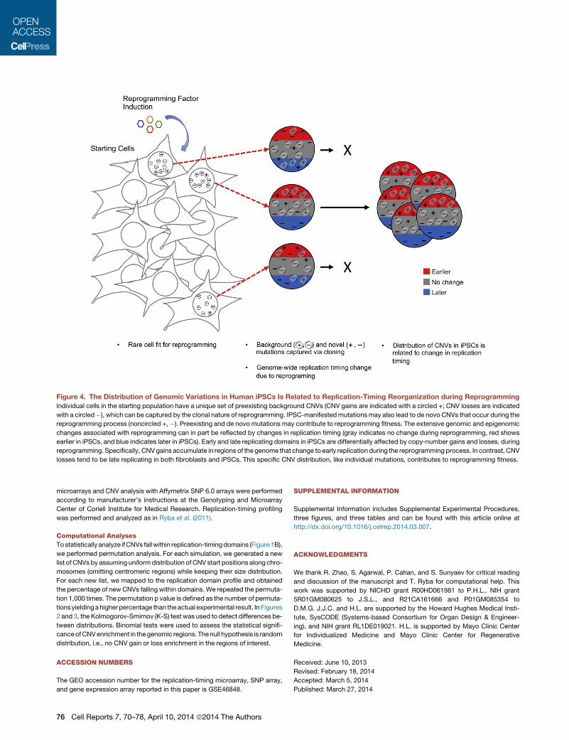
Figure 4. The Distribution of Genomic Variations in Human iPSCs Is Related to Replication-Timing Reorganization during Reprogramming
Individual cells in the starting population have a unique set of preexisting background CNVs (CNV gains are indicated with a circled +; CNV losses are indicated
with a circled�), which can be captured by the clonal nature of reprogramming. IPSC-manifestedmutationsmay also lead to de novo CNVs that occur during the
reprogramming process (noncircled +,�). Preexisting and de novo mutations may contribute to reprogramming fitness. The extensive genomic and epigenomic
changes associated with reprogramming can in part be reflected by changes in replication timing (gray indicates no change during reprogramming, red shows
earlier in iPSCs, and blue indicates later in iPSCs). Early and late replicating domains in iPSCs are differentially affected by copy-number gains and losses, during
reprogramming. Specifically, CNVgains accumulate in regions of the genome that change to early replication during the reprogramming process. In contrast, CNV
losses tend to be late replicating in both fibroblasts and iPSCs. This specific CNV distribution, like individual mutations, contributes to reprogramming fitness.
microarrays and CNV analysis with Affymetrix SNP 6.0 arrays were performed
according to manufacturer’s instructions at the Genotyping and Microarray
Center of Coriell Institute for Medical Research. Replication-timing profiling
was performed and analyzed as in Ryba et al. (2011).
Computational Analyses
Tostatistically analyze ifCNVs fallwithin replication-timingdomains (Figure1B),
we performed permutation analysis. For each simulation, we generated a new
list of CNVs by assuming uniformdistribution ofCNV start positions along chro-
mosomes (omitting centromeric regions) while keeping their size distribution.
For each new list, we mapped to the replication domain profile and obtained
the percentage of new CNVs falling within domains. We repeated the permuta-
tion 1,000 times. The permutation p value is defined as the number of permuta-
tions yieldingahigherpercentage than theactual experimental result. InFigures
2 and 3, the Kolmogorov-Smirnov (K-S) test was used to detect differences be-
tween distributions. Binomial tests were used to assess the statistical signifi-
canceofCNVenrichment in thegenomic regions. Thenull hypothesis is random
distribution, i.e., no CNV gain or loss enrichment in the regions of interest.
ACCESSION NUMBERS
The GEO accession number for the replication-timing microarray, SNP array,
and gene expression array reported in this paper is GSE46848.
76 Cell Reports 7, 70–78, April 10, 2014 ª2014 The Authors
SUPPLEMENTAL INFORMATION
Supplemental Information includes Supplemental Experimental Procedures,
three figures, and three tables and can be found with this article online at
http://dx.doi.org/10.1016/j.celrep.2014.03.007.
ACKNOWLEDGMENTS
We thank R. Zhao, S. Agarwal, P. Cahan, and S. Sunyaev for critical reading
and discussion of the manuscript and T. Ryba for computational help. This
work was supported by NICHD grant R00HD061981 to P.H.L., NIH grant
5R01GM080625 to J.S.L., and R21CA161666 and P01GM085354 to
D.M.G. J.J.C. and H.L. are supported by the Howard Hughes Medical Insti-
tute, SysCODE (Systems-based Consortium for Organ Design & Engineer-
ing), and NIH grant RL1DE019021. H.L. is supported by Mayo Clinic Center
for Individualized Medicine and Mayo Clinic Center for Regenerative
Medicine.
Received: June 10, 2013
Revised: February 18, 2014
Accepted: March 5, 2014
Published: March 27, 2014
REFERENCES
Abyzov, A., Mariani, J., Palejev, D., Zhang, Y., Haney, M.S., Tomasini, L.,
Ferrandino, A.F., Rosenberg Belmaker, L.A., Szekely, A., Wilson, M., et al.
(2012). Somatic copy number mosaicism in human skin revealed by induced
pluripotent stem cells. Nature 492, 438–442.
Amps, K., Andrews, P.W., Anyfantis, G., Armstrong, L., Avery, S., Baharvand,
H., Baker, J., Baker, D., Munoz, M.B., Beil, S., et al.; International Stem Cell
Initiative (2011). Screening ethnically diverse human embryonic stem cells
identifies a chromosome 20 minimal amplicon conferring growth advantage.
Nat. Biotechnol. 29, 1132–1144.
Apostolou, E., Ferrari, F., Walsh, R.M., Bar-Nur, O., Stadtfeld, M., Cheloufi, S.,
Stuart, H.T., Polo, J.M., Ohsumi, T.K., Borowsky, M.L., et al. (2013). Genome-
wide chromatin interactions of the Nanog locus in pluripotency, differentiation,
and reprogramming. Cell Stem Cell 12, 699–712.
Cavalli, G., and Misteli, T. (2013). Functional implications of genome topology.
Nat. Struct. Mol. Biol. 20, 290–299.
Chang, D.J., and Cimprich, K.A. (2009). DNA damage tolerance: when it’s OK
to make mistakes. Nat. Chem. Biol. 5, 82–90.
Cheng, L., Hansen, N.F., Zhao, L., Du, Y., Zou, C., Donovan, F.X., Chou, B.K.,
Zhou, G., Li, S., Dowey, S.N., et al.; NISC Comparative Sequencing Program
(2012). Low incidence of DNA sequence variation in human induced pluripo-
tent stem cells generated by nonintegrating plasmid expression. Cell Stem
Cell 10, 337–344.
De, S., and Michor, F. (2011). DNA replication timing and long-range DNA
interactions predict mutational landscapes of cancer genomes. Nat.
Biotechnol. 29, 1103–1108.
Dixon, J.R., Selvaraj, S., Yue, F., Kim, A., Li, Y., Shen, Y., Hu, M., Liu, J.S., and
Ren, B. (2012). Topological domains in mammalian genomes identified by
analysis of chromatin interactions. Nature 485, 376–380.
Fudenberg, G., Getz, G., Meyerson, M., and Mirny, L.A. (2011). High order
chromatin architecture shapes the landscape of chromosomal alterations in
cancer. Nat. Biotechnol. 29, 1109–1113.
Gilbert, D.M., Takebayashi, S.I., Ryba, T., Lu, J., Pope, B.D., Wilson, K.A., and
Hiratani, I. (2010). Space and time in the nucleus: developmental control of
replication timing and chromosome architecture. Cold Spring Harb. Symp.
Quant. Biol. 75, 143–153.
Goodarzi, A.A., Noon, A.T., Deckbar, D., Ziv, Y., Shiloh, Y., Lobrich, M., and
Jeggo, P.A. (2008). ATM signaling facilitates repair of DNA double-strand
breaks associated with heterochromatin. Mol. Cell 31, 167–177.
Gore, A., Li, Z., Fung, H.L., Young, J.E., Agarwal, S., Antosiewicz-Bourget, J.,
Canto, I., Giorgetti, A., Israel, M.A., Kiskinis, E., et al. (2011). Somatic coding
mutations in human induced pluripotent stem cells. Nature 471, 63–67.
Hansen, R.S., Thomas, S., Sandstrom, R., Canfield, T.K., Thurman, R.E.,
Weaver, M., Dorschner, M.O., Gartler, S.M., and Stamatoyannopoulos, J.A.
(2010). Sequencing newly replicated DNA reveals widespread plasticity in
human replication timing. Proc. Natl. Acad. Sci. USA 107, 139–144.
Helmrich, A., Ballarino, M., Nudler, E., and Tora, L. (2013). Transcription-repli-
cation encounters, consequences and genomic instability. Nat. Struct. Mol.
Biol. 20, 412–418.
Hiratani, I., Ryba, T., Itoh, M., Yokochi, T., Schwaiger, M., Chang, C.W., Lyou,
Y., Townes, T.M., Schubeler, D., and Gilbert, D.M. (2008). Global reorganiza-
tion of replication domains during embryonic stem cell differentiation. PLoS
Biol. 6, e245.
Hiratani, I., Ryba, T., Itoh, M., Rathjen, J., Kulik, M., Papp, B., Fussner, E.,
Bazett-Jones, D.P., Plath, K., Dalton, S., et al. (2010). Genome-wide dynamics
of replication timing revealed by in vitro models of mouse embryogenesis.
Genome Res. 20, 155–169.
Hu, M., Deng, K., Qin, Z., Dixon, J., Selvaraj, S., Fang, J., Ren, B., and Liu, J.S.
(2013). Bayesian inference of spatial organizations of chromosomes. PLoS
Comput. Biol. 9, e1002893.
Hussein, S.M., Batada, N.N., Vuoristo, S., Ching, R.W., Autio, R., Narva, E., Ng,
S., Sourour, M., Hamalainen, R., Olsson, C., et al. (2011). Copy number varia-
tion and selection during reprogramming to pluripotency. Nature 471, 58–62.
Koren, A., Polak, P., Nemesh, J., Michaelson, J.J., Sebat, J., Sunyaev, S.R.,
and McCarroll, S.A. (2012). Differential relationship of DNA replication timing
to different forms of human mutation and variation. Am. J. Hum. Genet. 91,
1033–1040.
Labib, K., and Hodgson, B. (2007). Replication fork barriers: pausing for a
break or stalling for time? EMBO Rep. 8, 346–353.
Laurent, L.C., Ulitsky, I., Slavin, I., Tran, H., Schork, A., Morey, R., Lynch, C.,
Harness, J.V., Lee, S., Barrero, M.J., et al. (2011). Dynamic changes in the
copy number of pluripotency and cell proliferation genes in human
ESCs and iPSCs during reprogramming and time in culture. Cell Stem Cell
8, 106–118.
Leonhardt, H., Rahn, H.P., Weinzierl, P., Sporbert, A., Cremer, T., Zink, D., and
Cardoso, M.C. (2000). Dynamics of DNA replication factories in living cells.
J. Cell Biol. 149, 271–280.
Liang, G., and Zhang, Y. (2013). Genetic and epigenetic variations in iPSCs:
potential causes and implications for application. Cell Stem Cell 13, 149–159.
Liu, L., De, S., and Michor, F. (2013). DNA replication timing and higher-order
nuclear organization determine single-nucleotide substitution patterns in
cancer genomes. Nat. Commun. 4, 1502.
MacDonald, J.R., Ziman, R., Yuen, R.K., Feuk, L., and Scherer, S.W. (2014).
The database of genomic variants: a curated collection of structural variation
in the human genome. Nucleic Acids Res. 42, D986–D992.
Martins-Taylor, K., Nisler, B.S., Taapken, S.M., Compton, T., Crandall, L.,
Montgomery, K.D., Lalande, M., and Xu, R.H. (2011). Recurrent copy number
variations in human induced pluripotent stem cells. Nat. Biotechnol. 29,
488–491.
Mayshar, Y., Ben-David, U., Lavon, N., Biancotti, J.C., Yakir, B., Clark, A.T.,
Plath, K., Lowry, W.E., and Benvenisty, N. (2010). Identification and classifica-
tion of chromosomal aberrations in human induced pluripotent stem cells. Cell
Stem Cell 7, 521–531.
Misteli, T. (2012). Parallel genome universes. Nat. Biotechnol. 30, 55–56.
Murga, M., Jaco, I., Fan, Y., Soria, R., Martinez-Pastor, B., Cuadrado, M.,
Yang, S.M., Blasco, M.A., Skoultchi, A.I., and Fernandez-Capetillo, O.
(2007). Global chromatin compaction limits the strength of the DNA damage
response. J. Cell Biol. 178, 1101–1108.
Orkin, S.H., and Hochedlinger, K. (2011). Chromatin connections to pluripo-
tency and cellular reprogramming. Cell 145, 835–850.
Park, I.H., Lerou, P.H., Zhao, R., Huo, H., and Daley, G.Q. (2008). Generation of
human-induced pluripotent stem cells. Nat. Protoc. 3, 1180–1186.
Phillips-Cremins, J.E., Sauria, M.E., Sanyal, A., Gerasimova, T.I., Lajoie, B.R.,
Bell, J.S., Ong, C.T., Hookway, T.A., Guo, C., Sun, Y., et al. (2013). Architec-
tural protein subclasses shape 3D organization of genomes during lineage
commitment. Cell 153, 1281–1295.
Quinlan, A.R., Boland, M.J., Leibowitz, M.L., Shumilina, S., Pehrson, S.M.,
Baldwin, K.K., and Hall, I.M. (2011). Genome sequencing of mouse induced
pluripotent stem cells reveals retroelement stability and infrequent DNA rear-
rangement during reprogramming. Cell Stem Cell 9, 366–373.
Rube, C.E., Lorat, Y., Schuler, N., Schanz, S., Wennemuth, G., and Rube, C.
(2011). DNA repair in the context of chromatin: new molecular insights by
the nanoscale detection of DNA repair complexes using transmission electron
microscopy. DNA Repair (Amst.) 10, 427–437.
Ryba, T., Hiratani, I., Lu, J., Itoh, M., Kulik, M., Zhang, J., Schulz, T.C., Robins,
A.J., Dalton, S., and Gilbert, D.M. (2010). Evolutionarily conserved replication
timing profiles predict long-range chromatin interactions and distinguish
closely related cell types. Genome Res. 20, 761–770.
Ryba, T., Battaglia, D., Pope, B.D., Hiratani, I., and Gilbert, D.M. (2011).
Genome-scale analysis of replication timing: from bench to bioinformatics.
Nat. Protoc. 6, 870–895.
Ryba, T., Battaglia, D., Chang, B.H., Shirley, J.W., Buckley, Q., Pope, B.D.,
Devidas, M., Druker, B.J., and Gilbert, D.M. (2012). Abnormal developmental
Cell Reports 7, 70–78, April 10, 2014 ª2014 The Authors 77
control of replication-timing domains in pediatric acute lymphoblastic leuke-
mia. Genome Res. 22, 1833–1844.
Schuster-Bockler, B., and Lehner, B. (2012). Chromatin organization is amajor
influence on regional mutation rates in human cancer cells. Nature 488,
504–507.
Schwaiger, M., Stadler, M.B., Bell, O., Kohler, H., Oakeley, E.J., and Schub-
eler, D. (2009). Chromatin state marks cell-type- and gender-specific replica-
tion of the Drosophila genome. Genes Dev. 23, 589–601.
Takahashi, K., Tanabe, K., Ohnuki, M., Narita, M., Ichisaka, T., Tomoda, K.,
and Yamanaka, S. (2007). Induction of pluripotent stem cells from adult human
fibroblasts by defined factors. Cell 131, 861–872.
Wang, P., Zhang, W., Yang, J., Qu, J., and Liu, G.H. (2012). Higher-order
genomic organization in pluripotent stem cells. Protein Cell 3, 483–486.
Watanabe, Y., Fujiyama, A., Ichiba, Y., Hattori, M., Yada, T., Sakaki, Y., and
Ikemura, T. (2002). Chromosome-wide assessment of replication timing for
78 Cell Reports 7, 70–78, April 10, 2014 ª2014 The Authors
human chromosomes 11q and 21q: disease-related genes in timing-switch
regions. Hum. Mol. Genet. 11, 13–21.
Watanabe, Y., Ikemura, T., and Sugimura, H. (2004). Amplicons on human
chromosome 11q are located in the early/late-switch regions of replication
timing. Genomics 84, 796–805.
Watanabe, A., Yamada, Y., and Yamanaka, S. (2013). Epigenetic regulation in
pluripotent stem cells: a key to breaking the epigenetic barrier. Philos. Trans.
R. Soc. Lond. B Biol. Sci. 368, 20120292.
Wei, Z., Gao, F., Kim, S., Yang, H., Lyu, J., An, W., Wang, K., and Lu,W. (2013).
Klf4 organizes long-range chromosomal interactions with the oct4 locus in
reprogramming and pluripotency. Cell Stem Cell 13, 36–47.
Zhang, H., Jiao,W., Sun, L., Fan, J., Chen,M.,Wang, H., Xu, X., Shen, A., Li, T.,
Niu, B., et al. (2013). Intrachromosomal looping is required for activation of
endogenous pluripotency genes during reprogramming. Cell Stem Cell 13,
30–35.


