Embed Size (px)
Citation preview

� ����� ��� � ��
Utilizing induced pluripotent stem cells (iPSCs) to understand the actions of
estrogens in human neurons
Carole Shum, Sara C. Macedo, Katherine Warre-Cornish, Graham Cocks,
Jack Price, Deepak P. Srivastava
PII: S0018-506X(15)30009-X
DOI: doi: 10.1016/j.yhbeh.2015.06.014
Reference: YHBEH 3909
To appear in: Hormones and Behavior
Please cite this article as: Shum, Carole, Macedo, Sara C., Warre-Cornish, Katherine,Cocks, Graham, Price, Jack, Srivastava, Deepak P., Utilizing induced pluripotent stemcells (iPSCs) to understand the actions of estrogens in human neurons, Hormones andBehavior (2015), doi: 10.1016/j.yhbeh.2015.06.014
This is a PDF file of an unedited manuscript that has been accepted for publication.As a service to our customers we are providing this early version of the manuscript.The manuscript will undergo copyediting, typesetting, and review of the resulting proofbefore it is published in its final form. Please note that during the production processerrors may be discovered which could affect the content, and all legal disclaimers thatapply to the journal pertain.
AC
CEPTED
MAN
USC
RIP
T
ACCEPTED MANUSCRIPT
1
Utilizing induced pluripotent stem cells (iPSCs) to understand the actions of
estrogens in human neurons.
Carole Shum1, Sara C. Macedo1,2, Katherine Warre-Cornish1, Graham Cocks1, Jack
Price1, Deepak P. Srivastava1*
1Department of Basic and Clinical Neuroscience, Cell and Behaviour Unit, The
James Black Centre, Institute of Psychiatry, Psychology and Neuroscience, King's
College London, London, SE5 8AF, UK; 2Faculty of Engineering, Universidade do
Porto, 4200-465 Porto, Portugal.
* = corresponding author: Deepak P. Srivastava, Department of Basic and Clinical
Neuroscience, The James Black Centre, King's College London, 125 Coldhabour
Lane, London, SE5 8AF, UK. Telephone (+44)2078485412; e-mail:
Key words: Stem Cells, 17β-estradiol, estrogen receptor, synapse, dendrite, dendritic
spines, psychiatric, schizophrenia, autism spectrum disorders, neurodevelopmental
disorders, neurodegenerative disease
AC
CEPTED
MAN
USC
RIP
T
ACCEPTED MANUSCRIPT
2
Abstract
Over recent years tremendous progress has been made towards
understanding the molecular and cellular mechanism by which estrogens exert
enhancing effects on cognition, and how they act as a neuroprotective or
neurotrophic agent in disease. Currently, much of this work has been carried out in
animal models with only a limited number of studies using native human tissue or
cells. Recent advances in stem cell technology now make it possible to reprogram
somatic cells from humans into induced pluripotent stem cells (iPSCs), which can
subsequently be differentiated in neurons of specific lineages. Importantly, the
reprogramming of cells allows for the generation of iPSCs that retains the genetic
"makeup" of the donor. Therefore, it is possible to generate iPSC-derived neurons
from patients diagnosed with specific diseases, that harbor the complex genetic
background associated with the disorder. Here, we review the iPSC technology and
how its currently being used to model neural development and neurological
diseases. Furthermore, we explore whether this cellular system could be used to
understand the role of estrogens in human neurons, and present preliminary data in
support of this. We further suggest that the use of iPSC technology offers a novel
system in which to not only further understand estrogens' effects in human cells, but
in which to investigate the mechanism by which estrogens are beneficial in disease.
Developing a greater understanding of these mechanisms in native human cells will
also aid in the development of safer and more effective estrogen-based therapeutics.
AC
CEPTED
MAN
USC
RIP
T
ACCEPTED MANUSCRIPT
3
Content:
Introduction
How do Estrogens influence cognition?
Estrogens and disease: therapeutic potential?
Generation and Differentiation of hiPSCs
iPSC reprogramming technology
Neuronal differentiation of hiPSCs
Using hiPSCs to investigate basic neurobiology
Using iPSCs to model and investigate disease
Limitations of iPSCs
Technical limitations
Reproducibility
Time
Modelling the effects of Estrogens in human stem cells and iPSCs
Estrogens and estrogen receptors in human stem cells
Characterising the effects of 17β-estradiol in hiPSCs
Conclusions
Acknowledgements
References
AC
CEPTED
MAN
USC
RIP
T
ACCEPTED MANUSCRIPT
4
Introduction
There are multiple lines of evidence that estrogens exert a powerful influence
over cognition (Brinton, 2009; Daniel, 2013; Frick, 2012; Galea et al., 2008; Luine,
2008, 2014). Studies using animal models have demonstrated that estrogens, in
particular 17β-estradiol, can influence hippocampal and cortical brain regions to
modulate cognitive function, including learning and memory (Frick, 2009; Galea et
al., 2008; Luine, 2008). This is in addition to the effects 17β-estradiol has on
reproductive and sexual behaviours, regulated by its actions in the hypothalamus
(Micevych et al., 2009; Roepke et al., 2011). At the cellular levels, the effects on
cognitive function are thought to be driven by 17β-estradiol's effects on synapse
structure and function (Brinton, 2009; Srivastava et al., 2013). In addition to these
neurotrophic effects, multiple studies have also indicated that 17β-estradiol has
potent neuroprotective actions (Arevalo et al., 2015), and has been suggested to be
a possible therapeutic avenue for the treatment of several neurodevelopmental,
psychiatric and neurodegenerative disorders (Gillies and McArthur, 2010; Hughes et
al., 2009; Srivastava and Penzes, 2011).
To date, much of our understanding of the molecular and cellular mechanisms
that underlie the effect of estrogen have come from animal based in vitro and in vivo
models. Conversely, our understanding of the mechanisms that underlie estrogens'
effects in human neurons is limited. Indeed, it has not been possible to investigate
the actions of estrogens at a molecular level in native human neurons, and thus to
validate whether or not the actions of estrogens as determined in animal models are
comparable to its actions in human neurons. This is in part due to the availability of,
and the ethical considerations when using human tissue. These issues result in the
lack of a suitable and reproducible cellular system that faithfully recapitulates a
AC
CEPTED
MAN
USC
RIP
T
ACCEPTED MANUSCRIPT
5
human neuronal cellular environment and that allows detailed cellular and molecular
studies to be carried out. It is also important to recognise that while animal studies
support a beneficial role for estrogens in a range of neurodevelopmental, psychiatric
and neurodegenerative disorders, how these data translates to humans is unclear.
This is particularly important, when considering that there has not been much
success in translating preclinical work into novel therapeutic agents to treat
debilitating neurological, neurodevelopmental or neurodegenerative disorders. This
lack of conversion is due to many factors, but are likely to include species
differences, differences in brain complexity and disease-specific phenotypes
(Dragunow, 2008). Another important factor to consider is the potential negative
effects of estrogen, or estrogen-based therapies such as increased risk of
cardiovascular problems and increased risk of developing cancers. An alternative
approach would be to mimic estrogenic-mediated positive effects by modulating
specific ERs (Hughes et al., 2009; Zhao et al., 2005) and/or regulating 17β-estradiol
intracellular molecular targets. Such strategies could exploit the beneficial effects of
estrogens without the harmful side effects. Therefore, in order to utilize estrogens or
estrogen-based therapeutic for the treatment of neurodevelopmental or
neurodegenerative disorders, a greater understanding of the effects these
compounds have on native human cells and in a disease context is critical (Gillies
and McArthur, 2010; Hughes et al., 2009; Srivastava and Penzes, 2011).
Recent advances in stem cell biology are now providing us the tools in which
to study basic and disease mechanisms in native human neurons (Brennand et al.,
2012; Cocks et al., 2014; Dolmetsch and Geschwind, 2011; Gaspard and
Vanderhaeghen, 2011). This has led to the ability to reprogram patient somatic cells
into human induced pluripotent stem cells (hiPSCs) and the subsequent
AC
CEPTED
MAN
USC
RIP
T
ACCEPTED MANUSCRIPT
6
differentiation into neurons of specific lineages (Dolmetsch and Geschwind, 2011;
Gaspard and Vanderhaeghen, 2011). Importantly, these cells encapsulate and
recapitulate the genetic landscape and cellular abnormalities associated with
complex disease (Durak and Tsai, 2014; Yu et al., 2013). Critically, this approach
provides a potentially limitless source of live human cells for understanding basic
neurobiology and disease pathophysiology, and for modelling the actions of potential
drug targets (Brennand et al., 2012; Cocks et al., 2014; Dolmetsch and Geschwind,
2011; Gaspard and Vanderhaeghen, 2011). In this review, we will review a) the
evidence that estrogens influence human cognition and maybe beneficial in the
treatment of neurodevelopment/psychiatric disorders; b) recent advances in our
ability to generate hiPSCs and their use in elucidating both basic and disease
relevant mechanisms; c) the current limitations and efforts to overcome them when
using iPSCs; and d) present some preliminary data demonstrating that neurons
differentiated from hiPSCs are responsive to 17β-estradiol treatment.
How do Estrogens influence cognition?
During early brain development 17β-estradiol has many roles, ranging from
the control of cell proliferation and apoptosis to synaptogenesis and neurogenesis
(McCarthy, 2008; Sakuma, 2009). In addition, 17β-estradiol is a critical factor in
determining sexual differentiation during development. It has an organizational role
which contributes to the establishment of sex differences by influencing the sexually
dimorphic formation of the neural circuitry that encodes reproductive and socio-
aggressive behaviours (Lee et al., 2014a; Ubuka and Tsutsui, 2014; Unger et al.,
2015; Yang and Shah, 2014). Accumulating evidence indicates that 17β-estradiol's
AC
CEPTED
MAN
USC
RIP
T
ACCEPTED MANUSCRIPT
7
ability to regulate synapse structure and function, and thus neural circuitry, underlies
its influence over cognitive function (Luine and Frankfurt, 2012; Sellers et al., 2014;
Srivastava et al., 2013). In the cortex and hippocampus, 17β-estradiol has been
shown to modulate dendritic spine and synapse formation and density (Luine and
Frankfurt, 2012; Srivastava et al., 2013), long-term potentiation (LTP) (Foy et al.,
1999; Kramar et al., 2009; Xiao et al., 2012) and long-term depression (LTD) (Mukai
et al., 2007). Indeed, regulation of these cellular parameters are thought to be key
events and cellular correlates of memory and learning (Fu and Zuo, 2011; Holtmaat
and Svoboda, 2009; Malenka and Bear, 2004; Morris, 2003).
The actions of 17β-estradiol are mediated by the classic estrogen receptors
(ERs) ERα, ERβ, as well as the G-protein coupled receptor, GPER1 (Brinton, 2009;
Sellers et al., 2014; Srivastava and Evans, 2013). These receptors mediate both
rapid, membrane-initiated signaling and longer-term/chronic actions via the
regulation of gene transcription (Brinton, 2009; McCarthy, 2008; Srivastava et al.,
2013). Both ERα and ERβ dimerize in response to 17β-estradiol binding, and
subsequently translocate to the nucleus, where they can bind and influence the
expression of certain genes (Greene et al., 1986). However, there is a growing
appreciation that 17β-estradiol can act via ERα, ERβ and GPER1 to rapidly regulate
non-classical signaling resulting in a modulation of cellular physiology (Spencer et
al., 2008; Srivastava et al., 2013; Woolley, 2007). Activation of these non-classical
pathways by 17β-estradiol can result in multiple cellular effects, including immediate
effects on cell physiology and even on protein synthesis or gene transcription
(Sellers et al., 2014). Importantly, signaling via specific ERs and the activation of
these pathways have also been shown to be required for 17β-estradiol-medaited
enhancements of cognitive function (Ervin et al., 2013; Frick, 2012; Gabor et al.,

AC
CEPTED
MAN
USC
RIP
T
ACCEPTED MANUSCRIPT
8
2015; Hawley et al., 2014; Luine and Frankfurt, 2012; Srivastava et al., 2013). It is
also important to note that the precise establishment of neural circuitry during
development, as well as the proper regulation and maintenance of synaptic
connectivity throughout the lifetime of an animal, is essential for normal
brain/cognitive function. Indeed disruptions in these process are thought to be a
major contributing factor to a number of neurodevelopmental and neurodegenerative
disorders (Penzes et al., 2011; Tau and Peterson, 2010; van Spronsen and
Hoogenraad, 2010). As such, the ability of 17β-estradiol to regulate synapse
structure and function may contribute to its beneficial effects in disease (Srivastava
and Penzes, 2011; Srivastava et al., 2013).
While the effects of estrogens on cognition have been well established in
animal models, the reported effects of estrogens on cognitive function in human
have been much more varied (Luine, 2014; Sherwin, 2012). Nevertheless, multiple
studies in human females have reported that administration of estrogens have a
positive effect on cognitive function, including memory (Duff and Hampson, 2000;
Hampson and Morley, 2013; Hogervorst and Bandelow, 2010; Sherwin, 2012; Smith
et al., 2006). In addition, several studies have suggested that 17β-estradiol levels
correlate with cognitive performance. For example, during the midluteal phase when
17β-estradiol levels are at their relative height, women have been shown to have a
transient increase in performance in typically female-favouring measures of cognition
such as verbal fluency. This is opposed to the menstrual phase in these same
women, during which 17β-estradiol decline correlates with a transient increase in
performance in typically male-favouring measures of cognition such as spatial ability
(Hampson, 1990). This relationship between estrogen concentration and cognition
has since been reiterated by several studies (Hampson and Morley, 2013;

AC
CEPTED
MAN
USC
RIP
T
ACCEPTED MANUSCRIPT
9
Hogervorst et al., 2004; Phillips and Sherwin, 1992b). In addition, the loss of
estrogens (and other steroids) following menopause has been suggested to
dramatically increases a woman’s risk of memory loss (Maki and Henderson,
2012; Ryan et al., 2012). Interestingly, it has also been shown that this decline can
be attenuated by administering exogenous estrogens relatively early in menopause
(Phillips and Sherwin, 1992a; Sherwin, 1988). However not all studies have reported
positive effects on cognition, with studies reporting no or even negative effects
(Daniel, 2013; Hogervorst and Bandelow, 2010; Luine, 2014; Sherwin, 2012). As
discussed by Luine (2014) in the primer for this special issue, the variation seen in
human studies could be due to difficulties in experimental design or potential
environmental cofounders. However, another possibility is that estrogens do not
affect human cognition in the same manner as that seen in animal models, due to
differences in the basic underlying molecular and cellular mechanisms.
Estrogens and disease: therapeutic potential?
There is also substantial evidence that estrogen exert neuroprotective effects
and may also have beneficial effects in animal models of disease (Arevalo et al.,
2015; Frick, 2012; Gillies and McArthur, 2010). Preclinical studies have provided
evidence that estrogen, or estrogen-based approaches are neuroprotective and
could be used in the treatment of neurodevelopmental and neurodegenerative
disorders such as schizophrenia (Gogos and van den Buuse, 2015; Labouesse et
al., 2015), depression (Bredemann and McMahon, 2014; Hajszan et al., 2010; Walf
et al., 2008), Parkinson's Disease (Bourque et al., 2012; Rodriguez-Perez et al.,
2013) and Alzheimer's disease (Logan et al., 2011; Zhang et al., 2012; Zhao et al.,
AC
CEPTED
MAN
USC
RIP
T
ACCEPTED MANUSCRIPT
10
2013). It has also been hypothesized that the beneficial effects of 17β-estradiol in
these disorders are mediated, in part, through the modulation of neural circuitry
(Frick, 2012; Gillies and McArthur, 2010; Hughes et al., 2009; Srivastava and
Penzes, 2011). For example, the antidepressive effect of 17β-estradiol in a learned
helplessness model of depression occurs concurrently with an increase in
spinogenesis and LTP in CA1 neurons (Bredemann and McMahon, 2014; Hajszan et
al., 2010). Furthermore, selective activation of ERβ has anti-depressive-like effects in
a number of cognitive tests (Walf et al., 2008); this is in addition to ERβ-mediated
modulation of synapse structure and function (Kramar et al., 2009; Srivastava et al.,
2010). Interestingly, Logan et al (2011), demonstrated that 17β-estradiol was
sufficient to rescue deficits in dendritic spine density induced by soluble beta amyloid
(Aβ) oligomers in neuronal cultures. Moreover, the authors reported that
administration of 17β-estradiol prevented Aβ oligomer-induced impairment of
inhibitory avoidance tasks (Logan et al., 2011), indicating that 17β-estradiol's
regulation of synapse structure contributes to its beneficial effects on Aβ-induced
cognitive deficits.
Several clinical studies and meta-analyses have been carried out
investigating the potential beneficial roles of 17β-estradiol or selective estrogen
receptor modulators (SERMs) in a range of disorders including schizophrenia
(Kindler et al., 2015; Kulkarni et al., 2014; Torrey and Davis, 2012; Weickert et al.,
2015), major depression (Kornstein et al., 2010; Young et al., 2007), and even
Alzheimer's disease (Maki, 2013; Wharton et al., 2011). For example, in a recent
large-scale, randomized-control study, Kulkarni et al. (2014) reported that
administration of 17β-estradiol to treatment-resistant female schizophrenic patients,
resulted in a clinically relevant amelioration of schizophrenic symptoms. In this study,
AC
CEPTED
MAN
USC
RIP
T
ACCEPTED MANUSCRIPT
11
200 µg of 17β-estradiol was delivered via a transdermal patch to females patients for
8 weeks; patients receiving this treatment showed a greater decrease in the positive
and negative syndrome scale (PANSS) than 100 µg 17β-estradiol or placebo.
Several recent studies have also investigated the therapeutic potential of the SERM
raloxifene in schizophrenia. In a 12 week double-blind, placebo-controlled study,
orally administered raloxifene improved probabilistic association learning and
significantly increased fMRI blood oxygen level-dependent (BOLD) activity in the
hippocampus and parahippocampal gyrus relative to placebo, in male and female
schizophrenic patients (Kindler et al., 2015). In a subsequent study by the same
group, raloxifene, administered orally, improved memory and attention/processing
speeds in male and female schizophrenic patients, compared to placebo (Weickert
et al., 2015).
While these studies do support a positive role for estrogens, or estrogen-
based therapies (Craig and Murphy, 2010; Cyr et al., 2000; Frick, 2009; Gillies and
McArthur, 2010; Kulkarni et al., 2008; Lee et al., 2014b; Maki and Henderson, 2012;
Sherwin, 2009; Torrey and Davis, 2012), they do not support estrogens as a long
term treatment. Previous studies from the Women’s’ Health Initiative (WHI)
investigated the potential of Hormone Replacement Therapy (HRT) as a therapeutic
avenue for the treatment of aging and dementia. The findings of the WHI studies
reported a decrease in cognitive function and an increased risk of dementia and
stroke in women over 65 years of age who received conjugated equine estrogens
(CEE) plus medroxyprogesterone acetate (MPA) compared to those who received
placebo (Espeland et al., 2004; Rossouw et al., 2002; Shumaker et al., 2004).
However, these studies were carried out in females that were postmenopasual for
~15-20 years, and the HRT studies used horse derived estrogens, made up mostly
AC
CEPTED
MAN
USC
RIP
T
ACCEPTED MANUSCRIPT
12
of estrone, an estrogen that has varied and often opposing effects to 17β-estradiol,
and progesterone on cognition in animal models (Barha et al., 2010; Barha and
Galea, 2013; Engler-Chiurazzi et al., 2012; McClure et al., 2013). Moreover, a direct
comparison of CEE and 17β-estradiol on verbal memory performance in
postmenopausal women indicates that 17β-estradiol has a more potent effect on
memory than CEE (Wroolie et al., 2011). It is also thought that the physiological
status of women is critical in determining the effectiveness of estrogen on cognition
and it has been hypothesized that postmenopausal women lose their
responsiveness to estrogens about 5 years after menopause (Asthana et al., 2009;
Craig et al., 2005; Hogervorst and Bandelow, 2010; Maki, 2013; Maki and
Henderson, 2012; Singh et al., 2013). Indeed, basic studies have also hypothesized
that there is a critical period, or "window of opportunity" following menopause or
surgical removal of ovaries, when the brain is still responsive to estrogens and the
hormone can exert positive effects (Singh et al., 2013; Vedder et al., 2014).
Conversely, treatment with estrogens after this time may exert negative, or adverse,
effects on cognition (Asthana et al., 2009; Maki, 2013; Sherwin, 2009).
As discussed above, our understanding of the potential beneficial effects of
estrogens, or in developing novel estrogen-based therapies, is limited due to the
inability to perform in depth molecular studies of estrogens in human neurons that
faithfully recapitulate the genetic, and therefore the cellular environment of specific
diseases. Understanding such mechanisms would not only enhance our
understandings of estrogens function in humans, but would enable us to develop
safer and more effective estrogen-based therapies. Other alternatives, such as
human post-mortem brain tissue and genetically-modified model organisms have
provided insights into how estrogens are beneficial in a number of disorders, but

AC
CEPTED
MAN
USC
RIP
T
ACCEPTED MANUSCRIPT
13
these approaches have limitations. Post-mortem tissue is not living tissue and does
not allow researchers to investigate the progression of the disorder. A limitation of
animal models is that they often do not faithfully reflect human pathophysiology.
Moreover, many disorders of the brain have a complex genetic underpinning, and
thus it is not currently possible to fully recapitulate the complex genetic landscape in
traditional animal models. Therefore, the ability to determine the potential
effectiveness of therapeutic agents, such as estrogens, or to identify and test novel
estrogen-based compounds/therapies is currently limited. If we are to fully recognise
the potential of estrogen-based therapies, whether it be for females or for males, it is
critical to investigate them in a cellular model which encapsulates the complex
nature of these diseases, and within in a native human cellular environment.
Generation and Differentiation of hiPSCs
iPSC reprogramming technology
The method of reprogramming adult somatic cells to pluripotent stem cells
was first described in 2006 by Takahashi and Yamanaka. They reported that dermal
fibroblasts from adult mice could be reprogrammed into a pluripotent state by
retroviral transduction of four transcription factors: OCT4, KLF4, c-MYC and SOX2
(Takahashi and Yamanaka, 2006). The reprogrammed cells were termed induced
pluripotent stem cells (iPSCs), and are similar to embryonic stem cells (ESCs) in
their morphology, proliferation, surface antigens, gene expression and capacity to
differentiate into the cell types of the three primordial germ layers. A year later,
Takahashi et al. (Takahashi et al., 2007b) applied the same technology to human
adult dermal fibroblasts to generate the first human iPSCs (hiPSCs). Yamanaka’s
AC
CEPTED
MAN
USC
RIP
T
ACCEPTED MANUSCRIPT
14
seminal studies provided an avenue to generate patient and disease-specific iPSCs
and led to his being awarded the Nobel Prize in Medicine and Physiology in 2012.
This discovery, combined with protocols for the directed differentiation of neurons,
enabled access to these cell types without the ethical issues involved with the use of
human embryonic stem cells.
Since this discovery, many others have shown that it is possible to generate
hiPSCs from other adult somatic cell types, including peripheral blood (Loh et al.,
2009), hair follicles (Aasen et al., 2008), amniotic cells (Li et al., 2009; Zhao et al.,
2010), cells present in urine (Zhou et al., 2012) and various other cell types (Aoki et
al., 2010; Bar-Nur et al., 2011; Eminli et al., 2009; Giorgetti et al., 2009; Haase et al.,
2009; Kim et al., 2009b; Liu et al., 2010; Nakagawa et al., 2008; Sugii et al., 2011;
Yu et al., 2007). Although a well-established cell type in many fields of research, due
to their ease of handling and the cost-effectiveness, there are disadvantages to the
use of fibroblasts as a starting cell type for producing hiPSCs. Patient dermal
fibroblasts are obtained from painful skin punch biopsies that present risk of
infections and allergic reactions to anesthetics, and must be performed by trained
professionals. In addition, fibroblasts are reprogrammed with a longer time frame
and less efficiency than other somatic cell types (Aasen et al., 2008).Thus, these
studies have advanced hiPSC research by enabling non-invasive methods of
acquiring starting material and reducing the time and costs, while increasing the
efficiency of reprogramming.
Conventional hiPSC reprogramming has made use of integrating viral vectors,
such as retroviral and lentiviral vectors, for the delivery of the four pluripotency
factors (OCT4, KLF4, c-MYC and SOX2) into the starting cell types (Takahashi et al.,
2007b; Yu et al., 2007). Integrating viral vectors were critical in the development of
AC
CEPTED
MAN
USC
RIP
T
ACCEPTED MANUSCRIPT
15
iPSC technology due to its ability to enable long-term transgene expression, but
result in the integration of viral DNA into the host genome. This type of transgene
delivery has disadvantages, such as the risk of insertional mutagenesis, residual
expression of integrated transgenes, and uncontrolled activation or inactivation of the
integrated transgenes, which is critical in the case of iPSC reprogramming, since all
four of the pluripotency factors are oncogenic (Hu, 2014). Tremendous effort has
since led to the development of alternative protocols to avoid the integration of the
pluripotency factors into the host genome. It is now possible to generate hiPSCs with
the use of episomal vectors (Sendai vectors; (Fusaki et al., 2009)), non-integrating
viral vectors (Sarkis et al., 2008), small molecules (Ichida, 2009; Lyssiotis et al.,
2009; Shi et al., 2008), protein transduction (Kim et al., 2009a) and microRNAs (Pfaff
et al., 2011; Subramanyam et al., 2011) (Figure 1). These methods have addressed
many of the issues associated with integrating viral vectors and advanced hiPSC
research by producing hiPSC lines with increased efficiency, low toxicity and free of
transgene footprints, making them feasible for clinical studies such as cell
replacement therapies. The efficiency of hiPSC generation has greatly improved
since Takahashi and Yamanaka’s initial breakthrough, and the technology continues
to develop.
Neuronal differentiation of hiPSCs
A key component of using hiPSCs to elucidate basic and disease relevant
mechanisms is the ability to direct the differentiation of stem cells to specific
neuronal cell types. The pathways involved in neural development were first
elucidated from studies of animal embryology. The first step in the development of
AC
CEPTED
MAN
USC
RIP
T
ACCEPTED MANUSCRIPT
16
the neural tube, neural induction, is believed to be the default pathway, involving the
bone morphogenetic proteins (BMPs), Wnt and fibroblast growth factor (FGF)
signalling pathways (Aubert et al., 2002; Chambers et al., 2009; LaVaute et al.,
2009). Neural induction leads to a default and primitive anterior identity, which is
subsequently patterned by extrinsic morphogens such as Wnts, FGFs, retinoic acid
and Sonic Hedgehog (Shh), to give rise forebrain, midbrain, hindbrain or spinal cord
domains.
Neuronal differentiation of hiPSCs follow the same pathways as in vivo to give
rise to mature neuronal populations (Shi et al., 2012) (Figure 2). The most efficient
neural induction of hiPSCs is achieved by dual inhibition of the SMAD signalling
pathway(Chambers et al., 2009). This involves the synergistic inhibition of the BMP
and TGFβ pathways to achieve rapid and uniform neural conversion of pluripotent
cells. Using small molecule antagonists or endogenous inhibitors, it is possible to
induce neural conversion to give rise to a population of neural progenitors (Boissart
et al., 2013; Chambers et al., 2009; Shi et al., 2012). Neural progenitors may then be
patterned into neuronal cell types with regional identities using specific combinations
of morphogens, small molecules, growth factors and transcription factors. Depending
on the combination and timing of these signals, a variety of neuronal cell types can
be obtained, including telecephalic precursors (Watanabe et al., 2005), midbrain
dopaminergic neurons (Kawasaki et al., 2000), basal forebrain cholinergic neurons
(Bissonnette et al., 2011), spinal motor neurons (Hu and Zhang, 2009), as well as
glial cells, such as astrocytes (Serio et al., 2013) and oligodendrocytes (Hu et al.,
2009).
It should be noted that protocols for the directed differentiation of hiPSCs into
neuronal subtypes are imperfect, often yielding a heterogeneous population of cell

AC
CEPTED
MAN
USC
RIP
T
ACCEPTED MANUSCRIPT
17
types. For instance, derivation of basal forebrain cholinergic neurons from human
pluripotent stem cells yields both cells of the basal forebrain cholinergic neuronal
lineage as well as interneurons and other cell types (Bissonnette et al., 2011). In
addition, each protocol differs in the efficiency of directed differentiation, with some
protocols generating highly pure neuronal subtypes (Du et al., 2015), and others
achieving lower yields (Kiskinis et al., 2014). Furthermore, many protocols are
focused on the directed differentiation of one particular neuronal subtype, neglecting
the role of neighbouring cell types, such as glia, that are present in the in vivo
environment. In fact, astroglia has been shown to have a critical role in the functional
maturation of hiPSC-derived neurons (Tang et al., 2013). Despite these caveats, the
last decade of research has seen a vast improvement in neuronal differentiation of
human pluripotent stem cells. These efforts have identified novel small molecules
that enhance neuronal differentiation and reduce costs, reducing the amount of time
required to yield highly enriched populations of specific neuronal subtypes (Kim et
al., 2010). In addition, they provide important guidelines and benchmarks for future
hiPSC studies of basic neurobiology and disease modelling.
Using hiPSCs to investigate basic neurobiology
Several studies have recently demonstrated the utility of hiPSCs for functional
studies of human neural development. Shi et al. (2012) showed that the distinct
steps of human cortical development can be recapitulated in an hiPSC system, from
cortical induction to the formation of functional excitatory synapses (Figure 3). This
system closely mirrored the in vivo cortex, in terms of the temporal order of
development, specificity of circuit formation, and laminar organisation of projection
AC
CEPTED
MAN
USC
RIP
T
ACCEPTED MANUSCRIPT
18
neurons (Shi et al., 2012). Importantly, this system enabled the generation of all
classes of cortical projection neurons for the first time, including the formation of
superficial layer neurons, which are absent in mouse cortical development. Similarly,
Espuny-Camacho et al. (2013) reported that human corticogenesis can be
recapitulated in hiPSCs in the absence of extrinsic morphogens, enabling the
generation of diverse pyramidal neurons with distinct patterns of axonal projections
and dendritic outgrowths that integrated functionally when grafted in neonatal mouse
brain (Espuny-Camacho et al., 2013). These studies show that hESCs and hiPSCs
both demonstrate a similar capacity to differentiate into all classes of cortical
projection neurons.
GABA interneurons are another major neuronal subtype of the human cortex.
Unlike cortical projection neurons, GABA interneurons mainly originate from the
medial ganglionic eminence and migrate to the cortex during development (Sussel et
al., 1999). Liu et al. (2013) used hiPSCs to examine the development of GABA
interneurons. This study showed that the differentiation of GABA interneurons from
hiPSCs follows the timeline of human GABA interneuron development, from
patterning of primitive neurepithelia to the generation of functional neurons with
inhibitory and excitatory inputs (Liu et al., 2013a). Furthermore, multiple GABA
interneuron subtypes were observed in this chemically defined system, with specific
GABA populations appearing according to their developmental scheudule (Liu et al.,
2013a). In a related study, this group reported the use of this method to successfully
direct the differentiation of hESCs to medial ganglionic eminence progenitors and
subsequently, GABA interneurons (Liu et al., 2013b). Interestingly, the
transplantation of hESC-derived medial ganglionic eminence progenitors to the
hippocampi of mice led to behavioural changes in learning and memory (Liu et al.,
AC
CEPTED
MAN
USC
RIP
T
ACCEPTED MANUSCRIPT
19
2013b). These findings show that in addition to the study of basic neurobiology,
hiPSCs and hESCs can be also used to investigate behaviours associated with
specific neuronal subtypes.
Similarly, others have investigated the development of other neuronal cell
types, including cerebellar Purkinje cells (Wang et al., 2015), retinal cells (Meyer et
al., 2009) and motor neurons (Toma et al., 2015). More recently, hiPSC-derived
neurons have been used to study specific aspects of cell biology, such as
mitochondrial biogenesis (O'Brien et al., 2015) and neuromuscular junction
development (Yoshida et al., 2015). These studies show that hiPSC-derived
neuronal cell types possess many of cellular and physiological characteristics as
hESC-derived and endogenous neuronal cell types. Furthermore, hiPSCs have been
used to generate a three-dimensional model of human neural tissue. Lancaster et al.
(2013) reported the development of brain tissue from hiPSCs, termed cerebral
organoids. Cerebral organoids consisted of discrete regions similar to the cerebral
cortex, ventricles and retina tissue, and recapitulated some key aspects of human
cortical development mentioned above (Lancaster et al., 2013). Similarly, Meyer et
al. (2011) showed that hiPSCs can be differentiated into 3D retinal structures
consisting of multiple retinal cell types, in a time frame similar to normal human
retinal development (Meyer et al., 2011). These studies provide models to engineer
human cortical circuits and investigate cortical function that had not previously been
possible with animal models due to species differences.
Using iPSCs to model and investigate disease
AC
CEPTED
MAN
USC
RIP
T
ACCEPTED MANUSCRIPT
20
One of the biggest challenges in the study of diseases of the nervous system
has been the inaccessibility of live human neural tissue from patients. hiPSC
technology combined with methods of directed differentiation provides a general
solution to this impediment. An obvious advantage of patient-specific hiPSCs is that
they carry the same genetic background as the patient, capturing the mutated genes,
as well as the known and unknown genetic modifiers that play important roles in
pathogenesis. In addition, patient-specific hiPSCs represent a more physiologically
relevant model system, negating the need to overexpress proteins at
superphysiological levels in current cellular and animal models. Thus far, a number
of groups have published studies of hiPSC neurons from patients with various
neurological conditions, including neuropsychiatric disorders schizophrenia (SCZ)
(Brennand et al., 2011) and autism spectrum disorders (ASD) (Marchetto et al.,
2010; Shcheglovitov et al., 2013), as well as neurodegenerative disorders
Alzheimer’s disease (AD) (Israel et al., 2012), Parkinson’s disease (PD) (Devine et
al., 2011) and amyotrophic lateral sclerosis (ALS) (Dimos et al., 2008).
Several groups have reported the generation of hiPSCs from patients with
Rett syndrome, an ASD caused by mutations of the MECP2 gene. Patient-specific
hiPSCs maintained the parental mutation and were pluripotent and able to
differentiate into the three germ layers (Ananiev et al., 2011; Cheung et al., 2011;
Marchetto et al., 2010). All three studies showed that neurons from Rett syndrome
hiPSC-derived neurons recapitulated a hallmark feature of ASD, reduction in soma
size. In addition, Marchetto et al. (2010) reported that Rett syndrome hiPSC-derived
neurons had fewer synapses, reduced spine density and alterations in calcium
signalling and defects in electrophysiology.
AC
CEPTED
MAN
USC
RIP
T
ACCEPTED MANUSCRIPT
21
Altered dendritic arborisation and synaptic density are characteristics that
appear to be shared between ASD and SCZ. The generation of hiPSCs from
patients with SCZ have also been reported by independent groups. Chiang et al.
(Chiang et al., 2011) were the first to generate patient-specific hiPSCs from a patient
with mutations in the DISC1 gene, albeit without reporting any SCZ-relevant
phenotypes. Soon after, Brennand et al. (2011) reported the generation of hiPSCs
from patients with childhood-onset SCZ or from families affected with psychiatric
disease, likely reflecting a genetic component to disease. Patient-specific hiPSCs
were indistinguishable from control hiPSCs in pluripotency and in their ability to
differentiate into functional neurons. However, patient-specific hiPSC-derived
neurons displayed decreased neuronal connectivity, reduced neurite outgrowth and
decreased PSD95 protein levels, despite normal spontaneous neuronal activity
(Brennand et al., 2011). Interestingly, a more recent study on hiPSCs generated
from patients with a family history of SCZ reported a discrepancy in these
observations. Robicsek et al. (2011) reported that patient-specific hiPSC-derived
neural progenitor cells (NPCs) exhibited abnormal morphology and a delay in
differentiation into dopaminergic neurons and an inability to differentiate into
glutamatergic neurons (Robicsek et al., 2013). hiPSCs have also been generated
from 22q13 deletion syndrome patients (Phelan-McDermid syndrome), who display
intellectual disability and an increase risk of ASDs. Amongst the gene deleted in this
region, SHANK3, which encodes a post-synaptic scaffold protein, has also been
associated with ASD, intellectual disability and SCZ. Neurons derived from these
patient-specific hiPSCs demonstrated reduced levels of Shank3 expression and
displayed deficits in excitatory transmission and synaptic number (Shcheglovitov et
al., 2013). Remarkably, the authors further demonstrated that overexpression of
AC
CEPTED
MAN
USC
RIP
T
ACCEPTED MANUSCRIPT
22
Shank3 or treatment with IGF1 was sufficient to restore synaptic deficits in these
patient-specific hiPSC-derived neurons (Shcheglovitov et al., 2013). Collectively,
these studies have provided proof of concept by demonstrating that cells derived
from patient-specific hiPSC are a cellular system in which to confirm and elucidate
the impact of genetic variations associated with complex disorder on cellular
phenotypes. However, more recently, several studies have now used patient-specific
hiPSCs as a tool to investigate novel mechanisms and cellular phenotypes that may
contribute to the pathophysiology of neurodevelopmental disorders. For example,
Wen et al. (2014) generated hiPSCs from 4 family members 2 of which had a
frameshift mutation in the DISC1 gene and were diagnosed with SCZ or major
depression, and 2 unaffected family members with the mutation. Neurons generated
from hiPSC of the affected family members displayed reduced levels of the DISC1
protein, abnormal synaptic transmission and reduced synapse number, consistent
with previous reports using animal models (Hayashi-Takagi et al., 2010; Wen et al.,
2014). However, the authors further discovered that reduction in DISC1 expression
also resulted in the dysregulation of multiple genes related to synaptic and
psychiatric disorder, hence uncovering a novel transcriptional regulatory action for
the DISC1 protein (Wen et al., 2014). Around the same time, Yoon et al. (2014)
generated hiPSCs from SCZ patients with a 15q11.2 microdeletion; one of the genes
expressed in this region is CYFIP1, which has been associated with SCZ and ASDs.
Using NPCs derived from these patient-specific hiPSCs, the authors identified that
haploinsufficiency of CFYIP1 resulted in a disruption in adherent junctions and apical
polarity, an effect that they further demonstrated to disrupt radial glial cells
localization in a mouse model (Yoon et al., 2014). This study further demonstrates
AC
CEPTED
MAN
USC
RIP
T
ACCEPTED MANUSCRIPT
23
how hiPSCs can be an entry point to discovering novel insights into disease
pathology (Durak and Tsai, 2014).
In addition to these studies, hiPSCs have also been used to successfully
model aspects of late-onset neurodegenerative disorders. Shortly after the
generation of the first hiPSCs, Dimos et al. (2008) was the first to report the
generation of patient-specific hiPSCs from an 82-year old woman with ALS, and their
differentiation into motor neurons. Although this study did not report any disease
phenotypes, a number of studies have since shown that patient-derived hiPSCs do
recapitulate disease-relevant phenotypes upon neuronal differentiation. After the
initial proof-of-principle study demonstrating the feasibility of generating patient-
specific hiPSCs and their differentiation into motor neurons, several groups reported
the generation of hiPSCs from ALS patients with known and well characterised
pathogenic mutations. These studies have shown that neurons and glia derived from
patient-specific hiPSCs recapitulate key pathological phenotypes of ALS, including
mutant cellular and biochemical features, and cellular vulnerability (Almeida et al.,
2013; Bilican et al., 2012; Egawa et al., 2012; Sareen et al., 2013). A comprehensive
study of hiPSCs generated from patients with familial AD and sporadic AD reported
key features of AD, such as higher levels of amyloid-beta and phosphorylated tau, in
both familial and sporadic AD hiPSC-derived neurons (Israel et al., 2012). Similarly,
midbrain dopaminergic neurons from hiPSCs generated from a familial PD patient
with a mutation in the gene encoding alpha-synuclein exhibit increased levels of
alpha-synuclein protein, recapitulating the cause of PD (Devine et al., 2011). Other
PD-relevant phenotypes have also been observed in hiPSC-derived neurons from
PD patients with mutations in the PINK1 and LRRK2 genes, including increased
AC
CEPTED
MAN
USC
RIP
T
ACCEPTED MANUSCRIPT
24
production of mitochondrial reactive oxygen species and abnormal mitochondrial
transport (Cooper et al., 2012).
Several recent studies have also combined genome editing tools with hiPSCs
to both model the cellular phenotypes associated with ALS, the most common adult-
onset motor neuron disease, and to investigate the genetic contribution to disease
pathogenesis. Two independent groups reported the generation of hiPSCs from ALS
patients with mutations in the SOD1 gene (Chen et al., 2014; Kiskinis et al., 2014).
Kiskinis et al. (2014) reported that patient-specific hiPSC-derived motor neurons
exhibited increased cell death and reduced soma size and shorter neurites, whereas
Sun et al. (2014) observed aggregation of mutant SOD1 protein and neurofilament
inclusions in patient-specific hiPSC-derived motor neurons (Chen et al., 2014;
Kiskinis et al., 2014). Kiskinis et al. (2014) and Sun et al. (2014) used zinc-finger
nuclease and TALENs, respectively, to target the correction of the SOD1 mutation in
the patient-specific hiPSCs. Both studies showed that genetic correction of the
SOD1 mutation rescued the ALS-associated phenotypes.
These studies provide support for the use of hiPSCs for modelling the
molecular pathogenesis of diseases of the nervous system. In addition to disease
modelling, patient-specific iPSCs may be used to investigate disease mechanisms
that may not be exposed in non-neuronal cell types. As iPSC technology and
protocols for directed differentiation continues to develop, hiPSC models have the
potential to greatly reduce the time and costs associated with clinical trials of drug
discovery.
Limitations of iPSCs
AC
CEPTED
MAN
USC
RIP
T
ACCEPTED MANUSCRIPT
25
Technical limitations
Despite the tremendous potential of hiPSCs, a number of technical limitations
currently restrict hiPSC-based studies. These limitations relate primarily to several
forms of variability arising from differences between hiPSC-neurons derived from a
single patient, from independent clones derived from a single patient and from
differences between hiPSC lines derived from different patients. In order to address
these constraints, large experiments consisting of multiple neuronal differentiations
from multiple independent hiPSC lines from multiple patients would have to be
performed. Due to the time constraints and costs involved with these large
experiments, most hiPSC studies have used a minimal number of cell lines and
neuronal differentiations for proof-of-principle experiments.
Most established methods of directed differentiation result in heterogeneous
populations of differentiated cells. These impure populations typically consist of
different subtypes of neurons, as well as non-neuronal cell types. This heterogeneity
leads to differences between individual hiPSC neurons derived from a single patient,
or neuron-to-neuron variability. Some studies have addressed this issue by using
fluorescent-activated cell sorting (FACS) to purify specific neuronal subtypes. To do
this, hiPSC-derived neuronal cultures may be transfected or transduced with a
plasmid or a virus encoding a fluorescent protein under the control of a subtype-
specific promoter, such that only the specific neuronal subtypes will express the
fluorescent protein, which can subsequently be sorted from the heterogeneous
population. This has been used to identify small molecules that improve survival of
hiPSC motor neurons from ALS patients (Yang et al., 2013). Although FACS
generates highly pure cultures and minimises neuron-to-neuron variability, this
method carries a risk of contamination and also results in a lower yield of cells.
AC
CEPTED
MAN
USC
RIP
T
ACCEPTED MANUSCRIPT
26
In addition to this, hiPSC clones derived from a single patient are known to
exhibit differences genetically and in differentiation propensities (Hu et al., 2010).
Genetic variability may arise from the reprogramming process, due to differences in
the number of viral integrations or spontaneous mutations (Gore et al., 2011). While
the former issue may be addressed by non-integrating viral vectors or the use of
small molecules, spontaneous mutations occur naturally and are an inherent
variability in hiPSC-based studies. Indeed, a major limitation of hiPSC research
stems from the accumulation of mutations in rapidly cycling cultured cells, such as
hiPSCs and human ESCs (Baker et al., 2007; Mayshar et al., 2010). Duplication of
chromosomal regions have been reported to occur in a proportion of the hiPSC and
human ESC lines analysed (Taapken et al., 2011). Biased gene expression
associated with such chromosomal aberrations has also been observed in some of
hiPSC lines analysed, as a result of culture adaptation, although biased gene
expression was also found at an early passage of one hiPSC line (Mayshar et al.,
2010). To work around these limitations, studies may be performed on early passage
hiPSC clones that have passed rigorous quality controls, including morphological,
gene expression and karyotype analyses, tests for contaminants, and with RNA and
DNA methylation profiling.
Another form of variability derives from differences between hiPSC lines
derived from different patients, or inter-patient variability. For instance, hiPSC lines
have been shown to differentiate into neuronal lineages with variability in
differentiation efficiency and in electrophysiological properties (Hu et al., 2010). Inter-
patient variability has also been associated with donor identity and sex (Boulting et
al., 2011). Some studies have taken advantage of the recent development of
genome editing technologies to circumvent such variability by generating isogenic
AC
CEPTED
MAN
USC
RIP
T
ACCEPTED MANUSCRIPT
27
hiPSC lines. Much of these differences continue to reduce with the development of
pluripotent stem cell technology. The use of commercially available cell culture
media, substrates and reprogramming kits has increased the quality and consistency
of hiPSC cultures. The development of protocols for the directed differentiation of
hiPSCs has led to better defined culture conditions and more reliable neuronal
cultures.
Reproducibility
In addition to the intra- and inter-patient variability discussed above, it is
important to consider the reproducibility of hiPSC-based studies. Unlike studies
involving the use of well-established cell lines, the experimental design in iPSC-
based studies is heavily debated. For instance, it is not clear how many patients or
iPSC lines should be included in a study, nor is it clear whether data from different
studies can be compared, due to differences in the origin of the donor tissue,
reprogramming method or differentiation protocol.
It has been suggested that iPSCs retain a memory of the tissue from which
they are derived, which could affect their ability to differentiate into certain cell
lineages (Marchetto et al., 2009). Subtle differences in global gene expression have
been reported between iPSCs derived from different somatic tissues from the same
individual, and between iPSCs derived the same individual, but reprogrammed by
different methods (Rouhani et al., 2014). Despite this, these differences are
considerably less than that observed for inter-individual transcriptional variation in
iPSCs (Rouhani et al., 2014). Therefore, it is likely that cellular phenotypes between
different iPSC lines are likely driven by different genetic backgrounds rather than

AC
CEPTED
MAN
USC
RIP
T
ACCEPTED MANUSCRIPT
28
donor tissue or reprogramming method. Indeed, an examination of multiple iPSC
lines derived from different individuals showed greater inter-individual than intra-
individual differences in expression of motor neuron markers (Boulting et al., 2011).
These findings suggest that iPSC-based studies should focus on working with iPSC
lines from different donors rather than multiple lines derived from the same individual
(Rouhani et al., 2014).
The issue regarding the use of differentiation protocols remains unclear.
Similar differentiation efficiencies have been obtained by independent laboratories
using the same standardised procedures (Boulting et al., 2011), supporting the
reproducibility of iPSC-based studies. However, many iPSC-based studies use
alternative differentiation protocols, which make it difficult to interpret findings from
different studies. Two independent studies using different differentiation protocols
have reported an identical biochemical phenotype in iPSC-derived neurons with the
same genotype (Bilican et al., 2012; Egawa et al., 2012). Alternatively, two
independent studies using different differentiation protocols recently reported
dissimilar rates of cell death in iPSC-derived neurons with the same genotype (Chen
et al., 2014; Kiskinis et al., 2014). As mentioned previously, the development of
simpler and more affordable methods for hiPSC culture and differentiation should
enable the generation of robust, large-scale neuronal cultures to enable
reproducibility of hiPSC studies.
Time
A notable constraint in the use of iPSCs for studies of basic and disease
mechanisms regards the time frame of in vitro experiments and the time required for
AC
CEPTED
MAN
USC
RIP
T
ACCEPTED MANUSCRIPT
29
the onset of psychiatric and neurological diseases. It has been shown that the gene
expression profiles of hiPSC-derived neural progenitors shared highly similar gene
expression profiles with brain tissue at 8-16 weeks post conception, whereas hiPSC-
derived neurons shared the most similarity gene expression profiles with brain tissue
at 8-24 weeks post conception (Brennand et al., 2014). These findings present a
challenge for the study of adult-onset neurological conditions. Indeed, most reports
of adult-onset disorders or late-onset neurodegenerative conditions have not been
able to model the loss of neurons that is typical in human disease. Rather, these
studies have identified susceptibility to particular cellular stressors (Bilican et al.,
2012; Israel et al., 2012), instead of explicit degeneration.
A recent study showed that it may be possible to circumvent the fetal identity
of iPSC-derived neurons. Progerin is a truncated form of a nuclear membrane
protein and is involved in Hutchinson-Gilford progeria syndrome, a genetic disorder
characterised by premature aging. Miller et al. (2013) found that the expression of
progerin protein leads to the induction of aging-related events in hiPSCs.
Importantly, the expression of progerin in hiPSCs from Parkinson’s disease patients
enabled the emergence of disease-relevant phenotypes (Brennand, 2013; Miller et
al., 2013). Although the expression of progerin appears to accelerate the maturation
of iPSC-derived neurons, it likely does not reflect all aspects of aging and alternative
approaches need to be developed to address the issue of age identity.
Modelling the effects of Estrogens in human stem cells and iPSCs
Estrogens and estrogen receptors in human stem cells
AC
CEPTED
MAN
USC
RIP
T
ACCEPTED MANUSCRIPT
30
To date there have been a select few studies that have investigated the
effects of estrogens in human neural stem cells (hNSCs) or neural progenitor cells
(hNPCs). In this review we define NSCs and NPCs as any self-renewing neural cell
capable of differentiation into neurons, astrocytes and/or oligodendrocytes, and will
use these terms interchangeably. The study by Hong et al. (2004), was one of the
first to investigate the expression of ERs and other steroid receptors in human
embryonic stem cells (hESCs), multipotent cells capable of differentiating into
various cell types from the 3 germ layers (Hong et al., 2004). This study
demonstrated that mRNA for ERα, ERβ, glucocorticoid receptor (GR), and
progesterone receptor (PR) were present in hESCs, but did not investigate ER
expression beyond this stage (Hong et al., 2004). However, it was not until Kishi et
al (2005) that the presence of these receptors and the effect of 17β-estradiol on
hNSC differentiation was demonstrated. In this study, both ERα and ERβ were found
to be expressed in hNSCs derived from fetal mesencephalic tissue. Differentiation of
these midbrain hNSCs further gave rise to a population of tyrosine hydroxlase
positive neurons in which a similar proportion of these neurons expressed ERα and
ERβ as determined by immunofluorescence (Kishi et al., 2005). Interestingly, 17β-
estradiol increased the number of tyrosine hydroxlase positive neurons following
differentiation in vitro, in a dose-dependent manner, and moreover, in vivo after
transplantation into mouse brains. Thus, these data indicated that 17β-estradiol
could influence the differentiation of mesencephalic hNSC into dopamine neurons in
vitro and in vivo. It should be noted that the effect of 17β-estradiol on the number of
grafted hNSCs in vivo could be due to an increase in cell survival following
transplantation (Kishi et al., 2005).
AC
CEPTED
MAN
USC
RIP
T
ACCEPTED MANUSCRIPT
31
In addition to regulating differentiation, 17β-estradiol has also been shown to
influence the proliferation of hNPCs. Using hNPCs derived from fetal cortex, Wang et
al. (2008) showed that treatment with 17β-estradiol increased hNSC proliferation in a
dose- and time-dependent manner. Assessment of ER expression revealed a
predominate expression of ERβ; ERα expressed was reported to be barely
detectable (Wang et al., 2008). Using ER selective agonists, the authors further
demonstrated that the ERβ agonist, DPN, but not the ERα agonist, PPT, was
capable of increasing hNPC proliferation. Treatment with 17β-estradiol and DPN, but
not PPT, also resulted in an increase in phosphorylation of the MAP kinases,
ERK1/2. Critically, 17β-estradiol and DPN induced cell proliferation was blocked in
the presence of the MEK kinase (a direct regulator of ERK1/2 phosphorylation)
inhibitor, U0126 (Wang et al., 2008). Therefore, the data from this study indicates
that ERβ is the predominate ER in cortical hNPCs, and mediates hNPC proliferation
via an MEK/ERK1/2-dependent pathway.
Several studies have also investigated ER expression, and the function of
17β-estradiol in human neurons derived from either fetal tissue or hESCs. Fried et al.
(2004) demonstrated that ERβ was highly expressed in primary cultures of neurons
and glial cells generated from human embryo cortex and spinal cord. Furthermore,
treatment with 17β-estradiol increased the expression of ERβ (Fried et al., 2004).
While this study suggested that young developing human neurons do express ERβ,
it was not clear whether ERα is also expressed in fetal neurons. More recently,
Zhang et al. (2010) have shown that ERβ, but not ERα is expressed in neurons
differentiated from the hESC lines, H7 and H9. In order to determine whether 17β-
estradiol, or activation of either ERα or ERβ could influence cell function, the authors
examined Ca2+ oscillations (Zhang et al., 2010). In both H7 and H9-derived neurons,
AC
CEPTED
MAN
USC
RIP
T
ACCEPTED MANUSCRIPT
32
17β-estradiol rapidly (within minutes) increased the frequency, amplitude and
synchrony of Ca2+ oscillations. Additionally, treatment of these neurons with 3
independent ERβ agonists, DPN, WAY-202041 or MF101, also increase the
frequency, amplitude and synchrony of Ca2+ oscillations within a few minutes (Zhang
et al., 2010). However, the ERα agonist, PPT, had no effect on Ca2+ oscillations,
mirroring the apparent lack of ERα expression in H7 or H9-derived neurons. Similar
results were also obtained in neurons derived from a mouse ESC line. Taken
together, the data from these studies suggest that ERβ can not only regulate hNPC
proliferation, but is also able to regulate Ca2+ signalling in human neurons derived
from an hESC source, and thus may play a predominate role during neuronal
development.
These studies are amongst the first to study the effects of 17β-estradiol on
cellular physiology in human neural tissue. However, a number of limitations are
associated with hNSC and hESCs. Firstly, a considerable amount of variation is
seen between different cell lines (Adewumi et al., 2007), making it difficult to
generalise and reproduce results across different lines. In addition, NSCs only
differentiate into specific neural lineage, determined by which brain region they were
a derived from. Critically, hNSCs and hESCs do not offer the ability to recapitulate
the ploygenic background associated with specific neurological diseases.
Nevertheless, these studies do offer us important insights into the actions of
estrogens in human neurons. For example, a number of animal studies, both in vivo
and in vitro, have reported similar effects of 17β-estradiol to that observed in hNSCs
and hESCs. Treatment of rat glioma and mouse hippocampal neurons with 17β-
estradiol has been shown to improve cell viability (Behl et al., 1995; Bishop and
Simpkins, 1994), and administration of 17β-estradiol was shown to reduce mortality

AC
CEPTED
MAN
USC
RIP
T
ACCEPTED MANUSCRIPT
33
from focal ischemia in rats (Zhang et al., 1998). In addition, increases in dendritic
spine density following exposure to estrogens have been observed in primary
hippocampal neurons (Murphy and Segal, 1996), cortical neurons (Srivastava et al.,
2008) and in cortical and hippocampal neurons of adult rats (Inagaki et al., 2012;
Woolley and McEwen, 1992). Moreover, Zhang et al. (2010) directly compared the
effect of 17β-estradiol on Ca2+ oscillations in neurons derived from hESCs and
mouse ESCs, and reported similar increases in Ca2+ signalling and activation of
kinase pathways in neurons from human or mouse ESCs suggesting a common
mechanism of action. Thus, these findings provide confidence that in vitro
experiments using hNSC/ESCs or hiPSC neurons would be able to model the
actions of 17β-estradiol within the human brain.
Characterising the effects of 17β-estradiol in hiPSCs
To our knowledge, no study has thus far investigated whether estrogens are
functionally active in neurons derived from hiPSCs. To this end we established an
iPSC line from hair keratinocytes of a healthy male individual. Hair keratinocytes
were reprogrammed using nonintegrating approach; cells were transduced with
Sendai virus expressing OCT4, SOX2, KLF4 and C-MYC (kind gift of M. Nakanishi,
AIST Japan) (Nishimura et al., 2011; Takahashi et al., 2007a) (also see (Cocks et
al., 2014)). Detailed quality control analyses of the hiPSC line was performed as
previously described (Cocks et al., 2014). In order to investigate the effects of 17β-
estradiol in neurons differentiated from this hiPSC line, we utilized a neuralization
protocol that has previously been used to generate forebrain/cortical neurons (Cocks
et al., 2014; Shi et al., 2012). Briefly, hiPSCs were differentiated in a monolayer, in
AC
CEPTED
MAN
USC
RIP
T
ACCEPTED MANUSCRIPT
34
the presence of SMAD inhibitors, Dorsomorphin, and SB431542 (Figure 3A). This
resulted in the generation of a relatively homogenous population of neuroepithelial
cells, which were positive for the NSC markers, nestin, an intermediate filament
protein, and SOX2, a marker of self-renewal, or pluripotency (Figure 3B)
(Chambers et al., 2009; Cocks et al., 2014; Shi et al., 2012; Yoon et al., 2014).
Subsequent passaging of this population of neuroepithelial cells resulted in the
generation of cortical neural rosettes (Figure 3C) (Chambers et al., 2009; Cocks et
al., 2014; Shi et al., 2012; Yoon et al., 2014). The apical lumen of neural rosettes
showed robust expression of the adhesion marker, ZO-1, representing the typical
formation of apical-basal polarity in hNPCs (Figure 3C) (Cocks et al., 2014; Shi et
al., 2012). Terminal differentiation of hNPCs into neurons was achieved by the
addition of the NOTCH inhibitor DAPT for 5 days (Cocks et al., 2014). This gave rise
to the generation of a large number of TBR1-positive neurons, a marker of layer V
cortical neurons (Espuny-Camacho et al., 2013; Shi et al., 2012), indicating that the
majority of cells had differentiated into excitatory forebrain/cortical projection neurons
(Figure 3D).
While the forebrain consists of both glutamatergic projection neurons and
GABAergic interneurons, previous studies have shown that forebrain excitatory
glutamatergic neurons are generated by cortical progenitor cells, whereas forebrain
GABAergic interneurons originate from the ganglionic eminence and migrate into the
cerebral cortex (Sussel et al., 1999). Several recent studies have shown that the
differentiation of hiPSCs into these two neuronal subtypes requires different
inductive signals. Shi et al. (2012) reported that the combination of retinoid signaling
and SMAD inhibition led to the induction of neuroepithelia with apicoc-basal polarity
unique to cortical progenitors, which subsequently differentiated into cortical
AC
CEPTED
MAN
USC
RIP
T
ACCEPTED MANUSCRIPT
35
projection neurons. In contrast, the differentiation of GABAergic interneurons
requires the ventralisation of primitive neuroepithelia into medial gangolionic
eminence-like progenitors, which subsequently differentiate into various GABAergic
interneuron subtypes (Liu et al., 2013a). In our lab, we have focused on the
generation of glutamatergic projection neurons, which represents the majority of
neurons in the cerebral cortex. However, the study of GABAergic interneurons, and
the co-culture of these two neuronal subtypes, is warranted to better recapitulate
cortical development.
An important characteristic of cortical glutamatergic neurons is their ability to
generate unipolar pyramidal neuronal morphology; interneurons develop multipolar
morphologies (Dotti et al., 1988; Gaspard et al., 2008; Markram et al., 2004).
Previously it has been shown that cortical neurons differentiated from hESCs also
generate unipolar pyramidal neuronal morphologies (Gaspard et al., 2008). After 35
days of differentiation, we found that the majority of MAP2-positive cells displayed a
unipolar morphology that would be associated with a young developing neuron
(Figure 4A). In order to examine the morphology of neurons differentiated for longer
periods of time, we transfected cells with eGFP to outline cell morphology. By day 45
hiPSC neurons had adopted a polarised neuronal morphology; Figure 4B shows a
representative image of an eGFP expressing hiPSC-neuron that has formed a single
primary dendrite with secondary and tertiary dendritic arborisations, and the
characteristic specification of an axonal process (Figures 4B). This morphology is
indicative of a young, yet maturing pyramidal neuron. During early postnatal
development, synaptic dendritic protrusions first appear as long, thin, highly motile
structures known as filopodia, which can initiate synaptic contacts with nearby axons
(Yoshihara et al., 2009; Ziv and Smith, 1996). This initial contact between pre- and
AC
CEPTED
MAN
USC
RIP
T
ACCEPTED MANUSCRIPT
36
post-synaptic sides is a key step in synaptogenesis. The subsequent maturation and
stabilisation of synapses is thought to involve filipodia transforming into highly
specialised dendritic protrusions known as dendritic spines (Yoshihara et al., 2009;
Ziv and Smith, 1996). Consistent with this hiPSC-neurons grown for 45 days also
displayed dendritic protrusions along the dendritic shaft (Figure 4C). Interestingly, a
number of these dendritic protrusions co-localized with the pre-synaptic marker,
synapsin1, suggesting that these protrusion maybe very immature dendritic spine
involved in synaptogenesis (Figure 4C).
Following the establishment of hiPSC neurons with a forebrain/cortical
lineage, we next asked whether these cells could respond to the application of 17β-
estradiol. Thus, we took advantage of the powerful effect that 17β-estradiol has on
regulating neuronal morphology during development (Arevalo et al., 2012; Srivastava
and Evans, 2013; Srivastava et al., 2013). To this end, we treated day 34 hiPSC-
neurons with either 17β-estradiol (10 nM) for 24 hours. Examination of neuronal
morphology of MAP2-positive day 35 neurons revealed that 17β-estradiol treatment
increased the number of dendritic branches (Figure 4D), an observation consistent
with previous studies (Arevalo et al., 2012). While this indicates that hiPSC-neurons
are indeed responsive to 17β-estradiol, it also demonstrates that male human
neurons are responsive to estrogenic signalling. These preliminary observations
provide evidence that 17β-estradiol is capable of inducing structural changes in
neurons differentiated from hiPSCs, derived from a healthy male individual. It will be
critical in the future to confirm that these neurons are indeed functional, and to
investigate the expression of ERs in these cells. Nevertheless, these data indicate
that hiPSCs are a suitable platform from which to investigate the role of estrogens
during neuronal development and even in disease.
AC
CEPTED
MAN
USC
RIP
T
ACCEPTED MANUSCRIPT
37
Conclusions
In this review we have attempted to highlight the recent advances in the field
of stem cell research and in particular iPSCs. It is clear that this field of study rapidly
developing and moreover, that this approach does hold much potential for
investigating the basic mechanisms of neural developments (Gaspard and
Vanderhaeghen, 2011). Critically, this approach is emerging as a key tool for further
developing our understanding of neurodevelopmental and degenerative disease by
revealing novel insight into disease pathophysiology or the screening of potential
therapeutic compounds (Brennand et al., 2012; Cocks et al., 2014; Dolmetsch and
Geschwind, 2011; Durak and Tsai, 2014; Gaspard and Vanderhaeghen, 2011; Yu et
al., 2013). A major advantage of hiPSCs over hNSCs or hESCs, is that they can be
generated using readily accessible tissue from individuals of any age and they can
be combined with methods of directed differentiation to enable accessibility to
multiple different neural cell types. Human NSCs and ESC are derived from fetal
tissue, and thus are much harder to access. It is also unclear how much variability
there are between hNSCs and hESCs, and there is considerable heterogeneity in
their ability to differentiate into neural tissue (Adewumi et al., 2007). Moreover,
neurons generated from NSC or ESCs are fate restricted, that is they are only
capable of generating neural cells of a specific lineage (Adewumi et al., 2007;
Gaspard and Vanderhaeghen, 2011). Critically, generation of iPSCs from patients
diagnosed with specific diseases, means that it is possible to model the disorder
using neurons that faithfully recapitulate the cellular environment of a disease state.
However, iPSC technology is still in its infancy with several limitations, although this
aspect of the technology is currently under being research by many laboratories.
AC
CEPTED
MAN
USC
RIP
T
ACCEPTED MANUSCRIPT
38
Moreover, this in vitro technology is unlikely to replace animal models of disease, but
as has already been shown, provides a complementary tool, and an entry point in
identifying novel pathogenic mechanisms that can subsequently be modelled in vivo
using animal models. Despite these caveats and limitations, several advances in our
understanding of neurological disorders have already been made using patient-
derived hiPSCs (Brennand et al., 2012; Durak and Tsai, 2014; Gaspard and
Vanderhaeghen, 2011).
As stated above, estrogens have a powerful influence over cognition.
Moreover, estrogens or estrogen-based therapies continue to be suggested as
potential therapeutic strategies, despite well described side effects. We propose that
one route to understanding the full potential of such strategies and in order to
develop more effective and safer estrogen-based therapies, is to utilize the ability of
hiPSCs to provide a native human cellular environment. This enables us to
investigate the actions of estrogens at a molecular level in native human neurons.
Moreover, it would allow for a greater understanding of how estrogens may affect
cellular processes in a physiologically relevant cellular model carrying the precise
genetic variants that contributed to neurological conditions. As our preliminary data
indicates that hiPSCs are indeed responsive to 17β-estradiol, we feel that this is an
achievable idea, although, this work needs to be carefully replicated and considered
in light of the limitations as an in vitro system. Patient-derived hiPSCs that
recapitulate the complex genetics associated with many disorders of the brain, have
the potential to fill a critical gap in determining both how estrogens, or altered levels
of estrogens, may contribute to pathogenetic mechanism, and moreover, provide
platform from which to develop more effective and safer estrogen-based
therapeutics, even in a sex-specific manner.
AC
CEPTED
MAN
USC
RIP
T
ACCEPTED MANUSCRIPT
39
Acknowledgements
Work described in this manuscript was supported by grants from the
Wellcome Trust ISSF Grant (No. 097819) and the King's Health Partners Research
and Development Challenge Fund a fund administered on behalf of King's Helath
Partners by Guy's and St Thomas' Charity, Medical Research Council (UK), Royal
Society UK, the Brain and Behavior Foundation (formally National Alliance for
Research on Schizophrenia and Depression (NARSAD)), and Psychiatric Research
Trust awarded to D.P.S. The research leading to these results has received support
from the Innovative Medicines Initiative Joint Undertaking under grant agreement no.
115300, resources of which are composed of financial contribution from the
European Union’s Seventh Framework Programme (FP7/2007–2013) and EFPIA
companies’ in kind contribution.
References
Aasen, T., Raya, A., Barrero, M.J., Garreta, E., Consiglio, A., Gonzalez, F., Vassena,
R., Bilic, J., Pekarik, V., Tiscornia, G., Edel, M., Boue, S., Belmonte, J.C.I., 2008.
Efficient and rapid generation of induced pluripotent stem cells from human
keratinocytes. Nat Biotech 26, 1276-1284.
Adewumi, O., Aflatoonian, B., Ahrlund-Richter, L., Amit, M., Andrews, P.W.,
Beighton, G., Bello, P.A., Benvenisty, N., Berry, L.S., Bevan, S., Blum, B., Brooking,
J., Chen, K.G., Choo, A.B., Churchill, G.A., Corbel, M., Damjanov, I., Draper, J.S.,
Dvorak, P., Emanuelsson, K., Fleck, R.A., Ford, A., Gertow, K., Gertsenstein, M.,
Gokhale, P.J., Hamilton, R.S., Hampl, A., Healy, L.E., Hovatta, O., Hyllner, J., Imreh,
AC
CEPTED
MAN
USC
RIP
T
ACCEPTED MANUSCRIPT
40
M.P., Itskovitz-Eldor, J., Jackson, J., Johnson, J.L., Jones, M., Kee, K., King, B.L.,
Knowles, B.B., Lako, M., Lebrin, F., Mallon, B.S., Manning, D., Mayshar, Y., McKay,
R.D., Michalska, A.E., Mikkola, M., Mileikovsky, M., Minger, S.L., Moore, H.D.,
Mummery, C.L., Nagy, A., Nakatsuji, N., O'Brien, C.M., Oh, S.K., Olsson, C.,
Otonkoski, T., Park, K.Y., Passier, R., Patel, H., Patel, M., Pedersen, R., Pera, M.F.,
Piekarczyk, M.S., Pera, R.A., Reubinoff, B.E., Robins, A.J., Rossant, J., Rugg-Gunn,
P., Schulz, T.C., Semb, H., Sherrer, E.S., Siemen, H., Stacey, G.N., Stojkovic, M.,
Suemori, H., Szatkiewicz, J., Turetsky, T., Tuuri, T., van den Brink, S., Vintersten, K.,
Vuoristo, S., Ward, D., Weaver, T.A., Young, L.A., Zhang, W., 2007.
Characterization of human embryonic stem cell lines by the International Stem Cell
Initiative. Nature biotechnology 25, 803-816.
Almeida, S., Gascon, E., Tran, H., Chou, H., Gendron, T., DeGroot, S., Tapper, A.,
Sellier, C., Charlet-Berguerand, N., Karydas, A., Seeley, W., Boxer, A., Petrucelli, L.,
Miller, B., Gao, F.-B., 2013. Modeling key pathological features of frontotemporal
dementia with C9ORF72 repeat expansion in iPSC-derived human neurons. Acta
Neuropathol 126, 385-399.
Ananiev, G., Williams, E.C., Li, H., Chang, Q., 2011. Isogenic pairs of wild type and
mutant induced pluripotent stem cell (iPSC) lines from Rett syndrome patients as in
vitro disease model. PLoS One 6, e25255.
Aoki, T., Ohnishi, H., Oda, Y., Tadokoro, M., Sasao, M., Kato, H., Hattori, K.,
Ohgushi, H., 2010. Generation of Induced Pluripotent Stem Cells from Human
Adipose-Derived Stem Cells Without c-MYC. Tissue Engineering Part A 16, 2197-
2206.
Arevalo, M.A., Azcoitia, I., Garcia-Segura, L.M., 2015. The neuroprotective actions of
oestradiol and oestrogen receptors. Nature reviews. Neuroscience 16, 17-29.

AC
CEPTED
MAN
USC
RIP
T
ACCEPTED MANUSCRIPT
41
Arevalo, M.A., Ruiz-Palmero, I., Scerbo, M.J., Acaz-Fonseca, E., Cambiasso, M.J.,
Garcia-Segura, L.M., 2012. Molecular mechanisms involved in the regulation of
neuritogenesis by estradiol: Recent advances. The Journal of steroid biochemistry
and molecular biology 131, 52-56.
Asthana, S., Brinton, R.D., Henderson, V.W., McEwen, B.S., Morrison, J.H.,
Schmidt, P.J., 2009. Frontiers proposal. National Institute on Aging "bench to
bedside: estrogen as a case study". Age (Dordr) 31, 199-210.
Aubert, J., Dunstan, H., Chambers, I., Smith, A., 2002. Functional gene screening in
embryonic stem cells implicates Wnt antagonism in neural differentiation. Nat
Biotech 20, 1240-1245.
Baker, D.E.C., Harrison, N.J., Maltby, E., Smith, K., Moore, H.D., Shaw, P.J., Heath,
P.R., Holden, H., Andrews, P.W., 2007. Adaptation to culture of human embryonic
stem cells and oncogenesis in vivo. Nat Biotech 25, 207-215.
Bar-Nur, O., Russ, Holger A., Efrat, S., Benvenisty, N., 2011. Epigenetic Memory
and Preferential Lineage-Specific Differentiation in Induced Pluripotent Stem Cells
Derived from Human Pancreatic Islet Beta Cells. Cell Stem Cell 9, 17-23.
Barha, C.K., Dalton, G.L., Galea, L.A., 2010. Low doses of 17alpha-estradiol and
17beta-estradiol facilitate, whereas higher doses of estrone and 17alpha- and
17beta-estradiol impair, contextual fear conditioning in adult female rats.
Neuropsychopharmacology 35, 547-559.
Barha, C.K., Galea, L.A., 2013. The hormone therapy, Premarin, impairs
hippocampus-dependent spatial learning and memory and reduces activation of new
granule neurons in response to memory in female rats. Neurobiology of aging 34,
986-1004.
AC
CEPTED
MAN
USC
RIP
T
ACCEPTED MANUSCRIPT
42
Behl, C., Widmann, M., Trapp, T., Holsboer, F., 1995. 17-β Estradiol Protects
Neurons from Oxidative Stress-Induced Cell Death in Vitro. Biochemical and
Biophysical Research Communications 216, 473-482.
Bilican, B., Serio, A., Barmada, S.J., Nishimura, A.L., Sullivan, G.J., Carrasco, M.,
Phatnani, H.P., Puddifoot, C.A., Story, D., Fletcher, J., Park, I.-H., Friedman, B.A.,
Daley, G.Q., Wyllie, D.J.A., Hardingham, G.E., Wilmut, I., Finkbeiner, S., Maniatis,
T., Shaw, C.E., Chandran, S., 2012. Mutant induced pluripotent stem cell lines
recapitulate aspects of TDP-43 proteinopathies and reveal cell-specific vulnerability.
Proceedings of the National Academy of Sciences 109, 5803-5808.
Bishop, J., Simpkins, J.W., 1994. Estradiol Treatment Increases Viability of Glioma
and Neuroblastoma Cells in Vitro. Molecular and Cellular Neuroscience 5, 303-308.
Bissonnette, C.J., Lyass, L., Bhattacharyya, B.J., Belmadani, A., Miller, R.J., Kessler,
J.A., 2011. The Controlled Generation of Functional Basal Forebrain Cholinergic
Neurons from Human Embryonic Stem Cells. STEM CELLS 29, 802-811.
Boissart, C., Poulet, A., Georges, P., Darville, H., Julita, E., Delorme, R., Bourgeron,
T., Peschanski, M., Benchoua, A., 2013. Differentiation from human pluripotent stem
cells of cortical neurons of the superficial layers amenable to psychiatric disease
modeling and high-throughput drug screening. Translational psychiatry 3, e294.
Boulting, G.L., Kiskinis, E., Croft, G.F., Amoroso, M.W., Oakley, D.H., Wainger, B.J.,
Williams, D.J., Kahler, D.J., Yamaki, M., Davidow, L., Rodolfa, C.T., Dimos, J.T.,
Mikkilineni, S., MacDermott, A.B., Woolf, C.J., Henderson, C.E., Wichterle, H.,
Eggan, K., 2011. A functionally characterized test set of human induced pluripotent
stem cells. Nature biotechnology 29, 279-286.
AC
CEPTED
MAN
USC
RIP
T
ACCEPTED MANUSCRIPT
43
Bourque, M., Dluzen, D.E., Di Paolo, T., 2012. Signaling pathways mediating the
neuroprotective effects of sex steroids and SERMs in Parkinson's disease. Frontiers
in neuroendocrinology 33, 169-178.
Bredemann, T.M., McMahon, L.L., 2014. 17beta Estradiol increases resilience and
improves hippocampal synaptic function in helpless ovariectomized rats.
Psychoneuroendocrinology 42, 77-88.
Brennand, K., Savas, J.N., Kim, Y., Tran, N., Simone, A., Hashimoto-Torii, K.,
Beaumont, K.G., Kim, H.J., Topol, A., Ladran, I., Abdelrahim, M., Matikainen-
Ankney, B., Chao, S.H., Mrksich, M., Rakic, P., Fang, G., Zhang, B., Yates, J.R., 3rd,
Gage, F.H., 2014. Phenotypic differences in hiPSC NPCs derived from patients with
schizophrenia. Molecular psychiatry.
Brennand, K.J., 2013. Inducing cellular aging: enabling neurodegeneration-in-a-dish.
Cell Stem Cell 13, 635-636.
Brennand, K.J., Simone, A., Jou, J., Gelboin-Burkhart, C., Tran, N., Sangar, S., Li,
Y., Mu, Y., Chen, G., Yu, D., McCarthy, S., Sebat, J., Gage, F.H., 2011. Modelling
schizophrenia using human induced pluripotent stem cells. Nature 473, 221-225.
Brennand, K.J., Simone, A., Tran, N., Gage, F.H., 2012. Modeling psychiatric
disorders at the cellular and network levels. Molecular psychiatry 17, 1239-1253.
Brinton, R.D., 2009. Estrogen-induced plasticity from cells to circuits: predictions for
cognitive function. Trends Pharmacol Sci 30, 212-222.
Chambers, S.M., Fasano, C.A., Papapetrou, E.P., Tomishima, M., Sadelain, M.,
Studer, L., 2009. Highly efficient neural conversion of human ES and iPS cells by
dual inhibition of SMAD signaling. Nat Biotech 27, 275-280.
Chen, H., Qian, K., Du, Z., Cao, J., Petersen, A., Liu, H., Blackbourn Iv, Lisle W.,
Huang, C.-L., Errigo, A., Yin, Y., Lu, J., Ayala, M., Zhang, S.-C., 2014. Modeling ALS
AC
CEPTED
MAN
USC
RIP
T
ACCEPTED MANUSCRIPT
44
with iPSCs Reveals that Mutant SOD1 Misregulates Neurofilament Balance in Motor
Neurons. Cell Stem Cell 14, 796-809.
Cheung, A.Y., Horvath, L.M., Grafodatskaya, D., Pasceri, P., Weksberg, R., Hotta,
A., Carrel, L., Ellis, J., 2011. Isolation of MECP2-null Rett Syndrome patient hiPS
cells and isogenic controls through X-chromosome inactivation. Hum Mol Genet 20,
2103-2115.
Chiang, C.H., Su, Y., Wen, Z., Yoritomo, N., Ross, C.A., Margolis, R.L., Song, H.,
Ming, G.l., 2011. Integration-free induced pluripotent stem cells derived from
schizophrenia patients with a DISC1 mutation. Molecular psychiatry 16, 358-360.
Cocks, G., Curran, S., Gami, P., Uwanogho, D., Jeffries, A.R., Kathuria, A.,
Lucchesi, W., Wood, V., Dixon, R., Ogilvie, C., Steckler, T., Price, J., 2014. The
utility of patient specific induced pluripotent stem cells for the modelling of Autistic
Spectrum Disorders. Psychopharmacology 231, 1079-1088.
Cooper, O., Seo, H., Andrabi, S., Guardia-Laguarta, C., Graziotto, J., Sundberg, M.,
McLean, J.R., Carrillo-Reid, L., Xie, Z., Osborn, T., Hargus, G., Deleidi, M., Lawson,
T., Bogetofte, H., Perez-Torres, E., Clark, L., Moskowitz, C., Mazzulli, J., Chen, L.,
Volpicelli-Daley, L., Romero, N., Jiang, H., Uitti, R.J., Huang, Z., Opala, G., Scarffe,
L.A., Dawson, V.L., Klein, C., Feng, J., Ross, O.A., Trojanowski, J.Q., Lee, V.M.-Y.,
Marder, K., Surmeier, D.J., Wszolek, Z.K., Przedborski, S., Krainc, D., Dawson,
T.M., Isacson, O., 2012. Pharmacological Rescue of Mitochondrial Deficits in iPSC-
Derived Neural Cells from Patients with Familial Parkinson’s Disease. Science
Translational Medicine 4, 141ra190.
Craig, M.C., Maki, P.M., Murphy, D.G., 2005. The Women's Health Initiative Memory
Study: findings and implications for treatment. Lancet neurology 4, 190-194.
AC
CEPTED
MAN
USC
RIP
T
ACCEPTED MANUSCRIPT
45
Craig, M.C., Murphy, D.G., 2010. Estrogen therapy and Alzheimer's dementia.
Annals of the New York Academy of Sciences 1205, 245-253.
Cyr, M., Calon, F., Morissette, M., Grandbois, M., Di Paolo, T., Callier, S., 2000.
Drugs with estrogen-like potency and brain activity: potential therapeutic application
for the CNS. Curr Pharm Des 6, 1287-1312.
Daniel, J.M., 2013. Estrogens, estrogen receptors, and female cognitive aging: the
impact of timing. Hormones and behavior 63, 231-237.
Devine, M.J., Ryten, M., Vodicka, P., Thomson, A.J., Burdon, T., Houlden, H.,
Cavaleri, F., Nagano, M., Drummond, N.J., Taanman, J.-W., Schapira, A.H., Gwinn,
K., Hardy, J., Lewis, P.A., Kunath, T., 2011. Parkinson's disease induced pluripotent
stem cells with triplication of the α-synuclein locus. Nat Commun 2, 440.
Dimos, J.T., Rodolfa, K.T., Niakan, K.K., Weisenthal, L.M., Mitsumoto, H., Chung,
W., Croft, G.F., Saphier, G., Leibel, R., Goland, R., Wichterle, H., Henderson, C.E.,
Eggan, K., 2008. Induced Pluripotent Stem Cells Generated from Patients with ALS
Can Be Differentiated into Motor Neurons. Science 321, 1218-1221.
Dolmetsch, R., Geschwind, D.H., 2011. The human brain in a dish: the promise of
iPSC-derived neurons. Cell 145, 831-834.
Dotti, C.G., Sullivan, C.A., Banker, G.A., 1988. The establishment of polarity by
hippocampal neurons in culture. J Neurosci 8, 1454-1468.
Dragunow, M., 2008. The adult human brain in preclinical drug development. Nature
reviews. Drug discovery 7, 659-666.
Du, Z.-W., Chen, H., Liu, H., Lu, J., Qian, K., Huang, C.-L., Zhong, X., Fan, F.,
Zhang, S.-C., 2015. Generation and expansion of highly pure motor neuron
progenitors from human pluripotent stem cells. Nat Commun 6.
AC
CEPTED
MAN
USC
RIP
T
ACCEPTED MANUSCRIPT
46
Duff, S.J., Hampson, E., 2000. A beneficial effect of estrogen on working memory in
postmenopausal women taking hormone replacement therapy. Hormones and
behavior 38, 262-276.
Durak, O., Tsai, L.H., 2014. Human induced pluripotent stem cells: now open to
discovery. Cell Stem Cell 15, 4-6.
Egawa, N., Kitaoka, S., Tsukita, K., Naitoh, M., Takahashi, K., Yamamoto, T.,
Adachi, F., Kondo, T., Okita, K., Asaka, I., Aoi, T., Watanabe, A., Yamada, Y.,
Morizane, A., Takahashi, J., Ayaki, T., Ito, H., Yoshikawa, K., Yamawaki, S., Suzuki,
S., Watanabe, D., Hioki, H., Kaneko, T., Makioka, K., Okamoto, K., Takuma, H.,
Tamaoka, A., Hasegawa, K., Nonaka, T., Hasegawa, M., Kawata, A., Yoshida, M.,
Nakahata, T., Takahashi, R., Marchetto, M.C.N., Gage, F.H., Yamanaka, S., Inoue,
H., 2012. Drug Screening for ALS Using Patient-Specific Induced Pluripotent Stem
Cells. Science Translational Medicine 4, 145ra104.
Eminli, S., Foudi, A., Stadtfeld, M., Maherali, N., Ahfeldt, T., Mostoslavsky, G., Hock,
H., Hochedlinger, K., 2009. Differentiation stage determines potential of
hematopoietic cells for reprogramming into induced pluripotent stem cells. Nat Genet
41, 968-976.
Engler-Chiurazzi, E.B., Talboom, J.S., Braden, B.B., Tsang, C.W., Mennenga, S.,
Andrews, M., Demers, L.M., Bimonte-Nelson, H.A., 2012. Continuous estrone
treatment impairs spatial memory and does not impact number of basal forebrain
cholinergic neurons in the surgically menopausal middle-aged rat. Hormones and
behavior 62, 1-9.
Ervin, K.S., Phan, A., Gabor, C.S., Choleris, E., 2013. Rapid oestrogenic regulation
of social and nonsocial learning. J Neuroendocrinol 25, 1116-1132.

AC
CEPTED
MAN
USC
RIP
T
ACCEPTED MANUSCRIPT
47
Espeland, M.A., Rapp, S.R., Shumaker, S.A., Brunner, R., Manson, J.E., Sherwin,
B.B., Hsia, J., Margolis, K.L., Hogan, P.E., Wallace, R., Dailey, M., Freeman, R.,
Hays, J., 2004. Conjugated equine estrogens and global cognitive function in
postmenopausal women: Women's Health Initiative Memory Study. JAMA 291,
2959-2968.
Espuny-Camacho, I., Michelsen, K.A., Gall, D., Linaro, D., Hasche, A., Bonnefont, J.,
Bali, C., Orduz, D., Bilheu, A., Herpoel, A., Lambert, N., Gaspard, N., Peron, S.,
Schiffmann, S.N., Giugliano, M., Gaillard, A., Vanderhaeghen, P., 2013. Pyramidal
neurons derived from human pluripotent stem cells integrate efficiently into mouse
brain circuits in vivo. Neuron 77, 440-456.
Foy, M.R., Xu, J., Xie, X., Brinton, R.D., Thompson, R.F., Berger, T.W., 1999.
17beta-estradiol enhances NMDA receptor-mediated EPSPs and long-term
potentiation. Journal of neurophysiology 81, 925-929.
Frick, K.M., 2009. Estrogens and age-related memory decline in rodents: what have
we learned and where do we go from here? Hormones and behavior 55, 2-23.
Frick, K.M., 2012. Building a better hormone therapy? How understanding the rapid
effects of sex steroid hormones could lead to new therapeutics for age-related
memory decline. Behavioral neuroscience 126, 29-53.
Fried, G., Andersson, E., Csoregh, L., Enmark, E., Gustafsson, J.A., Aanesen, A.,
Osterlund, C., 2004. Estrogen receptor beta is expressed in human embryonic brain
cells and is regulated by 17beta-estradiol. The European journal of neuroscience 20,
2345-2354.
Fu, M., Zuo, Y., 2011. Experience-dependent structural plasticity in the cortex.
Trends Neurosci 34, 177-187.

AC
CEPTED
MAN
USC
RIP
T
ACCEPTED MANUSCRIPT
48
Fusaki, N., Ban, H., Nishiyama, A., Saeki, K., Hasegawa, M., 2009. Efficient
induction of transgene-free human pluripotent stem cells using a vector based on
Sendai virus, an RNA virus that does not integrate into the host genome.
Proceedings of the Japan Academy, Series B 85, 348-362.
Gabor, C., Lymer, J., Phan, A., Choleris, E., 2015. Rapid effects of the G-protein
coupled oestrogen receptor (GPER) on learning and dorsal hippocampus dendritic
spines in female mice. Physiology & behavior 149, 53-60.
Galea, L.A., Uban, K.A., Epp, J.R., Brummelte, S., Barha, C.K., Wilson, W.L.,
Lieblich, S.E., Pawluski, J.L., 2008. Endocrine regulation of cognition and
neuroplasticity: our pursuit to unveil the complex interaction between hormones, the
brain, and behaviour. Canadian journal of experimental psychology = Revue
canadienne de psychologie experimentale 62, 247-260.
Gaspard, N., Bouschet, T., Hourez, R., Dimidschstein, J., Naeije, G., van den
Ameele, J., Espuny-Camacho, I., Herpoel, A., Passante, L., Schiffmann, S.N.,
Gaillard, A., Vanderhaeghen, P., 2008. An intrinsic mechanism of corticogenesis
from embryonic stem cells. Nature 455, 351-357.
Gaspard, N., Vanderhaeghen, P., 2011. From stem cells to neural networks: recent
advances and perspectives for neurodevelopmental disorders. Developmental
medicine and child neurology 53, 13-17.
Gillies, G.E., McArthur, S., 2010. Estrogen actions in the brain and the basis for
differential action in men and women: a case for sex-specific medicines.
Pharmacological reviews 62, 155-198.
Giorgetti, A., Montserrat, N., Aasen, T., Gonzalez, F., Rodríguez-Pizà, I., Vassena,
R., Raya, A., Boué, S., Barrero, M.J., Corbella, B.A., Torrabadella, M., Veiga, A.,

AC
CEPTED
MAN
USC
RIP
T
ACCEPTED MANUSCRIPT
49
Belmonte, J.C.I., 2009. Generation of induced pluripotent stem cells from human
cord blood using OCT4 and SOX2. Cell stem cell 5, 353-357.
Gogos, A., van den Buuse, M., 2015. Comparing the effects of 17beta-oestradiol and
the selective oestrogen receptor modulators, raloxifene and tamoxifen, on prepulse
inhibition in female rats. Schizophrenia research.
Gore, A., Li, Z., Fung, H.-L., Young, J.E., Agarwal, S., Antosiewicz-Bourget, J.,
Canto, I., Giorgetti, A., Israel, M.A., Kiskinis, E., Lee, J.-H., Loh, Y.-H., Manos, P.D.,
Montserrat, N., Panopoulos, A.D., Ruiz, S., Wilbert, M.L., Yu, J., Kirkness, E.F.,
Belmonte, J.C.I., Rossi, D.J., Thomson, J.A., Eggan, K., Daley, G.Q., Goldstein,
L.S.B., Zhang, K., 2011. Somatic coding mutations in human induced pluripotent
stem cells. Nature 471, 63-67.
Greene, G.L., Gilna, P., Waterfield, M., Baker, A., Hort, Y., Shine, J., 1986.
Sequence and expression of human estrogen receptor complementary DNA.
Science 231, 1150-1154.
Haase, A., Olmer, R., Schwanke, K., Wunderlich, S., Merkert, S., Hess, C.,
Zweigerdt, R., Gruh, I., Meyer, J., Wagner, S., Maier, L.S., Han, D.W., Glage, S.,
Miller, K., Fischer, P., Schöler, H.R., Martin, U., 2009. Generation of Induced
Pluripotent Stem Cells from Human Cord Blood. Cell Stem Cell 5, 434-441.
Hajszan, T., Szigeti-Buck, K., Sallam, N.L., Bober, J., Parducz, A., Maclusky, N.J.,
Leranth, C., Duman, R.S., 2010. Effects of estradiol on learned helplessness and
associated remodeling of hippocampal spine synapses in female rats. Biological
psychiatry 67, 168-174.
Hampson, E., 1990. Variations in sex-related cognitive abilities across the menstrual
cycle. Brain and cognition 14, 26-43.
AC
CEPTED
MAN
USC
RIP
T
ACCEPTED MANUSCRIPT
50
Hampson, E., Morley, E.E., 2013. Estradiol concentrations and working memory
performance in women of reproductive age. Psychoneuroendocrinology 38, 2897-
2904.
Hawley, W.R., Grissom, E.M., Moody, N.M., Dohanich, G.P., Vasudevan, N., 2014.
Activation of G-protein-coupled receptor 30 is sufficient to enhance spatial
recognition memory in ovariectomized rats. Behavioural brain research 262, 68-73.
Hayashi-Takagi, A., Takaki, M., Graziane, N., Seshadri, S., Murdoch, H., Dunlop,
A.J., Makino, Y., Seshadri, A.J., Ishizuka, K., Srivastava, D.P., Xie, Z., Baraban,
J.M., Houslay, M.D., Tomoda, T., Brandon, N.J., Kamiya, A., Yan, Z., Penzes, P.,
Sawa, A., 2010. Disrupted-in-Schizophrenia 1 (DISC1) regulates spines of the
glutamate synapse via Rac1. Nat Neurosci 13, 327-332.
Hogervorst, E., Bandelow, S., 2010. Sex steroids to maintain cognitive function in
women after the menopause: a meta-analyses of treatment trials. Maturitas 66, 56-
71.
Hogervorst, E., De Jager, C., Budge, M., Smith, A.D., 2004. Serum levels of estradiol
and testosterone and performance in different cognitive domains in healthy elderly
men and women. Psychoneuroendocrinology 29, 405-421.
Holtmaat, A., Svoboda, K., 2009. Experience-dependent structural synaptic plasticity
in the mammalian brain. Nature reviews. Neuroscience 10, 647-658.
Hong, S.H., Nah, H.Y., Lee, Y.J., Lee, J.W., Park, J.H., Kim, S.J., Lee, J.B., Yoon,
H.S., Kim, C.H., 2004. Expression of estrogen receptor-alpha and -beta,
glucocorticoid receptor, and progesterone receptor genes in human embryonic stem
cells and embryoid bodies. Molecules and cells 18, 320-325.
Hu, B.-Y., Weick, J.P., Yu, J., Ma, L.-X., Zhang, X.-Q., Thomson, J.A., Zhang, S.-C.,
2010. Neural differentiation of human induced pluripotent stem cells follows
AC
CEPTED
MAN
USC
RIP
T
ACCEPTED MANUSCRIPT
51
developmental principles but with variable potency. Proceedings of the National
Academy of Sciences 107, 4335-4340.
Hu, B.Y., Du, Z.W., Zhang, S.C., 2009. Differentiation of human oligodendrocytes
from pluripotent stem cells. Nature protocols 4, 1614-1622.
Hu, B.Y., Zhang, S.C., 2009. Differentiation of spinal motor neurons from pluripotent
human stem cells. Nature protocols 4, 1295-1304.
Hu, K., 2014. Vectorology and Factor Delivery in Induced Pluripotent Stem Cell
Reprogramming. Stem Cells and Development 23, 1301-1315.
Hughes, Z.A., Liu, F., Marquis, K., Muniz, L., Pangalos, M.N., Ring, R.H., Whiteside,
G.T., Brandon, N.J., 2009. Estrogen receptor neurobiology and its potential for
translation into broad spectrum therapeutics for CNS disorders. Curr Mol Pharmacol
2, 215-236.
Ichida, J., 2009. Reprogramming somatic cells to pluripotency using small
molecules. Rejuvenation Res 12, S34.
Inagaki, T., Frankfurt, M., Luine, V., 2012. Estrogen-induced memory enhancements
are blocked by acute bisphenol A in adult female rats: role of dendritic spines.
Endocrinology 153, 3357-3367.
Israel, M.A., Yuan, S.H., Bardy, C., Reyna, S.M., Mu, Y., Herrera, C., Hefferan, M.P.,
Van Gorp, S., Nazor, K.L., Boscolo, F.S., Carson, C.T., Laurent, L.C., Marsala, M.,
Gage, F.H., Remes, A.M., Koo, E.H., Goldstein, L.S.B., 2012. Probing sporadic and
familial Alzheimer/'s disease using induced pluripotent stem cells. Nature 482, 216-
220.
Kawasaki, H., Mizuseki, K., Nishikawa, S., Kaneko, S., Kuwana, Y., Nakanishi, S.,
Nishikawa, S.I., Sasai, Y., 2000. Induction of midbrain dopaminergic neurons from
ES cells by stromal cell-derived inducing activity. Neuron 28, 31-40.
AC
CEPTED
MAN
USC
RIP
T
ACCEPTED MANUSCRIPT
52
Kim, D.-S., Lee, J., Leem, J., Huh, Y., Kim, J., Kim, H.-S., Park, I.-H., Daley, G.,
Hwang, D.-Y., Kim, D.-W., 2010. Robust Enhancement of Neural Differentiation from
Human ES and iPS Cells Regardless of their Innate Difference in Differentiation
Propensity. Stem Cell Rev and Rep 6, 270-281.
Kim, D., Kim, C.-H., Moon, J.-I., Chung, Y.-G., Chang, M.-Y., Han, B.-S., Ko, S.,
Yang, E., Cha, K.Y., Lanza, R., Kim, K.-S., 2009a. Generation of Human Induced
Pluripotent Stem Cells by Direct Delivery of Reprogramming Proteins. Cell stem cell
4, 472-476.
Kim, J.B., Zaehres, H., Arauzo-Bravo, M.J., Scholer, H.R., 2009b. Generation of
induced pluripotent stem cells from neural stem cells. Nat. Protocols 4, 1464-1470.
Kindler, J., Weickert, C.S., Skilleter, A.J., Catts, S.V., Lenroot, R., Weickert, T.W.,
2015. Selective Estrogen Receptor Modulation Increases Hippocampal Activity
during Probabilistic Association Learning in Schizophrenia.
Neuropsychopharmacology.
Kishi, Y., Takahashi, J., Koyanagi, M., Morizane, A., Okamoto, Y., Horiguchi, S.,
Tashiro, K., Honjo, T., Fujii, S., Hashimoto, N., 2005. Estrogen promotes
differentiation and survival of dopaminergic neurons derived from human neural stem
cells. Journal of neuroscience research 79, 279-286.
Kiskinis, E., Sandoe, J., Williams, Luis A., Boulting, Gabriella L., Moccia, R.,
Wainger, Brian J., Han, S., Peng, T., Thams, S., Mikkilineni, S., Mellin, C., Merkle,
Florian T., Davis-Dusenbery, Brandi N., Ziller, M., Oakley, D., Ichida, J., Di
Costanzo, S., Atwater, N., Maeder, Morgan L., Goodwin, Mathew J., Nemesh, J.,
Handsaker, Robert E., Paull, D., Noggle, S., McCarroll, Steven A., Joung, J.K.,
Woolf, Clifford J., Brown, Robert H., Eggan, K., 2014. Pathways Disrupted in Human
AC
CEPTED
MAN
USC
RIP
T
ACCEPTED MANUSCRIPT
53
ALS Motor Neurons Identified through Genetic Correction of Mutant SOD1. Cell
Stem Cell 14, 781-795.
Kornstein, S.G., Young, E.A., Harvey, A.T., Wisniewski, S.R., Barkin, J.L., Thase,
M.E., Trivedi, M.H., Nierenberg, A.A., Rush, A.J., 2010. The influence of menopause
status and postmenopausal use of hormone therapy on presentation of major
depression in women. Menopause 17, 828-839.
Kramar, E.A., Chen, L.Y., Brandon, N.J., Rex, C.S., Liu, F., Gall, C.M., Lynch, G.,
2009. Cytoskeletal changes underlie estrogen's acute effects on synaptic
transmission and plasticity. The Journal of neuroscience : the official journal of the
Society for Neuroscience 29, 12982-12993.
Kulkarni, J., de Castella, A., Fitzgerald, P.B., Gurvich, C.T., Bailey, M.,
Bartholomeusz, C., Burger, H., 2008. Estrogen in severe mental illness: a potential
new treatment approach. Arch Gen Psychiatry 65, 955-960.
Kulkarni, J., Gavrilidis, E., Wang, W., Worsley, R., Fitzgerald, P.B., Gurvich, C., Van
Rheenen, T., Berk, M., Burger, H., 2014. Estradiol for treatment-resistant
schizophrenia: a large-scale randomized-controlled trial in women of child-bearing
age. Molecular psychiatry.
Labouesse, M.A., Langhans, W., Meyer, U., 2015. Effects of selective estrogen
receptor alpha and beta modulators on prepulse inhibition in male mice.
Psychopharmacology.
Lancaster, M.A., Renner, M., Martin, C.A., Wenzel, D., Bicknell, L.S., Hurles, M.E.,
Homfray, T., Penninger, J.M., Jackson, A.P., Knoblich, J.A., 2013. Cerebral
organoids model human brain development and microcephaly. Nature 501, 373-379.
AC
CEPTED
MAN
USC
RIP
T
ACCEPTED MANUSCRIPT
54
LaVaute, T.M., Yoo, Y.D., Pankratz, M.T., Weick, J.P., Gerstner, J.R., Zhang, S.-C.,
2009. Regulation of Neural Specification from Human Embryonic Stem Cells by BMP
and FGF. STEM CELLS 27, 1741-1749.
Lee, H., Kim, D.W., Remedios, R., Anthony, T.E., Chang, A., Madisen, L., Zeng, H.,
Anderson, D.J., 2014a. Scalable control of mounting and attack by Esr1+ neurons in
the ventromedial hypothalamus. Nature 509, 627-632.
Lee, J.H., Jiang, Y., Han, D.H., Shin, S.K., Choi, W.H., Lee, M.J., 2014b. Targeting
estrogen receptors for the treatment of Alzheimer's disease. Molecular neurobiology
49, 39-49.
Li, C., Zhou, J., Shi, G., Ma, Y., Yang, Y., Gu, J., Yu, H., Jin, S., Wei, Z., Chen, F.,
Jin, Y., 2009. Pluripotency can be rapidly and efficiently induced in human amniotic
fluid-derived cells. Human Molecular Genetics 18, 4340-4349.
Liu, H., Ye, Z., Kim, Y., Sharkis, S., Jang, Y.-Y., 2010. Generation of endoderm-
derived human induced pluripotent stem cells from primary hepatocytes. Hepatology
51, 1810-1819.
Liu, Y., Liu, H., Sauvey, C., Yao, L., Zarnowska, E.D., Zhang, S.-C., 2013a. Directed
differentiation of forebrain GABA interneurons from human pluripotent stem cells.
Nat. Protocols 8, 1670-1679.
Liu, Y., Weick, J.P., Liu, H., Krencik, R., Zhang, X., Ma, L., Zhou, G.-m., Ayala, M.,
Zhang, S.-C., 2013b. Medial ganglionic eminence-like cells derived from human
embryonic stem cells correct learning and memory deficits. Nat Biotech 31, 440-447.
Logan, S.M., Sarkar, S.N., Zhang, Z., Simpkins, J.W., 2011. Estrogen-induced
signaling attenuates soluble Abeta peptide-mediated dysfunction of pathways in
synaptic plasticity. Brain research 1383, 1-12.

AC
CEPTED
MAN
USC
RIP
T
ACCEPTED MANUSCRIPT
55
Loh, Y.-H., Agarwal, S., Park, I.-H., Urbach, A., Huo, H., Heffner, G.C., Kim, K.,
Miller, J.D., Ng, K., Daley, G.Q., 2009. Generation of induced pluripotent stem cells
from human blood. Blood 113, 5476-5479.
Luine, V.N., 2008. Sex steroids and cognitive function. J Neuroendocrinol 20, 866-
872.
Luine, V.N., 2014. Estradiol and cognitive function: past, present and future.
Hormones and behavior 66, 602-618.
Luine, V.N., Frankfurt, M., 2012. Estrogens facilitate memory processing through
membrane mediated mechanisms and alterations in spine density. Frontiers in
neuroendocrinology 33, 388-402.
Lyssiotis, C.A., Foreman, R.K., Staerk, J., Garcia, M., Mathur, D., Markoulaki, S.,
Hanna, J., Lairson, L.L., Charette, B.D., Bouchez, L.C., Bollong, M., Kunick, C.,
Brinker, A., Cho, C.Y., Schultz, P.G., Jaenisch, R., 2009. Reprogramming of murine
fibroblasts to induced pluripotent stem cells with chemical complementation of Klf4.
Proceedings of the National Academy of Sciences 106, 8912-8917.
Maki, P.M., 2013. Critical window hypothesis of hormone therapy and cognition: a
scientific update on clinical studies. Menopause 20, 695-709.
Maki, P.M., Henderson, V.W., 2012. Hormone therapy, dementia, and cognition: the
Women's Health Initiative 10 years on. Climacteric : the journal of the International
Menopause Society 15, 256-262.
Malenka, R.C., Bear, M.F., 2004. LTP and LTD: an embarrassment of riches.
Neuron 44, 5-21.
Marchetto, M.C., Carromeu, C., Acab, A., Yu, D., Yeo, G.W., Mu, Y., Chen, G.,
Gage, F.H., Muotri, A.R., 2010. A model for neural development and treatment of
Rett syndrome using human induced pluripotent stem cells. Cell 143, 527-539.
AC
CEPTED
MAN
USC
RIP
T
ACCEPTED MANUSCRIPT
56
Marchetto, M.C.N., Yeo, G.W., Kainohana, O., Marsala, M., Gage, F.H., Muotri, A.R.,
2009. Transcriptional Signature and Memory Retention of Human-Induced
Pluripotent Stem Cells. PLoS ONE 4, e7076.
Markram, H., Toledo-Rodriguez, M., Wang, Y., Gupta, A., Silberberg, G., Wu, C.,
2004. Interneurons of the neocortical inhibitory system. Nat Rev Neurosci 5, 793-
807.
Mayshar, Y., Ben-David, U., Lavon, N., Biancotti, J.-C., Yakir, B., Clark, A.T., Plath,
K., Lowry, W.E., Benvenisty, N., 2010. Identification and Classification of
Chromosomal Aberrations in Human Induced Pluripotent Stem Cells. Cell Stem Cell
7, 521-531.
McCarthy, M.M., 2008. Estradiol and the developing brain. Physiological reviews 88,
91-124.
McClure, R.E., Barha, C.K., Galea, L.A., 2013. 17beta-Estradiol, but not estrone,
increases the survival and activation of new neurons in the hippocampus in response
to spatial memory in adult female rats. Hormones and behavior 63, 144-157.
Meyer, J.S., Howden, S.E., Wallace, K.A., Verhoeven, A.D., Wright, L.S., Capowski,
E.E., Pinilla, I., Martin, J.M., Tian, S., Stewart, R., Pattnaik, B., Thomson, J.A.,
Gamm, D.M., 2011. Optic Vesicle-like Structures Derived from Human Pluripotent
Stem Cells Facilitate a Customized Approach to Retinal Disease Treatment. STEM
CELLS 29, 1206-1218.
Meyer, J.S., Shearer, R.L., Capowski, E.E., Wright, L.S., Wallace, K.A., McMillan,
E.L., Zhang, S.-C., Gamm, D.M., 2009. Modeling early retinal development with
human embryonic and induced pluripotent stem cells. Proceedings of the National
Academy of Sciences 106, 16698-16703.
AC
CEPTED
MAN
USC
RIP
T
ACCEPTED MANUSCRIPT
57
Micevych, P., Kuo, J., Christensen, A., 2009. Physiology of membrane oestrogen
receptor signalling in reproduction. J Neuroendocrinol 21, 249-256.
Miller, Justine D., Ganat, Yosif M., Kishinevsky, S., Bowman, Robert L., Liu, B., Tu,
Edmund Y., Mandal, P.K., Vera, E., Shim, J.-w., Kriks, S., Taldone, T., Fusaki, N.,
Tomishima, Mark J., Krainc, D., Milner, Teresa A., Rossi, Derrick J., Studer, L.,
2013. Human iPSC-Based Modeling of Late-Onset Disease via Progerin-Induced
Aging. Cell Stem Cell 13, 691-705.
Morris, R.G., 2003. Long-term potentiation and memory. Philosophical transactions
of the Royal Society of London. Series B, Biological sciences 358, 643-647.
Mukai, H., Tsurugizawa, T., Murakami, G., Kominami, S., Ishii, H., Ogiue-Ikeda, M.,
Takata, N., Tanabe, N., Furukawa, A., Hojo, Y., Ooishi, Y., Morrison, J.H., Janssen,
W.G., Rose, J.A., Chambon, P., Kato, S., Izumi, S., Yamazaki, T., Kimoto, T.,
Kawato, S., 2007. Rapid modulation of long-term depression and spinogenesis via
synaptic estrogen receptors in hippocampal principal neurons. J Neurochem 100,
950-967.
Murphy, D.D., Segal, M., 1996. Regulation of Dendritic Spine Density in Cultured Rat
Hippocampal Neurons by Steroid Hormones. The Journal of Neuroscience 16, 4059-
4068.
Nakagawa, M., Koyanagi, M., Tanabe, K., Takahashi, K., Ichisaka, T., Aoi, T., Okita,
K., Mochiduki, Y., Takizawa, N., Yamanaka, S., 2008. Generation of induced
pluripotent stem cells without Myc from mouse and human fibroblasts. Nat Biotech
26, 101-106.
Nishimura, K., Sano, M., Ohtaka, M., Furuta, B., Umemura, Y., Nakajima, Y.,
Ikehara, Y., Kobayashi, T., Segawa, H., Takayasu, S., Sato, H., Motomura, K.,
Uchida, E., Kanayasu-Toyoda, T., Asashima, M., Nakauchi, H., Yamaguchi, T.,
AC
CEPTED
MAN
USC
RIP
T
ACCEPTED MANUSCRIPT
58
Nakanishi, M., 2011. Development of defective and persistent Sendai virus vector: a
unique gene delivery/expression system ideal for cell reprogramming. The Journal of
biological chemistry 286, 4760-4771.
O'Brien, L.C., Keeney, P.M., Bennett, J.J.P., 2015. Differentiation of Human Neural
Stem Cells into Motor Neurons Stimulates Mitochondrial Biogenesis and Decreases
Glycolytic Flux. Stem Cells and Development.
Penzes, P., Cahill, M.E., Jones, K.A., VanLeeuwen, J.E., Woolfrey, K.M., 2011.
Dendritic spine pathology in neuropsychiatric disorders. Nat Neurosci 14, 285-293.
Pfaff, N., Fiedler, J., Holzmann, A., Schambach, A., Moritz, T., Cantz, T., Thum, T.,
2011. miRNA screening reveals a new miRNA family stimulating iPS cell generation
via regulation of Meox2. EMBO reports 12, 1153-1159.
Phillips, S.M., Sherwin, B.B., 1992a. Effects of estrogen on memory function in
surgically menopausal women. Psychoneuroendocrinology 17, 485-495.
Phillips, S.M., Sherwin, B.B., 1992b. Variations in memory function and sex steroid
hormones across the menstrual cycle. Psychoneuroendocrinology 17, 497-506.
Robicsek, O., Karry, R., Petit, I., Salman-Kesner, N., Muller, F.J., Klein, E., Aberdam,
D., Ben-Shachar, D., 2013. Abnormal neuronal differentiation and mitochondrial
dysfunction in hair follicle-derived induced pluripotent stem cells of schizophrenia
patients. Molecular psychiatry 18, 1067-1076.
Rodriguez-Perez, A.I., Dominguez-Meijide, A., Lanciego, J.L., Guerra, M.J.,
Labandeira-Garcia, J.L., 2013. Inhibition of Rho kinase mediates the neuroprotective
effects of estrogen in the MPTP model of Parkinson's disease. Neurobiology of
disease 58, 209-219.
AC
CEPTED
MAN
USC
RIP
T
ACCEPTED MANUSCRIPT
59
Roepke, T.A., Ronnekleiv, O.K., Kelly, M.J., 2011. Physiological consequences of
membrane-initiated estrogen signaling in the brain. Front Biosci (Landmark Ed) 16,
1560-1573.
Rossouw, J.E., Anderson, G.L., Prentice, R.L., LaCroix, A.Z., Kooperberg, C.,
Stefanick, M.L., Jackson, R.D., Beresford, S.A., Howard, B.V., Johnson, K.C.,
Kotchen, J.M., Ockene, J., 2002. Risks and benefits of estrogen plus progestin in
healthy postmenopausal women: principal results From the Women's Health
Initiative randomized controlled trial. JAMA 288, 321-333.
Rouhani, F., Kumasaka, N., de Brito, M.C., Bradley, A., Vallier, L., Gaffney, D., 2014.
Genetic Background Drives Transcriptional Variation in Human Induced Pluripotent
Stem Cells. PLoS Genet 10, e1004432.
Ryan, J., Stanczyk, F.Z., Dennerstein, L., Mack, W.J., Clark, M.S., Szoeke, C.,
Kildea, D., Henderson, V.W., 2012. Hormone levels and cognitive function in
postmenopausal midlife women. Neurobiology of aging 33, 1138-1147.
Sakuma, Y., 2009. Gonadal steroid action and brain sex differentiation in the rat. J
Neuroendocrinol 21, 410-414.
Sareen, D., O’Rourke, J.G., Meera, P., Muhammad, A.K.M.G., Grant, S.,
Simpkinson, M., Bell, S., Carmona, S., Ornelas, L., Sahabian, A., Gendron, T.,
Petrucelli, L., Baughn, M., Ravits, J., Harms, M.B., Rigo, F., Bennett, C.F., Otis, T.S.,
Svendsen, C.N., Baloh, R.H., 2013. Targeting RNA Foci in iPSC-Derived Motor
Neurons from ALS Patients with a C9ORF72 Repeat Expansion. Science
Translational Medicine 5, 208ra149.
Sarkis, C., Philippe, S., Mallet, J., Serguera, C., 2008. Non-Integrating Lentiviral
Vectors. Current Gene Therapy 8, 430-437.
AC
CEPTED
MAN
USC
RIP
T
ACCEPTED MANUSCRIPT
60
Sellers, K., Raval, P., Srivastava, D.P., 2014. Molecular signature of rapid estrogen
regulation of synaptic connectivity and cognition. Frontiers in neuroendocrinology.
Serio, A., Bilican, B., Barmada, S.J., Ando, D.M., Zhao, C., Siller, R., Burr, K., Haghi,
G., Story, D., Nishimura, A.L., Carrasco, M.A., Phatnani, H.P., Shum, C., Wilmut, I.,
Maniatis, T., Shaw, C.E., Finkbeiner, S., Chandran, S., 2013. Astrocyte pathology
and the absence of non-cell autonomy in an induced pluripotent stem cell model of
TDP-43 proteinopathy. Proceedings of the National Academy of Sciences 110, 4697-
4702.
Shcheglovitov, A., Shcheglovitova, O., Yazawa, M., Portmann, T., Shu, R.,
Sebastiano, V., Krawisz, A., Froehlich, W., Bernstein, J.A., Hallmayer, J.F.,
Dolmetsch, R.E., 2013. SHANK3 and IGF1 restore synaptic deficits in neurons from
22q13 deletion syndrome patients. Nature 503, 267-271.
Sherwin, B.B., 1988. Estrogen and/or androgen replacement therapy and cognitive
functioning in surgically menopausal women. Psychoneuroendocrinology 13, 345-
357.
Sherwin, B.B., 2009. Estrogen therapy: is time of initiation critical for
neuroprotection? Nature reviews. Endocrinology 5, 620-627.
Sherwin, B.B., 2012. Estrogen and cognitive functioning in women: lessons we have
learned. Behavioral neuroscience 126, 123-127.
Shi, Y., Kirwan, P., Smith, J., Robinson, H.P., Livesey, F.J., 2012. Human cerebral
cortex development from pluripotent stem cells to functional excitatory synapses. Nat
Neurosci 15, 477-486, S471.
Shi, Y., Tae Do, J., Desponts, C., Hahm, H.S., Schöler, H.R., Ding, S., 2008. A
Combined Chemical and Genetic Approach for the Generation of Induced Pluripotent
Stem Cells. Cell Stem Cell 2, 525-528.

AC
CEPTED
MAN
USC
RIP
T
ACCEPTED MANUSCRIPT
61
Shumaker, S.A., Legault, C., Kuller, L., Rapp, S.R., Thal, L., Lane, D.S., Fillit, H.,
Stefanick, M.L., Hendrix, S.L., Lewis, C.E., Masaki, K., Coker, L.H., 2004.
Conjugated equine estrogens and incidence of probable dementia and mild cognitive
impairment in postmenopausal women: Women's Health Initiative Memory Study.
JAMA 291, 2947-2958.
Singh, M., Simpkins, J.W., Bimonte-Nelson, H.A., Brinton, R.D., 2013. Window of
opportunity for estrogen and progestin intervention in brain aging and Alzheimer's
disease. Brain research 1514, 1-2.
Smith, Y.R., Love, T., Persad, C.C., Tkaczyk, A., Nichols, T.E., Zubieta, J.K., 2006.
Impact of combined estradiol and norethindrone therapy on visuospatial working
memory assessed by functional magnetic resonance imaging. The Journal of clinical
endocrinology and metabolism 91, 4476-4481.
Spencer, J.L., Waters, E.M., Romeo, R.D., Wood, G.E., Milner, T.A., McEwen, B.S.,
2008. Uncovering the mechanisms of estrogen effects on hippocampal function.
Frontiers in neuroendocrinology 29, 219-237.
Srivastava, D.P., Evans, P.D., 2013. G-protein oestrogen receptor 1: trials and
tribulations of a membrane oestrogen receptor. J Neuroendocrinol 25, 1219-1230.
Srivastava, D.P., Penzes, P., 2011. Rapid estradiol modulation of neuronal
connectivity and its implications for disease. Frontiers in endocrinology 2, 77.
Srivastava, D.P., Woolfrey, K.M., Jones, K.A., Shum, C.Y., Lash, L.L., Swanson,
G.T., Penzes, P., 2008. Rapid enhancement of two-step wiring plasticity by estrogen
and NMDA receptor activity. Proc Natl Acad Sci U S A 105, 14650-14655.
Srivastava, D.P., Woolfrey, K.M., Liu, F., Brandon, N.J., Penzes, P., 2010. Estrogen
receptor beta activity modulates synaptic signaling and structure. The Journal of
neuroscience : the official journal of the Society for Neuroscience 30, 13454-13460.
AC
CEPTED
MAN
USC
RIP
T
ACCEPTED MANUSCRIPT
62
Srivastava, D.P., Woolfrey, K.M., Penzes, P., 2013. Insights into rapid modulation of
neuroplasticity by brain estrogens. Pharmacological reviews 65, 1318-1350.
Subramanyam, D., Lamouille, S., Judson, R.L., Liu, J.Y., Bucay, N., Derynck, R.,
Blelloch, R., 2011. Multiple targets of miR-302 and miR-372 promote reprogramming
of human fibroblasts to induced pluripotent stem cells. Nat Biotech 29, 443-448.
Sugii, S., Kida, Y., Berggren, W.T., Evans, R.M., 2011. Feeder-dependent and
feeder-independent iPS cell derivation from human and mouse adipose stem cells.
Nat. Protocols 6, 346-358.
Sussel, L., Marin, O., Kimura, S., Rubenstein, J.L., 1999. Loss of Nkx2.1 homeobox
gene function results in a ventral to dorsal molecular respecification within the basal
telencephalon: evidence for a transformation of the pallidum into the striatum.
Development 126, 3359-3370.
Taapken, S.M., Nisler, B.S., Newton, M.A., Sampsell-Barron, T.L., Leonhard, K.A.,
McIntire, E.M., Montgomery, K.D., 2011. Karyotypic abnormalities in human induced
pluripotent stem cells and embryonic stem cells. Nat Biotech 29, 313-314.
Takahashi, K., Tanabe, K., Ohnuki, M., Narita, M., Ichisaka, T., Tomoda, K.,
Yamanaka, S., 2007a. Induction of pluripotent stem cells from adult human
fibroblasts by defined factors. Cell 131, 861-872.
Takahashi, K., Tanabe, K., Ohnuki, M., Narita, M., Ichisaka, T., Tomoda, K.,
Yamanaka, S., 2007b. Induction of Pluripotent Stem Cells from Adult Human
Fibroblasts by Defined Factors. Cell 131, 861-872.
Takahashi, K., Yamanaka, S., 2006. Induction of Pluripotent Stem Cells from Mouse
Embryonic and Adult Fibroblast Cultures by Defined Factors. Cell 126, 663-676.
Tang, X., Zhou, L., Wagner, A.M., Marchetto, M.C.N., Muotri, A.R., Gage, F.H.,
Chen, G., 2013. Astroglial cells regulate the developmental timeline of human
AC
CEPTED
MAN
USC
RIP
T
ACCEPTED MANUSCRIPT
63
neurons differentiated from induced pluripotent stem cells. Stem Cell Research 11,
743-757.
Tau, G.Z., Peterson, B.S., 2010. Normal development of brain circuits.
Neuropsychopharmacology 35, 147-168.
Toma, J.S., Shettar, B.C., Chipman, P.H., Pinto, D.M., Borowska, J.P., Ichida, J.K.,
Fawcett, J.P., Zhang, Y., Eggan, K., Rafuse, V.F., 2015. Motoneurons Derived from
Induced Pluripotent Stem Cells Develop Mature Phenotypes Typical of Endogenous
Spinal Motoneurons. The Journal of Neuroscience 35, 1291-1306.
Torrey, E.F., Davis, J.M., 2012. Adjunct treatments for schizophrenia and bipolar
disorder: what to try when you are out of ideas. Clinical schizophrenia & related
psychoses 5, 208-216.
Ubuka, T., Tsutsui, K., 2014. Review: neuroestrogen regulation of socio-sexual
behavior of males. Frontiers in neuroscience 8, 323.
Unger, E.K., Burke, K.J., Jr., Yang, C.F., Bender, K.J., Fuller, P.M., Shah, N.M.,
2015. Medial amygdalar aromatase neurons regulate aggression in both sexes. Cell
reports 10, 453-462.
van Spronsen, M., Hoogenraad, C.C., 2010. Synapse pathology in psychiatric and
neurologic disease. Curr Neurol Neurosci Rep 10, 207-214.
Vedder, L.C., Bredemann, T.M., McMahon, L.L., 2014. Estradiol replacement
extends the window of opportunity for hippocampal function. Neurobiology of aging
35, 2183-2192.
Walf, A.A., Koonce, C.J., Frye, C.A., 2008. Estradiol or diarylpropionitrile decrease
anxiety-like behavior of wildtype, but not estrogen receptor beta knockout, mice.
Behavioral neuroscience 122, 974-981.

AC
CEPTED
MAN
USC
RIP
T
ACCEPTED MANUSCRIPT
64
Wang, J.M., Liu, L., Brinton, R.D., 2008. Estradiol-17beta-induced human neural
progenitor cell proliferation is mediated by an estrogen receptor beta-phosphorylated
extracellularly regulated kinase pathway. Endocrinology 149, 208-218.
Wang, S., Wang, B., Pan, N., Fu, L., Wang, C., Song, G., An, J., Liu, Z., Zhu, W.,
Guan, Y., Xu, Z.-Q.D., Chan, P., Chen, Z., Zhang, Y.A., 2015. Differentiation of
human induced pluripotent stem cells to mature functional Purkinje neurons. Sci.
Rep. 5.
Watanabe, K., Kamiya, D., Nishiyama, A., Katayama, T., Nozaki, S., Kawasaki, H.,
Watanabe, Y., Mizuseki, K., Sasai, Y., 2005. Directed differentiation of telencephalic
precursors from embryonic stem cells. Nat Neurosci 8, 288-296.
Weickert, T.W., Weinberg, D., Lenroot, R., Catts, S.V., Wells, R., Vercammen, A.,
O'Donnell, M., Galletly, C., Liu, D., Balzan, R., Short, B., Pellen, D., Curtis, J., Carr,
V.J., Kulkarni, J., Schofield, P.R., Weickert, C.S., 2015. Adjunctive raloxifene
treatment improves attention and memory in men and women with schizophrenia.
Molecular psychiatry 20, 685-694.
Wen, Z., Nguyen, H.N., Guo, Z., Lalli, M.A., Wang, X., Su, Y., Kim, N.S., Yoon, K.J.,
Shin, J., Zhang, C., Makri, G., Nauen, D., Yu, H., Guzman, E., Chiang, C.H.,
Yoritomo, N., Kaibuchi, K., Zou, J., Christian, K.M., Cheng, L., Ross, C.A., Margolis,
R.L., Chen, G., Kosik, K.S., Song, H., Ming, G.L., 2014. Synaptic dysregulation in a
human iPS cell model of mental disorders. Nature 515, 414-418.
Wharton, W., Baker, L.D., Gleason, C.E., Dowling, M., Barnet, J.H., Johnson, S.,
Carlsson, C., Craft, S., Asthana, S., 2011. Short-term hormone therapy with
transdermal estradiol improves cognition for postmenopausal women with
Alzheimer's disease: results of a randomized controlled trial. Journal of Alzheimer's
disease : JAD 26, 495-505.
AC
CEPTED
MAN
USC
RIP
T
ACCEPTED MANUSCRIPT
65
Woolley, C., McEwen, B., 1992. Estradiol mediates fluctuation in hippocampal
synapse density during the estrous cycle in the adult rat [published erratum appears
in J Neurosci 1992 Oct;12(10):following table of contents]. The Journal of
Neuroscience 12, 2549-2554.
Woolley, C.S., 2007. Acute effects of estrogen on neuronal physiology. Annu Rev
Pharmacol Toxicol 47, 657-680.
Wroolie, T.E., Kenna, H.A., Williams, K.E., Powers, B.N., Holcomb, M., Khaylis, A.,
Rasgon, N.L., 2011. Differences in verbal memory performance in postmenopausal
women receiving hormone therapy: 17beta-estradiol versus conjugated equine
estrogens. The American journal of geriatric psychiatry : official journal of the
American Association for Geriatric Psychiatry 19, 792-802.
Xiao, X., Yang, Y., Zhang, Y., Zhang, X.M., Zhao, Z.Q., Zhang, Y.Q., 2012. Estrogen
in the Anterior Cingulate Cortex Contributes to Pain-Related Aversion. Cereb Cortex.
Yang, C.F., Shah, N.M., 2014. Representing sex in the brain, one module at a time.
Neuron 82, 261-278.
Yang, Yin M., Gupta, Shailesh K., Kim, Kevin J., Powers, Berit E., Cerqueira, A.,
Wainger, Brian J., Ngo, Hien D., Rosowski, Kathryn A., Schein, Pamela A., Ackeifi,
Courtney A., Arvanites, Anthony C., Davidow, Lance S., Woolf, Clifford J., Rubin,
Lee L., 2013. A Small Molecule Screen in Stem-Cell-Derived Motor Neurons
Identifies a Kinase Inhibitor as a Candidate Therapeutic for ALS. Cell Stem Cell 12,
713-726.
Yoon, K.J., Nguyen, H.N., Ursini, G., Zhang, F., Kim, N.S., Wen, Z., Makri, G.,
Nauen, D., Shin, J.H., Park, Y., Chung, R., Pekle, E., Zhang, C., Towe, M., Hussaini,
S.M., Lee, Y., Rujescu, D., St Clair, D., Kleinman, J.E., Hyde, T.M., Krauss, G.,
Christian, K.M., Rapoport, J.L., Weinberger, D.R., Song, H., Ming, G.L., 2014.
AC
CEPTED
MAN
USC
RIP
T
ACCEPTED MANUSCRIPT
66
Modeling a genetic risk for schizophrenia in iPSCs and mice reveals neural stem cell
deficits associated with adherens junctions and polarity. Cell Stem Cell 15, 79-91.
Yoshida, M., Kitaoka, S., Egawa, N., Yamane, M., Ikeda, R., Tsukita, K., Amano, N.,
Watanabe, A., Morimoto, M., Takahashi, J., Hosoi, H., Nakahata, T., Inoue, H.,
Saito, Megumu K., 2015. Modeling the Early Phenotype at the Neuromuscular
Junction of Spinal Muscular Atrophy Using Patient-Derived iPSCs. Stem Cell
Reports 4, 561-568.
Yoshihara, Y., De Roo, M., Muller, D., 2009. Dendritic spine formation and
stabilization. Curr Opin Neurobiol 19, 146-153.
Young, E.A., Kornstein, S.G., Harvey, A.T., Wisniewski, S.R., Barkin, J., Fava, M.,
Trivedi, M.H., Rush, A.J., 2007. Influences of hormone-based contraception on
depressive symptoms in premenopausal women with major depression.
Psychoneuroendocrinology 32, 843-853.
Yu, D.X., Marchetto, M.C., Gage, F.H., 2013. Therapeutic translation of iPSCs for
treating neurological disease. Cell Stem Cell 12, 678-688.
Yu, J., Vodyanik, M.A., Smuga-Otto, K., Antosiewicz-Bourget, J., Frane, J.L., Tian,
S., Nie, J., Jonsdottir, G.A., Ruotti, V., Stewart, R., Slukvin, I.I., Thomson, J.A., 2007.
Induced Pluripotent Stem Cell Lines Derived from Human Somatic Cells. Science
318, 1917-1920.
Zhang, L., Blackman, B.E., Schonemann, M.D., Zogovic-Kapsalis, T., Pan, X.,
Tagliaferri, M., Harris, H.A., Cohen, I., Pera, R.A., Mellon, S.H., Weiner, R.I.,
Leitman, D.C., 2010. Estrogen receptor beta-selective agonists stimulate calcium
oscillations in human and mouse embryonic stem cell-derived neurons. PLoS One 5,
e11791.
AC
CEPTED
MAN
USC
RIP
T
ACCEPTED MANUSCRIPT
67
Zhang, X., Wang, J., Xing, Y., Gong, L., Li, H., Wu, Z., Li, Y., Wang, Y., Dong, L., Li,
S., 2012. Effects of ginsenoside Rg1 or 17beta-estradiol on a cognitively impaired,
ovariectomized rat model of Alzheimer's disease. Neuroscience 220, 191-200.
Zhang, Y.-Q., Shi, J., Rajakumar, G., Day, A.L., Simpkins, J.W., 1998. Effects of
gender and estradiol treatment on focal brain ischemia. Brain research 784, 321-
324.
Zhao, H.-x., Li, Y., Jin, H.-f., Xie, L., Liu, C., Jiang, F., Luo, Y.-n., Yin, G.-w., Li, Y.,
Wang, J., Li, L.-s., Yao, Y.-q., Wang, X.-h., 2010. Rapid and efficient reprogramming
of human amnion-derived cells into pluripotency by three factors
OCT4/SOX2/NANOG. Differentiation 80, 123-129.
Zhao, L., Mao, Z., Chen, S., Schneider, L.S., Brinton, R.D., 2013. Early intervention
with an estrogen receptor beta-selective phytoestrogenic formulation prolongs
survival, improves spatial recognition memory, and slows progression of amyloid
pathology in a female mouse model of Alzheimer's disease. Journal of Alzheimer's
disease : JAD 37, 403-419.
Zhao, L., O'Neill, K., Diaz Brinton, R., 2005. Selective estrogen receptor modulators
(SERMs) for the brain: current status and remaining challenges for developing
NeuroSERMs. Brain research. Brain research reviews 49, 472-493.
Zhou, J., Wang, X., Zhang, S., Gu, Y., Yu, L., Wu, J., Gao, T., Chen, F., 2012.
Generation and Characterization of Human Cryptorchid-Specific Induced Pluripotent
Stem Cells from Urine. Stem Cells and Development 22, 717-725.
Ziv, N.E., Smith, S.J., 1996. Evidence for a role of dendritic filopodia in
synaptogenesis and spine formation. Neuron 17, 91-102.
AC
CEPTED
MAN
USC
RIP
T
ACCEPTED MANUSCRIPT
68
Figure legends.
Figure 1. Promise of hiPSCs. Schematic representation of how somatic cells taken
from a patient can be reprogrammed into induced pluripotent stem cells (iPSCs)
using the 'Yamanaka' factors, OCT4, KLF4, c-MYC and SOX2. Subsequent
differentiation of human iPSCs (hiPSCs) into neurons of define lineage allow for
investigations into disease pathophysiology and identification of potential drug
targets. In addition, hiPSC derived neurons may function as a cellular platform in
which drug screens can be carried out using disease relevant neurons.
Figure 2. Schematic representation of in vitro neural differentiation and human
neural development. Schematic representation of the mechanisms of in vitro neural
differentiation of hiPSCs and in vivo neural induction and early patterning of the
neural plate/tube. hiPSCs neural induction and neural progenitor specification follows
process as in vivo to give rise to well-defined neuronal populations. Before neural
induction, hiPSCs correspond to pluripotent cells within the inner cell mass of the
blastocyst. Neural induction, regulated by bone morphogenetic proteins (BMP), Wnt
and fibroblast growth factors, gives rise to neuroprogenitor cells (NPCs), and the
formation of neural rosette structures, which recapitulates a default anterior identity.
Subsequent patterning by addition of extrinsic morphogens including Wnts, FGFs,
retinoic acid and Sonic Hedgehog, give rise to forebrain, midbrain, hindbrain or
spinal cord progenitors and subsequently post-mitotic neurons. Scale bar = 10 μm.
AC
CEPTED
MAN
USC
RIP
T
ACCEPTED MANUSCRIPT
69
Figure 3. Corticogenesis in hiPSCs. (A) Schematic of neuralization protocol:
hiPSCs reprogrammed from keratinocytes of healthy individuals were neuralized as
a monolayer in the presence of SMAD inhibitors. (B) Following 8 days of
neuralization, a population of early neuroepithelial cells were formed, as determined
by positive staining for nestin and SOX2. (C) Subsequent formation of neural
progenitor cells (NPCs) was determined by formation of neural rosettes. This stage
recapitulates neural tube formation with a clear central pseudo-lumen between
concentrically elongated NPCs. Apical lumen of rosettes were positive for ZO-1 with
nestin positive cells forming a radial structure surrounding the lumen. (D) Terminal
differentiation of NPCs, induced by the addition of NOTCH inhibitor DAPT, resulted
in the differentiation of projecting glutamatergic neurons positive for MAP2 and
TBR1. Scale bar = 100 µm (B + C) and 50 µm (D).
Figure 4. Differentiated hiPSC-neurons display features of pyramidal neurons.
(A) Representative image of hiPSC-neurons differentiated for 35 days and stained
for MAP2 (green) with DAPI nuclear stain (blue). hiPSC-neurons display the
development of neuronal morphology with dendrites clearly present. Additional
smaller processes protruding from the cell soma or from main dendrites are also
evident, demonstrating a limited level of arborisation. (B) Expression of eGFP in 45
hiPSC-neurons differentiated for 45 days. By this age, hiPSC-neurons display a
polarized morphology; an axonal process can be identified, and a single, primary
dendrites with secondary and tertiary branches is also evident. (C) High
magnification images of dendrite of day 45 hiPSC-neurons expressing eGFP and co-
stained for the pre-synaptic marker. This reveals the presence of filipodia and
immature (thin) dendritic spines. Occasionally, immature dendritic spines co-localize

AC
CEPTED
MAN
USC
RIP
T
ACCEPTED MANUSCRIPT
70
with synapsin1 (yellow arrow heads) suggesting that these cells are undergoing
synaptogenesis. (D) Treatment with 17β-estradiol (10 nM) for 24 hours results in an
increase in dendritic branching. N = 18-23 cells per condition, from 3 experiments.
**p<0.01, Student t-test. Scale bar, 20 µm (A + B), 5 µm (C), 10 µm (D).
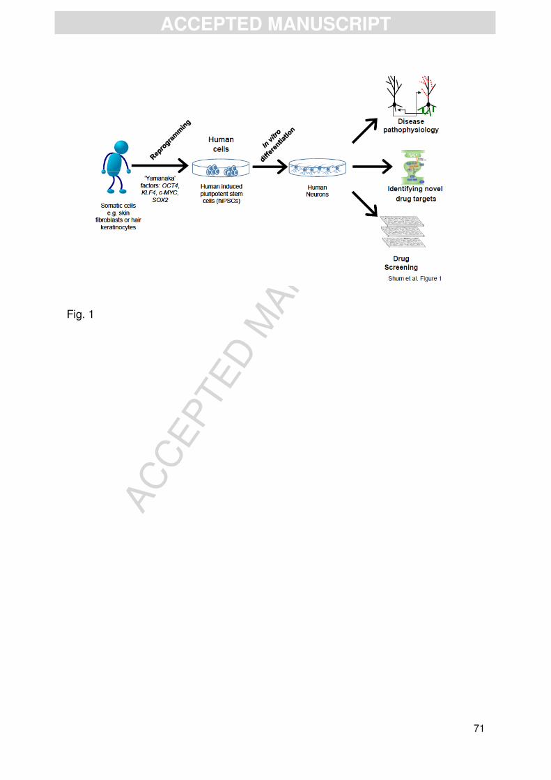
AC
CEPTED
MAN
USC
RIP
T
ACCEPTED MANUSCRIPT
71
Fig. 1

AC
CEPTED
MAN
USC
RIP
T
ACCEPTED MANUSCRIPT
72
Fig. 2
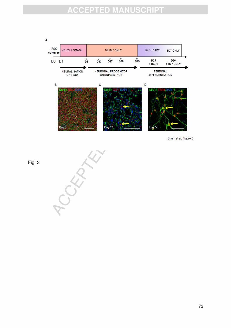
AC
CEPTED
MAN
USC
RIP
T
ACCEPTED MANUSCRIPT
73
Fig. 3
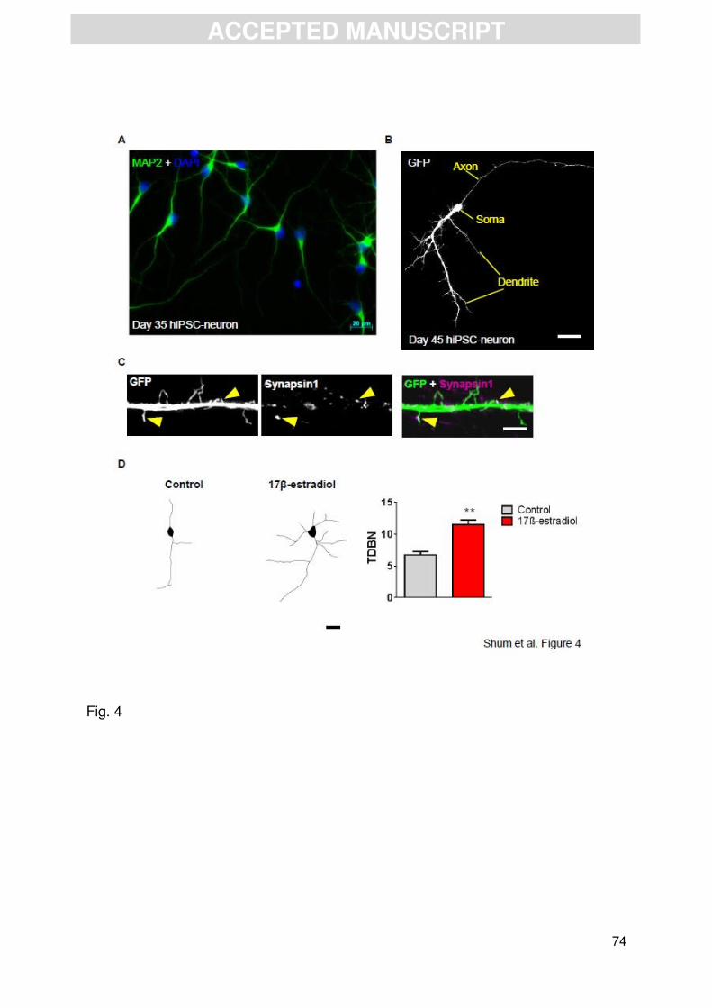
AC
CEPTED
MAN
USC
RIP
T
ACCEPTED MANUSCRIPT
74
Fig. 4

AC
CEPTED
MAN
USC
RIP
T
ACCEPTED MANUSCRIPT
75
Highlights
• Advances in stem cell biology allow for the reprogramming of somatic cells into induced pluripotent stem cells (iPSCs)
• Human iPSCs (hiPSCs) can be differentiated into defined neuronal lineages • hiPSCs have can used to investigate basic mechanisms of neural development • Reprogramming of somatic cells from patients has allowed investigations into
complex disorders of the CNS • Preliminary data indicates that hiPSCs are responsive to 17β-estradiol


