Embed Size (px)
Citation preview

Oct4 switches partnering from Sox2 to Sox17to reinterpret the enhancer code and specifyendoderm
Irene Aksoy1,6, Ralf Jauch2,6,Jiaxuan Chen1, Mateusz Dyla2,Ushashree Divakar1, Gireesh K Bogu1,Roy Teo1, Calista Keow Leng Ng2,3,Wishva Herath1, Sun Lili1, AndrewP Hutchins1,4, Paul Robson1,5,Prasanna R Kolatkar2,5,* andLawrence W Stanton1,3,5,*1Stem Cell and Developmental Biology, Genome Institute of Singapore,Singapore, 2Laboratory for Structural Biochemistry, Genome Instituteof Singapore, Singapore, 3School of Biological Sciences, NanyangTechnological University, Singapore, 4Immunology Frontier ResearchCentre, Osaka University, Osaka, Japan and 5Department of BiologicalSciences, National University of Singapore, Singapore
How regulatory information is encoded in the genome is
poorly understood and poses a challenge when studying
biological processes. We demonstrate here that genomic
redistribution of Oct4 by alternative partnering with Sox2
and Sox17 is a fundamental regulatory event of endodermal
specification. We show that Sox17 partners with Oct4 and
binds to a unique ‘compressed’ Sox/Oct motif that earmarks
endodermal genes. This is in contrast to the pluripotent
state where Oct4 selectively partners with Sox2 at ‘canoni-
cal’ binding sites. The distinct selection of binding sites by
alternative Sox/Oct partnering is underscored by our de-
monstration that rationally point-mutated Sox17 partners
with Oct4 on pluripotency genes earmarked by the canoni-
cal Sox/Oct motif. In an endodermal differentiation assay,
we demonstrate that the compressed motif is required for
proper expression of endodermal genes. Evidently, Oct4
drives alternative developmental programs by switching
Sox partners that affects enhancer selection, leading to
either an endodermal or pluripotent cell fate. This work
provides insights in understanding cell fate transcriptional
regulation by highlighting the direct link between the DNA
sequence of an enhancer and a developmental outcome.
The EMBO Journal (2013) 32, 938–953. doi:10.1038/
emboj.2013.31; Published online 8 March 2013Subject Categories: chromatin & transcription; developmentKeywords: endoderm; enhancer code; lineage specification;
pluripotency; Sox/Oct interaction
Introduction
The bulk of mammalian genomes consists of non-coding
regions whose function is only poorly annotated despite
extensive research (Birney et al, 2007; Gerstein et al, 2010;
Ernst et al, 2011). An important portion of the non-coding
genome encodes for enhancers that recruit sequence-specific
transcription factors (TFs), which in turn trigger changes in
the chromatin state, recruit co-activators, and therefore
regulate the expression of nearby genes. However,
the catalogue of functionally characterized enhancers is
slim and the mechanism of enhancer function remains
unclear. Specifically, ‘the enhancer code’, the sequence
features within enhancers that determine TF recruitment
and thereby gene expression and developmental programs,
is not well understood. It is believed that TFs partner to
combinatorially execute gene expression programs. However,
it is debated whether such partnerships require direct
and cooperative protein–protein interactions (Wilczynski
and Furlong, 2010; Biggin, 2011; Mirny, 2011). If TFs
physically cooperate, then the resulting protein complex
would target different genomic loci than individual proteins
in the absence of interaction partners. Such co-selectivity
would have profound consequences for the regulatory output
of TFs. Several genomic, biochemical, and structural studies
point to direct TF interactions and indicate that such
partnerships can alter the genomic binding and even the
sequence specificity of the participating factors (Hollenhorst
et al, 2009; Slattery et al, 2011). However, a system-wide
enquiry on how the genome binding and regulatory role of
TFs changes in the presence of alternative partner factors has
been missing. To this end, we studied the plasticity of Sox and
Oct TF partnerships during lineage commitment in early
mammalian development.
The Sox and Oct proteins comprise TF families of 20 and
14 members, respectively, and individual members function
in a wide range of biological processes (Phillips and Luisi,
2000; Kondoh and Kamachi, 2010; Bergsland et al, 2011).
Several Sox/Oct partnerships have been described in different
biological contexts (Kuhlbrodt et al, 1998; Tanaka et al, 2004;
Stefanovic et al, 2009); the synergistic action of Sox2 and
Oct4 in pluripotent stem cells being the most prominent
example of a functionally critical Sox/Oct partnership
(Nishimoto et al, 1999; Tomioka et al, 2002; Boyer et al,
2005). Other well-characterized examples are the Sox2/Brn2
partnership during neural development and the Sox11/Brn1
interaction that dictates glial cell identity (Kuhlbrodt et al,
1998; Tanaka et al, 2004). These observations led to the
hypothesis that a Sox/Oct partner code underlies many
critical cell fate choices (Wilson and Koopman, 2002;
Kondoh and Kamachi, 2010).
With the aim of better understanding and identifying
Sox/Oct partner codes in pluripotency and endodermal
contexts, we investigated the interaction of Oct4 with Sox2
*Corresponding authors. PR Kolatkar or LW Stanton, Stem Cell andDevelopmental Biology, Genome Institute of Singapore, 60 BiopolisStreet, #02-01 Genome Building, 138672 Singapore.Tel.: þ 65 6808 8006; Fax: þ 65 6808 8291;E-mail: [email protected] orTel.: þ 65 6808 8176; Fax: þ 65 6808 8291;E-mail: [email protected] authors contributed equally to this work.
Received: 16 November 2012; accepted: 24 January 2013; publishedonline: 8 March 2013
The EMBO Journal (2013) 32, 938–953
www.embojournal.org
EMBO
THE
EMBOJOURNAL
THE
EMBOJOURNAL
938 The EMBO Journal VOL 32 | NO 7 | 2013 &2013 European Molecular Biology Organization

and Sox17 on specific DNA enhancers. Oct4 partnership with
Sox2 in embryonic stem cells (ESCs) is well established.
However, there are reports suggesting that Oct4 might also
be involved in endodermal development, but its mechanisms
of action are not yet understood (Niwa et al, 2000). Sox17 is a
TF well known to play an important role in both primitive
(PrE) and definitive (DE) endoderm identity (Seguin et al,
2008; Morris et al, 2010; Niakan et al, 2010). Interestingly,
single-cell gene expression analysis of embryos showed
expression of Oct4 in PrE cells at comparable levels as in
the pluripotent epiblast (Kurimoto et al, 2006; Guo et al,
2010). Consequently, a state exists in the nascent inner cell
mass of the blastocyst, prior to the epiblast and PrE
formation, where Sox2, Sox17, and Pou5f1 (aka Oct4) are
co-expressed in the same cell (Guo et al, 2010), thereby
providing potential competition between Sox2 and Sox17
for Oct4 binding, which would influence subsequent cell
lineage decisions. Lineage segregation in the ICM during
preimplantation of mouse embryos is dependent on several
critical genetic and epigenetic events. The mechanisms that
initiate and control these processes are currently central
topics of investigation in developmental biology and despite
extensive research, very little is known. Indeed, the search
for the regulatory mechanisms involved in the control of the
earliest stages of mouse development has so far resulted in
very few candidates.
In this study, using a genome-wide ChIP (chromatin
immunoprecipitation)-sequencing approach we establish
that in a pluripotency context, Oct4 binds with Sox2 on a
distinctive canonical motif, whereas when Sox17 is intro-
duced into ESCs, Oct4 switches partners and interacts with
Sox17 on a compressed motif leading to the induction of a
specific endodermal differentiation program. Moreover, we
demonstrate that a reengineered Sox17 factor (Jauch et al,
2011) lacks its preference for the compressed motif and
thereby redistributes its binding from the compressed to the
canonical sites. Apparently, this genomic redistribution turns
Sox17EK into a potent inducer of pluripotency. Conversely,
the point-mutated Sox2KE has lost its preference for the
canonical motif. Moreover, using an in vitro model of PrE
development, we show that Oct4 is necessary for PrE
induction and that its interaction with Sox17 initiates
this specific cell fate choice. Finally, we show that genes
with a canonical or compressed motif are expressed in
the ICM of late mouse blastocyt where both the EPI and the
PrE specifications are occurring. We therefore identified
genes known to be important for PrE cell identity but also
new candidate genes that will help us to better understand
lineage segregation in early mouse preimplantation
development.
This work helps in our understanding of cell fate transcrip-
tional regulation and suggests a new partner code for
Sox/Oct, whereby the recruitment of Sox17/Oct4 to a
compressed Sox/Oct motif specifies the endoderm fate.
Results
Sox2 and Sox17 select disparate genomic loci by
selective partnering with Oct4 on canonical and
compressed DNA motifs
To establish that the genomic binding profile of Oct4 depends
on which Sox partner is present in cells, and to show that
different Sox/Oct combinations co-select distinctive sets of
target genes earmarked by specific composite motifs, we
generated ESC lines expressing, epitope-tagged (V5) Sox2
and Sox17 transgenes using a doxycycline-inducible cell
line (Beard et al, 2006; Figure 1A). Treatment of ESCs with
doxycycline for 48 h induced expression of Sox2-V5 and
Sox17-V5 proteins (Figure 1B) and mRNA levels
(Supplementary Figure S1). Using quantitative RT–PCR, we
observed the upregulation of Nanog and Oct4 in Sox2-V5
expressing cells and the upregulation of Gata4 and Lama1 in
Sox17-V5 expressing cells, validating that the transgenic Sox
proteins potently stimulate the expression of specific lineage
markers (Figure 1C). Genome-wide transcriptional analysis
of Sox17-V5 cells showed an increase in the expression of
several extra-embryonic endodermal genes including Col4a1,
Col4a2, Gata6, and Pdgfra (Supplementary Table S1).
To assess how Oct4 levels respond to elevated Sox17 expres-
sion, we performed western blots on these cells and found
that Oct4 protein levels remained unchanged 48 h after Sox17
induction (Figure 1D). By contrast, Sox2 protein levels de-
creased slightly (Figure 1D). These data established that 48 h
induction was suitable to compare the genomic binding
profiles of different Sox/Oct combinations in transitory states
occurring early in pluripotent cell differentiation.
To compare the genomic binding distribution of Sox2/Oct4
and Sox17/Oct4 pairs, we performed chromatin immunopre-
cipitation assays followed by genome-wide sequencing
(ChIP-seq) in two independent cell lines for each Sox factor
(Supplementary Table S2). Using antibodies against the
exogenous V5-tagged Sox factors and endogenous Oct4,
we identified binding sites using a stringent data analysis
pipeline (Supplementary Method). We first identified Oct4
sites (i) in the context of exogenous Sox2 expression
(Oct4Sox2) and (ii) in the context of exogenous Sox17 expres-
sion (Oct4Sox17). Next, we intersected these two Oct4 data
sets with Sox2 and Sox17 data sets. We found that Oct4Sox2
(Oct4 ChIP-seq in Sox2 expressing cells) is more likely to co-
bind genomic loci with Sox2 when compared with Sox17.
Conversely, Oct4Sox17 (Oct4 ChIP-seq in Sox17 expressing
cells) is more likely to co-bind with Sox17 when compared
to Sox2 (Figure 2A). This indicates that a substantial fraction
of Oct4 partnerships is dependent on whether Sox2 or Sox17
is expressed. Hence, changes in the Sox partner lead to a
partial genomic redistribution of Sox/Oct4 complexes. That
is, Sox2/Oct4 co-select a distinct set of genomic loci which is
different from the set co-selected by Sox17/Oct4. Next, we
performed a de novo motif analysis on all genomic loci co-
bound by either Sox2/Oct4Sox2 or Sox17/Oct4Sox17. For Sox2/
Oct4Sox2 sites, we identified the Sox/Oct canonical motif
(sequence related to CTTTGTCATGCAAAT) as most highly
enriched (Figure 2B(a)). This motif is well characterized and
has been functionally validated within the enhancers of
several prominent pluripotency genes, including the master
regulators Oct4, Sox2, and Nanog, which are jointly targeted
by the Sox2/Oct4 pair. However, for Sox17/Oct4Sox17 sites we
identified a variation of a Sox/Oct composite motif that is
related to a CTTTGTATGCAAAT sequence (Figure 2B(b)).
This ‘compressed’ motif shows spacing between the Sox
and Oct sites that is reduced by one base pair compared to
the canonical motif. We performed motif scans using position
weight matrices (PWMs) for canonical and compressed mo-
tifs (Figure 2C and D). In agreement with the de novo motif
Oct4/Sox17 interaction and endoderm specificationI Aksoy et al
939&2013 European Molecular Biology Organization The EMBO Journal VOL 32 | NO 7 | 2013

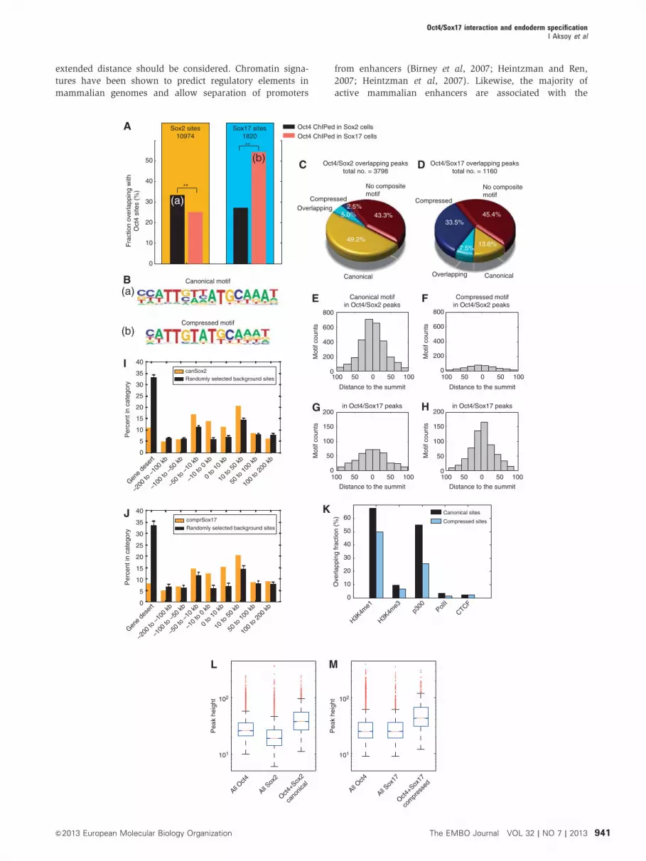
search, we observed a converse distribution of the motifs.
More than 50% of the 3798 Sox2/Oct4Sox2 sites contained a
canonical motif but only 7.5% contained a compressed
motif. In contrast, 440% of the 1160 Sox17/Oct4Sox17 sites
contained a compressed motif and 21.1% a canonical motif
(Supplementary Table S3). To further assess the significance
of the Sox/Oct occupancy at the alternative compressed motif
sites, we analysed the location of canonical and compressed
motifs with respect to ChIP-seq summits (Figure 2E–H). We
found that canonical motifs within Sox2/Oct4Sox2 sites cluster
more closely to the summit coordinate, the presumed binding
location. The same was observed for the compressed motifs
found at Sox17/Oct4Sox17 sites. In contrast, compressed mo-
tifs within Sox2/Oct4Sox2 sites and canonical motifs within
Sox17/Oct4Sox17 sites show a more widespread pattern, sug-
gesting that a portion of these motifs comprised motifs found
in the proximity of the actual binding site but are not directly
bound. Given these different motif preferences, and our aim
to decipher transcriptional regulation that set functionally
different gene sets apart, we focused further analysis on
Sox2/Oct4 bound canonical and Sox17/Oct4 bound com-
pressed sites. Earlier ChIP-seq analysis done on multiple
pluripotency TFs in mouse ESCs showed that Oct4 and
Sox2, together with Nanog and Stat3 tend to cluster quite
distally from promoters (Chen et al, 2008). Canonical and
compressed sites were likewise found in regions 10–50 kb
away from the transcription start sites (Figure 2I and J).
Therefore, when assigning motifs to target genes a more
�-Actin �-Actin
Sox2-V5 Sox17-V5
– + – +Dox Dox#1 #2
– + – +#1 #2
0
1
2
3
Sox2.1 Sox2.2 Sox17.1 Sox17.2
Rel
ativ
e m
RN
A e
xpre
ssio
nle
vel
Non-treated Nanog Oct4 Gata4 Lama1
FRT pgkNEOpA FRT HYGRO
pBS31-tetO-Sox-V5-SV40pApCAGGSflpE
Col1A1 locus
KH2 ES cells
Sox-V5 KH2 ES cells
tetO Sox-cDNA V5 HYGROSV40pA
Col1A1locus
ROSA26 �-Globin pArtTARosa26 locus
Doxycycline
Sox17-V5Oct4
Sox17-V5Sox2
– + – +Dox# 1 # 2
Sox17 KH2 ES cells
�-Actin 0
0.5
1
Oct4 Sox2
Sox17.1(–) Sox17.1(+) Sox17.2(–) Sox17.2(+)
Figure 1 Induced expression of Sox2 and Sox17 in ESC lines. (A) Schematic of the KH2-inducible vector system used to conditionally expressepitoge (V5)-tagged Sox proteins in ESCs (FRT (Flipase Recognition Target), pA (Polyadenylation signal), tetO (tetracycline/doxycyclineOperator)). (B) Anti-V5 western blot showing the expression of the V5-tagged Sox2 and Sox17 proteins in two independent cell clones treatedwith (þ ) or without (� ) doxycycline for 48 h. (C) Quantitative RT–PCR of Nanog, Oct4, Gata4 and Lama1 in Sox2- and Sox17-inducible ESCstreated with doxycycline for 48 h in two independent clones. (D) Western blot of Sox17-V5, Oct4 and Sox2 in two Sox17-V5 expressing ESClines and its quantification for Oct4 and Sox2. Proteins were (i) first incubated with the V5 and the Oct4 antibodies (upper panel), (ii) then themembrane was reprobed with the V5 and Sox2 antibodies (middle panel) and (iii) finally with the b-actin antibody (lower panel).
Figure 2 Sox2/Oct4 and Sox17/Oct4 pairs are recruited at different genomic loci. (A) Oct4 is redistributed to different genomic loci by Soxfactors. Co-occurrence of Sox2 or Sox17 with Oct4 in either Sox2-V5 or Sox17-V5 expressing KH2 cells. (a, b) Marks the intersect set of peaksused for de novo motif searching. Sox2 is more likely to co-occur with Oct4 ChiPed in a Sox2 OE background than with Oct4 ChIPed in a Sox17OE background (P-value 5.1e� 91, Z-score test). Inversely, Sox17 predominantly co-occurs on sites also bound by Oct4 ChIPed in Sox17 OEbackground whereas co-binding with Oct4 ChIPed in a Sox2 OE background much less likely (P-value 5.0e–116, Z-score test). (B) Matricespredicted by de novo motif analysis in (a) Sox2/Oct4Sox2 and (b) Sox17/Oct4Sox17 intersects. (C, D) Fraction of canonical, compressed,both (overlapping) and absence of motifs within (C) Sox2/Oct4Sox2 or (D) Sox17/Oct4Sox17 co-bound loci. (E–H) Motif counts binned bydistances of the actual motif coordinate to the summit of the ChIP-seq peak for the canonical and compressed motifs in Sox2/Oct4Sox2 sites andSox17/Oct4Sox17 sites. (I, J) Genome distribution of canonical motifs found in Sox2/Oct4Sox2 sites and compressed motifs in Sox17/Oct4Sox17
sites with respect to transcription start sites (TSS). (K) Fraction of Sox2/Oct4Sox2 bound canonical motifs in Sox2-V5 KH2 cells and Sox17/Oct4Sox17 bound compressed motifs in Sox17-V5 KH2 cells overlapping with H3K4m1 (enhancer mark), H3K4me3 (promoter mark), p300, PolIIand CTCF regions annotated by ChIP-seq for Bruce4 mouse ESC by the ENCODE consortium. (L) The subset of overlapped Oct4Sox2 and Sox2peaks with a motif is significantly higher than the total of Oct4Sox2 or Sox2 peaks (P-values 5.0e–209 and Poe–200; Wilcoxon rank sum test).(M) Similarly, the subset of peaks co-bound by Sox17/Oct4Sox17 containing a compressed motif is significantly higher than the total of Oct4Sox17
and Sox17 peaks (P-values 2.9e–93 and 1.2e–79; Wilcoxon rank sum test). Solid bars of boxes display the 25th–75th percentile of the peakswith the median indicated as an intersection. The box plots are shown on a logarithmic scale.
Oct4/Sox17 interaction and endoderm specificationI Aksoy et al
940 The EMBO Journal VOL 32 | NO 7 | 2013 &2013 European Molecular Biology Organization
extended distance should be considered. Chromatin signa-
tures have been shown to predict regulatory elements in
mammalian genomes and allow separation of promoters
from enhancers (Birney et al, 2007; Heintzman and Ren,
2007; Heintzman et al, 2007). Likewise, the majority of
active mammalian enhancers are associated with the
Oct4/Sox17 overlapping peakstotal no. = 1160
Oct4/Sox2 overlapping peakstotal no. = 3798
Canonical Canonical
Compressed CompressedOverlapping
Overlapping
No compositemotif
No compositemotif
(b)
(a)
All Oct4
All Sox
2
Oct4+S
ox2
cano
nical
All Oct4
All Sox
17
Oct4+S
ox17
com
pres
sed
Pea
k he
ight 102
101
Pea
k he
ight 102
101
Canonical motif
Compressed motif
H3K4m
e1
H3K4m
e3p3
00PolI
I
CTCF0
10
20
30
40
50
60
Ove
rlapp
ing
frac
tion
(%)
Canonical sites
Compressed sites
0
10
20
30
40
50
Fra
ctio
n ov
erla
ppin
g w
ithO
ct4
site
s (%
)
(b)
(a)2.5%
5.0%
49.2%
43.3%33.5%
45.4%
7.5%13.6%
Oct4 ChIPed in Sox2 cellsOct4 ChIPed in Sox17 cells
**
**
Sox2 sites10974
Sox17 sites1820
0
50
100
150
200
Compressed motifin Oct4/Sox2 peaks
in Oct4/Sox17 peaks
Distance to the summit
Mot
if co
unts
0
50
100
150
200
Canonical motifin Oct4/Sox2 peaks
in Oct4/Sox17 peaks
Distance to the summit
Mot
if co
unts
0
200
400
600
800
Distance to the summit
Mot
if co
unts
0
200
400
600
800
Distance to the summit
Mot
if co
unts
0 50 10050100 0 50 10050100
0 50 10050100 0 50 10050100
canSox2Randomly selected background sites
comprSox17
Randomly selected background sites
Per
cent
in c
ateg
ory
40
35
30
25
20
15
10
5
0
Per
cent
in c
ateg
ory
40
35
30
25
20
15
10
5
0
Gene
dese
rt
Gene
dese
rt
–200
to –
100
kb
–200
to –
100
kb
–100
to –
50 kb
–100
to –
50 kb
–50
to –
10 kb
–10
to 0
kb
0 to
10
kb
10 to
50
kb
50 to
100
kb
100
to 2
00 kb
–50
to –
10 kb
–10
to 0
kb
0 to
10
kb
10 to
50
kb
50 to
100
kb
100
to 2
00 kb
Oct4/Sox17 interaction and endoderm specificationI Aksoy et al
941&2013 European Molecular Biology Organization The EMBO Journal VOL 32 | NO 7 | 2013

histone acetyl transferase p300 (Visel et al, 2009). To relate
the compressed and canonical motifs to annotated regulatory
regions, we intersected our motif coordinates with sites
defined by the ENCODE consortium for Bruce4 ESC
(Figure 2K). We found that a higher proportion of both
canonical and compressed binding sites intersected with the
enhancer mark H3K4me1 as compared to the promoter mark
H3K4me3. Consistent with this, both the canonical and
compressed motifs overlapped an additional enhancer
mark, p300, but very few with the PolII promoter mark or
the CTCF insulator mark. Consistent with its function in
pluripotent cells, the canonical sites were proportionally
more enriched in ESC enhancers as compared to compressed
sites. We next analysed the magnitude of ChIP-seq summits
(‘pile-up’). We found that the subset of peaks containing
canonical motifs co-bound by Sox2/Oct4Sox2 was significantly
higher than the total of Sox2 and Oct4Sox2 peaks (Figure 2L
and M). Analogously, the subset of compressed motifs bound
by Sox17/Oct4Sox17 was stronger than all Sox17 or Oct4 Sox17
sites. These data suggest a cooperative recruitment of Sox2
and Oct4 to canonical and Sox17 and Oct4 to compressed
motifs in vivo.
Canonical and compressed motifs earmark distinctive
sets of genes that are differentially expressed
The rewiring of TF binding and gene regulatory networks in
general (Schmidt et al, 2010) and of Sox and Oct TFs in
particular (Kunarso et al, 2010) was suggested to be a key
driver of evolution. Nevertheless, some enhancer elements
were found to be under strong negative selection (Visel et al,
2008). We analysed the evolutionary conservation of
canonical and compressed motifs using the placental
mammalian PhastCons scores (Margulies et al, 2003).
Evolutionary conservation was noted for the canonical, and
to a lesser extent the compressed motif, indicating that a
subset of the observed binding events are conserved within
mammals (Figure 3A and B). To study gene expression
changes during endodermal differentiation, gene expression
profiling analysis was performed in ESCs before and after the
induction of Sox17 expression (Supplementary Table S1). The
genes were ranked by log2 transformed fold expression
changes and scanned for the occurrence of compressed and
canonical motifs in a distance ±50kb from the TSS (Figure
3C and D). A significant enrichment was observed for
the compressed motif near the TSS of genes upregulated
during endodermal differentiation; this was not observed
for downregulated or unaltered genes (Figure 3C). Among
the upregulated genes containing a Sox17/Oct4 bound com-
pressed motif are genes annotated to play a role during extra-
embryonic endoderm development such as Col4a1, Col4a2,
Lama1, Sall4, Pdgfra, Emb, and Hesx1 but also novel candi-
date regulators such as Tyro3 and Nr2f6. Canonical motif
densities were less correlated with expression changes
(Figure 3D). This observation strongly suggests that the
cooperative binding of Sox17 and Oct4 drives the expression
of genes during endodermal differentiation. To assess the
functionality of gene sets earmarked by canonical and com-
pressed motifs, gene ontology analysis was performed using
GREAT. As expected, the canonical motif predominates with-
in regulatory domains of genes expressed in the compacted
morula, inner cell mass and very early stages of mouse
development (Figure 3E). In contrast, compressed motifs
were found near a different set of genes, many of which act
at slightly later stages of development and contribute to germ
layer and organ system differentiation (Figure 3F). Because
so few genes are annotated with respect to the development
of extra-embryonic endoderm and early definitive endoderm
in gene ontology analysis, it is unlikely to find such ontology
terms. However, many of the terms associated with the
compressed motif are related to internal organs derived
from endoderm. Indeed, genes annotated as expressed in
TS19 are also known to function during extra-embryonic
and endoderm development. Consistently, the ontology
‘extra-embryonic component; endoderm’ was found to be
significantly enriched albeit not among the top 20 (Q-value
0.026). Namely genes such as Cyp26a1, Dab2, Hesx1, Hnf1b,
Fst, Lama1, Lama2, Otx2, Pdgfa, Pdgra, Sall4, Smad2, and
Srgn are jointly annotated by endodermal and TS19 ontology
terms and have a Sox/Oct compressed motif nearby.
These analyses highlight the functional divergence of the
Sox2/Oct4 canonical and Sox17/Oct4 compressed motifs
to drive cell fate choices. It appears that each motif governs
the expression of a distinct set of genes required for the
specification of either the endoderm lineage for the com-
pressed motif, or pluripotency for the canonical motif.
The genomic distribution of Sox factors can be altered
by mutagenesis of the Oct4 interface
We have previously demonstrated that strategic point muta-
tions introduced within Sox2 and Sox17 at the Oct4 interac-
tion domains altered their biochemical binding activities
in vitro (Jauch et al, 2011). To assess the genomic binding
profile of the reengineered Sox factors in vivo, we expressed
Sox2KE-V5 or Sox17EK-V5 in ESCs (Supplementary Figure
S2) and performed ChIP-seq analysis of the V5-tagged Sox
proteins, and of endogenous Oct4 in the context of the
expression of either Sox proteins (Oct4Sox2KE or Oct4Sox17EK)
(Supplementary Table S2). First, we conducted de novo motif
analysis on loci co-bound by either Sox2KE/Oct4Sox2KE or
Sox17EK/Oct4Sox17EK. In stark contrast to wild-type Sox17/
Oct4Sox17 (Figure 2B(b)), we found the canonical motif to be
most enriched in Sox17EK/Oct4Sox17EK co-bound sites
(Figure 4A(a)). Moreover, the Sox2KE/Oct4Sox2KE pair no
longer partners on canonical motif, but a single Sox element
was found to be the top scoring motif (Figure 4A(b)). This is
consistent with our previous quantitative electromobility
shift assay (EMSA) experiments indicating that Sox2KE
does not bind on the compressed motif as effectively as
Sox17 (Ng et al, 2012). Apparently, Sox2 requires further
mutations to engineer Sox17-like DNA recognition in vitro
and in ES cells. While simple homology models do not reveal
candidate amino acids that need to be modified to install
Sox17-like binding activities into Sox2, future structural or
molecular dynamics studies of Sox/Oct4 complexes on the
compressed motif can potentially resolve this question.
Motif scans showed an occurrence of over 60% of the
canonical motif within Sox17EK/Oct4Sox17EK sites compared
to only 6.8% of the compressed motif (Figure 4B). Notably,
Sox17EK/Oct4Sox17EK sites are more likely to contain canoni-
cal motifs than Sox2/Oct4Sox2 sites (Figure 2C). This is
consistent with our previous finding that Sox17EK cooperates
even more strongly with Oct4 than wild-type Sox2 on the
canonical motif (Ng et al, 2012). Likewise, Sox17EK has
consistently produced a larger number of pluripotent
Oct4/Sox17 interaction and endoderm specificationI Aksoy et al
942 The EMBO Journal VOL 32 | NO 7 | 2013 &2013 European Molecular Biology Organization
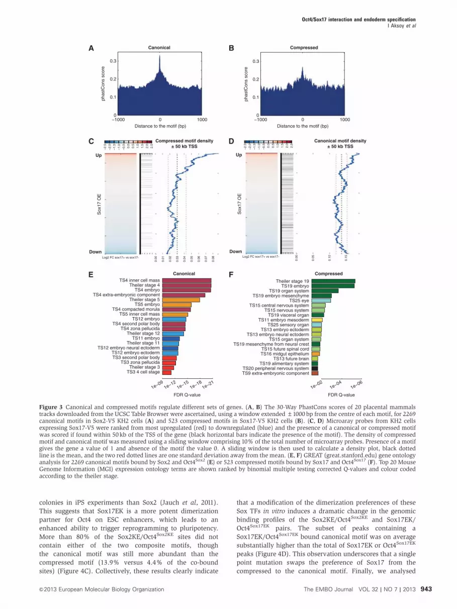
colonies in iPS experiments than Sox2 (Jauch et al, 2011).
This suggests that Sox17EK is a more potent dimerization
partner for Oct4 on ESC enhancers, which leads to an
enhanced ability to trigger reprogramming to pluripotency.
More than 80% of the Sox2KE/Oct4Sox2KE sites did not
contain either of the two composite motifs, though
the canonical motif was still more abundant than the
compressed motif (13.9% versus 4.4% of the co-bound
sites) (Figure 4C). Collectively, these results clearly indicate
that a modification of the dimerization preferences of these
Sox TFs in vitro induces a dramatic change in the genomic
binding profiles of the Sox2KE/Oct4Sox2KE and Sox17EK/
Oct4Sox17EK pairs. The subset of peaks containing a
Sox17EK/Oct4Sox17EK bound canonical motif was on average
substantially higher than the total of Sox17EK or Oct4Sox17EK
peaks (Figure 4D). This observation underscores that a single
point mutation swaps the preference of Sox17 from the
compressed to the canonical motif. Finally, we analysed
−1000 0 10000
0.1
0.2
0.3
Distance to the motif (bp)
phas
tCon
s sc
ore
−1000 0 10000
0.1
0.2
0.3
Distance to the motif (bp)
phas
tCon
s sc
ore
Canonical Compressed
1e−21
TS4 inner cell massTheiler stage 4
TS4 embryoTS4 extra-embryonic component
Theiler stage 5TS5 embryo
TS4 compacted morulaTS5 inner cell mass
TS12 embryoTS4 second polar body
TS4 zona pellucidaTheiler stage 12
TS11 embryoTheiler stage 11
TS12 embryo neural ectoderm
TS3 4 cell stageTheiler stage 3
TS3 zona pellucidaTS3 second polar bodyTS12 embryo ectoderm
FDR Q-value
1e−091e−12
1e−151e−18
Canonical
Theiler stage 19TS19 embryo
TS19 organ systemTS19 embryo mesenchyme
TS25 eyeTS15 central nervous system
TS15 nervous systemTS19 visceral organ
TS11 embryo mesodermTS25 sensory organ
TS13 embryo ectodermTS13 embryo neural ectoderm
TS15 organ systemTS19 mesenchyme from neural crest
TS15 future spinal cord
TS9 extra-embryonic componentTS20 peripheral nervous system
TS19 alimentary systemTS13 future brain
TS16 midgut epithelium
FDR Q-value
1e−021e−04
1e−06
Compressed
Compressed motif density± 50 kb TSS
Sox
17 O
E
Canonical motif density± 50 kb TSS
Down
Up
Down
Up
Sox
17 O
E
–2.5
–2.0
–1.5
–1.0
–0.5 0.0
0.5
1.0
1.5
2.0
2.5
–2.5
–2.0
–1.5
–1.0
–0.5 0.0
0.5
1.0
1.5
2.0
2.5
0.00Log2 FC sox17+ vs sox17- Log2 FC sox17+ vs sox17-
0.01
0.02
0.03
0.04
0.05
0.06
0.07
0.08 0.00
0.05
0.10
0.15
Figure 3 Canonical and compressed motifs regulate different sets of genes. (A, B) The 30-Way PhastCons scores of 20 placental mammalstracks downloaded from the UCSC Table Browser were ascertained, using a window extended±1000 bp from the centre of each motif, for 2269canonical motifs in Sox2-V5 KH2 cells (A) and 523 compressed motifs in Sox17-V5 KH2 cells (B). (C, D) Microaray probes from KH2 cellsexpressing Sox17-V5 were ranked from most upregulated (red) to downregulated (blue) and the presence of a canonical or compressed motifwas scored if found within 50kb of the TSS of the gene (black horizontal bars indicate the presence of the motif). The density of compressedmotif and canonical motif was measured using a sliding window comprising 10% of the total number of microarray probes. Presence of a motifgives the gene a value of 1 and absence of the motif the value 0. A sliding window is then used to calculate a density plot, black dottedline is the mean, and the two red dotted lines are one standard deviation away from the mean. (E, F) GREAT (great.stanford.edu) gene ontologyanalysis for 2269 canonical motifs bound by Sox2 and Oct4Sox2 (E) or 523 compressed motifs bound by Sox17 and Oct4Sox17 (F). Top 20 MouseGenome Information (MGI) expression ontology terms are shown ranked by binomial multiple testing corrected Q-values and colour codedaccording to the theiler stage.
Oct4/Sox17 interaction and endoderm specificationI Aksoy et al
943&2013 European Molecular Biology Organization The EMBO Journal VOL 32 | NO 7 | 2013

–2500 0 25000
1
2
3
4
5
Distance from motif (bp)
Mea
n pi
le-u
p pe
r m
illio
n
Sox2
CanonicalCompressed
Can
on
ical
Co
mp
ress
ed
–2500 0 25000
1
2
3
4
5
Mea
n pi
le-u
p pe
r m
illio
n
Sox17
CanonicalCompressed
–2500 0 25000
1
2
3
4
5
Mea
n pi
le-u
p pe
r m
illio
n
Sox2KE
CanonicalCompressed
–2500 0 25000
1
2
3
4
5
Mea
n pi
le-u
p pe
r m
illio
n
Sox17EK
CanonicalCompressed
Oct4/Sox17EK overlapping peakstotal no. of peaks = 3098
Oct4/Sox2KE overlapping peakstotal no. of peaks = 4602
Pea
k he
ight
AllOct4
AllSox17EK
Oct4+Sox17EKcanonical
(b)(a)
Canonical
CompressedOverlapping
No compositemotif
1.6%5.2% 36.7%
56.5%
Canonical
CompressedOverlapping
No compositemotif3.3%
1.1%12.8%
82.8%
101
102
Canonical motif Sox motif
Distance from motif (bp) Distance from motif (bp) Distance from motif (bp)
Sox17EK/Oct4Sox17EKA
B
E F G H
C D
Sox2KE/Oct4Sox2KE
Figure 4 Reengineered Sox2KE and Sox17EK factors target different loci than their wild-type counterparts. (A) Position weight matrices(PWMs) of motifs found de novo in peaks marked co-bound by Sox17EK/Oct4Sox17EK or Sox2KE/Oct4Sox2KE revealing a canonical Sox/Octmotifand a single Sox site. (B, C) Fraction of Sox17EK/Oct4Sox17EK or Sox2KE/Oct4Sox2KE peaks containing compressed, canonical, or both motifs(‘overlapping’). The total numbers of overlapped Oct4 and Sox2KE/Sox17EK peaks are shown. (D) Comparison of peak heights for canonicalmotifs co-bound by Sox17EK/Oct4Sox17EK, Oct4Sox17EK or Sox17EK. Solid bars of boxes display the 25th–75th percentile of the peaks with themedian indicated as an intersection. The subset peaks where Sox17EK/Oct4Sox17EK binds canonical motifs are significantly higher than the totalof Oct4 peaks (P-value o1e–200, Wilcoxon rank sum test). (E–H) Mean pile-up per million reads for both canonical and compressed data inSox2, Sox17, Sox2KE and Sox17EK expressing KH2 cells. Middle and bottom rows show density maps. Rows were ranked by motifquality scores with the highest scoring motifs at the top. The top panels show the mean pile-up per million reads for both canonical andcompressed data.
Figure 5 Oct4 expression is important for the differentiation of F9 cells into primitive endoderm (PrE). (A) Brightfield pictures ofundifferentiated F9 cells and differentiated F9 cells after retinoic acid (RA) treatment for 72 h. Relative mRNA expression as determined byQ-RT–PCR for PrE markers (B) Sox17, Gata4, Gata6, FoxA2, Col4a2 and Cxcr4; and pluripotency markers (C) Oct4, Nanog and Sox2 in F9 cellstreated with RA for 24–120 h and XEN cells. (D) Western blot of Oct4, Sox2 and Sox17 in F9 cells treated with RA for 1–4 days. (E) Oct4/Sox2and Oct4/Sox17 co-immunoprecipations performed in untreated and RA-treated F9 cells. An Oct4 antibody was used for immunoprecipitationand western blot was done using Sox2 and Sox17 antibodies. (F) Brightfield photos of F9 cells transfected with Oct4, Nanog and Non-targetingcontrol siRNAs and induced to differentiate at d1 after knock-down. Analysis of the expression levels of Oct4 Nanog, Sox17, Gata4 and Gata6 byQ-RT–PCR.
Oct4/Sox17 interaction and endoderm specificationI Aksoy et al
944 The EMBO Journal VOL 32 | NO 7 | 2013 &2013 European Molecular Biology Organization

Sox17
1
0.5
0
Rel
ativ
e m
RN
Aex
pre
ssio
n le
vel
1
0.5
0
Rel
ativ
e m
RN
Aex
pre
ssio
n le
vel
1
0.5
0
Rel
ativ
e m
RN
Aex
pre
ssio
n le
vel
1
0.5
0
Rel
ativ
e m
RN
Aex
pre
ssio
n le
vel
1
0.5
0
Rel
ativ
e m
RN
Aex
pre
ssio
n le
vel
1
0.5
0
Rel
ativ
e m
RN
Aex
pre
ssio
n le
vel
Sox17 Gata4
Gata4
Gata6
Gata6
siControl siNanog siOct4
F9 F9+RA 72 h
Oct4
Sox2
Sox17
Actin
Days after RA treatment
0 1 2 3 4
WB�-Sox2
WB�-Sox17
0
200
400
600
800
Sox17 Gata4 Gata6 FoxA2 Col4a2 Cxcr4
Rel
ativ
e m
RN
A e
xpre
ssio
n le
vel
F9
RA 24 h
RA 48 h
RA 72 h
RA 96 h
RA 120 h
Mefs
XEN
0
0.5
1
Oct4 Nanog Sox2
Rel
ativ
e m
RN
Aex
pre
ssio
n le
vel F9
RA 24 h
RA 48 h
RA 72 h
RA 96 h
RA 120 h
Mefs
0
Ct d1
Ct d3
Ct d2
siOct
4 d1
siOct
4 d3
Ct d4
siOct
4 d4
Ct d5
siOct
4 d5
Ct d6
siOct
4 d6
Ct d3
siOct
4 d3
Ct d4
siOct
4 d4
Ct d5
siOct
4 d5
Ct d6
siOct
4 d6
Ct d3
siOct
4 d3
Ct d4
siOct
4 d4Ct d
5
siOct
4 d5
Ct d6
siOct
4 d6
siOct
4 d2Ct d
3
siOct
4 d3Ct d
4
siOct
4 d4
Ct d5
siOct
4 d5Ct d
6
siOct
4 d6
Ct d6
siNan
og d6
Ct d6
siNan
og d6
Ct d5
siNan
og d5
Ct d4
siNan
og d4
Ct d3
siNan
og d3
Ct d6
siNan
og d6
Ct d5
siNan
og d5
Ct d4
siNan
og d4
Ct d3
siNan
og d3
Ct d6
siNan
og d6
Ct d5
siNan
og d5
Ct d4
siNan
og d4
Ct d3
siNan
og d3
Ct d5
siNan
og d5
Ct d4
siNan
og d4
Ct d3
siNan
og d3
Ct d2
siNan
og d2
Ct d1
siNan
og d1
0.5
1
Rel
ativ
e m
RN
Aex
pre
ssio
n le
vel
0
0.5
1
Rel
ativ
e m
RN
Aex
pre
ssio
n le
vel
Oct4 Nanog
Oct4 IP
0 h 24 h 48 h 72 h Ctrl
Oct4/Sox17 interaction and endoderm specificationI Aksoy et al
945&2013 European Molecular Biology Organization The EMBO Journal VOL 32 | NO 7 | 2013
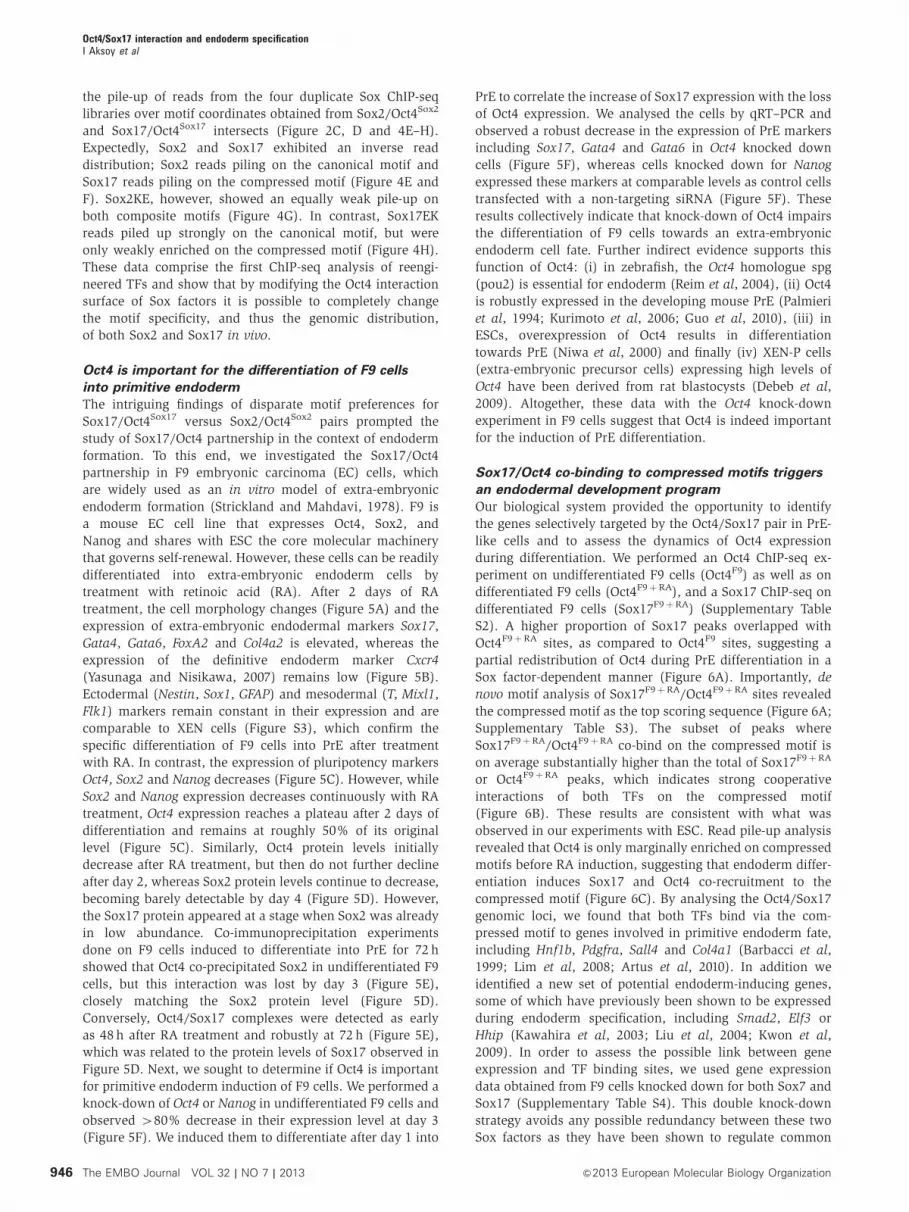
the pile-up of reads from the four duplicate Sox ChIP-seq
libraries over motif coordinates obtained from Sox2/Oct4Sox2
and Sox17/Oct4Sox17 intersects (Figure 2C, D and 4E–H).
Expectedly, Sox2 and Sox17 exhibited an inverse read
distribution; Sox2 reads piling on the canonical motif and
Sox17 reads piling on the compressed motif (Figure 4E and
F). Sox2KE, however, showed an equally weak pile-up on
both composite motifs (Figure 4G). In contrast, Sox17EK
reads piled up strongly on the canonical motif, but were
only weakly enriched on the compressed motif (Figure 4H).
These data comprise the first ChIP-seq analysis of reengi-
neered TFs and show that by modifying the Oct4 interaction
surface of Sox factors it is possible to completely change
the motif specificity, and thus the genomic distribution,
of both Sox2 and Sox17 in vivo.
Oct4 is important for the differentiation of F9 cells
into primitive endoderm
The intriguing findings of disparate motif preferences for
Sox17/Oct4Sox17 versus Sox2/Oct4Sox2 pairs prompted the
study of Sox17/Oct4 partnership in the context of endoderm
formation. To this end, we investigated the Sox17/Oct4
partnership in F9 embryonic carcinoma (EC) cells, which
are widely used as an in vitro model of extra-embryonic
endoderm formation (Strickland and Mahdavi, 1978). F9 is
a mouse EC cell line that expresses Oct4, Sox2, and
Nanog and shares with ESC the core molecular machinery
that governs self-renewal. However, these cells can be readily
differentiated into extra-embryonic endoderm cells by
treatment with retinoic acid (RA). After 2 days of RA
treatment, the cell morphology changes (Figure 5A) and the
expression of extra-embryonic endodermal markers Sox17,
Gata4, Gata6, FoxA2 and Col4a2 is elevated, whereas the
expression of the definitive endoderm marker Cxcr4
(Yasunaga and Nisikawa, 2007) remains low (Figure 5B).
Ectodermal (Nestin, Sox1, GFAP) and mesodermal (T, Mixl1,
Flk1) markers remain constant in their expression and are
comparable to XEN cells (Figure S3), which confirm the
specific differentiation of F9 cells into PrE after treatment
with RA. In contrast, the expression of pluripotency markers
Oct4, Sox2 and Nanog decreases (Figure 5C). However, while
Sox2 and Nanog expression decreases continuously with RA
treatment, Oct4 expression reaches a plateau after 2 days of
differentiation and remains at roughly 50% of its original
level (Figure 5C). Similarly, Oct4 protein levels initially
decrease after RA treatment, but then do not further decline
after day 2, whereas Sox2 protein levels continue to decrease,
becoming barely detectable by day 4 (Figure 5D). However,
the Sox17 protein appeared at a stage when Sox2 was already
in low abundance. Co-immunoprecipitation experiments
done on F9 cells induced to differentiate into PrE for 72 h
showed that Oct4 co-precipitated Sox2 in undifferentiated F9
cells, but this interaction was lost by day 3 (Figure 5E),
closely matching the Sox2 protein level (Figure 5D).
Conversely, Oct4/Sox17 complexes were detected as early
as 48 h after RA treatment and robustly at 72 h (Figure 5E),
which was related to the protein levels of Sox17 observed in
Figure 5D. Next, we sought to determine if Oct4 is important
for primitive endoderm induction of F9 cells. We performed a
knock-down of Oct4 or Nanog in undifferentiated F9 cells and
observed 480% decrease in their expression level at day 3
(Figure 5F). We induced them to differentiate after day 1 into
PrE to correlate the increase of Sox17 expression with the loss
of Oct4 expression. We analysed the cells by qRT–PCR and
observed a robust decrease in the expression of PrE markers
including Sox17, Gata4 and Gata6 in Oct4 knocked down
cells (Figure 5F), whereas cells knocked down for Nanog
expressed these markers at comparable levels as control cells
transfected with a non-targeting siRNA (Figure 5F). These
results collectively indicate that knock-down of Oct4 impairs
the differentiation of F9 cells towards an extra-embryonic
endoderm cell fate. Further indirect evidence supports this
function of Oct4: (i) in zebrafish, the Oct4 homologue spg
(pou2) is essential for endoderm (Reim et al, 2004), (ii) Oct4
is robustly expressed in the developing mouse PrE (Palmieri
et al, 1994; Kurimoto et al, 2006; Guo et al, 2010), (iii) in
ESCs, overexpression of Oct4 results in differentiation
towards PrE (Niwa et al, 2000) and finally (iv) XEN-P cells
(extra-embryonic precursor cells) expressing high levels of
Oct4 have been derived from rat blastocysts (Debeb et al,
2009). Altogether, these data with the Oct4 knock-down
experiment in F9 cells suggest that Oct4 is indeed important
for the induction of PrE differentiation.
Sox17/Oct4 co-binding to compressed motifs triggers
an endodermal development program
Our biological system provided the opportunity to identify
the genes selectively targeted by the Oct4/Sox17 pair in PrE-
like cells and to assess the dynamics of Oct4 expression
during differentiation. We performed an Oct4 ChIP-seq ex-
periment on undifferentiated F9 cells (Oct4F9) as well as on
differentiated F9 cells (Oct4F9þRA), and a Sox17 ChIP-seq on
differentiated F9 cells (Sox17F9þRA) (Supplementary Table
S2). A higher proportion of Sox17 peaks overlapped with
Oct4F9þRA sites, as compared to Oct4F9 sites, suggesting a
partial redistribution of Oct4 during PrE differentiation in a
Sox factor-dependent manner (Figure 6A). Importantly, de
novo motif analysis of Sox17F9þRA/Oct4F9þRA sites revealed
the compressed motif as the top scoring sequence (Figure 6A;
Supplementary Table S3). The subset of peaks where
Sox17F9þRA/Oct4F9þRA co-bind on the compressed motif is
on average substantially higher than the total of Sox17F9þRA
or Oct4F9þRA peaks, which indicates strong cooperative
interactions of both TFs on the compressed motif
(Figure 6B). These results are consistent with what was
observed in our experiments with ESC. Read pile-up analysis
revealed that Oct4 is only marginally enriched on compressed
motifs before RA induction, suggesting that endoderm differ-
entiation induces Sox17 and Oct4 co-recruitment to the
compressed motif (Figure 6C). By analysing the Oct4/Sox17
genomic loci, we found that both TFs bind via the com-
pressed motif to genes involved in primitive endoderm fate,
including Hnf1b, Pdgfra, Sall4 and Col4a1 (Barbacci et al,
1999; Lim et al, 2008; Artus et al, 2010). In addition we
identified a new set of potential endoderm-inducing genes,
some of which have previously been shown to be expressed
during endoderm specification, including Smad2, Elf3 or
Hhip (Kawahira et al, 2003; Liu et al, 2004; Kwon et al,
2009). In order to assess the possible link between gene
expression and TF binding sites, we used gene expression
data obtained from F9 cells knocked down for both Sox7 and
Sox17 (Supplementary Table S4). This double knock-down
strategy avoids any possible redundancy between these two
Sox factors as they have been shown to regulate common
Oct4/Sox17 interaction and endoderm specificationI Aksoy et al
946 The EMBO Journal VOL 32 | NO 7 | 2013 &2013 European Molecular Biology Organization

pan-endodermal genes (Seguin et al, 2008). By comparing
the compressed motif density to the genes that were
upregulated or downregulated in double knock-down F9
cells, a significant increase was observed in the compressed
motif density for genes that were downregulated after Sox7
and Sox17 double knock-down (Figure 6D). This indicates
that Sox17/Oct4 co-binding to compressed motifs is neces-
sary for the transcriptional activation of endodermal genes.
We compared Sox17 ChIP-seq data obtained from F9þRA
cells with Sox17 ChIP-chip data performed in XEN cells
which are embryo-derived PrE cells. In all, 1079 genes were
found to intersect between the two studies (Supplementary
Figure S4), but only 37 out of these 1079 genes harbour a
compressed motif (Supplementary Table S6), suggesting that
these sites are only bound by Sox17 and not by Oct4.
This is not surprising as Oct4 is expressed at low levels in
XEN cells compared to F9 cells induced to differentiate.
These data indicate that Oct4 and Sox17 co-recruitment on
the compressed motif is important for the induction
of PrE cell fate, but is likely unnecessary for continued
maintenance of PrE.
Endodermal genes are regulated by cooperative binding
of Sox17 and Oct4 to compressed motifs
There is evidence suggesting that Oct4 may play a role in the
establishment of the PrE within the mouse blastocyst as it is
expressed there and its forced expression in ESC leads to a
PrE differentiation program (Palmieri et al, 1994; Niwa et al,
2000). Based on our results, we asked whether Oct4 may
differentially target EPI and PrE genes within the ICM in the
particular context of its binding partners Sox2 and Sox17
which are also both expressed in the ICM. We reexamined
our previously generated single-cell gene expression data
(Guo et al, 2010) and focused this analysis on Oct4, Sox2
101
102
Compressed motif density
Sox7/17knock-down
0.00 0.01 0.02 0.03 0.04 0.05
i.e.Tyro3Hnf1bSrgn
Compressed motif
Log2 FC shSox7/17vs shCt
(a)
Pea
k he
ight
All Oct4 All Sox17 Oct4+Sox17
compressed
Down
Up2.4–3.2 3.2–2.4–1.6 1.61.8–0.8 0.0
−2500 −2500 −25000 25000
1
2
3
4
5
Distance from motif (bp)
Mea
n re
adpi
le-u
p pe
r m
illio
n
Oct4
0 25000
1
2
3
4
5
Distance from motif (bp)
Mea
n re
adpi
le-u
p pe
r m
illio
n
Oct4 RA
0 25000
1
2
3
4
5
Distance from motif (bp)
Mea
n re
adpi
le-u
p pe
r m
illio
n
Sox17 RA
Com
pres
sed
Sox17+RA
14 80710 20615 661
755 (a)1212358
3849
Oct4 +RAOct4
Figure 6 Oct4 and Sox17 co-bound the compressed motif in F9 cells induced to differentiate into primitive endoderm. (A) Fractional overlap ofendogenous Sox17 binding sites in RA-treated F9 cells co-occupied with Oct4 either before (red circle) or after adding RA (yellow circle). Upondifferentiation, Oct4 co-occupies significantly more sites with Sox17 than prior the RA addition (1987 versus 1133; P-value¼ 1.9e� 143; Z-scoretest) indicating that Sox17 and Oct4 are co-recruited to different genes. Importantly, many of these new genes are earmarked by compressedmotifs as found by de novo motif searching. (B) Distribution of peak heights in F9 cells treated with RA for all Oct4 sites, all Sox17 sites, andoverlapped Oct4 and Sox17 sites. Solid bars of boxes display the 25th–75th percentile of the peaks with the median indicated as an intersection.The box plots are shown in a logarithmic scale. The subset of peaks when Oct4 co-binds with Sox17 to compressed motifs comprisessignificantly higher cohort than the total of Oct4 peaks (P¼ 2.3e� 95, Wilcoxon rank sum test). (C) Mean pile-up per million for compressedmotifs in Oct4, Oct4 RA and Sox17 RA. Bottom row shows density maps of ChIP-seq data. (D) Compressed motif correlation with microarraygene expression of Sox7/Sox17 double knocked-down F9 cells. Motifs were assigned to genes if they are found within 50kb of a Refgene TSSand the motif density plot is generated as in Figure 3.
Oct4/Sox17 interaction and endoderm specificationI Aksoy et al
947&2013 European Molecular Biology Organization The EMBO Journal VOL 32 | NO 7 | 2013

and Sox17 expression within the ICM of the B64-cell stage
blastocyst. We ranked these ICM cells according to their
expression level of Sox17, the cells expressing high levels of
Sox17 are presumably presumptive of PrE, whereas cells
expressing low levels of Sox17 are presumptive of EPI
(Figure 7A). We observed that the expression pattern of
Sox2 is inversely correlated to Sox17 looking across all
cells. However, the expression level of Oct4 remained the
same in all cells regardless whether they are PrE-like or EPI-
like. This mRNA expression pattern is consistent with Oct4,
Sox2 and Sox17 protein levels, with Oct4 uniformly expressed
in all cells of the ICM, while Sox2 is initially expressed in all
cells of the ICM but then becomes restricted only to the
epiblast and Sox17 being initially expressed in a mixed sub-
population of ICM cells that later becomes restricted to the
blastocoel surface of the ICM (Figure 7B). These data suggest
that Oct4 may preferentially bind the canonical Sox/Oct
element in partnership with Sox2 in presumptive EPI while
in PrE, it may bind the compressed Sox/Oct element in
partnership with Sox17. We therefore generated RNA-seq
data from whole mouse embryos at E3.75 (stage where the
three cell types: TE, PrE and EPI are well resolved) and from
dissected ICMs in order to identify genes expressed specifi-
cally in the ICM (Supplementary Table S5). For each gene
identified, we measured the ratio of their expression level,
determined by the RPKM (reads per kilobase per million
mapped reads) value, in E3.75 blastocyst versus ICM.
Therefore, genes with a ratio lower than 1 were assigned as
ICM specific, whereas genes with a ratio higher than 1 were
assigned as TE specific. We found that among the ICM-
specific genes 67 are earmarked by a compressed motif and
133 by a canonical motif (Supplementary Table S5). These
gene sets are candidates for the differential transcriptional
regulation by Sox2/Oct4 versus Sox17/Oct4. The ratios are
shown for each motif in Figure 7C, in which were plotted as
controls Sox2, Sox17 and Oct4 for ICM-specific genes, and
Cdx2, Gata3 and Id2 for TE-specific genes. We then looked at
the expression level of these genes in ESC and PrE-like cells
by using the raw expression values obtained from genome-
wide gene expression analysis of mouse ESC and F9 cells
differentiated into PrE-like cells to better resolve EPI and PrE
genes within the ICM (Figure 7D; Supplementary Table S5).
0
4
8
12
0
4
8
12
0
4
8
12E
xpre
ssio
nE
xpre
ssio
nE
xpre
ssio
n
Sox17
Pou5f1
Sox2
1 2 3 4 5 6 7 8 9 10 11 12 13 14 15 16 17 18 19 20 21 22 23 24 25 26 27 28 29 30 31 33 34 35 36 37 38 39 43
Mid-stage blastocysts
0.01
0.1
1
10
100
E3.
75/IC
M e
xpre
ssio
n le
vel
ratio
s (lo
g sc
ale)
Genes with a compressed motif and a E3.75/ICM expression ratio <1
0.01
0.10
1.00
10.00
100.00
E3.
75/IC
M e
xpre
ssio
n le
vel
ratio
s (lo
g sc
ale)
Genes with a canonical motif and a E3.75/ICM expression ratio <1
Sox
2
Pou
5f1
Sox
17
Gat
a3C
dx2
Id2
TE Ct
TR
OP
HE
CT
OD
ER
MIC
MT
RO
PH
EC
TO
DE
RM
ICM
Gat
a3C
dx2
Id2
TECt
Sox
17
Sox
2
Pou
5f1
0
2000
4000
6000
8000
10 000
Exp
ress
ion
leve
l in
PrE
(ra
w)
Expression in PrE
Genes with a compressed motif Genes with a canonical motif
0
2000
4000
6000
8000
10 000
Exp
ress
ion
in E
S c
ells
(ra
w)
Expression in ES
Late-stage blastocysts
PrE-likeEPl-like
40 41 42 44 45 46 47 48 49 50 51 52 53 54 55 56 57 58 59 60 61 62 63 64
Figure 7 Oct4/Sox2 and Oct4/Sox17 complexes regulate specific genes in mouse embryos at the blastocyst stage. (A) Sox17, Sox2, Oct4, Nanogand Gata4 expression levels distribution across each cell of a 64-cell stage mouse embryo obtained by single-cell gene expression analysis (Guoet al, 2010). A background of Ct¼ 28 was used to obtain an absolute expression level. (B) Immunostainings of Oct4 and Sox17 in mouseembryos at mid and late blastocyst stage. Scale bar is 20mm. (C) Histogram plots representing E3.75 blastocyst versus ICM expression levelratios for genes harbouring a compressed or a canonical motif in their enhancer region. Expression levels were determined by RNA-seq inRPKM (reads per kilobase per million mapped reads). (D) Histogram plot of the raw expression levels of each genes in ES and PrE cells with acompressed or a canonical motif and an E3.75 blastocyst/ICM lower than 1.
Oct4/Sox17 interaction and endoderm specificationI Aksoy et al
948 The EMBO Journal VOL 32 | NO 7 | 2013 &2013 European Molecular Biology Organization

10 kb
Tyro
Sox2
Oct4 in Sox2
Sox17
Oct4 in Sox17
Sox17EK
Oct4 in Sox17EK
Oct4
Oct4 + RA
Sox17 + RA
control (KH2)
KH2
F9
10 kb
Hnf1b
Nr2f6 Srgn Zmat5 Hnf1b
Smad2 Tyro3 Sall4 Prdm10
10
20
30
40
50
Coo
pera
tivity
Scale Chr2: Scale Chr11:
Smad2 Tyro3 Sall4
Nr2f6 Srgn Zmat5 Hnf1b
Prdm1 Smad2 Tyro3 Sall4
Nr2f6 Srgn Zmat5 Hnf1b
Prdm1 Smad2 Tyro3 Sall4
Nr2f6 Srgn Zmat5 Hnf1b
Prdm1
Sox17 + Oct4 Sox2 + Oct4 Sox17EK + Oct4
Sox.Oct4.DNAOct.DNASox.DNA
Free DNA
Sox7 + Oct4
Smad2 Tyro3 Sall4 Prdm1
Nr2f6 Srgn Zmat5 Hnf1b
Lama1-positive control
F9 + RA
F9
Nr2f6 Tyro3
WT (a)
Degeneratingmutant (b)
Canonicalmutant (c)
Sox2-negative control
Canonicalmutant (d)
F9 + RA
F9
Prdm1Hnf1bSall4Zmat5SrgnSmad2
Sox2 Sox2
Sox2Sox2
Sox2
Sox2
Oct4
Oct4
Oct4
Oct4
Oct4Oct4Pluripotency genes
Canonical motif
Compressed motif
Endodermal genes
Canonical motif
Pluripotency genes
Compressed motif
Endodermal genes
Oct4
Oct4
Oct4
Sox17 Sox17
Sox17 Sox17
Sox17Sox2
CAGGS Transposase Min Prom GFPEnhancerTol2 Tol2
Sox17
Figure 8 Sox17 and Oct4 cooperatively bind on the compressed motif and regulate endodermal genes transcription. (A) Binding profiles of Soxfactors and Oct4 in KH2 and F9 cells at Hnf1b and Tyro3 genomic loci are shown. (B) EMSAs were performed using recombinant, DNA bindingdomains of Sox17, Sox2, Sox17EK and Sox7 with Oct4 on eight different DNA elements containing the compressed motif. Co-operativity factorsfor Sox17/Oct4 were represented as bar plots with±standard deviations error bars. (C) GFP reporter assay done on F9 cells treated (a–c) or not(d) with RAwith Wild-type (a), Degenerating mutants (b) and Canonical mutants (c, d) enhancers cloned in the Tol2 vector (Kawakami, 2007)(scale is 50mm). (D) Model describing the Sox2/Oct4 and Sox17/Oct4 partnerships in ESC. In undifferentiated ESC, where Oct4 and Sox2expression levels are high, both factors cooperate and target specifically the canonical motif to regulate the expression of specific pluripotencygenes. When the balance of Sox factors shifts in ES cells Oct4 switches from an interaction with Sox2 on canonical mofits towards aninteraction with Sox17, and targets specific genes containing a compressed motif to trigger endodermal specification.
Oct4/Sox17 interaction and endoderm specificationI Aksoy et al
949&2013 European Molecular Biology Organization The EMBO Journal VOL 32 | NO 7 | 2013

The genes with a compressed or a canonical motif
represented in the histograms are listed in Supplementary
Table S5. We found the compressed motif to be more
strongly associated with the presumed PrE subset of genes
while the canonical motif was more strongly associated
with presumed EPI genes (Figure 7C). This argues that the
earmark with a canonical motifs leads to Sox2/Oct4-mediated
EPI expression program whereas an earmark with a com-
pressed motif enables a Sox17/Oct4-mediated PrE expression
program.
To further study the function of the compressed motif
during endodermal differentiation, we selected eight genes
with prominent and unique Sox17 and Oct4 peaks in KH2
and/or F9 cells after RA induction (Figure 8A; Supplementary
Figure S5). Criteria for selecting genes for validation was
response during exogenous Sox17 expression in KH2 cells
(Nr2f6, Prdm1, Zmat5) or downregulation after Sox7/17
knock-down in F9 cells (Tyro3, Hnf1b, Srgn), the presence
of a compressed motif and, in some cases, literature support
for their function during extra-embryonic endoderm devel-
opment (Sall4, Smad2) (Liu et al, 2004; Lim et al, 2008). First,
we conducted EMSA experiments and showed that Oct4 and
Sox17 bound cooperatively on these selected enhancer
elements whereas the cooperation of Sox2 or Sox17EK with
Oct4 was abolished or markedly diminished (Figure 8B). We
also showed that Sox7 cooperatively binds with Oct4 on the
compressed motif (Figure 8B), like Sox17, which can explain
the redundancy between Sox7 and Sox17 in regulating en-
dodermal genes. Next, we conducted an in vivo reporter
assay (Kawakami et al, 2007). Selected enhancer regions
were cloned into the Tol2 vector, comprised of a minimal
promoter linked to GFP, and co-transfected with a vector
expressing a transposase for stable integration in F9 cells that
were induced, or not, to differentiate into PrE-like cells
(Figure 8C). An increase in GFP expression was observed
after 4 days of transfection only in F9 cells differentiated into
PrE-like cells (Figure 8C(a)) but not in undifferentiated F9
cells (data not shown), indicating a specific expression
pattern of these genes during endodermal differentiation.
Moreover, when we introduced rational mutations that de-
generated the compressed motif of the described enhancers
by the replacement of multiple critical nucleotides within the
Sox and Oct half-sites (degenerating mutants) reporter
activity upon differentiation was completely abolished
(Figure 8C(b)). Next, to more rigorously assess the relevance
of the compressed motif for reporter expression we generated
mutations by inserting a single nucleotide between Sox
and Oct half-sites to transform the compressed motif
into a ‘canonical motif’ (canonical mutants, Figure 8C(c);
Supplementary Figure S6). Notably, even this subtle distur-
bance of the enhancer architecture led to a complete loss of
the expression of GFP in differentiating F9 cells. This indi-
cates that cooperative binding of Sox17/Oct4 to the com-
pressed motif is indeed necessary to drive the expression of
these endodermal genes. In a reverse experiment, we also
tested the ‘canonical mutants’ in undifferentiated F9 cells and
observed a gain in GFP expression for Sall4- and Smad2-
driven reporters that would otherwise have been silent
(Figure 8C(d); Supplementary Figure S6). These results
indicate that a single nucleotide insertion can dramatically
alter enhancer activities by recruiting alternative Sox/Oct4
complexes.
DiscussionIn this study, the cooperation of Oct4 with Sox2 or Sox17
during the maintenance of the undifferentiated state of ESC
and PrE induction was explored. We found that Sox17/Oct4
partner to co-select specific target genes during commitment
to primitive endoderm. We employed a model of endodermal
specification through forced expression of Sox17 in ESC.
Sox17 has been shown to play a major role in endodermal
differentiation of both human (Seguin et al, 2008) and mouse
(Niakan et al, 2010) ESC. We focused in this study more
specifically on Oct4. Oct4 is well known to play a major role
in maintaining the pluripotent state of ESC but Oct4 has also
been shown to play a role in PrE differentiation of these cells;
however, the mechanisms involved in this process are still
poorly understood. By analysing Sox17/Oct4 co-binding sites
in this system, we first showed that these two TFs co-operate
and bind to a specific compressed Sox/Oct motif. We
identified a few hundred specific enhancer sites bound by
both Oct4 and Sox17. We found that one of the earliest events
occurring during endodermal induction is the redistribution
of Oct4 binding sites from the canonical motif in
undifferentiated ESC that express high levels of Sox2 to the
newly discovered compressed motif when the cells are
induced to differentiate into endoderm due to increasing
levels of Sox17. We subjected the compressed motif to
careful analysis to ascertain whether it contributes to an
endodermal enhancer code. We now provide evidence that
the genomic binding profile of Oct4 critically depends on
which Sox partner factor is present and that the enhancer
selection often relies on the nature of the composite motif.
To study the molecular mechanism of enhancer selection,
we performed ChIP-seq analysis using rationally engineered
Sox factors. These mutants harboured a single amino-
acid mutation at the Oct4 interaction site that inverts their
biological functions (Jauch et al, 2011). ChIP-seq experiments
done on these mutant factors inducibly expressed in ESC
using the same systems as their wild-type counterparts
showed a clear redistribution of Oct4 binding sites from the
compressed to the canonical motif for Sox17EK. Sox2KE lost
its ability to bind to the canonical motif but did not acquire
the ability to bind to the compressed motif. Potentially,
Sox2KE may only be kick starting differentiation towards an
endoderm fate by stimulating an as yet-unidentified
endoderm specifier (possibly one or all of Smad2, Nr2f6,
Srgn, Tyro3, Zmat4, Sall4, Hnf1 or Prdm1). Alternatively,
Sox2KE may simply be interfering with the normal function
of Sox2 while Oct4 remains broadly available for other Sox
factors to bind, predisposing the ESC to differentiate towards
endoderm once Sox17 is upregulated.
Overall, these results show that Sox/Oct partnerships
target specific loci in order to regulate cell fate determination
and that a strong cooperation between these TFs is important
to bind to their specific enhancer sites. As both Sox17 and
Oct4 are expressed in the primitive layers of embryos at the
blastocyst stage, we hypothesized that they might play a role
in the segregation of ICM cells into primitive endoderm cells.
We therefore utilized a well-characterized PrE in vitro model,
which consisted of F9 cells treated with RA. Oct4 was again
recruited to enhancers containing compressed motifs in the
presence of Sox17. Moreover, GFP-reporter assays further
established that co-binding of Sox17/Oct4 to the compressed
motif is necessary to drive the expression of genes during
Oct4/Sox17 interaction and endoderm specificationI Aksoy et al
950 The EMBO Journal VOL 32 | NO 7 | 2013 &2013 European Molecular Biology Organization

endoderm induction. Among the genes earmarked by
compressed motifs are several genes with reported roles
during endoderm specification, including Hnf1b, Pdgfra,
Smad2 and Sall4 but also a few hundreds of new candidate
genes, including Prdm1, Srgn, Tyro3, Nr2f6 and Zmat5.
Finally, we correlated the results we obtained using ESC
and F9 cells with data we generated from mouse embryos
in order to gain more insight into lineage segregation during
embryonic development. The inner cell mass of the mouse
blastocyst contains cells with two distinct lineage potential:
the epiblast and the primitive endoderm. The epiblast
gives rise to the embryo proper, whereas the PrE specifies
extra-embryonic tissues that support the development of
the embryo and act as a signalling source to pattern the
embryonic tissues prior to gastrulation. Till now, the
mechanisms that govern segregation between the epiblast
and the PrE remained unclear. ESC and F9 cells represent
tractable developmental systems to uncover these mechan-
isms as they can be converted to PrE-like cells. At embryonic
day 3.5, two populations of cells can be distinguished in the
ICM of mouse blastocysts, one that is Gata6þ and
supposedly at the origin of PrE cells and one Nanogþ that
will give rise to epiblast cells. In a single-cell study, Kurimoto
et al (2006) identified several genes specifically expressed in
each of these sub-populations. When we compared these lists
with our list of genes bound by Sox17/Oct4 on a compressed
motif we found 11 common genes, 10 of which were
expressed specifically in the Gata6þ cell population
(Pdgfra, Emb, Dusp4, Lhfpl2, Lama1, Pfkl, Pgam2, Tfpi,
Rap2c and an unknown gene) whereas only one (Bcl7a)
was expressed in the Nanogþ cell population. Additionally,
from our ICM-specific RNA-seq data the compressed motif is
enriched in PrE genes and the canonical motif in pluripotent
genes. These data emphasize the importance of the genes we
identified as Sox17/Oct4 targets and further functional
characterization of these genes will help for a better
understanding of PrE specification and segregation in vivo.
Mechanistically, we show here that one of the earliest events
occurring during endodermal induction is the redistribution
of Oct4 binding sites from the canonical motif in
undifferentiated ESC that expresses high levels of Sox2 to
the newly discovered compressed motif when the cells are
induced to differentiate into endoderm due to increasing
levels of Sox17.
Overall, these results show that Sox/Oct partnerships
target specific loci in order to regulate cell fate determination
and that a strong cooperation between these TFs is important
to bind to their specific enhancer sites. We propose here a
model by which, in pluripotent cells, Oct4 and Sox2 expres-
sion levels being high, both factors cooperate and target
specifically the canonical motif to regulate the expression of
genes involved in self-renewal and pluripotency (e.g.,
Nanog). When these cells are subjected to an endodermal
differentiation signal such as FGF4 within the ICM, Sox17
levels increase leading to a switch of Oct4 from an interaction
with Sox2 to an interaction with Sox17, and thereby targets
specific genes containing a compressed motif to trigger the
endodermal expression program (Figure 8D). Further func-
tional characterization of these newly identified genes will
help for a better understanding of PrE specification and
segregation in vivo. Together, we provide a conceptual frame-
work for the enhancer code that governs early cell fate
decision and demonstrate the molecular mechanism of selec-
tive Sox-Oct partnerships during this process.
Materials and methods
Generation of inducible KH2 ES cells expressingV5-tagged Sox factorsKH2 ES cells were obtained from Open Biosystems and culturedin DMEM supplemented with 15% fetal bovine serum (Invitrogen),0.055mM b-mercaptoethanol, 2mM L-glutamine, 0.1mMnon-essential amino acid and 1000U/ml of LIF. Cells were main-tained at 371C with 5% CO2. These cells were utilized to insert asingle copy of Sox2, Sox17, Sox2KE and Sox17EK cDNAs tagged witha V5 epitope using the method described in Beard et al (2006).Doxycycline was used at a final concentration of 1 mg/ml.
F9 cell cultureF9 cells were grown in media with DMEM (Invitrogen), 10% FBS,1% L-glutamine (Invitrogen) and 1% penicillin/streptomycin.To induce their differentiation into primitive endoderm cells, cellswere plated at a density of 300 000 cells/10 cm dish and grown inmedium containing 1mM of RA.
Microarray hybridization and data analysisMouse Ref-8 v2.0 Expression BeadChip microarrays (Illumina) wereused for genome-wide expression analysis. For hybridization,cRNAs, from duplicate or triplicate samples, were synthesized andlabelled using TotalPrep RNA Amplification Kit (Ambion), followingmanufacturer’s instructions. Scanned data from the BeadChip rawfiles for all samples were retrieved and background correctedusing BeadStudio, and subsequent analyses were completed inGeneSpring GX. Data were normalized both within and betweenarrays, and corrected for multiple testing according to Benjamini–Hochberg. We defined genes as significantly differentially expressedwhen the FDR is o0.05. The data may be obtained via GeneExpression Omnibus (GSE43234).
RNA sequencingRNA of E3.75 mouse embryo blastocysts and of immunosurgicallydissected ICMs (Solter and Knowles, 1975) were extracted using thePicoPure RNA Isolation Kit. Reverse transcription and amplificationsteps were done using the Nugen Ovation RNA-Seq system. TheSOLiD fragment library construction kit was used for the libraryconstruction from 1 mg of amplified cDNA. The resulting librarieswere sequenced using SOLiD technology. The respective manu-facturer’s protocol was followed at all stages. Sequenced reads werealigned to the mm9 mouse genome using the ABI MapReadsprogram. Gene expression in the form of RPKM values wasmeasured by normalizing the total reads mapping to a gene withits length and sequencing depth. All the transcripts were used forthis calculation. The data may be obtained via Gene ExpressionOmnibus (GSE44553).
Reporter assaysApproximately 105 cells untreated or treated with RA were trans-fected in 6-well plates with 2.5mg Tol2 GFP reporter plasmid, inwhich selected enhancers have been cloned using the primers listedin Supplementary Table S5, and 2.5mg of Transposase expressingplasmid using Lipofectamine 2000 reagent. After overnight incuba-tion, the transfection mix was replaced with F9 media with orwithout RA. The experiments were repeated twice. Mutant vectorswere generated by using the Quick XL mutagenesis kit (Stratagene)according to manufacturer’s instructions.
Electromobility shift assayEMSAs were carried out using purified recombinant proteinsaccording to the method as described previously (Ng et al, 2012).
Supplementary dataSupplementary data are available at The EMBO Journal Online(http://www.embojournal.org).
Oct4/Sox17 interaction and endoderm specificationI Aksoy et al
951&2013 European Molecular Biology Organization The EMBO Journal VOL 32 | NO 7 | 2013

Acknowledgements
We thank S Prabhakar and TL Huber for valuable comments onthe manuscript. We are grateful to Dr K Kawakami for providingthe Tol2 system. This work is supported by the Agency for Science,Technology and Research (A*STAR, www.a-star.edu.sg) Singapore.Author contributions: IA and RJ contributed to conception and
design, collection and/or assembly of data, data analysis andinterpretation, manuscript writing, final approval of manuscript;CJ, MD, UD, GKB, RT, CKLN and WH contributed to collection of
data; APH contributed to data analysis and interpretation; PR, PKand LWS contributed to financial support, administrative support,data analysis and interpretation, manuscript writing, final approvalof manuscript.
Conflict of interest
The authors declare that they have no conflict of interest.
ReferencesArtus J, Panthier JJ, Hadjantonakis AK (2010) A role for
PDGF signaling in expansion of the extra-embryonic endodermlineage of the mouse blastocyst. Development 137: 3361–3372
Barbacci E, Reber M, Ott MO, Breillat C, Huetz F, Cereghini S (1999)Variant hepatocyte nuclear factor 1 is required for visceralendoderm specification. Development 126: 4795–4805
Beard C, Hochedlinger K, Plath K, Wutz A, Jaenisch R (2006)Efficient method to generate single-copy transgenic mice bysite-specific integration in embryonic stem cells. Genesis 44:23–28
Bergsland M, Ramskold D, Zaouter C, Klum S, Sandberg R, Muhr J(2011) Sequentially acting Sox transcription factors in neurallineage development. Genes Dev 25: 2453–2464
Biggin MD (2011) Animal transcription networks as highlyconnected, quantitative continua. Dev Cell 21: 611–626
Birney E, Stamatoyannopoulos JA, Dutta A, Guigo R, Gingeras TR,Margulies EH, Weng Z, Snyder M, Dermitzakis ET, Thurman RE,Kuehn MS, Taylor CM, Neph S, Koch CM, Asthana S, Malhotra A,Adzhubei I, Greenbaum JA, Andrews RM, Flicek P et al (2007)Identification and analysis of functional elements in 1% ofthe human genome by the ENCODE pilot project. Nature 447:799–816
Boyer LA, Lee TI, Cole MF, Johnstone SE, Levine SS, Zucker JP,Guenther MG, Kumar RM, Murray HL, Jenner RG, Gifford DK,Melton DA, Jaenisch R, Young RA (2005) Core transcriptionalregulatory circuitry in human embryonic stem cells. Cell 122:947–956
Chen X, Xu H, Yuan P, Fang F, Huss M, Vega VB, Wong E, Orlov YL,Zhang W, Jiang J, Loh YH, Yeo HC, Yeo ZX, Narang V,Govindarajan KR, Leong B, Shahab A, Ruan Y, Bourque G,Sung WK et al (2008) Integration of external signaling pathwayswith the core transcriptional network in embryonic stem cells.Cell 133: 1106–1117
Debeb BG, Galat V, Epple-Farmer J, Iannaccone S, Woodward WA,Bader M, Iannaccone P, Binas B (2009) Isolation of Oct4-expres-sing extraembryonic endoderm precursor cell lines. PLoS ONE 4:e7216
Ernst J, Kheradpour P, Mikkelsen TS, Shoresh N, Ward LD,Epstein CB, Zhang X, Wang L, Issner R, Coyne M, Ku M,Durham T, Kellis M, Bernstein BE (2011) Mapping and analysisof chromatin state dynamics in nine human cell types. Nature473: 43–49
Gerstein MB, Lu ZJ, Van Nostrand EL, Cheng C, Arshinoff BI, Liu T,Yip KY, Robilotto R, Rechtsteiner A, Ikegami K, Alves P,Chateigner A, Perry M, Morris M, Auerbach RK, Feng X, Leng J,Vielle A, Niu W, Rhrissorrakrai K et al (2010) Integrative analysisof the Caenorhabditis elegans genome by the modENCODEproject. Science 330: 1775–1787
Guo G, Huss M, Tong GQ, Wang C, Li Sun L, Clarke ND, Robson P(2010) Resolution of cell fate decisions revealed by single-cellgene expression analysis from zygote to blastocyst. Dev Cell 18:675–685
Heintzman ND, Ren B (2007) The gateway to transcription:identifying, characterizing and understanding promoters in theeukaryotic genome. Cell Mol Life Sci 64: 386–400
Heintzman ND, Stuart RK, Hon G, Fu Y, Ching CW, Hawkins RD,Barrera LO, Van Calcar S, Qu C, Ching KA, Wang W, Weng Z,Green RD, Crawford GE, Ren B (2007) Distinct and predictivechromatin signatures of transcriptional promoters and enhancersin the human genome. Nat Genet 39: 311–318
Hollenhorst PC, Chandler KJ, Poulsen RL, Johnson WE, Speck NA,Graves BJ (2009) DNA specificity determinants associate
with distinct transcription factor functions. PLoS Genet 5:e1000778
Jauch R, Aksoy I, Hutchins AP, Ng CK, Tian XF, Chen J, PalasingamP, Robson P, Stanton LW, Kolatkar PR (2011) Conversion of Sox17into a pluripotency reprogramming factor by reengineering itsassociation with Oct4 on DNA. Stem Cells 29: 940–951
Kawahira H, Ma NH, Tzanakakis ES, McMahon AP, Chuang PT,Hebrok M (2003) Combined activities of hedgehog signalinginhibitors regulate pancreas development. Development 130:4871–4879
Kawakami K (2007) Tol2: a versatile gene transfer vector invertebrates. Genome Biol 8(Suppl 1): S7
Kondoh H, Kamachi Y (2010) SOX-partner code for cell specifica-tion: Regulatory target selection and underlying molecularmechanisms. Int J Biochem Cell Biol 42: 391–399
Kuhlbrodt K, Herbarth B, Sock E, Enderich J, Hermans-Borgmeyer I,Wegner M (1998) Cooperative function of POU proteins and SOXproteins in glial cells. J Biol Chem 273: 16050–16057
Kunarso G, Chia NY, Jeyakani J, Hwang C, Lu X, Chan YS, Ng HH,Bourque G (2010) Transposable elements have rewired the coreregulatory network of human embryonic stem cells. Nat Genet 42:631–634
Kurimoto K, Yabuta Y, Ohinata Y, Ono Y, Uno KD, Yamada RG, UedaHR, Saitou M (2006) An improved single-cell cDNA amplificationmethod for efficient high-density oligonucleotide microarray ana-lysis. Nucleic Acids Res 34: e42
Kwon MC, Koo BK, Kim YY, Lee SH, Kim NS, Kim JH, Kong YY(2009) Essential role of CR6-interacting factor 1 (Crif1) in E74-likefactor 3 (ELF3)-mediated intestinal development. J Biol Chem284: 33634–33641
Lim CY, Tam WL, Zhang J, Ang HS, Jia H, Lipovich L, Ng HH, WeiCL, Sung WK, Robson P, Yang H, Lim B (2008) Sall4 regulatesdistinct transcription circuitries in different blastocyst-derivedstem cell lineages. Cell Stem Cell 3: 543–554
Liu Y, Festing M, Thompson JC, Hester M, Rankin S, El-Hodiri HM,Zorn AM, Weinstein M (2004) Smad2 and Smad3 coordinatelyregulate craniofacial and endodermal development. Dev Biol 270:411–426
Margulies EH, Blanchette M, Haussler D, Green ED (2003)Identification and characterization of multi-species conservedsequences. Genome Res 13: 2507–2518
Mirny LA (2011) Nucleosome-mediated cooperativity between tran-scription factors. Proc Natl Acad Sci USA 107: 22534–22539
Morris SA, Teo RT, Li H, Robson P, Glover DM, Zernicka-Goetz M(2010) Origin and formation of the first two distinct cell types ofthe inner cell mass in the mouse embryo. Proc Natl Acad Sci USA107: 6364–6369
Ng CK, Li NX, Chee S, Prabhakar S, Kolatkar PR, Jauch R (2012)Deciphering the Sox-Oct partner code by quantitativecooperativity measurements. Nucleic Acids Res 40: 4933–4941
Niakan KK, Ji H, Maehr R, Vokes SA, Rodolfa KT, Sherwood RI,Yamaki M, Dimos JT, Chen AE, Melton DA, McMahon AP, EgganK (2010) Sox17 promotes differentiation in mouse embryonicstem cells by directly regulating extraembryonic gene expressionand indirectly antagonizing self-renewal. Genes Dev 24: 312–326
Nishimoto M, Fukushima A, Okuda A, Muramatsu M (1999) Thegene for the embryonic stem cell coactivator UTF1 carries aregulatory element which selectively interacts with a complexcomposed of Oct-3/4 and Sox-2. Mol Cell Biol 19: 5453–5465
Niwa H, Miyazaki J, Smith AG (2000) Quantitative expression ofOct-3/4 defines differentiation, dedifferentiation or self-renewalof ES cells. Nat Genet 24: 372–376
Oct4/Sox17 interaction and endoderm specificationI Aksoy et al
952 The EMBO Journal VOL 32 | NO 7 | 2013 &2013 European Molecular Biology Organization

Palmieri SL, Peter W, Hess H, Scholer HR (1994) Oct-4 transcriptionfactor is differentially expressed in the mouse embryo duringestablishment of the first two extraembryonic cell lineagesinvolved in implantation. Dev Biol 166: 259–267
Phillips K, Luisi B (2000) The virtuoso of versatility: POU proteinsthat flex to fit. J Mol Biol 302: 1023–1039
Reim G, Mizoguchi T, Stainier DY, Kikuchi Y, Brand M (2004) ThePOU domain protein spg (pou2/Oct4) is essential for endodermformation in cooperation with the HMG domain protein casano-va. Dev Cell 6: 91–101
Schmidt D, Wilson MD, Ballester B, Schwalie PC, Brown GD,Marshall A, Kutter C, Watt S, Martinez-Jimenez CP, Mackay S,Talianidis I, Flicek P, Odom DT (2010) Five-vertebrate ChIP-seqreveals the evolutionary dynamics of transcription factor binding.Science 328: 1036–1040
Seguin CA, Draper JS, Nagy A, Rossant J (2008) Establishment ofendoderm progenitors by SOX transcription factor expression inhuman embryonic stem cells. Cell Stem Cell 3: 182–195
Slattery M, Riley T, Liu P, Abe N, Gomez-Alcala P, Dror I, Zhou T,Rohs R, Honig B, Bussemaker HJ, Mann RS (2011) Cofactorbinding evokes latent differences in DNA binding specificitybetween Hox proteins. Cell 147: 1270–1282
Solter D, Knowles BB (1975) Immunosurgery of mouse blastocyst.Proc Natl Acad Sci USA 72: 5099–5102
Stefanovic S, Abboud N, Desilets S, Nury D, Cowan C, Puceat M(2009) Interplay of Oct4 with Sox2 and Sox17: a molecular switch
from stem cell pluripotency to specifying a cardiac fate. J Cell Biol186: 665–673
Strickland S, Mahdavi V (1978) The induction of differentiation interatocarcinoma stem cells by retinoic acid. Cell 15: 393–403
Tanaka S, Kamachi Y, Tanouchi A, Hamada H, Jing N, Kondoh H(2004) Interplay of SOX and POU factors in regulation ofthe Nestin gene in neural primordial cells. Mol Cell Biol 24:8834–8846
Tomioka M, Nishimoto M, Miyagi S, Katayanagi T, Fukui N,Niwa H, Muramatsu M, Okuda A (2002) Identification of Sox-2regulatory region which is under the control of Oct-3/4-Sox-2complex. Nucleic Acids Res 30: 3202–3213
Visel A, Prabhakar S, Akiyama JA, Shoukry M, Lewis KD, Holt A,Plajzer-Frick I, Afzal V, Rubin EM, Pennacchio LA (2008)Ultraconservation identifies a small subset of extremelyconstrained developmental enhancers. Nat Genet 40: 158–160
Visel A, Rubin EM, Pennacchio LA (2009) Genomic views of distant-acting enhancers. Nature 461: 199–205
Wilczynski B, Furlong EE (2010) Dynamic CRM occupancy reflects atemporal map of developmental progression. Mol Syst Biol 6: 383
Wilson M, Koopman P (2002) Matching SOX: partner proteins andco-factors of the SOX family of transcriptional regulators. CurrOpin Genet Dev 12: 441–446
Yasunaga M, Nisikawa S (2007) Production of endoderm-derivedvisceral organ cells from ES cells. Tanpakushitsu Kakusan Koso52: 57–66
Oct4/Sox17 interaction and endoderm specificationI Aksoy et al
953&2013 European Molecular Biology Organization The EMBO Journal VOL 32 | NO 7 | 2013


