Embed Size (px)
Citation preview
Distinct Sonic Hedgehog signalingdynamics specify floor plate and ventralneuronal progenitors in the vertebrateneural tube
Vanessa Ribes,1,4 Nikolaos Balaskas,1,4 Noriaki Sasai,1,4 Catarina Cruz,1 Eric Dessaud,1 Jordi Cayuso,2
Samuel Tozer,1 Lin Lin Yang,3 Ben Novitch,3 Elisa Marti,2 and James Briscoe1,5
1Developmental Neurobiology, Medical Research Council-National Institute for Medical Research, London NW7 1AA, UnitedKingdom; 2Instituto de Biologıa Molecular de Barcelona, Consejo Superior de Investigaciones Cientıficas (CSIC), Barcelona08028, Spain; 3Department of Neurobiology, Broad Center of Regenerative Medicine and Stem Cell Research, David GeffenSchool of Medicine at University of California at Los Angeles, Los Angeles, California 90095, USA
The secreted ligand Sonic Hedgehog (Shh) organizes the pattern of cellular differentiation in the ventral neuraltube. For the five neuronal subtypes, increasing levels and durations of Shh signaling direct progenitors toprogressively more ventral identities. Here we demonstrate that this mode of action is not applicable to thegeneration of the most ventral cell type, the nonneuronal floor plate (FP). In chick and mouse embryos, FPspecification involves a biphasic response to Shh signaling that controls the dynamic expression of keytranscription factors. During gastrulation and early somitogenesis, FP induction depends on high levels of Shhsignaling. Subsequently, however, prospective FP cells become refractory to Shh signaling, and this is a pre-requisite for the elaboration of their identity. This prompts a revision to the model of graded Shh signaling in theneural tube, and provides insight into how the dynamics of morphogen signaling are deployed to extend thepatterning capacity of a single ligand. In addition, we provide evidence supporting a common scheme for FPspecification by Shh signaling that reconciles mechanisms of FP development in teleosts and amniotes.
[Keywords: Shh signaling; neural tube; floor plate; FoxA2]
Supplemental material is available at http://www.genesdev.org.
Received September 30, 2009; revised version accepted April 7, 2010.
During development, an extraordinary array of differen-tiated cell types with diverse properties and functions areproduced in a spatially and temporally coordinated man-ner. Considerable progress has been made in elucidat-ing the molecular mechanisms that underlie this process.In broad terms, cell-autonomous determinants specifycell fate by regulating the transcriptional programs of ini-tially uncommitted progenitor populations (Jessell 2000;Davidson et al. 2002). Extrinsic signaling molecules,emanating from specialized organizer regions within de-veloping tissues, control the expression of these cellfate determinants (Jessell 2000). This signaling function,however, is performed by a remarkably small number ofsecreted molecules that are iteratively deployed duringembryonic development. Thus, understanding how cellsperceive and interpret these signals to produce the cellular
diversity characteristic of a tissue is a central question indevelopmental biology.
The vertebrate CNS represents a good model to addressthis issue. In this tissue, the organized generation of manydifferent cell types initiates the assembly of functionalneural circuits (Jessell 2000). In ventral regions of thecaudal neural tube, six progenitor domains arrayed alongthe dorsal ventral (DV) axis give rise to a set of neuronalcell types: V0, V1, V2, and V3 interneurons; motor neu-rons (MNs); and nonneuronal floor plate (FP) cells (Jessell2000). The FP, p3, and pMN domains—which generateFP, V3 neurons, and MNs, respectively—are arranged inthe most ventral part of the neural tube (Fig. 1A); FPcells are distinguished from the two neural cell types bytheir wedge-shaped morphology and organizer function(Schoenwolf et al. 1989; Ericson et al. 1996; Tessier-Lavigne and Goodman 1996). Furthermore, the identityand function of each of these three progenitor domainsare determined by the expression of a set of transcriptionfactors (Dessaud et al. 2008). The basic helix–loop–helix(bHLH) factor Olig2 is expressed by pMN cells and is
4These authors contributed equally to this work.5Corresponding author.E-MAIL [email protected]; FAX 44-20-8816-2523.Article is online at http://www.genesdev.org/cgi/doi/10.1101/gad.559910.
1186 GENES & DEVELOPMENT 24:1186–1200 � 2010 by Cold Spring Harbor Laboratory Press ISSN 0890-9369/10; www.genesdev.org
required for the generation of MNs, while the homeodo-main protein Nkx2.2 is expressed in p3 progenitors andinduces V3 neurons (Dessaud et al. 2008). FP cells express
the forkhead transcription factor FoxA2, which is suffi-cient to induce FP identity (Sasaki and Hogan 1994).Notably, however, the requirement for FoxA2 for FP gen-eration has been difficult to assess because mouse em-bryos lacking FoxA2 fail to gastrulate and die prior to theemergence of FP (Ang and Rossant 1994; Weinstein et al.1994).
The spatial expression of these transcription factorsin the neural tube—and, consequently the location of thepMN, p3, and FP domains—is regulated by the secretedsignaling molecule Sonic Hedgehog (Shh) (Dessaudet al. 2008). Initially produced by the node and notochord,lying ventral to neural tissue, and later by cells of the FP,Shh forms a ventral-to-dorsal gradient, the amplitude ofwhich increases with time as more Shh is produced fromthe notochord and FP (for example, see Chamberlain et al.2008). The prevailing model for how Shh signaling reg-ulates progenitor patterning suggests that higher concen-trations of ligand and longer durations of intracellular sig-naling induce the expression of increasingly more ventralgenes (Dessaud et al. 2008). In support of this, Nkx2.2 isinduced by a higher concentration and a longer duration ofShh signaling than Olig2 (Dessaud et al. 2007; Chamberlainet al. 2008). In this view, FP, as the most ventral cell type inthe neural tube, should require higher levels and longerdurations of Shh signaling than p3 cells. Consistent withthis, mice lacking the transmembrane protein Smo or Glitranscription factors, which are involved in the transduc-tion of Shh signal, demonstrated that FP specificationrequires Shh signaling (Chiang et al. 1996; Matise et al.1998; Zhang et al. 2001; Bai et al. 2002). In addition, highconcentrations of Shh appear to induce FP markers at theexpense of MN markers in neural explants (Placzek et al.1991; Roelink et al. 1995; Ericson et al. 1996). However,the ability of Shh to induce FP markers in neural pro-genitors seems to be context-dependent. For instance, gain-of-function experiments in chicks indicate that Shh issufficient to induce MNs and V3 neurons, but not FPcells, in the dorsal part of the neural tube (Placzek et al.1993; Charrier et al. 2002; Patten and Placzek 2002). In thiscontext, instead of acting as an inductive signal for FPspecification, permissive or survival functions have beenascribed to Shh (Charrier et al. 2001). In addition, therequirement for Shh signaling for FP formation does notappear to be conserved in all vertebrates. In particular,blockade of hedgehog signaling in zebrafish has little, ifany, effect on the emergence of the FP (Odenthal et al. 2000;Varga et al. 2001). Instead, Nodal signaling has beenimplicated in FP induction in zebrafish (Hatta et al. 1991;Sampath et al. 1998; Tian et al. 2003). This has led to theidea that different vertebrate species have adopted distinctmechanisms of FP induction, and has raised questions aboutthe function of Shh as a morphogen during FP development.
Here we test whether the Shh morphogen model of neu-ral tube patterning applies to FP development in chicksand mice. We confirm that high levels of Shh signaling areinitially necessary for FP specification. However, blockadeof Shh signaling after the initiation of somitogenesis in-hibits the patterning of neural progenitors in the ventralneural tube, but not FP specification. Moreover, following
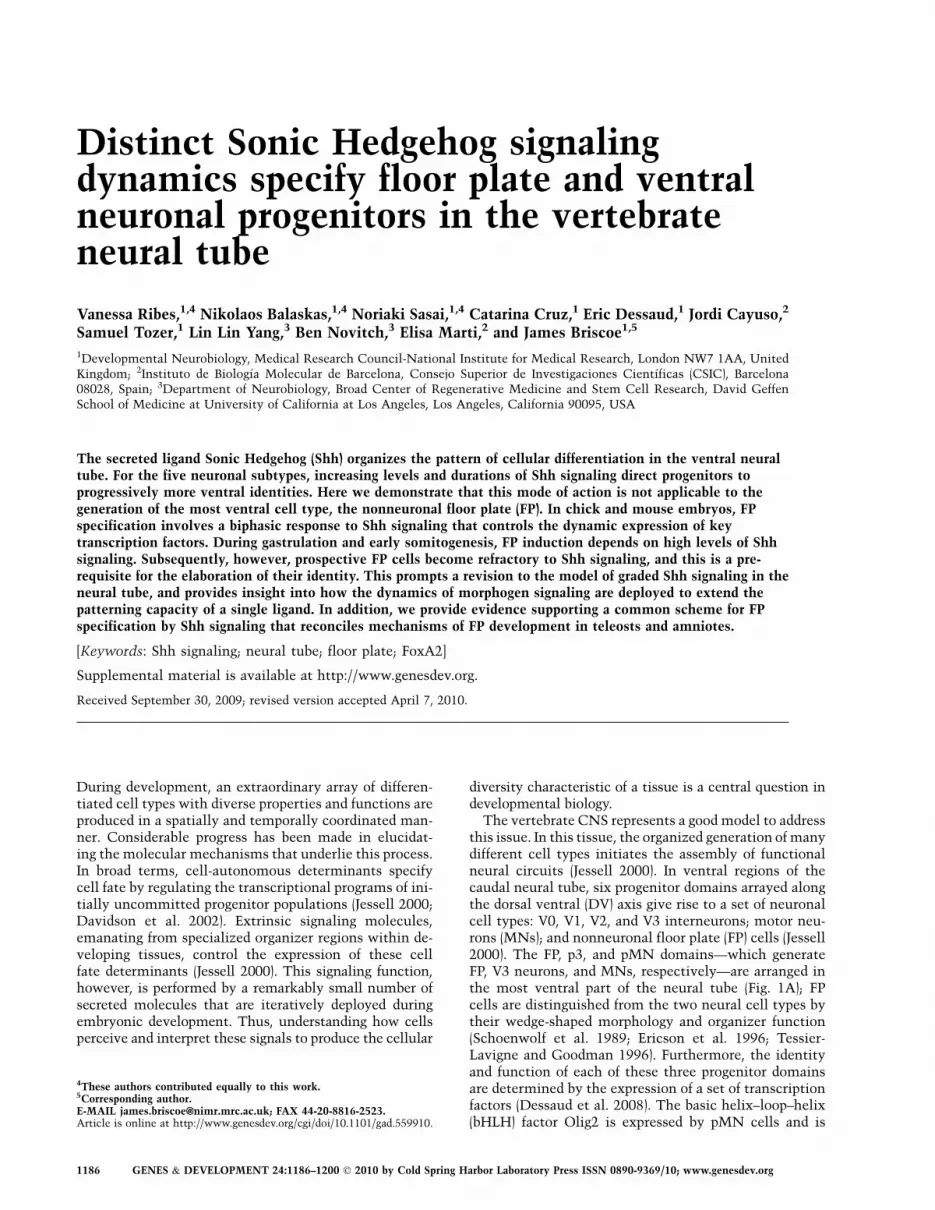
Figure 1. Gene expression profiles within midline cells of mouseneural tube. (A) Schematic depicting the dynamics of gene expres-sion and Shh gradient within the ventral neural tube. (nc) Noto-chord. (B–K) Expression ofFoxA2,Arx, Shh, Nkx2.2, andOlig2 (B–J),and Shh (K) mRNA showing the progressive appearance of FPmarkers at the midline of the neural tube from 7ss embryos. (B–K)Transverse sections at brachial levels of the neural tube. (D–D0)Lateral view of a whole embryo. (C–D0) FoxA2 protein is firstobserved from 7ss in ventral midline cells that express Nkx2.2.(D–D0) FoxA2 expression appears more robust than the scatteredNkx2.2 expression at posterior levels of the neural tube. (B) At thesestages, FoxA2 expression domain abuts Olig2-expressing cells.(E,E0) From E9.5 onward, the dorsal limit of FoxA2 is no longeradjacent to Olig2-expressing cells. Its expression is stronger at themidline, and weaker in cells that express Nkx2.2. (E–F9) Thispattern persists until E10.5. From this stage, additional FP markers,Arx (G–I) and Shh (J,K), appear in cells expressing high levels ofFoxA2. (G–H9) Concomitant to the induction of these markers,Nkx2.2 is no longer seen in the midline; hence, Arx and Shh arebarely coexpressed with Nkx2.2-expressing cells.
Signaling dynamics of floor plate specification
GENES & DEVELOPMENT 1187
initial induction of FP specification, Shh signaling isattenuated in presumptive FP cells. Maintaining Shh sig-naling at this time converts FP cells to ventral neural pro-genitors, demonstrating that the down-regulation of sig-naling is a prerequisite for the elaboration of FP identity.Thus, the specification of the FP and other ventral neuro-nal progenitors depends on distinct timing and duration ofShh signaling. Consistent with this, we demonstrate thatpresumptive FP cells display a dynamic transcriptionalcode that distinguishes FP precursors from ventral neuralprogenitors. We provide evidence that FoxA2 is requiredfor FP specification and the inhibition of p3 fate. Together,these data prompt a revision to the model of Shh-dependentpatterning of the neural tube, and offer new insight intohow the dynamics of morphogen signaling extend the pat-terning capacity of a single signal. Furthermore, we pro-vide evidence that the timing of Shh signaling during FPspecification is conserved between teleosts and amniotes,suggesting it represents a general feature of vertebrate FPdevelopment.
Results
A dynamic transcriptional code identifiesFP progenitors
We first assessed the dynamics of gene expression withinthe midline of the forming neural tube. The transcriptionfactor FoxA2 is commonly used as a marker of FP cells(Ericson et al. 1996; Patten and Placzek 2002; Nortonet al. 2005). FoxA2 is expressed in the ventral layer of thenode and in the notochord from embryonic day 7.5 (E7.5)in mice, where it orchestrates gastrulation (Ang andRossant 1994; Weinstein et al. 1994). It is then inducedin the ventral midline of the anterior neural plate fromthe 6-somite stage (ss), slightly later than the induction ofOlig2 and prior to Nkx2.2, markers of the pMN and p3progenitor domains, respectively (Fig. 1B–D0; data notshown). At these stages, the dorsal extent of FoxA2approximates the ventral boundary of Olig2 (Fig. 1B). By12–14ss, Nkx2.2 is induced in the midline of the neuraltube in FoxA2-expressing cells (Supplemental Fig. S1A,B;Jeong and McMahon 2005; data not shown). Subse-quently, as development proceeds, Nkx2.2 expression isextinguished from midline cells and becomes restrictedto the progenitors of the V3 neurons (Fig. 1E–E0; Supple-mental Fig. S1C–F). Low levels of FoxA2 expressionpersist in Nkx2.2-expressing cells (Fig. 1E–F9; Supplemen-tal Fig. S1C–E, arrowheads). In contrast, midline cellsdevoid of Nkx2.2 expression display high levels of FoxA2(Fig. 1E–F9; Supplemental Fig. S1A–F); at this stage, wetermed these FP cells.
The presence of FoxA2 in the p3 domain prompted usto examine whether we could identify other markers thatexclusively mark FP cells. The transcription factor Arxwas a good candidate, as it has been detected in the FP ofmouse embryos (Miura et al. 1997) and is restricted to themedial FP in zebrafish (Norton et al. 2005). Its expressionwas not detected before E9.5 (data not shown). From E9.5,Arx was limited to midline cells of the neural tube that
express FoxA2 and are negative for Nkx2.2 (Fig. 1G–I;Supplemental Fig. S1C–F). The induction of Arx corre-sponded to the time at which strong expression of Shhcould be detected in the FP (Fig. 1J,K). Together, thesedata suggest that FP specification is progressive: First,midline cells, which constitute the presumptive FP, ex-press a set of markers that are common to p3 progenitors,including FoxA2 and Nkx2.2. Then, Nkx2.2 is down-regulated, and late FP markers, including Shh and Arx, areinduced (Supplemental Fig. S1K).
The expression pattern of Olig2 suggests that the cellsthat become FP rarely, if ever, express Olig2. Consistentwith this, lineage analysis of cells that had expressed Olig2revealed that, although p3 progenitors descended fromOlig2-expressing cells, FP cells only very rarely had anOlig2 pedigree (Supplemental Fig. S1G–J9; Dessaud et al.2007). This indicates that the history of Olig2 expressioncan be used to discriminate cells destined to be FP cellsfrom p3 progenitors, both of which express Nkx2.2 duringtheir development. Together, therefore, these data provideevidence that a dynamic transcriptional code distinguishesventral progenitor populations, and that the FP lineagesegregates from neural progenitors prior to Olig2 induction(Supplemental Fig. S1K).
Transient high levels of Shh signaling are sufficientto induce FP cells
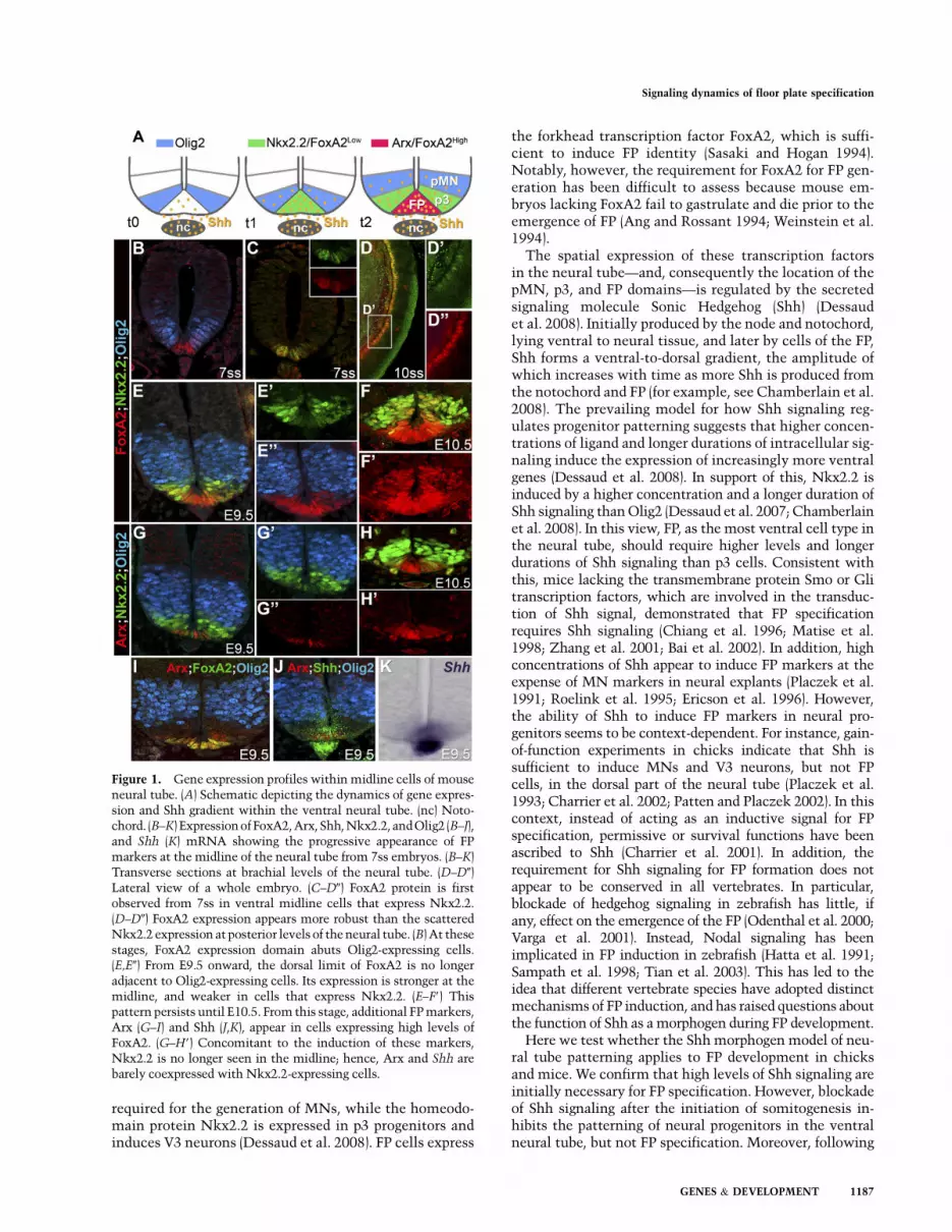
The progression from a p3 to FP gene expression profile inmidline cells would be consistent with higher levels and/or longer durations of Shh signaling regulating this switch(Dessaud et al. 2008). To better understand the dynamicsof Shh signaling associated with the emergence of FPidentity, we took advantage of chick ex vivo experiments(Fig. 2A). Naıve explants taken from the intermediateregion ([i] explants) of the neural plate induce FoxA2 inresponse to high concentrations of Shh, while lower con-centrations of Shh result in MN generation (Ericson et al.1996). In order to distinguish p3 and FP fates, we assayedthe expression of Nkx2.2 and Arx, specific markers ofp3 and FP, respectively. Exposure to 1 nM Shh for 48 hpredominantly induced Nkx2.2, and few, if any, Arx-expressing cells were detected (Fig. 2B,F). In contrast, ex-plants cultured in the presence of $4 nM Shh for 48 hcontained large numbers of Arx-expressing cells (Fig. 2C,F;data not shown), and the expression of Nkx2.2 was mark-edly reduced compared with cells exposed to 1 nM Shh (Fig.2C,F). These data indicate that the induction of FP identityrequires high concentrations of Shh, while lower concen-trations are sufficient to induce p3 identity.
To investigate the timing of FP induction, we nextassayed the expression of Arx and Nkx2.2 in [i] explantsexposed to 4–8 nM Shh for 24 h. In these conditions, fewcells expressed Arx, and most cells expressed Nkx2.2 (Fig.2F; data not shown). The expression profiles of Nkx2.2and Arx mRNA were consistent with this analysis (Fig.2G): In the presence of 4 nM Shh, Nkx2.2 was inducedwithin 12 h, while 1 nM Shh resulted in a delayedinduction of Nkx2.2. The levels of Nkx2.2 expression in[i] explants exposed to 4 nM decreased markedly from 24 h,
Ribes et al.
1188 GENES & DEVELOPMENT
concomitant with the induction of Arx. The induction ofFoxA2 mirrored that of Nkx2.2, but its expression wassustained after 24 h in explants exposed to 4 nM Shh (Fig.2G). These data suggest that the induction of FP identityin [i] explants requires high concentrations of Shh andtakes between 24 and 48 h.
We next asked what duration of Shh signaling wasrequired for the induction of FP. To address this, we ex-posed [i] explants to 4 nM Shh for 24 h, then replacedthe media with fresh media that either lacked Shh orcontained cyclopamine, an antagonist of the intracellulartransducer of the signaling pathway, Smo (Cooper et al.1998; Incardona et al. 1998). Explants were then culturedfor an additional 24 h (Fig. 2D–F). Blockade of Shh sig-naling at 24 h did not appear to affect the elaboration of FPidentity; the expression profile of Arx and Nkx2.2 wasindistinguishable from explants that were continuouslycultured in the presence of 4 nM Shh for 48 h (Fig. 2D–F).Thus the requirement for Shh signaling for the specifica-
tion of FP appears to be transient, and does not depend onsignaling being sustained beyond 24 h.
We next assayed for the expression profile of Ptch1 asa readout of Shh signaling (Marigo and Tabin 1996; Vokeset al. 2008). In the presence of 1 or 4 nM Shh, Ptch1 wasrapidly induced, and levels of expression peaked at 12 h(Fig. 2G). The peak levels of expression were higher in [i]explants exposed to 4 nM than 1 nM Shh, consistent withthe requirement for high levels of signaling for FP in-duction (Fig. 2F). Following the peak, however, the ex-pression levels of the Shh target gene began to decline.Compared with [i] explants exposed to 1 nM, in explantstreated with 4 nM Shh, the decrease of Ptch1 expressionwas more rapid and resulted in an ;70% drop in the lev-els of expression by 24 h (n = 4). Similarly, the expressionof another Shh target gene, Gli1 (Marigo et al. 1996; Leeet al. 1997; Vokes et al. 2008), displayed a higher peakthat decreased more rapidly in response to 4 nM com-pared with 1 nM Shh (Supplemental Fig. S4B). These data
Figure 2. Induction of FP markers by transient high levels of Shh signaling. (A) Schematic of HH10 chick embryos indicating theposition of [i] neural plate explants. (B–E) Expression of Arx and Nkx2.2 in [i] neural plate explants after 48 h of culture. Cells inexplants treated with 1 nM Shh exclusively express Nkx2.2 (B), while 4 nM Shh promotes FP fate, identified by Arx expression (C). Inexplants first exposed for 24 h to 4 nM Shh and then placed in a media either devoid of Shh (D) or containing 500 nM cyclopamine(cyclo) (E), FP induction is retained. (F) Quantification of cells expressing Arx and Nkx2.2 in [i] neural plate explants after 24 or 48 h ofculture (n $ 4 explants, number of cells per unit 6 SD). (G) Temporal dynamics of Nkx2.2, Arx, FoxA2, and Ptch1 expression relativeto Actin transcripts and normalized between sets of experiments assayed by quantitative PCR in [i] neural plate explants treated with1 nM (pale green) or 4 nM (pink) Shh (n $ 2 experiments, mean 6 SD). As definitive FP markers are induced, p3 markers and Ptch1 aredown-regulated. (H–P) Expression of Ptch1 (H–J,N–O) and Gli1 (K–M,P) in mouse embryos (H–M) and chick embryos (N–P). Insets inH and I indicate Arx, Nkx2.2, and Olig2 expression on sections adjacent to the main panel. Midline cells transiently express high levelsof Ptch1 and Gli1. The asterisks indicate the notochord.
Signaling dynamics of floor plate specification
GENES & DEVELOPMENT 1189
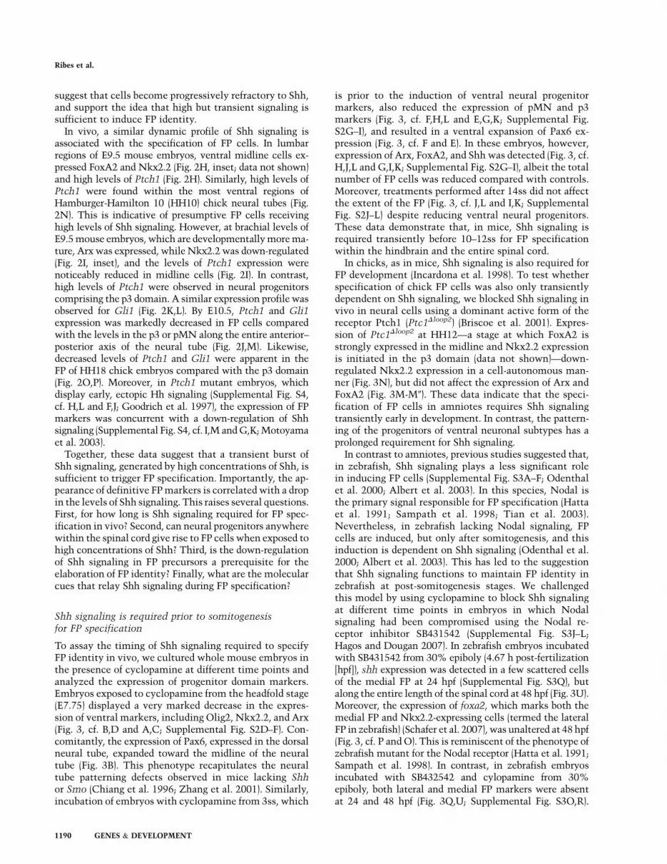
suggest that cells become progressively refractory to Shh,and support the idea that high but transient signaling issufficient to induce FP identity.
In vivo, a similar dynamic profile of Shh signaling isassociated with the specification of FP cells. In lumbarregions of E9.5 mouse embryos, ventral midline cells ex-pressed FoxA2 and Nkx2.2 (Fig. 2H, inset; data not shown)and high levels of Ptch1 (Fig. 2H). Similarly, high levels ofPtch1 were found within the most ventral regions ofHamburger-Hamilton 10 (HH10) chick neural tubes (Fig.2N). This is indicative of presumptive FP cells receivinghigh levels of Shh signaling. However, at brachial levels ofE9.5 mouse embryos, which are developmentally more ma-ture, Arx was expressed, while Nkx2.2 was down-regulated(Fig. 2I, inset), and the levels of Ptch1 expression werenoticeably reduced in midline cells (Fig. 2I). In contrast,high levels of Ptch1 were observed in neural progenitorscomprising the p3 domain. A similar expression profile wasobserved for Gli1 (Fig. 2K,L). By E10.5, Ptch1 and Gli1expression was markedly decreased in FP cells comparedwith the levels in the p3 or pMN along the entire anterior–posterior axis of the neural tube (Fig. 2J,M). Likewise,decreased levels of Ptch1 and Gli1 were apparent in theFP of HH18 chick embryos compared with the p3 domain(Fig. 2O,P). Moreover, in Ptch1 mutant embryos, whichdisplay early, ectopic Hh signaling (Supplemental Fig. S4,cf. H,L and F,J; Goodrich et al. 1997), the expression of FPmarkers was concurrent with a down-regulation of Shhsignaling (Supplemental Fig. S4, cf. I,M and G,K; Motoyamaet al. 2003).
Together, these data suggest that a transient burst ofShh signaling, generated by high concentrations of Shh, issufficient to trigger FP specification. Importantly, the ap-pearance of definitive FP markers is correlated with a dropin the levels of Shh signaling. This raises several questions.First, for how long is Shh signaling required for FP spec-ification in vivo? Second, can neural progenitors anywherewithin the spinal cord give rise to FP cells when exposed tohigh concentrations of Shh? Third, is the down-regulationof Shh signaling in FP precursors a prerequisite for theelaboration of FP identity? Finally, what are the molecularcues that relay Shh signaling during FP specification?
Shh signaling is required prior to somitogenesisfor FP specification
To assay the timing of Shh signaling required to specifyFP identity in vivo, we cultured whole mouse embryos inthe presence of cyclopamine at different time points andanalyzed the expression of progenitor domain markers.Embryos exposed to cyclopamine from the headfold stage(E7.75) displayed a very marked decrease in the expres-sion of ventral markers, including Olig2, Nkx2.2, and Arx(Fig. 3, cf. B,D and A,C; Supplemental Fig. S2D–F). Con-comitantly, the expression of Pax6, expressed in the dorsalneural tube, expanded toward the midline of the neuraltube (Fig. 3B). This phenotype recapitulates the neuraltube patterning defects observed in mice lacking Shhor Smo (Chiang et al. 1996; Zhang et al. 2001). Similarly,incubation of embryos with cyclopamine from 3ss, which
is prior to the induction of ventral neural progenitormarkers, also reduced the expression of pMN and p3markers (Fig. 3, cf. F,H,L and E,G,K; Supplemental Fig.S2G–I), and resulted in a ventral expansion of Pax6 ex-pression (Fig. 3, cf. F and E). In these embryos, however,expression of Arx, FoxA2, and Shh was detected (Fig. 3, cf.H,J,L and G,I,K; Supplemental Fig. S2G–I), albeit the totalnumber of FP cells was reduced compared with controls.Moreover, treatments performed after 14ss did not affectthe extent of the FP (Fig. 3, cf. J,L and I,K; SupplementalFig. S2J–L) despite reducing ventral neural progenitors.These data demonstrate that, in mice, Shh signaling isrequired transiently before 10–12ss for FP specificationwithin the hindbrain and the entire spinal cord.
In chicks, as in mice, Shh signaling is also required forFP development (Incardona et al. 1998). To test whetherspecification of chick FP cells was also only transientlydependent on Shh signaling, we blocked Shh signaling invivo in neural cells using a dominant active form of thereceptor Ptch1 (Ptc1Dloop2) (Briscoe et al. 2001). Expres-sion of Ptc1Dloop2 at HH12—a stage at which FoxA2 isstrongly expressed in the midline and Nkx2.2 expressionis initiated in the p3 domain (data not shown)—down-regulated Nkx2.2 expression in a cell-autonomous man-ner (Fig. 3N), but did not affect the expression of Arx andFoxA2 (Fig. 3M-M0). These data indicate that the speci-fication of FP cells in amniotes requires Shh signalingtransiently early in development. In contrast, the pattern-ing of the progenitors of ventral neuronal subtypes has aprolonged requirement for Shh signaling.
In contrast to amniotes, previous studies suggested that,in zebrafish, Shh signaling plays a less significant rolein inducing FP cells (Supplemental Fig. S3A–F; Odenthalet al. 2000; Albert et al. 2003). In this species, Nodal isthe primary signal responsible for FP specification (Hattaet al. 1991; Sampath et al. 1998; Tian et al. 2003).Nevertheless, in zebrafish lacking Nodal signaling, FPcells are induced, but only after somitogenesis, and thisinduction is dependent on Shh signaling (Odenthal et al.2000; Albert et al. 2003). This has led to the suggestionthat Shh signaling functions to maintain FP identity inzebrafish at post-somitogenesis stages. We challengedthis model by using cyclopamine to block Shh signalingat different time points in embryos in which Nodalsignaling had been compromised using the Nodal re-ceptor inhibitor SB431542 (Supplemental Fig. S3J–L;Hagos and Dougan 2007). In zebrafish embryos incubatedwith SB431542 from 30% epiboly (4.67 h post-fertilization[hpf]), shh expression was detected in a few scattered cellsof the medial FP at 24 hpf (Supplemental Fig. S3Q), butalong the entire length of the spinal cord at 48 hpf (Fig. 3U).Moreover, the expression of foxa2, which marks both themedial FP and Nkx2.2-expressing cells (termed the lateralFP in zebrafish) (Schafer et al. 2007), was unaltered at 48 hpf(Fig. 3, cf. P and O). This is reminiscent of the phenotype ofzebrafish mutant for the Nodal receptor (Hatta et al. 1991;Sampath et al. 1998). In contrast, in zebrafish embryosincubated with SB432542 and cylopamine from 30%epiboly, both lateral and medial FP markers were absentat 24 and 48 hpf (Fig. 3Q,U; Supplemental Fig. S3O,R).
Ribes et al.
1190 GENES & DEVELOPMENT
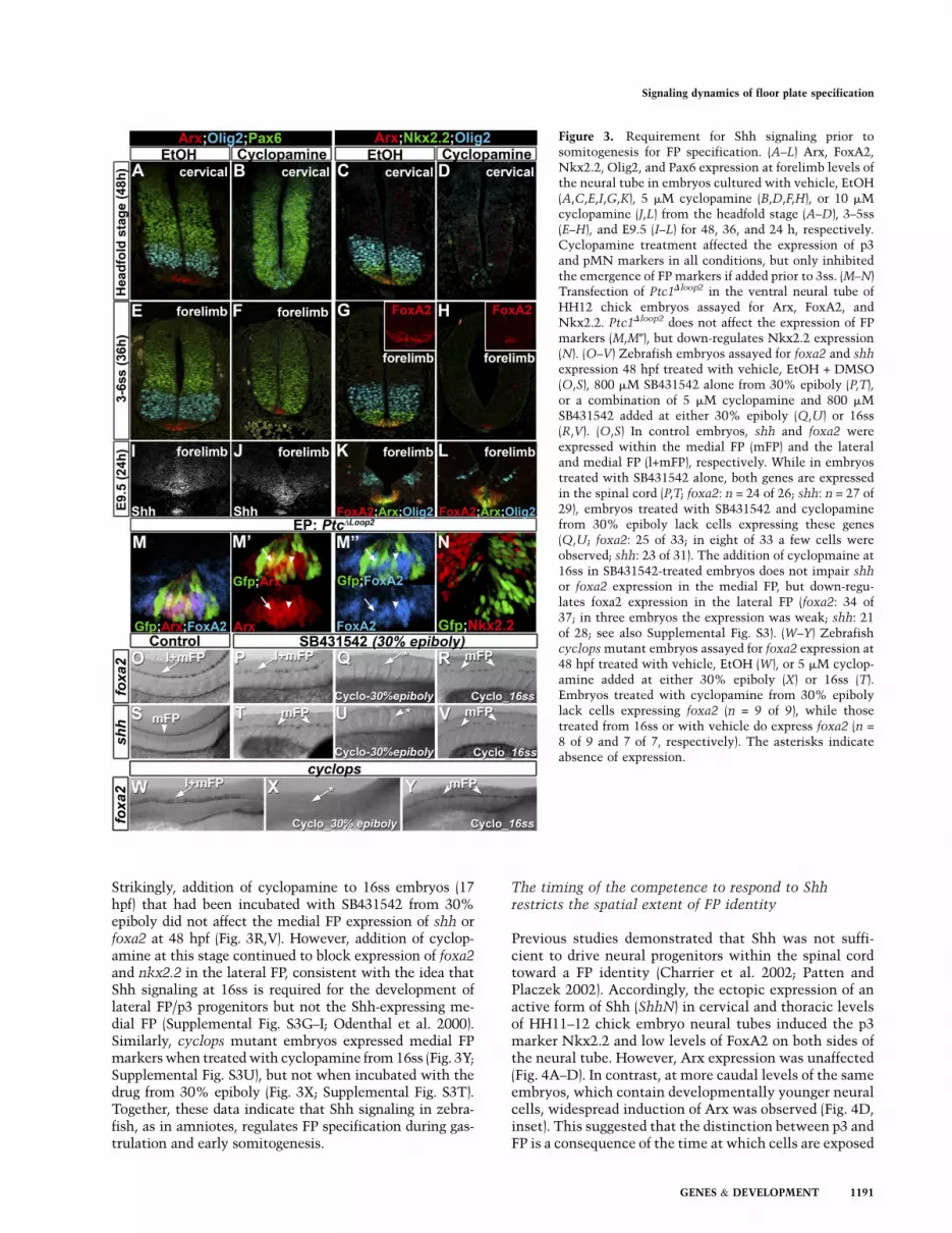
Strikingly, addition of cyclopamine to 16ss embryos (17hpf) that had been incubated with SB431542 from 30%epiboly did not affect the medial FP expression of shh orfoxa2 at 48 hpf (Fig. 3R,V). However, addition of cyclop-amine at this stage continued to block expression of foxa2and nkx2.2 in the lateral FP, consistent with the idea thatShh signaling at 16ss is required for the development oflateral FP/p3 progenitors but not the Shh-expressing me-dial FP (Supplemental Fig. S3G–I; Odenthal et al. 2000).Similarly, cyclops mutant embryos expressed medial FPmarkers when treated with cyclopamine from 16ss (Fig. 3Y;Supplemental Fig. S3U), but not when incubated with thedrug from 30% epiboly (Fig. 3X; Supplemental Fig. S3T).Together, these data indicate that Shh signaling in zebra-fish, as in amniotes, regulates FP specification during gas-trulation and early somitogenesis.
The timing of the competence to respond to Shhrestricts the spatial extent of FP identity
Previous studies demonstrated that Shh was not suffi-cient to drive neural progenitors within the spinal cordtoward a FP identity (Charrier et al. 2002; Patten andPlaczek 2002). Accordingly, the ectopic expression of anactive form of Shh (ShhN) in cervical and thoracic levelsof HH11–12 chick embryo neural tubes induced the p3marker Nkx2.2 and low levels of FoxA2 on both sides ofthe neural tube. However, Arx expression was unaffected(Fig. 4A–D). In contrast, at more caudal levels of the sameembryos, which contain developmentally younger neuralcells, widespread induction of Arx was observed (Fig. 4D,inset). This suggested that the distinction between p3 andFP is a consequence of the time at which cells are exposed
Figure 3. Requirement for Shh signaling prior tosomitogenesis for FP specification. (A–L) Arx, FoxA2,Nkx2.2, Olig2, and Pax6 expression at forelimb levels ofthe neural tube in embryos cultured with vehicle, EtOH(A,C,E,I,G,K), 5 mM cyclopamine (B,D,F,H), or 10 mMcyclopamine (J,L) from the headfold stage (A–D), 3–5ss(E–H), and E9.5 (I–L) for 48, 36, and 24 h, respectively.Cyclopamine treatment affected the expression of p3and pMN markers in all conditions, but only inhibitedthe emergence of FP markers if added prior to 3ss. (M–N)Transfection of Ptc1Dloop2 in the ventral neural tube ofHH12 chick embryos assayed for Arx, FoxA2, andNkx2.2. Ptc1Dloop2 does not affect the expression of FPmarkers (M,M0), but down-regulates Nkx2.2 expression(N). (O–V) Zebrafish embryos assayed for foxa2 and shh
expression 48 hpf treated with vehicle, EtOH + DMSO(O,S), 800 mM SB431542 alone from 30% epiboly (P,T),or a combination of 5 mM cyclopamine and 800 mMSB431542 added at either 30% epiboly (Q,U) or 16ss(R,V). (O,S) In control embryos, shh and foxa2 wereexpressed within the medial FP (mFP) and the lateraland medial FP (l+mFP), respectively. While in embryostreated with SB431542 alone, both genes are expressedin the spinal cord (P,T; foxa2: n = 24 of 26; shh: n = 27 of29), embryos treated with SB431542 and cyclopaminefrom 30% epiboly lack cells expressing these genes(Q,U; foxa2: 25 of 33; in eight of 33 a few cells wereobserved; shh: 23 of 31). The addition of cyclopmaine at16ss in SB431542-treated embryos does not impair shhor foxa2 expression in the medial FP, but down-regu-lates foxa2 expression in the lateral FP (foxa2: 34 of37; in three embryos the expression was weak; shh: 21of 28; see also Supplemental Fig. S3). (W–Y) Zebrafishcyclops mutant embryos assayed for foxa2 expression at48 hpf treated with vehicle, EtOH (W), or 5 mM cyclop-amine added at either 30% epiboly (X) or 16ss (T).Embryos treated with cyclopamine from 30% epibolylack cells expressing foxa2 (n = 9 of 9), while thosetreated from 16ss or with vehicle do express foxa2 (n =
8 of 9 and 7 of 7, respectively). The asterisks indicateabsence of expression.
Signaling dynamics of floor plate specification
GENES & DEVELOPMENT 1191
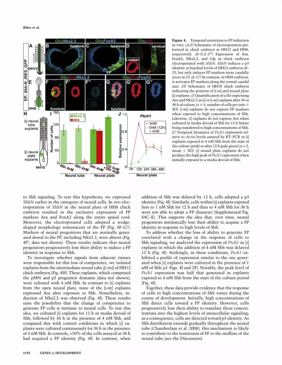
to Shh signaling. To test this hypothesis, we expressedShhN earlier in the ontogeny of neural cells. In ovo elec-troporation of ShhN in the neural plate of HH8 chickembryos resulted in the exclusive expression of FPmarkers Arx and FoxA2 along the entire spinal cord.Moreover, the electroporated cells adopted a wedge-shaped morphology reminiscent of the FP (Fig. 4F–G0).Markers of neural progenitors that are normally gener-ated dorsal to the FP, including Nkx2.2, were absent (Fig.4F0; data not shown). These results indicate that neuralprogenitors progressively lose their ability to induce a FPidentity in response to Shh.
To investigate whether signals from adjacent tissueswere responsible for this loss of competence, we isolatedexplants from the intermediate neural tube ([i-nt]) of HH12chick embryos (Fig. 4H). These explants, which comprisedthe pMN and p2 progenitor domains (data not shown),were cultured with 4 nM Shh. In contrast to [i] explantsfrom the open neural plate, none of the [i-nt] explantsexpressed Arx after exposure to Shh. Nonetheless, in-duction of Nkx2.2 was observed (Fig. 4I). These resultsraise the possibility that the change of competence togenerate FP cells is intrinsic to neural cells. To test thisidea, we cultured [i] explants for 12 h in media devoid ofShh, followed by 36 h in the presence of 4 nM Shh, andcompared this with control conditions in which [i] ex-plants were cultured continuously for 36 h in the presenceof 4 nM Shh. In controls, >50% of the cells assayed at 36 hhad acquired a FP identity (Fig. 4I). In contrast, when
addition of Shh was delayed by 12 h, cells adopted a p3identity (Fig. 4I). Similarly, cells within [i] explants exposedfirst to 1 nM Shh for 12 h and then to 4 nM Shh for 36 hwere not able to adopt a FP character (Supplemental Fig.S4C–E). This supports the idea that, over time, neuralprogenitors intrinsically lose their ability to acquire a FPidentity in response to high levels of Shh.
To address whether the loss of ability to generate FPcorrelated with a change in the response of cells toShh signaling, we analyzed the expression of Ptch1 in [i]explants in which the addition of 4 nM Shh was delayed12 h (Fig. 4J). Strikingly, in these conditions, Ptch1 ex-hibited a profile of expression similar to the one gener-ated when [i] explants were cultured in the presence of 1nM of Shh (cf. Figs. 4J and 2F). Notably, the peak level ofPtch1 expression was half that generated in explantsexposed to 4 nM Shh from the start of the culture period(Fig. 4J).
Together, these data provide evidence that the responseof cells to high concentrations of Shh varies during thecourse of development. Initially, high concentrations ofShh direct cells toward a FP identity. However, cellsprogressively lose their ability to translate these concen-trations into the highest levels of intracellular signaling;as a consequence, cells are directed toward p3 identity. AsShh distribution extends gradually throughout the neuraltube (Chamberlain et al. 2008), this mechanism is likelyto contribute to the restriction of FP to the midline of theneural tube (see the Discussion).
Figure 4. Temporal restriction to FP inductionin vivo. (A,E) Schematic of electroporation per-formed in chick embryos at HH12 and HH8,respectively. (B–D,E–F 0) Expression of Arx,FoxA2, Nkx2.2, and Gfp in chick embryoselectroporated with ShhN. ShhN induces a p3identity at brachial levels of HH12 embryos (B–D), but only induces FP markers more caudally(inset in D). (E–G0) In contrast, in HH8 embryos,it activates FP markers along the rostral–caudalaxis. (H) Schematic of HH10 chick embryosindicating the position of [i-nt] and neural plate[i] explants. (I) Quantification of cells expressingArx and Nkx2.2 in [i] or [i-nt] explants after 36 or48 h of culture (n $ 4, number of cells per unit 6
SD). [i-nt] explants do not express FP markerswhen exposed to high concentrations of Shh.Likewise, [i] explants do not express Arx whencultured in media devoid of Shh for 12 h beforebeing transferred to high concentrations of Shh.(J ) Temporal dynamics of Ptch1 expression rel-ative to Actin levels assayed by RT–PCR in [i]explants exposed to 4 nM Shh from the start ofthe culture (pink) or after 12 h (pale green) (n = 2,mean 6 SD). [i] neural plate explants do notproduce the high peak of Ptch1 expression wheninitially exposed to a media devoid of Shh.
Ribes et al.
1192 GENES & DEVELOPMENT
Specification of FP cells requires down-regulationof Shh signaling
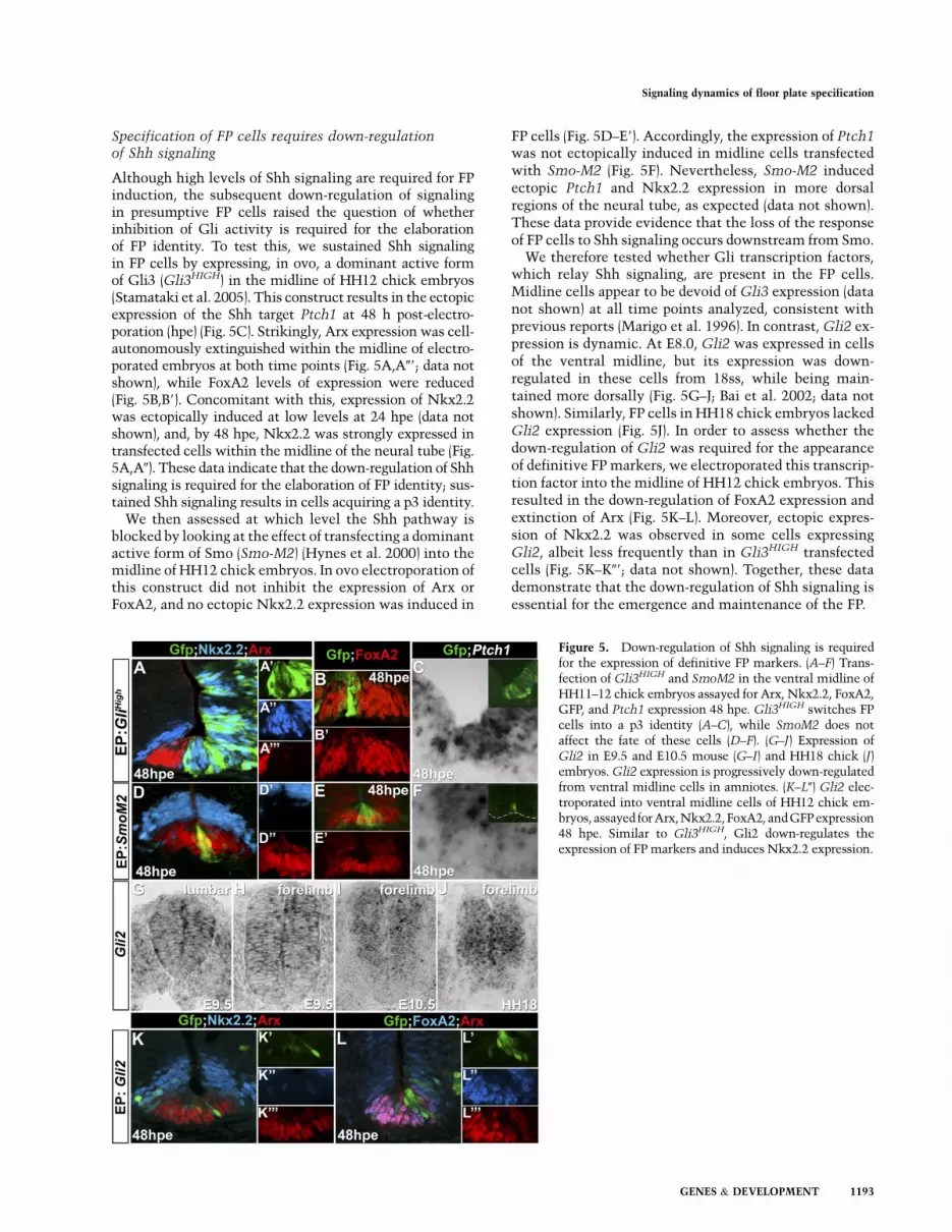
Although high levels of Shh signaling are required for FPinduction, the subsequent down-regulation of signalingin presumptive FP cells raised the question of whetherinhibition of Gli activity is required for the elaborationof FP identity. To test this, we sustained Shh signalingin FP cells by expressing, in ovo, a dominant active formof Gli3 (Gli3HIGH) in the midline of HH12 chick embryos(Stamataki et al. 2005). This construct results in the ectopicexpression of the Shh target Ptch1 at 48 h post-electro-poration (hpe) (Fig. 5C). Strikingly, Arx expression was cell-autonomously extinguished within the midline of electro-porated embryos at both time points (Fig. 5A,A09; data notshown), while FoxA2 levels of expression were reduced(Fig. 5B,B9). Concomitant with this, expression of Nkx2.2was ectopically induced at low levels at 24 hpe (data notshown), and, by 48 hpe, Nkx2.2 was strongly expressed intransfected cells within the midline of the neural tube (Fig.5A,A0). These data indicate that the down-regulation of Shhsignaling is required for the elaboration of FP identity; sus-tained Shh signaling results in cells acquiring a p3 identity.
We then assessed at which level the Shh pathway isblocked by looking at the effect of transfecting a dominantactive form of Smo (Smo-M2) (Hynes et al. 2000) into themidline of HH12 chick embryos. In ovo electroporation ofthis construct did not inhibit the expression of Arx orFoxA2, and no ectopic Nkx2.2 expression was induced in
FP cells (Fig. 5D–E9). Accordingly, the expression of Ptch1was not ectopically induced in midline cells transfectedwith Smo-M2 (Fig. 5F). Nevertheless, Smo-M2 inducedectopic Ptch1 and Nkx2.2 expression in more dorsalregions of the neural tube, as expected (data not shown).These data provide evidence that the loss of the responseof FP cells to Shh signaling occurs downstream from Smo.
We therefore tested whether Gli transcription factors,which relay Shh signaling, are present in the FP cells.Midline cells appear to be devoid of Gli3 expression (datanot shown) at all time points analyzed, consistent withprevious reports (Marigo et al. 1996). In contrast, Gli2 ex-pression is dynamic. At E8.0, Gli2 was expressed in cellsof the ventral midline, but its expression was down-regulated in these cells from 18ss, while being main-tained more dorsally (Fig. 5G–J; Bai et al. 2002; data notshown). Similarly, FP cells in HH18 chick embryos lackedGli2 expression (Fig. 5J). In order to assess whether thedown-regulation of Gli2 was required for the appearanceof definitive FP markers, we electroporated this transcrip-tion factor into the midline of HH12 chick embryos. Thisresulted in the down-regulation of FoxA2 expression andextinction of Arx (Fig. 5K–L). Moreover, ectopic expres-sion of Nkx2.2 was observed in some cells expressingGli2, albeit less frequently than in Gli3HIGH transfectedcells (Fig. 5K–K09; data not shown). Together, these datademonstrate that the down-regulation of Shh signaling isessential for the emergence and maintenance of the FP.
Figure 5. Down-regulation of Shh signaling is requiredfor the expression of definitive FP markers. (A–F) Trans-fection of Gli3HIGH and SmoM2 in the ventral midline ofHH11–12 chick embryos assayed for Arx, Nkx2.2, FoxA2,GFP, and Ptch1 expression 48 hpe. Gli3HIGH switches FPcells into a p3 identity (A–C), while SmoM2 does notaffect the fate of these cells (D–F). (G–J) Expression ofGli2 in E9.5 and E10.5 mouse (G–I) and HH18 chick (J)embryos. Gli2 expression is progressively down-regulatedfrom ventral midline cells in amniotes. (K–L0) Gli2 elec-troporated into ventral midline cells of HH12 chick em-bryos, assayed for Arx,Nkx2.2,FoxA2, andGFP expression48 hpe. Similar to Gli3HIGH, Gli2 down-regulates theexpression of FP markers and induces Nkx2.2 expression.
Signaling dynamics of floor plate specification
GENES & DEVELOPMENT 1193
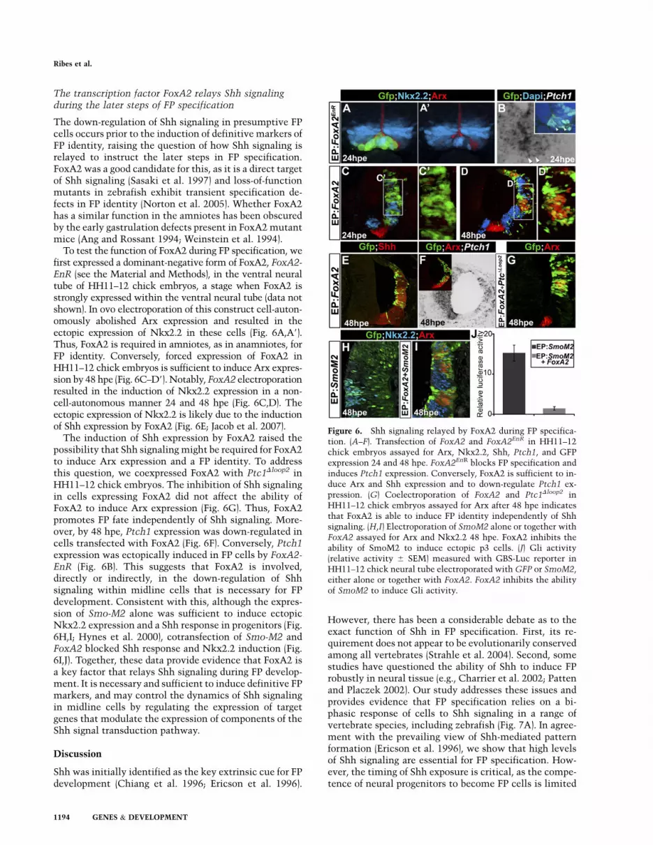
The transcription factor FoxA2 relays Shh signalingduring the later steps of FP specification
The down-regulation of Shh signaling in presumptive FPcells occurs prior to the induction of definitive markers ofFP identity, raising the question of how Shh signaling isrelayed to instruct the later steps in FP specification.FoxA2 was a good candidate for this, as it is a direct targetof Shh signaling (Sasaki et al. 1997) and loss-of-functionmutants in zebrafish exhibit transient specification de-fects in FP identity (Norton et al. 2005). Whether FoxA2has a similar function in the amniotes has been obscuredby the early gastrulation defects present in FoxA2 mutantmice (Ang and Rossant 1994; Weinstein et al. 1994).
To test the function of FoxA2 during FP specification, wefirst expressed a dominant-negative form of FoxA2, FoxA2-EnR (see the Material and Methods), in the ventral neuraltube of HH11–12 chick embryos, a stage when FoxA2 isstrongly expressed within the ventral neural tube (data notshown). In ovo electroporation of this construct cell-auton-omously abolished Arx expression and resulted in theectopic expression of Nkx2.2 in these cells (Fig. 6A,A9).Thus, FoxA2 is required in amniotes, as in anamniotes, forFP identity. Conversely, forced expression of FoxA2 inHH11–12 chick embryos is sufficient to induce Arx expres-sion by 48 hpe (Fig. 6C–D9). Notably, FoxA2 electroporationresulted in the induction of Nkx2.2 expression in a non-cell-autonomous manner 24 and 48 hpe (Fig. 6C,D). Theectopic expression of Nkx2.2 is likely due to the inductionof Shh expression by FoxA2 (Fig. 6E; Jacob et al. 2007).
The induction of Shh expression by FoxA2 raised thepossibility that Shh signaling might be required for FoxA2to induce Arx expression and a FP identity. To addressthis question, we coexpressed FoxA2 with Ptc1Dloop2 inHH11–12 chick embryos. The inhibition of Shh signalingin cells expressing FoxA2 did not affect the ability ofFoxA2 to induce Arx expression (Fig. 6G). Thus, FoxA2promotes FP fate independently of Shh signaling. More-over, by 48 hpe, Ptch1 expression was down-regulated incells transfected with FoxA2 (Fig. 6F). Conversely, Ptch1expression was ectopically induced in FP cells by FoxA2-EnR (Fig. 6B). This suggests that FoxA2 is involved,directly or indirectly, in the down-regulation of Shhsignaling within midline cells that is necessary for FPdevelopment. Consistent with this, although the expres-sion of Smo-M2 alone was sufficient to induce ectopicNkx2.2 expression and a Shh response in progenitors (Fig.6H,I; Hynes et al. 2000), cotransfection of Smo-M2 andFoxA2 blocked Shh response and Nkx2.2 induction (Fig.6I,J). Together, these data provide evidence that FoxA2 isa key factor that relays Shh signaling during FP develop-ment. It is necessary and sufficient to induce definitive FPmarkers, and may control the dynamics of Shh signalingin midline cells by regulating the expression of targetgenes that modulate the expression of components of theShh signal transduction pathway.
Discussion
Shh was initially identified as the key extrinsic cue for FPdevelopment (Chiang et al. 1996; Ericson et al. 1996).
However, there has been a considerable debate as to theexact function of Shh in FP specification. First, its re-quirement does not appear to be evolutionarily conservedamong all vertebrates (Strahle et al. 2004). Second, somestudies have questioned the ability of Shh to induce FProbustly in neural tissue (e.g., Charrier et al. 2002; Pattenand Placzek 2002). Our study addresses these issues andprovides evidence that FP specification relies on a bi-phasic response of cells to Shh signaling in a range ofvertebrate species, including zebrafish (Fig. 7A). In agree-ment with the prevailing view of Shh-mediated patternformation (Ericson et al. 1996), we show that high levelsof Shh signaling are essential for FP specification. How-ever, the timing of Shh exposure is critical, as the compe-tence of neural progenitors to become FP cells is limited
Figure 6. Shh signaling relayed by FoxA2 during FP specifica-tion. (A–F). Transfection of FoxA2 and FoxA2EnR in HH11–12chick embryos assayed for Arx, Nkx2.2, Shh, Ptch1, and GFPexpression 24 and 48 hpe. FoxA2EnR blocks FP specification andinduces Ptch1 expression. Conversely, FoxA2 is sufficient to in-duce Arx and Shh expression and to down-regulate Ptch1 ex-pression. (G) Coelectroporation of FoxA2 and Ptc1Dloop2 inHH11–12 chick embryos assayed for Arx after 48 hpe indicatesthat FoxA2 is able to induce FP identity independently of Shhsignaling. (H,I) Electroporation of SmoM2 alone or together withFoxA2 assayed for Arx and Nkx2.2 48 hpe. FoxA2 inhibits theability of SmoM2 to induce ectopic p3 cells. (J) Gli activity(relative activity 6 SEM) measured with GBS-Luc reporter inHH11–12 chick neural tube electroporated with GFP or SmoM2,either alone or together with FoxA2. FoxA2 inhibits the abilityof SmoM2 to induce Gli activity.
Ribes et al.
1194 GENES & DEVELOPMENT
to gastrulation and early somitogenesis stages. Progenitorsreceiving high levels of signaling outside this time windowadopt a p3 neural progenitor identity. Subsequently, how-ever, the elaboration of FP identity requires the duration ofShh signaling to be time-restricted, and sustained signalingwithin prospective FP progenitors leads them to adopt a p3
identity. Thus, three parameters—the level, timing, andduration of Shh signaling—distinguish FP cells from otherventral neural progenitors and control distinct transcrip-tional programs in these cell types.
A unified scheme for FP specification by Shhsignaling in vertebrates
Two contrasting models of FP development have beenformulated (for reviews, see Le Douarin and Halpern2000; Placzek et al. 2000; Strahle et al. 2004; Placzekand Briscoe 2005). In the first, FP cells are specified by aninductive signal, Shh, secreted from the notochord. Thismodel stipulates that Shh induces FP development in amanner analogous to its role in the induction and pat-terning of ventral neural progenitors (Placzek et al. 2000;Placzek and Briscoe 2005). Accordingly, FP induction isproposed to be the consequence of exposure to the highestlevels of Shh. In the second model, the induction of FPcells occurs within the organizer/node during gastrula-tion, and is independent of more anterior interactionsbetween the notochord and the ventral neural plate (LeDouarin and Halpern 2000; Strahle et al. 2004; Placzekand Briscoe 2005). In this model, Shh has little, if any, rolein inducing FP identity (Varga et al. 2001), or it acts as apermissive, or survival, signal that prevents apoptosis ofFP precursors (Charrier et al. 2001).
Our data strongly support the first model, and indicatethat Shh signaling is both necessary and sufficient for theinduction of FP identity in amniotes, consistent withprevious loss- and gain-of-function studies (Placzek et al.1991; Roelink et al. 1995; Chiang et al. 1996; Ericson et al.1996). Importantly, however, our data identify a restricteddevelopmental time window, spanning gastrulation andearly somitogenesis stages, when Shh signaling is able totrigger the emergence of FP identity within the openneural plate. This suggests an explanation for studies thatfailed to find a requirement of the notochord or Shh in FPinduction (Le Douarin and Halpern 2000; Strahle et al.2004). Indeed, exposure of chick neural cells to Shh afterthe initiation of somitogenesis does not induce FP for-mation (Teillet et al. 1998; Charrier et al. 2002; Patten andPlaczek 2002), and removal of notochord in chick em-bryos does not disrupt FP formation along most of theembryonic axis (van Straaten et al. 1989; Yamada et al.1991; Teillet et al. 1998).
How the time window during which Shh signaling isable to induce FP development is defined remains to bedetermined. However, the mechanism appears to be auto-nomous to neural tissue. Both anterior neural tube ex-plants deprived of surrounding tissues and neural plateexplants cultured for 12 h in medium lacking sufficientShh are unable to give rise to FP cells in response to Shhsignaling. One possibility is that an intrinsic clock isestablished within newly induced neural cells, and thissets a time limit for how long Shh is able to promote FPidentity. However, this does not exclude the possibilitythat the competence of cells to generate FP might dependon interactions with signaling pathways active withinneural tissue. For example, signals emanating dorsally,
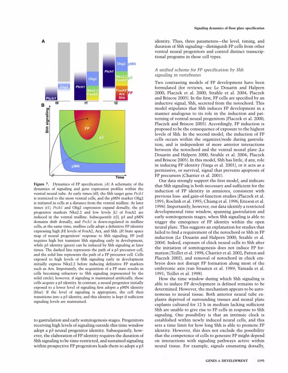
Figure 7. Dynamics of FP specification. (A) A schematic of thedynamics of signaling and gene expression profiles within theventral neural tube. At early times (t0), the Shh target gene Ptch1
is restricted to the most ventral cells, and the pMN marker Olig2is initiated in cells at a distance from the ventral midline. At latertimes (t1), Ptch1 and Olig2 expression expand dorsally; the p3progenitor markers Nkx2.2 and low levels (L) of FoxA2 areinduced in the ventral midline. Subsequently (t2), p3 and pMNdomains shift dorsally, and Ptch1 is down-regulated in midlinecells; at the same time, midline cells adopt a definitive FP identityexpressing high (H) levels of FoxA2, Arx, and Shh. (B) State spacemap of neural progenitors’ response to Shh signaling. FP (red)requires high but transient Shh signaling early in development,while p3 identity (green) can be induced by Shh signaling at latertimes. The dashed line represents the path of a p3 precursor cell,and the solid line represents the path of a FP precursor cell. Cellsexposed to high levels of Shh signaling early in developmentinitially express Nkx2.2 before inducing definitive FP markerssuch as Arx. Importantly, the acquisition of a FP state results incells becoming refractory to Shh signaling (represented by thesolid circle); however, if signaling is maintained artificially, thesecells acquire a p3 identity. In contrast, a neural progenitor initiallyexposed to a lower level of signaling first adopts a pMN identity(blue). If the level of signaling is appropriate, the cell thentransitions into a p3 identity, and this identity is kept if sufficientsignaling levels are maintained.
Signaling dynamics of floor plate specification
GENES & DEVELOPMENT 1195
such as Wnts and bone morphogenetic proteins (BMPs),might affect the ability of cells to respond to Shh (Daleet al. 1999; Patten and Placzek 2002; Joksimovic et al.2009); in this context, it is notable that exposure to theBMP antagonist Chordin enhances the ability of Shh toinduce FP (Dale et al. 1999; Patten and Placzek 2002).Moreover, regardless of how the change in competence ofneural tissue is achieved, this process is likely to be re-sponsible for the restriction of FP markers to the midlineof the neural tube. The amplitude of the Shh gradientincreases in the spinal cord as development proceeds(Chamberlain et al. 2008). Thus, prospective p3 cells areexposed to at least similar levels of ligand as prospective FPcells; however, the time at which p3 cells receive thesehigh concentrations of Shh is later than the presumptive FPcells (Chamberlain et al. 2008). This temporal–spatial con-straint on when cells are exposed to Shh would thereby pro-vide a mechanism to define the size of the FP, and is rem-iniscent of the way in which the timing of Wnt signalingdetermines the size of the heart field (Ueno et al. 2007).
The apparent species differences in FP induction havealso led to uncertainty over the molecular mechanism ofFP development. Although, in zebrafish, Nodal signalingis credited with the principal role in FP induction, inamniotes, it is likely to play only a minor role. On the onehand, Nodal potentiates the ability of Shh to induce FPidentity in the midline of the chick forebrain and mid-brain (Patten et al. 2003). On the other hand, FP markersare induced prematurely in the anterior epiblast of E7.5Nodal�/� mouse embryos (Camus et al. 2006). Moreover,FP induction is observed in mouse embryos formed fromcells deficient for nodal (Varlet et al. 1997) or its down-stream effector, Smad2 (Heyer et al. 1999), or treated witha nodal inhibitor (data not shown). In contrast to Nodal,our data suggest that the role of Shh signaling duringFP specification in teleosts and amniotes shares sev-eral features. The transience of Shh signaling within FPprogenitors indicated by the temporal profile of the ex-pression of the Shh target genes Gli1 and Ptch1 is sim-ilar in the FP of amniotes and zebrafish species (Fig. 2;Concordet et al. 1996; Lewis et al. 1999; Karlstrom et al.2003). Moreover, in each species tested, FP developmentrequires Shh signaling during a time window that spansgastrulation and early somitogenesis (Fig. 3). Importantly,this time window coincides with when Nodal signaling isrequired for zebrafish FP induction (Tian et al. 2003).Furthermore, FP-inducing signals appear to converge onthe regulation of FoxA2 in teleosts and amniotes (Strahleet al. 1993; Sasaki et al. 1997; Muller et al. 2000). Takentogether, the data indicate that, in all vertebrates, FPinduction takes place in a brief time window during thecourse of gastrulation, and the extrinsic signals involvedin this process regulate FoxA2 expression. The differencebetween species resides mainly in the relative contribu-tion of each signal. It is therefore tempting to hypothesizethat both Shh and Nodal signals were involved in FPspecification in the common ancestor of vertebrates. Sub-sequently, the relative importance of each signal changedduring the evolution of individual species (see alsoRastegar et al. 2002). Detailed analysis of the regulatory
elements directing expression of FoxA2 in different spe-cies should shed further light on this hypothesis (Sasakiet al. 1997).
Distinct dynamics of Shh signaling specify FP cells
FP development is triggered by high levels of Shh thatresult in the rapid induction of Nkx2.2 and Foxa2. In con-trast, cells fated to generate the adjacent p3 progenitor do-main are initially exposed to lower levels of signaling, andinduce Olig2 prior to Nkx2.2. However, the full elabora-tion of FP identity relies on a subsequent decrease in in-tracellular Shh signaling because FP cells switch to a p3identity if signaling is artificially sustained. Thus a dy-namic transcriptional program, produced by the differentlevels and timing of signaling, distinguishes FP cells fromp3 progenitors. How is the appropriate profile of Shh sig-naling achieved in the FP? During gastrulation and earlysomitogenesis, the concentration of Shh in neural tissuesis low (Chamberlain et al. 2008). Nevertheless, the lowlevels of the negative regulator of Shh signaling, Ptch1, inneural tissue at these early times may allow the induc-tion of high levels of signaling in response to low amountsof Shh. In addition, the Shh-interacting transmembraneproteins Gas1 and Cdo may sensitize progenitors to Shh(Allen et al. 2007; Martinelli and Fan 2007). Embryosmutant for both Cdo and Gas1 lack FP induction, and theforced expression of these factors in chick neural tube issufficient to direct cell-autonomous p3 cells toward a FPidentity (Tenzen et al. 2006; Allen et al. 2007; data notshown). Importantly, the expression of these proteins istightly regulated. In the ventral neural tube, Cdo tran-scripts are restricted to FP cells between E9.0 and E11.5.Gas1 is first induced throughout the neural tube at E8.5,and becomes progressively down-regulated within theventral neural tube. Prior to E9.5, therefore, prospectiveFP cells express the signal enhancers Cdo and Gas1(Tenzen et al. 2006; Allen et al. 2007).
The later decrease in signaling as FP identity is elabo-rated might be partly explained by the rapid down-regulation of Gas1. In addition, the repression of Gli2expression is likely to be essential. Gli2 plays the predo-minant role in relaying the Shh signal during FP induc-tion (Matise et al. 1998; Bai and Joyner 2001; Bai et al.2002), and its expression profile follows the dynamics ofShh signaling in the midline of both chicks and mice(Fig. 5; Bai and Joyner 2001; Lei et al. 2004). Thus repres-sion of Gli2, and possibly other components of the Shhsignaling pathway, in FP cells provides a mechanism thatwould result in the extinction of signaling in these cells.The down-regulation of these factors appears to be trig-gered by the transcription factor FoxA2 (Fig. 6; data notshown). This suggests a model in which Shh signalingdirectly induces expression of FoxA2 (Sasaki and Hogan1994), which then induces the expression of Shh itself(Ruiz i Altaba et al. 1995; Chang et al. 1997; Jeong andEpstein 2003) and participates in the extinction of Shhsignaling within these cells, perhaps by inducing targetgenes that directly regulate components of the Shh sig-nal transduction pathway. It is noteworthy that other
Ribes et al.
1196 GENES & DEVELOPMENT
forkhead transcription factors—including Foxp2, Foxp4,and Foxj1—are expressed in FP cells (Takahashi et al.2008; Yu et al. 2008; Morikawa et al. 2009; data notshown), raising the possibility that several members ofthis family participate in shaping the sensitivity of FPcells to Shh signaling.
A common strategy of morphogen interpretationfor the generation of cellular diversity
The distinct dynamics of Shh signaling that are requiredfor FP development add a new dimension to the interpre-tation of the Shh morphogen. Previous studies in the neu-ral tube suggested that concurrent increases in the leveland duration of Shh signaling lead to the progressive spec-ification of cell identities (Ericson et al. 1996; Jeong andMcMahon 2005; Dessaud et al. 2007). In contrast, the datapresented here indicate that cells exposed to the highestShh concentrations at early developmental times gener-ate a transient peak of signaling activity and produce FP.Then, at later times, cells transform increasing concen-trations of Shh into longer periods of signaling to generateprogressively more ventral neural progenitor domains(Dessaud et al. 2007). This identifies two modes of Shhmorphogen interpretation (Fig. 7A,B). These two modesalso appear to be deployed in other tissues. In the ver-tebrate limb bud, Gli activity exhibits a spatial and tem-poral gradient within the precursors of digits 2–4 (Ahnand Joyner 2004). Accordingly, the specification of thesestructures requires a prolonged exposure to Shh, suchthat digit 4 requires the longest exposure (Towers et al.2008; Zhu et al. 2008). In contrast, the specification ofdigit 5 appears similar to the specification of FP by Shhsignaling. The precursors of this digit, although exposedto high levels of Shh early in development, become inde-pendent of Shh signaling much sooner than other digitprecursors (Zhu et al. 2008). Moreover, a down-regulationof the Shh target genes Gli1 and Ptch1 in the precursorsof this digit (Ahn and Joyner 2004) is accompanied byan inhibition of Gli2 expression (Marigo et al. 1996).Furthermore, similar to FP cells, digit 5 precursors actas a source of Shh (Harfe et al. 2004). In the Drosophilawing disc, Hh protein secreted from the posterior com-partment induces the transcription factor engrailed(En) in a small territory in the anterior compartment(Strigini and Cohen 1997; Basler 2000). Expression of Enhas been associated with the down-regulation of Ptch andCi (Basler 2000). Thus, by analogy to vertebrates, it ispossible that cells responding to the highest levels of Hhsignaling in the anterior compartment become refractoryto the signal. Together, the data suggest a general meansto augment the role of a morphogen. This strategy canbe used in a wide range of development settings and canbe elaborated further, as exemplified by the triphasic(Off–On–Off) response of ventral pancreatic precursorsto BMP–Smad4 signaling (Wandzioch and Zaret 2009).Exploiting the dynamics of signal responses in this wayextends the potential of a single signal to control differ-ential transcriptional programs and cell identity duringembryogenesis.
Materials and methods
Immunohistochemistry and in situ hybridization
Antibody reagents and protocols have been described elsewhere(Ericson et al. 1997; Briscoe et al. 2000, 2001). The antibody forArx was kindly provided by J. Chelly (Poirier et al. 2004). Analysiswas carried out using a Leica TCS SP2 confocal microscope andprocessed with Adobe Photoshop 7.0 software (Adobe Systems). Insitu hybridization was performed as described (chicks and mice:Chotteau-Lelievre et al. 2006; zebrafish: Thisse et al. 1993) usingthe following probes: chick Ptch1 and Gli1-3 (C. Tabin); mousePtch1 and Gli1-3 (C.C. Hui); zebrafish foxa2 (axial) and shh
(P. Ingham).
Chick in ovo electroporation
All electroporation constructs were based on pCAGGS expres-sion vector engineered to bicistronically express nuclear targetedGFP (pCAGGS-IRES NLS-GFP). Gli3AHigh (Stamataki et al.2005), Gli2 (Schweitzer et al. 2000), Ptc1Dloop2 (Briscoe et al.2001), SmoM2 (Hynes et al. 2000), FoxA2 (Ferri et al. 2007),FoxA2EnR (Jacob et al. 2007), and ShhN (Roelink et al. 1995) havebeen described previously. HH8–12 embryos were electroporatedand incubated in ovo before dissecting and processing for eitherimmunohistochemistry or in situ hybridization.
Luciferase reporter assays
Luciferase measurements were performed using the Dual-Lucif-erase Reporter Assay System (Promega). Expression plasmids orempty vector were electroporated into HH10–12 chick embryostogether with the GBS-Luc firefly luciferase reporter (Sasaki et al.1997) and a Renilla luciferase reporter driven by a CMV promoter(Promega) for normalization. Assays were performed as describedpreviously (Stamataki et al. 2005).
Neural explants and RT–PCR
Explants isolated from HH10 chick embryos were cultured asdescribed (Yamada et al. 1993; Ericson et al. 1997). Cyclopamine(Toronto Research Chemicals) was dissolved in 96% ethanol.RNA was extracted using PicoPure RNA Isolation kit (Arcturus).cDNA was synthesized by SuperScriptII (Invitrogen) and quanti-tative PCR was performed using an ABI7500. The expressionof each gene was normalized to that of Actin. The following oli-gonucleotides were used: Actin, (fw) GAGAAATTGTGCGTGACATCA and (rev) CCTGAACCTCTCATTGCCA; Ptch1, (fw)TTTTTCTTTTCCTGGGCTTACTT and (rev) CATCTCTACCCGGGTAGTTC; Nkx2.2, (fw) ACCTTCCAAACGGGCATCand (rev) TGTAATGGGCGTTGTATTGC; Arx, (fw) TGGCCTCAGTAGCCTTACCT and (rev) AGCATTGAGAAACCTTCCAAA; FoxA2, (fw) AGCAGTCGCCCAACAAGATG and (rev)TCTGGCGGTAGAAGGGGAAG.
Embryo culture experiments
Mouse embryos with intact yolk sacs, dissected from timed preg-nant females, were cultured for 24 h in medium (rat serum, Tyrodesolution; 1:1 between E7.5 and E8.5; 2:1 at E9.5) in 8-mL tubes(2.5 mL of medium per tube; three embryos per tube). Cyclopaminewas used at a concentration of 5–10 mM as indicated. Cultureswere performed in a water-saturated roller-tube incubator at 37°C,5% CO2 and 5% O2 at E7.5, 20% O2 at E8.5, and 65% O2 at E9.5.After culture, embryos were fixed and processed. Gene expressionpatterns were always compared between embryos processed inthe same culture experiment in appropriate control conditions.
Signaling dynamics of floor plate specification
GENES & DEVELOPMENT 1197
Zebrafish embryos and drug treatments
Wild-type zebrafish embryos or cyclops mutant fish (Hatta et al.1991) were treated with vehicle (EtOH+ and/or DMSO) and 5 mMcyclopamine (Toronto Research Chemicals; stock solution at 10mM was prepared in 96% EtOH) at either 30% epiboly stage orat 16ss, and with 800 mM SB431542 at 30% epiboly stage (Sigma;stock solution at 60 mM in DMSO; Hagos and Dougan 2007)dissolved in Danieau solution. There were no differences in theneural expression of axial and shh between untreated and vehicle-treated embryos.
Acknowledgments
We thank members of the laboratory, especially NataschaBushati, John Jacob, and Euginia Piddini for helpful commentsand discussions. We are grateful to C.C. Hui for Ptch1 mutantembryos. We also thank Biological Services staff at NIMR forhelp with the mouse colonies. We thank T. Jessell, B. Han, andM. Mendelsohn for generating Olig2-cre mice, and the UCL FishFacility for supplying cyclops mutant fish. V.R. and S.T. aresupported by EMBO LTFs and N.S. is supported by Marie CurieFellowship. Work in the laboratory of J.B. is supported by theMRC (UK) and the Wellcome Trust (grant 080630). Work in theE.M. laboratory is supported by the Spanish Ministry of Educa-tion grant BFU2004-00455/BMC.
References
Ahn S, Joyner AL. 2004. Dynamic changes in the response ofcells to positive hedgehog signaling during mouse limbpatterning. Cell 118: 505–516.
Albert S, Muller F, Fischer N, Biellmann D, Neumann C, BladerP, Strahle U. 2003. Cyclops-independent floor plate differen-tiation in zebrafish embryos. Dev Dyn 226: 59–66.
Allen BL, Tenzen T, McMahon AP. 2007. The Hedgehog-bindingproteins Gas1 and Cdo cooperate to positively regulate Shhsignaling during mouse development. Genes Dev 21: 1244–1257.
Ang SL, Rossant J. 1994. HNF-3b is essential for node andnotochord formation in mouse development. Cell 78: 561–574.
Bai CB, Joyner AL. 2001. Gli1 can rescue the in vivo function ofGli2. Development 128: 5161–5172.
Bai CB, Auerbach W, Lee JS, Stephen D, Joyner AL. 2002. Gli2,but not Gli1, is required for initial Shh signaling and ectopicactivation of the Shh pathway. Development 129: 4753–4761.
Basler K. 2000. EMBO gold medal 1999. Waiting periods, in-structive signals and positional information. EMBO J 19:1168–1175.
Briscoe J, Pierani A, Jessell TM, Ericson J. 2000. A homeodomainprotein code specifies progenitor cell identity and neuronalfate in the ventral neural tube. Cell 101: 435–445.
Briscoe J, Chen Y, Jessell TM, Struhl G. 2001. A hedgehog-insensitive form of patched provides evidence for direct long-range morphogen activity of sonic hedgehog in the neuraltube. Mol Cell 7: 1279–1291.
Camus A, Perea-Gomez A, Moreau A, Collignon J. 2006.Absence of Nodal signaling promotes precocious neuraldifferentiation in the mouse embryo. Dev Biol 295: 743–755.
Chamberlain CE, Jeong J, Guo C, Allen BL, McMahon AP. 2008.Notochord-derived Shh concentrates in close associationwith the apically positioned basal body in neural target cellsand forms a dynamic gradient during neural patterning.Development 135: 1097–1106.
Chang BE, Blader P, Fischer N, Ingham PW, Strahle U. 1997.Axial (HNF3b) and retinoic acid receptors are regulators ofthe zebrafish sonic hedgehog promoter. EMBO J 16: 3955–3964.
Charrier JB, Lapointe F, Le Douarin NM, Teillet MA. 2001. Anti-apoptotic role of Sonic hedgehog protein at the early stages ofnervous system organogenesis. Development 128: 4011–4020.
Charrier JB, Lapointe F, Le Douarin NM, Teillet MA. 2002. Dualorigin of the floor plate in the avian embryo. Development129: 4785–4796.
Chiang C, Litingtung Y, Lee E, Young KE, Corden JL, WestphalH, Beachy PA. 1996. Cyclopia and defective axial patterningin mice lacking Sonic hedgehog gene function. Nature 383:407–413.
Chotteau-Lelievre A, Dolle P, Gofflot F. 2006. Expression anal-ysis of murine genes using in situ hybridization with radio-active and non radioactively labeled probes. In Methods of
molecular biology: In situ hybridization protocols 3rd ed.(ed.IA Darby, TD Hewitson), pp. 61–87. Humana Press, Totowa, NJ.
Concordet JP, Lewis KE, Moore JW, Goodrich LV, Johnson RL,Scott MP, Ingham PW. 1996. Spatial regulation of a zebrafishpatched homologue reflects the roles of sonic hedgehog andprotein kinase A in neural tube and somite patterning.Development 122: 2835–2846.
Cooper MK, Porter JA, Young KE, Beachy PA. 1998. Teratogen-mediated inhibition of target tissue response to Shh signal-ing. Science 280: 1603–1607.
Dale K, Sattar N, Heemskerk J, Clarke JD, Placzek M, Dodd J.1999. Differential patterning of ventral midline cells by axialmesoderm is regulated by BMP7 and chordin. Development
126: 397–408.Davidson EH, Rast JP, Oliveri P, Ransick A, Calestani C, Yuh
CH, Minokawa T, Amore G, Hinman V, Arenas-Mena C,et al. 2002. A genomic regulatory network for development.Science 295: 1669–1678.
Dessaud E, Yang LL, Hill K, Cox B, Ulloa F, Ribeiro A, Mynett A,Novitch BG, Briscoe J. 2007. Interpretation of the sonichedgehog morphogen gradient by a temporal adaptationmechanism. Nature 450: 717–720.
Dessaud E, McMahon AP, Briscoe J. 2008. Pattern formation in thevertebrate neural tube: A sonic hedgehog morphogen-regulatedtranscriptional network. Development 135: 2489–2503.
Ericson J, Morton S, Kawakami A, Roelink H, Jessell TM. 1996.Two critical periods of Sonic Hedgehog signaling required forthe specification of motor neuron identity. Cell 87: 661–673.
Ericson J, Rashbass P, Schedl A, Brenner-Morton S, KawakamiA, van Heyningen V, Jessell TM, Briscoe J. 1997. Pax6controls progenitor cell identity and neuronal fate in re-sponse to graded Shh signaling. Cell 90: 169–180.
Ferri AL, Lin W, Mavromatakis YE, Wang JC, Sasaki H, WhitsettJA, Ang SL. 2007. Foxa1 and Foxa2 regulate multiple phasesof midbrain dopaminergic neuron development in a dosage-dependent manner. Development 134: 2761–2769.
Goodrich LV, Milenkovic L, Higgins KM, Scott MP. 1997.Altered neural cell fates and medulloblastoma in mousepatched mutants. Science 277: 1109–1113.
Hagos EG, Dougan ST. 2007. Time-dependent patterning of themesoderm and endoderm by Nodal signals in zebrafish. BMC
Dev Biol 7: 22. doi: 10.1186/1471-213X-7-22.Harfe BD, Scherz PJ, Nissim S, Tian H, McMahon AP, Tabin CJ.
2004. Evidence for an expansion-based temporal Shh gradientin specifying vertebrate digit identities. Cell 118: 517–528.
Hatta K, Kimmel CB, Ho RK, Walker C. 1991. The cyclopsmutation blocks specification of the floor plate of the zebra-fish central nervous system. Nature 350: 339–341.
Ribes et al.
1198 GENES & DEVELOPMENT
Heyer J, Escalante-Alcalde D, Lia M, Boettinger E, Edelmann W,Stewart CL, Kucherlapati R. 1999. Postgastrulation Smad2-deficient embryos show defects in embryo turning and anteriormorphogenesis. Proc Natl Acad Sci 96: 12595–12600.
Hynes M, Ye W, Wang K, Stone D, Murone M, Sauvage F,Rosenthal A. 2000. The seven-transmembrane receptorsmoothened cell-autonomously induces multiple ventral celltypes. Nat Neurosci 3: 41–46.
Incardona JP, Gaffield W, Kapur RP, Roelink H. 1998. Theteratogenic Veratrum alkaloid cyclopamine inhibits sonichedgehog signal transduction. Development 125: 3553–3562.
Jacob J, Ferri AL, Milton C, Prin F, Pla P, Lin W, Gavalas A, AngSL, Briscoe J. 2007. Transcriptional repression coordinatesthe temporal switch from motor to serotonergic neurogene-sis. Nat Neurosci 10: 1433–1439.
Jeong Y, Epstein DJ. 2003. Distinct regulators of Shh transcriptionin the floor plate and notochord indicate separate origins forthese tissues in the mouse node. Development 130: 3891–3902.
Jeong J, McMahon AP. 2005. Growth and pattern of themammalian neural tube are governed by partially overlap-ping feedback activities of the hedgehog antagonists patched1 and Hhip1. Development 132: 143–154.
JessellTM. 2000.Neuronalspecification inthespinalcord: Inductivesignals and transcriptional codes. Nat Rev Genet 1: 20–29.
Joksimovic M, Yun BA, Kittappa R, Anderegg AM, Chang WW,Taketo MM, McKay RD, Awatramani RB. 2009. Wnt antag-onism of Shh facilitates midbrain floor plate neurogenesis.Nat Neurosci 12: 125–131.
Karlstrom RO, Tyurina OV, Kawakami A, Nishioka N, TalbotWS, Sasaki H, Schier AF. 2003. Genetic analysis of zebrafishgli1 and gli2 reveals divergent requirements for gli genes invertebrate development. Development 130: 1549–1564.
Le Douarin NM, Halpern ME. 2000. Discussion point. Originand specification of the neural tube floor plate: Insights fromthe chick and zebrafish. Curr Opin Neurobiol 10: 23–30.
Lee J, Platt KA, Censullo P, Ruiz i Altaba A. 1997. Gli1 is atarget of Sonic hedgehog that induces ventral neural tubedevelopment. Development 124: 2537–2552.
Lei Q, Zelman AK, Kuang E, Li S, Matise MP. 2004. Trans-duction of graded Hedgehog signaling by a combination ofGli2 and Gli3 activator functions in the developing spinalcord. Development 131: 3593–3604.
Lewis KE, Concordet JP, Ingham PW. 1999. Characterisation ofa second patched gene in the zebrafish Danio rerio and thedifferential response of patched genes to Hedgehog signal-ling. Dev Biol 208: 14–29.
Marigo V, Tabin CJ. 1996. Regulation of patched by sonichedgehog in the developing neural tube. Proc Natl Acad
Sci USA 93: 9346–9351.Marigo V, Johnson RL, Vortkamp A, Tabin CJ. 1996. Sonic
hedgehog differentially regulates expression of GLI andGLI3 during limb development. Dev Biol 180: 273–283.
Martinelli DC, Fan CM. 2007. Gas1 extends the range ofHedgehog action by facilitating its signaling. Genes Dev
21: 1231–1243.Matise MP, Epstein DJ, Park HL, Platt KA, Joyner AL. 1998. Gli2
is required for induction of floor plate and adjacent cells, butnot most ventral neurons in the mouse central nervoussystem. Development 125: 2759–2770.
Miura H, Yanazawa M, Kato K, Kitamura K. 1997. Expression ofa novel aristaless related homeobox gene ‘Arx’ in the verte-brate telencephalon, diencephalon and floor plate. Mech Dev
65: 99–109.Morikawa Y, Hisaoka T, Senba E. 2009. Characterization of
Foxp2-expressing cells in the developing spinal cord.Neuroscience 162: 1150–1162.
Motoyama J, Milenkovic L, Iwama M, Shikata Y, Scott MP, HuiCC. 2003. Differential requirement for Gli2 and Gli3 inventral neural cell fate specification. Dev Biol 259: 150–161.
Muller F, Albert S, Blader P, Fischer N, Hallonet M, Strahle U.2000. Direct action of the nodal-related signal cyclops ininduction of sonic hedgehog in the ventral midline of theCNS. Development 127: 3889–3897.
Norton WH, Mangoli M, Lele Z, Pogoda HM, Diamond B,Mercurio S, Russell C, Teraoka H, Stickney HL, Rauch GJ,et al. 2005. Monorail/Foxa2 regulates floorplate differentia-tion and specification of oligodendrocytes, serotonergic ra-phe neurones and cranial motoneurones. Development 132:645–658.
Odenthal J, van Eeden FJ, Haffter P, Ingham PW, Nusslein-Volhard C. 2000. Two distinct cell populations in the floorplate of the zebrafish are induced by different pathways. Dev
Biol 219: 350–363.Patten I, Placzek M. 2002. Opponent activities of Shh and BMP
signaling during floor plate induction in vivo. Curr Biol 12:47–52.
Patten I, Kulesa P, Shen MM, Fraser S, Placzek M. 2003. Distinctmodes of floor plate induction in the chick embryo.Development 130: 4809–4821.
Placzek M, Briscoe J. 2005. The floor plate: Multiple cells,multiple signals. Nat Rev Neurosci 6: 230–240.
Placzek M, Yamada T, Tessier-Lavigne M, Jessell T, Dodd J.1991. Control of dorsoventral pattern in vertebrate neuraldevelopment: Induction and polarizing properties of the floorplate. Dev Suppl 2: 105–122.
Placzek M, Jessell TM, Dodd J. 1993. Induction of floor platedifferentiation by contact-dependent, homeogenetic signals.Development 117: 205–218.
Placzek M, Dodd J, Jessell TM. 2000. Discussion point. Thecase for floor plate induction by the notochord. Curr Opin
Neurobiol 10: 15–22.Poirier K, Van Esch H, Friocourt G, Saillour Y, Bahi N, Backer S,
Souil E, Castelnau-Ptakhine L, Beldjord C, Francis F, et al.2004. Neuroanatomical distribution of ARX in brain and itslocalisation in GABAergic neurons. Brain Res Mol Brain Res
122: 35–46.Rastegar S, Albert S, Le Roux I, Fischer N, Blader P, Muller F,
Strahle U. 2002. A floor plate enhancer of the zebrafishnetrin1 gene requires Cyclops (Nodal) signalling and thewinged helix transcription factor FoxA2. Dev Biol 252: 1–14.
Roelink H, Porter JA, Chiang C, Tanabe Y, Chang DT, BeachyPA, Jessell TM. 1995. Floor plate and motor neuron in-duction by different concentrations of the amino-terminalcleavage product of sonic hedgehog autoproteolysis. Cell 81:445–455.
Ruiz i Altaba A, Jessell TM, Roelink H. 1995. Restrictions tofloor plate induction by hedgehog and winged-helix genes inthe neural tube of frog embryos. Mol Cell Neurosci 6: 106–121.
Sampath K, Rubinstein AL, Cheng AM, Liang JO, Fekany K,Solnica-Krezel L, Korzh V, Halpern ME, Wright CV. 1998.Induction of the zebrafish ventral brain and floorplate re-quires cyclops/nodal signalling. Nature 395: 185–189.
Sasaki H, Hogan BL. 1994. HNF-3b as a regulator of floor platedevelopment. Cell 76: 103–115.
Sasaki H, Hui C, Nakafuku M, Kondoh H. 1997. A binding sitefor Gli proteins is essential for HNF-3b floor plate enhanceractivity in transgenics and can respond to Shh in vitro.Development 124: 1313–1322.
Schafer M, Kinzel D, Winkler C. 2007. Discontinuous organi-zation and specification of the lateral floor plate in zebrafish.Dev Biol 301: 117–129.
Signaling dynamics of floor plate specification
GENES & DEVELOPMENT 1199
Schoenwolf GC, Bortier H, Vakaet L. 1989. Fate mapping theavian neural plate with quail/chick chimeras: Origin ofprospective median wedge cells. J Exp Zool 249: 271–278.
Schweitzer R, Vogan KJ, Tabin CJ. 2000. Similar expression andregulation of Gli2 and Gli3 in the chick limb bud. Mech Dev
98: 171–174.Stamataki D, Ulloa F, Tsoni SV, Mynett A, Briscoe J. 2005. A
gradient of Gli activity mediates graded Sonic Hedgehogsignaling in the neural tube. Genes Dev 19: 626–641.
Strahle U, Blader P, Henrique D, Ingham PW. 1993. Axial,a zebrafish gene expressed along the developing body axis,shows altered expression in cyclops mutant embryos. Genes
Dev 7: 1436–1446.Strahle U, Lam CS, Ertzer R, Rastegar S. 2004. Vertebrate floor-
plate specification: Variations on common themes. Trends
Genet 20: 155–162.Strigini M, Cohen SM. 1997. A Hedgehog activity gradient
contributes to AP axial patterning of the Drosophila wing.Development 124: 4697–4705.
Takahashi K, Liu FC, Hirokawa K, Takahashi H. 2008. Expres-sion of Foxp4 in the developing and adult rat forebrain.J Neurosci Res 86: 3106–3116.
Teillet MA, Lapointe F, Le Douarin NM. 1998. The relationshipsbetween notochord and floor plate in vertebrate develop-ment revisited. Proc Natl Acad Sci USA 95: 11733–11738.
Tenzen T, Allen BL, Cole F, Kang JS, Krauss RS, McMahon AP.2006. The cell surface membrane proteins Cdo and Boc arecomponents and targets of the Hedgehog signaling pathwayand feedback network in mice. Dev Cell 10: 647–656.
Tessier-Lavigne M, Goodman CS. 1996. The molecular biologyof axon guidance. Science 274: 1123–1133.
Thisse C, Thisse B, Schilling TF, Postlethwait JH. 1993. Struc-ture of the zebrafish snail1 gene and its expression in wild-type, spadetail and no tail mutant embryos. Development
119: 1203–1215.Tian J, Yam C, Balasundaram G, Wang H, Gore A, Sampath K.
2003. A temperature-sensitive mutation in the nodal-relatedgene cyclops reveals that the floor plate is induced duringgastrulation in zebrafish. Development 130: 3331–3342.
Towers M, Mahood R, Yin Y, Tickle C. 2008. Integration ofgrowth and specification in chick wing digit-patterning.Nature 452: 882–886.
Ueno S, Weidinger G, Osugi T, Kohn AD, Golob JL, Pabon L,Reinecke H, Moon RT, Murry CE. 2007. Biphasic role forWnt/b-catenin signaling in cardiac specification in zebrafishand embryonic stem cells. Proc Natl Acad Sci USA 104:9685–9690.
van Straaten HW, Hekking JW, Beursgens JP, Terwindt-Rouwen-horst E, Drukker J. 1989. Effect of the notochord on pro-liferation and differentiation in the neural tube of the chickembryo. Development 107: 793–803.
Varga ZM, Amores A, Lewis KE, Yan YL, Postlethwait JH, EisenJS, Westerfield M. 2001. Zebrafish smoothened functions inventral neural tube specification and axon tract formation.Development 128: 3497–3509.
Varlet I, Collignon J, Robertson EJ. 1997. Nodal expression inthe primitive endoderm is required for specification of theanterior axis during mouse gastrulation. Development 124:1033–1044.
Vokes SA, Ji H, Wong WH, McMahon AP. 2008. A genome-scaleanalysis of the cis-regulatory circuitry underlying sonichedgehog-mediated patterning of the mammalian limb.Genes Dev 22: 2651–2663.
Wandzioch E, Zaret KS. 2009. Dynamic signaling network forthe specification of embryonic pancreas and liver progeni-tors. Science 324: 1707–1710.
Weinstein DC, Ruiz i Altaba A, Chen WS, Hoodless P, PreziosoVR, Jessell TM, Darnell JE Jr. 1994. The winged-helix tran-scription factor HNF-3b is required for notochord develop-ment in the mouse embryo. Cell 78: 575–588.
Yamada T, Placzek M, Tanaka H, Dodd J, Jessell TM. 1991.Control of cell pattern in the developing nervous system:Polarizing activity of the floor plate and notochord. Cell 64:635–647.
Yamada T, Pfaff SL, Edlund T, Jessell TM. 1993. Control of cellpattern in the neural tube: Motor neuron induction bydiffusible factors from notochord and floor plate. Cell 73:673–686.
Yu X, Ng CP, Habacher H, Roy S. 2008. Foxj1 transcriptionfactors are master regulators of the motile ciliogenic pro-gram. Nat Genet 40: 1445–1453.
Zhang XM, Ramalho-Santos M, McMahon AP. 2001. Smooth-ened mutants reveal redundant roles for Shh and Ihh signal-ing including regulation of L/R symmetry by the mousenode. Cell 106: 781–792.
Zhu J, Nakamura E, Nguyen MT, Bao X, Akiyama H, Mackem S.2008. Uncoupling Sonic hedgehog control of pattern andexpansion of the developing limb bud. Dev Cell 14: 624–632.
Ribes et al.
1200 GENES & DEVELOPMENT