Embed Size (px)
Citation preview

Paracrine Hedgehog signaling in stomach and intestine: new rolesfor Hedgehog in gastrointestinal patterning
Åsa Kolterud1,*, Ann S. Grosse1, William J. Zacharias1, Katherine D. Walton1, Katherine E.Kretovich1, Blair Madison2, Meghna Waghray1, Jennifer E. Ferris3, Chunbo Hu1, Juanita L.Merchant4, Andrzej Dlugosz3, Andreas H. Kottmann5, and Deborah L. Gumucio11Department of Cell and Developmental Biology, University of Michigan Medical School, Ann Arbor,MI 48109-22002Department of Genetics, University of Pennsylvania School of Medicine, Philadelphia,Pennsylvania 191043Department of Dermatology, University of Michigan Medical School, Ann Arbor, MI 48109-22004Department of Physiology and Internal Medicine, University of Michigan Medical School, AnnArbor, MI 48109-22005Department of Psychiatry, Genome Center and Center for Motor Neuron Biology and Disease,Columbia University, New York, NY 10032
AbstractBackground & Aims—Hedgehog signaling is critical in gastrointestinal patterning. Mice deficientin Hedgehog signaling exhibit abnormalities that mirror deformities seen in the human VACTERL(vertebral, anal, cardiac, tracheal, esophageal, renal, limb) association. However, the direction ofHedgehog signal flow is controversial and the cellular targets of Hedgehog signaling change withtime during development. We profiled cellular Hedgehog response patterns from embryonic day 10.5(E10.5) to adult in murine antrum, pyloric region, small intestine and colon.
Methods—Hedgehog signaling was profiled using Hedgehog pathway reporter mice and in situhybridization. Cellular targets were identified by immunostaining. Ihh-overexpressing transgenicanimals were generated and analyzed.
Results—Hedgehog signaling is strictly paracrine from antrum to colon throughout embryonic andadult life. Novel findings include: mesothelial cells of the serosa transduce Hedgehog signals in fetallife; the hindgut epithelium expresses Ptch but not Gli1 at E10.5; the two layers of the muscularisexterna respond differently to Hedgehog signals; organogenesis of the pyloric sphincter is associatedwith robust Hedgehog signaling; dramatically different Hedgehog responses characterize stomachand intestine at E16; after birth, the muscularis mucosa and villus smooth muscle (SM) consistprimarily of Hedgehog responsive cells and Hh levels actively modulate villus core SM.
Corresponding author: Deborah L. Gumucio, Ph.D., Department of Cell and Developmental Biology, University of Michigan MedicalSchool, 109 Zina Pitcher Place, 2045 BSRB, Ann Arbor, MI 48109-2200.*Present Address: Karolinska Institutet, Department of Biosciences and Nutrition, Novum, SE-141 57 Huddinge, SwedenFinancial Disclosures: Nothing to disclosePublisher's Disclaimer: This is a PDF file of an unedited manuscript that has been accepted for publication. As a service to our customerswe are providing this early version of the manuscript. The manuscript will undergo copyediting, typesetting, and review of the resultingproof before it is published in its final citable form. Please note that during the production process errors may be discovered which couldaffect the content, and all legal disclaimers that apply to the journal pertain.
NIH Public AccessAuthor ManuscriptGastroenterology. Author manuscript; available in PMC 2010 August 1.
Published in final edited form as:Gastroenterology. 2009 August ; 137(2): 618–628. doi:10.1053/j.gastro.2009.05.002.
NIH
-PA Author Manuscript
NIH
-PA Author Manuscript
NIH
-PA Author Manuscript

Conclusions—These studies reveal a previously unrecognized association of paracrine Hedgehogsignaling with several gastrointestinal patterning events involving the serosa, pylorus and villussmooth muscle. The results may have implications for several human anomalies and could potentiallyexpand the spectrum of the human VACTERL association.
IntroductionOrganogenesis of the gut relies on soluble signals that pass bi-directionally betweenendodermal and mesodermal layers (reviewed in 1). The Hedgehog (Hh) signaling pathwayparticipates in this process at multiple sites along the developing gut2. Indeed, Hedgehogsignaling is part of an ancient gut sculpting program, as components of this pathway in guttissues of Drosophila3, Amphioxus4, leech5, sea urchin6, zebrafish7, Xenopus8, chicken9 andmouse10 11 coordinate morphogenic patterning events that are specific to each regional addressalong the anterior/posterior axis of the gut tube.
In vertebrates, Hh ligands include Sonic Hedgehog (Shh), Indian Hedgehog (Ihh) and DesertHedgehog (Dhh). All three are expressed in the developing gut tube. Shh and Ihh are epitheliallyexpressed and do not overlap with Dhh, which is expressed in Schwann cells, peripheral nervesand endotheial cells10. The three ligands bind to the receptors, Patched-1 (Ptch-1) andPatched-2 (Ptch-2). In the absence of ligand, the unoccupied Ptch receptor inhibits anothermembrane protein, Smoothened (Smo), deactivating the pathway. Hh ligand binding to Ptchrelieves this repression, activating the pathway as measured by transcriptional modulation oftarget genes. The Gli transcription factors (Gli1, Gli2 and Gli3) represent the downstreameffectors of Hh signaling in vertebrates (reviewed in 12) All three of these factors are expressedin the gastrointestinal tract13.
Significant GI pathology results from reduction of Hh ligand levels. Shh-/- or Ihh-/- mice exhibitmalrotation of the GI tract, decreased muscularis propria and enteric neuron abnormalities14,15. Other aspects of the phenotypes of these two ligand knockouts are distinct and include:esophageal atresia with tracheal esophageal fistula, gastric overgrowth and imperforate anusin Shh deficient animals; and Hirschprung's-like dilation of the colon as well as epithelial stemcell defects in Ihh null mice16. Reducing the combined (Shh+Ihh) Hh signal from theepithelium either by expression of a soluble form of the Hedgehog inhibitor protein, Hhip 17
or by injection of an anti-Hedgehog antibody18, results in a distinct phenotype that includesbranched villi and vacuolated epithelium as well as disrupted mesenchymal patterning.
Loss of any of the Gli factors also has pathological consequences. Gli2 null animals exhibitmalformations of the esophagus and hindgut while Gli3 deficient mice present with analstenosis and overgrowth of the distal stomach, without apparent small intestinal phenotype19. Gli1-/- mice show no apparent gut abnormalities 20, but a full complement of Gli1 activityis important in coping with inflammatory stress: a Gli1 variant in the human population(E1100Q) is implicated in inflammatory bowel disease and the Gli1+/- mouse is highly sensitiveto chemically-induced colitis21.
Likewise, in humans, perturbed Hedgehog signaling is implicated in malformations of the GItract. Pallister-Hall syndrome, which includes limb defects, hypothalamic hamartomas andimperforate anus, is due to a frameshift in the Gli3 protein22. In large part, however, theassociation of human deformities with the Hedgehog pathway has been based on the similarityof these malformations to those described in mouse Hh pathway mutants: the humanVACTERL association (which includes vertebral, cardiac, tracheo-esophageal and/oranorectal malformations) mirrors foregut and/or hindgut phenotypes seen in Shh, Gli2 or Gli3null mice14, 23.
Kolterud et al. Page 2
Gastroenterology. Author manuscript; available in PMC 2010 August 1.
NIH
-PA Author Manuscript
NIH
-PA Author Manuscript
NIH
-PA Author Manuscript
Despite the importance of the Hedgehog signaling program to gut development and disease,conflicting reports exist as to the direction of Hh signals. Some studies support a strictlyparacrine Hh signal (from epithelium to mesenchyme)10, 14, 17, while others suggest that theepithelium can also participate in an autocrine signaling program, especially during laterdevelopment and adulthood24-26. Indeed, autocrine Hh signals have been proposed to mediatePaneth cell differentiation26, control colonic epithelial cell differentiation25 and promoteepithelial regeneration in a setting of chronic inflammation24.
In this study, we mapped regional expression of Hh signaling molecules (Shh, Ihh, Ptch1, Gli1,Gli2, Gli3) throughout development in the antrum, small intestine and large intestine. The datareveal that the Hh signaling pathway is strictly paracrine at all times. The dynamic expressiondomains of Hh pathway activators suggest that in addition to previously assigned roles, Hhsignaling might be involved in formation of the pylorus, patterning of antral epithelium,emergence of intestinal cell identity, and development of the serosal layer. Analysis oftransgenic mice that overexpress Ihh revealed that Hh levels control SM populations in thecores of the villi.
Material and methodsMice
Gli1+/LacZ, Gli2 +/LacZ, Ptch1+/LacZ mice have been described20, 27-29. Shh+/LacZ mice wereused in a previous study30; their derivation will be described elsewhere (Gonzalez andKottman, in preparation). Heterozygous mice were mated with C57Bl/6 mice and the morningof vaginal plug was counted as E0.5. Genotyping was performed as previously described13,17, 31, 32. Protocols for X-gal staining, immunostaining, in situ hybridization, Q-RT-PCR andgeneration of Ihh transgenic mice are detailed in Supplementary Methods(www.gastrojournal.org).
ResultsAlthough Gli1, Gli2 and Ptch1 are all components of the Hh pathway, they reveal differentaspects of the Hh signaling network. Gli1 is a direct target of Hh and its expression is dependentupon active Hh signaling27. Ptch1 is also a Hh target gene, but its transcription is not solelydependent upon Hh. Finally, Gli2 is an important mediator of activation27, but its expressionis not transcriptionally regulated by Hh. Thus, Ptch1LacZ/+ and Gli2LacZ/+ mice indicate cellscapable of responding to Hh signals while Gli1LacZ/+ mice reveal the cells that are activelyresponding. We used these reporters to map Hh pathway components in the developing andadult GI tract. In situ hybridization was used to confirm the reporter findings and examineadditional Hh pathway molecules (Gli3, Ptch2).
E10.5: Ptch1, but not Gli1 is expressed in hindgut epitheliumBoth Ihh and Shh are expressed robustly in the multilayered E10.5 endoderm throughoutmidgut and hindgut as documented previously10. Ptch1LacZ/+ is expressed in mesenchymalcells of the presumptive antrum (data not shown) and midgut (Figure 1A). However, on thedorsal side of the hindgut epithelium and post-anal portion of the tail gut, Ptch1LacZ/+ stainingis clearly seen in the epithelium (Figure 1B,C). In contrast, Gli1LacZ/+ staining is strictlymesenchymal in all of these tissues (Figure D-F). Since Gli1 expression requires Hh ligandswhile Ptch1 may be expressed independently33, 34, we conclude that Hh signaling is paracrinein these tissues at this time. Gli3 is also highly expressed in the early intestinal mesenchymeand is progressively down-regulated during fetal development (Supplementary Information 1).
Kolterud et al. Page 3
Gastroenterology. Author manuscript; available in PMC 2010 August 1.
NIH
-PA Author Manuscript
NIH
-PA Author Manuscript
NIH
-PA Author Manuscript
E14.5: Novel aspects of Hh signaling in stomach, pyloric sphincter, muscularis externa,enteric neurons and serosa
An opposing gradient of Shh and Ihh expression has been previously documented in E11stomach10. We detected three different patterns of Hh expression in E14.5 stomach: inforestomach epithelium, Shh is expressed strongly while Ihh is undetectable; in distal stomachthat will give rise to corpus and antrum, Ihh expression is strong, and overlaps with the weaker,but detectable expression of Shh (Figure 2A, B); finally, on the lesser curvature near theesophagus, a small region of epithelium expresses both Shh and Ihh at high levels (line, Figure2A,B). Ptch1 expression is also high in this region (Figure 2C), as observed previously35. Thelesser curvature of the stomach derives from the ventral gut tube36 and is clinically importantsince it is a common site for the development of gastric tumors37.
Ptch1, Gli1 and Gli2 are all exclusively mesenchymal at E14.5 in stomach and proximalduodenum (Figure 2C-H). The inner layer of the muscularis externa (ME) is positive for Gli1(Figure 2D, F), while both layers are positive for Gli2 (Figure 2E,G). The signals for Ptch1(Figure 2C, H), Gli1 (Figure 2D) and Gli2 (Figure 2E) are prominent surrounding the pyloricborder. The forming pyloric sphincter is seen as a spur of Gli1, Gli2 and Ptch1 positive nucleithat extends inward from the inner circular layer of the ME. Interestingly, enteric neurons inthe stomach and small intestine are unstained in all three reporter models (SupplementaryInformation 2).
As previously reported10, both Shh and Ihh are highly expressed in the intestinal epithelium(Figure 3A,B). Ptch1, Gli1 and Gli2, are expressed only in the mesenchyme (Figure 3C-G).Both in situ hybridization using a Ptch1 specific probe (Figure 3C) and X-gal stained sectionsfrom the Ptch1LacZ/+ reporter mice (Figure 3D) reveal that Ptch1 expression is high near theepithelium, falls off sharply in mesenchymal cells that lie 8-10 cell diameters away from theepithelium, and then rises again at the inner side of the ME (Figure 3C,D, asterisks). Gli1 isexpressed throughout the loose mesenchyme, with particularly robust expression at theepithelial surface and at the inner layer of the ME (Figure 3E,F). Gli1 expression is alsoapparent in some cells of the serosal layer (arrowheads).
The pattern of Gli2 expression is similar to that of Gli1, but more cells are stained. In addition,both inner and outer muscle layers are strongly positive for Gli2 (Figure 3G), while Gli1 isexpressed only in the inner muscular layer (Figure 3E, F). Most, but not all cells of the serosaare also positive for Gli2. Enteric neurons are strikingly negative (Figure 3G, arrow). Gli3expression is much weaker at E14.5 than at E12.5, but remains detectable in the ME(Supplemental Information 1E,F). In E14.5 colon, staining patterns for Gli1, Gli2 and Ptch1are entirely similar to those of the small intestine; no epithelial expression is detectable (datanot shown).
E16.5: Dynamic alterations in Hh signal transduction in stomach and intestineAs antral gland morphogenesis begins, both Shh and Ihh mRNA are detected throughout theepithelium of the antral villus-like structures (Figure 4A,B). Ptch1, Gli1 and Gli2 are restrictedto the underlying mesenchyme (Figure 4C-F). Ptch1 is expressed between the invaginatingepithelial folds, in the submucosa and in the innermost cells of the inner circular layer of theME (Figure 4C). Gli1 is expressed similarly, but is additionally found within entire circularmuscle as well as some cells of the serosa (Figure 4D). Gli2 is detected throughout the laminapropria and in both layers of the ME (Figure 4E). In cross sections of the pyloric border region,Gli1 expression and, by implication, active Hh signaling, is now much more prominent in theantrum and pyloric border region itself than in the adjacent duodenal tissue (Figure 4F). Thisdramatic difference in Hh signaling between antrum and duodenum was not present at E14.5.
Kolterud et al. Page 4
Gastroenterology. Author manuscript; available in PMC 2010 August 1.
NIH
-PA Author Manuscript
NIH
-PA Author Manuscript
NIH
-PA Author Manuscript
Intestinal villus morphogenesis is initiated at approximately E15.5 and proceeds in an anteriorto posterior wave. As villi grow, proliferating epithelial cells become concentrated at theintervillus base (Figure 5A). Shh expression resolves to encompass only the region occupiedby the proliferative cells at the base of forming villi (Figure 5B); this differs from the patternseen in antral epithelium (Figure 4A). In contrast, Ihh is expressed throughout the epitheliumin newly formed villi; as villi lengthen, Ihh message is reduced slightly at the tips (Figure 5C).
Ptch2, Gli1, Ptch1 and Gli2 are expressed in the cores of growing villi (Figure 5D-I). Ptch2(Figure 5D) and Gli3 (Supplementary Information 1H) are weakly expressed in mesenchyme.Gli1 is expressed in the circular SM of the ME (Figure 5E,H) while Gli2 is expressed in bothexterna layers (Figure 5I). Both Gli1 and Gli2 are expressed in some serosal cells. Entericneurons of Auerbach's plexus are negative for Hh signal transduction components (Figure 5G-I, Supplementary Information 2). Colonic expression of all reporters at E16.5 is alsomesenchymal and is similar to the pattern seen in small intestine (Supplementary Information3).
P0-P10: Muscularis mucosa (MM) and intervillus muscle contain Hh responsive cellsAt birth (P0), Ptch1, Gli1 and Gli2 are robustly expressed in mesenchymal cells, with prominentactivity in antrum and pylorus. At postnatal day 10, a distinct muscularis mucosa, containingmany PtchLacZ/+ and Gli1LacZ/+ stained cells also becomes clearly visible (SupplementaryInformation 4). By P0, Gli3 activity is undetectable in antral stomach (SupplementaryInformation 1I,J).
In newborn intestine, a layer of Gli1 positive cells lies just below the intervillus epitheliumprior to crypt formation (Figure 6A, arrows). Ten days later, the intervillus epithelium hasstarted to reshape to form crypt like structures. A nearly continuous layer of X-gal positivecells with elongated nuclei is seen immediately beneath the emerging crypts; this is thedeveloping MM (Figure 6B, arrowhead). Some Hh responsive cells within the villus cores areconnected with this layer; these cells express desmin and αSMA and represent the developingSM cores within the villi (Figure 6C-E). Budding crypts remain in close contact with a cuff ofHh responsive cells (Figure 6F-H). Enteric neurons are devoid of Gli1 and Gli2, but for thefirst time, a subset of these cells is positive for Ptch1 (Supplementary Information 2). Gli3 isundetectable after P0 (Supplementary Information 1K,L).
Adult mice: paracrine signaling in antrum, small intestine and colonAs in the fetus, analysis of adult mice revealed no evidence for autocrine Hh signaling in theseportions of the gut tube (Supplementary Information 5-8). Though antral stomach waspreviously thought to be devoid of Hh signaling38, we found consistent evidence of Shhexpression and Hh signal transduction in this tissue (Supplementary Information 5 and 6). Inadult intestine (Figure 7) and colon (Supplementary Figure 7), Hh responsive cells includedSM precursors, differentiated SM cells of the villus cores and MM, myofibroblasts andpericytes. Gli3 was not detectable in adult tissues, though high background in colon could haveobscured weak expression (data not shown).
Overexpression of Hh results in amplification of villus core smooth muscleThe studies above indicated that SM precursors and differentiated SMCs in the MM and villuscores transduce Hh signals, suggesting that hedgehog might be important in the developmentor maintenance of these populations. Indeed, our earlier studies of a mouse model of Hhinhibition indicated that reduced Hh signaling results in loss of differentiated SMC from villuscores17. To examine whether increased levels of Hh ligand would promote SM differentiation,we used the mouse villin promoter31 to generate transgenic mice that overexpress Ihh in smallintestinal epithelium. Figure 7J shows that 12.4KVil-Ihh mice exhibit expanded smooth muscle
Kolterud et al. Page 5
Gastroenterology. Author manuscript; available in PMC 2010 August 1.
NIH
-PA Author Manuscript
NIH
-PA Author Manuscript
NIH
-PA Author Manuscript
in villus cores. Together, these findings indicate that Hh signals control the size of the smoothmuscle population in the villus cores.
DiscussionThis analysis of Hh signal response in the developing and mature GI tract provides a cellularbasis for Hh function in this tissue and suggests new avenues for exploration. Novel findingsinclude: a) shortly after gut tube formation, epithelial cells of the hindgut and tailgut expressPtch1, but not Gli1; b) serosal cells respond to Hh signals during fetal life; c) the developingMM contains Hh responsive cells at P10 and continues to receive Hh signals during adult life;d) Hh levels control the amount of smooth muscle in villus cores; e) enteric neurons are notresponsive to Hh signals in fetal life; they express Ptch1, but not Gli1 in neonatal and adultlife; f) Hh responding cells are concentrated at the pyloric border during formation of thatsphincter and are prominent in the antrum, and much less so in the intestine after E16.5; g)epithelial cells of the antrum, small intestine and colon do not express Gli1 and therefore donot respond to Hh signals at any point during embryonic, fetal or adult life. This last findingis in conflict with previous reports that propose autocrine epithelial hedgehog signaling25, 26
(discussed in Supplementary Information 6-8). Here, we further enlarge upon the potentialimplications of these data in the context of gastrointestinal development and disease.
It is interesting that transient expression of Ptch1, but not Gli1 is detectable in E10.5 hindgutand tailgut epithelium as well as in postnatal enteric neurons. Ptch1 expression in the absenceof Gli1 has also been seen in neural tissue33, 34. Although Ptch1 is normally a Hh target gene,it can also act as a dependence receptor, to promote apoptosis in the absence of a Hh signal. Ithas been proposed that such apoptotic activity may help to shape the neural tube duringdevelopment34. Perhaps transient expression of Ptch1 in these hindgut cells that clearly are nottransducing Hh signals (since they are Gli1 negative) plays a role in preventing the apoptosisof these cells.
Hh signal transduction has not previously been demonstrated in the MM, but our data indicatethat the cells of the forming MM are Hh responsive. Morphological studies indicate that theMM arises from the inner circular layer of ME39. Indeed, we detected apparent connectionsbetween the MM and cells of the inner circular layer of ME (Figure 6) and the SM cells of thevillus core. It will be important to lineage trace this developing SM network to confirm theseapparent origins. Our transgenic studies further reveal that villus core SM is highly sensitiveto the level of Hh ligand. Though MM did not appear amplified, this might be due to the factthat the strength of the villin promoter is greater in villus tips than in crypts31. Alternatively,MM cells might be under separate control, even though these cells are also responsive to Hhsignals as measured by their expression of Gli1.
Interestingly, from an early time point, the signaling properties of the inner circular and outerlongitudinal muscles of the ME are different: only the former is Gli1 positive, indicative ofactive Hh signaling. Such different properties could potentially play a role in the differentialresponse of these two muscles in disease. For example, familial type IV visceral myopathypresents with hypertophy of the inner circular and atrophy of the outer longitudinal muscle ofthe small bowel and is a rare cause of chronic intestinal pseudoobstruction40, 41. Given theability of Hh signaling to modulate villus core smooth muscle as shown above and in our earlierstudies17, it will be important to examine whether increased Hedgehog signaling plays any rolein the etiology of this rare but usually fatal pathological condition.
The finding that the serosal mesothelium responds to Hh signals throughout developmentsuggests that a source of Hh ligand may lie in the peritoneal cavity and/or that these cells receiveHh signals as they migrate onto the surface of the gut tube at E11.542. Recent studies indicate
Kolterud et al. Page 6
Gastroenterology. Author manuscript; available in PMC 2010 August 1.
NIH
-PA Author Manuscript
NIH
-PA Author Manuscript
NIH
-PA Author Manuscript

that mesothelial cells undergo epithelial to mesenchymal transition and migrate into the guttube, differentiating into endothelial cells, vascular SM cells and pericytes42. Whether thevasculogenic activity of gut serosal mesothelium requires Hh signaling has not been directlytested, but it is noteworthy that Smo null embryos, which lack Hh response, have major vasculardefects43 and administration of Shh blocking antibodies or a chemical inhibitor of Hh signalingproduces vascular malformations and impaired vascular remodeling44, 45. Confirmation of theconnection between Hedgehog signaling and the serosal mesothelium of the gut that issuggested here could potentially have therapeutic implications in the context of a raredevelopmental anomaly called “apple peel bowel”, a syndrome of intestinal wasting that isassociated with loss of the serosa and its associated blood vessels46, 47.
Our data also suggest a possible indirect role for Hh in the establishment of intestinal vs.stomach epithelial identity. Exactly at E16.5, the precise time when a clear-cut epithelialboundary is being generated between stomach and intestine32, a dramatic difference in Hhsignaling is generated in the mesenchyme: Hh signal transduction is robust in the antrum andmuch less prominent in small intestine. This difference is not visible at E14.5. This finding fitswell with previous functional studies in Xenopus, where Hh signaling is also downregulatedduring differentiation of the intestine48. Indeed, constitutive activation of Hh signaling in themidgut results in arrested cytodifferentiation and poor growth of that tissue49. Thus, there maybe an evolutionarily conserved requirement for downregulation of Hh signaling to permitintestinal cell differentiation.
We show here that in contrast to earlier conclusions50, Hh signal transduction is quite activein the antrum. Though its function in this adult tissue is not clear, a role for Hh in patterningthe perinatal antral epithelium was previously suggested by the antral overgrowth phenotypeseen in Gli3 and Shh null mice14, 19. Since signal transduction in the antrum is exclusivelyparacrine, this effect must represent an altered relay from epithelium to mesenchyme and backto epithelium. In this context, it is of interest to consider a common developmental abnormalityof the pyloric stomach known as hypertrophic pyloric stenosis (HPS). In a study of over 100infants with HPS, Hernanz-Schulman et al. found that robust overgrowth of the antral mucosawas responsible for obstruction of the pyloric opening51. Interestingly, a survey of the literatureshows that HPS is often associated with other anomalies51, 52, including: abdominalmalrotation or volvulus53, 54, cardiac anomalies55, imperforate anus, tracheal esophagealfistula56-58 and hydronephrosis of the kidney52, 59. Each of these malformations is seen in theVACTERL association, a constellation of abnormalities involving vertebral, anal, tracheal,esophageal, renal and limb development that are thought to be linked to defects in Hhsignaling23. Since both Shh null mice and humans with HPS exhibit antral epithelialovergrowth, HPS might represent yet another associated abnormality within the VACTERLspectrum.
Supplementary MaterialRefer to Web version on PubMed Central for supplementary material.
AcknowledgmentsGrant support: NIH P01 DK62041, NIH R01 DK065850, T32-HD007505, T32-HL07622
The authors acknowledge support from NIH R01 DK065850 (DG), NIH P01 DK62041 (DG, JM, AD); theOrganogenesis Training Program, T32-HD007505 (WZ and AG and ÅK); and the Hematology Training Program,T32-HL07622 (KW). Excellent technical support was provided by the Organogenesis Morphology Core and theMicroscopy and Image Analysis Laboratory.
Kolterud et al. Page 7
Gastroenterology. Author manuscript; available in PMC 2010 August 1.
NIH
-PA Author Manuscript
NIH
-PA Author Manuscript
NIH
-PA Author Manuscript

References1. Roberts DJ. Molecular mechanisms of development of the gastrointestinal tract. Dev Dyn
2000;219:109–20. [PubMed: 11002332]2. Lees C, Howie S, Sartor RB, et al. The hedgehog signalling pathway in the gastrointestinal tract:
implications for development, homeostasis, and disease. Gastroenterology 2005;129:1696–710.[PubMed: 16285967]
3. Mohler J, Vani K. Molecular organization and embryonic expression of the hedgehog gene involvedin cell-cell communication in segmental patterning of Drosophila. Development 1992;115:957–71.[PubMed: 1280560]
4. Shimeld SM. The evolution of the hedgehog gene family in chordates: insights from amphioxushedgehog. Dev Genes Evol 1999;209:40–7. [PubMed: 9914417]
5. Kang D, Huang F, Li D, et al. A hedgehog homolog regulates gut formation in leech (Helobdella).Development 2003;130:1645–57. [PubMed: 12620988]
6. Walton KD, Croce JC, Glenn TD, et al. Genomics and expression profiles of the Hedgehog and Notchsignaling pathways in sea urchin development. Dev Biol 2006;300:153–64. [PubMed: 17067570]
7. Strahle U, Blader P, Ingham PW. Expression of axial and sonic hedgehog in wildtype and midlinedefective zebrafish embryos. Int J Dev Biol 1996;40:929–40. [PubMed: 8946241]
8. Ekker SC, McGrew LL, Lai CJ, et al. Distinct expression and shared activities of members of thehedgehog gene family of Xenopus laevis. Development 1995;121:2337–47. [PubMed: 7671800]
9. Sukegawa A, Narita T, Kameda T, et al. The concentric structure of the developing gut is regulated bySonic hedgehog derived from endodermal epithelium. Development 2000;127:1971–80. [PubMed:10751185]
10. Bitgood MJ, McMahon AP. Hedgehog and Bmp genes are coexpressed at many diverse sites of cell-cell interaction in the mouse embryo. Dev Biol 1995;172:126–38. [PubMed: 7589793]
11. Echelard Y, Epstein DJ, St-Jacques B, et al. Sonic hedgehog, a member of a family of putativesignaling molecules, is implicated in the regulation of CNS polarity. Cell 1993;75:1417–30.[PubMed: 7916661]
12. Varjosalo M, Taipale J. Hedgehog: functions and mechanisms. Genes Dev 2008;22:2454–72.[PubMed: 18794343]
13. Li X, Madison BB, Zacharias W, et al. Deconvoluting the intestine: molecular evidence for a majorrole of the mesenchyme in the modulation of signaling cross talk. Physiol Genomics 2007;29:290–301. [PubMed: 17299133]
14. Ramalho-Santos M, Melton DA, McMahon AP. Hedgehog signals regulate multiple aspects ofgastrointestinal development. Development 2000;127:2763–72. [PubMed: 10821773]
15. Litingtung Y, Lei L, Westphal H, et al. Sonic hedgehog is essential to foregut development. Nat Genet1998;20:58–61. [PubMed: 9731532]
16. Chiang C, Litingtung Y, Lee E, et al. Cyclopia and defective axial patterning in mice lacking Sonichedgehog gene function. Nature 1996;383:407–13. [PubMed: 8837770]
17. Madison BB, Braunstein K, Kuizon E, et al. Epithelial hedgehog signals pattern the intestinal crypt-villus axis. Development 2005;132:279–89. [PubMed: 15590741]
18. Wang LC, Nassir F, Liu ZY, et al. Disruption of hedgehog signaling reveals a novel role in intestinalmorphogenesis and intestinal-specific lipid metabolism in mice. Gastroenterology 2002;122:469–82. [PubMed: 11832461]
19. Kim JH, Huang Z, Mo R. Gli3 null mice display glandular overgrowth of the developing stomach.Dev Dyn 2005;234:984–91. [PubMed: 16247775]
20. Park HL, Bai C, Platt KA, et al. Mouse Gli1 mutants are viable but have defects in SHH signaling incombination with a Gli2 mutation. Development 2000;127:1593–605. [PubMed: 10725236]
21. Lees CW, Zacharias WJ, Tremelling M, et al. Analysis of germline GLI1 variation implicateshedgehog signalling in the regulation of intestinal inflammatory pathways. PLoS Med 2008;5:e239.[PubMed: 19071955]
22. Johnston JJ, Olivos-Glander I, Killoran C, et al. Molecular and clinical analyses of Greigcephalopolysyndactyly and Pallister-Hall syndromes: robust phenotype prediction from the type andposition of GLI3 mutations. Am J Hum Genet 2005;76:609–22. [PubMed: 15739154]
Kolterud et al. Page 8
Gastroenterology. Author manuscript; available in PMC 2010 August 1.
NIH
-PA Author Manuscript
NIH
-PA Author Manuscript
NIH
-PA Author Manuscript

23. Kim PC, Mo R, Hui Cc C. Murine models of VACTERL syndrome: Role of sonic hedgehog signalingpathway. J Pediatr Surg 2001;36:381–4. [PubMed: 11172440]
24. Nielsen CM, Williams J, van den Brink GR, et al. Hh pathway expression in human gut tissues andin inflammatory gut diseases. Lab Invest 2004;84:1631–42. [PubMed: 15502857]
25. van den Brink GR, Bleuming SA, Hardwick JC, et al. Indian Hedgehog is an antagonist of Wntsignaling in colonic epithelial cell differentiation. Nat Genet 2004;36:277–82. [PubMed: 14770182]
26. Varnat F, Heggeler BB, Grisel P, et al. PPARbeta/delta regulates paneth cell differentiation viacontrolling the hedgehog signaling pathway. Gastroenterology 2006;131:538–53. [PubMed:16890607]
27. Bai CB, Auerbach W, Lee JS, et al. Gli2, but not Gli1, is required for initial Shh signaling and ectopicactivation of the Shh pathway. Development 2002;129:4753–61. [PubMed: 12361967]
28. Bai CB, Joyner AL. Gli1 can rescue the in vivo function of Gli2. Development 2001;128:5161–72.[PubMed: 11748151]
29. Goodrich LV, Milenkovic L, Higgins KM, et al. Altered neural cell fates and medulloblastoma inmouse patched mutants. Science 1997;277:1109–13. [PubMed: 9262482]
30. Jeong J, Mao J, Tenzen T, et al. Hedgehog signaling in the neural crest cells regulates the patterningand growth of facial primordia. Genes Dev 2004;18:937–51. [PubMed: 15107405]
31. Madison BB, Dunbar L, Qiao XT, et al. Cis elements of the villin gene control expression in restricteddomains of the vertical (crypt) and horizontal (duodenum, cecum) axes of the intestine. J Biol Chem2002;277:33275–83. [PubMed: 12065599]
32. Braunstein EM, Qiao XT, Madison B, et al. Villin: A marker for development of the epithelial pyloricborder. Dev Dyn 2002;224:90–102. [PubMed: 11984877]
33. Romer JT, Kimura H, Magdaleno S, et al. Suppression of the Shh pathway using a small moleculeinhibitor eliminates medulloblastoma in Ptc1(+/-)p53(-/-) mice. Cancer Cell 2004;6:229–40.[PubMed: 15380514]
34. Thibert C, Teillet MA, Lapointe F, et al. Inhibition of neuroepithelial patched-induced apoptosis bysonic hedgehog. Science 2003;301:843–6. [PubMed: 12907805]
35. Spencer-Dene B, Sala FG, Bellusci S, et al. Stomach development is dependent on fibroblast growthfactor 10/fibroblast growth factor receptor 2b-mediated signaling. Gastroenterology 2006;130:1233–44. [PubMed: 16618415]
36. Tremblay KD, Zaret KS. Distinct populations of endoderm cells converge to generate the embryonicliver bud and ventral foregut tissues. Dev Biol 2005;280:87–99. [PubMed: 15766750]
37. Odze RD. Unraveling the mystery of the gastroesophageal junction: a pathologist's perspective. AmJ Gastroenterol 2005;100:1853–67. [PubMed: 16144130]
38. van den Brink GR, Hardwick JC, Tytgat GN, et al. Sonic hedgehog regulates gastric glandmorphogenesis in man and mouse. Gastroenterology 2001;121:317–28. [PubMed: 11487541]
39. Masumoto K, Nada O, Suita S, et al. The formation of the chick ileal muscle layers as revealed byalpha-smooth muscle actin immunohistochemistry. Anat Embryol (Berl) 2000;201:121–9. [PubMed:10672364]
40. Kansu A, Ensari A, Kalayci AG, et al. A very rare cause of intestinal pseudoobstruction: familialvisceral myopathy type IV. Acta Paediatr 2000;89:733–6. [PubMed: 10914974]
41. Jacobs E, Ardichvili D, Perissino A, et al. A case of familial visceral myopathy with atrophy andfibrosis of the longitudinal muscle layer of the entire small bowel. Gastroenterology 1979;77:745–50. [PubMed: 467930]
42. Wilm B, Ipenberg A, Hastie ND, et al. The serosal mesothelium is a major source of smooth musclecells of the gut vasculature. Development 2005;132:5317–28. [PubMed: 16284122]
43. Byrd N, Becker S, Maye P, et al. Hedgehog is required for murine yolk sac angiogenesis. Development2002;129:361–72. [PubMed: 11807029]
44. Nagase T, Nagase M, Yoshimura K, et al. Defects in aortic fusion and craniofacial vasculature in theholoprosencephalic mouse embryo under inhibition of sonic hedgehog signaling. J Craniofac Surg2006;17:736–44. [PubMed: 16877927]
45. Kolesova H, Roelink H, Grim M. Sonic hedgehog is required for the assembly and remodeling ofbranchial arch blood vessels. Dev Dyn 2008;237:1923–34. [PubMed: 18570256]
Kolterud et al. Page 9
Gastroenterology. Author manuscript; available in PMC 2010 August 1.
NIH
-PA Author Manuscript
NIH
-PA Author Manuscript
NIH
-PA Author Manuscript

46. Dickson JA. Apple peel small bowel: an uncommon variant of duodenal and jejunal atresia. J PediatrSurg 1970;5:595–600. [PubMed: 5502887]
47. Pumberger W, Birnbacher R, Pomberger G, et al. Duodeno-jejunal atresia with volvulus, absent dorsalmesentery, and absent superior mesenteric artery: a hereditary compound structure in duodenalatresia? Am J Med Genet 2002;109:52–5. [PubMed: 11932992]
48. Bailey TJ, El-Hodiri H, Zhang L, et al. Regulation of vertebrate eye development by Rx genes. Int JDev Biol 2004;48:761–70. [PubMed: 15558469]
49. Zhang J, Rosenthal A, de Sauvage FJ, et al. Downregulation of Hedgehog signaling is required fororganogenesis of the small intestine in Xenopus. Dev Biol 2001;229:188–202. [PubMed: 11133163]
50. van den Brink GR, Hardwick JC, Nielsen C, et al. Sonic hedgehog expression correlates with fundicgland differentiation in the adult gastrointestinal tract. Gut 2002;51:628–33. [PubMed: 12377798]
51. Hernanz-Schulman M, Lowe LH, Johnson J, et al. In vivo visualization of pyloric mucosalhypertrophy in infants with hypertrophic pyloric stenosis: is there an etiologic role? AJR Am JRoentgenol 2001;177:843–8. [PubMed: 11566686]
52. Bidair M, Kalota SJ, Kaplan GW. Infantile hypertrophic pyloric stenosis and hydronephrosis: is therean association? J Urol 1993;150:153–5. [PubMed: 8510237]
53. Anagnostara A, Koumanidou C, Vakaki M, et al. Chronic gastric volvulus and hypertrophic pyloricstenosis in an infant. J Clin Ultrasound 2003;31:383–6. [PubMed: 12923885]
54. Oguzkurt P, Senocak ME, Hicsonmez A. A rare coexistence of two gastric outlet obstructive lesions:infantile hypertrophic pyloric stenosis and organoaxial gastric volvulus. Turk J Pediatr 2000;42:87–9. [PubMed: 10731880]
55. Mehta AV, Ambalavanan SK. Infantile hypertrophic pyloric stenosis and congenital heart disease:an under-recognized association. Tenn Med 1997;90:496–7. [PubMed: 9409170]
56. Kilic N, Gurpinar A, Kiristioglu I, et al. Association of oesophageal atresia and hypertrophic pyloricstenosis. Acta Paediatr 2000;89:118–9. [PubMed: 10677071]
57. Magilner AD. Esophageal atresia and hypertrophic pyloric stenosis: sequential coexistence of disease(case report). AJR Am J Roentgenol 1986;147:329–30. [PubMed: 3487951]
58. Oguzkurt P, Tanyel FC, Haliloglu M, et al. An uncommon association of H-type tracheoesophagealfistula with infantile hypertrophic pyloric stenosis. Turk J Pediatr 1999;41:143–6. [PubMed:10770691]
59. Atwell JD, Levick P. Congenital hypertrophic pyloric stenosis and associated anomalies in thegenitourinary tract. J Pediatr Surg 1981;16:1029–35. [PubMed: 6279815]
AbbreviationsVACTERL
vertebral, anal, cardiac, tracheal, esophageal, renal, limb
Hh Hedgehog
Shh Sonic Hedgehog
Ihh Indian Hedgehog
Dhh Desert Hedgehog
Ptch-1 Patched-1
Ptch-2 Patched-2
Kolterud et al. Page 10
Gastroenterology. Author manuscript; available in PMC 2010 August 1.
NIH
-PA Author Manuscript
NIH
-PA Author Manuscript
NIH
-PA Author Manuscript

Smo Smoothened
Hhip Hedgehog inhibitor protein
ME muscularis externa
MM muscularis mucosa
α-SMA alpha smooth muscle actin
HPS hypertrophic pyloric stenosis
SM Smooth Muscle
Kolterud et al. Page 11
Gastroenterology. Author manuscript; available in PMC 2010 August 1.
NIH
-PA Author Manuscript
NIH
-PA Author Manuscript
NIH
-PA Author Manuscript

Figure 1. Epithelial expression of Ptch1 in E10.5 hindgutX-gal staining of Ptch1LacZ/+ (A-C) and Gli1LacZ/+ (D-F) mice reveals mesenchymalexpression of both reporters. Dorsal epithelium of the hindgut (B, arrow) and tailgut (C, arrow),but not the midgut (A) is Ptch1 positive. All epithelial tissues are Gli1 negative. Bars = 200μm.
Kolterud et al. Page 12
Gastroenterology. Author manuscript; available in PMC 2010 August 1.
NIH
-PA Author Manuscript
NIH
-PA Author Manuscript
NIH
-PA Author Manuscript
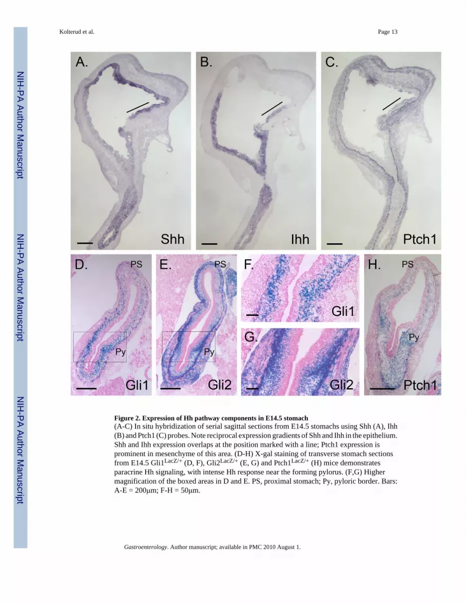
Figure 2. Expression of Hh pathway components in E14.5 stomach(A-C) In situ hybridization of serial sagittal sections from E14.5 stomachs using Shh (A), Ihh(B) and Ptch1 (C) probes. Note reciprocal expression gradients of Shh and Ihh in the epithelium.Shh and Ihh expression overlaps at the position marked with a line; Ptch1 expression isprominent in mesenchyme of this area. (D-H) X-gal staining of transverse stomach sectionsfrom E14.5 Gli1LacZ/+ (D, F), Gli2LacZ/+ (E, G) and Ptch1LacZ/+ (H) mice demonstratesparacrine Hh signaling, with intense Hh response near the forming pylorus. (F,G) Highermagnification of the boxed areas in D and E. PS, proximal stomach; Py, pyloric border. Bars:A-E = 200μm; F-H = 50μm.
Kolterud et al. Page 13
Gastroenterology. Author manuscript; available in PMC 2010 August 1.
NIH
-PA Author Manuscript
NIH
-PA Author Manuscript
NIH
-PA Author Manuscript
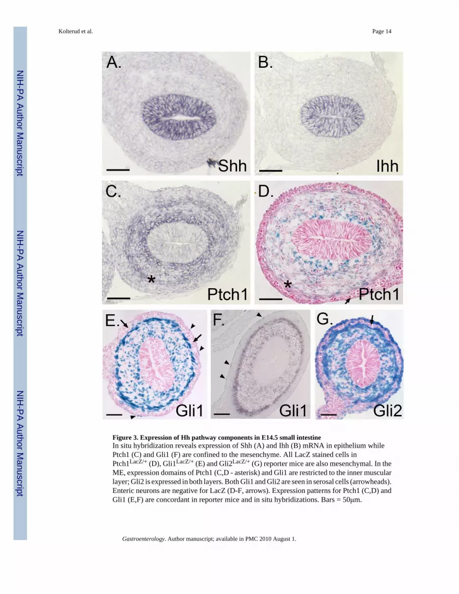
Figure 3. Expression of Hh pathway components in E14.5 small intestineIn situ hybridization reveals expression of Shh (A) and Ihh (B) mRNA in epithelium whilePtch1 (C) and Gli1 (F) are confined to the mesenchyme. All LacZ stained cells inPtch1LacZ/+ (D), Gli1LacZ/+ (E) and Gli2LacZ/+ (G) reporter mice are also mesenchymal. In theME, expression domains of Ptch1 (C,D - asterisk) and Gli1 are restricted to the inner muscularlayer; Gli2 is expressed in both layers. Both Gli1 and Gli2 are seen in serosal cells (arrowheads).Enteric neurons are negative for LacZ (D-F, arrows). Expression patterns for Ptch1 (C,D) andGli1 (E,F) are concordant in reporter mice and in situ hybridizations. Bars = 50μm.
Kolterud et al. Page 14
Gastroenterology. Author manuscript; available in PMC 2010 August 1.
NIH
-PA Author Manuscript
NIH
-PA Author Manuscript
NIH
-PA Author Manuscript

Figure 4. Hh pathway activity across the E16.5 pyloric border(A, B) In situ hybridization of antrum using Shh and Ihh specific probes. (C-F) X-gal stainedantral sections from E16.5 Ptch1LacZ/+, Gli1LacZ/+, and Gli2LacZ/+ antrum. (F) Cross sectionof the forming pyloric border in a Gli1 LacZ/+ mouse reveals a drastic difference in active Hhsignaling between the stomach (S, left) and intestine (I, right); Py = pylorus. Arrowheads =Gli1 positive serosal cells. Bars = 100μm.
Kolterud et al. Page 15
Gastroenterology. Author manuscript; available in PMC 2010 August 1.
NIH
-PA Author Manuscript
NIH
-PA Author Manuscript
NIH
-PA Author Manuscript
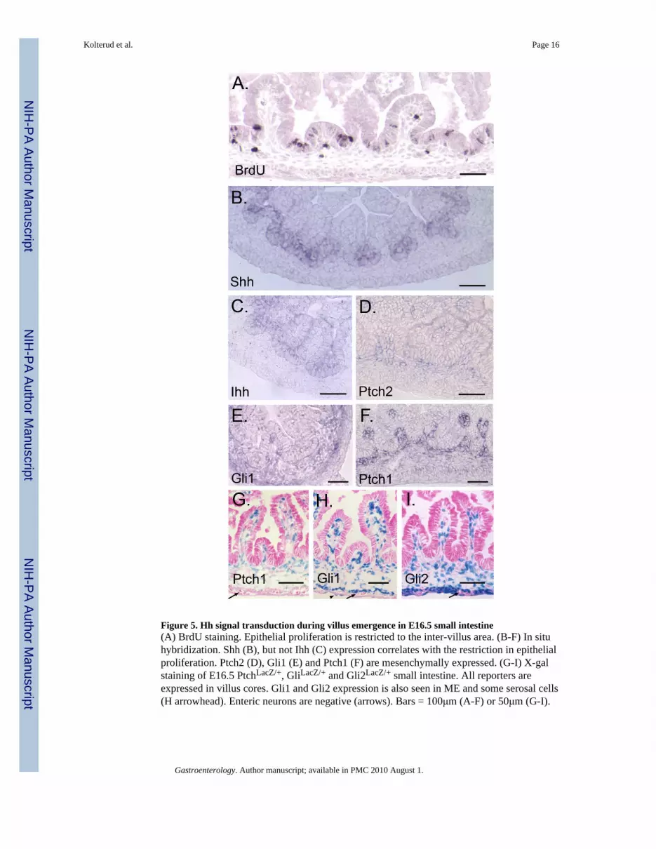
Figure 5. Hh signal transduction during villus emergence in E16.5 small intestine(A) BrdU staining. Epithelial proliferation is restricted to the inter-villus area. (B-F) In situhybridization. Shh (B), but not Ihh (C) expression correlates with the restriction in epithelialproliferation. Ptch2 (D), Gli1 (E) and Ptch1 (F) are mesenchymally expressed. (G-I) X-galstaining of E16.5 PtchLacZ/+, GliLacZ/+ and Gli2LacZ/+ small intestine. All reporters areexpressed in villus cores. Gli1 and Gli2 expression is also seen in ME and some serosal cells(H arrowhead). Enteric neurons are negative (arrows). Bars = 100μm (A-F) or 50μm (G-I).
Kolterud et al. Page 16
Gastroenterology. Author manuscript; available in PMC 2010 August 1.
NIH
-PA Author Manuscript
NIH
-PA Author Manuscript
NIH
-PA Author Manuscript
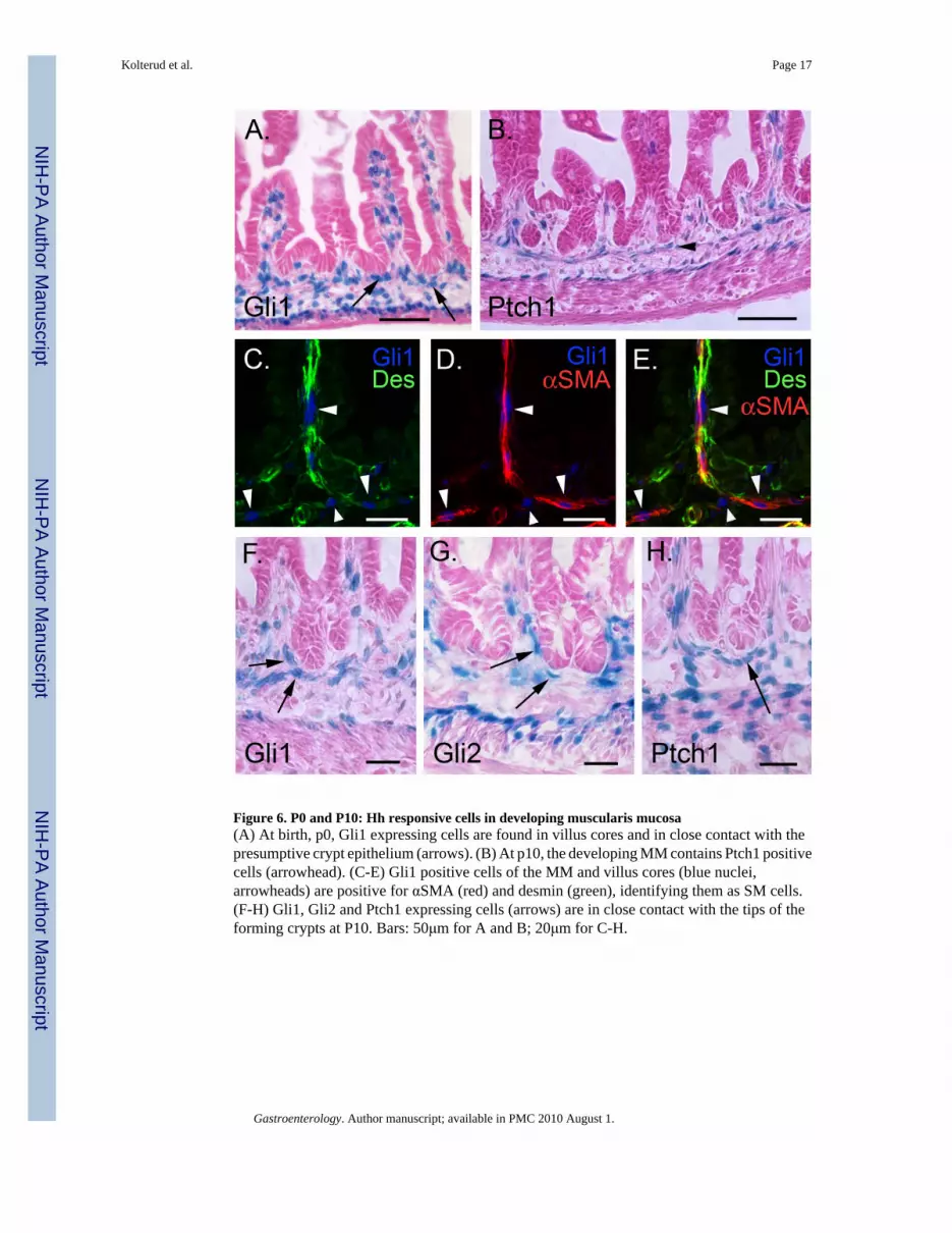
Figure 6. P0 and P10: Hh responsive cells in developing muscularis mucosa(A) At birth, p0, Gli1 expressing cells are found in villus cores and in close contact with thepresumptive crypt epithelium (arrows). (B) At p10, the developing MM contains Ptch1 positivecells (arrowhead). (C-E) Gli1 positive cells of the MM and villus cores (blue nuclei,arrowheads) are positive for αSMA (red) and desmin (green), identifying them as SM cells.(F-H) Gli1, Gli2 and Ptch1 expressing cells (arrows) are in close contact with the tips of theforming crypts at P10. Bars: 50μm for A and B; 20μm for C-H.
Kolterud et al. Page 17
Gastroenterology. Author manuscript; available in PMC 2010 August 1.
NIH
-PA Author Manuscript
NIH
-PA Author Manuscript
NIH
-PA Author Manuscript

Figure 7. Hedgehog controls smooth muscle development in postnatal miceX-gal staining of jejunum from adult Ptch1 LacZ/+ (A), Gli1 LacZ/+ (B,D-J), and Gli2 LacZ/+ (C)mice. (A-D) Hh responsive cells are located within villus cores, at the crypt villus junction, inthe submucosa and in the muscularis externa. (E-H) Sections were stained with X-gal (E,G)and then co-stained with antibodies against α-SMA (red) and desmin (green). Immunostainedimages taken on the confocal were overlaid (F,H) with images of the same field taken underbright field illumination (X-gal pattern, E,G). Hh responsive cells included desmin and α-SMAdouble positive smooth muscle cells (yellow) in the villus cores (arrowheads in H), at the cryptvillus junction (arrows in E,F) and in the MM (arrowheads in E,F). Gli1 expressing cells arealso detected among the desmin positive, α-SMA negative muscle precursors (green) locatedinside the villi close to the epithelium (arrow in H) and among the α-SMA positive, desminnegative (red) subepithelial myofibroblasts that line the crypts (not shown). (I,J) One monthold wild type (WT) mice (I) and their littermates transgenic for 12.4KVil-Ihh (J). Transgenicanimals show dramatic increase in villus smooth muscle and reduced smooth muscleprecursors. Scale bars: (A-C, I,J) = 100μm; (D) = 50μm; (E-H) = 20μm.
Kolterud et al. Page 18
Gastroenterology. Author manuscript; available in PMC 2010 August 1.
NIH
-PA Author Manuscript
NIH
-PA Author Manuscript
NIH
-PA Author Manuscript