Embed Size (px)
Citation preview

Double sensitization toenzymes in a baker
M. Santaolalla*, M. De Barrio, C. De Frutos,M. Gandolfo, J. Zubeldia, M. Rubio, A. Rodr�guez,M. L. Baeza
Key words: alpha-amylase; bronchial challenge;lysozyme.
The enzymes alpha-amylase and lyso-
zyme have been
identified as
inhalative aller-
gens in baker’s
asthma (1–4),
and in egg-pro-
cessing or pharmaceutical workers
(5–10), respectively.
We report the case of a 41-year-old
man who worked in a bakery for
25 years. After 22 years of glazing rolls
with fresh egg, he began to use a spraying
device for the egg mixture. A few months
later he began to develop rhinoconjunc-
tivitis and asthma just minutes after this
activity. He remained asymptomatic
during weekends and holidays.
He then worked on mixing wheat flour
to make bread. Initially he was free of
symptoms, but 9 months later he pre-
sented with rhinoconjunctivitis and asth-
ma. He had no symptoms on ingestion of
egg or wheat flour. At home, he had
seven birds and a cat, but his symptoms
did not worsen on exposure to these pets,
to house dust, or pollen.
Physical examination, blood cell count,
biochemistry, and chest radiographs were
all normal. Skin prick test (SPT) with
commercially available extracts (Leti,
Spain), were positive to egg white, egg
yolk, ovomucoid, wheat flour, barley
flour, Dermatophagoides pteronyssinus,
D. farinae, G. fusca, L. destructor,
T. putrescentiae, and cockroach, as well
as to a lysozyme extract (Wassermann) at
10 mg/ml, and to alpha-amylase extract
(1 mg/ml). SPT were negative to a bat-
tery of pollen and molds, cat and dog
dander, canary and pigeon feathers,
ovalbumin, and rye flour.
Total serum IgE was 1142 kU/l. IgE
antibodies were detected to egg white
(2.07 kU/l), egg yolk (0.86), ovomucoid
(2.26), wheat flour (3.21), barley flour
(1.95), alpha-amylase (0.39), lysozyme
(2.05), D. pteronyssinus (5.60), D. farinae
(5.48), L. destructor (3.70) and
T. putrescentiae (5.58) (CAP Pharmacia,
Sweden). The test was negative to oval-
bumin, and to canary and pigeon feathers.
Spirometry revealed a forced vital
capacity (FVC) of 105%, forced expira-
tory volume in 1s (FEV1) of 94%, FEV1/
FVC of 93%, and forced expiratory flow
(FEF25)75) of 64%.
Specific bronchial challenge to lyso-
zyme (tidal breathing method) elicited an
early asthmatic response (22% fall in
FEV1 in 10 min). Specific bronchial
challenge to alpha-amylase elicited an
early asthmatic response with a 24% fall
in FEV1 in 20 min.
We have demonstrated IgE sensitiza-
tion and clinical sensitivity to two
enzymes from different sources by SPT,
IgE antibody determination, and bron-
chial provocation test. The two enzymes
are lysozyme (from egg used to decorate
rolls) and fungal alpha-amylase (used to
improve bread qualities). This patient
was also strongly sensitized to dust mites,
although no symptoms have yet been
produced.
*Marcela Santaolalla Montoya
C./Jazmın 40, 6� A28033 Madrid, Spain
Tel: +34 91 302 80 15
E-mail: [email protected]
Accepted for publication 29 April 2002
Allergy 2002: 57:957
Copyright � Blackwell Munksgaard 2002
ISSN 0105-4538
References1. Baur X, Posch A. Characterized allergens
causing baker’s asthma. Allergy
1998;53:562–566.
2. Baur X, Degens P, Sander I. Baker’s
asthma: still among the most frequent
occupational respiratory disorders.
J Allergy Clin Immunol 1998;102:984–997.
3. Baur X, Chen Z, Liebers V. Exposure-
response relationships of occupational
inhalative allergens. Clin Exp Allergy
1998;28:537–544.
4. Losada E, Hinojosa M, Quirce S,
Sanchez-Cano M, Moneo I. Occupa-
tional asthma caused by alpha-amylase
inhalation: clinical and immunologic find-
ings and bronchial response patterns.
J Allergy Clin Immunol 1992;89:118–125.
5. Leser C, Hartmann AL, Praml G,
Wuthrich B. The �egg–egg� syndrome:
occupational respiratory allergy to
airborne egg proteins with consecutive
ingestive egg allergy in the bakery and
confectionery industry. J Invest Allergol
Clin Immunol 2001;11:89–93.
6. Fremont S, Kanny G, Nicolas JP,
Moneret-Vautrin DA. Prevalence of
lysozyme sensitization in an egg-allergic
population. Allergy 1997;52:224–228.
7. Boeniger MF, Lummus ZL, Biagini RE
et al. Exposure to protein aeroallergens in
egg processing facilities. Appl Occup
Environ Hyg 2001;16:660–670.
8. Quirce S, Dıez- Gomez ML, Eiras P,
Cuevas M, Baz G, Losada E. Inhalant
allergy to egg yolk and egg white pro-
teins. Clin Exp Allergy 1998;28:478–485.
9. Bernstein D, Smith A, Moller D et al.
Clinical and immunologic studies among
egg processing workers with occupational
asthma. J Allergy Clin Immunol
1987;80:791–797.
10. Bernstein A, Kraut A, Bernstein D
et al. Occupational asthma induced by
inhaled egg lysozyme. Chest
1993;103:532–535.
Allergy to garlic
G. Pires*, E. Pargana, V. Loureiro, M. M. Almeida,J. R. Pinto
Key words: children; garlic; food allergy; urticaria.
AL LERGY 2 0 0 2 : 5 7 : 9 5 7 – 9 6 8 • COPYR IGHT ª 2002 BLACKWELL MUNKSGAARD • I SSN 0105 - 4 538 • ALL R IGHTS RESERVED
• CONTRIBUT IONS TO THIS SECT ION WILL NOT UNDERGO PEER REV IEW. BUT WILL BE REV IEWED BY THE ASSOCIATE EDITORS •
A case of occupationalallergy to non-crossre-activity allergens.
957
ALLERGY Net

Garlic belongs to
the Liliaceae
family. This
family is divided
into two sub-
families, Asparagoidea (asparagus) and
Aliolidea (garlic, onion, chive, leek and
aloes). Hypersensitivity to garlic has been
described as a cause of contact dermatitis
and occupational asthma and rhinitis.
Immediate allergic reactions to garlic
ingestion (1,2) or contact (1) are rare,
especially in children.
We report a case of IgE-mediated
urticaria after contact and ingestion of
raw garlic in a child.
A 16-month-old boy with a personal
history of cow’s milk and egg-white al-
lergy, developed urticaria on the face and
neck immediately after local contact with
raw garlic. From the age of 12 months he
had ingested cooked garlic with tolerance.
He had ingested onion from the age of
6 months with no symptoms.
Skin prick and prick-prick tests were
positive with commercial garlic extract
(Stallergenes) and fresh raw garlic with a
wheal diameter of 15 · 9 mm and
17 · 8 mm, respectively. The prick-prick
test was negative with fresh cooked
garlic. Skin prick and prick-prick tests
were negative with both commercial
onion extract (Stallergenes) and fresh raw
and cooked onion. We also tested 10
children, followed in our Immunoallergy
Department, with fresh raw and cooked
garlic and onion, as controls, all being
negative. Specific IgE to garlic and onion
(Pharmacia CAP System) were negative
(< 0.35 kU/l).
Contact challenge was positive (urtic-
aria) with raw garlic and negative with
cooked garlic. Oral challenge with raw
garlic was positive with generalized urtic-
aria within 10 min after ingestion. Oral
challenge with cooked garlic was negative.
The child is on raw and cooked garlic
eviction, being asymptomatic.
The immunoblotting analysis (Ala-
BLOT DPC-Amerlab) showed two pro-
teinic bands with molecular masses of
approximately 12 kDa and around
40–50 kDa, probably corresponding to
the previously identified garlic proteins
mannose-binding lectin (3) and alliinase
(4), respectively.
Perez-Pimiento et al. (5) also identified
a band with a molecular mass of 12 kDa
to young garlic, garlic, onion and leek
extracts in a woman allergic to young
garlic. Asero et al. (1) also found bands
with molecular mass of 10, 20 and 40 kDa
in a woman with urticaria after ingestion
and contact with raw and cooked garlic.
We found no cross-reactivity between
onion and garlic, two foods that belong
to the same family. Asero et al. (1) also
reported a case without cross-reactivity
between these foods. However, some
authors found immunological cross-
reactivity between the Liliaceae family
(6,7). Probably the level of cross-reactiv-
ity among the different Liliaceae vegeta-
bles varies among individuals.
Since our child tolerated cooked garlic,
we suggest that the allergenic fraction to
which he is sensitized is denatured by
heat treatment. Nevertheless, a case of
systemic urticaria associated with inges-
tion of both raw and cooked garlic has
been described by Asero et al. (1). Prob-
ably this patient had a garlic allergy
caused by heat-resistant allergens.
By allowing cooked garlic in the child’s
diet, we might delay complete immuno-
logical tolerance to garlic proteins. Or
might the consumption of cooked garlic
even favour acquisition of complete oral
tolerance and recovery from garlic
allergy? More data will be necessary to
answer this question.
*Servico de Imunoalergologia
Hospital de Dona Estefania
Rua Jacinta Marto
1169-045 Lisboa
Portugal
Fax: +351 213126654
E-mail: [email protected]
Accepted for publication 6 May 2002
Allergy 2002: 57:957
Copyright � Blackwell Munksgaard 2002
ISSN 0105-4538
References
1. Asero R, Mistrello G, Roncarolo D,
Antoniotti PL, Falagiani P. A case of
garlic allergy. J Allergy Clin Immunol
1998;101:427–428.
2. Rance F, Dutau G. Labial food challenge
in children with food allergy. Pediatr
Allergy Immunol 1997;8:41–44.
3. Smeets K, Van Damme EJ, Verhaert P
et al. Isolation characterization and
molecular cloning of the mannose-binding
lectins from leaves and roots of garlic
(Allium sativum L). Plant Mol Biol
1997;33:223–234.
4. Rabinkov A, Zhu XZ, Grafi G, Galili G,
Mirelman D. Alliin lyase (Allium sati-
vum). Biochemical characterization and
cDNA cloning. Appl Biochem Biotechnol
1994;48:149–171.
5. Perez-Pimiento AJ, Moneo I, Santaola-
lla M, Paz S, Fernandez-Parra B,
Domınguez-Lazaro AR. Anaphylactic
reaction to young garlic. Allergy
1999;54:626–629.
6. Lybarger JA, Gallagher JS, Pulver
AW, Litwin A, Brooks S, Bernstein L.
Occupational asthma induced by inhalation
and ingestion of garlic. J Allergy Clin
Immunol 1982;69:448–454.
7. Eng PA, Yman L, Maaninen E, Wuth-
rich B. Inhalant allergy to fresh asparagus.
Clin Exp Allergy 1996;26:330–334.
Eotaxin in IgE-mediatedrhinoconjunctivitis
K. Jahnz-R+z_yk*, T. Targowski, E. Głodzinska-Wyszogrodzka, T. Płusa
Key words: allergic inflammation; allergicrhinoconjunctivitis; eotaxin.
Recruitment of eosinophils to the site of
inflammation is mediated by numerous
cytokines (CC). Among these CC, the
chemokine eotaxin has been shown to
possess unusual
selective chem-
otactic activity
(1,2). In a
number of
studies it has been shown that an increase
in the serum level of eotaxin is induced by
allergen exposure (3,4). However others
factors influencing the content of eotaxin
in serum are still unclear. In this study we
examine the role of age and sex on serum
eotaxin in healthy volunteers (H) and
subjects with intermittent allergic rhino-
conjunctivitis (AR) (5).
The level of serum eotaxin was meas-
ured by ELISA (KITS, R&D, USA,
pg/ml) in 64 healthy people (46 women;
18 men) and 122 with AR (44 women; 78
men) before the pollen season. Data were
analyzed with the statistical package
(statistica (version 5.1) for windows).
The student t-test was used for inter-
group comparison.
A 16-month-old boywith urticaria andpositive SPT.
A relation with age andsex only in allergies.
958
ALLERGY Net

There was no significant difference in
the mean age of the allergic and healthy
groups (AR: 30.0 years (SD 10.6); H:
28.2 years (SD 8.9); P ¼ 0.2). The mean
serum eotaxin concentration was higher
in AR, but the difference between the
groups was not statistically significant
(AR: 114.1 (SD 45.5); H: 100.6 (SD 50.5);
P ¼ 0.07). A relationship between serum
eotaxin level and sex and age in AR was
noted; in this group the lowest concen-
tration of eotaxin was observed in
women younger than 25 years (81.8: SD
35.3) and the highest concentration was
observed in men older than 35 years
(145.5; SD 52.6).
A multiple regression model was
derived for the influence of age and sex
on the serum level of eotaxin in the
allergic and healthy groups using two
formulae:
Formula A: eotaxin ð0:7Þ¼ 2:6� age ð19:6Þ þ 31:8� 47:1
ðR ¼ 0:43; P<0:00038Þ
Formula B: eotaxin ð0:3Þ¼ 1:4� age ð7:2Þ þ 31:7
� sex ð16:2Þ þ 21:26� 38:2
ðR ¼ 0:47; P<0:000001Þ
A statistically significant multiple
linear correlation was found (Fig. 1)
(formula B) between age (semipartial
correlation a¼ 0.33, P ¼ 0.00003) and
sex (semipartial correlation a¼ 0.35,
P ¼ 0.00008) on the one side, and the
serum level of eotaxin in on the other in
the AR group. In the H group there was a
linear correlation between age (semipar-
tial correlation coefficient a¼ 0.414,
P ¼ 0.0006), while no significant linear
correlation was found for sex (semipartial
correlation factor a¼ 0.18, P ¼ 0.16).
The regression model describing the
influence of age on the serum concentra-
tion of eotaxin in healthy subjects is
presented as Formula A.
To our knowledge, no study concern-
ing a correlation between the serum level
of eotaxin and age in allergic or healthy
people has been published so far. Neither
have we found any publications about a
relationship between the serum concen-
tration of eotaxin and sex. However, it is
known that male sex is related to allergic
sensitization and higher production of
Ag-specific IgE (6,7). In our opinion, this
study suggests that age and sex are
important determinants of the serum
content of eotaxin in patients with inter-
mittent IgE-mediated rhinoconjunctivitis.
Further studies should elucidate why sex
has different effects in people with aller-
gies and those without.
*Department of Internal Medicine
Pneumonology and Allergology
Central Hospital of Military
School of Medicine
Szaserow 128 Street
00–909 Warsaw
Poland
E-mail: [email protected]
Accepted for publication 29 April 2002
Allergy 2002: 57:958
Copyright � Blackwell Munksgaard 2002
ISSN 0105-4538
References
1. Honda K, Chihara J. Eosinophil activa-
tion by eotaxin—eotaxin primes the
production of reactive oxygen species from
eosinophils. J Allergy Clin Immunol
1999;54:1262–1269.
2. Morita E, Kameyoshi Y, Hiragun T,
Mihara T, Yamamoto S. The C-C
chemokines, RANTES and eotaxin in
atopic dermatitis. J Allergy Clin Immunol
2001;56:194–195.
3. Yamada H, Yamaguchi M, Nakajima T,
et al. Eotaxin in induced sputum of asth-
matics: relationship with eosinophils and
eosinophil cationic protein in sputum.
J Allergy Clin Immunol 2000;55:392–397.
4. Jahnz-Roz_yk K. Eotaxin—marker of
allergic inflammation in asthma. Int Rev
Allergol Clin Immunol 2000;6:5–10.
5. Johansson SGO, O’B Hourihane J,
Bousquet J, et al. A revised nomenclature
for allergy. An EAACI position statement
from the EAACI nomenclature task force.
Allergy 2001;56:813–824.
6. Linneberg A, Nielsen NH, Madsen F,
Frolund L, Dirksen A, Jorgensen T.
Factors related to allergic sensitization to
aeroallergens in a cross-sectional study in
adults: the Copenhagen Allergy Study. Clin
Exp Allergy 2001;31:1409–1417.
7. Yamatomo T, Okano M, Ono T, et al.
Sex-related differences in the initiation of
allergic rhinitis in mice. Allergy
2001;56:525–531.
The biology of cypress allergy
E. Agea, O. Bistoni, A. Russano, L. Corazzi, L. Minelli,G. Bassotti, F.M. de Benedictis, F. Spinozzi*
Key words: allergy; asthma; Cupressaceae;epidemiology; pollinosis.
Winter rhinoconjunctivitis caused by
cypress allergy represents an emerging
cause of morbidity in young adults living
in the Mediterranean area, where orna-
mental use of
both Cupressus
arizonica and C.
sempervirens has
spread in recent
years (1,2).
Figure 1. Regression models of influence of age and sex on the serum level of eotaxin in healthy (Formula
A) and allergic people (Formula B).
Approx. half of IgE-sen-sitizedaresymptomfree.
959
ALLERGY Net

Despite emphasis in describing this
pollinosis as an emerging one, popula-
tion-based studies on cypress allergy and
sensitization are lacking. Our epidemio-
logical survey was designed to assess the
prevalence of symptomatic oculorhinitis
and asymptomatic cypress sensitization in
a cohort of 3434 subjects living in central
Italy (Perugia county, Umbria region).
Data were collected using a modified
questionnaire of the European Commu-
nity Respiratory Health Survey (3). Sub-
jects had skin prick tests (SPT) with
Dermatophagoides, Alternaria tenuis,
Parietaria officinalis, Graminaceae mix,
Olea europaea, Cupressus arizonica and
C. sempervirens extracts (ALK-Abello
and Lofarma, Milan, Italy), by a standard
procedure. Specific IgE levels were deter-
mined with commercially available
reagents.
To categorize enrolled subjects (4),
we applied a clinical score for absence
(0 points) or presence (3 points) of
symptoms, and a laboratory score from 0
to 3 points for cutaneous wheal and flare
reactions plus 0–3 points for IgE anti-
body levels (RAST class from I to IV) to
cypress extracts.
Table 1 summarizes the distribution of
cypress allergy and sensitization in our
study population. Among group A sub-
jects, 103 (84.4%) were mono-sensitive to
cypress pollen, whereas the remaining 19
(15.6%) were also sensitive to perennial
and/or seasonal allergens. The frequency
of self-reported asthmatic symptoms was
less than 1% (one subject). Group A
patients had fewer atopic members in
their first-degree relatives, compared to
other groups.
SPT and/or RAST give information
merely on sensitivity to individual aller-
gens (5); 4.9% of our subjects were
clinically asymptomatic but, nonetheless,
had positive SPT and/or measurable IgE
antibody levels. Therefore, assuming
sensitization to cypress pollen as study
end-point, would lead to a disease pre-
valence over-estimation (up to 8% of the
total study population), similar to that
previously published (1,2).
The virtual absence of asthma has been
noted for other Cupressaceae species,
such as the respiratory allergy to Junipe-
rus sabinoides, and recently confirmed in
a survey on asthmatic children living in
the south-east of France (6,7).
In conclusion, in areas (such as central
Italy) in which cypress trees have been a
common feature for centuries, cypress
hypersensitivity is relatively low. None-
theless, the widespread use of such trees
justifies the major attention we now pose
in diagnosing this particular winter
allergy.
*Department of Clinical and Experimental
Medicine
University of Perugia
Policlinico Monteluce
I-06122 Perugia
Italy
E-mail: [email protected]
Accepted for publication 23 May 2002
Allergy 2002: 57:959
Copyright � Blackwell Munksgaard 2002
ISSN 0105-4538
References
1. Mari A, Di Felice G, Affermi C,
Barletta B, Tinghino R, Pini C. Cypress
allergy: an underestimated pollinosis.
Allergy 1997;52:355–356.
2. Ariano R, Panzani RC, Chiappella M,
Augeri G. Pollinosis in a Mediterranean
area (Riviera Ligure, Italy): ten years of
pollen counts, correlation with clinical
sensitization and meteorological data. J
Invest Allergol Clin Immunol 1994;4:81–86.
3. European Community Respiratory
Health Survey. Prevalence of asthma and
asthma symptoms in a general population
sample from northern Italy. Allergy
1995;50:755–759.
4. Johansson SGO, Hourihane J O’B,
Bousquet J et al. Position paper. A revised
nomenclature for allergy. Allergy
2001;56:813–824.
5. Tschopp JM, Sistek D, Schindler C et al.
Current allergic asthma and rhinitis: diag-
nostic efficiency of three commonly used
atopic markers (IgE, skin prick tests, and
Phadiatop). Results from 8329 randomized
adults from the SAPALDIA Study. Swiss
Study on Air Pollution and Lung Diseases
in Adults. Allergy 1998;53:608–613.
6. Ramirez DA. The natural history of
mountain cedar pollinosis. Allergol Immu-
nol 2000;32:86–91.
7. Dubus J-C, Melluso J-P, Bodiu AC,
Stremler-Lebel N. Allergy to cypress
pollen. Allergy 2000;55:410–411.
Food allergy to pork meat
M. Atanaskovic-Markovic*, M. Gavrovic-Jankulovic,R. M. Jankov, O. Vu6kovic, B. Nestorovic
Key words: childhood asthma; dust mites;pork allergy.
Although pork allergy is rare in child-
hood it may manifest in a variety of ways:
urticaria, oral allergy syndrome (OAS)
and exacerbation of an atopic eczema/
dermatitis syndrome (1).
We report on a four-year-old boy with
atopic eczema/dermatitis syndrome,
AEDS and asthma due to sensitization to
the house-dust mite Dermatophagoides
pteronyssinus,
and who had an
anaphylactic
reaction a few
minutes after
eating porkmeat.
Table 1. Distribution of cypress allergy or sensitization in a population of 3434 young adults of central
Italy
Number Prevalence (%) Features of cypress sensitivity Familial atopy
Group A 122 3.6 Oculo-rhinitis, positive SPT
and detectable IgE antibody
19.5%*
Group B 167 4.9 Positive SPT and detectable
IgE antibody
40.3%
Group C 539 15.7 None 38.9%
Group D 2606 75.8 None No
Allergic subjects (group A) reached a total score ¼ 7, by adding the 3 clinical symptom points to the
sum of the laboratory score. Group B consisted of atopic subjects (0 point clinical score), identified
as cypress-sensitized on the basis of the laboratory score only. Control groups were atopic subjects
(group C) suffering from seasonal or perennial rhinitis due to other inhalant allergens; group D
nonatopic healthy subjects. Statistical evaluation was performed by two dimensional contingency
tables and Pearson’s chi-squared tests with spss software.
*P < 0.001 vs. groups B and C.
Unusual case with IgEantibodies to cookedand roasted meat
960
ALLERGY Net
Examination of the patient included
cutaneous (skin prick test – SPTs, prick
prick test – PPT), serological and provoc-
ative diagnostic tests with commercially
available allergenic extracts (Torlak,
Yugoslavia). Open challenge with food
was carried out according to instructions
given in an EAACI position paper (2).
Pork meat (cooked and roasted) was
homogenized and extracted as a 20%
suspension for 3 h in 0.2 M PBS, pH 7.2.
Allergenic molecules were detected by
Western immunoblot according to
Harlow & Lane (3).
SPTs were positive for pork meat
(+ +) and dust mite allergens (+). PPT
with fresh food was positive only to pork
meat. Open challenge with the pork meat
induced, after a few minutes, generalized
urticaria with intensive itching in the
throat and eyes, and swelling of the lip,
tongue, palate and throat.
The patient’s serum reacted moderately
toDp (13.1 kU/l, class 3), whereas specific
IgE to pork meat was undetectable. The
total IgE was five times higher (223 kU/l)
than the value usual for his age.
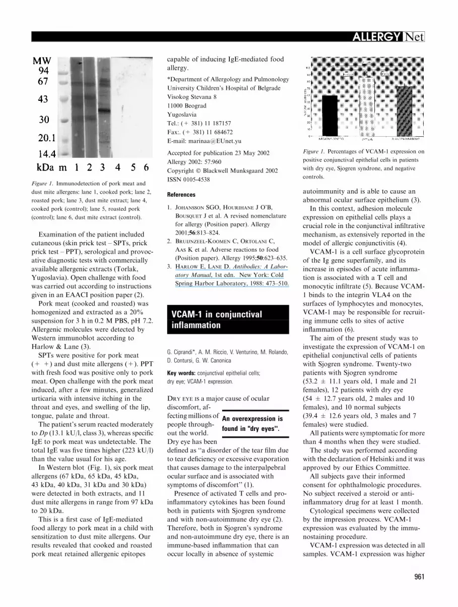
In Western blot (Fig. 1), six pork meat
allergens (67 kDa, 65 kDa, 45 kDa,
43 kDa, 40 kDa, 31 kDa and 30 kDa)
were detected in both extracts, and 11
dust mite allergens in range from 97 kDa
to 20 kDa.
This is a first case of IgE-mediated
food allergy to pork meat in a child with
sensitization to dust mite allergens. Our
results revealed that cooked and roasted
pork meat retained allergenic epitopes
capable of inducing IgE-mediated food
allergy.
*Department of Allergology and Pulmonology
University Children’s Hospital of Belgrade
Visokog Stevana 8
11000 Beograd
Yugoslavia
Tel.: (+ 381) 11 187157
Fax:. (+ 381) 11 684672
E-mail: [email protected]
Accepted for publication 23 May 2002
Allergy 2002: 57:960
Copyright � Blackwell Munksgaard 2002
ISSN 0105-4538
References
1. Johansson SGO, Hourihane J O’B,
Bousquet J et al. A revised nomenclature
for allergy (Position paper). Allergy
2001;56:813–824.
2. Bruijnzeel-Koomen C, Ortolani C,
Aas K et al. Adverse reactions to food
(Position paper). Allergy 1995;50:623–635.
3. Harlow E, Lane D. Antibodies: A Labor-
atory Manual, 1st edn. New York: Cold
Spring Harbor Laboratory, 1988: 473–510.
VCAM-1 in conjunctivalinflammation
G. Ciprandi*, A. M. Riccio, V. Venturino, M. Rolando,D. Contursi, G. W. Canonica
Key words: conjunctival epithelial cells;dry eye; VCAM-1 expression.
Dry eye is a major cause of ocular
discomfort, af-
fectingmillions of
people through-
out the world.
Dry eye has been
defined as ‘‘a disorder of the tear film due
to tear deficiency or excessive evaporation
that causes damage to the interpalpebral
ocular surface and is associated with
symptoms of discomfort’’ (1).
Presence of activated T cells and pro-
inflammatory cytokines has been found
both in patients with Sjogren syndrome
and with non-autoimmune dry eye (2).
Therefore, both in Sjogren’s syndrome
and non-autoimmune dry eye, there is an
immune-based inflammation that can
occur locally in absence of systemic
autoimmunity and is able to cause an
abnormal ocular surface epithelium (3).
In this context, adhesion molecule
expression on epithelial cells plays a
crucial role in the conjunctival infiltrative
mechanism, as extensively reported in the
model of allergic conjunctivitis (4).
VCAM-1 is a cell surface glycoprotein
of the Ig gene superfamily, and its
increase in episodes of acute inflamma-
tion is associated with a T cell and
monocytic infiltrate (5). Because VCAM-
1 binds to the integrin VLA4 on the
surfaces of lymphocytes and monocytes,
VCAM-1 may be responsible for recruit-
ing immune cells to sites of active
inflammation (6).
The aim of the present study was to
investigate the expression of VCAM-1 on
epithelial conjunctival cells of patients
with Sjogren syndrome. Twenty-two
patients with Sjogren syndrome
(53.2 ± 11.1 years old, 1 male and 21
females), 12 patients with dry eye
(54 ± 12.7 years old, 2 males and 10
females), and 10 normal subjects
(39.4 ± 12.6 years old, 3 males and 7
females) were studied.
All patients were symptomatic for more
than 4 months when they were studied.
The study was performed according
with the declaration of Helsinki and it was
approved by our Ethics Committee.
All subjects gave their informed
consent for ophthalmologic procedures.
No subject received a steroid or anti-
inflammatory drug for at least 1 month.
Cytological specimens were collected
by the impression process. VCAM-1
expression was evaluated by the immu-
nostaining procedure.
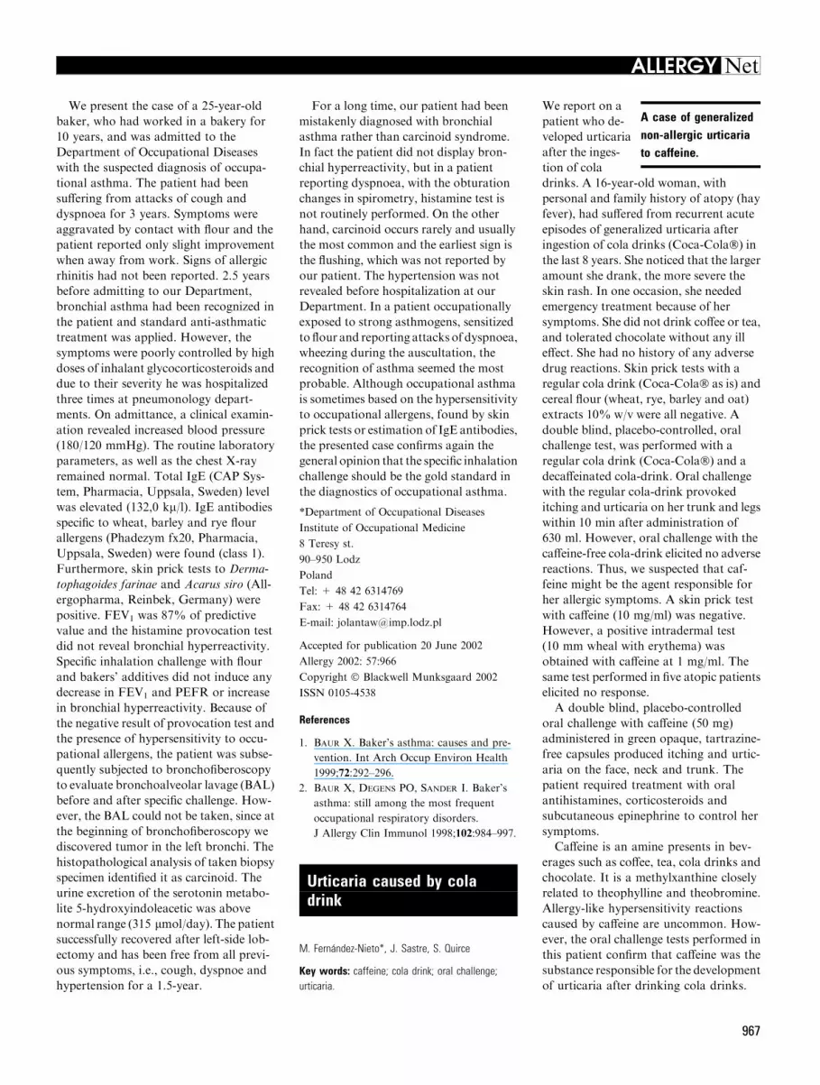
VCAM-1 expression was detected in all
samples. VCAM-1 expression was higher
Figure 1. Immunodetection of pork meat and
dust mite allergens: lane 1, cooked pork; lane 2,
roasted pork; lane 3, dust mite extract; lane 4,
cooked pork (control); lane 5, roasted pork
(control); lane 6, dust mite extract (control).
Figure 1. Percentages of VCAM-1 expression on
positive conjunctival epithelial cells in patients
with dry eye, Sjogren syndrone, and negative
controls.
An overexpression isfound in "dry eyes''.
961
ALLERGY Net

in patients with Sjogren syndrome and
dry eye than in normal subjects. More-
over, patients with dry eye showed a
significant difference in comparison with
normal subjects (P ¼ 0.0006) and with
patients suffering from Sjogren syndrome
(P ¼ 0.03). Patients with Sjogren syn-
drome showed a significant difference in
comparison with normal subjects
(P ¼ 0.04).
VCAM-1 is an adhesion molecules
involved in cellular recruitment: in fact, its
expressionmay be regarded as amarker of
a broad range of inflammatory diseases.
The meaning of our findings is consis-
tent with the pathophysiological mecha-
nisms involved in the immuno-mediated
inflammation occurring in dry eye disor-
ders.
The overexpression of VCAM-1 in all
dry eye disorders is dependent on the
presence of lymphocyte activation, which
has been previously demonstrated in all
dry eye disorders (1,2).
In conclusion, this study is the first
evidence concerning the overexpression
of VCAM-1 involved in the inflammatory
response associated with dry eye.
*Clinica delle Malattie Allergiche ed
Immunologiche Padiglione Maragliano
(piano terra) Ospedale San Martino
Largo R. Benzi 10
16132 Genoa
Italy
Tel: +39 10 3537929
Fax: +39 10 3538904
E-mail: [email protected]
Accepted for publication 23 April 2002
Allergy 2002: 57:961
Copyright � Blackwell Munksgaard 2002
ISSN 0105-4538
References
1. Pflugfelder SC, Solomon A, Stern MC.
The diagnosis and management of dry eye.
Cornea 2000;19:644–649.
2. Stern ME, Beuerman RW, Fox RI, et al.
The pathology of dry eye: the interaction
between the ocular surface and lachrymal
glands. Cornea 1998;17:589–589.
3. AugustinAJ, SpitznasM,KavianiN, et al.
Oxidative reactions in the tear fluid of pa-
tients suffering from dry eyes. Graefes Arch
Clin Exp Ophthalmol 1995;233:694–698.
4. Johansson SGO, O’B Hourihane J,
Bousquet J, et al. A revised nomenclature
for allergy. Allergy 2001;56:813–824.
5. Pisella PJ, Brignole F, Debbasch C, et al.
Flow cytometric analysis of conjunctival
epithelium in ocular rosacea and
keratoconjunctivitis sicca. Ophthalmology
2000;107:1841–1849.
6. Tu Z, Kelley VR, Collins T, Lee FS.
IkB kinase is critical for TNFa-inducedVCAM-1 gene expression in renal tubular
epithelial cells. J Immunol 2001;166:6839–
6846.
Severe cow's milk allergy
R. Shaoul*, O. Mesner, A. Kessel, M. Jaffe
Key words: allergy; infants; life-threatening;milk protein.
Cow’s milk protein allergy (CMPA) is a
form of food-induced allergic reaction
that usually appears during infancy (1,2).
CMPA may involve the gastrointestinal
tract, the respiratory tract, or the skin (1).
Symptoms may manifest as an acute
episode —at times life-threatening—or
may have a more chronic form (1, 3).
Immediate hy-
persensitivity re-
actions to cow’s
milk (CM) are
usually IgE-me-
diated. However, the food protein-in-
duced enterocolitis syndrome (FPIES) is
not IgE-mediated (3).
We present a series of four babies that
presented with severe life-threatening
episodes, all related to unsupervised
self-challenge with either a CM-based
formula or a dairy product. Parental
decisions, physician recommendations, or
inadvertent ingestion resulted in these
serious clinical presentations.
Case 1: A 5-month-old boy presented
with vomiting, bloody diarrhea, severe
lethargy, and shock, that developed
within hours of exposure to a dairy
product. Previous exposure to a CM-
based formula had resulted in respiratory
distress, wheezing, and hematochezia. A
presumptive diagnosis of CMPA was
made and a semi-elemental formula for
feeding was recommended. He subse-
quently had two similar episodes on
re-exposure to CM. Parental consent to a
skin test could not be obtained.
Case 2: A 6-week-old girl presented
with emesis and diarrhea a few hours
after ingesting a CM-based formula. A
presumed diagnosis of CMPA was made
and a semi-elemental formula diet was
started. A trial of CM-based formula
feeding on a pediatrician’s recommenda-
tion at the age of 4 months led to a state
of lethargy, peripheral cyanosis, vomit-
ing, and profuse diarrhea, within 15 min
of ingestion. A skin prick test was neg-
ative for CM and soy proteins.
Case 3: A 6-month-old- boy presented
with multiple episodes of vomiting, pro-
fuse watery diarrhea, and severe lethargy.
This began 2 h after exposure to a com-
mercial baby pudding containing CM.
Past history included several episodes of
fever, vomiting, and diarrhea after expo-
sure to a CM-based formula. A skin test
was negative for CM or soy proteins.
Case 4: A 5-month-old boy presented
with fever, lethargy, and recurrent vom-
iting which started 2 h after ingesting a
dairy product. Before this episode he had
been fed solely on breast milk, except for
a single trial of CM-based formula at age
3 months, which was followed by fever,
vomiting, and a skin rash. A skin test was
positive for CM and soy protein antigens.
In all four cases other possible etiologies
with similar presentations were excluded.
Avoidance of CM-based formula was
recommended in all cases. None of the
children had a personal or family history
of allergic diseases.
In a recent position paper (4), Johans-
son et al. suggested an updated nomen-
clature for food allergies. They suggested
that an adverse reaction to food should be
called food hypersensitivity. When
immunologic mechanisms are demon-
strated, the appropriate term is food
allergy; and if the role of IgE is highlight-
ed, the term is IgE-mediated food allergy.
The common element for all these
cases was that all the children had dem-
onstrated a previous serious reaction to
CM protein. Despite warnings given to
the parents of the dangers of CM inges-
tion, all the children been re-challenged
with CM protein either because of a
parent’s decision or a physician’s advice.
One infant (case 4) was confirmed to
have IgE-mediated immediate hypersen-
sitivity to CM and soy proteins by a
positive skin test. Another infant’s clin-
ical picture (case 1), although not proven
by skin test, was compatible with this type
of hypersensitivity. The two other cases
Near-fatal reactionsin four children.
962
ALLERGY Net

(2 and 3), whose skin tests were negative,
were compatible with the diagnosis of
FPIES. This is a non-IgE-mediated form
of acute allergy to CM or other allergens.
It is a disorder of early infancy, mani-
festing within hours of exposure with
vomiting and diarrhea, and it may result
in dehydration. Hypotension is not
uncommon, fecal occult blood is often
positive, and an elevated polymorphonu-
clear blood count is typical. Skin prick
tests are negative in most cases (3).
Diagnosis of this disorder is based on
clinical and challenge criteria (3).
In conclusion, food allergy and FPIES
in particular must be considered in the
differential diagnosis of repeated shock-
like episodes, or severe gastrointestinal
symptomatology related to feeding in
infants.
The appropriate guidance to families
regarding strict elimination of CM or soy
protein from the diet is crucial.
*Pediatric Day Care Unit
Department of Paediatrics
Bnai Zion Medical Center
47 Golomb St
POB 4940
Haifa 31048
Israel
Tel: +972 4 835 9662
Fax: +972 4 837 1393
E-mail: [email protected]
Accepted for publication 14 May 2002
Allergy 2002: 57:962
Copyright � Blackwell Munksgaard 2002
ISSN 0105-4538
References
1. Sampson HA. Food allergy. Part 1: im-
munopathogenesis and clinical disorders.
J Allergy Clin Immunol, 1999;103:
717–728.
2. Businco L, Benincori N, Cantani A.
Epidemiology, incidence and clinical
aspects of food allergy. Ann Allergy,
1984;53:615–622.
3. Sicherer SH. Food protein–induced ente-
rocolitis syndrome: clinical perspectives.
J Pediatr Gastroenterol Nutr, 2000;30
(Suppl.): S45–S49.
4. Johansson SGO, Hourihane J O’B,
Bousquet J et al. A revised nomenclature
for allergy. An EAACI position statement
from the EAACI nomenclature task force.
Allergy, 2001;56:813–824.
Simultaneous drug allergies
E. Gonz9lez-Mancebo, M. Fern9ndez-Rivas*,M. Cuevas, E. Gonz9lez Gonz9lez,C. Lara C9tedra, M. Dolores Alonso
Key words: clavulanic acid; moxifloxacin;betalactams; penicillin; quinolones; drug allergy.
We report the case of a 32-year-old
woman who developed a generalized
urticaria 15 min
after the intake
of amoxycilin-
clavulanic acid
(AX/CL) (500/
125 mg) (Augmentine�, GlaxoSmith
Kline, Madrid, Spain). The reaction
subsided with methylprednisolone and
dexclorfeniramine administered at the
emergency room. After this reaction the
patient tolerated a 7-day course of AX.
One year later, she presented a nonit-
ching micropapular exanthema some
hours after the intake of 400 mg moxi-
floxacin (MX) (Octegra�, Grupo Vita,
Spain), which resolved in 3 days with
dexclorfeniramine. One year after the
latter reaction the following allergologi-
cal study was performed. Written in-
formed consent was given by the patient.
Skin prick tests (SPTs) and intradermal
tests (IDTs) with benzylpenicilloyl poly
L-lysine (PPL) and minor determinant
mixture (MDM) (Allergopen�, Allergo-
pharma, Reinbek, Germany), benzylpen-
icillin (BP) (10, 000 UI/ml), AX (25 mg/
ml) and AX/CL (25/5 mg/ml) yielded
negative results. Specific IgE (Pharmacia
CAP System, Uppsala, Sweden) to ben-
zylpenicilloyl, fenoxymethylpenicilloyl,
AX and ampicillin were also negative. An
oral challenge with increasing doses of
AX/CL up to 500/125 mg was well
tolerated.
Fifteen days after this first negative
study the patient was re-evaluated. An
immediate positive IDT with AX/CL was
found (10 mm wheal, 27 mm erythema).
CL (Beecham, Spain) was tested sepa-
rately in a saline solution at 1 and 10 mg/
ml. These two concentrations do not
induce nonspecific skin tests reactions in
controls (1,3). SPTs with CL were nega-
tive, but an immediate IDT reaction was
observed with 10 mg/ml (12 mm wheal,
28 mm erythema). The remaining skin
tests and specific IgE determinations were
negative. A histamine release test (HRT)
with BP, AX and CL was performed
following an automated fluorometric
method previously described (1). No
release was observed with BP and AX,
whereas CL induced a dose-related re-
sponse with a maximum release of 50%
with 200 lg. The CL concentrations used
did not induce histamine release in con-
trols (1). One year later IDT and HRT
with CL yielded again positive results.
SPTs performed with MX at 1 and
10 mg/ml, and a patch test (10% in
petrolatum) were negative. Higher con-
centrations of MX induced nonspecific
SPT reactions in control subjects. HRT
with MX and ciprofloxacin yielded high
peaks in the patient and control tests,
probably because of an interference with
the fluorometric method of histamine
determination, as already described with
quinolones (2). Twenty hours after the
oral administration ofMX (up to 400 mg)
the patient developed a mild pruritic
micropapular exanthema which resolved
with oral prednisone and cetirizine in
48 h. No reaction was observed with the
related quinolones pipemidic acid, nor-
floxacin and ciprofloxacin, neither in
SPTs (1 mg/ml), IDTs (0.001–1 mg/ml)
and patch tests (10% in petrolatum), nor
after their oral administration.
The results of the allergological study
performed (positive IDT andHRT to CL)
suggest that the immediate reaction
induced by AX/CL in our patient was due
to a specific immunologic mechanism
towards CL. Clavulanic acid is a betalac-
tam antibiotic with weak antibacterial
activity, but a potent inhibitor of betalac-
tamases. Despite its wide use in associa-
tionwithAX, there are only three reported
cases of allergic reactions toCL (1,3), in all
of which an immediate immunologic
mechanism was also involved. As with our
patient, two of the previously reported
cases (1) had no reactivity to other bet-
alactams. This specificity in the immuno-
logic response to CL might be related to
differences in its structure and metabo-
lism, which can generate metabolites not
related to those derived from BP (4).
Moxifloxacin is a fluorquinolone
antimicrobial agent recently marketed,
and only two anaphylactic reactions
have been reported so far (5). MX
induced in our patient a delayed reaction
A patient with hyper-sensitivity to clavulanicacid and moxiflocacin.
963
ALLERGY Net

(exanthema) in which an immunologic
mechanism, probably directed to a spe-
cific MX metabolite, seems to be
involved. This could explain the negativ-
ity of the tests performed with the native
drug, as well as the lack of cross-reac-
tivity with other quinolones, in contrast
to previous reports (2).
In summary, this patient combines
two exceptional selective immunologic
reactions to two unrelated drugs: an
immediate reaction to the betalactam
CL, and a delayed reaction to the
quinolone MX.
*Fundacion Hospital Alcorcon
Unidad de Alergia
C/ Budapest, 1
28922 Alcorcon (Madrid)
Spain
Tel: + 34 91 621 96 95
Fax: + 34 91 621 99 75
E-mail: [email protected]
Accepted for publication 2 July 2002
Allergy 2002: 57:963
Copyright � Blackwell Munksgaard 2002
ISSN 0105-4538
References
1. Fernandez-Rivas M, Perez Carral C,
Cuevas M, Marti C, Moral A, Senent C.
Selective allergic reactions to clavulanic acid.
J Allergy Clin Immunol 1995;95:748–750.
2. Davila I, Dıez ML,Quirce S, Fraj J, de la
Hoz B, Lazaro M.Cross-reactivity between
quinolones. Allergy 1993;48:388–390.
3. Cahen YD, Wuthrich B. Drug allergy
to the b-lactam antibiotics clavulanic
acid and amoxycillin. Allergy 1997;52:
117–118.
4. Haginaka J, Yasuda H, Uno T, Nakag-
awa T. Degradation of clavulanic acid in
aqueous alkaline solution: isolation and
structural investigation of degradation
products. Chem Pharm Bull (Tokyo)
1985;33:218–244.
5. Moxifloxacin. In: MICROMEDEX�Healthcare Series, Vol. 108. MICROME-
DEX Inc. 1974–2001.
Adverse reactions topyrazinamide
C. Ribi*, C. Hauser
Key words: Tuberculosis; pyrazinamide; hyper-sensitivity; rash; desensitization.
Cutaneous
reactions to
antituberculosis
drugs are com-
mon. We de-
scribe a case of immediate cutaneous
reaction to pyrazinamide without previ-
ous exposure.
A 41-year-old Latino-American wom-
an without prior antituberculous treat-
ment was diagnosed having active lung
tuberculosis. Rifampicin (600 mg/day),
isoniazid (INH) (300 mg/day), pyrazina-
mide (1500 mg/day), ethambutol
(800 mg/day), and pyridoxine (40 mg/
day) were prescribed. Treatment was
started with a single dose of INH and
rifampicin. The three other drugs were
added the following morning. Within
minutes, she developed a flush with
nausea, dyspnea and abdominal discom-
fort followed by an itchy rash. All drugs
were stopped and clemastine was admin-
istered. The skin eruption disappeared
within 24 h and did not recur after
sequential challenge with rifampicine and
isoniazid first at reduced, then at full
dosage. After administration of 500 mg
of pyrazinamide, she developed an iden-
tical skin rash within half an hour.
Challenge with ethambutol was well tol-
erated. Pyrazinamide was replaced by
ciprofloxacin in our case.
Pyrazinamide has gained importance
in the past years as the incidence of
multiresistant tuberculosis is rising. It
was synthesized in the 1950’s following
the observation that nicotinamide was
effective in the treatment of experimental
tuberculous infections. The mechanism of
antituberculous action is not completely
understood. Liver injury is the most
serious side-effect. Other side-effects
include hyperuricemia, anorexia, nausea,
vomiting, arthralgia and fever. Photo-
sensitivity, skin rashes, flushing and pel-
lagra have been reported.
Hypersensitivity reactions to antitu-
berculous drugs usually appear within
3–7 weeks after initiation of treatment.
The incidence of cutaneous reactions due
to pyrazinamide is not quantified. It
seems, however, that the skin reactions to
other antituberculous drugs are far more
frequent (1): 2% of more than 2000
patients treated with isoniazid presented
a skin rash as the most common adverse
effect. Another series with almost 2000
patients treated with ethambutol revealed
an incidence of 0.5%. Rifampicin caused
skin rash in 0.8% of patients. Cutaneous
hypersensitivity reactions to antitubercu-
lous drugs are more frequent and more
severe in HIV-positive patients (2).
Nicotinamide from which pyrazina-
mide has been synthesized regularly
causes truncal and facial flushing and
itching. These reactions appear to be
prostaglandin-mediated and can be pre-
vented by aspirin (3). One could hypoth-
esize that a similar mechanism is
responsible for pyrazinamide-induced
flushing and skin rash.
Pyrazinamide accounted for four other
cases of immediate skin rash after first
dose of antituberculous regimen (4–6).
Flushing has been described in one
patient (4). In two cases re-administra-
tion of pyrazinamide caused a similar
rash. Pyrazinamide can be tolerated after
reintroduction at a lower dose followed
by a stepwise dose increment (6).
In conclusion, pyrazinamide should be
suspected if an immediate skin rash
develops at initiation of antituberculosis
chemotherapy. If the skin involvement is
not severe, sequential reintroduction of
the drugs first at low, then at full dosage
should be attempted. Pyrazinamide
should be reintroduced last and at low
dose.
*University Hospital Geneva,
Division of Immunology and Allergy
Rue Micheli-du-Crest 24
CH-1211 Geneva 14, Switzerland
Email: [email protected]
Accepted for publication 2 July 2002
Allergy 2002: 57:964
Copyright � Blackwell Munksgaard 2002
ISSN 0105-4538
References
1. Patel AM, Mckean J. Avoidance and
management of adverse reactions to
antituberculosis drugs. Drug Saf 1995
January;12:1–25.
2. Kuaban C, Bercion R, Koula-Shiro S.
Current HIV seroprevalence rate and inci-
dence of adverse skin reactions in adults
with pulmonary tuberculosis receiving
thiacetazone-free antituberculosis treat-
ment in Yaounde, Cameroon. Cent Afr J
Med 1998 February;44:34–37.
3. Jungnickel PW et al. Effect of two aspirin
pretreatment regimens on niacin-induced
Immediate skin rashat initiation of anti-tuberculosis therapy.
964
ALLERGY Net

cutaneous reactions. J General Intern Med
1997 October;12:591–596.
4. Radal M et al. Eruption apres la 1ere prise
d’une chimiotherapie standard antituber-
culeuse. Penser au pyrazinamide. Rev Mal
Respir 1998 June;15:305–306.
5. Olivier C et al. Eruption apres une 1ere
prise d’une quadritherapie antitubercule-
use: penser au pyrazinamide. Arch Pediatr
1998 March;5:289–290.
6. Shorr AF, Trotta RF. PZA Hypersensi-
tivity. Chest 1996 March;109:855–856.
IgE antibodies to penicillin inskin test negative patients
M. J. Torres, C. Mayorga, J. A. Cornejo-Garc�a,A. Romano, M. Blanca*
Key words: IgE antibodies; immediate reactions;in vitro test; penicillin allergy; skin test.
Skin tests (ST) are reported to be more
sensitive than in
vitro tests for
determining IgE
antibodies to
beta-lactams.
However,
evidence exists of in vitro positivity with
ST negativity in patients allergic to pen-
icillins, but the number of cases was
limited and the report is more than 20
years old (1), so that a re-evaluation
seems necessary.
We studied a group of subjects with an
immediate allergic reaction to a penicillin
derivative who were ST negative with a
validated standard of classical and side
chain penicillin determinants, butwhohad
in their serum IgEantibodies to apenicillin
derivative. STs were performed using PPL
5 · 10)5 mmol/l, MDM 2 · 10)2 mmol/l
(Allergopharma, Merck, Darmstadt,
Germany), AX 20 mg/ml (Beecham,
Toledo, Spain) and AMP 20 mg/ml
(Antibiotic SA, Leon, Spain) (2). IgE
antibodies were measured using RAST
to benzylpenicilloyl-poly L-lysine
(BPO-PLL) and amoxicylloyl-poly
L-lysine (AXO-PLL) (3). A drug provo-
cation test (DPT) was made administering
increasing doses of benzylpenicillin (BP)
and amoxicillin (AX) at 1-hour intervals
(2). Two control groups were evaluated:
negative, 40 nonallergic subjects with
negative STs to all haptens and good
tolerance to BP and AX; and positive, 40
patients with ST positive to at least one of
the haptens and RAST positive to BPO-
PLL or AXO-PLL.
Of 290 patients with immediate allergic
reactions to a penicillin derivative, 40
(13.8%) were ST negative and RAST
positive. The diagnosis was confirmed
because five developed systemic symp-
toms after ST, 11 developed more than
one reaction with penicillin derivatives
according to the clinical history, and in 24
it was confirmed by DPT. Twenty-three
were female and 17 male, the mean age
was 44.5 ± 14.6 years, 28 had developed
anaphylaxis and 12 urticaria. AX was the
drug involved in 26 cases, un-recalled
penicillin in 6, ampicillin in 5, BP in 2 and
cloxacillin in 1. Themean interval between
the occurrence of the reaction and the
study was 252 ± 281 days. In 13 cases
(32.5%) the RAST was positive to both
haptens, in nine (22.5%) only to BPO-
PLL and in 18 (45%) only to AXO-PLL.
Comparison with the positive control
group showed no significant differences in
age, sex, time interval, or RAST values.
Linear regression analysis showed no
significant association between the RAST
value and the time interval, either in the
patients or in the positive control group.
None of the subjects in the negative
control group had a positive RAST.
It is generally considered that a subject
with a positive history of penicillin
allergy but who is ST negative is likely to
tolerate the drug (4), but patients allergic
to penicillins may be ST negative and
develop an allergic response after
exposure (2). Confirmation of the diag-
nosis in these subjects is difficult since the
DPT may involve significant risks. These
subjects not detected by skin testing led
us to consider alternative methods to
confirm the diagnosis whilst avoiding
DPT. A specific IgE antibody assay is a
good alternative approach in persons
with a history of penicillin allergy but
who are ST negative. Whether this can
be extended to other beta-lactams, such
as cephalosporins, is at present being
studied.
Acknowledgments
This work was supported in part by the FIS(00/0838) and the Junta de Andalucıa (165/00). We thank Ian Johnstone for his helpwith the English language version.
*Servicio de Alergia
Hospital Universitario La Paz
Paseo de La Castellana 2612
8046 Madrid
Spain
Tel.: +34 91 7277080
Fax: +34 91 7277050
E-mail: [email protected]
Accepted for publication 21 May 2002
Allergy 2002: 57:965
Copyright � Blackwell Munksgaard 2002
ISSN 0105-4538
References
1. Kraft D, Roth A, Mischer P, Pichler H,
Ebner H. Specific and total serum IgE
measurements in the diagnosis of penicillin
allergy. A long term follow-up study. Clin
Allergy 1977;7:21–28.
2. Torres MJ, Romano A, Mayorga C et al.
Diagnostic evaluation of a large group of
patients with immediate allergy to penicil-
lins: the role of skin testing. Allergy
2001;56:850–856.
3. Blanca M, Mayorga C, Perez E et al.
Determination of IgE antibodies to the
benzylpenicilloyl determinant: a compari-
son between poly-l-lysine and human
serum albumin as carriers. J Immunol Meth
1992;153:99–105.
4. Weiss ME, Adkinson NF. Immediate
hypersensitivity reactions to penicillin and
related antibiotics. Clin Allergy
1988;18:515–540.
AAE and IgA myeloma
C. Martin-Garcia*, M. L. D�ez-G+mez, E. Camacho,P. Berges, R. G. Rodriguez, L. M. Villar, M. C. Sadaba,O. Ordo=ez
Key words: acquired angioedema; C1-inhibitordeficiency; carcinoid tumor; monoclonal gammopathy.
A 72-year-old woman was admitted for
evaluation of
recurrent,
non-pruritic,
edematous
attacks occurring
on the face, lips and uvula, and which did
not affect the airways. The patient had no
personal allergic history or family his-
tory of angioedema. Attacks resolved
Acquired Angioedematype II in a case ofcarcinoid tumor
In-vitro assay of IgEantibody is important inpenicillin allergy.
Abbreviations: AAE: acquired angioedema;C1-INH: C1 esterase-inhibitor.
965
ALLERGY Net

spontaneously or after treatment with low
doses of oral corticosteroids for 2–3 days.
A complement profile revealed: CH50 less
than 25 U/ml, C4 less than 1.38 mg/dl,
C1 inhibitor 12 mg/dl (15–40) and normal
C3 at 121 mg/dl. Serum IgA 1050 mg/dl
(n: 70–400), IgG and IgM were normal.
Electrophoresis showed a distinct abnor-
mal band in the beta region. Serum
immunoelectrophoresis revealed an IgA
lambda paraprotein. Spirometric values
showed a moderate restriction with forced
vital capacity of 71%. A chest radiograph
revealed a loss in volume of the left
hemithorax with a left upwards hilar
traction and poor delimitation of the
lower left cardiac edge, compatible with
an atelectasis of the left upper lobe.
Computerized axial tomography of
the chest confirmed the presence of
the atelectasis. Computerized axial
tomography of the abdomen was normal.
Analysis of bone marrow revealed a
monoclonal IgA-lambda population of
plasma cells, without evidence of overt
myeloma. One hour before broncho-
fiberscopy, replacement therapy with
1000 units of C1-INH concentrate (Ber-
inert P. Aventis Behring GmbHMarburg,
Germany) was administered to the
patient. Bronchofiberscope revealed a
smooth and fungate rounded mass pro-
truding into the left main bronchus,
suggestive of a carcinoid tumor. A pul-
monary biopsy was also carried out and it
was compatible with carcinoid tumor.
Because of the potentially lethal nature of
these attacks, 2 mg stanazolol twice daily
was started. In January 2001, the patient
underwent surgery for her carcinoid
tumor. Six months after the operation,
the patient had not had attacks of
angioedema, while complement compo-
nent levels and IgA levels continued to be
abnormal. She is still under treatment
with 2 mg stanazolol once daily.
To demonstrate that there was an agent
in the patient’s plasma that inhibited the
C1 inhibitor, we incubated the patient’s
plasma with plasma from a healthy blood
donor (1/1) for 18 h at 4�C. Another
serum diluted 1/1 in PBS and incubated in
the same conditions was used as a control.
To assess whether monoclonal antibody
was responsible for C1 inhibitor inacti-
vation, we incubated separately the iso-
lated IgA paraprotein and polyclonal IgG
obtained from the patient, with a plasma
specimen from a healthy blood donor
(1/1) for 18 h at 4�C. Monoclonal IgA
completely abrogated C1 inhibitor activ-
ity of the control plasma. The results are
expressed in Table 1.
AAE type II is characterized by the
presence of autoantibodies to C1 inhib-
itor (1,2). Indirect evidence suggests that
these antibodies could be paraproteins in
some cases, but so far this assessment has
not been completely demonstrated (3).
We have studied a patient with acquired
autoimmune C1-INH deficiency. We
could demonstrated that IgA paraprotein
was responsible for C1 inhibitor inacti-
vation. To the best of our knowledge, this
is the first case of AAE that directly
demonstrates the anti C1-INH activity of
a monoclonal paraprotein. A review of
the literature suggests that this is the first
case of acquired angioedema associated
with a carcinoid tumor.
c/ Jesus 5
5�1 Madrid 28034
Spain
Tel: + 34 91 420 16 44
Fax: + 34 91 3368693
E-mail: [email protected]
Accepted for publication 23 May 2002
Allergy 2002: 57:965
Copyright � Blackwell Munksgaard 2002
ISSN 0105-4538
References
1. Jackson J, Sim RB, Whelan A, Feighery
C. An IgG autoantibody which inactivates
C1-inhibitor. Nature 1986;323:722–724.
2. Cicardi M, Bisiani G, Cugno M, Spaeth
P, Agostini A. Autoimmune C1 inhibitor
deficiency: report of eight patients. Am J
Med 1993;95:169–175.
3. Cicardi M, Beretta A, Colombo M,
Gioffre D, Cugno M, Agostini A. Rele-
vance of lymphoproliferative disorders and
of anti-C1 inhibitor autoantibodies in
acquired angio-oedema. Clin Exp Immunol
1996;106:475–480.
Carcinoid behind baker'sasthma
J. Walusiak*, C. Palczynski
Key words: bakers' asthma; carcinoid; hypersensi-tivity to flour.
Bakers’ asthma is one of the most
common occupational allergic diseases.
It is estimated
that 5–10% of
bakers suffer
from asthma
and 14–39%
from allergic
rhinitis (1).
However, it has
been estimated that about 30% of bakers
sensitized to occupational allergens do
not display work-related symptoms (2).
The prevalence of IgE antibodies has
been reported to be high, e.g., in Baur
et al. study 53% of examined bakers had
IgE antiwheat flour (1). It should be also
noted that about 30% of bakers report-
ing work-related chest symptoms do not
demonstrate IgE antibodies to occupa-
tional allergens (1).
Bronchial provocationand bronchoscopymight be considered inunusual cases.
Table 1. Characteristics of C1 inhibitor, complement, IgA and IgG
Patient’s values Normal range
C3 (mg/dl) 121 90–180
C4 (mg/dl). <1.38 10–45
CH50 (U/ml) <25 150–250
C1INH (mg/dl) 12 15–40
IgA (mg/dl) 1050 70–400
IgG (mg/dl) 723 700–1600
Functional activity of C1 inhibitor 80–100%
Control plasma 100%
Patient’s plasma 0%
Patient’s plasma with control plasma 7%
Patient’s IgA with control plasma 0%
Patient’s IgG with control plasma 80%
966
ALLERGY Net
We present the case of a 25-year-old
baker, who had worked in a bakery for
10 years, and was admitted to the
Department of Occupational Diseases
with the suspected diagnosis of occupa-
tional asthma. The patient had been
suffering from attacks of cough and
dyspnoea for 3 years. Symptoms were
aggravated by contact with flour and the
patient reported only slight improvement
when away from work. Signs of allergic
rhinitis had not been reported. 2.5 years
before admitting to our Department,
bronchial asthma had been recognized in
the patient and standard anti-asthmatic
treatment was applied. However, the
symptoms were poorly controlled by high
doses of inhalant glycocorticosteroids and
due to their severity he was hospitalized
three times at pneumonology depart-
ments. On admittance, a clinical examin-
ation revealed increased blood pressure
(180/120 mmHg). The routine laboratory
parameters, as well as the chest X-ray
remained normal. Total IgE (CAP Sys-
tem, Pharmacia, Uppsala, Sweden) level
was elevated (132,0 kl/l). IgE antibodies
specific to wheat, barley and rye flour
allergens (Phadezym fx20, Pharmacia,
Uppsala, Sweden) were found (class 1).
Furthermore, skin prick tests to Derma-
tophagoides farinae and Acarus siro (All-
ergopharma, Reinbek, Germany) were
positive. FEV1 was 87% of predictive
value and the histamine provocation test
did not reveal bronchial hyperreactivity.
Specific inhalation challenge with flour
and bakers’ additives did not induce any
decrease in FEV1 and PEFR or increase
in bronchial hyperreactivity. Because of
the negative result of provocation test and
the presence of hypersensitivity to occu-
pational allergens, the patient was subse-
quently subjected to bronchofiberoscopy
to evaluate bronchoalveolar lavage (BAL)
before and after specific challenge. How-
ever, the BAL could not be taken, since at
the beginning of bronchofiberoscopy we
discovered tumor in the left bronchi. The
histopathological analysis of taken biopsy
specimen identified it as carcinoid. The
urine excretion of the serotonin metabo-
lite 5-hydroxyindoleacetic was above
normal range (315 lmol/day). The patient
successfully recovered after left-side lob-
ectomy and has been free from all previ-
ous symptoms, i.e., cough, dyspnoe and
hypertension for a 1.5-year.
For a long time, our patient had been
mistakenly diagnosed with bronchial
asthma rather than carcinoid syndrome.
In fact the patient did not display bron-
chial hyperreactivity, but in a patient
reporting dyspnoea, with the obturation
changes in spirometry, histamine test is
not routinely performed. On the other
hand, carcinoid occurs rarely and usually
the most common and the earliest sign is
the flushing, which was not reported by
our patient. The hypertension was not
revealed before hospitalization at our
Department. In a patient occupationally
exposed to strong asthmogens, sensitized
to flour and reporting attacks of dyspnoea,
wheezing during the auscultation, the
recognition of asthma seemed the most
probable. Although occupational asthma
is sometimes based on the hypersensitivity
to occupational allergens, found by skin
prick tests or estimation of IgE antibodies,
the presented case confirms again the
general opinion that the specific inhalation
challenge should be the gold standard in
the diagnostics of occupational asthma.
*Department of Occupational Diseases
Institute of Occupational Medicine
8 Teresy st.
90–950 Lodz
Poland
Tel: + 48 42 6314769
Fax: + 48 42 6314764
E-mail: [email protected]
Accepted for publication 20 June 2002
Allergy 2002: 57:966
Copyright � Blackwell Munksgaard 2002
ISSN 0105-4538
References
1. Baur X. Baker’s asthma: causes and pre-
vention. Int Arch Occup Environ Health
1999;72:292–296.
2. Baur X, Degens PO, Sander I. Baker’s
asthma: still among the most frequent
occupational respiratory disorders.
J Allergy Clin Immunol 1998;102:984–997.
Urticaria caused by coladrink
M. Fern9ndez-Nieto*, J. Sastre, S. Quirce
Key words: caffeine; cola drink; oral challenge;urticaria.
We report on a
patient who de-
veloped urticaria
after the inges-
tion of cola
drinks. A 16-year-old woman, with
personal and family history of atopy (hay
fever), had suffered from recurrent acute
episodes of generalized urticaria after
ingestion of cola drinks (Coca-Cola�) in
the last 8 years. She noticed that the larger
amount she drank, the more severe the
skin rash. In one occasion, she needed
emergency treatment because of her
symptoms. She did not drink coffee or tea,
and tolerated chocolate without any ill
effect. She had no history of any adverse
drug reactions. Skin prick tests with a
regular cola drink (Coca-Cola� as is) and
cereal flour (wheat, rye, barley and oat)
extracts 10% w/v were all negative. A
double blind, placebo-controlled, oral
challenge test, was performed with a
regular cola drink (Coca-Cola�) and a
decaffeinated cola-drink. Oral challenge
with the regular cola-drink provoked
itching and urticaria on her trunk and legs
within 10 min after administration of
630 ml. However, oral challenge with the
caffeine-free cola-drink elicited no adverse
reactions. Thus, we suspected that caf-
feine might be the agent responsible for
her allergic symptoms. A skin prick test
with caffeine (10 mg/ml) was negative.
However, a positive intradermal test
(10 mm wheal with erythema) was
obtained with caffeine at 1 mg/ml. The
same test performed in five atopic patients
elicited no response.
A double blind, placebo-controlled
oral challenge with caffeine (50 mg)
administered in green opaque, tartrazine-
free capsules produced itching and urtic-
aria on the face, neck and trunk. The
patient required treatment with oral
antihistamines, corticosteroids and
subcutaneous epinephrine to control her
symptoms.
Caffeine is an amine presents in bev-
erages such as coffee, tea, cola drinks and
chocolate. It is a methylxanthine closely
related to theophylline and theobromine.
Allergy-like hypersensitivity reactions
caused by caffeine are uncommon. How-
ever, the oral challenge tests performed in
this patient confirm that caffeine was the
substance responsible for the development
of urticaria after drinking cola drinks.
A case of generalizednon-allergic urticariato caffeine.
967
ALLERGY Net

We have no evidence to implicate an
immune mechanism as being responsible
for this reaction to caffeine, and a phar-
macological mechanism is also very
unlikely since xanthines themselves are
inhibitors of the release of chemical
mediators (1). Therefore, a personal
susceptibility or non-allergic hypersensi-
tivity pathway may be the underlying
mechanism. In the previous case reports
of urticaria caused by caffeine (2–4), the
prick test with caffeine was positive in
two out of three cases (2,4). However, the
passive cutaneous transfer test
(Prausnitz–Kustner) performed on one
patient was negative (2). Urticarial reac-
tions caused by aminophylline (5,6) have
been reported, and have been considered
secondary to the ethylene diamine por-
tion of the aminophylline molecule (6).
Caffeine should be considered as a
potential causative agent of urticaria
induced by the ingestion of caffeine-
containing beverages and drugs.
*Fundacion Jimenez Dıaz
Allergy Department
Av. Reyes Catolicos, 2
28040 Madrid
Spain
Fax: + 34 91 549 94 98
E-mail: [email protected]
Accepted for publication 7 May 2002
Allergy 2002: 57:967
Copyright � Blackwell Munksgaard 2002
ISSN 0105-4538
References
1. Lichtestein LM, Margolis S. Histamine
release in vitro: inhibition by catecholam-
ines and methylxantines. Science
1968;161:902–909.
2. Pola J, Subiza J, Armentia A et al.
Urticaria caused by caffeine. Ann Allergy
1988;60:207–208.
3. Quirce S, Freire P, Fernandez-Rivas M,
Davila I, Losada E. Urticaria from
caffeine. J Allergy Clin Immunol
1991;88:680–681.
4. Caballero T, Garcıa-Ara C, Pascual C,
Dıaz-Pena JM, Ojeda A. Urticaria induced
by caffeine. J Invest Allergol Clin Immunol
1993;3:160–162.
5. Wong D, Lopapa AF, Haddad ZH.
Immediate hypersensitivity reaction to
aminophylline. J Allergy Clin Immunol
1971;48:165–170.
6. Booth BH, Coleman WP, Mitchell DQ.
Urticaria following intravenous amino-
phylline. Ann Allergy 1979;43:289–290.
The major allergen of linseed
F. Le+n, M. Rodr�guez*, M. Cuevas
Key words: allergy; blot; dimer; flax; IgE; linseed;Linum.
Linum usitatissimum (flax) is a plant
belonging to the
Linaceae family.
Its seeds have
been used from
the beginning of
civilization for
human and cattle consumption, and its
fibres have always been of important
commercial value in the textile industry.
In our time, linseed oil is increasingly
used as a laxative (1). Anaphylaxis in-
duced by flax has been described (2,3),
and a multimeric protein has been sug-
gested as the allergen (2). We studied the
IgE antibody specificity of a 39-year-old
woman who suffered an anaphylactic
reaction immediately after the ingestion
of the first spoonful of grains of linseed,
prescribed as a laxative.
A skin prick test with linseed was
positive and IgE antibodies to linseed in
serum (20 KU/l), with total serum IgE of
221 KU/l, was demonstrated by Immuno
CAP assay (Pharmacia Diagnostics) and
by a positive Leukocyte Histamine
Release Test. SDS-PAGE of linseed
extract, stained with Coomassie (Fig. 1,
panel A) showed clear differences between
the b-mercaptoethanol-reduced (lane 1,
main antigen approx. 55 kDa) and the
non-reduced extracts (lane 2,main antigen
c. 30 kDa). Western Blotting (Fig. 1,
panel B), revealed specific reactivity to-
wards one major allergen of approxi-
mately 56 kDa prior to reduction (lane 1).
This reactivity was greatly abolished in the
reduced sample, in which a 28-kDa band
was the major allergen (lane 2).
This study strongly suggests that the
implicated allergen is a dimer, consisting
of monomers (28 kDa) bound by SH2
groups. A candidate might be the malate
dehydrogenase MDH-1 found in flax
seeds, a dimer of identical subunits in the
35 kDa range (4). From a clinical point
of view, the increasing use of linseed as
laxative in herbal medicine (usually
without detailed labelling) and in multi-
grain bread, implies a growing risk of
hypersensitivity that should be taken into
account.
*Servicio de Alergologıa
Hospital Prıncipe de Asturias
Ctra. Alcala-Meco s/n
28805 Alcala de Henares (Madrid)
Spain
Fax: +91 880 18 25
E-mail: [email protected]
Accepted for publication 3 May 2002
Allergy 2002: 57:968
Copyright � Blackwell Munksgaard 2002
ISSN 0105-4538
References
1. Merier P, Seiler W, Stahelin H. Bulk-
forming agents as laxatives in geriatric
patients. Schweiz Med Wochenschr
1990;120:314–317.
2. Lezaun A, Fraj J, Colas C et al. Anaphy-
laxis from linseed. Allergy 1998;53:105–106.
3. Alonso L, Marcos ML, Blanco JM et al.
Anaphylaxis caused by linseed (flaxseed)
intake. J Allergy Clin Immunol
1996;98:469–470.
4. Fieldes MA, Dixon B. Malate dehydro-
genase isozymes in flax genotroph leaves:
Differences in apparent molecular weight
and charge between and within L and S.
Biochem Genet 1998;26:249–260.
Figure 1. Panel A. Protein separation by 10%
SDS-PAGE of linseed extract (40 lg/lane), in the
absence (lane 1) or presence (lane 2) of b2-mer-
captoethanol. Coomassie-blue staining of the gel.
Panel B. IgE-immunoblotting of linseed extract
(40 lg/lane) after protein transfer to PVDF
membranes. Lanes 1 and 3, unreduced; lanes 2–5,
reduced with b2-mercaptoethanol. Lanes 1 and 2,
patient’s serum; lanes 3 and 4, control serum.
Lane 5, secondary antibody alone (goat antihu-
man IgE).
A 28-kDa dimericprotein identified inLinum usitatissimum.
968
ALLERGY Net