Embed Size (px)
Citation preview
Ocular SurfaceInflammation and
Allergy
Dr Alexandra CrawfordSenior Fellow in Cornea & Anterior Segment
Dr David PendergrastConsultant Ophthalmologist
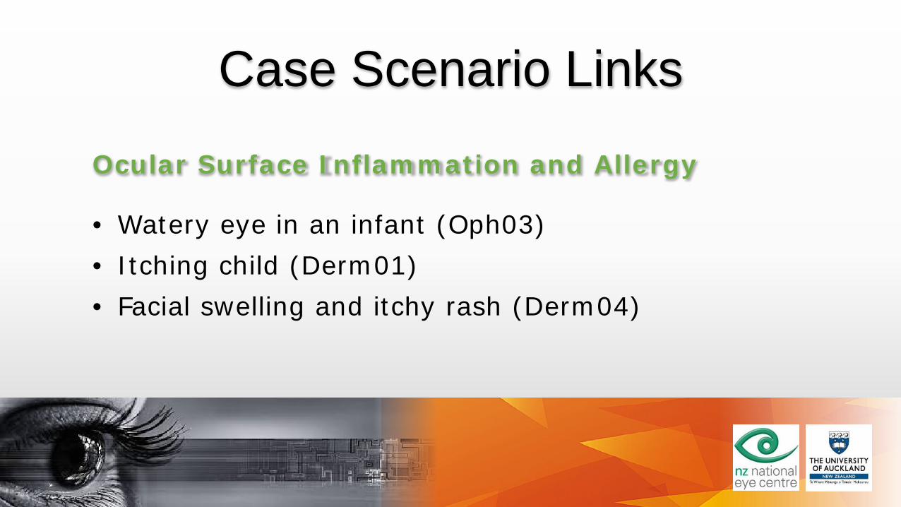
Case Scenario Links
Ocular Surface Inflammation and Allergy
• Watery eye in an infant (Oph03)• Itching child (Derm01)• Facial swelling and itchy rash (Derm04)
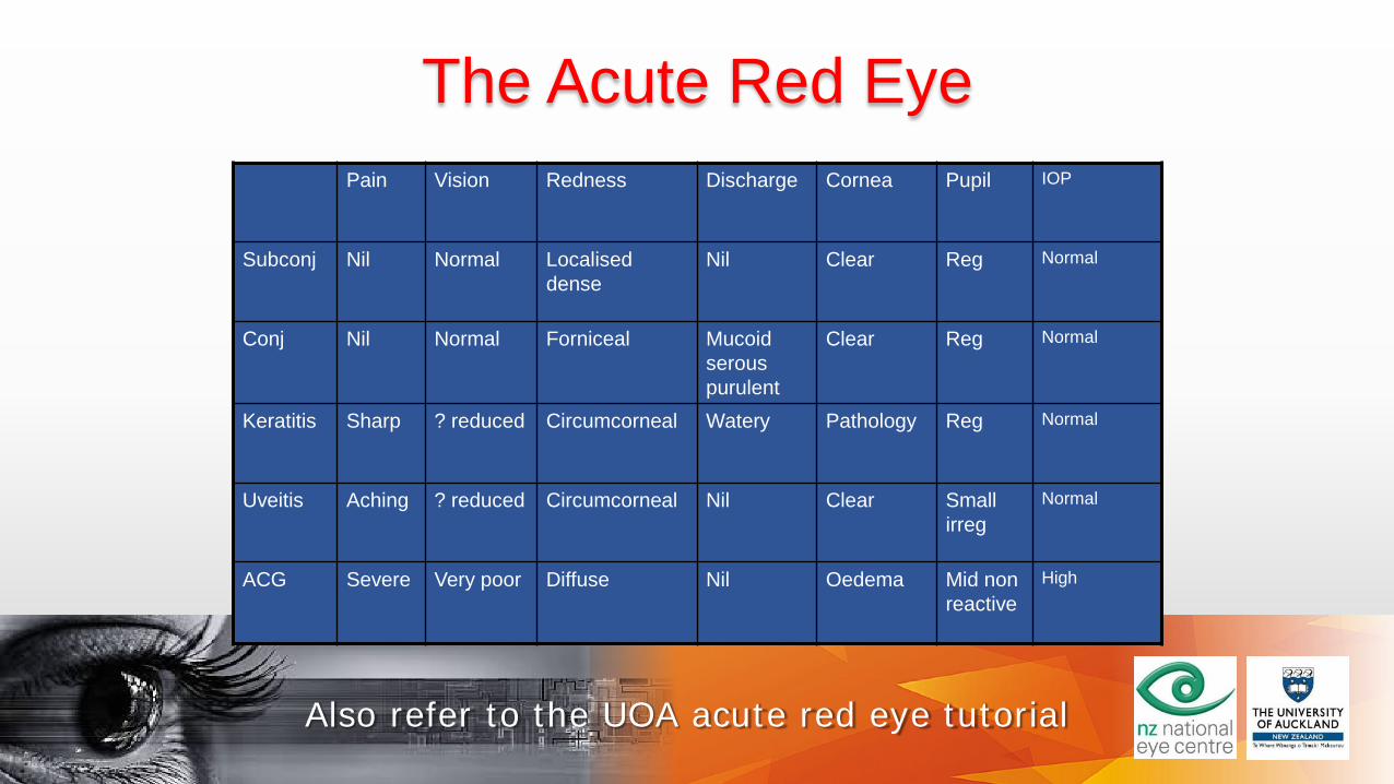
The Acute Red EyePain Vision Redness Discharge Cornea Pupil IOP
Subconj Nil Normal Localiseddense
Nil Clear Reg Normal
Conj Nil Normal Forniceal Mucoid serous purulent
Clear Reg Normal
Keratitis Sharp ? reduced Circumcorneal Watery Pathology Reg Normal
Uveitis Aching ? reduced Circumcorneal Nil Clear Small irreg
Normal
ACG Severe Very poor Diffuse Nil Oedema Mid non reactive
High
Also refer to the UOA acute red eye tutorial
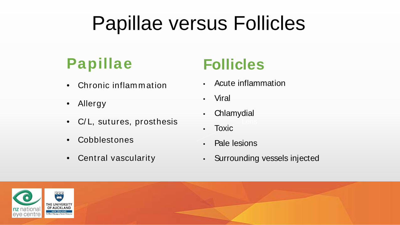
Papillae versus Follicles
Lens
Papillae• Chronic inflammation
• Allergy
• C/L, sutures, prosthesis
• Cobblestones
• Central vascularity
Follicles• Acute inflammation• Viral• Chlamydial• Toxic• Pale lesions• Surrounding vessels injected
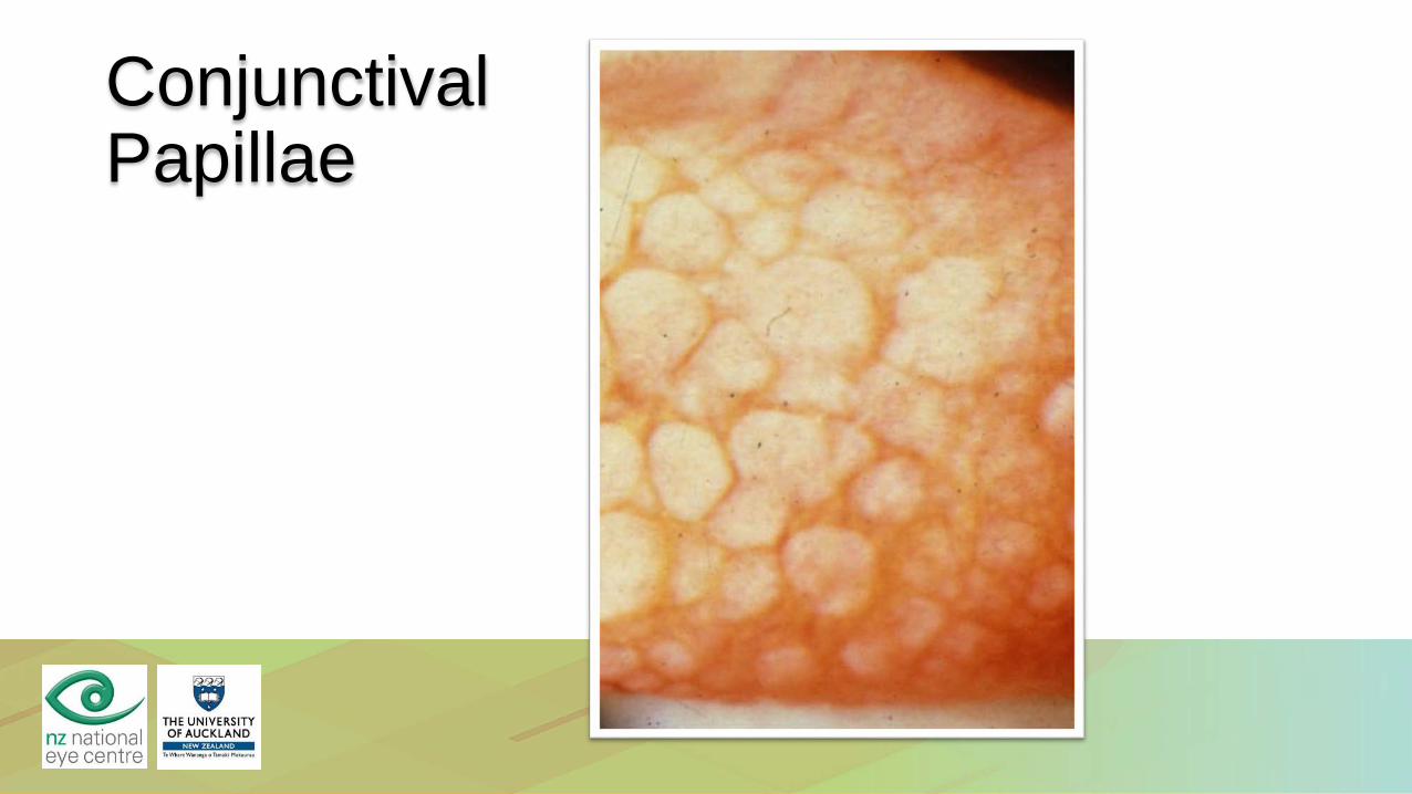
ConjunctivalPapillae
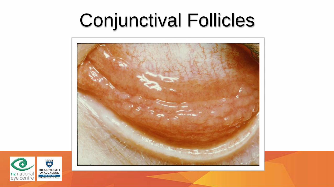
Conjunctival Follicles
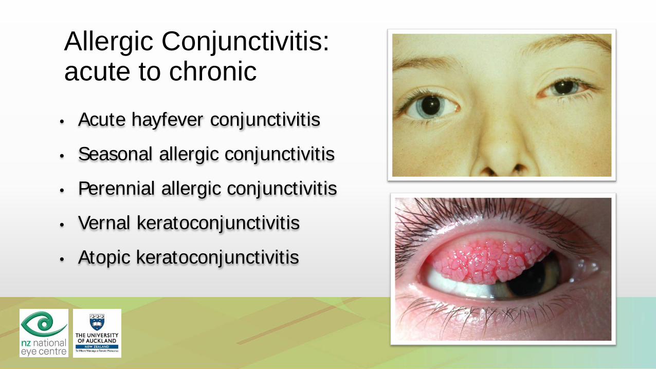
Allergic Conjunctivitis: acute to chronic
• Acute hayfever conjunctivitis• Seasonal allergic conjunctivitis• Perennial allergic conjunctivitis• Vernal keratoconjunctivitis• Atopic keratoconjunctivitis
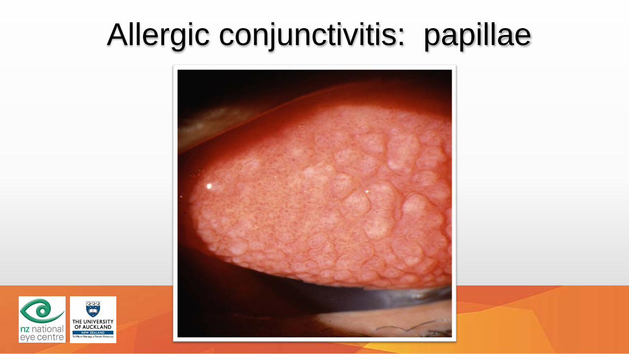
Allergic conjunctivitis: papillae
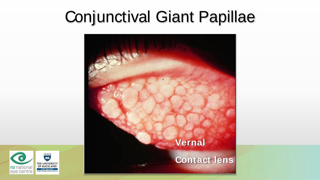
Conjunctival Giant Papillae
Vernal
Contact lens
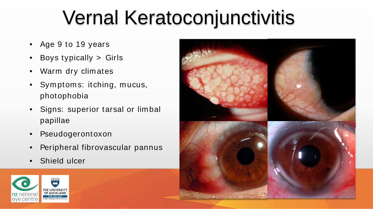
Vernal Keratoconjunctivitis• Age 9 to 19 years
• Boys typically > Girls
• Warm dry climates
• Symptoms: itching, mucus, photophobia
• Signs: superior tarsal or limbalpapillae
• Pseudogerontoxon
• Peripheral fibrovascular pannus
• Shield ulcer
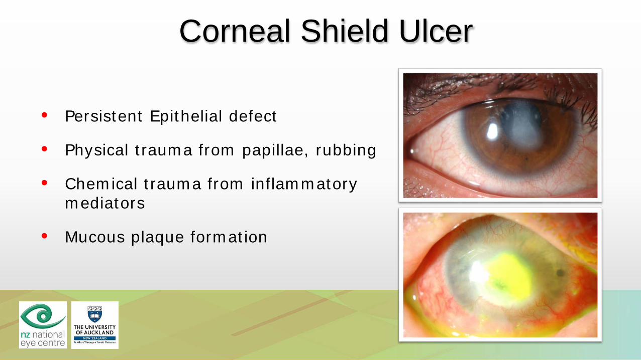
Corneal Shield Ulcer
• Persistent Epithelial defect
• Physical trauma from papillae, rubbing
• Chemical trauma from inflammatory mediators
• Mucous plaque formation
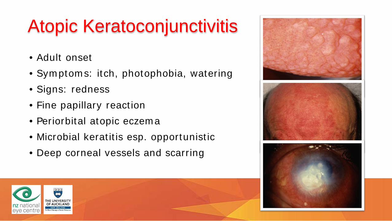
Atopic Keratoconjunctivitis• Adult onset• Symptoms: itch, photophobia, watering• Signs: redness• Fine papillary reaction• Periorbital atopic eczema• Microbial keratitis esp. opportunistic• Deep corneal vessels and scarring
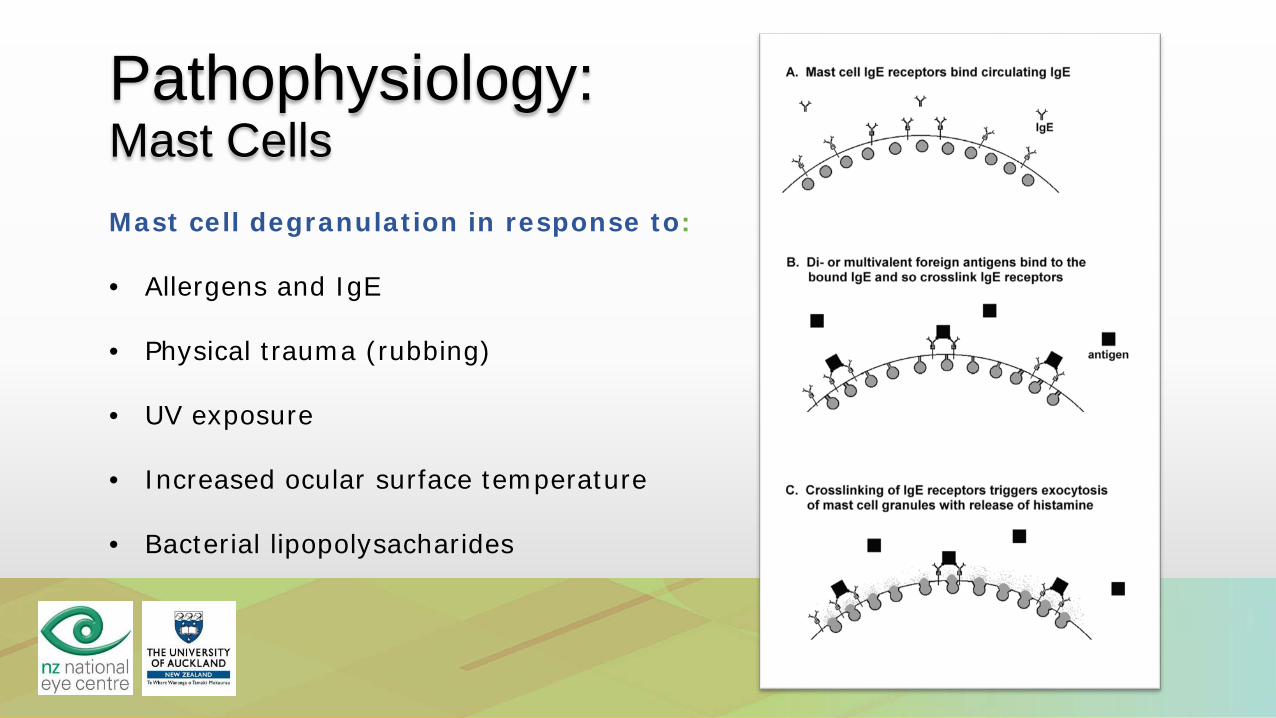
Pathophysiology: Mast CellsMast cell degranulation in response to:
• Allergens and IgE
• Physical trauma (rubbing)
• UV exposure
• Increased ocular surface temperature
• Bacterial lipopolysacharides
Therapeutic options: I(for milder disease)
• Avoidance of allergens and rubbing
• Cold compresses
• Topical antihistamines: rapid onset
• Systemic antihistamines: slower onset
• Mast cell stabilisers: preventative use
• Topical NSAIDs: Acular has some effect
• Dual action agents: best current therapy e.g. Patanol
Therapeutic options II(for vision threatening disease)
• Topical corticosteroids
o Introduce at high frequency, tail off rapidly
• Topical cyclosporine 2% ointment
• Systemic immunosuppression
• Surgery:
o Excision of papillae
o Superficial keratectomy
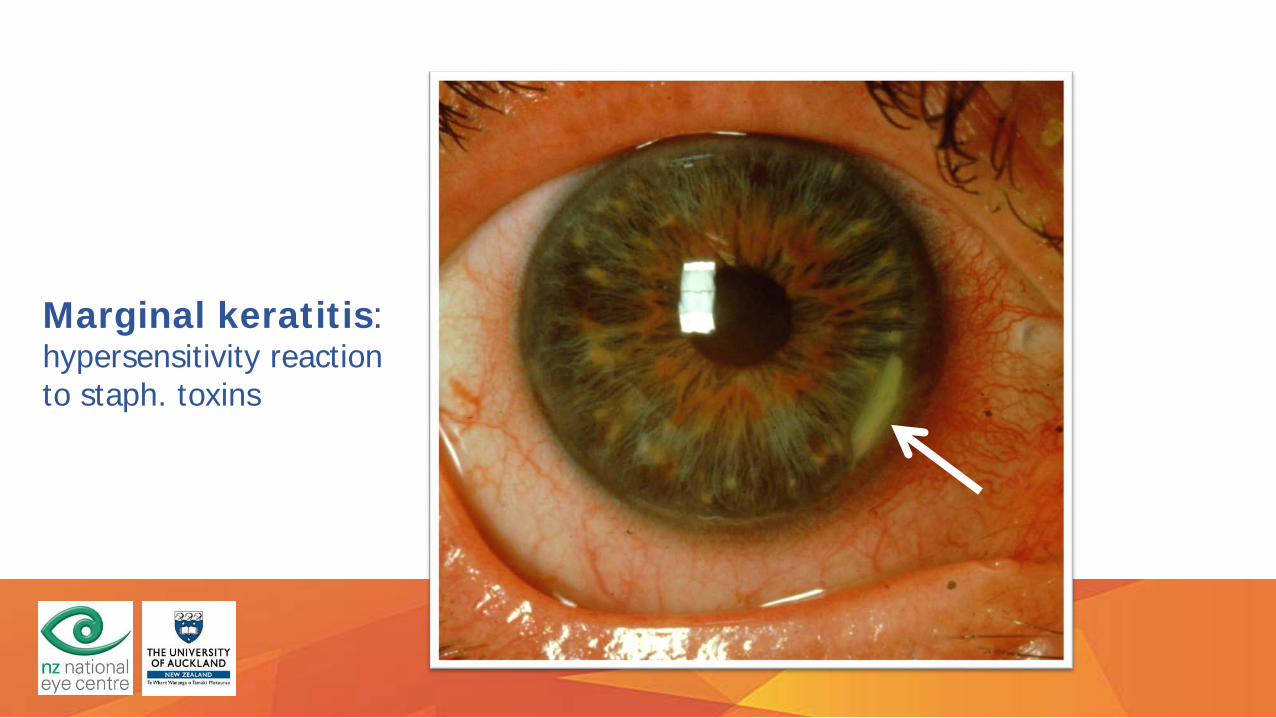
Image used with patient permission
Marginal keratitis: hypersensitivity reaction to staph. toxins
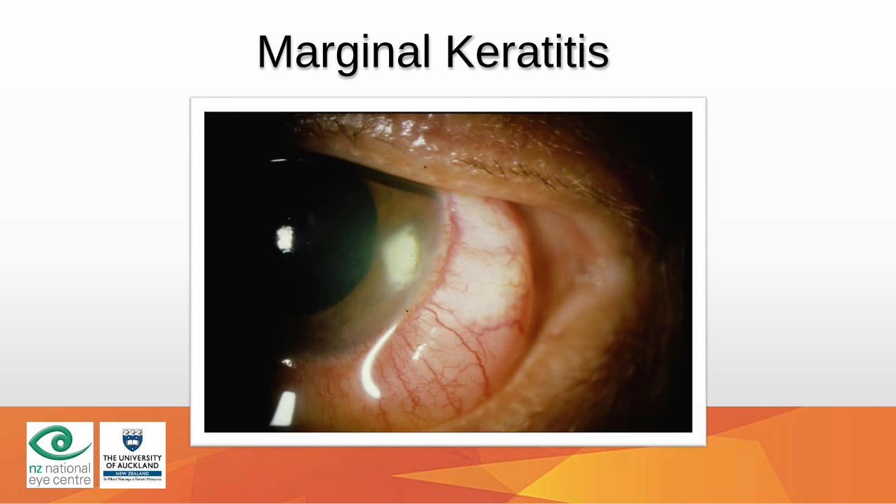
Marginal Keratitis
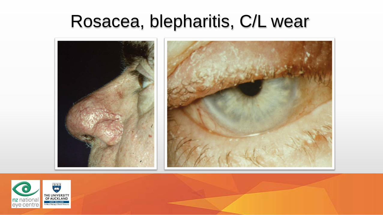
Rosacea, blepharitis, C/L wear
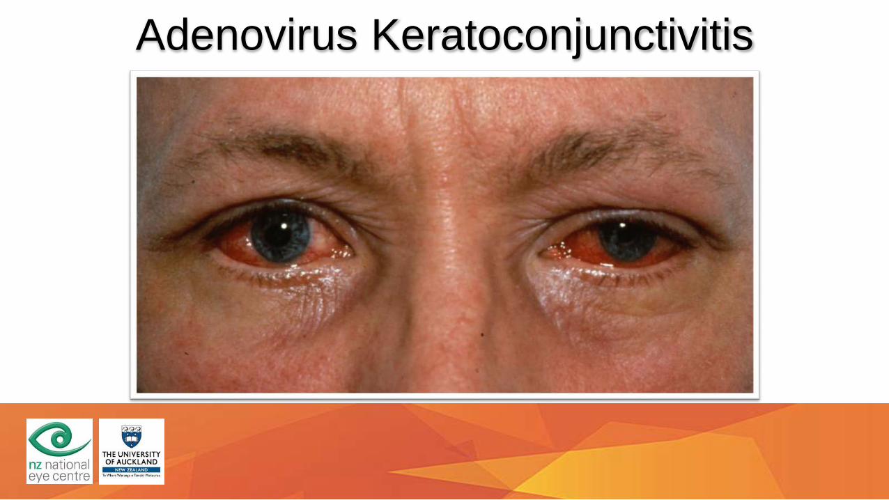
Adenovirus Keratoconjunctivitis
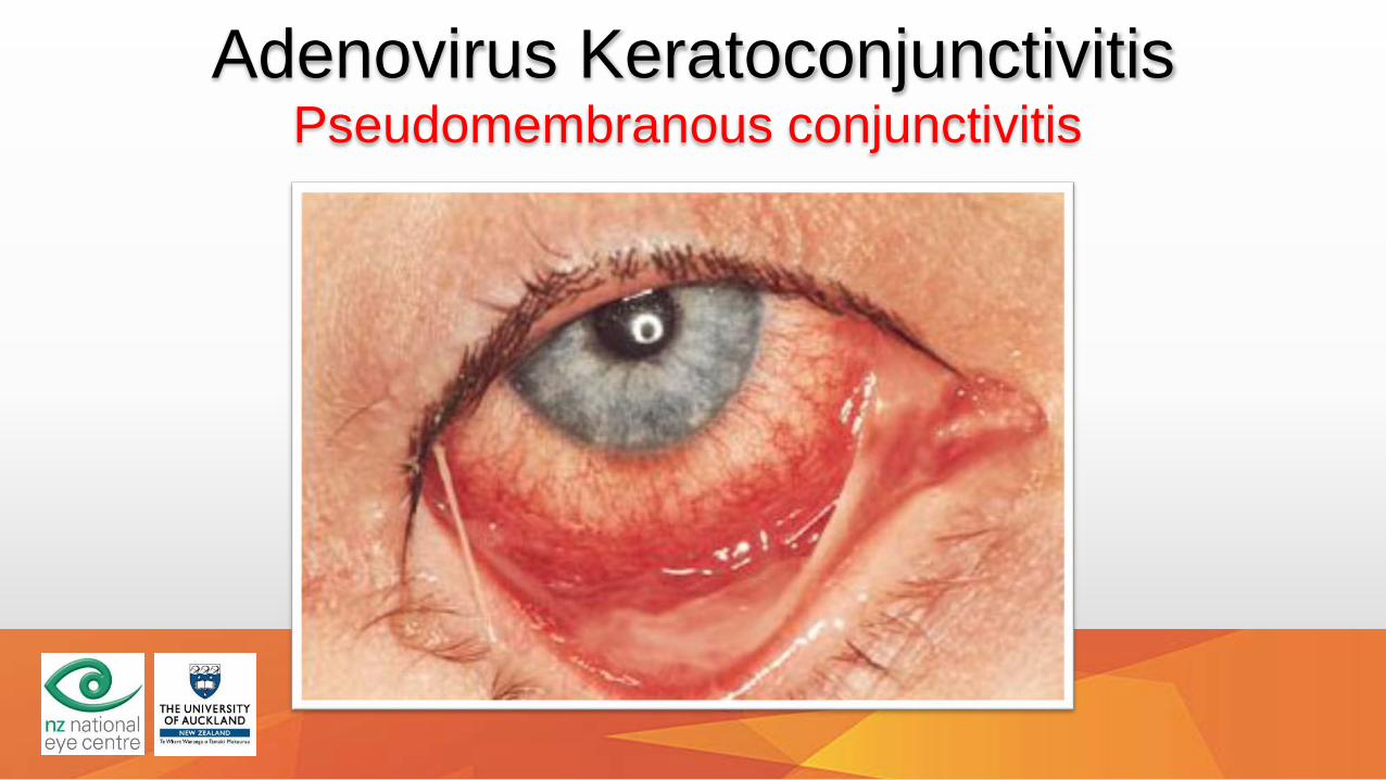
Pseudomembranous conjunctivitisAdenovirus Keratoconjunctivitis
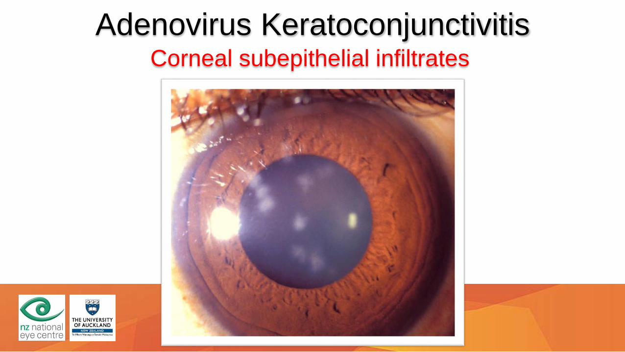
Corneal subepithelial infiltratesAdenovirus Keratoconjunctivitis
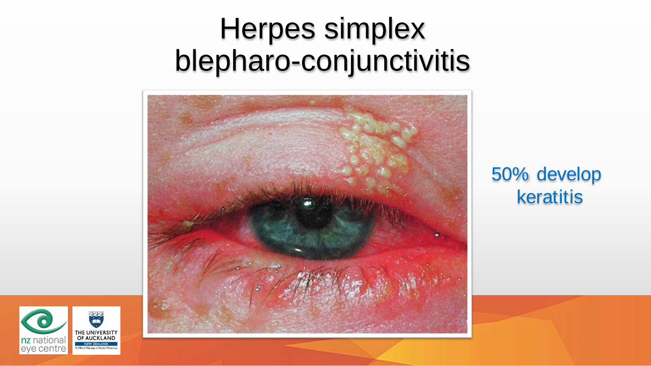
Herpes simplex blepharo-conjunctivitis
50% develop keratitis
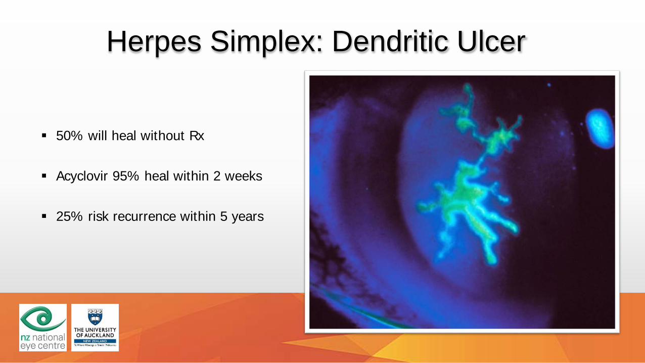
Herpes Simplex: Dendritic Ulcer
50% will heal without Rx
Acyclovir 95% heal within 2 weeks
25% risk recurrence within 5 years
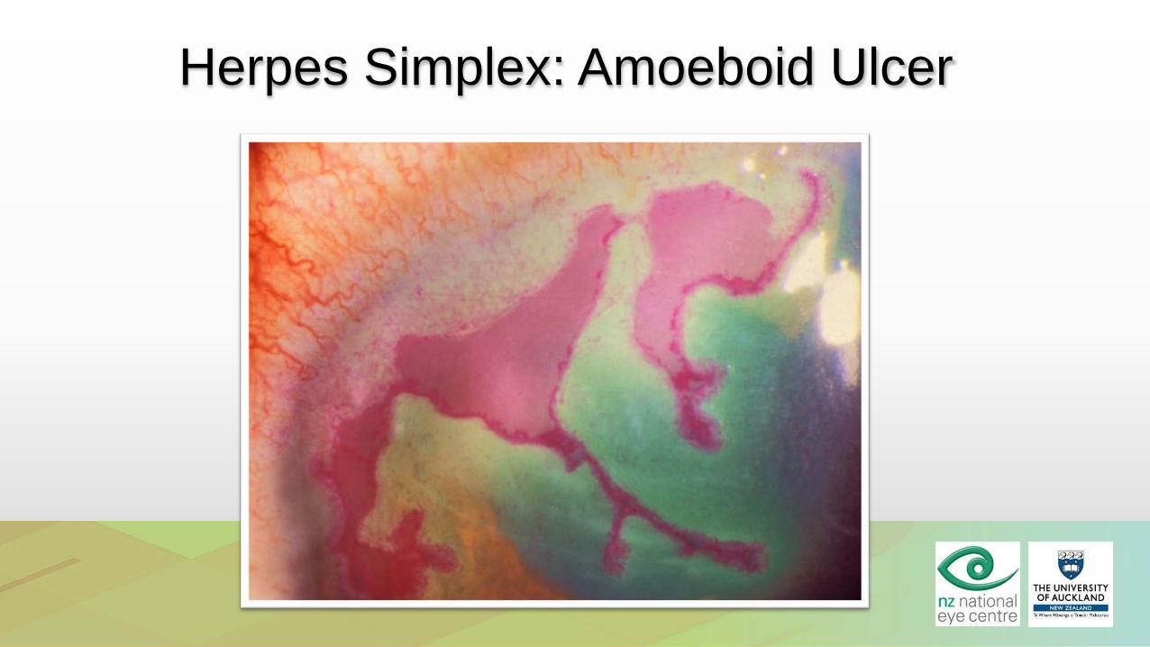
Herpes Simplex: Amoeboid Ulcer
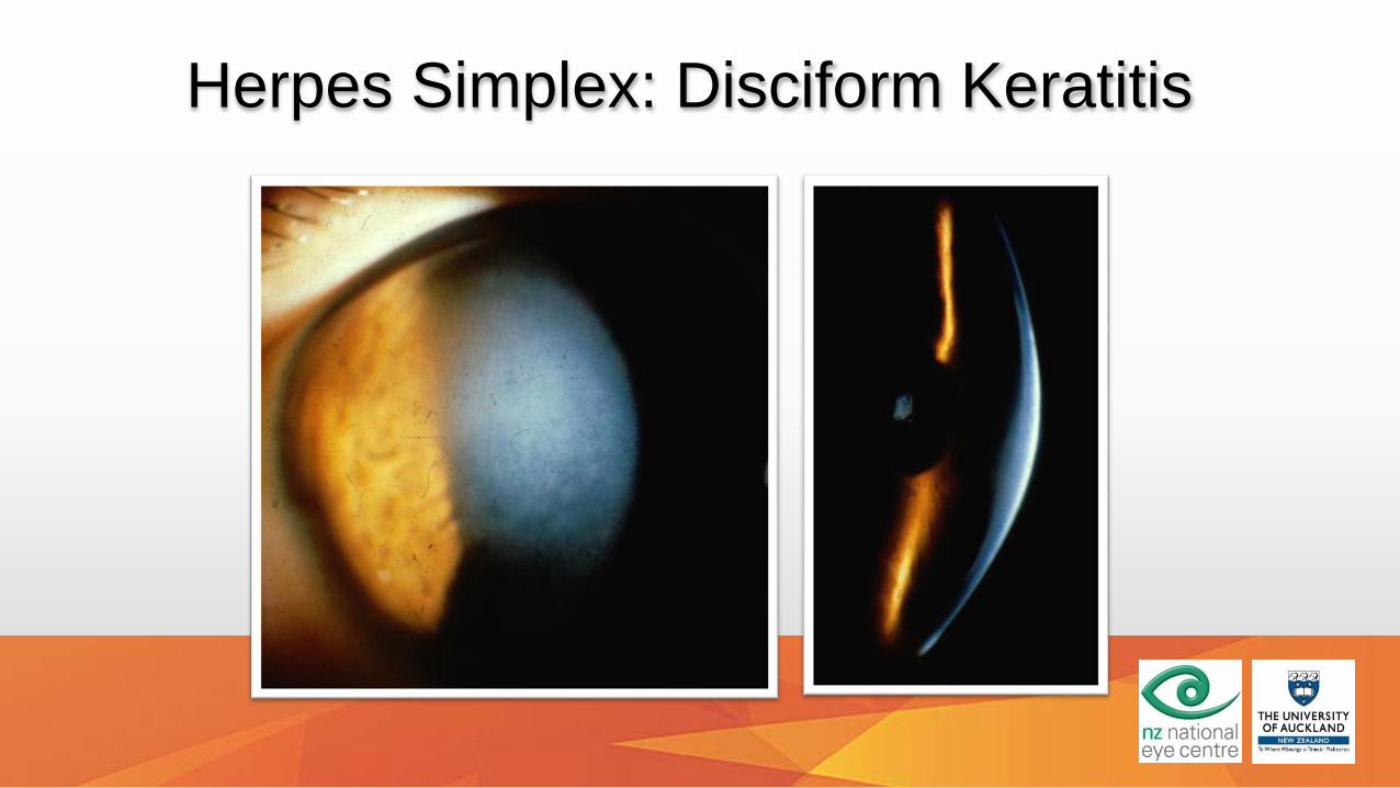
Herpes Simplex: Disciform Keratitis

HSV: Anaesthetic (denervated),Scarred, Vascularised Cornea
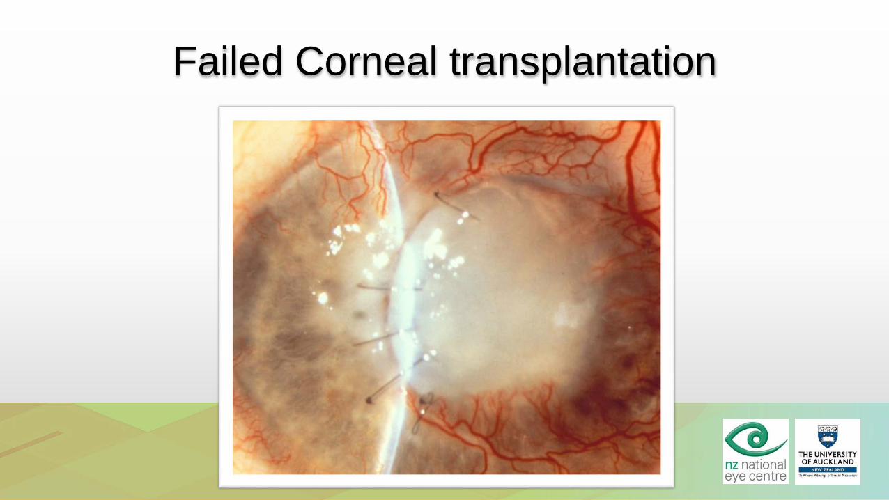
Failed Corneal transplantation
The EndMaterial contained in this lecture presentation is
copyright of The Department of Ophthalmology, New Zealand National Eye Centre, University of Auckland, and should not be reproduced without first obtaining
written permission