Embed Size (px)
Citation preview
Citation: Alamri, M.A.; Mirza, M.U.;
Adeel, M.M.; Ashfaq, U.A.; Tahir ul
Qamar, M.; Shahid, F.; Ahmad, S.;
Alatawi, E.A.; Albalawi, G.M.;
Allemailem, K.S.; et al. Structural
Elucidation of Rift Valley Fever Virus
L Protein towards the Discovery of
Its Potential Inhibitors.
Pharmaceuticals 2022, 15, 659.
https://doi.org/10.3390/
ph15060659
Academic Editor: Marialuigia
Fantacuzzi
Received: 23 April 2022
Accepted: 20 May 2022
Published: 25 May 2022
Publisher’s Note: MDPI stays neutral
with regard to jurisdictional claims in
published maps and institutional affil-
iations.
Copyright: © 2022 by the authors.
Licensee MDPI, Basel, Switzerland.
This article is an open access article
distributed under the terms and
conditions of the Creative Commons
Attribution (CC BY) license (https://
creativecommons.org/licenses/by/
4.0/).
pharmaceuticals
Article
Structural Elucidation of Rift Valley Fever Virus L Proteintowards the Discovery of Its Potential InhibitorsMubarak A. Alamri 1,† , Muhammad Usman Mirza 2,† , Muhammad Muzammal Adeel 3,† ,Usman Ali Ashfaq 4 , Muhammad Tahir ul Qamar 4,* , Farah Shahid 4, Sajjad Ahmad 5 , Eid A. Alatawi 6,Ghadah M. Albalawi 7,8, Khaled S. Allemailem 7,* and Ahmad Almatroudi 7
1 Department of Pharmaceutical Chemistry, College of Pharmacy, Prince Sattam Bin Abdulaziz University,Al-Kharj 16273, Saudi Arabia; [email protected]
2 Department of Chemistry and Biochemistry, University of Windsor, Windsor, ON N9B 3P4, Canada;[email protected]
3 3D Genomics Research Center, College of Informatics, Huazhong Agricultural University,Wuhan 430070, China; [email protected]
4 Department of Bioinformatics and Biotechnology, Government College University Faisalabad,Faisalabad 38000, Pakistan; [email protected] (U.A.A.); [email protected] (F.S.)
5 Department of Health and Biological Sciences, Abasyn University, Peshawar 25000, Pakistan;[email protected]
6 Department of Medical Laboratory Technology, Faculty of Applied Medical Sciences, University of Tabuk,Tabuk 71491, Saudi Arabia; [email protected]
7 Department of Medical Laboratories, College of Applied Medical Sciences, Qassim University,Buraydah 51452, Saudi Arabia; [email protected] (G.M.A.); [email protected] (A.A.)
8 Department of Laboratory and Blood Bank, King Fahd Specialist Hospital, Tabuk 47717, Saudi Arabia* Correspondence: [email protected] (M.T.u.Q.); [email protected] (K.S.A.)† These authors contributed equally to this work.
Abstract: Rift valley fever virus (RVFV) is the causative agent of a viral zoonosis that causes asignificant clinical burden in domestic and wild ruminants. Major outbreaks of the virus occur inlivestock, and contaminated animal products or arthropod vectors can transmit the virus to humans.The viral RNA-dependent RNA polymerase (RdRp; L protein) of the RVFV is responsible for viralreplication and is thus an appealing drug target because no effective and specific vaccine againstthis virus is available. The current study reported the structural elucidation of the RVFV-L proteinby in-depth homology modeling since no crystal structure is available yet. The inhibitory bindingmodes of known potent L protein inhibitors were analyzed. Based on the results, further moleculardocking-based virtual screening of Selleckchem Nucleoside Analogue Library (156 compounds)was performed to find potential new inhibitors against the RVFV L protein. ADME (Absorption,Distribution, Metabolism, and Excretion) and toxicity analysis of these compounds was also per-formed. Besides, the binding mechanism and stability of identified compounds were confirmed by a50 ns molecular dynamic (MD) simulation followed by MM/PBSA binding free energy calculations.Homology modeling determined a stable multi-domain structure of L protein. An analysis of knownL protein inhibitors, including Monensin, Mycophenolic acid, and Ribavirin, provide insights intothe binding mechanism and reveals key residues of the L protein binding pocket. The screeningresults revealed that the top three compounds, A-317491, Khasianine, and VER155008, exhibited ahigh affinity at the L protein binding pocket. ADME analysis revealed good pharmacodynamics andpharmacokinetic profiles of these compounds. Furthermore, MD simulation and binding free energyanalysis endorsed the binding stability of potential compounds with L protein. In a nutshell, thepresent study determined potential compounds that may aid in the rational design of novel inhibitorsof the RVFV L protein as anti-RVFV drugs.
Keywords: RVFV; RdRp; structural modeling; virtual screening; docking; MD simulation
Pharmaceuticals 2022, 15, 659. https://doi.org/10.3390/ph15060659 https://www.mdpi.com/journal/pharmaceuticals
Pharmaceuticals 2022, 15, 659 2 of 22
1. Introduction
Rift Valley fever is a hemorrhagic ailment caused by the Rift Valley Viral Infection(RVVI), affecting humans and livestock. Mosquitos are the vectors for this disease. The firstcase of RVVI was reported in East Africa’s Rift Valley in Kenya, which made it known asRVF. In its first instance, a significant population of farmed sheep was killed from 1930 to1931. Aedex and cules are the two common vectors (mosquitoes) transmitting this virus [1].Post RVVI, flu-like common symptoms have lasted up to seven days. Although mosthuman infections are mild, approximately four percent of the cases have been severe andshow symptoms such as Hemorrhagic fever, meningoencephalitis, and ocular type [2].
The virus responsible for this disease is Rift Valley Fever Virus (RVFV), a single-stranded RNA virus belonging to the Phenuviridae Family and the genus Phlebovirus.Rift Valley fever (RVF) is caused by RVFV and has a wide range of clinical symptoms. Amild febrile illness is common in humans and can lead to more severe illnesses such asencephalitis, hemorrhagic fever, and liver disease. Neurological issues including dizziness,paralysis, headaches, hallucinations, vertigo, and delirium are also observed [3]. Up to10% of RVFV-infected humans experience ophthalmologic complications, including retinalhemorrhaging and photophobia. Sheep, cattle, and goats are more susceptible to RVFV,with a 20–30% mortality rate in adult ruminants and 70% in young animals. Spontaneousabortion in infected pregnant animals is alarmingly high, ranging from 40–100 percent [4].
RVF outbreaks are sporadic and linked to meteorological, hydrological, and socioe-conomic factors. RVF outbreaks have been reported to produce considerable numbersof infected human cases, which have substantially impacted the healthcare system [5].According to an analysis by the Center for Disease Control and Prevention (CDC), severalRVF outbreaks have occurred in various countries, resulting in hundreds of thousandsof human illnesses and human casualties [6]. This disease has spread from the Africanregion to the Arabian to the USA, making it a grave concern to affected regions and thewhole world [7]. Epizootics that occur in livestock frequently precede human epidemics. Inother words, reducing RVVI in animals is expected to break the transmission cycle and helpavoid human sickness [8]. The CDC and the U.S Department of Agriculture (USDA) bothclassify RVFV as a select agent because of its ability to cause morbidity and mortality andits potential use as a bioterrorism agent [4,9]. Unfortunately, there are no FDA-approvedvaccines for humans, but some approved vaccines are available for veterinary use. Similarly,there are no therapeutics available to treat RVFV, requiring additional research in this area.
As per the structure of RVF, it is a high-molecular-weight, single-stranded, linearvirion (2–2.5 nm in diameter, 200–300 nm in length, and displaying helical symmetry)enclosed within the ribonucleo-capsid [10]. Three segments of the RVFV genome (S, M, andL-segments) have previously been described as having three separate open reading frames(ORFs) coding for three distinct proteins (viral polymerase, L-protein, and S-protein) [11,12].The L segment, which encodes only one protein, RNA-dependent RNA polymerase (RdRp),commonly referred to as L protein, is particularly important to our research. The L proteinis involved in genomic replication and viral mRNA transcription. The middle region ofthe L protein contains an RdRp domain, which is found in all RNA viral polymerasesand is required for viral RNA production. The RdRp domain retains the six characteris-tic conserved structural motifs, including PreA/F, A, B, C, D, and E in the central coreregion [12–15]. Most of these motifs are located in the palm subdomain and characterizethe active site chamber formation [14]. These host mRNAs are employed as primers forviral transcription and suppress viral-RNA-induced immune responses [16]. L protein hasan endonuclease domain at its N-terminus that is necessary for cap-snatching, in whichit cleaves 50 m7G caps from host mRNAs [17,18]. Viruses translate and reproduce theirgenome in the cell cytoplasm for survival and growth. During the replication, the virususes a technique termed cap-snatching, which means taking advantage of two known viralL protein functions: the capability to bind cap structures and cut off the cellular mRNAattached to [19].
Pharmaceuticals 2022, 15, 659 3 of 22
The rational drug design process is greatly accelerated by using different computer-aided drug designing applications for in silico drug screening [20–27]. A virtual screening-based drug discovery strategy has been identified as one of the most effective ways todiscover and develop new drugs [28]. RVFV L protein was selected for the current studyas a potential target for which no drug has been reported so far. Besides evolutionarilyconserved motifs in the core region, RdRp has channels/tunnels that link the active sitechamber with the exterior and therefore emerge as a potential target for developing anti-viral inhibitors [29–32], which is evident from the inhibitor design against many deadlyviruses such as Zika virus (ZIKV) [33–36], Japanese encephalitis virus (JEV) [37], West Nile virus(WNV) [38], Dengue virus (DENV) [39–41], HCV [24,42–44], HIV [45], SARS-CoV-2 [25],and most of the drugs have been reported against Ebola polymerase L (EBOV) [26]. Thecurrent study aimed to use the virtual screening approach to identify potential RVFV Lprotein inhibitors and inhibitory binding modes of known potent inhibitors, followed bymolecular docking analysis to discover novel inhibitors that could be used as potentialleads for RVVI treatment.
2. Results and Discussion2.1. Protein Structural Analysis
Homology modeling is a well-established technique in modern drug discovery, andwith more advancement in machine learning approaches, it is now possible to build ahomology model with high accuracy even from a template with a low identity. Recent ad-vances in homology modeling have proven their effectiveness as an alternative [46,47] andretrospective analysis, and validate the usefulness of homology modeling in SBVS. [48–51].In the current study, the 3D structure of RVFV L protein was predicted by using the ho-mology modeling approach via Modeller.v9.11. It was observed that the crystal structureand cryo-EM structure of severe fever with thrombocytopenia syndrome virus L (SFTSVL) protein (PDB IDs: 6L42 and 6Y6K) were the best hits based on query coverage andpercentage identity. 6L42 showed a 34.41% sequence identity and a 91% query coverageduring the sequence to structure alignment, while 6Y6K showed a 34.28% sequence identityand a 91% query coverage against the target sequence (Figure S1). Hence, these modelswere considered the best templates for homology modeling. Chain “A” of both templateswere used for downstream structure prediction steps. Studies have suggested that morethan 90% confidence indicates that the core model is precise and correct, deviating 2–4 Åin RMSD from the native protein structure [52]. Moreover, a good percentage of identitywith maximum coverage between the template and the query sequence indicates a highlevel of accuracy in the model. The structural superposition/RMSD of the model with thetemplates is depicted in (Figure S2).
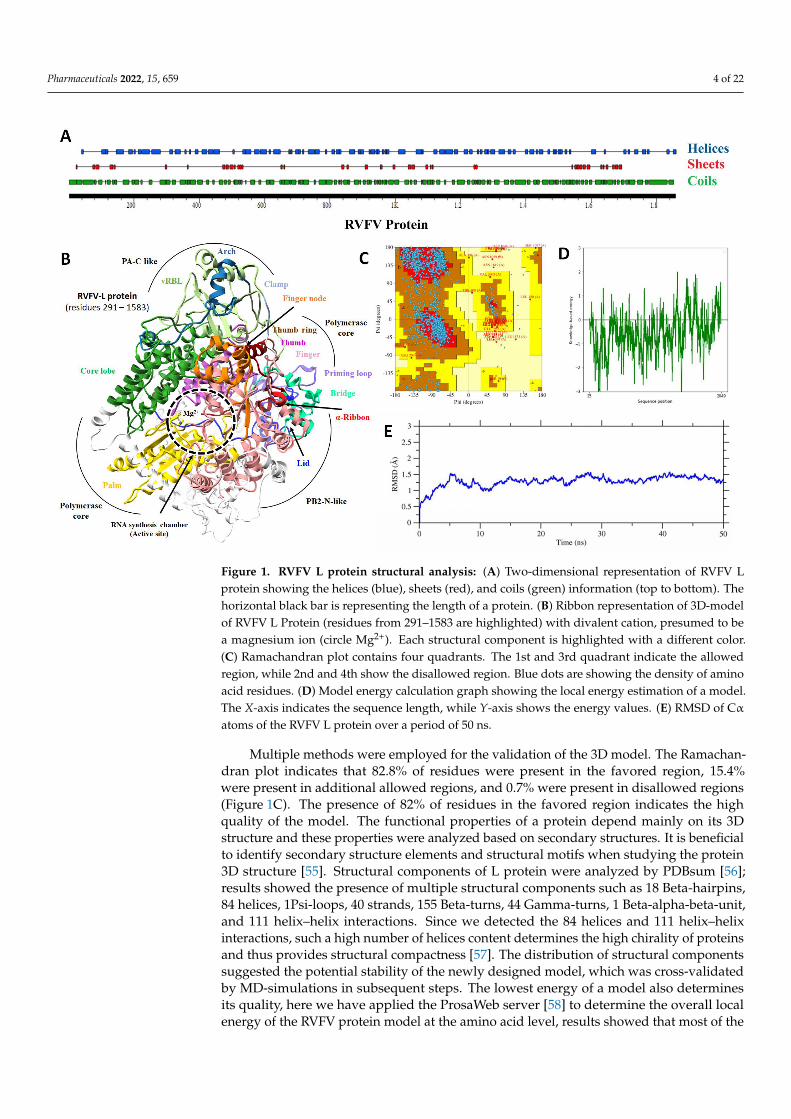
Overall, the newly predicted structure showed a pretty stable arrangement of amino-acid residues; apparently, no structural distortion was observed, and a total of 82 helices,40 sheets, and 118 coils were detected (Figure 1A). Deep structural analysis suggested thatthe computational model of RVFV protein contains seven domains, i.e., the endoN domain(amino acids 25–205), separated by a linker of span 206–295; the PA-C-like domain from 296to 762 amino acid; the RdRp core from 763 to 1345 amino acid; the PB2-N-like domain withresidues 1346–1571 span; the arm domain contains two spans 1615–1696 and 1811–1932that are separated by a blocker motif of 1811–1852 residues; the next domain was CBD withthe residues 1697–1810, and on the C-terminal, a lariat domain (1933–2049 amino acid) wasobserved as previously reported [53] (Figure 1B). Some of the RVFV protein model domainswere reported as structurally similar to SFTSV-L (severe fever with thrombocytopaeniasyndrome virus –L protein) [54], which seems evident because these proteins shared thesame values as the Phenuiviridae protein family. The orientation of each domain determinesthe functional specificity of protein and facilitates inter-molecular interactions for domainorganization [19,53].
Pharmaceuticals 2022, 15, 659 4 of 22
Pharmaceuticals 2022, 15, x FOR PEER REVIEW 4 of 22
each domain determines the functional specificity of protein and facilitates inter-molecu-
lar interactions for domain organization [19,53].
Figure 1. RVFV L protein structural analysis: (A) Two-dimensional representation of RVFV L pro-
tein showing the helices (blue), sheets (red), and coils (green) information (top to bottom). The hor-
izontal black bar is representing the length of a protein. (B) Ribbon representation of 3D-model of
RVFV L Protein (residues from 291–1583 are highlighted) with divalent cation, presumed to be a
magnesium ion (circle Mg2+). Each structural component is highlighted with a different color. (C)
Ramachandran plot contains four quadrants. The 1st and 3rd quadrant indicate the allowed region,
while 2nd and 4th show the disallowed region. Blue dots are showing the density of amino acid
residues. (D) Model energy calculation graph showing the local energy estimation of a model. The
X-axis indicates the sequence length, while Y-axis shows the energy values. (E) RMSD of Cα atoms
of the RVFV L protein over a period of 50 ns.
Multiple methods were employed for the validation of the 3D model. The Ramachan-
dran plot indicates that 82.8% of residues were present in the favored region, 15.4% were
present in additional allowed regions, and 0.7% were present in disallowed regions (Fig-
ure 1C). The presence of 82% of residues in the favored region indicates the high quality
of the model. The functional properties of a protein depend mainly on its 3D structure
and these properties were analyzed based on secondary structures. It is beneficial to iden-
tify secondary structure elements and structural motifs when studying the protein 3D
structure [55]. Structural components of L protein were analyzed by PDBsum [56]; results
showed the presence of multiple structural components such as 18 Beta-hairpins, 84 heli-
ces, 1Psi-loops, 40 strands, 155 Beta-turns, 44 Gamma-turns, 1 Beta-alpha-beta-unit, and
111 helix–helix interactions. Since we detected the 84 helices and 111 helix–helix interac-
tions, such a high number of helices content determines the high chirality of proteins and
thus provides structural compactness [57]. The distribution of structural components sug-
gested the potential stability of the newly designed model, which was cross-validated by
MD-simulations in subsequent steps. The lowest energy of a model also determines its
Figure 1. RVFV L protein structural analysis: (A) Two-dimensional representation of RVFV Lprotein showing the helices (blue), sheets (red), and coils (green) information (top to bottom). Thehorizontal black bar is representing the length of a protein. (B) Ribbon representation of 3D-modelof RVFV L Protein (residues from 291–1583 are highlighted) with divalent cation, presumed to bea magnesium ion (circle Mg2+). Each structural component is highlighted with a different color.(C) Ramachandran plot contains four quadrants. The 1st and 3rd quadrant indicate the allowedregion, while 2nd and 4th show the disallowed region. Blue dots are showing the density of aminoacid residues. (D) Model energy calculation graph showing the local energy estimation of a model.The X-axis indicates the sequence length, while Y-axis shows the energy values. (E) RMSD of Cαatoms of the RVFV L protein over a period of 50 ns.
Multiple methods were employed for the validation of the 3D model. The Ramachan-dran plot indicates that 82.8% of residues were present in the favored region, 15.4%were present in additional allowed regions, and 0.7% were present in disallowed regions(Figure 1C). The presence of 82% of residues in the favored region indicates the highquality of the model. The functional properties of a protein depend mainly on its 3Dstructure and these properties were analyzed based on secondary structures. It is beneficialto identify secondary structure elements and structural motifs when studying the protein3D structure [55]. Structural components of L protein were analyzed by PDBsum [56];results showed the presence of multiple structural components such as 18 Beta-hairpins,84 helices, 1Psi-loops, 40 strands, 155 Beta-turns, 44 Gamma-turns, 1 Beta-alpha-beta-unit,and 111 helix–helix interactions. Since we detected the 84 helices and 111 helix–helixinteractions, such a high number of helices content determines the high chirality of proteinsand thus provides structural compactness [57]. The distribution of structural componentssuggested the potential stability of the newly designed model, which was cross-validatedby MD-simulations in subsequent steps. The lowest energy of a model also determinesits quality, here we have applied the ProsaWeb server [58] to determine the overall localenergy of the RVFV protein model at the amino acid level, results showed that most of the
Pharmaceuticals 2022, 15, 659 5 of 22
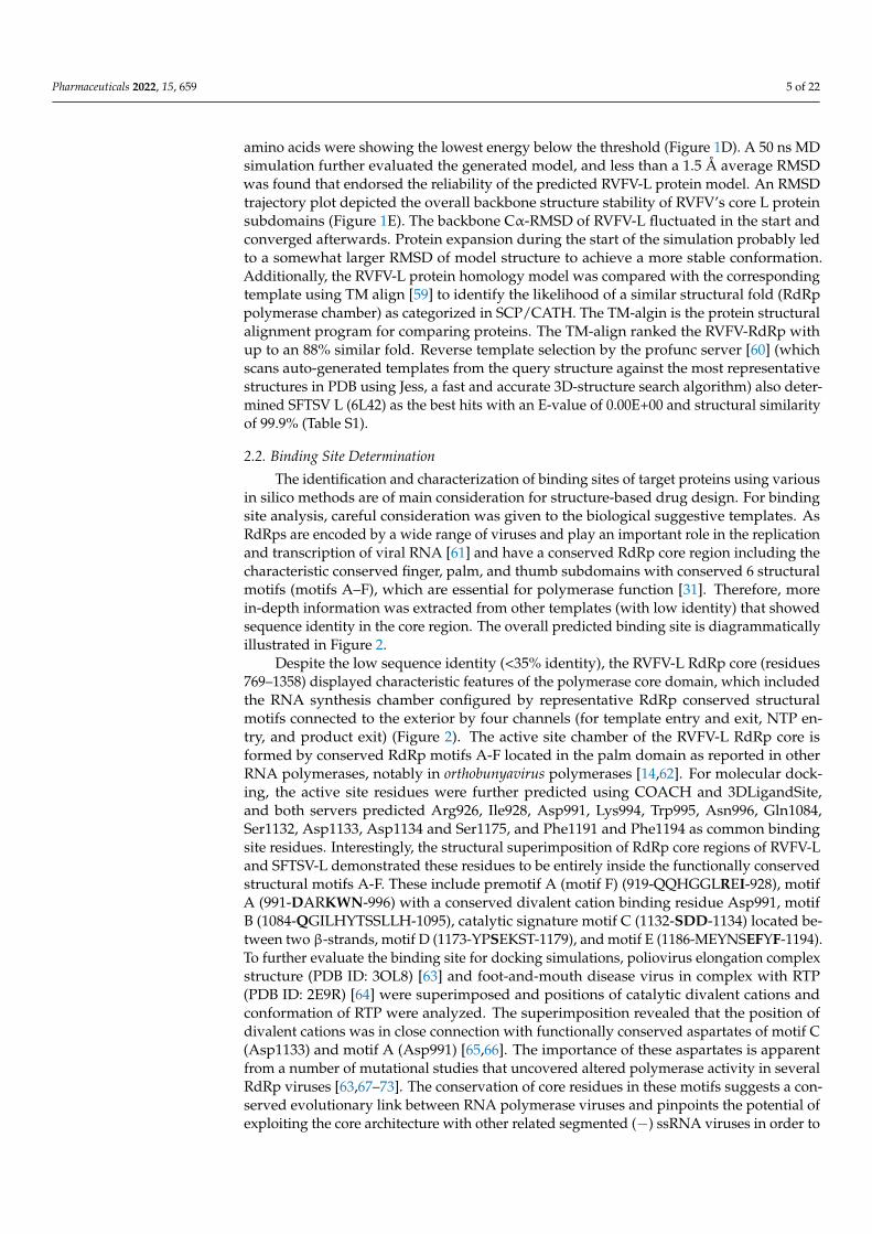
amino acids were showing the lowest energy below the threshold (Figure 1D). A 50 ns MDsimulation further evaluated the generated model, and less than a 1.5 Å average RMSDwas found that endorsed the reliability of the predicted RVFV-L protein model. An RMSDtrajectory plot depicted the overall backbone structure stability of RVFV’s core L proteinsubdomains (Figure 1E). The backbone Cα-RMSD of RVFV-L fluctuated in the start andconverged afterwards. Protein expansion during the start of the simulation probably ledto a somewhat larger RMSD of model structure to achieve a more stable conformation.Additionally, the RVFV-L protein homology model was compared with the correspondingtemplate using TM align [59] to identify the likelihood of a similar structural fold (RdRppolymerase chamber) as categorized in SCP/CATH. The TM-algin is the protein structuralalignment program for comparing proteins. The TM-align ranked the RVFV-RdRp withup to an 88% similar fold. Reverse template selection by the profunc server [60] (whichscans auto-generated templates from the query structure against the most representativestructures in PDB using Jess, a fast and accurate 3D-structure search algorithm) also deter-mined SFTSV L (6L42) as the best hits with an E-value of 0.00E+00 and structural similarityof 99.9% (Table S1).
2.2. Binding Site Determination
The identification and characterization of binding sites of target proteins using variousin silico methods are of main consideration for structure-based drug design. For bindingsite analysis, careful consideration was given to the biological suggestive templates. AsRdRps are encoded by a wide range of viruses and play an important role in the replicationand transcription of viral RNA [61] and have a conserved RdRp core region including thecharacteristic conserved finger, palm, and thumb subdomains with conserved 6 structuralmotifs (motifs A–F), which are essential for polymerase function [31]. Therefore, morein-depth information was extracted from other templates (with low identity) that showedsequence identity in the core region. The overall predicted binding site is diagrammaticallyillustrated in Figure 2.
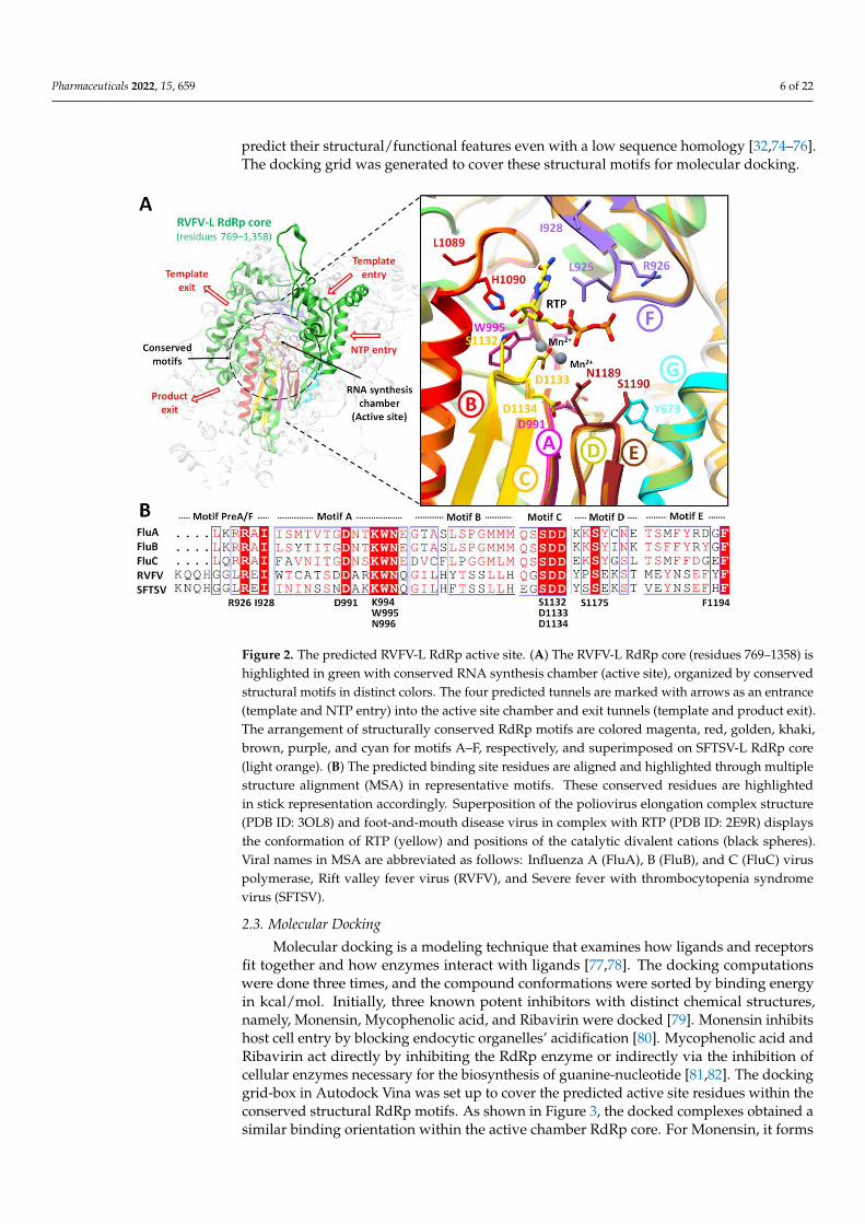
Despite the low sequence identity (<35% identity), the RVFV-L RdRp core (residues769–1358) displayed characteristic features of the polymerase core domain, which includedthe RNA synthesis chamber configured by representative RdRp conserved structuralmotifs connected to the exterior by four channels (for template entry and exit, NTP en-try, and product exit) (Figure 2). The active site chamber of the RVFV-L RdRp core isformed by conserved RdRp motifs A-F located in the palm domain as reported in otherRNA polymerases, notably in orthobunyavirus polymerases [14,62]. For molecular dock-ing, the active site residues were further predicted using COACH and 3DLigandSite,and both servers predicted Arg926, Ile928, Asp991, Lys994, Trp995, Asn996, Gln1084,Ser1132, Asp1133, Asp1134 and Ser1175, and Phe1191 and Phe1194 as common bindingsite residues. Interestingly, the structural superimposition of RdRp core regions of RVFV-Land SFTSV-L demonstrated these residues to be entirely inside the functionally conservedstructural motifs A-F. These include premotif A (motif F) (919-QQHGGLREI-928), motifA (991-DARKWN-996) with a conserved divalent cation binding residue Asp991, motifB (1084-QGILHYTSSLLH-1095), catalytic signature motif C (1132-SDD-1134) located be-tween two β-strands, motif D (1173-YPSEKST-1179), and motif E (1186-MEYNSEFYF-1194).To further evaluate the binding site for docking simulations, poliovirus elongation complexstructure (PDB ID: 3OL8) [63] and foot-and-mouth disease virus in complex with RTP(PDB ID: 2E9R) [64] were superimposed and positions of catalytic divalent cations andconformation of RTP were analyzed. The superimposition revealed that the position ofdivalent cations was in close connection with functionally conserved aspartates of motif C(Asp1133) and motif A (Asp991) [65,66]. The importance of these aspartates is apparentfrom a number of mutational studies that uncovered altered polymerase activity in severalRdRp viruses [63,67–73]. The conservation of core residues in these motifs suggests a con-served evolutionary link between RNA polymerase viruses and pinpoints the potential ofexploiting the core architecture with other related segmented (−) ssRNA viruses in order to
Pharmaceuticals 2022, 15, 659 6 of 22
predict their structural/functional features even with a low sequence homology [32,74–76].The docking grid was generated to cover these structural motifs for molecular docking.
Pharmaceuticals 2022, 15, x FOR PEER REVIEW 6 of 22
Figure 2. The predicted RVFV-L RdRp active site. (A) The RVFV-L RdRp core (residues 769–1358)
is highlighted in green with conserved RNA synthesis chamber (active site), organized by conserved
structural motifs in distinct colors. The four predicted tunnels are marked with arrows as an en-
trance (template and NTP entry) into the active site chamber and exit tunnels (template and product
exit). The arrangement of structurally conserved RdRp motifs are colored magenta, red, golden,
khaki, brown, purple, and cyan for motifs A–F, respectively, and superimposed on SFTSV-L RdRp
core (light orange). (B) The predicted binding site residues are aligned and highlighted through
multiple structure alignment (MSA) in representative motifs. These conserved residues are high-
lighted in stick representation accordingly. Superposition of the poliovirus elongation complex
structure (PDB ID: 3OL8) and foot-and-mouth disease virus in complex with RTP (PDB ID: 2E9R)
displays the conformation of RTP (yellow) and positions of the catalytic divalent cations (black
spheres). Viral names in MSA are abbreviated as follows: Influenza A (FluA), B (FluB), and C (FluC)
virus polymerase, Rift valley fever virus (RVFV), and Severe fever with thrombocytopenia syn-
drome virus (SFTSV).
Despite the low sequence identity (<35% identity), the RVFV-L RdRp core (residues
769–1358) displayed characteristic features of the polymerase core domain, which in-
cluded the RNA synthesis chamber configured by representative RdRp conserved struc-
tural motifs connected to the exterior by four channels (for template entry and exit, NTP
entry, and product exit) (Figure 2). The active site chamber of the RVFV-L RdRp core is
formed by conserved RdRp motifs A-F located in the palm domain as reported in other
RNA polymerases, notably in orthobunyavirus polymerases [14,62]. For molecular docking,
the active site residues were further predicted using COACH and 3DLigandSite, and both
servers predicted Arg926, Ile928, Asp991, Lys994, Trp995, Asn996, Gln1084, Ser1132,
Asp1133, Asp1134 and Ser1175, and Phe1191 and Phe1194 as common binding site resi-
dues. Interestingly, the structural superimposition of RdRp core regions of RVFV-L and
SFTSV-L demonstrated these residues to be entirely inside the functionally conserved
structural motifs A-F. These include premotif A (motif F) (919-QQHGGLREI-928), motif
A (991-DARKWN-996) with a conserved divalent cation binding residue Asp991, motif B
(1084-QGILHYTSSLLH-1095), catalytic signature motif C (1132-SDD-1134) located be-
tween two β-strands, motif D (1173-YPSEKST-1179), and motif E (1186-MEYNSEFYF-
Figure 2. The predicted RVFV-L RdRp active site. (A) The RVFV-L RdRp core (residues 769–1358) ishighlighted in green with conserved RNA synthesis chamber (active site), organized by conservedstructural motifs in distinct colors. The four predicted tunnels are marked with arrows as an entrance(template and NTP entry) into the active site chamber and exit tunnels (template and product exit).The arrangement of structurally conserved RdRp motifs are colored magenta, red, golden, khaki,brown, purple, and cyan for motifs A–F, respectively, and superimposed on SFTSV-L RdRp core(light orange). (B) The predicted binding site residues are aligned and highlighted through multiplestructure alignment (MSA) in representative motifs. These conserved residues are highlightedin stick representation accordingly. Superposition of the poliovirus elongation complex structure(PDB ID: 3OL8) and foot-and-mouth disease virus in complex with RTP (PDB ID: 2E9R) displaysthe conformation of RTP (yellow) and positions of the catalytic divalent cations (black spheres).Viral names in MSA are abbreviated as follows: Influenza A (FluA), B (FluB), and C (FluC) viruspolymerase, Rift valley fever virus (RVFV), and Severe fever with thrombocytopenia syndromevirus (SFTSV).
2.3. Molecular Docking
Molecular docking is a modeling technique that examines how ligands and receptorsfit together and how enzymes interact with ligands [77,78]. The docking computationswere done three times, and the compound conformations were sorted by binding energyin kcal/mol. Initially, three known potent inhibitors with distinct chemical structures,namely, Monensin, Mycophenolic acid, and Ribavirin were docked [79]. Monensin inhibitshost cell entry by blocking endocytic organelles’ acidification [80]. Mycophenolic acid andRibavirin act directly by inhibiting the RdRp enzyme or indirectly via the inhibition ofcellular enzymes necessary for the biosynthesis of guanine-nucleotide [81,82]. The dockinggrid-box in Autodock Vina was set up to cover the predicted active site residues within theconserved structural RdRp motifs. As shown in Figure 3, the docked complexes obtained asimilar binding orientation within the active chamber RdRp core. For Monensin, it forms
Pharmaceuticals 2022, 15, 659 7 of 22
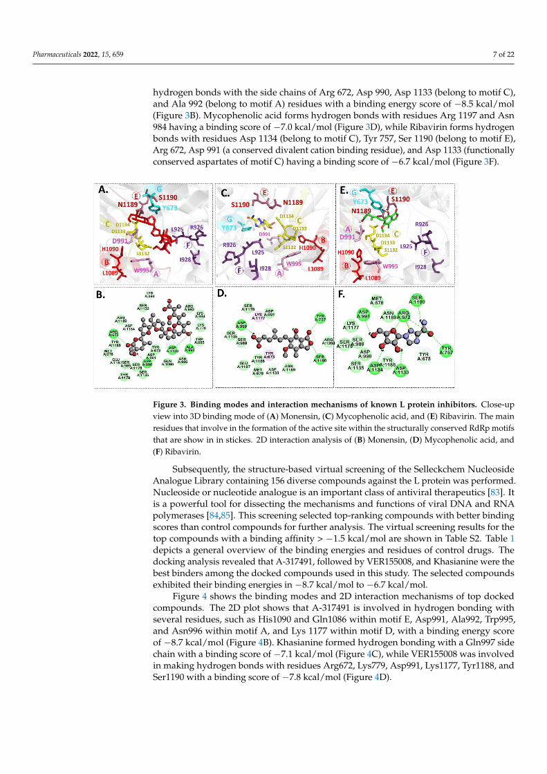
hydrogen bonds with the side chains of Arg 672, Asp 990, Asp 1133 (belong to motif C),and Ala 992 (belong to motif A) residues with a binding energy score of −8.5 kcal/mol(Figure 3B). Mycophenolic acid forms hydrogen bonds with residues Arg 1197 and Asn984 having a binding score of −7.0 kcal/mol (Figure 3D), while Ribavirin forms hydrogenbonds with residues Asp 1134 (belong to motif C), Tyr 757, Ser 1190 (belong to motif E),Arg 672, Asp 991 (a conserved divalent cation binding residue), and Asp 1133 (functionallyconserved aspartates of motif C) having a binding score of −6.7 kcal/mol (Figure 3F).
Pharmaceuticals 2022, 15, x FOR PEER REVIEW 7 of 22
1194). To further evaluate the binding site for docking simulations, poliovirus elongation
complex structure (PDB ID: 3OL8) [63] and foot-and-mouth disease virus in complex with
RTP (PDB ID: 2E9R) [64] were superimposed and positions of catalytic divalent cations
and conformation of RTP were analyzed. The superimposition revealed that the position
of divalent cations was in close connection with functionally conserved aspartates of motif
C (Asp1133) and motif A (Asp991) [65,66]. The importance of these aspartates is apparent
from a number of mutational studies that uncovered altered polymerase activity in several
RdRp viruses [63,67–73]. The conservation of core residues in these motifs suggests a con-
served evolutionary link between RNA polymerase viruses and pinpoints the potential of
exploiting the core architecture with other related segmented (−) ssRNA viruses in order
to predict their structural/functional features even with a low sequence homology [32,74–
76]. The docking grid was generated to cover these structural motifs for molecular dock-
ing.
2.3. Molecular Docking
Molecular docking is a modeling technique that examines how ligands and receptors
fit together and how enzymes interact with ligands [77,78]. The docking computations
were done three times, and the compound conformations were sorted by binding energy
in kcal/mol. Initially, three known potent inhibitors with distinct chemical structures,
namely, Monensin, Mycophenolic acid, and Ribavirin were docked [79]. Monensin inhib-
its host cell entry by blocking endocytic organelles’ acidification [80]. Mycophenolic acid
and Ribavirin act directly by inhibiting the RdRp enzyme or indirectly via the inhibition
of cellular enzymes necessary for the biosynthesis of guanine-nucleotide [81,82]. The
docking grid-box in Autodock Vina was set up to cover the predicted active site residues
within the conserved structural RdRp motifs. As shown in Figure 3, the docked complexes
obtained a similar binding orientation within the active chamber RdRp core. For
Monensin, it forms hydrogen bonds with the side chains of Arg 672, Asp 990, Asp 1133
(belong to motif C), and Ala 992 (belong to motif A) residues with a binding energy score
of −8.5 kcal/mol (Figure 3B). Mycophenolic acid forms hydrogen bonds with residues Arg
1197 and Asn 984 having a binding score of −7.0 kcal/mol (Figure 3D), while Ribavirin
forms hydrogen bonds with residues Asp 1134 (belong to motif C), Tyr 757, Ser 1190 (be-
long to motif E), Arg 672, Asp 991 (a conserved divalent cation binding residue), and Asp
1133 (functionally conserved aspartates of motif C) having a binding score of −6.7 kcal/mol
(Figure 3F).
Figure 3. Binding modes and interaction mechanisms of known L protein inhibitors. Close-upview into 3D binding mode of (A) Monensin, (C) Mycophenolic acid, and (E) Ribavirin. The mainresidues that involve in the formation of the active site within the structurally conserved RdRp motifsthat are show in in stickes. 2D interaction analysis of (B) Monensin, (D) Mycophenolic acid, and(F) Ribavirin.
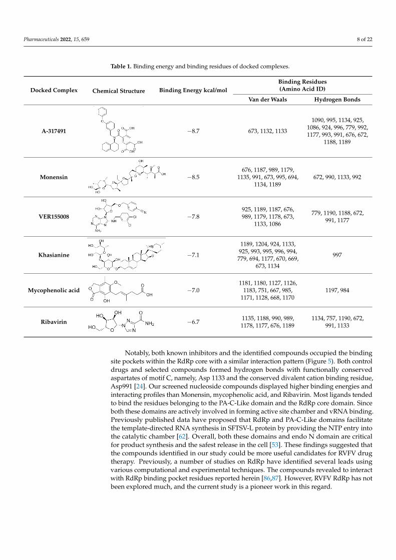
Subsequently, the structure-based virtual screening of the Selleckchem NucleosideAnalogue Library containing 156 diverse compounds against the L protein was performed.Nucleoside or nucleotide analogue is an important class of antiviral therapeutics [83]. Itis a powerful tool for dissecting the mechanisms and functions of viral DNA and RNApolymerases [84,85]. This screening selected top-ranking compounds with better bindingscores than control compounds for further analysis. The virtual screening results for thetop compounds with a binding affinity > −1.5 kcal/mol are shown in Table S2. Table 1depicts a general overview of the binding energies and residues of control drugs. Thedocking analysis revealed that A-317491, followed by VER155008, and Khasianine were thebest binders among the docked compounds used in this study. The selected compoundsexhibited their binding energies in −8.7 kcal/mol to −6.7 kcal/mol.
Figure 4 shows the binding modes and 2D interaction mechanisms of top dockedcompounds. The 2D plot shows that A-317491 is involved in hydrogen bonding withseveral residues, such as His1090 and Gln1086 within motif E, Asp991, Ala992, Trp995,and Asn996 within motif A, and Lys 1177 within motif D, with a binding energy scoreof −8.7 kcal/mol (Figure 4B). Khasianine formed hydrogen bonding with a Gln997 sidechain with a binding score of −7.1 kcal/mol (Figure 4C), while VER155008 was involvedin making hydrogen bonds with residues Arg672, Lys779, Asp991, Lys1177, Tyr1188, andSer1190 with a binding score of −7.8 kcal/mol (Figure 4D).
Pharmaceuticals 2022, 15, 659 8 of 22
Table 1. Binding energy and binding residues of docked complexes.
Docked Complex Chemical Structure Binding Energy kcal/molBinding Residues(Amino Acid ID)
Van der Waals Hydrogen Bonds
A-317491
Pharmaceuticals 2022, 15, x FOR PEER REVIEW 8 of 22
Figure 3. Binding modes and interaction mechanisms of known L protein inhibitors. Close-up
view into 3D binding mode of (A) Monensin, (C) Mycophenolic acid, and (E) Ribavirin. The main
residues that involve in the formation of the active site within the structurally conserved RdRp mo-
tifs that are show in in stickes. 2D interaction analysis of (B) Monensin, (D) Mycophenolic acid, and
(F) Ribavirin.
Subsequently, the structure-based virtual screening of the Selleckchem Nucleoside
Analogue Library containing 156 diverse compounds against the L protein was per-
formed. Nucleoside or nucleotide analogue is an important class of antiviral therapeutics
[83]. It is a powerful tool for dissecting the mechanisms and functions of viral DNA and
RNA polymerases [84,85]. This screening selected top-ranking compounds with better
binding scores than control compounds for further analysis. The virtual screening results
for the top compounds with a binding affinity > −1.5 kcal/mol are shown in Table S2. Table
1 depicts a general overview of the binding energies and residues of control drugs. The
docking analysis revealed that A-317491, followed by VER155008, and Khasianine were
the best binders among the docked compounds used in this study. The selected com-
pounds exhibited their binding energies in −8.7 kcal/mol to −6.7 kcal/mol.
Table 1. Binding energy and binding residues of docked complexes.
Docked
Complex Chemical Structure
Binding
Energy
kcal/mol
Binding Residues
(Amino Acid ID)
Van der Waals Hydrogen Bonds
A-317491
−8.7 673, 1132, 1133
1090, 995, 1134, 925, 1086,
924, 996, 779, 992, 1177,
993, 991, 676, 672, 1188,
1189
Monensin
−8.5 676, 1187, 989, 1179, 1135,
991, 673, 995, 694, 1134, 1189 672, 990, 1133, 992
VER155008
−7.8 925, 1189, 1187, 676, 989,
1179, 1178, 673, 1133, 1086
779, 1190, 1188, 672, 991,
1177
Khasianine
−7.1
1189, 1204, 924, 1133, 925,
993, 995, 996, 994, 779, 694,
1177, 670, 669, 673, 1134
997
Mycophenolic
acid
−7.0
1181, 1180, 1127, 1126, 1183,
751, 667, 985, 1171, 1128, 668,
1170
1197, 984
Ribavirin
−6.7 1135, 1188, 990, 989, 1178,
1177, 676, 1189
1134, 757, 1190, 672, 991,
1133
Figure 4 shows the binding modes and 2D interaction mechanisms of top docked
compounds. The 2D plot shows that A-317491 is involved in hydrogen bonding with sev-
eral residues, such as His1090 and Gln1086 within motif E, Asp991, Ala992, Trp995, and
Asn996 within motif A, and Lys 1177 within motif D, with a binding energy score of −8.7
kcal/mol (Figure 4B). Khasianine formed hydrogen bonding with a Gln997 side chain with
a binding score of −7.1 kcal/mol (Figure 4C), while VER155008 was involved in making
−8.7 673, 1132, 1133
1090, 995, 1134, 925,1086, 924, 996, 779, 992,1177, 993, 991, 676, 672,
1188, 1189
Monensin
Pharmaceuticals 2022, 15, x FOR PEER REVIEW 8 of 22
Figure 3. Binding modes and interaction mechanisms of known L protein inhibitors. Close-up
view into 3D binding mode of (A) Monensin, (C) Mycophenolic acid, and (E) Ribavirin. The main
residues that involve in the formation of the active site within the structurally conserved RdRp mo-
tifs that are show in in stickes. 2D interaction analysis of (B) Monensin, (D) Mycophenolic acid, and
(F) Ribavirin.
Subsequently, the structure-based virtual screening of the Selleckchem Nucleoside
Analogue Library containing 156 diverse compounds against the L protein was per-
formed. Nucleoside or nucleotide analogue is an important class of antiviral therapeutics
[83]. It is a powerful tool for dissecting the mechanisms and functions of viral DNA and
RNA polymerases [84,85]. This screening selected top-ranking compounds with better
binding scores than control compounds for further analysis. The virtual screening results
for the top compounds with a binding affinity > −1.5 kcal/mol are shown in Table S2. Table
1 depicts a general overview of the binding energies and residues of control drugs. The
docking analysis revealed that A-317491, followed by VER155008, and Khasianine were
the best binders among the docked compounds used in this study. The selected com-
pounds exhibited their binding energies in −8.7 kcal/mol to −6.7 kcal/mol.
Table 1. Binding energy and binding residues of docked complexes.
Docked
Complex Chemical Structure
Binding
Energy
kcal/mol
Binding Residues
(Amino Acid ID)
Van der Waals Hydrogen Bonds
A-317491
−8.7 673, 1132, 1133
1090, 995, 1134, 925, 1086,
924, 996, 779, 992, 1177,
993, 991, 676, 672, 1188,
1189
Monensin
−8.5 676, 1187, 989, 1179, 1135,
991, 673, 995, 694, 1134, 1189 672, 990, 1133, 992
VER155008
−7.8 925, 1189, 1187, 676, 989,
1179, 1178, 673, 1133, 1086
779, 1190, 1188, 672, 991,
1177
Khasianine
−7.1
1189, 1204, 924, 1133, 925,
993, 995, 996, 994, 779, 694,
1177, 670, 669, 673, 1134
997
Mycophenolic
acid
−7.0
1181, 1180, 1127, 1126, 1183,
751, 667, 985, 1171, 1128, 668,
1170
1197, 984
Ribavirin
−6.7 1135, 1188, 990, 989, 1178,
1177, 676, 1189
1134, 757, 1190, 672, 991,
1133
Figure 4 shows the binding modes and 2D interaction mechanisms of top docked
compounds. The 2D plot shows that A-317491 is involved in hydrogen bonding with sev-
eral residues, such as His1090 and Gln1086 within motif E, Asp991, Ala992, Trp995, and
Asn996 within motif A, and Lys 1177 within motif D, with a binding energy score of −8.7
kcal/mol (Figure 4B). Khasianine formed hydrogen bonding with a Gln997 side chain with
a binding score of −7.1 kcal/mol (Figure 4C), while VER155008 was involved in making
−8.5676, 1187, 989, 1179,
1135, 991, 673, 995, 694,1134, 1189
672, 990, 1133, 992
VER155008
Pharmaceuticals 2022, 15, x FOR PEER REVIEW 8 of 22
Figure 3. Binding modes and interaction mechanisms of known L protein inhibitors. Close-up
view into 3D binding mode of (A) Monensin, (C) Mycophenolic acid, and (E) Ribavirin. The main
residues that involve in the formation of the active site within the structurally conserved RdRp mo-
tifs that are show in in stickes. 2D interaction analysis of (B) Monensin, (D) Mycophenolic acid, and
(F) Ribavirin.
Subsequently, the structure-based virtual screening of the Selleckchem Nucleoside
Analogue Library containing 156 diverse compounds against the L protein was per-
formed. Nucleoside or nucleotide analogue is an important class of antiviral therapeutics
[83]. It is a powerful tool for dissecting the mechanisms and functions of viral DNA and
RNA polymerases [84,85]. This screening selected top-ranking compounds with better
binding scores than control compounds for further analysis. The virtual screening results
for the top compounds with a binding affinity > −1.5 kcal/mol are shown in Table S2. Table
1 depicts a general overview of the binding energies and residues of control drugs. The
docking analysis revealed that A-317491, followed by VER155008, and Khasianine were
the best binders among the docked compounds used in this study. The selected com-
pounds exhibited their binding energies in −8.7 kcal/mol to −6.7 kcal/mol.
Table 1. Binding energy and binding residues of docked complexes.
Docked
Complex Chemical Structure
Binding
Energy
kcal/mol
Binding Residues
(Amino Acid ID)
Van der Waals Hydrogen Bonds
A-317491
−8.7 673, 1132, 1133
1090, 995, 1134, 925, 1086,
924, 996, 779, 992, 1177,
993, 991, 676, 672, 1188,
1189
Monensin
−8.5 676, 1187, 989, 1179, 1135,
991, 673, 995, 694, 1134, 1189 672, 990, 1133, 992
VER155008
−7.8 925, 1189, 1187, 676, 989,
1179, 1178, 673, 1133, 1086
779, 1190, 1188, 672, 991,
1177
Khasianine
−7.1
1189, 1204, 924, 1133, 925,
993, 995, 996, 994, 779, 694,
1177, 670, 669, 673, 1134
997
Mycophenolic
acid
−7.0
1181, 1180, 1127, 1126, 1183,
751, 667, 985, 1171, 1128, 668,
1170
1197, 984
Ribavirin
−6.7 1135, 1188, 990, 989, 1178,
1177, 676, 1189
1134, 757, 1190, 672, 991,
1133
Figure 4 shows the binding modes and 2D interaction mechanisms of top docked
compounds. The 2D plot shows that A-317491 is involved in hydrogen bonding with sev-
eral residues, such as His1090 and Gln1086 within motif E, Asp991, Ala992, Trp995, and
Asn996 within motif A, and Lys 1177 within motif D, with a binding energy score of −8.7
kcal/mol (Figure 4B). Khasianine formed hydrogen bonding with a Gln997 side chain with
a binding score of −7.1 kcal/mol (Figure 4C), while VER155008 was involved in making
−7.8925, 1189, 1187, 676,989, 1179, 1178, 673,
1133, 1086
779, 1190, 1188, 672,991, 1177
Khasianine
Pharmaceuticals 2022, 15, x FOR PEER REVIEW 8 of 22
Figure 3. Binding modes and interaction mechanisms of known L protein inhibitors. Close-up
view into 3D binding mode of (A) Monensin, (C) Mycophenolic acid, and (E) Ribavirin. The main
residues that involve in the formation of the active site within the structurally conserved RdRp mo-
tifs that are show in in stickes. 2D interaction analysis of (B) Monensin, (D) Mycophenolic acid, and
(F) Ribavirin.
Subsequently, the structure-based virtual screening of the Selleckchem Nucleoside
Analogue Library containing 156 diverse compounds against the L protein was per-
formed. Nucleoside or nucleotide analogue is an important class of antiviral therapeutics
[83]. It is a powerful tool for dissecting the mechanisms and functions of viral DNA and
RNA polymerases [84,85]. This screening selected top-ranking compounds with better
binding scores than control compounds for further analysis. The virtual screening results
for the top compounds with a binding affinity > −1.5 kcal/mol are shown in Table S2. Table
1 depicts a general overview of the binding energies and residues of control drugs. The
docking analysis revealed that A-317491, followed by VER155008, and Khasianine were
the best binders among the docked compounds used in this study. The selected com-
pounds exhibited their binding energies in −8.7 kcal/mol to −6.7 kcal/mol.
Table 1. Binding energy and binding residues of docked complexes.
Docked
Complex Chemical Structure
Binding
Energy
kcal/mol
Binding Residues
(Amino Acid ID)
Van der Waals Hydrogen Bonds
A-317491
−8.7 673, 1132, 1133
1090, 995, 1134, 925, 1086,
924, 996, 779, 992, 1177,
993, 991, 676, 672, 1188,
1189
Monensin
−8.5 676, 1187, 989, 1179, 1135,
991, 673, 995, 694, 1134, 1189 672, 990, 1133, 992
VER155008
−7.8 925, 1189, 1187, 676, 989,
1179, 1178, 673, 1133, 1086
779, 1190, 1188, 672, 991,
1177
Khasianine
−7.1
1189, 1204, 924, 1133, 925,
993, 995, 996, 994, 779, 694,
1177, 670, 669, 673, 1134
997
Mycophenolic
acid
−7.0
1181, 1180, 1127, 1126, 1183,
751, 667, 985, 1171, 1128, 668,
1170
1197, 984
Ribavirin
−6.7 1135, 1188, 990, 989, 1178,
1177, 676, 1189
1134, 757, 1190, 672, 991,
1133
Figure 4 shows the binding modes and 2D interaction mechanisms of top docked
compounds. The 2D plot shows that A-317491 is involved in hydrogen bonding with sev-
eral residues, such as His1090 and Gln1086 within motif E, Asp991, Ala992, Trp995, and
Asn996 within motif A, and Lys 1177 within motif D, with a binding energy score of −8.7
kcal/mol (Figure 4B). Khasianine formed hydrogen bonding with a Gln997 side chain with
a binding score of −7.1 kcal/mol (Figure 4C), while VER155008 was involved in making
−7.1
1189, 1204, 924, 1133,925, 993, 995, 996, 994,779, 694, 1177, 670, 669,
673, 1134
997
Mycophenolic acid
Pharmaceuticals 2022, 15, x FOR PEER REVIEW 8 of 22
Figure 3. Binding modes and interaction mechanisms of known L protein inhibitors. Close-up
view into 3D binding mode of (A) Monensin, (C) Mycophenolic acid, and (E) Ribavirin. The main
residues that involve in the formation of the active site within the structurally conserved RdRp mo-
tifs that are show in in stickes. 2D interaction analysis of (B) Monensin, (D) Mycophenolic acid, and
(F) Ribavirin.
Subsequently, the structure-based virtual screening of the Selleckchem Nucleoside
Analogue Library containing 156 diverse compounds against the L protein was per-
formed. Nucleoside or nucleotide analogue is an important class of antiviral therapeutics
[83]. It is a powerful tool for dissecting the mechanisms and functions of viral DNA and
RNA polymerases [84,85]. This screening selected top-ranking compounds with better
binding scores than control compounds for further analysis. The virtual screening results
for the top compounds with a binding affinity > −1.5 kcal/mol are shown in Table S2. Table
1 depicts a general overview of the binding energies and residues of control drugs. The
docking analysis revealed that A-317491, followed by VER155008, and Khasianine were
the best binders among the docked compounds used in this study. The selected com-
pounds exhibited their binding energies in −8.7 kcal/mol to −6.7 kcal/mol.
Table 1. Binding energy and binding residues of docked complexes.
Docked
Complex Chemical Structure
Binding
Energy
kcal/mol
Binding Residues
(Amino Acid ID)
Van der Waals Hydrogen Bonds
A-317491
−8.7 673, 1132, 1133
1090, 995, 1134, 925, 1086,
924, 996, 779, 992, 1177,
993, 991, 676, 672, 1188,
1189
Monensin
−8.5 676, 1187, 989, 1179, 1135,
991, 673, 995, 694, 1134, 1189 672, 990, 1133, 992
VER155008
−7.8 925, 1189, 1187, 676, 989,
1179, 1178, 673, 1133, 1086
779, 1190, 1188, 672, 991,
1177
Khasianine
−7.1
1189, 1204, 924, 1133, 925,
993, 995, 996, 994, 779, 694,
1177, 670, 669, 673, 1134
997
Mycophenolic
acid
−7.0
1181, 1180, 1127, 1126, 1183,
751, 667, 985, 1171, 1128, 668,
1170
1197, 984
Ribavirin
−6.7 1135, 1188, 990, 989, 1178,
1177, 676, 1189
1134, 757, 1190, 672, 991,
1133
Figure 4 shows the binding modes and 2D interaction mechanisms of top docked
compounds. The 2D plot shows that A-317491 is involved in hydrogen bonding with sev-
eral residues, such as His1090 and Gln1086 within motif E, Asp991, Ala992, Trp995, and
Asn996 within motif A, and Lys 1177 within motif D, with a binding energy score of −8.7
kcal/mol (Figure 4B). Khasianine formed hydrogen bonding with a Gln997 side chain with
a binding score of −7.1 kcal/mol (Figure 4C), while VER155008 was involved in making
−7.01181, 1180, 1127, 1126,
1183, 751, 667, 985,1171, 1128, 668, 1170
1197, 984
Ribavirin
Pharmaceuticals 2022, 15, x FOR PEER REVIEW 8 of 22
Figure 3. Binding modes and interaction mechanisms of known L protein inhibitors. Close-up
view into 3D binding mode of (A) Monensin, (C) Mycophenolic acid, and (E) Ribavirin. The main
residues that involve in the formation of the active site within the structurally conserved RdRp mo-
tifs that are show in in stickes. 2D interaction analysis of (B) Monensin, (D) Mycophenolic acid, and
(F) Ribavirin.
Subsequently, the structure-based virtual screening of the Selleckchem Nucleoside
Analogue Library containing 156 diverse compounds against the L protein was per-
formed. Nucleoside or nucleotide analogue is an important class of antiviral therapeutics
[83]. It is a powerful tool for dissecting the mechanisms and functions of viral DNA and
RNA polymerases [84,85]. This screening selected top-ranking compounds with better
binding scores than control compounds for further analysis. The virtual screening results
for the top compounds with a binding affinity > −1.5 kcal/mol are shown in Table S2. Table
1 depicts a general overview of the binding energies and residues of control drugs. The
docking analysis revealed that A-317491, followed by VER155008, and Khasianine were
the best binders among the docked compounds used in this study. The selected com-
pounds exhibited their binding energies in −8.7 kcal/mol to −6.7 kcal/mol.
Table 1. Binding energy and binding residues of docked complexes.
Docked
Complex Chemical Structure
Binding
Energy
kcal/mol
Binding Residues
(Amino Acid ID)
Van der Waals Hydrogen Bonds
A-317491
−8.7 673, 1132, 1133
1090, 995, 1134, 925, 1086,
924, 996, 779, 992, 1177,
993, 991, 676, 672, 1188,
1189
Monensin
−8.5 676, 1187, 989, 1179, 1135,
991, 673, 995, 694, 1134, 1189 672, 990, 1133, 992
VER155008
−7.8 925, 1189, 1187, 676, 989,
1179, 1178, 673, 1133, 1086
779, 1190, 1188, 672, 991,
1177
Khasianine
−7.1
1189, 1204, 924, 1133, 925,
993, 995, 996, 994, 779, 694,
1177, 670, 669, 673, 1134
997
Mycophenolic
acid
−7.0
1181, 1180, 1127, 1126, 1183,
751, 667, 985, 1171, 1128, 668,
1170
1197, 984
Ribavirin
−6.7 1135, 1188, 990, 989, 1178,
1177, 676, 1189
1134, 757, 1190, 672, 991,
1133
Figure 4 shows the binding modes and 2D interaction mechanisms of top docked
compounds. The 2D plot shows that A-317491 is involved in hydrogen bonding with sev-
eral residues, such as His1090 and Gln1086 within motif E, Asp991, Ala992, Trp995, and
Asn996 within motif A, and Lys 1177 within motif D, with a binding energy score of −8.7
kcal/mol (Figure 4B). Khasianine formed hydrogen bonding with a Gln997 side chain with
a binding score of −7.1 kcal/mol (Figure 4C), while VER155008 was involved in making
−6.7 1135, 1188, 990, 989,1178, 1177, 676, 1189
1134, 757, 1190, 672,991, 1133
Notably, both known inhibitors and the identified compounds occupied the bindingsite pockets within the RdRp core with a similar interaction pattern (Figure 5). Both controldrugs and selected compounds formed hydrogen bonds with functionally conservedaspartates of motif C, namely, Asp 1133 and the conserved divalent cation binding residue,Asp991 [24]. Our screened nucleoside compounds displayed higher binding energies andinteracting profiles than Monensin, mycophenolic acid, and Ribavirin. Most ligands tendedto bind the residues belonging to the PA-C-Like domain and the RdRp core domain. Sinceboth these domains are actively involved in forming active site chamber and vRNA binding.Previously published data have proposed that RdRp and PA-C-Like domains facilitatethe template-directed RNA synthesis in SFTSV-L protein by providing the NTP entry intothe catalytic chamber [62]. Overall, both these domains and endo N domain are criticalfor product synthesis and the safest release in the cell [53]. These findings suggested thatthe compounds identified in our study could be more useful candidates for RVFV drugtherapy. Previously, a number of studies on RdRp have identified several leads usingvarious computational and experimental techniques. The compounds revealed to interactwith RdRp binding pocket residues reported herein [86,87]. However, RVFV RdRp has notbeen explored much, and the current study is a pioneer work in this regard.
Pharmaceuticals 2022, 15, 659 9 of 22
Pharmaceuticals 2022, 15, x FOR PEER REVIEW 9 of 22
hydrogen bonds with residues Arg672, Lys779, Asp991, Lys1177, Tyr1188, and Ser1190
with a binding score of −7.8 kcal/mol (Figure 4D).
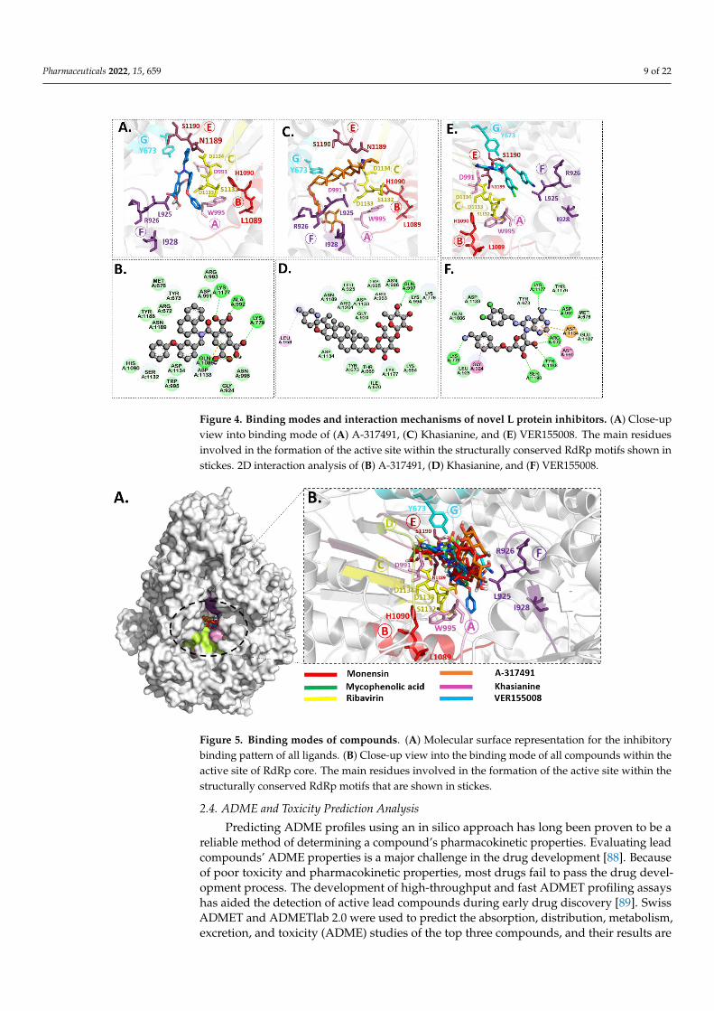
Figure 4. Binding modes and interaction mechanisms of novel L protein inhibitors. (A) Close-up
view into binding mode of (A) A-317491, (C) Khasianine, and (E) VER155008. The main residues
involved in the formation of the active site within the structurally conserved RdRp motifs shown in
stickes. 2D interaction analysis of (B) A-317491, (D) Khasianine, and (F) VER155008.
Notably, both known inhibitors and the identified compounds occupied the binding
site pockets within the RdRp core with a similar interaction pattern (Figure 5). Both con-
trol drugs and selected compounds formed hydrogen bonds with functionally conserved
aspartates of motif C, namely, Asp 1133 and the conserved divalent cation binding resi-
due, Asp991 [24]. Our screened nucleoside compounds displayed higher binding energies
and interacting profiles than Monensin, mycophenolic acid, and Ribavirin. Most ligands
tended to bind the residues belonging to the PA-C-Like domain and the RdRp core do-
main. Since both these domains are actively involved in forming active site chamber and
vRNA binding. Previously published data have proposed that RdRp and PA-C-Like do-
mains facilitate the template-directed RNA synthesis in SFTSV-L protein by providing the
NTP entry into the catalytic chamber [62]. Overall, both these domains and endo N do-
main are critical for product synthesis and the safest release in the cell [53]. These findings
suggested that the compounds identified in our study could be more useful candidates
for RVFV drug therapy. Previously, a number of studies on RdRp have identified several
leads using various computational and experimental techniques. The compounds re-
vealed to interact with RdRp binding pocket residues reported herein [86,87]. However,
RVFV RdRp has not been explored much, and the current study is a pioneer work in this
regard.
Figure 4. Binding modes and interaction mechanisms of novel L protein inhibitors. (A) Close-upview into binding mode of (A) A-317491, (C) Khasianine, and (E) VER155008. The main residuesinvolved in the formation of the active site within the structurally conserved RdRp motifs shown instickes. 2D interaction analysis of (B) A-317491, (D) Khasianine, and (F) VER155008.
Pharmaceuticals 2022, 15, x FOR PEER REVIEW 10 of 22
Figure 5. Binding modes of compounds. (A) Molecular surface representation for the inhibitory
binding pattern of all ligands. (B) Close-up view into the binding mode of all compounds within
the active site of RdRp core. The main residues involved in the formation of the active site within
the structurally conserved RdRp motifs that are shown in stickes.
2.4. ADME and Toxicity Prediction Analysis
Predicting ADME profiles using an in silico approach has long been proven to be a
reliable method of determining a compound’s pharmacokinetic properties. Evaluating
lead compounds’ ADME properties is a major challenge in the drug development [88].
Because of poor toxicity and pharmacokinetic properties, most drugs fail to pass the drug
development process. The development of high-throughput and fast ADMET profiling
assays has aided the detection of active lead compounds during early drug discovery [89].
Swiss ADMET and ADMETlab 2.0 were used to predict the absorption, distribution, me-
tabolism, excretion, and toxicity (ADME) studies of the top three compounds, and their
results are presented in Table 2. Gastro-intestinal absorption (GI) and blood-brain barrier
(BBB) permeation indicate drug absorption and distribution of drug molecules [71]. One
of the primary factors optimising drug discovery is information about drug distribution
via BBB [90]. According to Table 2 results, all compounds show low gastro-intestinal ab-
sorption and no BBB permeation. The compounds cannot cross the blood-brain barrier
(blood-brain barrier negative), so their consumption is not linked to the onset of neuro-
logical disorders. The absorption of the compounds was further revealed by caco-2 per-
meability values ranging from –6.019 to –5.727 log unit. A permeability of > −5.15 log unit
in the ADMETlab 2.0 server indicates optimal caco-2 absorption. Oral bioavailability is
frequently viewed as crucial in determining the drug-likeness of active compounds as
therapeutic agents [91]. Furthermore, a variety of cytochromes (CYPs) regulate drug me-
tabolism, with CYP2C19, CYP1A2, CYP2C9, CYP3A4, and CYP2D6 being critical for the
biotransformation of drug molecules. The ability of a drug to inhibit or act as a substrate
of the cytochrome P450 (CYP450) subfamily determines its therapeutic action [92]. A-
317491 is an inhibitor of CYP2C9 while being a non-inhibitor and a non-substrate of other
isoforms, while Khasianine is a non-inhibitor and a non-substrate of all isoforms, and
VER155008 is an inhibitor of CYP3A4 and a non-inhibitor and non-substrates of other
isoforms. Besides, p-glycoprotein inhibitors reduce the bioavailability of drugs that are
known to be transported by it [93]. Except for Khasianine, all of the compounds in our
analysis are inhibitors and negative substrates of p-glycoprotein, which explains the good
absorption profile of the compounds. All of the compounds studied were nontoxic in
terms of AMES toxicity. Following that, the safety profile of the three compounds was
assessed by conducting toxicity prediction studies with an online tool: ProTox-II. This
server classified substances into six toxicity classes (1–6), with class one being the most
Figure 5. Binding modes of compounds. (A) Molecular surface representation for the inhibitorybinding pattern of all ligands. (B) Close-up view into the binding mode of all compounds within theactive site of RdRp core. The main residues involved in the formation of the active site within thestructurally conserved RdRp motifs that are shown in stickes.
2.4. ADME and Toxicity Prediction Analysis
Predicting ADME profiles using an in silico approach has long been proven to be areliable method of determining a compound’s pharmacokinetic properties. Evaluating leadcompounds’ ADME properties is a major challenge in the drug development [88]. Becauseof poor toxicity and pharmacokinetic properties, most drugs fail to pass the drug devel-opment process. The development of high-throughput and fast ADMET profiling assayshas aided the detection of active lead compounds during early drug discovery [89]. SwissADMET and ADMETlab 2.0 were used to predict the absorption, distribution, metabolism,excretion, and toxicity (ADME) studies of the top three compounds, and their results are
Pharmaceuticals 2022, 15, 659 10 of 22
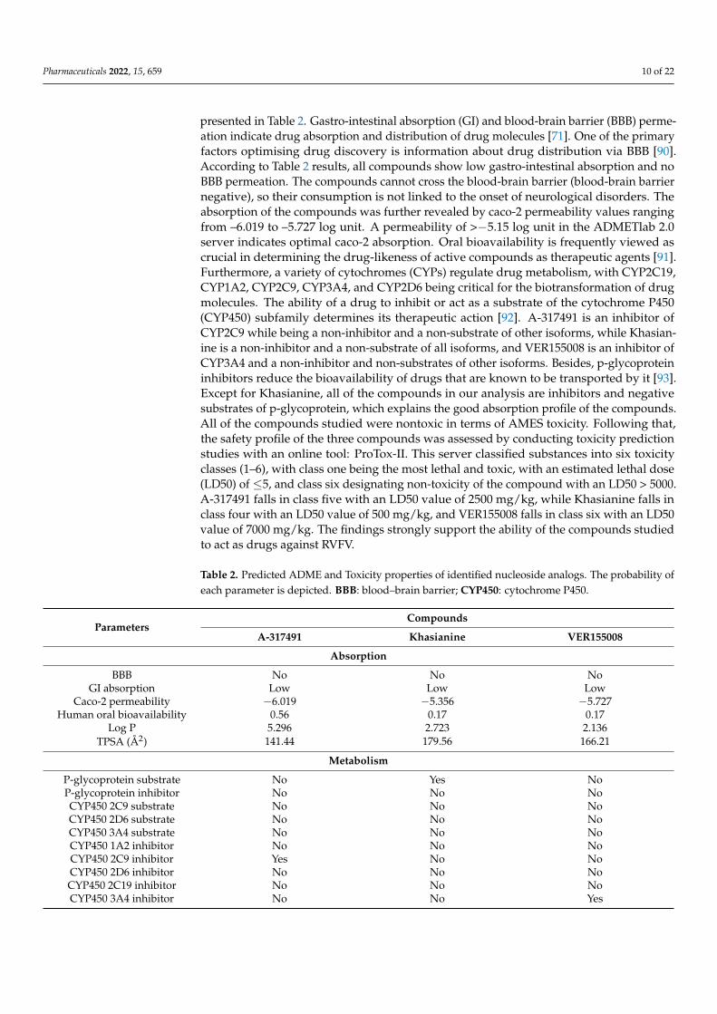
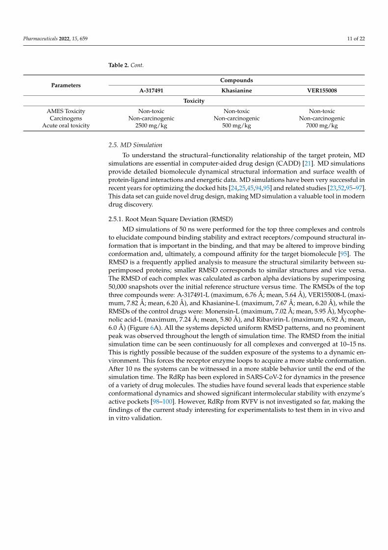
presented in Table 2. Gastro-intestinal absorption (GI) and blood-brain barrier (BBB) perme-ation indicate drug absorption and distribution of drug molecules [71]. One of the primaryfactors optimising drug discovery is information about drug distribution via BBB [90].According to Table 2 results, all compounds show low gastro-intestinal absorption and noBBB permeation. The compounds cannot cross the blood-brain barrier (blood-brain barriernegative), so their consumption is not linked to the onset of neurological disorders. Theabsorption of the compounds was further revealed by caco-2 permeability values rangingfrom –6.019 to –5.727 log unit. A permeability of >−5.15 log unit in the ADMETlab 2.0server indicates optimal caco-2 absorption. Oral bioavailability is frequently viewed ascrucial in determining the drug-likeness of active compounds as therapeutic agents [91].Furthermore, a variety of cytochromes (CYPs) regulate drug metabolism, with CYP2C19,CYP1A2, CYP2C9, CYP3A4, and CYP2D6 being critical for the biotransformation of drugmolecules. The ability of a drug to inhibit or act as a substrate of the cytochrome P450(CYP450) subfamily determines its therapeutic action [92]. A-317491 is an inhibitor ofCYP2C9 while being a non-inhibitor and a non-substrate of other isoforms, while Khasian-ine is a non-inhibitor and a non-substrate of all isoforms, and VER155008 is an inhibitor ofCYP3A4 and a non-inhibitor and non-substrates of other isoforms. Besides, p-glycoproteininhibitors reduce the bioavailability of drugs that are known to be transported by it [93].Except for Khasianine, all of the compounds in our analysis are inhibitors and negativesubstrates of p-glycoprotein, which explains the good absorption profile of the compounds.All of the compounds studied were nontoxic in terms of AMES toxicity. Following that,the safety profile of the three compounds was assessed by conducting toxicity predictionstudies with an online tool: ProTox-II. This server classified substances into six toxicityclasses (1–6), with class one being the most lethal and toxic, with an estimated lethal dose(LD50) of ≤5, and class six designating non-toxicity of the compound with an LD50 > 5000.A-317491 falls in class five with an LD50 value of 2500 mg/kg, while Khasianine falls inclass four with an LD50 value of 500 mg/kg, and VER155008 falls in class six with an LD50value of 7000 mg/kg. The findings strongly support the ability of the compounds studiedto act as drugs against RVFV.
Table 2. Predicted ADME and Toxicity properties of identified nucleoside analogs. The probability ofeach parameter is depicted. BBB: blood–brain barrier; CYP450: cytochrome P450.
ParametersCompounds
A-317491 Khasianine VER155008
Absorption
BBB No No NoGI absorption Low Low Low
Caco-2 permeability −6.019 −5.356 −5.727Human oral bioavailability 0.56 0.17 0.17
Log P 5.296 2.723 2.136TPSA (Å2) 141.44 179.56 166.21
Metabolism
P-glycoprotein substrate No Yes NoP-glycoprotein inhibitor No No NoCYP450 2C9 substrate No No NoCYP450 2D6 substrate No No NoCYP450 3A4 substrate No No NoCYP450 1A2 inhibitor No No NoCYP450 2C9 inhibitor Yes No NoCYP450 2D6 inhibitor No No NoCYP450 2C19 inhibitor No No NoCYP450 3A4 inhibitor No No Yes
Pharmaceuticals 2022, 15, 659 11 of 22
Table 2. Cont.
ParametersCompounds
A-317491 Khasianine VER155008
Toxicity
AMES Toxicity Non-toxic Non-toxic Non-toxicCarcinogens Non-carcinogenic Non-carcinogenic Non-carcinogenic
Acute oral toxicity 2500 mg/kg 500 mg/kg 7000 mg/kg
2.5. MD Simulation
To understand the structural–functionality relationship of the target protein, MDsimulations are essential in computer-aided drug design (CADD) [21]. MD simulationsprovide detailed biomolecule dynamical structural information and surface wealth ofprotein-ligand interactions and energetic data. MD simulations have been very successful inrecent years for optimizing the docked hits [24,25,45,94,95] and related studies [23,52,95–97].This data set can guide novel drug design, making MD simulation a valuable tool in moderndrug discovery.
2.5.1. Root Mean Square Deviation (RMSD)
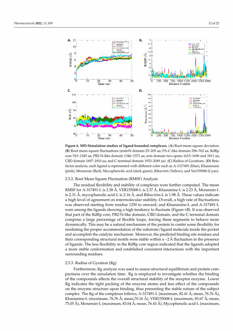
MD simulations of 50 ns were performed for the top three complexes and controlsto elucidate compound binding stability and extract receptors/compound structural in-formation that is important in the binding, and that may be altered to improve bindingconformation and, ultimately, a compound affinity for the target biomolecule [95]. TheRMSD is a frequently applied analysis to measure the structural similarity between su-perimposed proteins; smaller RMSD corresponds to similar structures and vice versa.The RMSD of each complex was calculated as carbon alpha deviations by superimposing50,000 snapshots over the initial reference structure versus time. The RMSDs of the topthree compounds were: A-317491-L (maximum, 6.76 Å; mean, 5.64 Å), VER155008-L (maxi-mum, 7.82 Å; mean, 6.20 Å), and Khasianine-L (maximum, 7.67 Å; mean, 6.20 Å), while theRMSDs of the control drugs were: Monensin-L (maximum, 7.02 Å; mean, 5.95 Å), Mycophe-nolic acid-L (maximum, 7.24 Å; mean, 5.80 Å), and Ribavirin-L (maximum, 6.92 Å; mean,6.0 Å) (Figure 6A). All the systems depicted uniform RMSD patterns, and no prominentpeak was observed throughout the length of simulation time. The RMSD from the initialsimulation time can be seen continuously for all complexes and converged at 10–15 ns.This is rightly possible because of the sudden exposure of the systems to a dynamic en-vironment. This forces the receptor enzyme loops to acquire a more stable conformation.After 10 ns the systems can be witnessed in a more stable behavior until the end of thesimulation time. The RdRp has been explored in SARS-CoV-2 for dynamics in the presenceof a variety of drug molecules. The studies have found several leads that experience stableconformational dynamics and showed significant intermolecular stability with enzyme’sactive pockets [98–100]. However, RdRp from RVFV is not investigated so far, making thefindings of the current study interesting for experimentalists to test them in in vivo andin vitro validation.
Pharmaceuticals 2022, 15, 659 12 of 22
Pharmaceuticals 2022, 15, x FOR PEER REVIEW 12 of 22
(maximum, 7.82 Å; mean, 6.20 Å), and Khasianine-L (maximum, 7.67 Å; mean, 6.20 Å),
while the RMSDs of the control drugs were: Monensin-L (maximum, 7.02 Å; mean, 5.95
Å), Mycophenolic acid-L (maximum, 7.24 Å; mean, 5.80 Å), and Ribavirin-L (maximum,
6.92 Å; mean, 6.0 Å) (Figure 6A). All the systems depicted uniform RMSD patterns, and
no prominent peak was observed throughout the length of simulation time. The RMSD
from the initial simulation time can be seen continuously for all complexes and converged
at 10–15 ns. This is rightly possible because of the sudden exposure of the systems to a
dynamic environment. This forces the receptor enzyme loops to acquire a more stable
conformation. After 10 ns the systems can be witnessed in a more stable behavior until the
end of the simulation time. The RdRp has been explored in SARS-CoV-2 for dynamics in
the presence of a variety of drug molecules. The studies have found several leads that
experience stable conformational dynamics and showed significant intermolecular stabil-
ity with enzyme’s active pockets [98–100]. However, RdRp from RVFV is not investigated
so far, making the findings of the current study interesting for experimentalists to test
them in in vivo and in vitro validation.
Figure 6. MD-Simulation studies of ligand bounded complexes. (A) Root mean square deviation.
(B) Root mean square fluctuations (endoN domain 25–205 aa; PA-C-like domain 296–762 aa; RdRp
core 763–1345 aa; PB2-N-like domain 1346–1571 aa; arm domain two spans 1615–1696 and 1811 aa;
CBD domain 1697–1810 aa; and C-terminal domain 1933–2049 aa). (C) Radius of Gyrations. (D) Beta-
factor analysis, each ligand is represented with different color such as A-1317491 (blue), Khasianane
(pink), Monensin (Red), Mycophenolic acid (dark green), Ribavirin (Yellow), and Ver155008 (Cyan).
2.5.2. Root Mean Square Fluctuation (RMSF) Analysis
The residual flexibility and stability of complexes were further computed. The mean
RMSF for A-317491-L is 2.38 Å, VER155008-L is 2.37 Å, Khasianine-L is 2.23 Å, Monensin-
L is 2.31 Å, mycophenolic acid-L is 2.16 Å, and Ribavirin-L is 1.98 Å. These values indicate
a high level of agreement on intermolecular stability. Overall, a high rate of fluctuations
was observed starting from residue 1250 to onward, and Khasianine-L and A-317491-L
were among the ligands showing a high tendency to fluctuate (Figure 6B). It was observed
that part of the RdRp core, PB2-N-like domain, CBD domain, and the C-terminal domain
comprise a large percentage of flexible loops, forcing these segments to behave more
Figure 6. MD-Simulation studies of ligand bounded complexes. (A) Root mean square deviation.(B) Root mean square fluctuations (endoN domain 25–205 aa; PA-C-like domain 296–762 aa; RdRpcore 763–1345 aa; PB2-N-like domain 1346–1571 aa; arm domain two spans 1615–1696 and 1811 aa;CBD domain 1697–1810 aa; and C-terminal domain 1933–2049 aa). (C) Radius of Gyrations. (D) Beta-factor analysis, each ligand is represented with different color such as A-1317491 (blue), Khasianane(pink), Monensin (Red), Mycophenolic acid (dark green), Ribavirin (Yellow), and Ver155008 (Cyan).
2.5.2. Root Mean Square Fluctuation (RMSF) Analysis
The residual flexibility and stability of complexes were further computed. The meanRMSF for A-317491-L is 2.38 Å, VER155008-L is 2.37 Å, Khasianine-L is 2.23 Å, Monensin-Lis 2.31 Å, mycophenolic acid-L is 2.16 Å, and Ribavirin-L is 1.98 Å. These values indicatea high level of agreement on intermolecular stability. Overall, a high rate of fluctuationswas observed starting from residue 1250 to onward, and Khasianine-L and A-317491-Lwere among the ligands showing a high tendency to fluctuate (Figure 6B). It was observedthat part of the RdRp core, PB2-N-like domain, CBD domain, and the C-terminal domaincomprise a large percentage of flexible loops, forcing these segments to behave moredynamically. This may be a natural mechanism of the protein to confer some flexibility formediating the proper accommodation of the substrate/ligand molecule inside the pocketand accomplish the catalytic mechanism. Moreover, the predicted binding site residues andtheir corresponding structural motifs were stable within a ~2 Å fluctuation in the presenceof ligands. The less flexibility in the RdRp core region indicated that the ligands adopteda more stable conformation and established consistent interactions with the importantsurrounding residues.
2.5.3. Radius of Gyration (Rg)
Furthermore, Rg analysis was used to assess structural equilibrium and protein com-pactness over the simulation time. Rg is employed to investigate whether the bindingof the compounds affects the overall structural stability of the receptor enzyme. LowerRg indicates the tight packing of the enzyme atoms and less effect of the compoundson the enzyme structure upon binding, thus presenting the stable nature of the subjectcomplex. The Rg of the complexes follows; A-317491-L (maximum, 82.41 Å; mean, 76.76 Å),Khasianine-L (maximum, 76.76 Å; mean,70.16 Å), VER155008-L (maximum, 83.67 Å; mean,75.05 Å), Monensin-L (maximum, 83.64 Å; mean, 76.43 Å), Mycophenolic acid-L (maximum,
Pharmaceuticals 2022, 15, 659 13 of 22
85.40 Å; mean, 77.29 Å), and Ribavirin-L (maximum, 83.76 Å; mean, 74.09 Å) (Figure 6C).All six complexes are quite stable and remain compact. These Rg results complement theRMSD result in interpreting the docked complex stability.
2.5.4. B-Factor Analysis
B-factors were also derived from simulation trajectories to probe highly mobile regionsof the complexes. The B-factor monitors the thermal motion of protein atoms, side chains,and whole regions. The B-factor is commonly used to identify internal protein motions,probing rigid and flexible regions important in proteins/enzyme functionality. The averagevalues of the B-factor for the top three complexes A-317491-L, VER155008-L Khasianine-L,and control drugs Monensin-L, Mycophenolic acid-L, and Ribavirin-L were: 201.68 Å2,210.62 Å2,187.20 Å2, 183.67 Å2, 155.86 Å2, and 139.35 Å2, respectively (Figure 6D). Itdemonstrates that these complexes have good stability throughout the 50 ns simulationperiod. From the simulation results, it can be concluded that all the studied systems arestructurally stable, and the intermolecular interactions remained strong throughout thesimulation time.
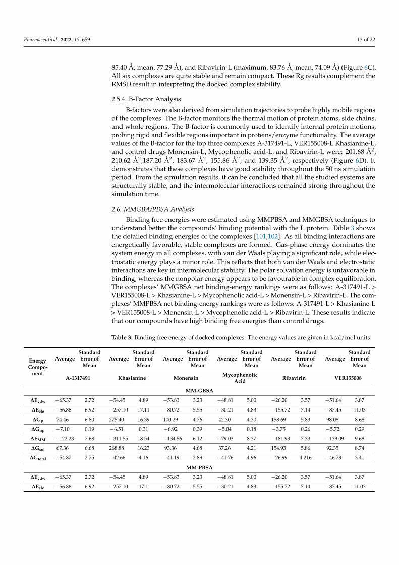
2.6. MMGBA/PBSA Analysis
Binding free energies were estimated using MMPBSA and MMGBSA techniques tounderstand better the compounds’ binding potential with the L protein. Table 3 showsthe detailed binding energies of the complexes [101,102]. As all binding interactions areenergetically favorable, stable complexes are formed. Gas-phase energy dominates thesystem energy in all complexes, with van der Waals playing a significant role, while elec-trostatic energy plays a minor role. This reflects that both van der Waals and electrostaticinteractions are key in intermolecular stability. The polar solvation energy is unfavorable inbinding, whereas the nonpolar energy appears to be favourable in complex equilibration.The complexes’ MMGBSA net binding-energy rankings were as follows: A-317491-L >VER155008-L > Khasianine-L > Mycophenolic acid-L > Monensin-L > Ribavirin-L. The com-plexes’ MMPBSA net binding-energy rankings were as follows: A-317491-L > Khasianine-L> VER155008-L > Monensin-L > Mycophenolic acid-L > Ribavirin-L. These results indicatethat our compounds have high binding free energies than control drugs.
Table 3. Binding free energy of docked complexes. The energy values are given in kcal/mol units.
EnergyCompo-
nent
AverageStandardError ofMean
AverageStandardError ofMean
AverageStandardError ofMean
AverageStandardError ofMean
AverageStandardError ofMean
AverageStandardError ofMean
A-1317491 Khasianine Monensin MycophenolicAcid Ribavirin VER155008
MM-GBSA
∆Evdw −65.37 2.72 −54.45 4.89 −53.83 3.23 −48.81 5.00 −26.20 3.57 −51.64 3.87
∆Eele −56.86 6.92 −257.10 17.11 −80.72 5.55 −30.21 4.83 −155.72 7.14 −87.45 11.03
∆Gp 74.46 6.80 275.40 16.39 100.29 4.76 42.30 4.30 158.69 5.83 98.08 8.68
∆Gnp −7.10 0.19 −6.51 0.31 −6.92 0.39 −5.04 0.18 −3.75 0.26 −5.72 0.29
∆EMM −122.23 7.68 −311.55 18.54 −134.56 6.12 −79.03 8.37 −181.93 7.33 −139.09 9.68
∆Gsol 67.36 6.68 268.88 16.23 93.36 4.68 37.26 4.21 154.93 5.86 92.35 8.74
∆Gtotal −54.87 2.75 −42.66 4.16 −41.19 2.89 −41.76 4.96 −26.99 4.216 −46.73 3.41
MM-PBSA
∆Evdw −65.37 2.72 −54.45 4.89 −53.83 3.23 −48.81 5.00 −26.20 3.57 −51.64 3.87
∆Eele −56.86 6.92 −257.10 17.1 −80.72 5.55 −30.21 4.83 −155.72 7.14 −87.45 11.03
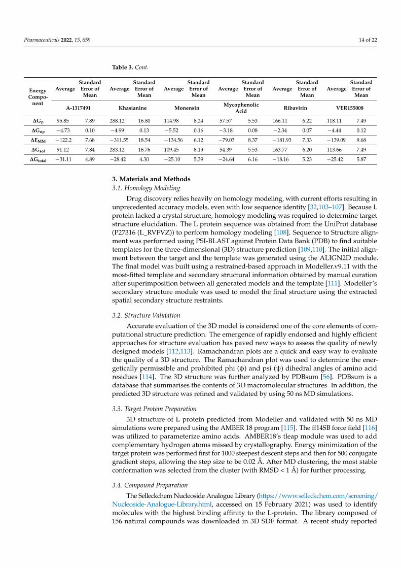
Pharmaceuticals 2022, 15, 659 14 of 22
Table 3. Cont.
EnergyCompo-
nent
AverageStandardError ofMean
AverageStandardError ofMean
AverageStandardError ofMean
AverageStandardError ofMean
AverageStandardError ofMean
AverageStandardError ofMean
A-1317491 Khasianine Monensin MycophenolicAcid Ribavirin VER155008
∆Gp 95.85 7.89 288.12 16.80 114.98 8.24 57.57 5.53 166.11 6.22 118.11 7.49
∆Gnp −4.73 0.10 −4.99 0.13 −5.52 0.16 −3.18 0.08 −2.34 0.07 −4.44 0.12
∆EMM −122.2 7.68 −311.55 18.54 −134.56 6.12 −79.03 8.37 −181.93 7.33 −139.09 9.68
∆Gsol 91.12 7.84 283.12 16.76 109.45 8.19 54.39 5.53 163.77 6.20 113.66 7.49
∆Gtotal −31.11 4.89 −28.42 4.30 −25.10 5.39 −24.64 6.16 −18.16 5.23 −25.42 5.87
3. Materials and Methods3.1. Homology Modeling
Drug discovery relies heavily on homology modeling, with current efforts resulting inunprecedented accuracy models, even with low sequence identity [32,103–107]. Because Lprotein lacked a crystal structure, homology modeling was required to determine targetstructure elucidation. The L protein sequence was obtained from the UniProt database(P27316 (L_RVFVZ)) to perform homology modeling [108]. Sequence to Structure align-ment was performed using PSI-BLAST against Protein Data Bank (PDB) to find suitabletemplates for the three-dimensional (3D) structure prediction [109,110]. The initial align-ment between the target and the template was generated using the ALIGN2D module.The final model was built using a restrained-based approach in Modeller.v9.11 with themost-fitted template and secondary structural information obtained by manual curationafter superimposition between all generated models and the template [111]. Modeller’ssecondary structure module was used to model the final structure using the extractedspatial secondary structure restraints.
3.2. Structure Validation
Accurate evaluation of the 3D model is considered one of the core elements of com-putational structure prediction. The emergence of rapidly endorsed and highly efficientapproaches for structure evaluation has paved new ways to assess the quality of newlydesigned models [112,113]. Ramachandran plots are a quick and easy way to evaluatethe quality of a 3D structure. The Ramachandran plot was used to determine the ener-getically permissible and prohibited phi (φ) and psi (ψ) dihedral angles of amino acidresidues [114]. The 3D structure was further analyzed by PDBsum [56]. PDBsum is adatabase that summarises the contents of 3D macromolecular structures. In addition, thepredicted 3D structure was refined and validated by using 50 ns MD simulations.
3.3. Target Protein Preparation
3D structure of L protein predicted from Modeller and validated with 50 ns MDsimulations were prepared using the AMBER 18 program [115]. The ff14SB force field [116]was utilized to parameterize amino acids. AMBER18’s tleap module was used to addcomplementary hydrogen atoms missed by crystallography. Energy minimization of thetarget protein was performed first for 1000 steepest descent steps and then for 500 conjugategradient steps, allowing the step size to be 0.02 Å. After MD clustering, the most stableconformation was selected from the cluster (with RMSD < 1 Å) for further processing.
3.4. Compound Preparation
The Selleckchem Nucleoside Analogue Library (https://www.selleckchem.com/screening/Nucleoside-Analogue-Library.html, accessed on 15 February 2021) was used to identifymolecules with the highest binding affinity to the L-protein. The library composed of156 natural compounds was downloaded in 3D SDF format. A recent study reported
Pharmaceuticals 2022, 15, 659 15 of 22
Monensin, Mycophenolic acid, and Ribavirin as potent inhibitors of RVFV-L protein [79].To get an insight into the mechanism of the binding of known inhibitors, these three potentinhibitors were docked as controls in the present study. These drugs were retrieved fromthe PubChem database (https://pubchem.ncbi.nlm.nih.gov, accessed on 15 February 2021)in 3D SDF format [117].
3.5. Structure-Based Virtual Screening
The AutoDock Vina implicated in the PyRx (version 0.8) was used to perform thevirtual screening of the compounds against the target protein [118,119]. The control drugsand chemical library of 156 compounds in SDF format were imported in the Open-babelprogram of PyRx for further energy minimization, followed by conversion of all the ligandsinto AutoDock PDBQT format. Further, the top three ligands were subjected to dockingagainst the L protein of RVFV using AutoDock Vina. The grid box was set to cover the entirebinding site within the core chamber of RdRp with box_size of (x = 20, y = 20, z = 20 Å) anddimensions of (x = 122.005, y = 105.326, and z = 137.824). Dockings were run in triplicatesto ensure absolute consistency of the results. The docked solutions were clustered using anRMSD of 1 Å. The 3D structural alignment, visualization and analysis, and docking figuresproduction were carried out using Discovery Studio 2019 software [120], PyMOL [121], andUCSF Chimera 1.14 [122].
3.6. ADME and Toxicity Prediction Analysis
The analysis of the pharmacological activity of drug candidates is a critical stepin drug discovery [93]. Therefore, in silico prediction of pharmacophore properties ofactive compounds is critical for accelerating the drug development process. Absorption,distribution, metabolism, excretion, and toxicity (ADMET) are the most plausible drug-likeproperties to evaluate virtual hits. ADME analysis was carried out by submitting thecompounds’ canonical simplified molecular-input line-entry system (SMILES) to onlineservers; ADMETlab 2.0 [123] and Swiss ADME [124]. Acute oral toxicity was predicted bythe Protox II web server [125].
3.7. MD Simulations
AMBER18 was used to perform MD simulations of the docked solutions [115]. Thesame MD simulation protocol was adopted in the current study as described in previ-ous studies [22,94,126–128]. The Antechamber package of AmberTools was employedto generate the general AMBER force field (GAFF) parameters for the studied ligandsusing AM1–BCC charge definitions. Each top complex was explicitly solvated with wa-ter molecules, and then counter ions were added to create a neutral system. A waterbox with a thickness of 12 Å was created using the TIP3P solvent model to encircle thecomplex [129,130]. The complex was simulated using periodic boundary conditions, withelectrostatic interactions modeled using the particle mesh Ewald procedure [131]. Fornonbounded interactions, a threshold value of 8 Å was defined during the procedure.Water molecules were minimized for 500 cycles, and then the entire system was minimizedfor 1000 rounds. The temperature of each system was then steadily increased to 300 K.Solutes in the first phase were restrained for 50 ps during equilibration of counterions andwater molecules, while protein side chains were relaxed afterwards. At 300 K and 1 atm,a 50 ns MD simulation was performed, and coordinate trajectories were collected every2 ps during the simulation under the NPT ensemble. While the SHAKE algorithm [132]constrained covalent and hydrogen bonds, Langevin dynamics [133] was used to controlthe system temperature. The original structure was used as a reference, and AMBER’s CPP-TRAJ [134,135] was used to generate an RMSD (root mean square deviation) plot to assessthe system’s MD simulation convergence [136]. The structural flexibility of ligands wascalculated using ligand RMSD. The radius of gyration (RoG) was analyzed to determine thecomplex’s compactness and three-dimensional packing. RMSF reflects the root mean squareaveraged distance between an atom and its average geometric position [137]. The Term
Pharmaceuticals 2022, 15, 659 16 of 22
β–factor, which is closely related to the RMSF, measures the spatial displacement of atomsaround their mean positions, as well as a thermal and local vibrational movement [138].
3.8. MMGBA/PBSA Analysis
AMBER18 MM/PBSA and MM/PBSA methods were used to estimate the binding freeenergy (G binding) of the complexes [101,139]. The method has been well-documented inour previous studies, implemented in AMBER 18. The free energy difference was calculatedusing 100 snapshots from simulated trajectories at regular intervals. The total bindingfree energy is calculated as a sum of the molecular mechanics binding energy (∆EMM) andsolvation free energy (∆Gsol) as follows:
∆EMM = ∆Eint + ∆Eele + ∆Evdw
∆Gsol = ∆Gp + ∆Gnp
∆Gtotal = ∆EMM + ∆Gsol
∆Gbind = ∆EMM + ∆Gsol − T∆S
In these equations, ∆EMM is further divided into internal energy (∆Eint), electro-static energy (∆Eele), and van der Waals energy (∆Evdw), and the total solvation freeenergy (∆Gsol) is contributed by the sum of polar (∆Gp) and non-polar (∆Gnp) components.∆Gbind is the free energy of binding of the ligand evaluated after entropic calculations(T∆S). We excluded entropic term, T∆S, from the calculation due to its intensive computa-tional cost [101,102,140,141]. These methods have been well documented [101,128,140] andconsidered reliable end-point binding free energy estimations [102,141].
4. Conclusions
A combination of virtual screening and molecular docking analysis was used to iden-tify inhibitors of RVFV L protein in the present study. The 3D structure of the RVFV-Lenzyme was predicted, and the findings could aid researchers working on RVFV drug de-velopment. The Selleckchem Nucleoside Analogue Library (156 compounds) was screened,and the top three compounds, A-317491, VER155008, and Khasianine exhibit a high bindingaffinity compared to the control drugs chosen that may inhibit RVFV-L enzyme activity andhence virus replication. Compounds also show good pharmacodynamic and pharmacoki-netic profiles based on toxicity and ADME studies. Overall, the results reveal that all of thecompounds fit into the same binding pocket of the protein, suggesting that they could begood therapeutic candidates for RVFV. However, further in-vivo and in-vitro experimentsare required to convert these potential inhibitors into clinical drugs. We anticipate thatthe findings of this study will be useful in the future for developing and exploring novelnatural anti-RVFV therapeutic agents.
Supplementary Materials: The following supporting information can be downloaded at: https://www.mdpi.com/article/10.3390/ph15060659/s1, Figure S1. Sequence alignment of RVFV Lprotein with the templates (Structure of the alpha-Synuclein (PDB ID: 6I42 and severe fever withthrombocytopenia syndrome virus L protein (PDB ID: 6Y6K). Figure S2. Structural superposition ofthe RVFV L protein model with the templates. The RMSD values between the model and 6I42 and6Y6K are 0.179 and 1.165 Å, respectively. Table S1. Reverse template comparison of RVFV L proteinmodel with the representative structures in PDB using ProFunc reverse template search program.Table S2. Virtual Screening result of Selleckchem Nucleoside Analogue Library (compounds withbinding affinity > −1.5 kcal/mol) against model of RVFV-L RdRp core.
Author Contributions: Conceptualization, U.A.A., M.T.u.Q., S.A. and K.S.A.; data curation, M.T.u.Q.,F.S., S.A., E.A.A., G.M.A. and A.A.; formal analysis, M.A.A., M.U.M., M.M.A., U.A.A. and S.A.;funding acquisition, K.S.A.; investigation, M.A.A., M.U.M., M.M.A., F.S., E.A.A. and G.M.A.; method-ology, M.A.A., M.U.M., M.M.A., U.A.A., M.T.u.Q., S.A., E.A.A., G.M.A., K.S.A. and A.A.; projectadministration, M.T.u.Q. and K.S.A.; resources, K.S.A.; software, M.A.A., M.U.M., M.M.A. and S.A.;
Pharmaceuticals 2022, 15, 659 17 of 22
supervision, U.A.A. and K.S.A.; validation, U.A.A., M.T.u.Q., F.S., S.A., E.A.A., G.M.A., K.S.A. andA.A.; visualization, M.A.A., M.U.M. and M.M.A.; writing—original draft, M.A.A., M.U.M., M.M.A.and F.S.; writing—review and editing, U.A.A., M.T.u.Q., S.A., E.A.A., G.M.A., K.S.A. and A.A. Allauthors have read and agreed to the published version of the manuscript.
Funding: This research received no external funding.
Institutional Review Board Statement: Not applicable.
Informed Consent Statement: Not applicable.
Data Availability Statement: The data presented in this study are available within the article andSupplementary Material.
Acknowledgments: The researchers would like to thank the Deanship of Scientific Research, QassimUniversity for funding the publication of this project.
Conflicts of Interest: The author declares no conflict of interest.
References1. Pepin, M.; Bouloy, M.; Bird, B.H.; Kemp, A.; Paweska, J. Rift Valley fever virus (Bunyaviridae: Phlebovirus): An update on
pathogenesis, molecular epidemiology, vectors, diagnostics and prevention. Vet. Res. 2010, 41, 61. [CrossRef] [PubMed]2. Bird, B.H.; Ksiazek, T.G.; Nichol, S.T.; MacLachlan, N.J. Rift Valley fever virus. J. Am. Vet. Med. Assoc. 2009, 234, 883–893.
[CrossRef] [PubMed]3. Hartman, A. Rift valley fever. Clin. Lab. Med. 2017, 37, 285–301. [CrossRef] [PubMed]4. Ikegami, T.; Makino, S. The pathogenesis of Rift Valley fever. Viruses 2011, 3, 493–519. [CrossRef]5. Drake, J.M.; Hassan, A.N.; Beier, J.C. A statistical model of Rift Valley fever activity in Egypt. J. Vector Ecol. 2013, 38, 251–259.
[CrossRef]6. Helmy, Y.A.; El-Adawy, H.; Abdelwhab, E.M. A comprehensive review of common bacterial, parasitic and viral zoonoses at the
human-animal interface in Egypt. Pathogens 2017, 6, 33. [CrossRef]7. Gaudreault, N.N.; Indran, S.V.; Bryant, P.; Richt, J.A.; Wilson, W.C. Comparison of Rift Valley fever virus replication in North
American livestock and wildlife cell lines. Front. Microbiol. 2015, 6, 664. [CrossRef]8. McElroy, A.K.; Albariño, C.G.; Nichol, S.T. Development of a RVFV ELISA that can distinguish infected from vaccinated animals.
Virol. J. 2009, 6, 1–11. [CrossRef]9. Lumley, S.; Horton, D.L.; Hernandez-Triana, L.L.; Johnson, N.; Fooks, A.R.; Hewson, R. Rift Valley fever virus: Strategies for
maintenance, survival and vertical transmission in mosquitoes. J. Gen. Virol. 2017, 98, 875–887. [CrossRef]10. Ikegami, T. Molecular biology and genetic diversity of Rift Valley fever virus. Antiviral Res. 2012, 95, 293–310. [CrossRef]11. Gerrard, S.R.; Nichol, S.T. Synthesis, proteolytic processing and complex formation of N-terminally nested precursor proteins of
the Rift Valley fever virus glycoproteins. Virology 2007, 357, 124–133. [CrossRef] [PubMed]12. Müller, R.; Poch, O.; Delarue, M.; Bishop, D.; Bouloy, M. Rift Valley fever virus L segment: Correction of the sequence and
possible functional role of newly identified regions conserved in RNA-dependent polymerases. J. Gen. Virol. 1994, 75, 1345–1352.[CrossRef] [PubMed]
13. Poch, O.; Sauvaget, I.; Delarue, M.; Tordo, N. Identification of four conserved motifs among the RNA-dependent polymeraseencoding elements. EMBO J. 1989, 8, 3867–3874. [CrossRef] [PubMed]
14. Gerlach, P.; Malet, H.; Cusack, S.; Reguera, J. Structural insights into bunyavirus replication and its regulation by the vRNApromoter. Cell 2015, 161, 1267–1279. [CrossRef] [PubMed]
15. Kinsella, E.; Martin, S.G.; Grolla, A.; Czub, M.; Feldmann, H.; Flick, R. Sequence determination of the Crimean–Congo hemorrhagicfever virus L segment. Virology 2004, 321, 23–28. [CrossRef]
16. Bouloy, M.; Weber, F. Molecular biology of Rift Valley fever virus. Open Virol. J. 2010, 4, 8. [CrossRef]17. Morin, B.; Coutard, B.; Lelke, M.; Ferron, F.; Kerber, R.; Jamal, S.; Frangeul, A.; Baronti, C.; Charrel, R.; De Lamballerie, X. The
N-terminal domain of the arenavirus L protein is an RNA endonuclease essential in mRNA transcription. PLoS Pathog. 2010,6, e1001038. [CrossRef]
18. Patterson, J.; Holloway, B.; Kolakofsky, D. La Crosse virions contain a primer-stimulated RNA polymerase and a methylatedcap-dependent endonuclease. J. Virol. 1984, 52, 215–222. [CrossRef]
19. Gogrefe, N.; Reindl, S.; Günther, S.; Rosenthal, M. Structure of a functional cap-binding domain in Rift Valley fever virus Lprotein. PLoS Pathog. 2019, 15, e1007829. [CrossRef]
20. Sarwar, S.; Abdul Qadir, M.; Alharthy, R.D.; Ahmed, M.; Ahmad, S.; Vanmeert, M.; Mirza, M.U.; Hameed, A. Folate ConjugatedPolyethylene Glycol Probe for Tumor-Targeted Drug Delivery of 5-Fluorouracil. Molecules 2022, 27, 1780. [CrossRef]
21. Salo-Ahen, O.M.H.; Alanko, I.; Bhadane, R.; Bonvin, A.M.J.J.; Honorato, R.V.; Hossain, S.; Juffer, A.H.; Kabedev, A.; Lahtela-Kakkonen, M.; Larsen, A.S. Molecular Dynamics Simulations in Drug Discovery and Pharmaceutical Development. Processes2021, 9, 71. [CrossRef]
Pharmaceuticals 2022, 15, 659 18 of 22
22. Ahmad, S.; Lee, Y.K.; Nazir, M.; Abdul Rahman, N.; Trant, J.F.; Abdullah, I. Fragment-based in silico design of SARS CoV-2 mainprotease inhibitors. Chem. Biol. Drug Des. 2021, 98, 604–619. [CrossRef] [PubMed]
23. Durrani, F.G.; Gul, R.; Mirza, M.U.; Kaderbhai, N.N.; Froeyen, M.; Saleem, M. Mutagenesis of DsbAss is Crucial for the SignalRecognition Particle Mechanism in Escherichia coli: Insights from Molecular Dynamics Simulations. Biomolecules 2019, 9, 133.[CrossRef] [PubMed]
24. Khalid, H.; Landry, K.B.; Ijaz, B.; Ashfaq, U.A.; Ahmed, M.; Kanwal, A.; Froeyen, M.; Mirza, M.U. Discovery of novel Hepatitis Cvirus inhibitor targeting multiple allosteric sites of NS5B polymerase. Infect. Genet. Evol. 2020, 84, 104371. [CrossRef] [PubMed]
25. Mirza, M.U.; Ahmad, S.; Abdullah, I.; Froeyen, M. Identification of novel human USP2 inhibitor and its putative role in treatmentof COVID-19 by inhibiting SARS-CoV-2 papain-like (PLpro) protease. Comput. Biol. Chem. 2020, 89, 107376. [CrossRef]
26. Mirza, M.U.; Vanmeert, M.; Ali, A.; Iman, K.; Froeyen, M.; Idrees, M. Perspectives towards antiviral drug discovery against Ebolavirus. J. Med. Virol. 2019, 91, 2029–2048. [CrossRef]
27. ul Qamar, M.T.; Ahmad, S.; Khan, A.; Mirza, M.U.; Ahmad, S.; Abro, A.; Chen, L.-L.; Almatroudi, A.; Wei, D.-Q. Structuralprobing of HapR to identify potent phytochemicals to control Vibrio cholera through integrated computational approaches.Comput. Biol. Med. 2021, 138, 104929. [CrossRef]
28. Sliwoski, G.; Kothiwale, S.; Meiler, J.; Lowe, E.W. Computational methods in drug discovery. Pharmacol. Rev. 2014, 66, 334–395.[CrossRef]
29. Jácome, R.; Becerra, A.; de León, S.P.; Lazcano, A. Structural analysis of monomeric RNA-dependent polymerases: Evolutionaryand therapeutic implications. PLoS ONE 2015, 10, e0139001. [CrossRef]
30. Van Der Linden, L.; Vives-Adrián, L.; Selisko, B.; Ferrer-Orta, C.; Liu, X.; Lanke, K.; Ulferts, R.; De Palma, A.M.; Tanchis, F.;Goris, N. The RNA template channel of the RNA-dependent RNA polymerase as a target for development of antiviral therapy ofmultiple genera within a virus family. PLoS Pathog. 2015, 11, e1004733. [CrossRef]
31. Ferrer-Orta, C.; Arias, A.; Escarmís, C.; Verdaguer, N. A comparison of viral RNA-dependent RNA polymerases. Curr. Opin.Struct. Biol. 2006, 16, 27–34. [CrossRef] [PubMed]
32. Mirza, M.U.; Vanmeert, M.; Froeyen, M.; Ali, A.; Rafique, S.; Idrees, M. In silico structural elucidation of RNA-dependent RNApolymerase towards the identification of potential Crimean-Congo Hemorrhagic Fever Virus inhibitors. Sci. Rep. 2019, 9, 6809.[CrossRef] [PubMed]
33. Godoy, A.S.; Lima, G.M.; Oliveira, K.I.; Torres, N.U.; Maluf, F.V.; Guido, R.V.; Oliva, G. Crystal structure of Zika virus NS5RNA-dependent RNA polymerase. Nat. Commun. 2017, 8, 14764. [CrossRef] [PubMed]
34. Zhang, C.; Feng, T.; Cheng, J.; Li, Y.; Yin, X.; Zeng, W.; Jin, X.; Li, Y.; Guo, F.; Jin, T. Structure of the NS5 methyltransferase fromZika virus and implications in inhibitor design. Biochem. Biophys. Res. Commun. 2017, 492, 624–630. [CrossRef] [PubMed]
35. Hercík, K.; Kozak, J.; Šála, M.; Dejmek, M.; Hrebabecký, H.; Zborníková, E.; Smola, M.; Ruzek, D.; Nencka, R.; Boura, E. Adenosinetriphosphate analogs can efficiently inhibit the Zika virus RNA-dependent RNA polymerase. Antivir. Res. 2017, 137, 131–133.[CrossRef]
36. Pattnaik, A.; Palermo, N.; Sahoo, B.R.; Yuan, Z.; Hu, D.; Annamalai, A.S.; Vu, H.L.; Correas, I.; Prathipati, P.K.; Destache, C.J.Discovery of a non-nucleoside RNA polymerase inhibitor for blocking Zika virus replication through in silico screening. Antivir.Res. 2018, 151, 78–86. [CrossRef]
37. Lu, G.; Gong, P. Crystal structure of the full-length Japanese encephalitis virus NS5 reveals a conserved methyltransferase-polymerase interface. PLoS Pathog. 2013, 9, e1003549. [CrossRef] [PubMed]
38. Malet, H.; Egloff, M.-P.; Selisko, B.; Butcher, R.E.; Wright, P.J.; Roberts, M.; Gruez, A.; Sulzenbacher, G.; Vonrhein, C.; Bricogne, G.Crystal structure of the RNA polymerase domain of the West Nile virus non-structural protein 5. J. Biol. Chem. 2007, 282,10678–10689. [CrossRef]
39. Noble, C.G.; Lim, S.P.; Chen, Y.-L.; Liew, C.W.; Yap, L.; Lescar, J.; Shi, P.-Y. Conformational flexibility of the Dengue virusRNA-dependent RNA polymerase revealed by a complex with an inhibitor. J. Virol. 2013, 87, 5291–5295. [CrossRef]
40. Noble, C.G.; Chen, Y.-L.; Dong, H.; Gu, F.; Lim, S.P.; Schul, W.; Wang, Q.-Y.; Shi, P.-Y. Strategies for development of dengue virusinhibitors. Antivir. Res. 2010, 85, 450–462. [CrossRef] [PubMed]
41. El Sahili, A.; Lescar, J. Dengue virus non-structural protein 5. Viruses 2017, 9, 91. [CrossRef] [PubMed]42. De Francesco, R.; Tomei, L.; Altamura, S.; Summa, V.; Migliaccio, G. Approaching a new era for hepatitis C virus therapy:
Inhibitors of the NS3-4A serine protease and the NS5B RNA-dependent RNA polymerase. Antivir. Res. 2003, 58, 1–16. [CrossRef]43. Dhanak, D.; Duffy, K.J.; Johnston, V.K.; Lin-Goerke, J.; Darcy, M.; Shaw, A.N.; Gu, B.; Silverman, C.; Gates, A.T.; Nonnemacher, M.R.
Identification and biological characterization of heterocyclic inhibitors of the hepatitis C virus RNA-dependent RNA polymerase.J. Biol. Chem. 2002, 277, 38322–38327. [CrossRef]
44. Gemma, S.; Brogi, S.; Novellino, E.; Campiani, G.; Maga, G.; Brindisi, M.; Butini, S. HCV-targeted antivirals: Current status andfuture challenges. Curr. Pharm. Des. 2014, 20, 3445–3464. [CrossRef] [PubMed]
45. Mirza, M.U.; Saadabadi, A.; Vanmeert, M.; Salo-Ahen, O.M.H.; Abdullah, I.; Claes, S.; De Jonghe, S.; Schols, D.; Ahmad, S.;Froeyen, M. Discovery of HIV entry inhibitors via a hybrid CXCR4 and CCR5 receptor pharmacophore-based virtual screeningapproach. Eur. J. Pharm. Sci. 2020, 155, 105537. [CrossRef] [PubMed]
46. Cavasotto, C.N.; Phatak, S.S. Homology modeling in drug discovery: Current trends and applications. Drug Discov. Today 2009,14, 676–683. [CrossRef]
Pharmaceuticals 2022, 15, 659 19 of 22
47. Hillisch, A.; Pineda, L.F.; Hilgenfeld, R. Utility of homology models in the drug discovery process. Drug Discov. Today 2004, 9,659–669. [CrossRef]
48. Oshiro, C.; Bradley, E.K.; Eksterowicz, J.; Evensen, E.; Lamb, M.L.; Lanctot, J.K.; Putta, S.; Stanton, R.; Grootenhuis, P.D.J.Performance of 3D-database molecular docking studies into homology models. J. Med. Chem. 2004, 47, 764–767. [CrossRef]
49. Kairys, V.; Fernandes, M.X.; Gilson, M.K. Screening drug-like compounds by docking to homology models: A systematic study. J.Chem. Inf. Model 2006, 46, 365–379. [CrossRef]
50. Fernandes, M.X.; Kairys, V.; Gilson, M.K. Comparing ligand interactions with multiple receptors via serial docking. J. Chem. Inf.Comput. Sci. 2004, 44, 1961–1970. [CrossRef]
51. McGovern, S.L.; Shoichet, B.K. Information decay in molecular docking screens against holo, apo, and modeled conformations ofenzymes. J. Med. Chem. 2003, 46, 2895–2907. [CrossRef] [PubMed]
52. Mirza, M.U.; Rafique, S.; Ali, A.; Munir, M.; Ikram, N.; Manan, A.; Salo-Ahen, O.M.; Idrees, M. Towards peptide vaccines againstZika virus: Immunoinformatics combined with molecular dynamics simulations to predict antigenic epitopes of Zika viralproteins. Sci. Rep. 2016, 6, 1–17. [CrossRef] [PubMed]
53. Wang, P.; Liu, L.; Liu, A.; Yan, L.; He, Y.; Shen, S.; Hu, M.; Guo, Y.; Liu, H.; Liu, C. Structure of severe fever with thrombocytopeniasyndrome virus L protein elucidates the mechanisms of viral transcription initiation. Nat. Microbiol. 2020, 5, 864–871. [CrossRef][PubMed]
54. Vogel, D.; Thorkelsson, S.R.; Quemin, E.R.; Meier, K.; Kouba, T.; Gogrefe, N.; Busch, C.; Reindl, S.; Günther, S.; Cusack, S.Structural and functional characterization of the severe fever with thrombocytopenia syndrome virus L protein. Nucleic Acids Res.2020, 48, 5749–5765. [CrossRef] [PubMed]
55. Eisenberg, D. The discovery of the α-helix and β-sheet, the principal structural features of proteins. Proc. Natl. Acad. Sci. USA2003, 100, 11207–11210. [CrossRef] [PubMed]
56. Laskowski, R.A.; Jabłonska, J.; Pravda, L.; Vareková, R.S.; Thornton, J.M. PDBsum: Structural summaries of PDB entries. ProteinSci. 2018, 27, 129–134. [CrossRef]
57. Sidorova, A.E.; Malyshko, E.V.; Lutsenko, A.O.; Shpigun, D.K.; Bagrova, O.E. Protein Helical Structures: Defining Handednessand Localization Features. Symmetry 2021, 13, 879. [CrossRef]
58. Wiederstein, M.; Sippl, M.J. ProSA-web: Interactive web service for the recognition of errors in three-dimensional structures ofproteins. Nucleic Acids Res. 2007, 35, W407–W410. [CrossRef]
59. Zhang, Y.; Skolnick, J. TM-align: A protein structure alignment algorithm based on the TM-score. Nucleic Acids Res. 2005, 33,2302–2309. [CrossRef]
60. Laskowski, R.A.; Watson, J.D.; Thornton, J.M. ProFunc: A server for predicting protein function from 3D structure. Nucleic AcidsRes. 2005, 33, W89–W93. [CrossRef]
61. Sun, Y.; Li, J.; Gao, G.F.; Tien, P.; Liu, W. Bunyavirales ribonucleoproteins: The viral replication and transcription machinery. Crit.Rev. Microbiol. 2018, 44, 522–540. [CrossRef] [PubMed]
62. Pflug, A.; Guilligay, D.; Reich, S.; Cusack, S. Structure of influenza A polymerase bound to the viral RNA promoter. Nature 2014,516, 355. [CrossRef] [PubMed]
63. Arnold, J.J.; Ghosh, S.K.B.; Cameron, C.E. Poliovirus RNA-dependent RNA polymerase (3Dpol): Divalent cation modulation ofprimer, template, and nucleotide selection. J. Biol. Chem. 1999, 274, 37060–37069. [CrossRef]
64. Ferrer-Orta, C.; Arias, A.; Pérez-Luque, R.; Escarmís, C.; Domingo, E.; Verdaguer, N. Sequential structures provide insights intothe fidelity of RNA replication. Proc. Natl. Acad. Sci. USA 2007, 104, 9463–9468. [CrossRef] [PubMed]
65. Amroun, A.; Priet, S.; de Lamballerie, X.; Quérat, G. Bunyaviridae RdRps: Structure, motifs, and RNA synthesis machinery. Crit.Rev. Microbiol. 2017, 43, 753–778. [CrossRef]
66. Rothwell, P.J.; Waksman, G. Structure and mechanism of DNA polymerases. Adv. Protein Chem. 2005, 71, 401–440.67. Biswas, S.K.; Nayak, D.P. Mutational analysis of the conserved motifs of influenza A virus polymerase basic protein 1. J. Virol.
1994, 68, 1819–1826. [CrossRef]68. Sánchez, A.B.; de la Torre, J.C. Genetic and biochemical evidence for an oligomeric structure of the functional L polymerase of the
prototypic arenavirus lymphocytic choriomeningitis virus. J. Virol. 2005, 79, 7262–7268. [CrossRef]69. Vázquez, A.L.; Alonso, J.M.M.; Parra, F. Mutation analysis of the GDD sequence motif of a calicivirus RNA-dependent RNA
polymerase. J. Virol. 2000, 74, 3888–3891. [CrossRef]70. Zhou, Y.; Zheng, H.; Gao, F.; Tian, D.; Yuan, S. Mutational analysis of the SDD sequence motif of a PRRSV RNA-dependent RNA
polymerase. Sci. China Life Sci. 2011, 54, 870–879. [CrossRef]71. Abdelrheem, D.A.; Rahman, A.A.; Elsayed, K.N.; Abd El-Mageed, H.; Mohamed, H.S.; Ahmed, S.A. Isolation, characterization,
in vitro anticancer activity, dft calculations, molecular docking, bioactivity score, drug-likeness and admet studies of eightphytoconstituents from brown alga sargassum platycarpum. J. Mol. Struct. 2021, 1225, 129245. [CrossRef]
72. Bergeron, É.; Albariño, C.G.; Khristova, M.L.; Nichol, S.T. Crimean-Congo hemorrhagic fever virus-encoded ovarian tumorprotease activity is dispensable for virus RNA polymerase function. J. Virol. 2010, 84, 216–226. [CrossRef]
73. Dunn, E.F.; Pritlove, D.C.; Jin, H.; Elliott, R.M. Transcription of a recombinant bunyavirus RNA template by transiently expressedbunyavirus proteins. Virology 1995, 211, 133–143. [CrossRef] [PubMed]
74. Beerens, N.; Selisko, B.; Ricagno, S.; Imbert, I.; Van Der Zanden, L.; Snijder, E.J.; Canard, B. De novo initiation of RNA synthesisby the arterivirus RNA-dependent RNA polymerase. J. Virol. 2007, 81, 8384–8395. [CrossRef]
Pharmaceuticals 2022, 15, 659 20 of 22
75. Boonrod, K.; Chotewutmontri, S.; Galetzka, D.; Krczal, G. Analysis of tombusvirus revertants to identify essential amino acidresidues within RNA-dependent RNA polymerase motifs. J. Gen. Virol. 2005, 86, 823–826. [CrossRef] [PubMed]
76. Pasternak, A.O.; Spaan, W.J.M.; Snijder, E.J. Nidovirus transcription: How to make sense? J. Gen. Virol. 2006, 87, 1403–1421.[CrossRef] [PubMed]
77. Qamar, M.T.; Ashfaq, U.A.; Tusleem, K.; Mumtaz, A.; Tariq, Q.; Goheer, A.; Ahmed, B. In-silico identification and evaluation ofplant flavonoids as dengue NS2B/NS3 protease inhibitors using molecular docking and simulation approach. Pak. J. Pharm. Sci.2017, 30, 2119–2137. [PubMed]
78. Muhseen, Z.T.; Hameed, A.R.; Al-Hasani, H.M.; Ahmad, S.; Li, G. Computational Determination of Potential MultiproteinTargeting Natural Compounds for Rational Drug Design Against SARS-COV-2. Molecules 2021, 26, 674. [CrossRef]
79. Piper, M.E.; Gerrard, S.R. A novel system for identification of inhibitors of Rift Valley fever virus replication. Viruses 2010, 2,731–747. [CrossRef]
80. Ausubel, F.M. Current Protocols in Molecular Biology; Greene Pub. Associates and Wiley-Interscience: New York, NY, USA, 1987;Volume 1.
81. Graci, J.D.; Cameron, C.E. Mechanisms of action of ribavirin against distinct viruses. Rev. Med. Virol. 2006, 16, 37–48. [CrossRef]82. Allison, A.C.; Eugui, E.M. Mycophenolate mofetil and its mechanisms of action. Immunopharmacology 2000, 47, 85–118. [CrossRef]83. Seley-Radtke, K.L.; Yates, M.K. The evolution of nucleoside analogue antivirals: A review for chemists and non-chemists. Part 1:
Early structural modifications to the nucleoside scaffold. Antivir. Res. 2018, 154, 66–86. [CrossRef] [PubMed]84. Kuchta, R.D. Nucleotide Analogues as Probes for DNA and RNA Polymerases. Curr. Protoc. Chem. 2010, 2, 111–124. [CrossRef]
[PubMed]85. Ju, J.; Kumar, S.; Li, X.; Jockusch, S.; Russo, J.J. Nucleotide analogues as inhibitors of viral polymerases. BioRxiv 2020.86. Ghazwani, M.Y.; Bakheit, A.H.; Hakami, A.R.; Alkahtani, H.M.; Almehizia, A.A. Virtual Screening and Molecular Docking
Studies for Discovery of Potential RNA-Dependent RNA Polymerase Inhibitors. Crystals 2021, 11, 471. [CrossRef]87. Gajjar, N.D.; Dhameliya, T.M.; Shah, G.B. In search of RdRp and Mpro inhibitors against SARS CoV-2: Molecular docking,
molecular dynamic simulations and ADMET analysis. J. Mol. Struct. 2021, 1239, 130488. [CrossRef]88. Lin, J.H.; Lu, A.Y. Role of pharmacokinetics and metabolism in drug discovery and development. Pharmacol. Rev. 1997, 49,
403–449.89. Tsaioun, K.; Bottlaender, M.; Mabondzo, A. ADDME–Avoiding Drug Development Mistakes Early: Central nervous system drug
discovery perspective. BMC Neurol. 2009, 9, 1–11. [CrossRef]90. Alavijeh, M.S.; Chishty, M.; Qaiser, M.Z.; Palmer, A.M. Drug metabolism and pharmacokinetics, the blood-brain barrier, and
central nervous system drug discovery. NeuroRx 2005, 2, 554–571. [CrossRef]91. Thomas, V.H.; Bhattachar, S.; Hitchingham, L.; Zocharski, P.; Naath, M.; Surendran, N.; Stoner, C.L.; El-Kattan, A. The road map
to oral bioavailability: An industrial perspective. Expert Opin. Drug Metab. Toxicol. 2006, 2, 591–608. [CrossRef]92. Das, P.; Majumder, R.; Mandal, M.; Basak, P. In-Silico approach for identification of effective and stable inhibitors for COVID-19
main protease (Mpro) from flavonoid based phytochemical constituents of Calendula officinalis. J. Biomol. Struct. Dyn. 2020, 39,6265–6280. [CrossRef] [PubMed]
93. Mohammed, I. Virtual screening of Microalgal compounds as potential inhibitors of Type 2 Human Transmembrane serineprotease (TMPRSS2). arXiv 2021, arXiv:2108.13764.
94. Anwar, F.; Saleem, U.; Ahmad, B.; Ashraf, M.; Rehman, A.U.; Froeyen, M.; Kee, L.Y.; Abdullah, I.; Mirza, M.U.; Ahmad, S. Newnaphthalene derivative for cost-effective AChE inhibitors for Alzheimer’s treatment: In silico identification, in vitro and in vivovalidation. Comput. Biol. Chem. 2020, 89, 107378. [CrossRef] [PubMed]
95. Guterres, H.; Im, W. Improving protein-ligand docking results with high-throughput molecular dynamics simulations. J. Chem.Inf. Model 2020, 60, 2189–2198. [CrossRef]
96. Vanmeert, M.; Razzokov, J.; Mirza, M.U.; Weeks, S.D.; Schepers, G.; Bogaerts, A.; Rozenski, J.; Froeyen, M.; Herdewijn, P.;Pinheiro, V.B. Rational design of an XNA ligase through docking of unbound nucleic acids to toroidal proteins. Nucleic Acids Res.2019, 47, 7130–7142. [CrossRef]
97. Rehman, H.M.; Mirza, M.U.; Ahmad, M.A.; Saleem, M.; Froeyen, M.; Ahmad, S.; Gul, R.; Alghamdi, H.A.; Aslam, M.S.; Sajjad, M.A putative prophylactic solution for COVID-19: Development of novel multiepitope vaccine candidate against SARS-COV-2 bycomprehensive immunoinformatic and molecular modelling approach. Biology 2020, 9, 296. [CrossRef]
98. Koulgi, S.; Jani, V.; Uppuladinne, M.V.; Sonavane, U.; Joshi, R. Remdesivir-bound and ligand-free simulations reveal the probablemechanism of inhibiting the RNA dependent RNA polymerase of severe acute respiratory syndrome coronavirus 2. RSC Adv.2020, 10, 26792–26803. [CrossRef]
99. Hosseini, M.; Chen, W.; Xiao, D.; Wang, C. Computational molecular docking and virtual screening revealed promising SARS-CoV-2 drugs. Precis. Clin. Med. 2021, 4, 1–16. [CrossRef]
100. Rudrapal, M.; Gogoi, N.; Chetia, D.; Khan, J.; Banwas, S.; Alshehri, B.; Alaidarous, M.A.; Laddha, U.D.; Khairnar, S.J.; Walode, S.G.Repurposing of phytomedicine-derived bioactive compounds with promising anti-SARS-CoV-2 potential: Molecular docking,MD simulation and drug-likeness/ADMET studies. Saudi J. Biol. Sci. 2021, 29, 2432–2446. [CrossRef]
101. Genheden, S.; Ryde, U. The MM/PBSA and MM/GBSA methods to estimate ligand-binding affinities. Expert Opin. Drug Discov.2015, 10, 449–461. [CrossRef]
Pharmaceuticals 2022, 15, 659 21 of 22
102. Wang, E.; Sun, H.; Wang, J.; Wang, Z.; Liu, H.; Zhang, J.Z.H.; Hou, T. End-point binding free energy calculation with MM/PBSAand MM/GBSA: Strategies and applications in drug design. Chem. Rev. 2019, 119, 9478–9508. [CrossRef] [PubMed]
103. Fiser, A.; Sali, A. Modeller: Generation and refinement of homology-based protein structure models. Methods Enzym. 2003, 374,461–491. [CrossRef]
104. Fan, H.; Irwin, J.J.; Webb, B.M.; Klebe, G.; Shoichet, B.K.; Sali, A. Molecular Docking Screens Using Comparative Models ofProteins. J. Chem. Inf. Model 2009, 49, 2512–2527. [CrossRef] [PubMed]
105. Mariani, V.; Kiefer, F.; Schmidt, T.; Haas, J.; Schwede, T. Assessment of template based protein structure predictions in CASP9.Proteins 2011, 79 (Suppl. 10), 37–58. [CrossRef] [PubMed]
106. Huang, Y.J.; Mao, B.; Aramini, J.M.; Montelione, G.T. Assessment of template-based protein structure predictions in CASP10.Proteins 2014, 82 (Suppl. 2), 43–56. [CrossRef]
107. Webb, B.; Sali, A. Protein Structure Modeling with MODELLER. Methods Mol. Biol. 2014, 1137, 1–15. [CrossRef]108. Consortium, U. UniProt: A worldwide hub of protein knowledge. Nucleic Acids Res. 2019, 47, D506–D515. [CrossRef]109. Sussman, J.L.; Lin, D.; Jiang, J.; Manning, N.O.; Prilusky, J.; Ritter, O.; Abola, E.E. Protein Data Bank (PDB): Database of three-
dimensional structural information of biological macromolecules. Acta Crystallogr. Sect. D Biol. Crystallogr. 1998, 54, 1078–1084.[CrossRef]
110. Altschul, S.F.; Madden, T.L.; Schäffer, A.A.; Zhang, J.; Zhang, Z.; Miller, W.; Lipman, D.J. Gapped BLAST and PSI-BLAST: A newgeneration of protein database search programs. Nucleic Acids Res. 1997, 25, 3389–3402. [CrossRef]
111. Eswar, N.; Eramian, D.; Webb, B.; Shen, M.-Y.; Sali, A. Protein structure modeling with MODELLER. In Structural Proteomics;Springer: Berlin/Heidelberg, Germany, 2008; pp. 145–159.
112. Querin, O.; Young, V.; Steven, G.; Xie, Y. Computational efficiency and validation of bi-directional evolutionary structuraloptimisation. Comput. Methods Appl. Mech. Eng. 2000, 189, 559–573. [CrossRef]
113. Tahir ul Qamar, M.; Mirza, M.U.; Song, J.-M.; Rao, M.J.; Zhu, X.; Chen, L.-L. Probing the structural basis of Citrus phytochrome Busing computational modelling and molecular dynamics simulation approaches. J. Mol. Liq. 2021, 340, 116895. [CrossRef]
114. Gopalakrishnan, K.; Sowmiya, G.; Sheik, S.; Sekar, K. Ramachandran plot on the web (2.0). Protein Pept. Lett. 2007, 14, 669–671.[CrossRef] [PubMed]
115. Case, D.; Darden, T.; Cheatham, T., III; Simmerling, C.; Wang, J.; Duke, R.; Luo, R.; Crowley, M.; Walker, R.; Zhang, W. AMBER 10;University of California: San Francisco, CA, USA, 2008.
116. Maier, J.A.; Martinez, C.; Kasavajhala, K.; Wickstrom, L.; Hauser, K.E.; Simmerling, C. ff14SB: Improving the accuracy of proteinside chain and backbone parameters from ff99SB. J. Chem. Theory Comput. 2015, 11, 3696–3713. [CrossRef]
117. Kim, S.; Thiessen, P.A.; Bolton, E.E.; Chen, J.; Fu, G.; Gindulyte, A.; Han, L.; He, J.; He, S.; Shoemaker, B.A. PubChem substanceand compound databases. Nucleic Acids Res. 2016, 44, D1202–D1213. [CrossRef] [PubMed]
118. Dallakyan, S.; Olson, A.J. Small-Molecule Library Screening by Docking with PyRx. In Chemical Biology; Springer:Berlin/Heidelberg, Germany, 2015; pp. 243–250.
119. Trott, O.; Olson, A.J. AutoDock Vina: Improving the speed and accuracy of docking with a new scoring function, efficientoptimization, and multithreading. J. Comput. Chem. 2010, 31, 455–461. [CrossRef]
120. Jejurikar, B.L.; Rohane, S.H. Drug Designing in Discovery Studio. Asian J. Res. Chem. 2021, 14, 135–138.121. DeLano, W.L. Pymol: An open-source molecular graphics tool. CCP4 Newsl. Protein Crystallogr. 2002, 40, 82–92.122. Pettersen, E.F.; Goddard, T.D.; Huang, C.C.; Couch, G.S.; Greenblatt, D.M.; Meng, E.C.; Ferrin, T.E. UCSF Chimera—A visualiza-
tion system for exploratory research and analysis. J. Comput. Chem. 2004, 25, 1605–1612. [CrossRef]123. Xiong, G.; Wu, Z.; Yi, J.; Fu, L.; Yang, Z.; Hsieh, C.; Yin, M.; Zeng, X.; Wu, C.; Lu, A. ADMETlab 2.0: An integrated online platform
for accurate and comprehensive predictions of ADMET properties. Nucleic Acids Res. 2021, 49, W5–W14. [CrossRef]124. Daina, A.; Michielin, O.; Zoete, V. SwissADME: A free web tool to evaluate pharmacokinetics, drug-likeness and medicinal
chemistry friendliness of small molecules. Sci. Rep. 2017, 7, 1–13. [CrossRef]125. Banerjee, P.; Eckert, A.O.; Schrey, A.K.; Preissner, R. ProTox-II: A webserver for the prediction of toxicity of chemicals. Nucleic
Acids Res. 2018, 46, W257–W263. [CrossRef] [PubMed]126. Malik, A.; Arooj, M.; Butt, T.T.; Zahid, S.; Zahid, F.; Jafar, T.H.; Waquar, S.; Gan, S.H.; Ahmad, S.; Mirza, M.U. In silico and in vivo
characterization of cabralealactone, solasodin and salvadorin in a rat model: Potential anti-inflammatory agents. Drug Des. Dev.Ther. 2018, 12, 1431. [CrossRef] [PubMed]
127. Naheed, N.; Maher, S.; Saleem, F.; Khan, A.; Wadood, A.; Rasheed, S.; Choudhary, M.I.; Froeyen, M.; Abdullah, I.; Mirza, M.U.New isolate from Salvinia molesta with antioxidant and urease inhibitory activity. Drug Dev. Res. 2021, 82, 1169–1181. [CrossRef][PubMed]
128. Alamri, M.A.; Tahir ul Qamar, M.; Mirza, M.U.; Bhadane, R.; Alqahtani, S.M.; Muneer, I.; Froeyen, M.; Salo-Ahen, O.M.H.Pharmacoinformatics and molecular dynamics simulation studies reveal potential covalent and FDA-approved inhibitors ofSARS-CoV-2 main protease 3CLpro. J. Biomol. Struct. Dyn. 2020, 39, 4936–4948. [CrossRef] [PubMed]
129. Man, V.H.; Wu, X.; He, X.; Xie, X.-Q.; Brooks, B.R.; Wang, J. Determination of van der Waals Parameters Using a DoubleExponential Potential for Nonbonded Divalent Metal Cations in TIP3P Solvent. J. Chem. Theory Comput. 2021, 17, 1086–1097.[CrossRef]
Pharmaceuticals 2022, 15, 659 22 of 22
130. Amin, S.A.; Ghosh, K.; Gayen, S.; Jha, T. Chemical-informatics approach to COVID-19 drug discovery: Monte Carlo based QSAR,virtual screening and molecular docking study of some in-house molecules as papain-like protease (PLpro) inhibitors. J. Biomol.Struct. Dyn. 2021, 39, 4764–4773. [CrossRef]
131. Ngo, S.T.; Quynh Anh Pham, N.; Thi Le, L.; Pham, D.-H.; Vu, V.V. Computational determination of potential inhibitors ofSARS-CoV-2 main protease. J. Chem. Inf. Model 2020, 60, 5771–5780. [CrossRef]
132. Kräutler, V.; Van Gunsteren, W.F.; Hünenberger, P.H. A fast SHAKE algorithm to solve distance constraint equations for smallmolecules in molecular dynamics simulations. J. Comput. Chem. 2001, 22, 501–508. [CrossRef]
133. Bussi, G.; Parrinello, M. Accurate sampling using Langevin dynamics. Phys. Rev. E 2007, 75, 056707. [CrossRef]134. Bhowmik, D.; Nandi, R.; Jagadeesan, R.; Kumar, N.; Prakash, A.; Kumar, D. Identification of potential inhibitors against SARS-
CoV-2 by targeting proteins responsible for envelope formation and virion assembly using docking based virtual screening, andpharmacokinetics approaches. Infect. Genet. Evol. 2020, 84, 104451. [CrossRef]
135. Roe, D.R.; Cheatham III, T.E. PTRAJ and CPPTRAJ: Software for processing and analysis of molecular dynamics trajectory data. J.Chem. Theory Comput. 2013, 9, 3084–3095. [CrossRef] [PubMed]
136. Kouznetsova, V.L.; Zhang, A.; Tatineni, M.; Miller, M.A.; Tsigelny, I.F. Potential COVID-19 papain-like protease PLpro inhibitors:Repurposing FDA-approved drugs. PeerJ 2020, 8, e9965. [CrossRef] [PubMed]
137. Alamri, M.A.; ul Qamar, M.T.; Afzal, O.; Alabbas, A.B.; Riadi, Y.; Alqahtani, S.M. Discovery of anti-MERS-CoV small covalentinhibitors through pharmacophore modeling, covalent docking and molecular dynamics simulation. J. Mol. Liq. 2021, 330, 115699.[CrossRef]
138. Kuzmanic, A.; Zagrovic, B. Determination of ensemble-average pairwise root mean-square deviation from experimental B-factors.Biophys. J. 2010, 98, 861–871. [CrossRef]
139. Homeyer, N.; Gohlke, H. Free energy calculations by the molecular mechanics Poisson— Boltzmann surface area method. Mol.Inform. 2012, 31, 114–122. [CrossRef] [PubMed]
140. Sun, H.; Duan, L.; Chen, F.; Liu, H.; Wang, Z.; Pan, P.; Zhu, F.; Zhang, J.Z.H.; Hou, T. Assessing the performance of MM/PBSAand MM/GBSA methods. 7. Entropy effects on the performance of end-point binding free energy calculation approaches. Phys.Chem. Chem. Phys. 2018, 20, 14450–14460. [CrossRef] [PubMed]
141. Tuccinardi, T. What is the current value of MM/PBSA and MM/GBSA methods in drug discovery? Expert Opin. Drug Discov.2021, 16, 1233–1237. [CrossRef] [PubMed]


![[Blood cultures when fever?]](https://img.dokumen.tips/doc/110x75/6349c3447442d262850fb1e0/blood-cultures-when-fever.jpg)


