Embed Size (px)
Citation preview
molecules
Article
Elucidation of the Structure of Lignin–Carbohydrate Complexesin Ginkgo CW-DHP by 13C-2H Dual Isotope Tracer
Kai Zhang 1 , Yanchao Liu 1, Sheng Cui 1 and Yimin Xie 1,2,*
�����������������
Citation: Zhang, K.; Liu, Y.; Cui, S.;
Xie, Y. Elucidation of the Structure of
Lignin–Carbohydrate Complexes in
Ginkgo CW-DHP by 13C-2H Dual
Isotope Tracer. Molecules 2021, 26,
5740. https://doi.org/10.3390/
molecules26195740
Academic Editors: Xiaohui Wang and
Chuanfu Liu
Received: 12 August 2021
Accepted: 17 September 2021
Published: 22 September 2021
Publisher’s Note: MDPI stays neutral
with regard to jurisdictional claims in
published maps and institutional affil-
iations.
Copyright: © 2021 by the authors.
Licensee MDPI, Basel, Switzerland.
This article is an open access article
distributed under the terms and
conditions of the Creative Commons
Attribution (CC BY) license (https://
creativecommons.org/licenses/by/
4.0/).
1 Research Institute of Pulp & Paper Engineering, Hubei University of Technology, Wuhan 430068, China;[email protected] (K.Z.); [email protected] (Y.L.); [email protected] (S.C.)
2 Hubei Provincial Key Laboratory of Green Materials for Light Industry, Hubei University of Technology,Wuhan 430068, China
* Correspondence: [email protected] or [email protected]
Abstract: To elucidate the chemical linkages between lignin and carbohydrates in ginkgo cell walls,13C-2H-enriched cell wall-dehydrogenation polymers (CW-DHP) were selectively prepared withcambial tissue from Ginkgo biloba L. by feeding D-glucose-[6-2H2], coniferin-[α-13C], and pheny-lalanine ammonia-lyase (PAL) inhibitor. The abundant detection of 13C and 2H confirmed thatD-glucose-[6-2H2] and coniferin-[α-13C] were involved in the normal metabolism of ginkgo cambialcells that had been effectively labelled with dual isotopes. In the ginkgo CW-DHP, ketal and etherlinkages were formed between the C-α of lignin side chains and carbohydrates, as revealed by solidstate CP/MAS 13C-NMR differential spectroscopy. Furthermore, the DMSO/TBAH ionic liquidssystem was used to fractionate the ball-milled CW-DHP into three lignin-carbohydrate complex(LCC) fractions: glucan–lignin complex (GL), glucomannan–lignin complex (GML), and xylan–lignincomplex (XL). The XRD determination indicated that the cellulose type I of the GL was convertedinto cellulose type II during the separation process. The molecular weight was in the order ofAc-GL > Ac-GML > XL. The 13C-NMR and 1H-NMR differential spectroscopy of 13C-2H-enrichedGL fraction indicated that lignin was linked with cellulose C-6 by benzyl ether linkages. It was alsofound that there were benzyl ether linkages between the lignin side chain C-α and glucomannan C-6in the 13C-2H-enriched GML fraction. The formation of ketal linkages between the C-α of lignin andxylan was confirmed in the 13C-2H-enriched XL fraction.
Keywords: lignin–carbohydrate complexes; CW-DHP; isotope labelling; ginkgo; NMR
1. Introduction
Lignocellulosic biomass has attracted widespread attention as one of the most impor-tant raw materials for energy, chemicals, and materials because of its unique sustainabilityand renewability. Lignocellulose is made up of three types of biopolymers: cellulose,hemicelluloses, and lignin. These components are cross-linked in various covalent andnon-covalent ways to form a tightly bonded network. The lignin–carbohydrate com-plexes (LCC) [1,2] were formed by covalently linking hydrophobic lignin with hydrophilicpolysaccharides. The natural LCC found in plant cell walls and the LCC formed during theacquired processing seriously hinder delignification during chemical pulping [3–5]. Whenconverting lignocellulose into bioethanol, the presence of the LCC structure restricts theenzymatic hydrolysis [6–8] and fermentation processes [9,10].
Three types of lignin–carbohydrate bonding have been proposed in the literature:benzyl ether, ester and phenyl glycosidic linkages [11–13]. The ether linkage was formedbetween the quinone methide intermediate of lignin and the alcoholic hydroxyl group ofthe sugar residue, whereas the ester linkage was formed between the quinone methideintermediate of lignin and the carboxylic group of uronic acid [11,14]. Traditional analysismethods, such as oxidation [15], acid hydrolysis [16] and alkali-catalysed hydrolysis [17]
Molecules 2021, 26, 5740. https://doi.org/10.3390/molecules26195740 https://www.mdpi.com/journal/molecules

Molecules 2021, 26, 5740 2 of 20
can also provide useful information about the LCC structure, but they may cause damageto the structure of lignin or polysaccharides and are ineffective in elucidating the structuralcharacteristics of LCC. Therefore, the non-destructive and complete separation of the ligninand carbohydrate bonding structures is a prerequisite for the study of the LCC structure.Some researchers [18–20] have suggested a fractionation method in which ball milling iscombined with dimethyl sulfoxide (DMSO)/tetrabutylammonium hydroxide (TBAH) ionicliquids to completely dissolve LCC and separate different LCC fractions. Even thoughresearchers have proven the existence of a covalent bond between lignin and carbohydrates,further research into the form and quantification of the covalent bond is still needed.
LCC is a macromolecular polymer with a small content of LC bonds; moreover,because of the low natural abundance of 13C isotope (about 1.1%), there is an extensiveoverlapping of carbohydrate and lignin signals in carbon-13 nuclear magnetic resonance(13C-NMR). The combination of 13C isotope tracing with 13C-NMR determination is anon-destructive analytical approach that can solve the issues mentioned above. Importantinformation on the polymerisation process and the chemical cross-linking of cell wallpolysaccharides and lignin may be properly obtained using this type of analysis. The 13Cisotope labelling technique has been successfully used to study the chemical structure ofLCC in ginkgo (Ginkgo biloba L.), oleander (Nerium oleander), rice stalk (Oryza sativa) andwheat straw (Triticum aestivum) [11,21–26]. Through the preceding research, the existence ofbenzyl ether, benzyl ester, and ketal linkages between lignin and carbohydrates have beenconfirmed. However, the work mentioned above can only find information on linkagesfrom one side of the LCC structure, such as polysaccharide structures or lignin structures,while the information about the other side, without labelling, relies only on speculation andchemical analysis. Moreover, the one-sided isotope labelling can hardly obtain quantitativeinformation about the relationship between lignin and polysaccharides.
In this study, D-glucose-[6-2H2] and coniferin-[α-13C] dual isotopic tracer methodswere applied to trace glucose and lignin, respectively. The selective 13C-2H-enrichedcell wall-dehydrogenation polymers (CW-DHP) were prepared from ginkgo soft cambialtissues and fractionated to acquire three different LCC fractions, which were systematicallycharacterised to evaluate the complex chemical bonds between lignin and carbohydrates.
2. Results and Discussion2.1. Preparation of Ginkgo CW-DHP and Lignin–Carbohydrate Complex Fractions
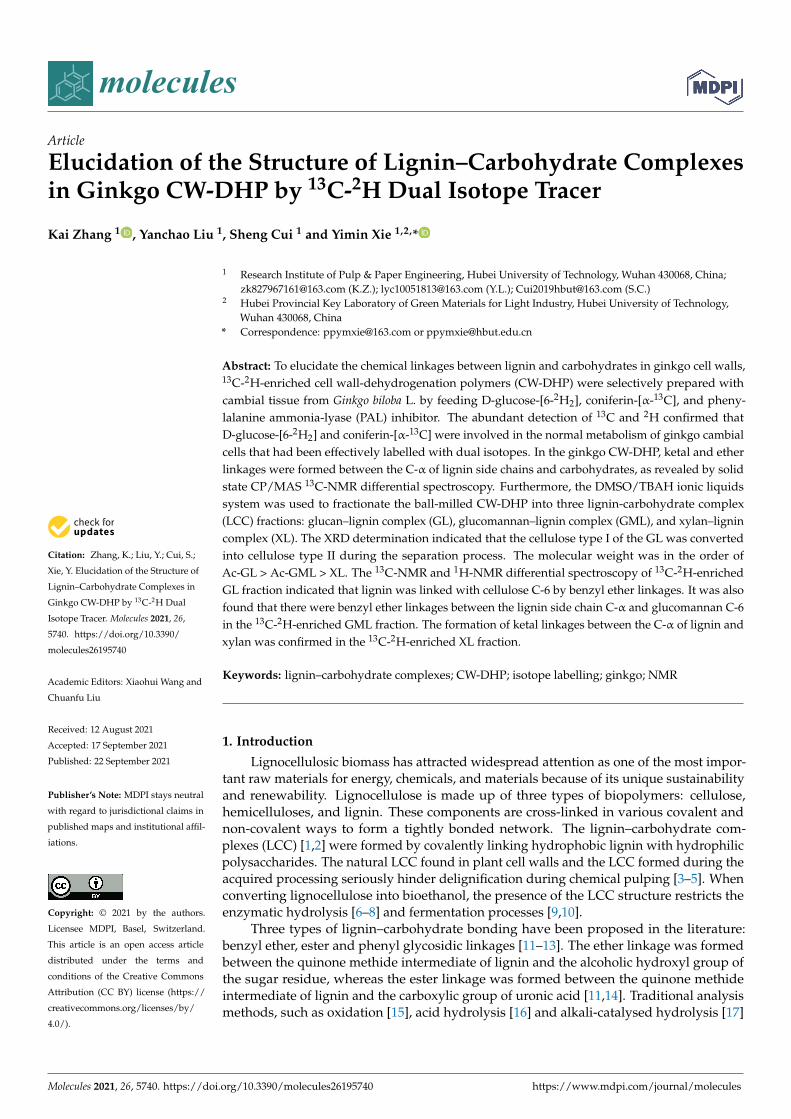
The preparation and analysis of the specifically 13C-2H-enriched lignin–carbohydratecomplex fractions in ginkgo CW-DHP are schematically illustrated in Figure 1. The original13C-2H double stable isotope technique was applied to trace lignin and carbohydrates viaconiferin-[α-13C] and D-glucose-[6-2H2], respectively.
First, the selectively 13C-2H-enriched CW-DHP was prepared with cambial tissue fromGinkgo biloba L. β-glucosidase can promote the hydrolysis of coniferin-[α-13C] into coniferylalcohol monomers and glucose units. Glucose can generate hydrogen peroxide in situ byglucose oxidase in cambial cells. Hydrogen peroxide combined with the phenol oxidaseof wood cells can polymerize coniferyl alcohol monomers into CW-DHP. Wood cambialtissues naturally contain the enzymes necessary for the polymerization of coniferin into CW-DHP [27]. The ginkgo CW-DHP was then dissolved in ionic solutions and fractionated intothree different LCC fractions. Finally, 13C-NMR and 1H-NMR were used to characterise thecovalent bonds of LCC from the two perspectives of lignin and carbohydrates, respectively.
Molecules 2021, 26, 5740 3 of 20
Figure 1. Schematic illustration of the preparation and analysis of specifically 13C-2H-enriched LCC fractions in ginkgo CW-DHP.
2.2. Characterisation of Gingko CW-DHP
2.2.1. Abundance Characterization of 13C-2H-Enriched Ginkgo CW-DHP
According to Table 1, the δ13C (VPDB) and δD (VSMOW) of the experimental group,B, were much larger than those of the control group, A. The 13Cα/12Cα ratio of the13C-2H-enriched ginkgo CW-DHP was 6.6 times that of group A, and the D6/H6 ratioof group B was 37 times that of the unenriched ginkgo CW-DHP. These two sets of dataindicate that exogenous coniferin-[α-13C] and D-glucose-[6-2H2] are involved in the normalmetabolism of ginkgo cambial cells, and that the lignin and polysaccharides of ginkgoCW-DHP were successfully labeled by 13C and D, respectively, after cultivation. Theseresults were consistent with previous studies [11,26,28,29], and ginkgo CW-DHP could beused in subsequent experiments.
Table 1. 13C and D abundance of Ginkgo CW-DHP.
Sample 13C(VPDB) 13Cα/12Cα (%) D(VSMOW) D6/H6 (%)
A −27.18 (±2.21) 1.08 −132.90 (±4.20) 0.01B 81.69 (±5.46) 7.10 1902.75 (±30.25) 0.37
Note: A: unenriched ginkgo CW-DHP; B: 13C-2H-enriched ginkgo CW-DHP.
2.2.2. Evaluation of the Lignification of Ginkgo CW-DHP
The total lignin content of the natural Ginkgo biloba L. xylem sample was measuredto be 31.03% (±0.15%). The lignin content of the ginkgo soft cambial tissues determinedby the acetyl bromide method before culture was 14.47% (±0.12%), which indicated thatthe degree of lignification was low and that the differentiation ability was strong, asshown in Table 2. After biological culture in the laboratory, the lignin content of theunenriched sample and the isotope-enriched sample increased by 19.74% (±0.16%) and19.89% (±0.14%), respectively. This showed that when coniferin-[α-13C], D-glucose-[6-2H2]and AOA were administered, the ginkgo cambial tissues could be metabolised normallyand differentiated into lignin and polysaccharides during 18 days of culture. Therefore, thelignification degree of ginkgo CW-DHP improved significantly. Furthermore, the lignincontent of the unenriched and enriched CW-DHP samples were similar, indicating that theexogenous isotopes added to the culture medium did not inhibit the normal growth anddevelopment of ginkgo cambial tissues.
Molecules 2021, 26, 5740 4 of 20
Table 2. Comparison of lignin contents before and after the culture of the soft cambial tissues of aginkgo tree.
SamplesLignin Contents
Cambial Tissues CW-DHP Increase
unenriched sample 14.47% (±0.12%) 19.74% (±0.16%) 5.42%13C-2H-enriched sample 14.47% (±0.12%) 19.89% (±0.14%) 5.27%
2.2.3. Solid-State CP/MAS 13C-NMR Analysis
The dehydrogenation polymer (DHP) formed by the polymerisation of the coniferylalcohol monomer was called “conventional DHP”. Compared to natural lignin or milled-wood lignin (MWL), conventional DHP was characterised by significant amounts of β-βand β-5 structures, as well as end-groups which were mainly of the coniferyl alcohol type,while the β-O-4 linkages were present to a lesser extent [30–32]. The CW-DHP preparedby simulating natural lignification conditions contains complete wood cell walls, and itsmolecular weight was hundreds of times larger than that of conventional DHP. In CW-DHP,the frequency of β-O-4 substructures was higher than that of conventional DHP, which wascloser to native lignin. The combined frequency of β-5, β-β, and β-1 and the frequency ofthe coniferyl alcohol/coniferaldehyde side chain were lower than that of conventional DHPand slightly higher than that estimated for ginkgo lignin [27,31,33]. Therefore, CW-DHPwas more similar to natural lignin than conventional DHP.
Stable isotope enrichment and subsequent solid or liquid NMR analysis have pre-viously been applied to wood xylem [23–25,28,34]. To distinguish the isotope-labelledsignals of the carbon atoms found in the lignin side chain from those found in non-labelledsamples, solid-state cross-polarization magic angle spinning (CP/MAS) 13C-NMR differ-ential spectroscopy was used. In the difference spectra, all carbon signals, except the 13Cenhanced signal, are eliminated. Thus, peak areas in the difference spectra represent thefrequency of enriched α-13C and are assigned to different types of α-carbon according tochemical shifts.
The solid-state CP/MAS 13C-NMR spectra of enriched and unenriched ginkgo CW-DHP and their difference spectrum are shown in Figure 2. The assignment of their signalsis shown in Table 3. The difference spectrum showed five broad peaks. From the integratedarea in the difference spectrum, 80.1–67.9 ppm accounted for 39.6% of the total area, whichmainly consisted of β-O-4 substructures [11,22,35]; the area at 93.1–80.7 ppm was 29.1%,which was mainly β-5, β-β, and Cα-O-R (R was glycosyl) in lignin [26], and the area at67.9–58.0 ppm which accounted for 12.9%, and which mainly could be assigned to the β-1substructures in lignin. The above results confirmed that the lignin produced in ginkgoCW-DHP had the structural characteristics of typical DHP. The area at 100.5–110.2 ppmaccounted for 7.6%, demonstrating that ginkgo CW-DHP had a limited amount of ketal link-ages formed between carbohydrates and lignin C-α [26,36]. The area at 140.5–124.7 ppmwas 10.8%, and this region was principally CαH = CH in coniferyl alcohol [37], indicatinga higher amount of coniferyl alcohol than protolignin in the CW-DHP.
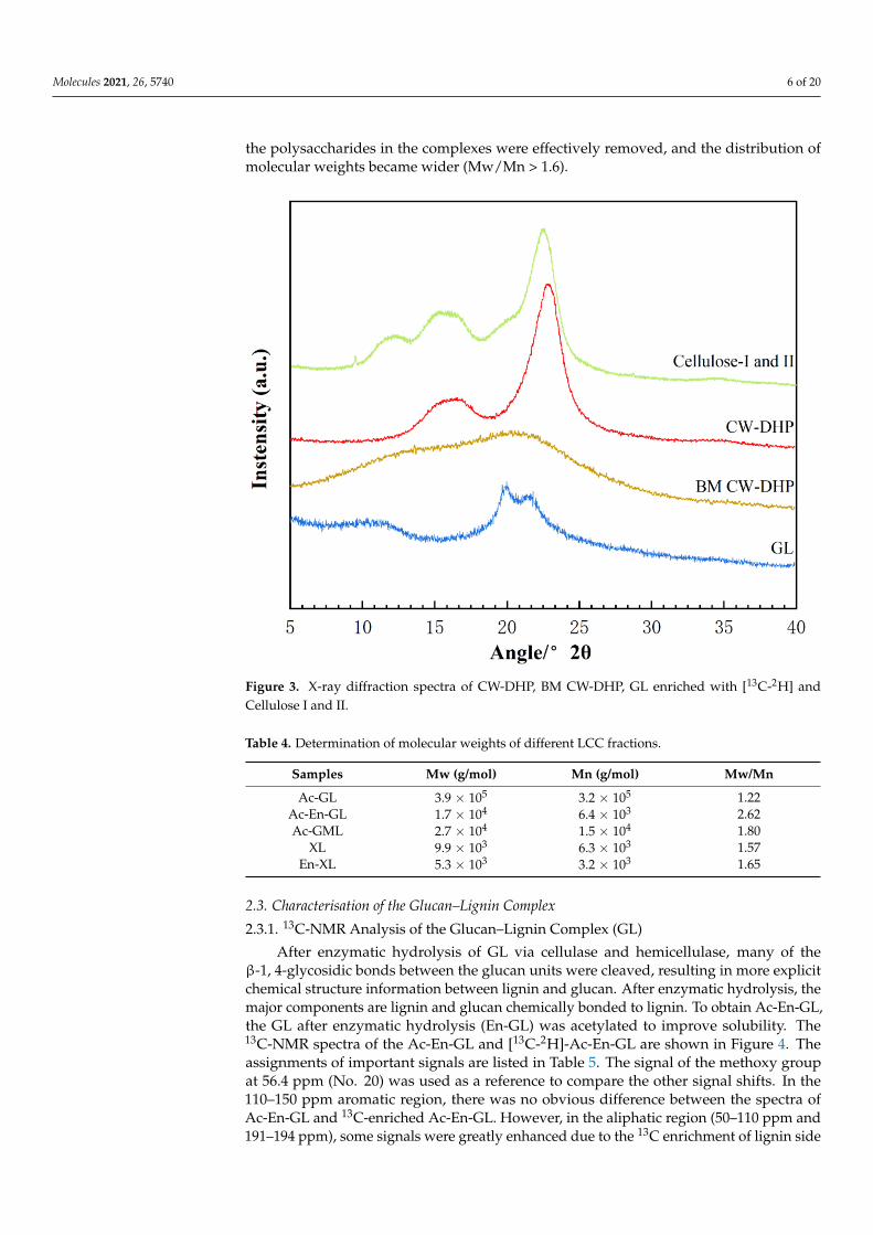
2.2.4. XRD Characterisation of CW-DHP and the Fractions
The X-ray diffraction (XRD) spectra observed of CW-DHP, ball-milled (BM) CW-DHP,glucan–lignin complex (GL) labelled with dual isotope, and cellulose I and II were shownin Figure 3. The characteristic peaks of CW-DHP were located at 2θ = 15◦, 16.5◦ and22.5◦, respectively, corresponding to the (1 −1 0), (1 −1 0), and (1 −1 0) crystal planesof cellulose I [38,39]. After 12 h of complete ball milling, the CW-DHP was physicallyaltered, with the majority of the crystalline structure in the cellulose being destroyed [18].The CW-DHP after ball milling was completely dissolved in the DMSO/TBAH system,and the crystalline morphology of the cellulose I of the original CW-DHP was convertedinto cellulose II in the precipitated fraction GL, suggesting that cellulose is the majorglucan component in LCC. The new characteristic peaks of cellulose II appeared in the

Molecules 2021, 26, 5740 5 of 20
XRD spectra of GL at 2θ = 12.5◦, 20◦ and 22.3◦, corresponding to (1 −1 0), (1 1 0) and(0 2 0), respectively [40,41].
Figure 2. Solid-state CP/MAS 13C-NMR spectra of Ginkgo CW-DHP: (A) 13C-2H-enriched ginkgoCW-DHP; (B) unenriched ginkgo CW-DHP; and (C) difference spectrum obtained by subtractingspectra (B) from spectra (A).
Table 3. Assignment of signals of CP/MAS 13C-NMR difference spectrum of Ginkgo CW-DHP.
Shift (δ, ppm) Assignments Ratio of Integrated Area
140.5–124.7 -CαH = CH- of coniferyl alcohol 10.8%
100.5–110.2 ketal linkages in carbohydrates andlignin C-α 7.6%
93.1–80.7 β-5, β-β and Cα-O-R (R was glycosyl) 29.1%80.1–67.9 Cα in β-O-4 39.6%67.9–58.0 Cα in β-1 12.9%
2.2.5. Molecular Weight Evaluation of LCC Fractions
The molecular weights and polydispersity results of different LCC fractions are shown inTable 4. The molecular weights’ order was acetylated GL (Ac-GL) > acetylated glucomannan–lignin complex (Ac-GML) > xylan–lignin complex (XL). These results are in agreementwith the common knowledge that the order of the molecular size of the carbohydrates iscellulose > glucomannan > xylan. Due to its high molecular weight, the GL fraction wasprecipitated promptly after dispersion in water. The hemicelluloses in softwood mainly con-sist of glucomannan. Barium ions readily form insoluble complexes with mannans throughinteractions between the ions and the vicinal cis-hydroxyl groups on carbons 2 and 3 of themannose units. Other polysaccharides, e.g., xylan, are not precipitated because they haveno such cis-hydroxyl group structure [42]. The polydispersity of Ac-GL was the least, whichindicated that the molecular weights of Ac-GL were generally too large. After enzymatichydrolysis, the molecular weights of Ac-En-GL and En-XL were greatly reduced, part of
Molecules 2021, 26, 5740 6 of 20
the polysaccharides in the complexes were effectively removed, and the distribution ofmolecular weights became wider (Mw/Mn > 1.6).
Figure 3. X-ray diffraction spectra of CW-DHP, BM CW-DHP, GL enriched with [13C-2H] andCellulose I and II.
Table 4. Determination of molecular weights of different LCC fractions.
Samples Mw (g/mol) Mn (g/mol) Mw/Mn
Ac-GL 3.9 × 10⁵ 3.2 × 10⁵ 1.22Ac-En-GL 1.7 × 10⁴ 6.4 × 10³ 2.62Ac-GML 2.7 × 10⁴ 1.5 × 10⁴ 1.80
XL 9.9 × 10³ 6.3 × 10³ 1.57En-XL 5.3 × 10³ 3.2 × 10³ 1.65
2.3. Characterisation of the Glucan–Lignin Complex
2.3.1. 13C-NMR Analysis of the Glucan–Lignin Complex (GL)
After enzymatic hydrolysis of GL via cellulase and hemicellulase, many of theβ-1, 4-glycosidic bonds between the glucan units were cleaved, resulting in more explicitchemical structure information between lignin and glucan. After enzymatic hydrolysis, themajor components are lignin and glucan chemically bonded to lignin. To obtain Ac-En-GL,the GL after enzymatic hydrolysis (En-GL) was acetylated to improve solubility. The13C-NMR spectra of the Ac-En-GL and [13C-2H]-Ac-En-GL are shown in Figure 4. Theassignments of important signals are listed in Table 5. The signal of the methoxy groupat 56.4 ppm (No. 20) was used as a reference to compare the other signal shifts. In the110–150 ppm aromatic region, there was no obvious difference between the spectra ofAc-En-GL and 13C-enriched Ac-En-GL. However, in the aliphatic region (50–110 ppm and191–194 ppm), some signals were greatly enhanced due to the 13C enrichment of lignin side

Molecules 2021, 26, 5740 7 of 20
chains. This indicated that exogenously supplied coniferin-[α-13C] participates in ligninmetabolism without interfering with the normal lignin biosynthesis.
Figure 4. 13C-NMR spectra of Ac-En-GL and Ac-En-GL-[13C-2H] fractions from ginkgo CW-DHP.
Table 5. Assignments of 13C-NMR spectra of Ac-En-GL from ginkgo CW-DHP.
Signalδ13C (ppm)
AssignmentsAc-En-GL13C-Enriched Control
1 194.6 194.3 α-CO, and α-CHO in vanillin2 191.5 191.7 α-CHO3 170.7 169.9 -C = O in acetyl group
4 153.0 153.2 C-4 in G, α-etherified; C-α incinnamaldehyde
5 150.1 150.6 C-3 in G, α-etherified; C-4 in G,non-etherified

Molecules 2021, 26, 5740 8 of 20
Table 5. Cont.
Signalδ13C (ppm)
AssignmentsAc-En-GL13C-Enriched Control
6 147.5 148.5 C-3 in G, non-etherified
7 143.7 143.3 C-α in cinnamic acid; C-4 inphenylcoumaran substructures
8 135.6 - C-1 in G, non-etherified
9 129.7 - C-1 in G with C-α in -CαH = CH-of coniferyl alcohols
10 119.5 119.3 C-6 in G, non-etherified11 115.7 115.7 C-6 in β-5; C-5 in G, non-etherified12 111.5 111.3 C-2 in G, non-etherified
13 105.8 - C-αwith ketal linkages; C-1in glucose
14 87.4 87.2 C-α in phenylcoumaran
15 84.0 84.1 C-β in β-arylether; C-α inpinoresinol
16 82.5 - C-α etherified to glucan
17 72.1 72.5 C-α in β-arylether; C-6 in glucanwith ether linkage; C-2 in glucose
18 63.6 63.3 C-γ in phenylcoumaran19 60.5 61.1 C-γ in β-arylether; C-6 in glucose20 56.4 56.2 -OCH321 29.5 29.3 Unknown22 21.3 21.9 -CH3 in acetyl group
Comparing the spectrum of Ac-En-GL-[13C-2H] with that of Ac-En-GL, 194.4 ppm(No. 1) and 191.0 ppm (No. 2), signal enhancements could be assigned to the α-CO andvanillin α-CHO, respectively, and the enhanced signal at 153.0 ppm (No. 4) could beassigned to cinnamaldehyde C-α. An enhanced signal that appeared at 143.7 ppm (No. 7)could be assigned to cinnamic acid C-α. An enhanced signal at 129.7 ppm (No. 9) wasrelated to the -CαH = CH- in the guaiacyl side chain. The signal at 105.8 ppm (No. 13) wasenhanced by 13C enrichment; according to the 13C-NMR spectra of the model compound,the signal was considered to be the ketal linkages between C-α of the lignin side chain andglucan, the signal could also be assigned to C-1 in glucose [11,43]. The enhanced signals at87.4 ppm (No. 14) and 84.0 ppm (No. 15) were assigned to C-α in phenylcoumaran andpinoresinol substructure. The signal at 82.0 ppm (No. 16) was obvious in 13C enrichedAc-En-GL, which was considered as C-α with benzyl ether linkages to glucan [11,26]. Thesignal at 72.1 ppm (No. 17) was significantly enhanced, and could be assigned to C-2 inglucose, C-α in β-aryl ether substructure and C-6 in glucan with ether linkage to lignin [29].The (No. 18) and (No. 19) enhanced signal peaks near 70–60 ppm corresponded to C-γ inβ-5, C-γ in β-arylether and C-6 in glucose, respectively.
2.3.2. 1H-NMR Analysis of the GL
To further investigate the chemical bonds between the lignin side chain and glucanC-6, the acetyl group (δ = 1.9–2.4 ppm) was used as a reference to compare the othersignal shifts. The 1H-NMR differential spectrum of Ac-En-GL (Figure 5) was obtained bysubtracting the 1H-NMR spectrum of the experimental group from the 1H-NMR spectrumof the control group. According to the 1H-NMR analysis of processed hexose [44], a pair ofshoulder-shaped signal peaks at 4.68 ppm (No. 2) and 4.59 ppm (No. 3) was identified inbenzyl ether with glucan C-6 and designated as D-6a and D-6b, indicating benzyl etherlinkages between glucan C-6 and lignin [26,45]. The resonance signal at 4.15 ppm (No. 4)
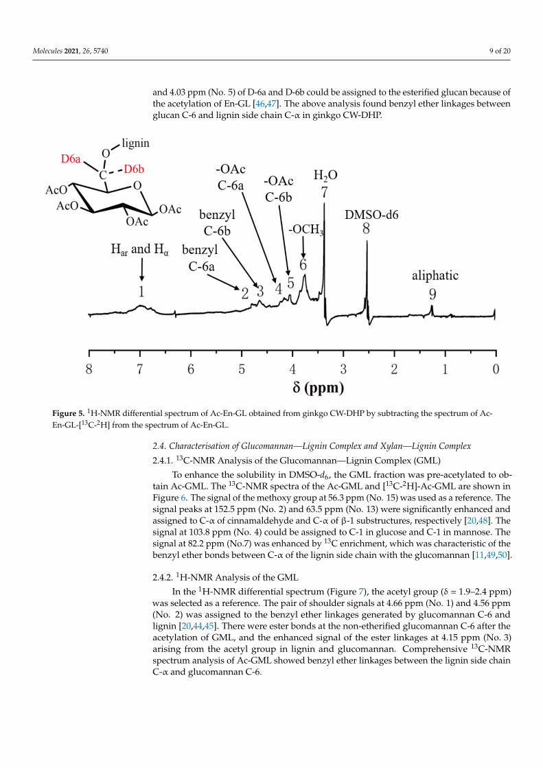
Molecules 2021, 26, 5740 9 of 20
and 4.03 ppm (No. 5) of D-6a and D-6b could be assigned to the esterified glucan because ofthe acetylation of En-GL [46,47]. The above analysis found benzyl ether linkages betweenglucan C-6 and lignin side chain C-α in ginkgo CW-DHP.
Figure 5. 1H-NMR differential spectrum of Ac-En-GL obtained from ginkgo CW-DHP by subtracting the spectrum of Ac-En-GL-[13C-2H] from the spectrum of Ac-En-GL.
2.4. Characterisation of Glucomannan—Lignin Complex and Xylan—Lignin Complex
2.4.1. 13C-NMR Analysis of the Glucomannan—Lignin Complex (GML)
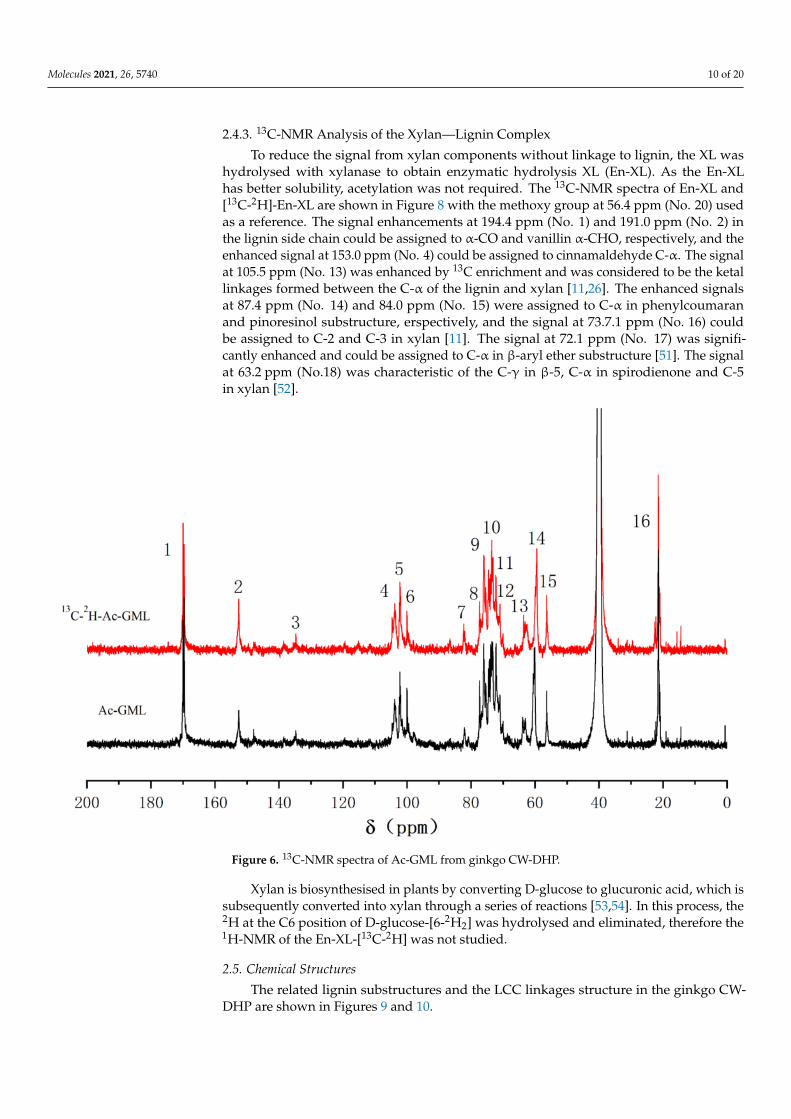
To enhance the solubility in DMSO-d6, the GML fraction was pre-acetylated to ob-tain Ac-GML. The 13C-NMR spectra of the Ac-GML and [13C-2H]-Ac-GML are shown inFigure 6. The signal of the methoxy group at 56.3 ppm (No. 15) was used as a reference. Thesignal peaks at 152.5 ppm (No. 2) and 63.5 ppm (No. 13) were significantly enhanced andassigned to C-α of cinnamaldehyde and C-α of β-1 substructures, respectively [20,48]. Thesignal at 103.8 ppm (No. 4) could be assigned to C-1 in glucose and C-1 in mannose. Thesignal at 82.2 ppm (No.7) was enhanced by 13C enrichment, which was characteristic of thebenzyl ether bonds between C-α of the lignin side chain with the glucomannan [11,49,50].
2.4.2. 1H-NMR Analysis of the GML
In the 1H-NMR differential spectrum (Figure 7), the acetyl group (δ = 1.9–2.4 ppm)was selected as a reference. The pair of shoulder signals at 4.66 ppm (No. 1) and 4.56 ppm(No. 2) was assigned to the benzyl ether linkages generated by glucomannan C-6 andlignin [20,44,45]. There were ester bonds at the non-etherified glucomannan C-6 after theacetylation of GML, and the enhanced signal of the ester linkages at 4.15 ppm (No. 3)arising from the acetyl group in lignin and glucomannan. Comprehensive 13C-NMRspectrum analysis of Ac-GML showed benzyl ether linkages between the lignin side chainC-α and glucomannan C-6.
Molecules 2021, 26, 5740 10 of 20
2.4.3. 13C-NMR Analysis of the Xylan—Lignin Complex
To reduce the signal from xylan components without linkage to lignin, the XL washydrolysed with xylanase to obtain enzymatic hydrolysis XL (En-XL). As the En-XLhas better solubility, acetylation was not required. The 13C-NMR spectra of En-XL and[13C-2H]-En-XL are shown in Figure 8 with the methoxy group at 56.4 ppm (No. 20) usedas a reference. The signal enhancements at 194.4 ppm (No. 1) and 191.0 ppm (No. 2) inthe lignin side chain could be assigned to α-CO and vanillin α-CHO, respectively, and theenhanced signal at 153.0 ppm (No. 4) could be assigned to cinnamaldehyde C-α. The signalat 105.5 ppm (No. 13) was enhanced by 13C enrichment and was considered to be the ketallinkages formed between the C-α of the lignin and xylan [11,26]. The enhanced signalsat 87.4 ppm (No. 14) and 84.0 ppm (No. 15) were assigned to C-α in phenylcoumaranand pinoresinol substructure, erspectively, and the signal at 73.7.1 ppm (No. 16) couldbe assigned to C-2 and C-3 in xylan [11]. The signal at 72.1 ppm (No. 17) was signifi-cantly enhanced and could be assigned to C-α in β-aryl ether substructure [51]. The signalat 63.2 ppm (No.18) was characteristic of the C-γ in β-5, C-α in spirodienone and C-5in xylan [52].
Figure 6. 13C-NMR spectra of Ac-GML from ginkgo CW-DHP.
Xylan is biosynthesised in plants by converting D-glucose to glucuronic acid, which issubsequently converted into xylan through a series of reactions [53,54]. In this process, the2H at the C6 position of D-glucose-[6-2H2] was hydrolysed and eliminated, therefore the1H-NMR of the En-XL-[13C-2H] was not studied.
2.5. Chemical Structures
The related lignin substructures and the LCC linkages structure in the ginkgo CW-DHP are shown in Figures 9 and 10.

Molecules 2021, 26, 5740 11 of 20
Figure 7. 1H-NMR differential spectrum of Ac-GML obtained from ginkgo CW-DHP by subtracting the spectrum ofAc-GML-[13C-2H] from the spectrum of Ac-GML.
Figure 8. 13C-NMR spectra of En-XL from ginkgo CW-DHP.
Molecules 2021, 26, 5740 12 of 20
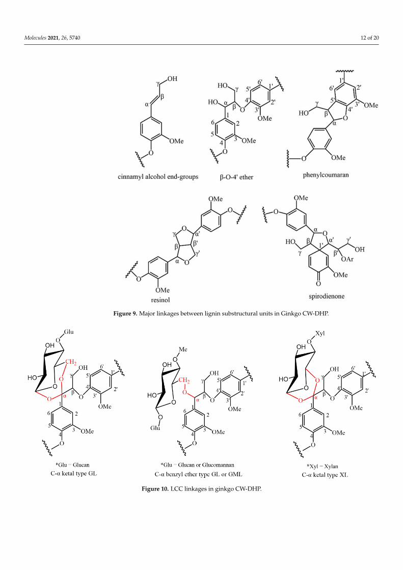
Figure 9. Major linkages between lignin substructural units in Ginkgo CW-DHP.
Molecules 2021, 26, x. https://doi.org/10.3390/xxxxx www.mdpi.com/journal/molecules
Figure 9. Major linkages between lignin substructural units in Ginkgo CW-DHP.
Figure 10. LCC linkages in ginkgo CW-DHP. Figure 10. LCC linkages in ginkgo CW-DHP.
Molecules 2021, 26, 5740 13 of 20
3. Materials and Methods3.1. Materials
Five-year-old Ginkgo biloba L. trees were obtained from the Wuhan Botanical Garden(Wuhan, China). Sodium acetate-1-13C, D-glucose-[6-2H2], hemicellulase (Aspergillus niger,≥1500 unites/g), β-glucosidase (almond, ≥7000 unites/g) and dual antibiotics (penicillin-streptomycin) were purchased from Sigma-Aldrich (St. Louis, MO, USA). Cellulase (OnozukaRS, 16,000 unites/g) was purchased from Aladdin Reagent (Shanghai, China). Acetyl bro-mide, tetrabutylammonium hydroxide (TBAH, 40% w/w in water), N-methylimidazole car-boxymethoxyamine hemihydrochloride (AOA) and xylanase (Aspergillus oryzae, ≥2500 unites/g)were purchased from Macklin Reagent (Shanghai, China). All other chemicals are of ana-lytical grade.
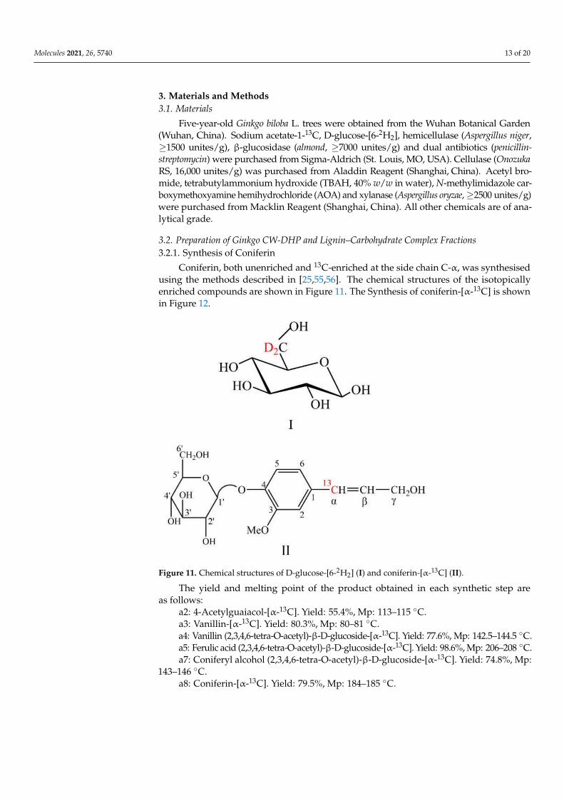
3.2. Preparation of Ginkgo CW-DHP and Lignin–Carbohydrate Complex Fractions3.2.1. Synthesis of Coniferin
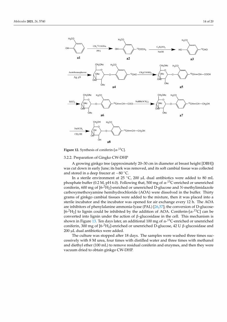
Coniferin, both unenriched and 13C-enriched at the side chain C-α, was synthesisedusing the methods described in [25,55,56]. The chemical structures of the isotopicallyenriched compounds are shown in Figure 11. The Synthesis of coniferin-[α-13C] is shownin Figure 12.
Figure 11. Chemical structures of D-glucose-[6-2H2] (I) and coniferin-[α-13C] (II).
The yield and melting point of the product obtained in each synthetic step areas follows:
a2: 4-Acetylguaiacol-[α-13C]. Yield: 55.4%, Mp: 113–115 ◦C.a3: Vanillin-[α-13C]. Yield: 80.3%, Mp: 80–81 ◦C.a4: Vanillin (2,3,4,6-tetra-O-acetyl)-β-D-glucoside-[α-13C]. Yield: 77.6%, Mp: 142.5–144.5 ◦C.a5: Ferulic acid (2,3,4,6-tetra-O-acetyl)-β-D-glucoside-[α-13C]. Yield: 98.6%, Mp: 206–208 ◦C.a7: Coniferyl alcohol (2,3,4,6-tetra-O-acetyl)-β-D-glucoside-[α-13C]. Yield: 74.8%, Mp:
143–146 ◦C.a8: Coniferin-[α-13C]. Yield: 79.5%, Mp: 184–185 ◦C.
Molecules 2021, 26, 5740 14 of 20
Figure 12. Synthesis of coniferin-[α-13C].
3.2.2. Preparation of Gingko CW-DHP
A growing ginkgo tree (approximately 20–30 cm in diameter at breast height [DBH])was cut down in early June; its bark was removed, and its soft cambial tissue was collectedand stored in a deep freezer at −80 ◦C.
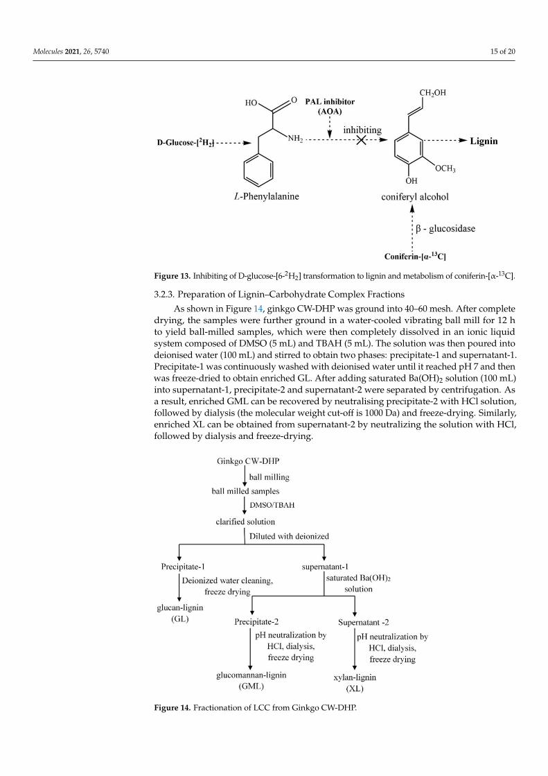
In a sterile environment at 25 ◦C, 200 µL dual antibiotics were added to 80 mLphosphate buffer (0.2 M, pH 6.0). Following that, 500 mg of α-13C-enriched or unenrichedconiferin, 600 mg of [6-2H2]-enriched or unenriched D-glucose and N-methylimidazolecarboxymethoxyamine hemihydrochloride (AOA) were dissolved in the buffer. Thirtygrams of ginkgo cambial tissues were added to the mixture, then it was placed into asterile incubator and the incubator was opened for air exchange every 12 h. The AOAare inhibitors of phenylalanine ammonia-lyase (PAL) [26,57]; the conversion of D-glucose-[6-2H2] to lignin could be inhibited by the addition of AOA. Coniferin-[α-13C] can beconverted into lignin under the action of β-glucosidase in the cell. This mechanism isshown in Figure 13. Ten days later, an additional 100 mg of α-13C-enriched or unenrichedconiferin, 300 mg of [6-2H2]-enriched or unenriched D-glucose, 42 U β-glucosidase and200 µL dual antibiotics were added.
The culture was stopped after 18 days. The samples were washed three times suc-cessively with 8 M urea, four times with distilled water and three times with methanoland diethyl ether (100 mL) to remove residual coniferin and enzymes, and then they werevacuum dried to obtain ginkgo CW-DHP.
Molecules 2021, 26, 5740 15 of 20
Figure 13. Inhibiting of D-glucose-[6-2H2] transformation to lignin and metabolism of coniferin-[α-13C].
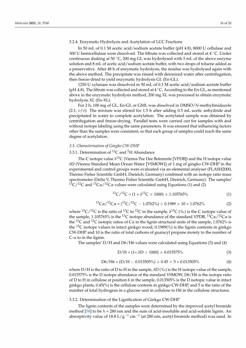
3.2.3. Preparation of Lignin–Carbohydrate Complex Fractions
As shown in Figure 14, ginkgo CW-DHP was ground into 40–60 mesh. After completedrying, the samples were further ground in a water-cooled vibrating ball mill for 12 hto yield ball-milled samples, which were then completely dissolved in an ionic liquidsystem composed of DMSO (5 mL) and TBAH (5 mL). The solution was then poured intodeionised water (100 mL) and stirred to obtain two phases: precipitate-1 and supernatant-1.Precipitate-1 was continuously washed with deionised water until it reached pH 7 and thenwas freeze-dried to obtain enriched GL. After adding saturated Ba(OH)2 solution (100 mL)into supernatant-1, precipitate-2 and supernatant-2 were separated by centrifugation. Asa result, enriched GML can be recovered by neutralising precipitate-2 with HCl solution,followed by dialysis (the molecular weight cut-off is 1000 Da) and freeze-drying. Similarly,enriched XL can be obtained from supernatant-2 by neutralizing the solution with HCl,followed by dialysis and freeze-drying.
Figure 14. Fractionation of LCC from Ginkgo CW-DHP.
Molecules 2021, 26, 5740 16 of 20
3.2.4. Enzymatic Hydrolysis and Acetylation of LCC Fractions
In 50 mL of 0.1 M acetic acid/sodium acetate buffer (pH 4.8), 8000 U cellulase and300 U hemicellulase were dissolved. The filtrate was collected and stored at 4 ◦C. Undercontinuous shaking at 50 ◦C, 200 mg GL was hydrolysed with 5 mL of the above enzymesolution and 8 mL of acetic acid/sodium acetate buffer, with two drops of toluene added asa preservative. After 48 h of enzymatic hydrolysis, the residue was hydrolysed again withthe above method. The precipitate was rinsed with deionized water after centrifugation,then freeze-dried to yield enzymatic hydrolysis GL (En-GL).
1250 U xylanase was dissolved in 50 mL of 0.1 M acetic acid/sodium acetate buffer(pH 4.8). The filtrate was collected and stored at 4 ◦C. According to the En-GL, as mentionedabove in the enzymatic hydrolysis method, 200 mg XL was processed to obtain enzymatichydrolysis XL (En-XL).
For 2 h, 100 mg of GL, En-GL or GML was dissolved in DMSO/N-methylimidazole(2:1, v/v). The mixture was stirred for 1.5 h after adding 0.5 mL acetic anhydride andprecipitated in water to complete acetylation. The acetylated sample was obtained bycentrifugation and freeze-drying. Parallel tests were carried out for samples with andwithout isotope labeling using the same parameters. It was ensured that influencing factorsother than the samples were consistent, so that each group of samples could reach the samedegree of acetylation.
3.3. Characterisation of Gingko CW-DHP
3.3.1. Determination of 13C and 2H Abundance
The C isotope value δ13C (Vienna Pee Dee Belemnite [VPDB]) and the H isotope valueδD (Vienna Standard Mean Ocean Water [VSMOW]) of 1 mg of gingko CW-DHP in theexperimental and control groups were evaluated via an elemental analyser (FLASH2000,Thermo Fisher Scientific GmbH, Dreieich, Germany) combined with an isotope ratio massspectrometer (Delta V, Thermo Fisher Scientific GmbH, Dreieich, Germany). The samples’13C/12C and 13Cα/12Cα values were calculated using Equations (1) and (2)
13C/12C = (1 + δ13C ÷ 1000) × 1.105765% (1)
13Cα/12Cα = (13C/12C − 1.0762%) ÷ 0.1989 × 10 + 1.0762% (2)
where 13C/12C is the ratio of 13C to 12C in the sample, δ13C (‰) is the C isotope value ofthe sample, 1.105765% is the 13C isotope abundance of the standard VPDB, 13Cα/12Cα isthe 13C and 12C isotopic ratios of Cα in the lignin structural units of the sample, 1.0762% isthe 13C isotope values in intact ginkgo wood, 0.1989(%) is the lignin contents in ginkgoCW-DHP and 10 is the ratio of total carbons of guaiacyl propane moiety to the number ofC-α to in the lignin.
The samples’ D/H and D6/H6 values were calculated using Equations (3) and (4)
D/H = (1+ δD ÷ 1000) × 0.015575% (3)
D6/H6 = (D/H − 0.013505%) ÷ 0.45 × 5 + 0.013505% (4)
where D/H is the ratio of D to H in the sample, δD (‰) is the H isotope value of the sample,0.015575% is the D isotope abundance of the standard VSMOW, D6/H6 is the isotope ratioof D to H in cellulose at position 6 in the sample, 0.013505% is the D isotopic value in intactginkgo plants, 0.45(%) is the cellulose contents in ginkgo CW-DHP, and 5 is the ratio of thenumber of total hydrogens in a glucose unit in cellulose to H6 in the cellulose structures.
3.3.2. Determination of the Lignification of Ginkgo CW-DHP
The lignin contents of the samples were determined by the improved acetyl bromidemethod [58] to be λ = 280 nm and the sum of acid-insoluble and acid-soluble lignin. Anabsorptivity value of 18.8 L/g−1 cm−1 (at 280 nm, acetyl bromide method) was used. In
Molecules 2021, 26, 5740 17 of 20
this experiment, ginkgo wood mill was used as the standard sample and lignin values inthe ginkgo CW-DHP were calculated using Equation (5)
X = (K × A)/B × 100% (5)
where B is the absorbance of the standard ginkgo wood mill, K is the total lignin contentsof the standard ginkgo wood mill, and A is the absorbance of the CW-DHP.
3.3.3. Determination of Solid-State 13C-NMR Spectroscopy
The solid-state CP/MAS 13C-NMR was recorded on an Avance III 600-MHz solid-state NMR spectrometer with a solid probe (Bruker, Billerica, MA, USA). The experimentalconditions were as follows: a temperature of 14.1 ◦C, 3 ms contact time, 0.05 s receptiontime, the magic angle spinning frequency fixed at 10 kHz, a pulse width of 35 kHz anda pulse delay of 2 s. Each sample was accumulated approximately 5000 times. All of thespinning side bands are very small, by fixing the spinning frequency, the small spinningside bands will not contribute to the difference spectra [59].
3.3.4. Determination of XRD
The X-ray Diffraction system (Empyrean Sharp, Panaco, The Netherlands) was usedto analyse the crystallisation characteristics of fully dried samples with a CuKα radiationwavelength of 0.154 nm. Diffractograms were achieved by scanning from 5◦ to 40◦ (2θ) at arate of 5◦/min, a step size of 0.02◦ and a divergence slit width of 1◦.
3.3.5. Determination of Molecular Weights
Gel permeation chromatography (GPC) was determined by a Shimadzu LC-20ADHPLC system (Shimadzu, Kyoto, Japan) equipped with a shim pack GPC-803D column(Shimadzu, Kyoto, Japan) and a refractive index (RI) detector (Shimadzu, Kyoto, Japan)at a flow rate of 1 mL/min at 40 ◦C with N, N-dimethylformamide (DMF) as eluent. Thesample (2 mg) was dissolved in 1 mL DMF, and 25 µL of this solution was injected into GPC.Calibration curves were established based on four monodisperse polystyrene standardswith molecular weights ranging from 2900 to 19,800 g/mol. The data were analysed usingliquid chromatography solution software (Shimadzu, Kyoto, Japan).
3.3.6. Determination of 13C-NMR and 1H-NMR Spectroscopy
First, 80 mg of the samples was dissolved in 0.5 mL of DMSO-d6. Then, all NMR spec-tra were recorded on a Bruker Avance III 500-MHz spectrometer (Fällanden, Switzerland)equipped with aϕ5 mm broad-band fluorine observation (BBFO) probe at a temperature of25 ◦C. The 13C NMR spectra were recorded in the FT mode at 100.6 MHz with the followingconditions: 1.75-s pulse delay and 0.94-s reception time, with the data points collected at32 kbit and after accumulating 20,000 scans. The 1H-NMR spectrum was recorded with thefollowing conditions: 4.3-s pulse delay and 0.74-s acquisition time, with the data pointscollected at 32 kbit and after the accumulation of 500 scans.
4. Conclusions
1. Detection of the 13C and 2H abundance of ginkgo CW-DHP showed that exogenousconiferin-[α-13C] and D-glucose-[6-2H2] were involved in the normal metabolism of thesoft ginkgo cambial tissues. Therefore, the lignin and cell wall polysaccharides weresuccessfully labelled by 13C and D, respectively. Furthermore, the degree of lignification ofginkgo CW-DHP was increased by biological culture.
2. Solid-state CP/MAS 13C-NMR analysis revealed that the main lignin linkagesinclude β-O-4, β-5, β-1 and β-β substructures, of which β-O-4 was the most abundant. Inaddition, ketal and ether linkages were formed between the C-α of lignin side chains andcarbohydrates in ginkgo CW-DHP.

Molecules 2021, 26, 5740 18 of 20
3. XRD examination indicated that the cellulose I of GL was converted into cellulose IIduring the separation process. The molecular weight was in the order of Ac-GL > Ac-GML > XL.After enzymatic hydrolysis, the molecular weight was significantly reduced, indicating theremoval of carbohydrates without chemical bonds to the lignin macromolecule.
4. There were Cα-ketal linkages and Cα-benzyl ether linkages between glucan andlignin, while the lignin was linked with the glucomannan through Cα-benzyl ether linkages.Xylan and lignin were linked mainly by Cα-ketal linkages.
Author Contributions: Conceptualization, K.Z., Y.L. and Y.X.; methodology, K.Z., Y.L. and Y.X.;writing—original draft preparation, K.Z.; writing—review and editing, S.C. and Y.X.; visualization,K.Z.; supervision, Y.X.; project administration, Y.X.; funding acquisition, Y.X. All authors have readand agreed to the published version of the manuscript.
Funding: This research was funded by the National Natural Science Foundation of China (Grant No.21878070), and the Outstanding Young and Middle-aged Technological Innovation Team Project ofHubei Provincial Universities (Grant No. T201205).
Institutional Review Board Statement: Not applicable.
Informed Consent Statement: Not applicable.
Data Availability Statement: The data presented in this study are available in the manuscript.
Conflicts of Interest: The authors declare no conflict of interest.
Sample Availability: Samples of the compounds are not available from the authors.
References1. Björkman, A. Studies on finely divided wood. Part 1. Extraction of lignin with neutral solvents. Sven. Papp. 1956, 59, 477–485.2. Koshijima, T.; Watanabe, T. Association between Lignin and Carbohydrates in Wood and Other Plant Tissues; Springer:
Berlin/Heidelberg, Germany, 2003.3. Gierer, J.; Wannström, S. The formation of ether bonds between lignins and carbohydrates during alkaline pulping processes.
Holzforschung 1986, 40, 347–352. [CrossRef]4. Iverson, T.; Wannström, S. Lignin-carbohydrate bonds in a residual lignin isolated from pine kraft pulp. Holzforschung 1986, 40,
19–22. [CrossRef]5. Choi, J.W.; Choi, D.-H.; Faix, O. Characterization of lignin–carbohydrate linkages in the residual lignins isolated from chemical
pulps of spruce (Picea abies) and beech wood (Fagus sylvatica). J. Wood Sci. 2007, 53, 309–313. [CrossRef]6. Berlin, A.; Balakshin, M.; Gilkes, N.; Kadla, J.; Maximenko, V.; Kubo, S.; Saddler, J. Inhibition of cellulase, xylanase and
β-glucosidase activities by softwood lignin preparations. J. Biotechnol. 2006, 125, 198–209. [CrossRef] [PubMed]7. Nakagame, S.; Chandra, R.P.; Kadla, J.F.; Saddler, J.N. The isolation, characterization and effect of lignin isolated from steam
pretreated Douglas-fir on the enzymatic hydrolysis of cellulose. Bioresour. Technol. 2011, 102, 4507–4517. [CrossRef]8. Yang, H.; Xie, Y.; Zheng, X.; Pu, Y.; Huang, F.; Meng, X.; Wu, W.; Ragauskas, A.; Yao, L. Comparative study of lignin characteristics
from wheat straw obtained by soda AQ and kraft pretreatment and effect on the following enzymatic hydrolysis process. Bioresour.Technol. 2016, 207, 361–369. [CrossRef]
9. Min, D.-Y.; Li, Q.; Chiang, V.; Jameel, H.; Chang, H.-M.; Lucia, L. The influence of lignin-carbohydrate complexes on thecellulase-mediated saccharification I: Transgenic black cottonwood (western balsam poplar, California poplar) P. trichocarpaincluding the xylan down-regulated and the lignin downregulated lines. Fuel 2014, 119, 207–213. [CrossRef]
10. Min, D.-Y.; Yang, C.; Chiang, V.; Hasan, J.; Chang, H.-M. The influence of lignin-carbohydrate complexes on the cellulose-mediatedsaccharification II: Transgenic hybrid poplars (Populus nigra L. and Populus maximowiczii A.). Fuel 2014, 116, 56–62. [CrossRef]
11. Xie, Y.; Yasuda, S.; Wu, H.; Liu, H. Analysis of the structure of lignin-carbohydrate complexes by the specific 13C tracer method.J. Wood Sci. 2000, 46, 130–136. [CrossRef]
12. Yuan, T.Q.; Sun, S.N.; Xu, F.; Sun, R.C. Characterization of lignin structures and lignin-carbohydrate complex (LCC) linkages byquantitative 13C and 2D HSQC NMR spectroscopy. J. Agric. Food Chem. 2011, 59, 10604–10614. [CrossRef]
13. Nishimura, H.; Kamiya, A.; Nagata, T.; Katahira, M.; Watanabe, T. Direct evidence for a ether linkage between lignin andcarbohydrates in wood cell walls. Sci. Rep. 2018, 8, 6538. [CrossRef] [PubMed]
14. Balakshin, M.; Capanema, E.; Chang, H.-M. A fraction of MWL with high concentration of lignin–carbohydrate linkages: Isolationand analysis with 2D NMR spectroscopic techniques. Holzforschung 2007, 61, 1–7. [CrossRef]
15. Watanabe, T.; Koshijima, T. Evidence for an ester linkage between lignin and glucuronic acid in lignin-carbohydrate complexesby DDQ-Oxidation. Agric. Biol. Chem. 1988, 52, 2953–2955. [CrossRef]
16. Eriksson, Ö.; Goring, D.A.I.; Lindgren, B.O. Structural studies on the chemical bonds between lignin and carbohydrates in spruce.Wood Sci. Technol. 1980, 14, 267–279. [CrossRef]
Molecules 2021, 26, 5740 19 of 20
17. Takakashi, N.; Koshijima, T. Ester linkages between lignin and glucuronoxylan in lignin-carbohydrate complex from beech (Faguscrenata) wood. Wood Sci. Technol. 1988, 22, 231–241. [CrossRef]
18. Li, J.; Martin-Sampedro, R.; Pedrazzi, C.; Gellerstedt, G. Fractionation and characterization of lignin-carbohydrate complexes(LCCs) from eucalyptus fibers. Holzforschung 2011, 65, 43–50. [CrossRef]
19. Du, X.; Gellerstedt, G.; Li, J. Universal fractionation of lignin–carbohydrate complexes (LCCs) from lignocellulosic biomass: Anexample using spruce wood. Plant J. 2013, 74, 328–338. [CrossRef]
20. Du, X.; Pérez-Boada, M.; Fernández, C.; Rencoret, J.; del Río, J.C.; Jiménez-Barbero, J.; Li, J.; Gutiérrez, A.; Martínez, A. Analysis oflignin–carbohydrate and lignin–lignin linkages after hydrolase treatment of xylan–lignin, glucomannan–lignin and glucan–lignincomplexes from spruce wood. Planta 2014, 239, 1079–1090. [CrossRef]
21. Lewis, N.G.; Yamamoto, E.; Wooten, J.B.; Just, G.; Ohashi, H.; Towers, G.H.N. Monitoring biosynthesis of wheat cell-wallphenylpropanoids in situ. Science 1987, 237, 1344–1346. [CrossRef]
22. Lewis, N.G.; Razal, R.A.; Dhara, K.P.; Yamamoto, E.; Bokelmann, G.H.; Wooten, J.B. Incorporation of [2-13C] ferulic acid, a ligninprecursor, into Leucaena leucocephala and its analysis by solid state 13C NMR spectroscopy. J. Chem. Soc. Com. Chem. Commun.1988, 24, 1626–1628. [CrossRef]
23. Terashima, N.; Hafrén, J.; Westermark, U.; VanderHart, D.L. Structure of lignin in Ginkgo wood determined by a combination ofspecific 13C-enrichment technique and solid state NMR spectroscopy. In Proceedings of the 9th International Symposium onWood and Pulping Chemistry, Montréal, QC, Canada, June 9–12 1997; pp. H1–H5.
24. Terashima, N.; Atalla, R.H.; VanderHart, D.L. Solid state NMR spectroscopy of specifically 13C-enriched lignin in wheat strawfrom coniferin. Phytochemistry 1997, 46, 863–870. [CrossRef]
25. Xie, Y.; Yasuda, S.; Terashima, N. Selective carbon 13 enrichment of side chain carbons of oleander lignin traced by carbon 13nuclear magnetic resonance. Mokuzai Gakkaishi 1994, 40, 191–198.
26. Xie, Y.; Liu, Y.; Jiang, C.; Wu, H. The Existence of Cellulose and Lignin Chemical Connections in Ginkgo Traced by 2H-13C DualIsotopes. Bioresources 2020, 15, 9028–9044. [CrossRef]
27. Hafrén, J.; Westermark, U.; Lennholm, H.; Terashima, N. Formation of 13C-Enriched Cell-Wall DHP Using Isolated Soft Xylemfrom Picea abies. Holzforschung 2002, 56, 585–591. [CrossRef]
28. Xie, Y.; Terashima, N. Selective carbon 13-enrichment of side chain carbons of ginkgo lignin traced by carbon 13 nuclear magneticresonance. Mokuzai Gakkaishi 1991, 37, 935–941.
29. Xiang, S.; Xie, Y.; Yang, H.; Yao, L. Analysis of the association between cellulose and lignin by carbon 13 tracer method. Spectrosc.Spectr. Anal. 2013, 33, 2488–2491.
30. Brunow, G.; Lundquist, K. Comparison of a synthetic dehydrogenation polymer of coniferyl alcohol with milled wood ligninfrom spruce, using 1H-NMR spectroscopy. Pap. Puu. 1980, 62, 669–672.
31. Terashima, N.; Atalla, R.H.; Ralph, S.A.; Landucci, L.L.; Lapierre, C.; Monties, B. New preparations of lignin polymer modelsunder conditions that approximate cell wall lignification. I. Synthesis of novel lignin polymer models and their structuralcharacterization by 13C-NMR. Holzforschung 1995, 49, 521–527. [CrossRef]
32. Terashima, N.; Atalla, R.H.; Ralph, S.A.; Landucci, L.L.; Lapierre, C.; Monties, B. New preparations of lignin polymer models underconditions that approximate cell wall lignification. II. Structural characterization of the models by thioacidolysis. Holzforschung1996, 50, 9–14. [CrossRef]
33. Terashima, N.; Hafrén, J.; Westermark, U. Preparation of 13C-enriched DHP on unlignified spruce cell walls (in Japanese). InProceedings of the Annual Meeting of the Japan Wood Research Society, Shizuoka, Japan, 3–5 April 1998; Volume 3, p. 389.
34. Terashima, N.; Hafrén, J.; Westermark, U.; VanderHart, D.L. Nondestructive analysis of lignin structure by NMR spectroscopy ofspecifically 13C-enriched lignins. Part 1. Solid state study of ginkgo wood. Holzforschung 2002, 56, 43–50. [CrossRef]
35. Sipilä, J.; Brunow, G. On the mechanism of formation of non-cyclic benzyl ethers during lignin biosynthesis. Part 4. The reactionsof a β-O-4 type quinone methide with carboxylic acids in the presence of phenols. The formation and stability of benzyl estersbetween lignin and carbohydrates. Holzforschung 1991, 45, 9–14.
36. Jacques, D.; Haslam, E.; Bedford, G.R.; Greatbanks, D. Plant proanthocyanidins. Part II. Proanthocyanidin-A2 and its derivatives.J. Chem. Soc. Perkin Trans. 1974, 1, 2663–2671. [CrossRef]
37. Lüdemann, H.D.; Nimz, H. Carbon-13 nuclear magnetic resonance spectra of lignins. Biochem. Biophys. Res. Commun. 1973, 52,1162–1169. [CrossRef]
38. Henrique, M.A.; Flauzino Neto, W.P.; Silvério, H.A.; Martins, D.F.; Alves Gurgel, L.V.; Silva Barud, H.; Morais, L.C.; Pasquinia, D.Kinetic study of the thermal decomposition of cellulose nanocrystals with different polymorphs, cellulose I and II, extracted fromdifferent sources and using different types of acids. Ind. Crop. Prod. 2015, 76, 128–140. [CrossRef]
39. Ahmed-Haras, M.R.; Kao, N.; Ward, L. Single-step heterogeneous catalysis production of highly monodisperse sphericalnanocrystalline cellulose. Int. J. Biol. Macromol. 2020, 154, 246–255. [CrossRef]
40. French, A.D. Idealized powder diffraction patterns for cellulose polymorphs. Cellulose 2014, 21, 885–896. [CrossRef]41. Jin, E.; Guo, J.; Yang, F.; Zhu, Y.; Song, J.; Jin, Y.; Orlando, J.R. On the polymorphic and morphological changes of cellulose
nanocrystals (CNC-I) upon mercerization and conversion to CNC-II. Carbohydr. Polym. 2016, 143, 327–335. [CrossRef]42. Meier, H. Barium hydroxide as a selective precipitating agent for hemicelluloses. Acta. Chem. Scand. 1958, 12, 144–146. [CrossRef]43. Taneda, H.; Nakano, J.; Hosoya, S.; Chang, H.-M. Stability of α-ether type model compounds during chemical pulping processes.
J. Wood Chem. Technol. 1987, 7, 485–497.
Molecules 2021, 26, 5740 20 of 20
44. Yoshihiro, N.; Hiroshi, O.; Hiroshi, M. 1H-NMR studies of (6r)-and (6s)-deuterated d-hexoses: Assignment of the preferredrotamers about C5-C6 bond of D-glucose and D-galactose derivatives in solutions. Tetrahedron Lett. 1984, 25, 1575–1578.
45. Balakshin, M.; Capanema, E.; Gracz, H.; Chang, H.-M.; Jameel, H. Quantification of lignin-carbohydrate linkages with high-resolution NMR spectroscopy. Planta 2011, 233, 1097–1110. [CrossRef] [PubMed]
46. Deus, C.; Friebolin, H. Partiell acetylierte cellulose-synthese und bestimmung der substituentenverteilung mit hilfe der 1HNMR-spektroskopie. Makromol. Chem. 1991, 192, 75–83. [CrossRef]
47. Hikichi, K.; Kakuta, Y.; Katoh, T. 1H NMR study on substituent distribution of cellulose diacetate. Polym. J. 1995, 27, 659–663.[CrossRef]
48. Miyagawa, Y.; Kamitakahara, H.; Takano, T.; Nakatsubo, F. Fractionation and characterization of lignin carbohydrate complexes(LCCs) of Eucalyptus globulus in residues left after MWL isolation. Part I: Analyses of hemicellulose-lignin fraction (HC-L).Holzforschung 2012, 66, 459–465. [CrossRef]
49. Grasdalen, H.; Painter, T. NMR studies of composition and sequence in legume-seed galactomannans. Carbohydr. Res. 1980, 81,59–66. [CrossRef]
50. Bi, H.; Gao, T.; Li, Z.; Ji, L.; Yang, W.; Iteku Jeff, B.; Liu, E.; Zhou, Y. Structural elucidation and antioxidant activity of awater-soluble polysaccharide from the fruit bodies of Bulgaria inquinans (Fries). Food Chem. 2013, 138, 1470–1475. [CrossRef]
51. Miyagawa, Y.; Kamitakahara, H.; Takano, T. Fractionation and characterization of lignin-carbohydrate complexes (LCCs) ofEucalyptus globulus in residues left after MWL isolation. Part II: Analyses of xylan-lignin fraction (X-L). Holzforschung 2013, 67,629–642. [CrossRef]
52. Yao, L.; Chen, C.; Zheng, X.; Peng, Z.; Yang, H.; Xie, Y. Determination of Lignin-Carbohydrate Complexes Structure of WheatStraw using Carbon-13 Isotope as a Tracer. Bioresources 2016, 11, 6692–6707. [CrossRef]
53. Slater, W.G.; Beevers, H. Utilization of D-Glucuronate by Corn Coleoptiles. Plant Physiol. 1958, 33, 146–151. [CrossRef]54. Bailey, R.W.; Hassid, W.Z. Xylan synthesis from uridine-diphosphate-d-xylose by particulate preparations from immature
corncobs. Proc. Natl. Acad. Sci. USA 1966, 56, 1586–1593. [CrossRef] [PubMed]55. Terashima, N.; Ralph, S.A.; Landucci, L.L. New facile syntheses of monolignol glucosides; p-glucocoumaryl alcohol, coniferin
and syringin. Holzforschung 1996, 50, 151–155.56. Terashima, N.; Koa, C.; Matsushita, Y.; Westermark, U. Monolignol glucosides as intermediate compounds in lignin biosynthesis.
Revisiting the cell wall lignification and new 13C-tracer experiments with Ginkgo biloba and Magnolia liliiflora. Holzforschung 2016,70, 801–810. [CrossRef]
57. Imai, T.; Terashima, N. Determination of the distribution and reaction of polysaccharides in wood cell walls by the isotope tracertechnique. IV. Selective radio-labeling of xylan in magnolia (Magnolia kobus) and visualization of its distribution in differentiatingxylem by microautoradiography. Mokuzai Gakkaishi 1992, 38, 693–699.
58. Iiyama, K.; Wallis, A.F.A. An improved acetyl bromide procedure for determining lignin in woods and wood pulps. Wood Sci.Technol. 1988, 22, 271–280. [CrossRef]
59. Parkås, J.; Paulsson, M.; Westermark, U.; Terashima, N. Solid State NMR Analysis of β-13C-Enriched Lignocellulosic MaterialDuring Light-Induced Yellowing. Holzforschung 2001, 55, 276–282. [CrossRef]


