Embed Size (px)
Citation preview

VETERINARY RESEARCHBalenghien et al. Veterinary Research 2013, 44:78http://www.veterinaryresearch.org/content/44/1/78
REVIEW Open Access
Towards a better understanding of Rift Valleyfever epidemiology in the south-west of theIndian OceanThomas Balenghien1†, Eric Cardinale1,2†, Véronique Chevalier3†, Nohal Elissa4†, Anna-Bella Failloux5*†,Thiery Nirina Jean Jose Nipomichene4†, Gaelle Nicolas3†, Vincent Michel Rakotoharinome6†, Matthieu Roger1†
and Betty Zumbo7†
Abstract
Rift Valley fever virus (Phlebovirus, Bunyaviridae) is an arbovirus causing intermittent epizootics and sporadic epidemicsprimarily in East Africa. Infection causes severe and often fatal illness in young sheep, goats and cattle. Domesticanimals and humans can be contaminated by close contact with infectious tissues or through mosquito infectiousbites. Rift Valley fever virus was historically restricted to sub-Saharan countries. The probability of Rift Valley feveremerging in virgin areas is likely to be increasing. Its geographical range has extended over the past years. As a recentexample, autochthonous cases of Rift Valley fever were recorded in 2007–2008 in Mayotte in the Indian Ocean. It hasbeen proposed that a single infected animal that enters a naive country is sufficient to initiate a major outbreak beforeRift Valley fever virus would ever be detected. Unless vaccines are available and widely used to limit its expansion, RiftValley fever will continue to be a critical issue for human and animal health in the region of the Indian Ocean.
Table of contents
1. Disease and transmission2. Mosquito vectors3. Virus-vector interactions4. Diagnosis and surveillance5. Prevention and control6. Future of RVF on islands of the Indian Ocean7. Competing interests8. Authors’ contributions9. Acknowledgments10. References
1. Disease and transmissionRift Valley fever (RVF) is an emerging zoonotic vector-borne disease representing a threat to animal and humanhealth, and livestock production. Abortions and high mor-talities in newborns are observed in animals [1,2]. In
* Correspondence: [email protected]†Equal contributors5Institut Pasteur, Department of Virology, Arboviruses and Insect Vectors,25–28 rue du Dr Roux, 75724 Paris, cedex 15, FranceFull list of author information is available at the end of the article
© 2013 Balenghien et al.; licensee BioMed CenCreative Commons Attribution License (http:/distribution, and reproduction in any medium
humans, symptoms vary from a flu-like syndrome to en-cephalitic, ocular or hemorrhagic syndrome. The case fa-tality rate of the latter form can be as high as 50% [3].Since its first isolation in 1930 in Kenya [4], RVF out-
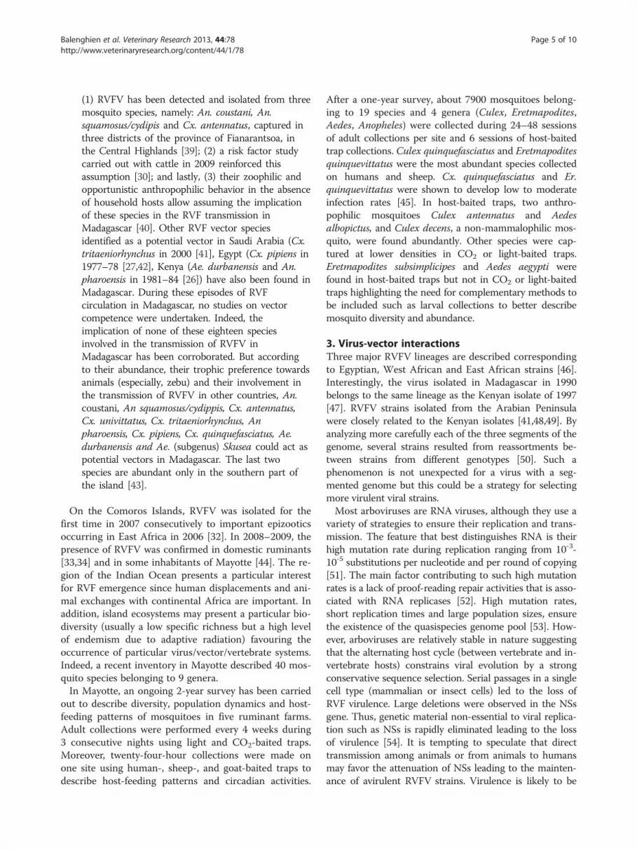
breaks have been reported in most sub-Saharan coun-tries, especially the Rift Valley in Kenya and Tanzania [5](Figure 1). Subsequent outbreaks with human cases havebeen reported in South Africa [6] and the Nile Valleyfrom Sudan to the Egyptian delta [7]. The disease spreadfrom continental Africa to Madagascar in 1991 [8-11] andin the Arabian Peninsula in 2000 [12]. In Madagascar,RVFV was isolated for the first time in 1979 from pools ofmosquitoes captured during the rainy season in the pri-mary rain forest of Perinet, Moramanga district [13]. Themost recent RVF outbreaks were detected in Somalia(2006–2007) [14], Kenya (2006–2007) [15], Tanzania(2007) [14], Sudan (2007–2008) [16], Madagascar (2008–2009) [17], South Africa (2008, 2009, and 2010) [18],Mauritania (2010) [19], Botswana (2010) [20], andNamibia (2010) [21]. In Mayotte, sporadic cases in live-stock have been recorded since 2004 with human casesdetected in 2007–2008 and 2011. While RVF was originally
tral Ltd. This is an Open Access article distributed under the terms of the/creativecommons.org/licenses/by/2.0), which permits unrestricted use,, provided the original work is properly cited.

Figure 1 Geographical distribution of Rift Valley fever. Rift valley fever is historically endemic to many countries of sub-Saharan Africa. Itspread from continental Africa to Madagascar in 1991 and in the Arabian Peninsula in 2000. In Mayotte in the Indian Ocean, RVF cases have beenreported since 2004.
Balenghien et al. Veterinary Research 2013, 44:78 Page 2 of 10http://www.veterinaryresearch.org/content/44/1/78

Balenghien et al. Veterinary Research 2013, 44:78 Page 3 of 10http://www.veterinaryresearch.org/content/44/1/78
associated with livestock mortality, recent outbreaks haveresulted in increased fatality rates in humans [16].Rift Valley fever virus (RVFV) is transmitted among
ruminants by mosquito bites mainly belonging to theAedes and Culex genera and by direct contact with bodyfluids of viremic animals. Moreover, biological or mech-anical transmission of RVFV was reproduced experimen-tally with other hematophagous flies but field relevanceof these transmission routes are still unclear [22,23].Humans are mainly infected by close contact with blood,excreta of infected animals, consumption of raw milk[24], and in some rare cases, through mosquito bites[25] (Figure 2).RVFV circulates between animals within an enzootic
cycle during most years, but may become epizootic dur-ing wet years in regions such as East Africa. Virus ismaintained during dry seasons in desiccation-resistanteggs of several Aedes species which have acquired thevirus by vertical transmission [26]. For example, in EastAfrica, flooding of natural excavations lead to the hatch-ing of large numbers of Aedes (Aedimorphus andNeomelaniconion subgenera) eggs initiating viral circula-tion. Movements of viremic animals along trade routeshave been suspected to be responsible for the virusspreading [27]. Unless vaccines are used on a large scalein Africa, RVF will continue to be a significant problemwith the fear of being introduced into western countries.In the Indian Ocean, RVF was described in Madagascar,
Mayotte and other islands of Comoros. In Madagascar,RVFV was first isolated in 1979 from mosquitoes [28].The isolated viral strains were closely related to Egyptianstrains. Later, epizootics in 1990 involved viral strains gen-etically close to strains from Zimbabwe [11,29]. The firsthuman cases were only reported during the 2008 out-break; 417 cases were suspected with 59 laboratory con-firmed, and 19 deaths [17]. The proximity with East Africamay favor recurrent RVF introduction via livestock trade[29]. A survey conducted in 2009 in cattle from a pilotarea in Madagascar highlands demonstrated a recurrentcirculation of RVFV [30]. In this temperate and mountain-ous region, the climate is not favourable to RVFV vectors.Therefore, the involvement of cattle in virus circulationand persistence was suspected. In the same area, two dis-tinct trading practices have been described [31]: usualtrade and a traditional barter practice named kapsile. Toconclude the barter, the applicant has to exhibit his cattleallowing frequent contact between animals and people.Social network analysis methods suggested that networkscould be formed by preferential attachment mechanisms,due to a better reputation of some breeders or villages.The results highlighted the need for a careful descriptionof exchange practices for the understanding of the RVFVcirculation mechanisms. Due to links with markets locatedin RVFV-affected areas during the 2008–2009 outbreak,
the usual trade network could support virus introductionfrom other parts of Madagascar. A protective effect of thevillage distance to the nearest water point which suggesteda vector-borne transmission could partly support diseasetransmission in the highlands area.In Mayotte, a human case was reported in August
2007 suggesting an autochthonous circulation of RVF onthis island of the Comoros archipelago [32]. Amongstthe 488 sera sampled from ruminants, 32.8% were foundto be serologically positive during the 2009 dry season[33]. Serological surveys of ruminants showed a RVF cir-culation on Mayotte as early as 2004 without any humanor animal cases detected [34].
2. Mosquito vectorsEighteen out of more than 65 mosquito species de-scribed as potential vectors of RVFV worldwide, havebeen found in Madagascar [28]. Aedes species of thesubgenus Neomelaniconion, incriminated in the verticaltransmission of RVFV in Kenya were also present on theisland [28]. Four periods can be defined in RVF historyin Madagascar:
(i) 1979 with the first detection of RVFV. The viruswas first isolated in 1979 from multispecific pools ofAnopheles (An. coustani, An. fuscicolor, An. pauliani,An. squamosus), Culex (Culex simpsoni, Cx.vansomereni, Cx. antennatus, Cx. quinquefasciatus,Cx. annulioris, Cx. univittatus), Mansonia uniformisand Coquillettidia grandidieri [35] during the rainyseason in the forest area of Périnet in the district ofMoramanga. However, this study was not able togive the accurate implication of each species in theRVF transmission or their role in the maintenanceof the virus during the inter-epizootic period.
(ii)1990–1991 with the first epizootics. During the firstepizootics of RVF in the Malagasy livestock describedin the east coast and the highlands, respectively, in1990 and 1991, thousands of mosquitoes werecollected but no virus was isolated [8-11].
(iii)1993–2008 with RVFV detected in Malagasy cattleduring the inter-epizootic period. From 1993 to 2008,the virus was not detected in more than 150 000mosquitoes tested (Anopheles, Culex, Aedes,Mansonia, Aedemomyia, Coquillettidia,Eretmapodites, Mansonia, and Uranotaenia) despiteits circulation in livestock and its maintenance at alow level [28,36,37].
(iv) 2008–2009 with the last epizootic. Recently, in2008–2009, RVF was recorded in humans and cattlein several districts of Madagascar [17] with a broaddistribution of the virus on the island [38]. Thesere-emergences during two rainy seasons suggestedthe role of mosquitoes in the transmission of RVF:

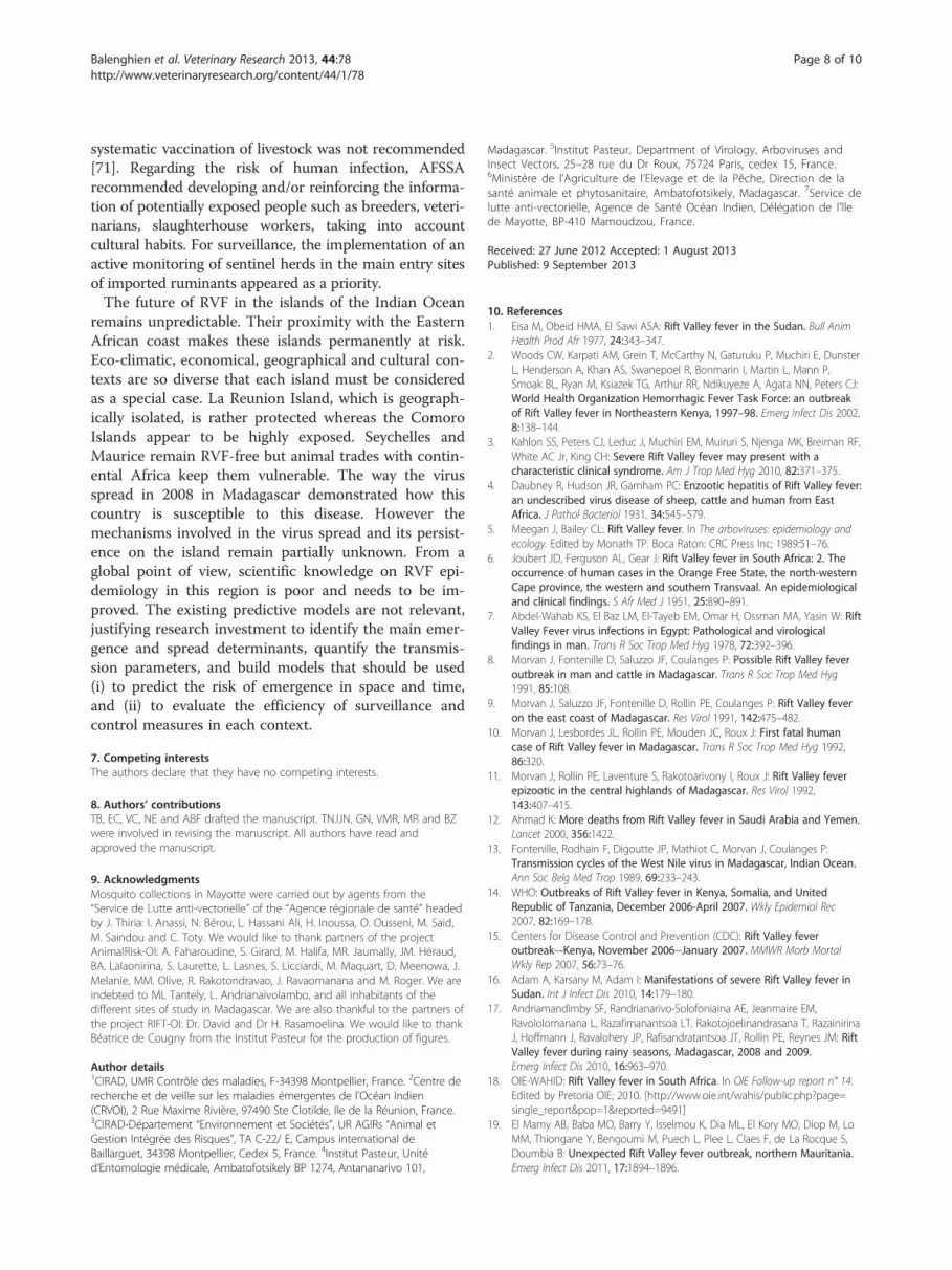
Figure 2 Cycle of Rift Valley fever. The virus can be maintained in an enzootic cycle involving Aedes mosquitoes which are able to transmit thevirus vertically to their offspring. Epizootic outbreaks are often linked with unusual rains or warm seasons, favoring the hatching of infected Aedeseggs that are then able to initiate the virus circulation. Subsequently, large numbers of secondary vectors belonging to the Culex genus could beinfected and induce the emergence of epidemic/epizootic outbreaks. Transmission to humans occurs through direct contact with high virusloads when aborted fetuses are manipulated.
Balenghien et al. Veterinary Research 2013, 44:78 Page 4 of 10http://www.veterinaryresearch.org/content/44/1/78
Balenghien et al. Veterinary Research 2013, 44:78 Page 5 of 10http://www.veterinaryresearch.org/content/44/1/78
(1) RVFV has been detected and isolated from threemosquito species, namely: An. coustani, An.squamosus/cydipis and Cx. antennatus, captured inthree districts of the province of Fianarantsoa, inthe Central Highlands [39]; (2) a risk factor studycarried out with cattle in 2009 reinforced thisassumption [30]; and lastly, (3) their zoophilic andopportunistic anthropophilic behavior in the absenceof household hosts allow assuming the implicationof these species in the RVF transmission inMadagascar [40]. Other RVF vector speciesidentified as a potential vector in Saudi Arabia (Cx.tritaeniorhynchus in 2000 [41], Egypt (Cx. pipiens in1977–78 [27,42], Kenya (Ae. durbanensis and An.pharoensis in 1981–84 [26]) have also been found inMadagascar. During these episodes of RVFcirculation in Madagascar, no studies on vectorcompetence were undertaken. Indeed, theimplication of none of these eighteen speciesinvolved in the transmission of RVFV inMadagascar has been corroborated. But accordingto their abundance, their trophic preference towardsanimals (especially, zebu) and their involvement inthe transmission of RVFV in other countries, An.coustani, An squamosus/cydippis, Cx. antennatus,Cx. univittatus, Cx. tritaeniorhynchus, Anpharoensis, Cx. pipiens, Cx. quinquefasciatus, Ae.durbanensis and Ae. (subgenus) Skusea could act aspotential vectors in Madagascar. The last twospecies are abundant only in the southern part ofthe island [43].
On the Comoros Islands, RVFV was isolated for thefirst time in 2007 consecutively to important epizooticsoccurring in East Africa in 2006 [32]. In 2008–2009, thepresence of RVFV was confirmed in domestic ruminants[33,34] and in some inhabitants of Mayotte [44]. The re-gion of the Indian Ocean presents a particular interestfor RVF emergence since human displacements and ani-mal exchanges with continental Africa are important. Inaddition, island ecosystems may present a particular bio-diversity (usually a low specific richness but a high levelof endemism due to adaptive radiation) favouring theoccurrence of particular virus/vector/vertebrate systems.Indeed, a recent inventory in Mayotte described 40 mos-quito species belonging to 9 genera.In Mayotte, an ongoing 2-year survey has been carried
out to describe diversity, population dynamics and host-feeding patterns of mosquitoes in five ruminant farms.Adult collections were performed every 4 weeks during3 consecutive nights using light and CO2-baited traps.Moreover, twenty-four-hour collections were made onone site using human-, sheep-, and goat-baited traps todescribe host-feeding patterns and circadian activities.
After a one-year survey, about 7900 mosquitoes belong-ing to 19 species and 4 genera (Culex, Eretmapodites,Aedes, Anopheles) were collected during 24–48 sessionsof adult collections per site and 6 sessions of host-baitedtrap collections. Culex quinquefasciatus and Eretmapoditesquinquevittatus were the most abundant species collectedon humans and sheep. Cx. quinquefasciatus and Er.quinquevittatus were shown to develop low to moderateinfection rates [45]. In host-baited traps, two anthro-pophilic mosquitoes Culex antennatus and Aedesalbopictus, and Culex decens, a non-mammalophilic mos-quito, were found abundantly. Other species were cap-tured at lower densities in CO2 or light-baited traps.Eretmapodites subsimplicipes and Aedes aegypti werefound in host-baited traps but not in CO2 or light-baitedtraps highlighting the need for complementary methods tobe included such as larval collections to better describemosquito diversity and abundance.
3. Virus-vector interactionsThree major RVFV lineages are described correspondingto Egyptian, West African and East African strains [46].Interestingly, the virus isolated in Madagascar in 1990belongs to the same lineage as the Kenyan isolate of 1997[47]. RVFV strains isolated from the Arabian Peninsulawere closely related to the Kenyan isolates [41,48,49]. Byanalyzing more carefully each of the three segments of thegenome, several strains resulted from reassortments be-tween strains from different genotypes [50]. Such aphenomenon is not unexpected for a virus with a seg-mented genome but this could be a strategy for selectingmore virulent viral strains.Most arboviruses are RNA viruses, although they use a
variety of strategies to ensure their replication and trans-mission. The feature that best distinguishes RNA is theirhigh mutation rate during replication ranging from 10-3-10-5 substitutions per nucleotide and per round of copying[51]. The main factor contributing to such high mutationrates is a lack of proof-reading repair activities that is asso-ciated with RNA replicases [52]. High mutation rates,short replication times and large population sizes, ensurethe existence of the quasispecies genome pool [53]. How-ever, arboviruses are relatively stable in nature suggestingthat the alternating host cycle (between vertebrate and in-vertebrate hosts) constrains viral evolution by a strongconservative sequence selection. Serial passages in a singlecell type (mammalian or insect cells) led to the loss ofRVF virulence. Large deletions were observed in the NSsgene. Thus, genetic material non-essential to viral replica-tion such as NSs is rapidly eliminated leading to the lossof virulence [54]. It is tempting to speculate that directtransmission among animals or from animals to humansmay favor the attenuation of NSs leading to the mainten-ance of avirulent RVFV strains. Virulence is likely to be

Balenghien et al. Veterinary Research 2013, 44:78 Page 6 of 10http://www.veterinaryresearch.org/content/44/1/78
restored when alternation is initiated again. Indeed, verte-brates are subject to acute infections with clearance of thevirus triggered by immune defense when vectors sustainpersistent viral replication becoming the site of geneticchanges such as reassortments. Such rearrangements maylead to restore virulence with the acquisition of a completeNSs gene in the course of virus replication [50]. This re-sult agrees with observations on the natural evolution ofRVF outbreaks whose intensity declines with increasingherd immunity and declining vector populations.
4. Diagnosis and surveillanceAs described in many African countries, the virus maycirculate at a very low level, silently, without or with fewclinical signs: this cryptic transmission is extremely diffi-cult to detect [55]. In Mayotte, in 2004, where a sero-prevalence of up to 22% (130 animals tested) wasestimated in small ruminants without any clinical symp-toms, RVF was diagnosed in a human case presentingbrain disorders. Susceptible animals were present indensities high enough to ensure virus circulation but toolow to induce waves of abortions and animal mortalities.To detect RVFV, several approaches are available: mo-
lecular detection (RT-PCR, real-time quantitative RT-PCR),virus antigen detection, and anti-RVF IgM or IgG anti-body detection [56]. The main drawback of medical la-boratories in the Indian Ocean region remains theweakness of diagnostic tools. Indeed, some diagnosis ofRVF can only be achieved in well-furnished laboratories,with a range of standardized diagnostic reagents involvingexperienced personal used to manipulating serial numbersof strains. Another issue is the difficulty in maintainingthe cold chain, particularly when distances are importantas in Madagascar, between the field and the laboratory;samples must be stored at −80 °C for further viral isola-tion. Thus, appropriate containers and ice packs fortransportation allowing keeping serum samples between−20 °C and +4 °C, should be provided to veterinary ser-vices in charge of sample collections. This highlights theneed of an early warning system to improve transfer be-tween the field and the laboratory.Depending on the epidemiological status of the coun-
try, surveillance has its main purpose. In La Reunion,Maurice and Seychelles which are RVF-free islands butat risk for introduction, early detection of the disease isa priority. In Mayotte, Comoros and Madagascar whereRVF is probably endemic, the priority is the detection ofany increased incidences preceding outbreaks. The epi-demiological surveillance was heterogeneous betweenthe different islands: some countries had their own net-work such as Madagascar, Mayotte or La Reunion Islandand some others received direct epidemiological informa-tion from the field (Comoros, Maurice, and Seychelles).Since then, a regional epidemio-surveillance network
called “AnimalRisk – OI” has been set up. All stakeholdersin the Indian Ocean involved in animal health are in-cluded in this network (veterinary public health and re-search institutes). A steering committee composed by thechief veterinary officers or their deputy is in charge of de-fining the diseases under surveillance, RVF mainly, anddata management. Once a month, the steering committeedisseminates collected epidemiological data through webconferences and later, a quarterly epidemiological report isproduced. In endemic countries, sentinel herds have beenfollowed up for one year and half are used to assess theseroconversion rate, to detect any clinical cases that couldbe attributed to RVF and finally, to confirm the circulationof RVFV despite very few notified symptoms [57]. Once thedisease is detected in humans, it is usually well-establishedin animal populations. Nevertheless in Madagascar andMayotte in 2008, livestock infections were detected aftersevere human cases were reported [32,58], highlightingthe lack of collaboration between human and animalhealth services.Given the frequent low specificity of clinical signs, syn-
dromic surveillance may be a useful and cost-effectivetool to help in controlling RVF. Indeed, this methodologyallows minimizing the main limitations of the passive sur-veillance systems: (i) reducing the time lag between theonset of the outbreak and the diagnosis, (ii) using anonspecific case definition to increase the sensitivity, and(iii) minimizing the under-reporting by the systematic andcontinuous screening of information at an earlier stage ofthe disease process. On the human side in Mayotte in2009, the surveillance of acute febrile syndromes inhumans allowed to detect 10 human RVF cases [59].The development of models based on climatic indica-
tors and vegetation index remains promising for risk-based surveillance implementation. These models areavailable for Eastern Africa and allow forecasting RVF out-breaks many weeks before. However, their predictive per-formance was shown to be low for Madagascar and SouthAfrica, and needs to be improved and adapted to specificMalagasy ecological and climatic conditions [60,61].
5. Prevention and controlSeveral control measures are described usually includingthe following: (i) control of livestock movements withrespect to trade and export; (ii) vector control with anemphasis on larvicides in vector breeding sites ratherthan aerial sprayings targeting adults or (iii) vaccinationof livestock.The role of livestock movements in RVF spread at
short or long distances -trade, transhumance- has alreadybeen shown [31,48,62]. As a matter of fact, a large live-stock trade exists or existed between countries in the In-dian Ocean and countries of Eastern Africa where thedisease is endemic, and phylogenetic studies strongly

Balenghien et al. Veterinary Research 2013, 44:78 Page 7 of 10http://www.veterinaryresearch.org/content/44/1/78
suggest that RVFV has been introduced in the IndianOcean by ruminant trade [29,63].Because RVF could be introduced through the import-
ation of domestic ruminants from infected countries, al-though this could only occur if importation took placewithin the short incubation period for the disease, adop-tion of the recommended guidelines of the OIE Inter-national Animal Health Code for such importationswould prevent this risk. When importing from infectedcountries, veterinarian authorities should require for do-mestic ruminants the presentation of a veterinary certifi-cate attesting the following:
1. Vaccinated animals (a) showed no clinical signs ofRVF on the day of shipment; (b) were vaccinatedusing a vaccine complying with the standardsdescribed in the OIE Manual not less than 21 daysand not more than 90 days prior to shipment; (c) werekept in a quarantine station in the country of originfor the 30 days prior to shipment and showed noclinical sign of RVF during that period.
2. Unvaccinated animals (a) showed no clinical sign ofRVF on the day of shipment; (b) were subjected tothe diagnostic tests for RVF with negative resultswithin 30 days before entry into quarantine; (c) werekept in a quarantine station in the country of originfor the 30 days prior to shipment and showed noclinical sign of RVF during that period; (d) weresubjected to the diagnostic tests for RVF withnegative results not less than 14 days after entry intoquarantine; (e) were protected from insect vectorsduring quarantine and transportation to the place ofshipment.
But to date, all these recommendations are often dis-regarded between Tanzania and Comoros. Indeed, since2002, importation of live animals in Comoros fromTanzania has been common, increasing the risk of intro-ducing continental pathogens or vectors as illustratedwith outbreaks of East Coast fever in 2003 and 2004 inGrande Comore [62]. Once RVFV is introduced, it isquite difficult to prevent its spread (i) between theneighboring islands because of no quarantine park andillegal trade and (ii) throughout countries, such asMadagascar, where the large-scale movement of cattle iscommon and often uncontrolled.The timing of the events associated with the 2007–
2008 outbreak in the Indian Ocean and molecular epi-demiological studies also support RVFV importationfrom East Africa, possibly even during the 2006–2007outbreak. Although most cases in the Indian Ocean(Comoros, Mayotte and Madagascar) were reportedfrom January through April 2008 [32,64], epidemiologicevidence has linked the 2008–2009 epizootics to that
occurring on the mainland a few years earlier. Especially,clinical signs of RVF (human or animal cases) were ob-served in early 2007 on these islands, and retrospectiveinvestigations revealed that RVFV had been circulatingin livestock at least since December 2007 [38]. Mauricemust also be considered as an island at-risk because ofregular importations of goats from Kenya or cows fromSouth Africa; even if there are severe quarantine mea-sures at Richelieu station, such as surveillance of anyclinical signs, random serological sampling, and quaran-tine duration from 15 days to 2 months, an emphasisshould be made on surveillance to detect any sign ofRVF disease.Beside the trade control, a safe vaccine is now available
for livestock [65-67], which is probably an efficient wayto protect both animals and humans interrupting thevirus transmission in endemic areas; even if it could beresponsible for the generation of recombinant viruseswhen used in ongoing infection areas [68]. However,socio-economic studies are needed to assess the sustain-ability and the acceptability of measures by breeders inthe Indian Ocean context. Other ways to control thespread of RVF involve control of the vector and protec-tion against their bites. Larvicide controls of mosquitobreeding sites are the most effective measure of vectorcontrol but are applicable only if breeding sites can beclearly identified and are limited in size and extent. Dur-ing periods of flooding, however, the number and extentof breeding sites are usually too extended for insecticidetreatments. Besides the financial cost [69], ecologicaland health issues associated with the extensive use of in-secticides should be considered. Lastly, since the majorroute of human infection is direct exposure to infectedanimals [69], information of people may also ensure ap-propriate slaughtering and consumption practices, thusdecreasing the risk of infection to humans.
6. Future of RVF on islands of the Indian OceanFollowing the 2007 Eastern Africa outbreak and the de-tection of indigenous human cases in Mayotte in 2008[32], a global qualitative assessment of the risk of outbreakfor Mayotte and La Reunion Island, was performed by theFrench Agency for Food Safety (AFSSA, now ANSES)[70]. The conclusions of this assessment were that the fol-lowing: (i) the risk of outbreak in these two islands wouldbe rather due to the introduction of a viremic animal com-ing either from Madagascar, Comoros or Eastern Africancoasts; (ii) this risk would be rather reduced for LaReunion Island located far from endemic areas and wherecontrols of imports are drastic; (iii) in Mayotte, illegal in-troductions of ruminants are frequent enough to justifymore controls of livestock imports; (iv) in Mayotte again,due to low animal densities, the risk for RVF to becomeendemic was estimated to be very low. Therefore, a
Balenghien et al. Veterinary Research 2013, 44:78 Page 8 of 10http://www.veterinaryresearch.org/content/44/1/78
systematic vaccination of livestock was not recommended[71]. Regarding the risk of human infection, AFSSArecommended developing and/or reinforcing the informa-tion of potentially exposed people such as breeders, veteri-narians, slaughterhouse workers, taking into accountcultural habits. For surveillance, the implementation of anactive monitoring of sentinel herds in the main entry sitesof imported ruminants appeared as a priority.The future of RVF in the islands of the Indian Ocean
remains unpredictable. Their proximity with the EasternAfrican coast makes these islands permanently at risk.Eco-climatic, economical, geographical and cultural con-texts are so diverse that each island must be consideredas a special case. La Reunion Island, which is geograph-ically isolated, is rather protected whereas the ComoroIslands appear to be highly exposed. Seychelles andMaurice remain RVF-free but animal trades with contin-ental Africa keep them vulnerable. The way the virusspread in 2008 in Madagascar demonstrated how thiscountry is susceptible to this disease. However themechanisms involved in the virus spread and its persist-ence on the island remain partially unknown. From aglobal point of view, scientific knowledge on RVF epi-demiology in this region is poor and needs to be im-proved. The existing predictive models are not relevant,justifying research investment to identify the main emer-gence and spread determinants, quantify the transmis-sion parameters, and build models that should be used(i) to predict the risk of emergence in space and time,and (ii) to evaluate the efficiency of surveillance andcontrol measures in each context.
7. Competing interestsThe authors declare that they have no competing interests.
8. Authors’ contributionsTB, EC, VC, NE and ABF drafted the manuscript. TNJJN, GN, VMR, MR and BZwere involved in revising the manuscript. All authors have read andapproved the manuscript.
9. AcknowledgmentsMosquito collections in Mayotte were carried out by agents from the“Service de Lutte anti-vectorielle” of the “Agence régionale de santé” headedby J. Thiria: I. Anassi, N. Bérou, L. Hassani Ali, H. Inoussa, O. Ousseni, M. Said,M. Saindou and C. Toty. We would like to thank partners of the projectAnimalRisk-OI: A. Faharoudine, S. Girard, M. Halifa, MR. Jaumally, JM. Héraud,BA. Lalaonirina, S. Laurette, L. Lasnes, S. Licciardi, M. Maquart, D. Meenowa, J.Melanie, MM. Olive, R. Rakotondravao, J. Ravaomanana and M. Roger. We areindebted to ML Tantely, L. Andrianaivolambo, and all inhabitants of thedifferent sites of study in Madagascar. We are also thankful to the partners ofthe project RIFT-OI: Dr. David and Dr H. Rasamoelina. We would like to thankBéatrice de Cougny from the Institut Pasteur for the production of figures.
Author details1CIRAD, UMR Contrôle des maladies, F-34398 Montpellier, France. 2Centre derecherche et de veille sur les maladies émergentes de l’Océan Indien(CRVOI), 2 Rue Maxime Rivière, 97490 Ste Clotilde, Ile de la Réunion, France.3CIRAD-Département “Environnement et Sociétés”, UR AGIRs “Animal etGestion Intégrée des Risques”, TA C-22/ E, Campus international deBaillarguet, 34398 Montpellier, Cedex 5, France. 4Institut Pasteur, Unitéd’Entomologie médicale, Ambatofotsikely BP 1274, Antananarivo 101,
Madagascar. 5Institut Pasteur, Department of Virology, Arboviruses andInsect Vectors, 25–28 rue du Dr Roux, 75724 Paris, cedex 15, France.6Ministère de l’Agriculture de l’Elevage et de la Pêche, Direction de lasanté animale et phytosanitaire, Ambatofotsikely, Madagascar. 7Service delutte anti-vectorielle, Agence de Santé Océan Indien, Délégation de l’îlede Mayotte, BP-410 Mamoudzou, France.
Received: 27 June 2012 Accepted: 1 August 2013Published: 9 September 2013
10. References1. Eisa M, Obeid HMA, El Sawi ASA: Rift Valley fever in the Sudan. Bull Anim
Health Prod Afr 1977, 24:343–347.2. Woods CW, Karpati AM, Grein T, McCarthy N, Gaturuku P, Muchiri E, Dunster
L, Henderson A, Khan AS, Swanepoel R, Bonmarin I, Martin L, Mann P,Smoak BL, Ryan M, Ksiazek TG, Arthur RR, Ndikuyeze A, Agata NN, Peters CJ:World Health Organization Hemorrhagic Fever Task Force: an outbreakof Rift Valley fever in Northeastern Kenya, 1997–98. Emerg Infect Dis 2002,8:138–144.
3. Kahlon SS, Peters CJ, Leduc J, Muchiri EM, Muiruri S, Njenga MK, Breiman RF,White AC Jr, King CH: Severe Rift Valley fever may present with acharacteristic clinical syndrome. Am J Trop Med Hyg 2010, 82:371–375.
4. Daubney R, Hudson JR, Garnham PC: Enzootic hepatitis of Rift Valley fever:an undescribed virus disease of sheep, cattle and human from EastAfrica. J Pathol Bacteriol 1931, 34:545–579.
5. Meegan J, Bailey CL: Rift Valley fever. In The arboviruses: epidemiology andecology. Edited by Monath TP. Boca Raton: CRC Press Inc; 1989:51–76.
6. Joubert JD, Ferguson AL, Gear J: Rift Valley fever in South Africa: 2. Theoccurrence of human cases in the Orange Free State, the north-westernCape province, the western and southern Transvaal. An epidemiologicaland clinical findings. S Afr Med J 1951, 25:890–891.
7. Abdel-Wahab KS, El Baz LM, El-Tayeb EM, Omar H, Ossman MA, Yasin W: RiftValley Fever virus infections in Egypt: Pathological and virologicalfindings in man. Trans R Soc Trop Med Hyg 1978, 72:392–396.
8. Morvan J, Fontenille D, Saluzzo JF, Coulanges P: Possible Rift Valley feveroutbreak in man and cattle in Madagascar. Trans R Soc Trop Med Hyg1991, 85:108.
9. Morvan J, Saluzzo JF, Fontenille D, Rollin PE, Coulanges P: Rift Valley feveron the east coast of Madagascar. Res Virol 1991, 142:475–482.
10. Morvan J, Lesbordes JL, Rollin PE, Mouden JC, Roux J: First fatal humancase of Rift Valley fever in Madagascar. Trans R Soc Trop Med Hyg 1992,86:320.
11. Morvan J, Rollin PE, Laventure S, Rakotoarivony I, Roux J: Rift Valley feverepizootic in the central highlands of Madagascar. Res Virol 1992,143:407–415.
12. Ahmad K: More deaths from Rift Valley fever in Saudi Arabia and Yemen.Lancet 2000, 356:1422.
13. Fontenille, Rodhain F, Digoutte JP, Mathiot C, Morvan J, Coulanges P:Transmission cycles of the West Nile virus in Madagascar, Indian Ocean.Ann Soc Belg Med Trop 1989, 69:233–243.
14. WHO: Outbreaks of Rift Valley fever in Kenya, Somalia, and UnitedRepublic of Tanzania, December 2006-April 2007. Wkly Epidemiol Rec2007, 82:169–178.
15. Centers for Disease Control and Prevention (CDC): Rift Valley feveroutbreak-–Kenya, November 2006--January 2007. MMWR Morb MortalWkly Rep 2007, 56:73–76.
16. Adam A, Karsany M, Adam I: Manifestations of severe Rift Valley fever inSudan. Int J Infect Dis 2010, 14:179–180.
17. Andriamandimby SF, Randrianarivo-Solofoniaina AE, Jeanmaire EM,Ravololomanana L, Razafimanantsoa LT, Rakotojoelinandrasana T, RazainirinaJ, Hoffmann J, Ravalohery JP, Rafisandratantsoa JT, Rollin PE, Reynes JM: RiftValley fever during rainy seasons, Madagascar, 2008 and 2009.Emerg Infect Dis 2010, 16:963–970.
18. OIE-WAHID: Rift Valley fever in South Africa. In OIE Follow-up report n° 14.Edited by Pretoria OIE; 2010. [http://www.oie.int/wahis/public.php?page=single_report&pop=1&reported=9491]
19. El Mamy AB, Baba MO, Barry Y, Isselmou K, Dia ML, El Kory MO, Diop M, LoMM, Thiongane Y, Bengoumi M, Puech L, Plee L, Claes F, de La Rocque S,Doumbia B: Unexpected Rift Valley fever outbreak, northern Mauritania.Emerg Infect Dis 2011, 17:1894–1896.

Balenghien et al. Veterinary Research 2013, 44:78 Page 9 of 10http://www.veterinaryresearch.org/content/44/1/78
20. OIE-WAHID: Rift Valley fever, Bostwana; 2010. [http://www.oie.int/wahis/public.php?page=singlereport$pop=1$ reported=9947]
21. OIE-WAHID: Rift Valley fever, Namibia; 2010. [http://web.oie.int/wahis/public.php?page=single_report&pop=1&reported=925811]
22. Dohm DJ, Rowton ED, Lawyer PG, O’Guinn M, Turell MJ: Laboratorytransmission of Rift Valley fever virus by Phlebotomus duboscqi,Phlebotomus papatasi, Phlebotomus sergenti, and Sergentomyia schwetzi(Diptera: Psychodidae). J Med Entomol 2000, 37:435–438.
23. Hoch AL, Gargan TP 2nd, Bailey CL: Mechanical transmission of Rift Valleyfever virus by hematophagous Diptera. Am J Trop Med Hyg 1985, 34:188–193.
24. LaBeaud AD, Kazura JW, King CH: Advances in Rift Valley fever research:insights for disease prevention. Curr Opin Infect Dis 2010, 23:403–408.
25. Seufi AM, Galal FH: Role of Culex and Anopheles mosquito species aspotential vectors of rift valley fever virus in Sudan outbreak, 2007.BMC Infect Dis 2010, 10:65.
26. Linthicum KJ, Davies FG, Kairo A, Bailey CL: Rift Valley fever virus (familyBunyaviridae, genus Phlebovirus). Isolations from Diptera collectedduring an inter-epizootic period in Kenya. J Hyg (Lond) 1985, 95:197–209.
27. Hoogstraal H, Meegan JM, Khalil GM, Adham FK: The Rift Valley feverepizootic in Egypt 1977–78. 2. Ecological and entomological studies.Trans R Soc Trop Med Hyg 1979, 73:624–629.
28. Fontenille D: Transmission cycles of arboviruses in Madagascar. Arch InstPasteur Madagascar 1989, 55:7–317 (in French).
29. Carroll SA, Reynes JM, Khristova ML, Andriamandimby SF, Rollin PE, NicholST: Genetic evidence for Rift Valley fever outbreaks in Madagascarresulting from virus introductions from the East African mainland ratherthan enzootic maintenance. J Virol 2011, 85:6162–6167.
30. Chevalier V, Rakotondrafara T, Jourdan M, Heraud JM, Andriamanivo HR,Durand B, Ravaomanana J, Rollin PE, Rakotondravao R: An unexpectedrecurrent transmission of Rift Valley fever virus in cattle in a temperateand mountainous area of Madagascar. PLoS Negl Trop Dis 2011, 5:e1423.
31. Nicolas G, Durand B, Durboz R, Rakotondravao R, Chevalier V: Descriptionand analysis of the cattle trade network in the Madagascar highlands:potential role in the diffusion of Rift Valley fever virus. Acta Trop 2013,126:19–27.
32. Sissoko D, Giry C, Gabrie P, Tarantola A, Pettinelli F, Collet L, D’Ortenzio E,Renault P, Pierre V: Rift Valley Fever, Mayotte, 2007–2008. Emerg Infect Dis2009, 15:568–570.
33. Roger M, Girard S, Faharoudine A, Halifa M, Bouloy M, Cetre-Sossah C,Cardinale E: Rift valley fever in ruminants, Republic of Comoros, 2009.Emerg Infect Dis 2011, 17:1319–1320.
34. Cêtre-Sossah C, Pédarrieu A, Guis H, Defernez C, Bouloy M, Favre J, Girard S,Cardinale E, Albina E: Prevalence of Rift Valley Fever among Ruminants,Mayotte. Emerg Infect Dis 2012, 18:972–975.
35. Clerc Y, Coulanges P: Rapport du Laboratoire des arbovirus 1980. Arch InstPasteur Madag 1981, 49:65–69 (in French).
36. Morvan J, Rakoto-Andrianarivelo M, Laventure S: Fièvre de la vallée du Rift.Rapport annuel d’activités: Institut Pasteur de Madagascar; 1993:36–38.
37. Zeller H, Duchemin JB, Rakoto-Andrianarivelo J: Fièvre de la Vallée du Rift.Institut Pasteur de Madagascar: Rapport annuel d’activités; 1998:62–64.
38. Jeanmaire EM, Rabenarivahiny R, Biarmann M, Rabibisoa L, Ravaomanana F,Randriamparany T, Andriamandimby SF, Diaw CS, Fenozara P, de La RocqueS, Reynes JM: Prevalence of Rift Valley Fever infection in ruminants inMadagascar after the 2008 outbreak. Vector Borne Zoonotic Dis 2011,11:395–402.
39. Ratovonjato J, Olive MM, Tantely LM, Andrianaivolambo L, Tata E, RazainirinaJ, Jeanmaire E, Reynes JM, Elissa N: Detection, Isolation, and Geneticcharacterization of Rift Valley Fever Virus from Anopheles (Anopheles)coustani, Anopheles (Cellia) squamosus, and Culex (Culex) antennatus ofthe Haute Matsiatra Region, Madagascar. Vector Borne Zoonotic Dis 2011,11:753–759.
40. Tantely ML, Rakotoniaina J-C, Tata E, Andrianaivolambo L, Razafindrasata F,Fontenille D, Elissa N: Biology of mosquitoes that are potential vectors ofRift Valley Fever virus in different biotopes of the Central Highlands ofMadagascar. J Med Entomol 2013, 50:603–610.
41. Jup PG, Kemp A, Grobbelaar A, Leman P, Burt FJ, Alahmed AM, Al Mujalli D,Al Khamees M, Swanepoel R: The 2000 epidemic of Rift Valley Fever inSaudi Arabia: mosquito vector studies. Med Vet Entomol 2002, 16:245–252.
42. Meegan JM: The Rift Valley fever epizootic in Egypt 1977–78 1.Description of the epizootic and virological studies. Trans R Soc Trop MedHyg 1979, 73:618–629.
43. Jean Jose Nepomichene TN, Tantely ML, Andrianaivolambo L, Tata E,Ramihangihajason T, Rakotoniaina JC, Ratovonjato J, Elissa N: Fièvre de laVallée du Rift, Investigation Entomologiques 2008–2011. Antananarivo,Madagascar (in French): Congrès conjoint parasito-vetérinaire; 2011.
44. Pépin M: Rift Valley fever. Med Mal Infect 2011, 41:322–329 (in French).45. Mcintosh BM, Jupp PG: Epidemiological aspects of Rift Valley fever in
South Africa with reference to vectors. Contrib Epidemiol Biostat 1981,3:92–99.
46. Sall AA, De A, Zanotto PM, Zeller HG, Digoutte JP, Thiongane Y, Bouloy M:Variability of the NS(S) protein among Rift Valley fever virus isolates.J Gen Virol 1997, 78:2853–2858.
47. Sall AA, De A, Zanotto PM, Vialat P, Sene OK, Bouloy M: Origin of 1997–98Rift Valley fever outbreak in East Africa. Lancet 1998, 352:1596–1597.
48. Shoemaker T, Boulianne C, Vincent MJ, Pezzanite L, Al-Qahtani MM, Al-Mazrou Y, Khan AS, Rollin PE, Swanepoel R, Ksiazek TG, Nichol ST: Geneticanalysis of viruses associated with emergence of Rift Valley fever inSaudi Arabia and Yemen, 2000–2001. Emerg Infect Dis 2002, 8:1415–1420.
49. Miller BR, Godsey MS, Crabtree MB, Savage HM, Al-Mazrao Y, Al-Jeffri MH,Abdoon AM, Al-Seghayer SM, Al-Shahrani AM, Ksiazek TG: Isolation andgenetic characterization of Rift Valley fever virus from Aedes vexansarabiensis, Kingdom of Saudi Arabia. Emerg Infect Dis 2002, 8:1492–1494.
50. Sall AA, Zanotto PM, Sene OK, Zeller HG, Digoutte JP, Thiongane Y, BouloyM: Genetic reassortment of Rift Valley fever virus in nature. J Virol 1999,73:8196–8200.
51. Drake JW: Rates of spontaneous mutations among RNA viruses. Proc NatlAcad Sci U S A 1993, 90:4171–4175.
52. Steinhauer DA, Domingo E, Holland JJ: Lack of evidence for proofreadingmechanisms associated with an RNA virus polymerase. Gene 1992,122:281–288.
53. Domingo E, Holland JJ, Biebricher C, Eigen M: Quasi-species: the conceptand the word. In Molecular evolution of viruses. Edited by Gibbs A, CalisherC, Garcia-Arenal F. Cambridge: University Press; 1995:171–180.
54. Moutailler S, Roche B, Thiberge JM, Caro V, Rougeon F, Failloux AB: Hostalternation is necessary to maintain the genome stability of rift valleyfever virus. PLoS Negl Trop Dis 2011, 5:e1156.
55. Food and Agriculture Organization of the United Nations: In Preparation ofRift Valley Fever Contingency Plans. Edited by William A, Geering F, Davies G,Martin V. Rome: Food and Agriculture Organization of the United Nations;2003.
56. Pepin M, Bouloy M, Bird BH, Kemp A, Paweska J: Rift Valley fever virus(Bunyaviridae: Phlebovirus): an update on pathogenesis, molecularepidemiology, vectors, diagnostics and prevention. Vet Res 2010, 41:61.
57. Cardinale E, Roger M, Elissa N, Faharoudine A, Girard S, Halifa M, JaumallyMR, Héraud JM, Lalaonirina BA, Laurette S, Lasnes L, Licciardi S, Maquart M,Melanie J, Meenowa D, Olive MM, Rakotoharinome M, Rakotondravao R,Ravaomanana J: Le réseau régional AnimalRisk dans l’Océan Indien.Bulletin épidémiologique de l’Anses 2011, 43:8–12. Spécial DOM-TOM.
58. Rakotoarivelo RA, Andrianasolo R, Razafimahefa SH, RandremandrantoRazafimbelo NS, Randria MJ: Severe presentations of Rift Valley Fever inMadagascar. Med Mal Infect 2011, 41:318–321.
59. Lernout T, Zumbo B, Girard S, Aubert L, Toty C, Coroller F, Balenghien T,Lajoinie G, Filleul L: Etat des lieux sur la fièvre de la vallée du Rift àMayotte. Bulletin de Veille Sanitaire 2011, 12:9–10.
60. Anyamba A, Chretien JP, Small J, Tucker CJ, Formenty PB, Richardson JH,Britch SC, Schnabel DC, Erickson RL, Linthicum KJ: Prediction of a RiftValley fever outbreak. Proc Natl Acad Sci U S A 2009, 106:955–959.
61. Anyamba A, Linthicum KJ, Small J, Britch SC, Pak E, de La Rocque S,Formenty P, Hightower AW, Breiman RF, Chretien JP, Tucker CJ, Schnabel D,Sang R, Haagsma K, Latham M, Lewandowski HB, Magdi SO, Mohamed MA,Nguku PM, Reynes JM, Swanepoel R: Prediction, assessment of the RiftValley fever activity in East and Southern Africa 2006–2008 and possiblevector control strategies. Am J Trop Med Hyg 2010, 83:43–51.
62. Cêtre-Sossah C, Albina E: Rift Valley Fever: veterinary aspects and impactfor human health. Med Trop 2009, 69:358–361.
63. Abd El-Rahim IHA, El-Hakim UA, Hussein M: An epizootic of Rift Valleyfever in Egypt in 1997. Rev Sci Tech Off Int Epi 1999, 18:741–748.
64. Bird BH, Ksiazek TG, Nichol ST, MacLachlan NJ: Rift Valley fever virus.J Am Vet Med Assoc 2009, 234:883–893.
65. Dungu B, Louw I, Lubisi A, Hunter P, von Teichman B, Bouloy M: Evaluationof the efficacy and safety of the Rift Valley Fever Clone 13 vaccine insheep. Vaccine 2010, 28:4581–4587.

Balenghien et al. Veterinary Research 2013, 44:78 Page 10 of 10http://www.veterinaryresearch.org/content/44/1/78
66. Muller R, Saluzzo JF, Lopez N, Dreier T, Turell M, Smith J, Bouloy M:Characterization of clone 13, a naturally attenuated avirulent isolate ofRift Valley fever virus, which is altered in the small segment. Am J TropMed Hyg 1995, 53:405–411.
67. von Teichman B, Engelbrecht A, Zulu G, Dungu B, Pardini A, Bouloy M:Safety and efficacy of Rift Valley fever Smithburn and Clone 13 vaccinesin calves. Vaccine 2011, 29:5771–5777.
68. Grobbelaar AA, Weyer J, Leman PA, Kemp A, Paweska JT, Swanepoel R:Molecular epidemiology of Rift Valley fever virus. Emerg Infect Dis 2011,17:2270–2276.
69. Anyangu A, Gould L, Sharif S, Nguku P, Omolo J, Mutonga D, Rao CY,Lederman ER, Schnabel D, Paweska JT, Katz M, Hightower A, Njenga MK,Feikin DR, Breiman RF: Risk factors for severe Rift Valley fever infection inKenya, 2007. Am J Trop Med Hyg 2010, 83:14–21.
70. AFSSA: Avis de l’Agence Française de Sécurité Sanitaire des Aliments sur lerisque de propagation de la fièvre de la vallée du Rift dans un département etune collectivité départementale française de l’Océan Indien (La Réunion etMayotte). Maisons-Alfort: AFSSA; 2008:156.
71. AFSSA: Avis de l’Agence Française de Sécurité Sanitaire des Aliments sur lerisque de propagation et de pérennisation de la FVR à Mayotte. Maisons-alfort:AFSSA; 2008:12. n°2008-SA-0074.
doi:10.1186/1297-9716-44-78Cite this article as: Balenghien et al.: Towards a better understanding ofRift Valley fever epidemiology in the south-west of the Indian Ocean.Veterinary Research 2013 44:78.
Submit your next manuscript to BioMed Centraland take full advantage of:
• Convenient online submission
• Thorough peer review
• No space constraints or color figure charges
• Immediate publication on acceptance
• Inclusion in PubMed, CAS, Scopus and Google Scholar
• Research which is freely available for redistribution
Submit your manuscript at www.biomedcentral.com/submit