Embed Size (px)
Citation preview

GENERAL AND COMPARATIVE ENDOCRINOLOGY 27, 71-83 (1975)
Stimulation of In Vitro Steroid Production in Turtle Ovarian Tissue by Reptilian, Amphibian,
and Mammalian Gonadotropins
DAVID P.CREWSAND PAULLICHT
Department of Zoology, University of California, Berkeley, California 94720
Accepted May 8, 1975
Purified gonadotropins (FSH and LH) from a reptile, the snapping turtle, (Chelydra ser- pentina), sheep, and bullfrog (Rana catesbeiana) were used to investigate the hormonal con- trol of ovarian steroidogenesis in vitro in four species of turtles (Kinosternon, Sternotherus, Gopherus, and Chelydra). Follicle wall membrane (FW) from large follicles and stromal connective tissue (stroma) interspersed with small follicles were incubated separately for 4-16 hr, and steroids in extracted medium measured by radioimmunoassay after chroma- tography. Both FSH and LH from the turtle, mammal, and amphibian stimulated proges- terone production in the four species of turtles. Turtle and ovine hormones also stimulated estradiol secretion. Experiments with LH antisera indicated that the steroidogenic actions of turtle and ovine FSH are intrinsic to the molecule.
Although the reptilian ovary is ap- parently under pituitary control and pro- duces many of the same major steroids as in the mammal (Licht, 1974), it is not clear whether steroidogenesis is regulated by the same gonadotropins in both Classes. Both progesterone and estradiol have been iso- lated and identified in the ovary (Lupo di Prisco et al., 1968; Ozon, 1972; Chan and Callard, 1974) and peripheral plasma (Cal- lard et al., 1972a, c; Chan et al., 1973) of reptiles, and several studies have demon- strated the capacity of reptilian ovaries to synthesize progesterone in vitro (Callard and Leathem, 1964, 1965; Callard, 1966; Klicka and Mahmoud, 1972, 1973; Co- lombo et al., 1974; Chan and Callard, 1974). These steroids have been shown to be important in the control of a variety of morphological, physiological and behav- ioral processes (Cooper and Ferguson, 1972a, b; Kimbel and Erpino, 1971; Me- dica et al., 1973; Callard et al., 1972a-c; Chester-Jones et al., 1972; Ferguson, 1966). Patterns of progesterone produc- tion, in particular, have been related to
both the mode of reproduction (Bragdon et al., 1954; Callard and Leathem, 1965; Cal- lard, 1966) and the stage of the reproduc- tive cycle (Callard and Leathem, 1965; Callard et al., 1972~). There is also good evidence for a gonadal steroid feedback system involving the hypothalamus and hypophysis in reptiles that resembles that observed in mammals (Lisk, 1967; Callard et al., 1972a-c; Callard and Doolittle, 1973). However, the relationship between ovarian steroidogenesis and the different hormones produced by the reptilian pitu- itary has not been established.
There is still some controversy re- garding the respective roles of follicle stim- ulating hormone (FSH) and luteinizing hormone (LH) in the control of ovarian activity in mammals, but it is generally ac- cepted that FSH is primarily responsible for follicular growth and development, whereas LH regulates ovulation and ovarian steroid secretion (see review by Schwartz, 1974; Lidner et al., 1974). This apparent specificity for LH in certain func- tions of the mammalian ovary is of special
71 Cqyrieht @ 1975 by Academic Press. Inc. All rights of repmduction in any form reserved.

72 CREWS AND LICHT
interest for comparative studies on rep- tilian reproductive endocrinology since a similar dependence on LH in reptiles has been difficult of demonstrate. Fractiona- tion studies with the snapping turtle (Chelydra serpentina) demonstrated the existence of two distinct pituitary gonado- tropins that appear homologous to the FSH and LH of mammals (Licht and Pap- koff, 1974a, b; Licht et al., 1974), but it does not follow that these hormones play the same role in the two Classes. For ex- ample, injections of purified mammalian FSH alone in female lizards stimulates ovarian and oviducal growth, ovulation, and presumably ovarian steroid secretion, while the mammalian LH is relatively in- active (Licht, 1970; Licht and Hartree, 1971; Licht and Tsui, 1975; Jones, 1969; Eyeson, 1971; Callard et al., 1972~). Use of specific antisera confirmed that these results with ovine FSH reflected intrinsic activity and not contamination with LH (Licht and Tsui, 1975). Similar results were obtained in female lizards using the purified snapping turtle FSH and LH (Licht and Crews, 1975). On the other hand, Chan and Callard (1974) recently reported that mammalian LH, but not FSH, stimulated progesterone production by turtle ovarian tissue in vitro. Thus, the possibility of important species differences between chelonians (turtles) and squa- mates (snakes and lizards) must be consid- ered; in particular, squamates may be unique among reptiles in lacking a depen- dence on LH, since this activity appears to be very low or lacking in their pituitary (Licht, 1974; Licht et al., 1974). It is clearly essential to examine the actions of reptilian hormones in homologous species or at least members of the same order.
The present investigation examines the actions of the two hormones from the snapping turtle on ovarian steroid secre- tion in four genera of turtles including the homologous species. For comparative pur- poses, we also examined purified gonado-
tropins from a mammal (ovine) and an an- uran amphibian (bullfrog). These studies focused on the in vitro production of progesterone and estrogen using several components of the ovarian tissue from in- tact and hypophysectomized animals.
MATERIALS AND METHODS
Animals Sexually mature musk turtles, Sternotherus odor-
atus, and mud turtles, Kinosternon subrubrum (Family Kinosternidae), were collected in Southern Louisiana in autumn. Turtles were maintained in out- door ponds in Berkeley on a diet of chopped fish and beef heart. Twenty-four hours prior to the experi- ment, animals were brought into the laboratory and kept at room temperature (21-23”). Except in Expt 3, all animals were found to have a number of large yolked follicles and oviducal eggs with corresponding corpora lutea. Since this species lays multiple clutches each season, these remaining enlarged fol- licles presumably represent the next to be ovulated (Mahmoud and Klicka, 1972).
Large, sexually mature gopher tortoises, Gopherus agassizi, were provided by the California Fish and Wildlife Service in September 1974. Tortoises were kept at 28” and maintained on lettuce and apples until sacrificed. These females were found to have only medium-sized follicles.
Two tests were performed with snapping turtles, Chelydra serpentina, obtained from Wisconsin; this was the source for the snapping turtle pituitary glands used for gonadotropin fractionation. The first female, received in December, had a series of 19 uniformly enlarged follicles (13-14 mm). The second, in March, had a series of 25 mm follicles.
Experimental Procedures Animals were decapitated and the ovarian tissue
removed immediately and placed in incubation medium. Ovaries were then separated into two com- ponents for incubation: (1) The encapsulating mem- brane from large (>7.0 mm) preovulatory follicles were obtained by expressing the yolk; these consisted of theta and granulosa cells and zona pellucida and were designated follicle wall (FW). (2) The remaining tissue consisting of very small (< 1.0 mm) yolking follicles, previtellogenic follicles, connective epithe- lium, and possibly germinal bed was designated “stroma.” Except in the experiment with Chelydra, the small follicles in the stroma were left intact; with the Chelydra, the yolk was also expressed from these before incubation. Each type of tissue was minced and portions from each follicle or stroma were distrib-

REPTILIAN OVARIAN STEROIDOGENESIS 73
uted evenly among the various incubation tubes. Tissues were rinsed in several changes of medium (without glucose) at 20” for at least 30 min before in- cubation.
Incubations were carried out in Krebs-Ringer bi- carbonate buffer (pH 7.2-7.4) containing 2 mg/ml glucose (except in the last experiment), with shaking under an atmosphere of 95% O,-5% CO, for 4-16 hr. Bovine serum albumin (1%) was used in Expt 3. The different incubation times were tested in an effort to improve the yield of estrogen which tended to be low. Special attention was given to incubation tem- perature since this variable may have pronounced ef- fects on gonadal responses to gonadotropins in rep- tiles (e.g., Licht, 1972b). In most cases, 28” was employed since this appears to be within the range that is easily tolerated or preferred by the various species (Brattstrom, 1965; Licht, 1972a). Gopherus tissues were also studied at 36” (Expt 9), since this desert species may also be active at this level (Bratt- Strom, 1965; McGinnis and Voigt, 1971). Incuba- tions were carried out in 15 x 85 mm disposalbe cul- ture tubes with 0.5 or 1.0 ml of medium.
At the end of each incubation, the tissue was re- moved, weighed, dried in a vacuum oven at 56”. defatted, dissolved in 1 N NaOH, and protein deter- mined calorimetrically by the Biuret method. Incuba- tion medium was either extracted immediately (see below) or stored at -20” for later analysis. In one experiment (Expt lo), the FW was homogenized in distilled water and extracted for determination of steroid content immediately following incubation. To facilitate comparison with other determinations based on protein content of tissue, the protein content of the homogenized tissue was estimated as 5% of wet weight- the average value obtained for all other experiments.
Hormones
Our studies were based largely on a series of puri- fied gonadotropins obtained from the snapping turtle, Chelydra serpentina, and bioassayed as described in Licht and Papkoff (1974a). Three such fractions were employed: the partially purified FSH (T-38-A) had a relative potency of 0.5 X NIH-FSH-Sl in Anolis lizard assay. Although much less potent than the best turtle FSH (see below), T-38-A is highly purified with regard to other hormone activities, especially LH (< 0.004 x NIH-LH-Sl in the anuran ovulation test which is equivalent to < 0.2% contamination relative to the turtle LH preparation). A more highly purified FSH (T-48-B and T-48-C) was obtained by gel filtra- tion on Sephadex G-100; these were approximately 25 X NIH-FSH-Sl with nondetectable LH activity. Tests with turtle LH utilized a highly purified fraction (T-28B,37C) which was 2.1 x NIH-FSH-Sl in an- uran ovulation test. In Anolis assay, the activity of
this turtle LH was 0.1 x NIH-FSH-Sl, but it is not clear whether this reflects contamination with FSH or an intrinsic activity of the LH (see Licht and Pap- koff, 1973).
Mammalian (ovine) preparations (NIH-FSH-S9 and SlO and NIH-LH-Sl8) were generously supplied by the Hormone Distribution Officer, National Insti- tute of Arthritis and Metabolic Diseases, National In- stitutes of Health, Bethesda, Maryland. Gonado- tropins from the bullfrog (Rana catesbeiana) were prepared as described in Licht and Papkoff (1974~) with additional purification of LH on Sephadex G-100. The LH(C-53-C) is considered highly puri- fied: potency by anuran ovulation test, 0.33 x NIH- LH-Sl ; and activity in Anolis assay, 0.1 X NIH- FSH-S 1. The bullfrog FSH(C-6 1-B) was an interme- diate fraction, but it has relatively high activity in Anolis (2.5 X NIH-FSH-Sl) and low LH contami- nation (< 0.001 X NIH-LH-SI or < 0.6% of the ho- mologous LH).
In all assays, controls consisted of samples of tissue incubated in medium without hormone. Each hormone was generally tested at several doses. Con- trols and each dose of hormone were tested with two to four replicates. The small quantity of tissue in kinosternids and Gopherus usually allowed only two to three replicates.
Immunologic Purification of FSH
An antiserum raised in monkeys against ovine LH (prepared by Ellen Daniels, Department of Physiol- ogy and Anatomy, University of California, Berke- ley) was used in some experiments to test for pos- sible contributions of cross-contamination between FSH and LH. Previous biological tests demonstrated the ability of this antiserum to neutralize the activity of both mammalian and Chelydra LH (Licht et a/., 1974). Treatment of gonadotropins with antiserum and independent verification of LH neutralization by anuran ovulation test were as described previously (Licht et al., 1974).
Radioimmunoassay
Steroids (estradiol and progesterone) were mea- sured by radioimmunoassay (RIA) according to the method of Mikhail et al. (1970) and Wu and Lundy (197 1) with the following modifications. Measured aliquots of tissue homogenates or incubation medium (0.5-0.9 ml) together with 0.1 ml each of tritiated progesterone (2700 cpm of 96 p Ci/mM Proges- terone [ 1 ,2,6,7-3H] from New England Nuclear) and estradiol-17P (2300 cpm of 99 p Ci/mM Es- tradio1[2,3,6,7-3H] from New England Nuclear) for internal standards were extracted with 6 ml dichloro- methane (Mallinckrodt). Tracers were purified by chromatography on Sephadex LH-20 shortly before

74 CREWS AND LICHT
use. After high speed centrifugation (14,6OOg), the organic phase was removed and dried under nitrogen. The dried extract was then applied to Sephadex LH-20 (Pharmacia) microcolumns (0.5 x 20 cm) and eluted using 85 : 15 benzene : methanol (Spectrograde) as solvent. Progesterone (fractions 3 and 4 ml) and estradiol (fractions 7-9 ml) were isolated and dried under nitrogen. Samples were then redissolved in 0.75 ml of 0.1% gelatin phosphate assay buffer (pH 7.4) and divided as follows: 0.1 ml for determination of recovery, and two aliquots-0.1 and 0.5 ml-for RIA. All values were corrected for recovery which ranged from 70 to 90%. Testing of medium samples at two dilutions verified parallelism with the stan- dards.
Aliquots of the progesterone fraction were in- cubated with progesterone-specific antiserum (S-85#5 obtained from Cl Abraham) and 10,000 cpm tritiated progesterone for 12-16 hr. Lyophilized an- tiserum was dissolved in 2 ml and subsequently di- luted 1 : 12,500. Aliquots of the estradiol fraction were incubated with an estradiol antiserum (obtained from Drs. T. M. Nett and V. L. Estergreen) and 10,000 cpm of tritiated estradiol-17/3 for 18 hr. The preparation and characterization of this antiserum are described by Nett et al. (1973). The greatest cross- reaction with other estrogens with respect to es- tradiol-17P (100%) was 62% for estrone (however, this was separated chromatographically from es- tradiol in the present study). Progesterone and other nonestrogenic steroids do not cross-react with this antiserum. The antiserum was diluted 1 :50,000 for use.
Bound steroid was separated from free steroid with Dextran-coated charcoal (0.6% Dextran-T; 6% Norit-A). Following centrifugation, the supematant was suspended in 10 ml TritonlToluene scintillation cocktail (1 liter Triton X-100, 2 liter Toluene, 16 g NEN Omnifluor) and counted in a Beckman LS-100 C Liquid scintillation counter at 2% error.
Standard curves for progesterone ranged from lo- 1000 pg and 5-100 pg for estradiol; the minimum detectable dose on the standard curves were 10 and 5 pg. respectively. However, blank values for incuba- tion medium and tissue homogenates using these assay procedures were 25-50 pg for progesterone and 10 pg for estradiol, and values below these levels were considered nondetectable.
Incubations
Experiment 1. Comparison of ovaries from intact and hypophysectomized Stemotherus. In a prelimi- nary experiment to determine whether hypophysec- tomy affects the endogenous activity and respon- siveness of ovarian tissues to reptilian gonadotropin, tissues from two intact Sternotherus were compared
with those from two hypophysectomized 48 hr prior to incubation of ovaries. Tissues were incubated with turtle FSH or LH for 8 hr. Hypophysectomy was performed as described in Licht (1972a).
Experiments 2 and 3. Steroid production by ovarian stroma and FW from hypophysectomized Kinosternon. Two Kinosternon were hypophysec- tomized 3 days prior to incubation. Pooled tissues were incubated for 7.5 hr with varying amounts of partially purified turtle FSH or LH alone or in combi- nation.
The third test was conducted with stroma from two Kinosternon that had been hypophysectomized 24 hr before sacrifice. Tissues were pooled and incubated with either partially purified turtle FSH, purified turtle FSH, or turtle LH for 5 hr.
Experiments 4-8. Influence of turtle and ovine gon- adotropins on ovarian tissue from intact Sternotherus and Kinosternon. In Expt 4, turtle FSH and LH were added alone or in combination to pooled stroma and FW from six intact Sternotherus. To determine if longer incubation would result in increased steroid production, expecially estradiol, pooled ovarian tissue from two Kinosternon was incubated for 16 hr with turtle hormones. Specificity for mammalian gonado- tropins was tested with two Kinosternon using the NIH preparations.
Antiserum against LH (LH A/S) was used in con- junction with both ovine and turtle gonadotropins in Expts 8 and 9, respectively, to ascertain whether the actions of FSHs were due to contamination with LH. Normal sheep serum was added to controls. Tissues were incubated for 5 hr.
Experiments 9-10. InJIuence of turtle gonado- tropins on Gopherus ovaries. In the first experiment with Gopherus (Expt 9), FW tissue was incubated at 28” for 5 hr with three doses of either partially puri- fied turtle FSH or the LH. In Expt 10, turtle LH was compared with the highly purified turtle FSH at 36” for 4 hr. Antiserum was also used in conjunction with LH. Both medium and tissue were assayed in this experiment.
Experiment Il. Effects of turtle gonadotropins in the homologous species. Using tissues from Chelydra serpentina, all three Chelydra gonadotropin fractions were tested on FW tissue at three doses with four replicates at each dose. The LH and partially purified FSH were also tested in duplicate or triplicate with stroma. All yolk from small follicles in the stromal tissue was removed prior to incubations. Incubations were performed at 28” for 5 hr.
Experiment 12. Effect of amphibian hormone on Chelydra ovaries. This test examined the effects of a single dose (2 pg) of FSH (C-61-B) and LH (C-53-C) from the bullfrog on progesterone production in FW from Chelydra. Incubation conditions were the same asinExpt 11.

REPTILIAN OVARIAN STEROIDOGENESIS 75
RESULTS
Both stromal and FW tissue from ovaries of all three turtle species studied had measurable levels of steroid produc- tion in vitro in the absence of gonado- tropin; in all cases, steroid production was enhanced by the addition of either turtle or ovine hormones. The level of steroid production and the degree of response to hormones were variable among individuals and species but in all cases, both types of gonadotropin-FSH and LH - increased steroid levels. In gen- eral, progesterone production was consid- erably higher and more consistently stimu- lated than was estradiol. In fact, estradiol could not be detected with tissues from Gopherus.
Kinosternidae
Although individual variation in the first experiment with intact and hypophysec- tomized Sternotherus prevented accurate comparisons among turtles and hormones, the single dose of turtle FSH and LH (20
pg/cc) clearly stimulated progesterone pro- duction by stroma and FW and the average response was greater for tissues from the hypophysectomized animals. Tur- tle gonadotropins also stimulated proges- terone as well as estradiol in ovarian tissues from hypophysectomized Kinost- ernon (Expt 2; Table 1). The response was especially striking in the FW tissue. The level of steroid production by these tissues was greater than observed in sub- sequent tests with ovaries from intact Kinosternon (Expt 3). The apparently higher protein specific levels of steroid production in FW tissue may be mislead- ing, since part of the protein content of the stroma (in all experiments with Steronotherus, Kinosternon, and Goph- eras) was due to yolk in small follicles which probably did not contribute to ster- oidogenesis.
Turtle FSH and LH both stimulated in vitro progesterone production by ovarian stroma and FW tissue from intact Ster- notherus (Expt 4) and Kinosternum (Expts 3,5 and 8; Table 2). Partially purified FSH
TABLE 1 STIMULATION OF OVARIAN STEROID PRODUCTION in Vitro BY TURTLE FSH (T-38-A) AND
LH (T-28B, 37C) IN HYPOPHYSECTOMIZED Kinosternon Expr 2)
Dose Progesterone Estradiol Tissue Hormone GudW (ng/lOO mg tissue protein) (ng/lOO mg tissue protein)
Stroma Control - 5.2” 0.35” FSH .2.5 5.2 0.55
1.25 7.9 0.30 5.0 13.8 0.38
50.0 14.4 0.86 LH .25 3.8 0.20
1.25 3.9 0.40 5.0 13.0 0.80
50.0 11.3 0.36 FSH + LH .5 + .75 5.6 0.28
Follicle wall Control - 6.4 <.20 FSH 5 38.7 0.95
50 57.1 0.50 LH 5 18.1 <.30
50 15.0 <.30
a Values represent means for duplicate samples.

76 CREWS AND LICHT
TABLE 2 STIMULATION OF in Vitro PROGESTERONE PRODUCTION BY OVARIAN TISSUES FROM
INTACT TURTLES BY REPTILIAN (TURTLE) GONADOTROPINS
Species Hormone Dose
OLgIcc)
Stroma FW Progesterone Progesterone
@g/100 mg tissue protein) (ng/lOO mg tissue protein)
Sternotherus (Expt 4)
Control
FSH (T-38-A)
FSH (T-48-B)
LH (T-28B ,37C)
Kinosternon (Expt 5)
Control
FSH (T-38-A)
FSH (T-48-B)
LH (T-28B,37C)
Kinosternon (Expt 8)
Control
FSH (T-38-A)
FSH + LH A/S LH (T-28B,37C)
LH + LH A/S
-
2.5 25
0.1 1.0 0.5 2.5 5.0
25.0 -
.5 5.0 0.05 0.1 0.5 2.5
.5 1.0 5.0 -
.5 3.38 5.0 5.56 5.0 5.84
.5 3.18 5.0 7.02 5.0 4.51
1.9a
4.2 5.2 3.2 4.9
3.6
4.6 8.4
9.8 13.1
20.3
3.9”
12.1 20.1 23.3
40.4
2.33
8.19
5.84 30.4 11.0
4.74 26.2
1.82
a Values represent means for triplicate samples. * Values represent means for duplicate samples.
(T-38-A) generally gave the same or a greater response than the highly purified LH (T-28B, 37C), and the highly purified FSH(T-48-B or C) gave equal or greater stimulation but at much lower doses (Expts 3,4,5). When LH antiserum was employed in connection with Kinosternon (Expt 8), the potency of turtle LH was reduced but the action of FSH was unaf- fected (Table 2).
Mammalian FSH was clearly more po- tent than mammalian LH in stimulating progesterone production in Kinosternon
stroma (Expts 6 and 7, Table 3) and FW (Expt 6, Table 3). In both experiments, the amount produced in response to the low dose of NIH-FSH-S9 was equivalent to or greater than that produced with the higher dose of NIH-LH-S18. Addition of LH an- tiserum (Expt 7) decreased the stromal response to both hormones: The LH was fully inactivated but FSH still produced a clear stimulation of progesterone in the presence of antiserum (Table 3).
Estradiol was detectable in only three experiments and there were no consistent

REPTILIAN OVARIAN STEROIDOGENESIS 77
TABLE 3 STIMULATION OF TURTLE OVARIAN PROGESTERONE PRODUCTION BY MAMMALIAN
(OVINE) GONAD~TROPINS
Species
Kinosternon
(Expt 6)
Kinosternon
(EW 7)
Hormone
Control
NIH-FSH-S9
NIH-LH-S 18
Control
NIH-FSH-SlO
NIH-FSH + LH A/S NIH-LH-S 18
NIH-LH + LH A/S
Dose OLdcc)
-
10 100
5 50 -
.5 5.0 5.0 .5
5.0 5.0
Stroma (ng/lOO mg
tissue protein)
0.43*
8.55 8.83 1.79 2.59 4.81
9.70 32.16 15.53 1.52 5.86 2.22
Dose (&cc)
-
10
10
Follicle wall (ndl~ mg
tissue protein)
3.54”
10.01
3.25
LI Values represent means for duplicate samples.
differences in potency between partially purified turtle FSH and LH in either tissue (Table 5). As with progesterone, low doses of the highly purified turtle FSH (T-48-B) tended to give the same response as higher doses of the other two preparations.
Gopherus
Results with ovarian tissue from Gopherus paralleled those observed with the two kinostemids. With FW tissue the
TABLE 4 PROGESTERONE LEVELS IN TISSUE AND MEDIUM
FOLLOWING 5 HR INCUBATION OF OVARIAN
FOLLICLE WALL FROM Gopherus agassizi (EXPT 10) WITH TURTLE FSH (T-48-C)
AND LH (T-28-B,37C)
Medium Tissue Dose (ng/l@J mg WI00 mg
Hormone OLPICC) tissue protein) tissue protein)
Control - 21.6” 134.2’ FSH .Ol 127.2 148.2
.20 56.8 115.4 LH .05 39.0 83.0
1.0 135.8 145.8 LH+LH A/S 1.0 39.0 64.0
a Values represent means for duplicate samples.
stimulatory effect of turtle LH and par- tially purified FSH (T-38-A) were gener- ally similar (Expt 9; Fig. 1). The highly purified turtle FSH (T-48-C) appeared to be more potent than the LH, especially at very low doses (Expt 10; Table 4). Addi- tion of LH antiserum greatly reduced the activity of the turtle LH (Expt 10; Table 4). Although accurate determinations could not be made because of lack of par- allelism between sample aliquots, proges- terone content in stimulated tissue was comparable to control, unstimulated tissue (Table 4).
Chelydra
Results with the snapping turtle hor- mones in the same species were similar to those observed in other turtles. Both es- tradiol and progesterone production were stimulated in FW and stromal tissue with all three gonadotropin preparations tested. The most striking increase in steroid was evident in progesterone secretion by the FW tissue (Fig. 2). A significant dose response was evident between the two lower doses of the highly purified FSH
78 CREWS AND LICHT
TABLE 5 STIMULATION OF in Vitro ESTRADIOL-17P PRODUCTION BY OVARIAN TISSUE FROM INTACT TURTLES BY
REPTILIAN (TURTLE) AND MAMMALIAN (OVINE) GONADOTROPINS
Species Hormone Dose
b%/cc)
Stroma (ng/lOO mg
tissue protein) Dose
(P&C)
Follicle wall (ng/lOO mg
tissue protein)
Sternotherm (Em 4)
Kinosternon (Expt 3)
Kinosternon (Expt 5)
Kinosternon (Expt 6)
Control
FSH (T-38-A)
FSH (T-48-B)
LH (T-28B,37C)
Control
FSH (T-38-A)
FSH (T-48-B)
LH (T-28B,37C)
Control
FSH (T-38-A)
FSH (T-48-B)
LH (T-28B,37C)
Control
NIH-FSH-S9
NIH-LH-Sl8
- <0.5” - 7.48O
2.5 3.8 25 5.5
.I 4.0 1.0 3.7 2.5 2.5
25 7.1 - 2.15
.5 5.0
.l 2.5
.5 5.0
-
10 1.56 100 1.18
5 0.56 50 0.30
3.6 3.4 2.8 4.7 1.8 4.4 2.4
2.43 3.20 2.85 7.45 1.22 4.50
<.13
.l 6.28 1.0 7.54
.5 7.94 5.0 7.82
- 3.40
.05 7.75
.50 6.65 1.0 11.75
a Values represent means for duplicate samples
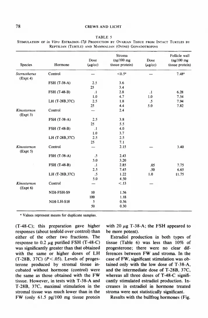
(T-48-C); this preparation gave higher responses (about tenfold over control) than either of the other two fractions. The response to 0.2 pg purified FSH (T-48-C) was significantly greater than that obtained with the same or higher doses of LH (T-28B, 37C) (Pc.05). Levels of proges- terone produced by stromal tissue in- cubated without hormone (control) were the same as those obtained with the FW tissue. However, in tests with T-38-A and T-28B, 37C, maximal stimulation in the stromal tissue was much lower than in the FW (only 6 1.5 pg/lOO mg tissue protein
with 20 pg T-38-A; the FSH appeared to be more potent).
Estradiol production in both types of tissue (Table 6) was less than 10% of progesterone; there were no clear dif- ferences between FW and stroma. In the case of FW, significant stimulation was ob- tained only with the low dose of T-38-A, and the intermediate dose of T-28B, 37C, whereas all three doses of T-48-C signifi- cantly stimulated estradiol production. In- creases in estradiol in hormone treated stroma were not statistically significant.
Results with the bullfrog hormones (Fig.

REPTILIAN OVARIAN STEROIDOGENESIS 79
350 Gopherus Follicle Wall
128°C. 5hrs)
0.1 05 I.0 20 5 10 20 Dose (jq 1
FIG. 1. Influence of turtle gonadotropins on progesterone production by follicle wall membranes from the gopher tortoise, Gopherus ngassizi. Curve represents mean of duplicate samples shown by individual points.
3) indicate that both gonadotropins stimu- late progesterone production by Chelydra FW. The LH appears more potent at the one dose tested.
DISCUSSION
Despite the use of four species of turtles (representing three families) in different ovarian states and variation in incuba- tion times and temperature, mammalian, amphibian, and reptilian FSH and LH
consistently stimulated progesterone pro- duction by both types of ovarian tissue examined. Also, although stimulation of estradiol was more variable when it oc- curred, both types of gonadotropic hor- mone were effective. Variation in control levels of steroid production and in the magnitude of the steroidogenic responses to gonadotropins may reflect the dif- ferences in ovarian state and incubation conditions. Since tissue progesterone con-
3oo Che&&o Follicle Wall - (28°C.5hrsJ
g so- 0 h s .g zoo-
F
8 <
150-
E
s loo- & -z $ i so-
0.0, 0.02 0.10 0.2 1.0 2.0 10.0 20 Dose Lug/cc)
FIG. 2. Inthtence of snapping turtle gonadotropins on progesterone production by follicle wall membranes of ovaries from the homologous species. Mean and SE for four replicates are shown.
80 CREWS AND LICHT
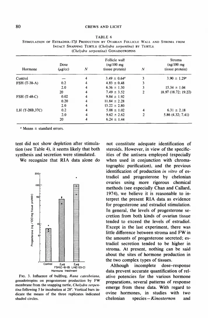
TABLE 6 STIMULATION OF ESTRADIOL-I7P PRODUCTION BY OVARIAN FOLLICLE WALL AND STROMA FROM
INTACT SNAPPING TURTLE (Chelydra serpentina) BY TURTLE (Chelydra serpentina) GONADOTROPINS
Hormone
Control FSH (T-38-A)
FSH (T-48-C)
LH (T-28B ,37C)
Dose
b&4
- 0.2 2.0
20 0.02 0.20 2.0 0.2 2.0
20
N
4 4 4 4 4 4 4 4 4 4
Follicle wall (ngi100 mg
tissue protein)
3.49 f 0.w 4.83 2 0.48 6.36 2 1.30 7.49 k 3.52 9.84 zk 1.92
11.84 2 2.28 13.22 T 2.80
5.08 k 1.02 9.62 f 2.62 6.24 2 1.44
Stroma (ngi100 mg
tissue protein)
3.90 f 1.29’
15.34 2 1.04 18.97 (18.72; 19.22)
6.31 -+ 2.18 5.86 (4.32; 7.41)
a Means rt standard errors.
tent did not show depletion after stimula- tion (see Table 4), it seems likely that both synthesis and secretion were stimulated.
We recognize that RIA data alone do
2ua FSHtd-&I-B, LHiC-53-C)
Hormone treatment
FIG. 3. Influence of bullfrog, Rana cafesbeiana, gonadotropins on progesterone production by FW membrane from the snapping turtle, Chelydra serpen- tina following 5 hr incubation at 28”. Vertical bars in- dicate the means of the three replicates indicated shaded circles.
not constitute adequate identification of steroids. However, in view of the specific- ities of the antisera employed (especially when used in conjunction with chroma- tographic purification), and the previous identification of production in vitro of es- tradiol and progesterone by chelonian ovaries using more rigorous chemical methods (see especially Chan and Callard, 1974), we believe it is reasonable to in- terpret the present RIA data as evidence for progesterone and estradiol stimulation. In general, the levels of progesterone se- cretion from both kinds of ovarian tissue tended to exceed the levels of estradiol. Except in the last experiment, there was little difference between stroma and FW in the amounts of progesterone secreted; es- tradiol secretion tended to be higher in stroma. At present, nothing can be said about the sites of hormone production in the two complex types of tissues.
Although incomplete dose-response data prevent accurate quantification of rel- ative potencies for the various hormone preparations, several patterns of response emerge from these data. With regard to ovine hormones, in studies with two chelonian species - Kinosternon and

REPTILIAN OVARIAN STEROIDOGENESIS 81
Gopherus (Expts 6, 7 and 9) -even the relatively impure NIH-FSH gave consid- erably greater stimulation than the more purified NIH-LH (e.g., Tables 3 and 5). The discrepancy between the potencies of the two turtle hormones was less pro- nounced, but the LH also appears gener- ally less effective than FSH. The small number of replicates for individual Kinost- ernon, Sternotherus, and Gopherus stud- ies precludes detailed statistical analy- ses for each experiment. However, com- parisons of the magnitudes of all responses for FSH and LH by a conservative non- parametric statistical test (e.g., Sign test; Siegel, 1956) reveals that the responses to T-38-A were significantly greater than that for equivalent doses of T-28B, 37C (N = 33, Z = 3.13 for two-tailed test, P < .002). The highly purified turtle FSH (T-48-B or C) appears to be even more po- tent, since it produced the same responses as the other two preparations but at much lower doses (P > .05). In the experiment with Chelydra, the results indicate that the highly purified turtle FSH (T-48-C) is more potent in stimulating progesterone than either the LH or the less purified FSH; the latter two preparations gave sim- ilar responses. The one test with frog hor- mones indicated that purified LH might be more potent than a partially purified FSH (Fig. 3). Preliminary tests with gonado- tropins derived from the green sea turtle (Chelonia mydas), which represents a dif- ferent superfamily from Chelydra, also in- dicate that both purified FSH and LH stimulate large increases in progesterone production (approx 25X control levels) in Chelydra FW. Thus, although the four species of turtle appeared to vary in their capacity to produce steroids and in their sensitivity to hormonal stimulation (in fact, the greatest differences were between the two kinosternids), no interspecific dif- ference was evident in the pattern of res- ponsiveness to the two types of go- nadotropin. The conclusion we wish to
emphasize here is that there is no distinct specificity for either type of gonado- tropin, independent of the species from which the hormones are derived.
These results with steroid secretion by turtle ovary constitute the first demon- stration of the action of reptilian hormones in a homologous order and species. These results are consistent with previous find- ings using ovine and turtle hormones in female lizards; i.e., an apparent lack of specificity for the two gonadotropins. Lack of specificity for LH in the lizard may be correlated with the possible lack of this activity in the squamate pituitary (Licht, 1974). However, this explanation is inade- quate to account for the lack of specificity in Reptilia generally, since chelonian rep- tiles do possess a distinct LH molecule that appears homologous to mammalian LH.
Our results with the turtle ovary are not in agreement with the recent report of Chan and Callard (1974) that ovine LH was more potent than FSH in stimulating the conversion of labeled precursors to progesterone in vitro with ovaries of Pseu- demys scripta. Our survey of species sug- gests that this discrepancy is not likely to be due to interspecific differences (Pseud- emys belongs to the same superfamily as the four genera tested here). Since Chan and Callard measured conversion rather than total secretion, the possibility must be considered that the two hormones may act differentially on some aspect of ovarian steroid metabolism other than those mea- sured in our studies. However, since they used only a single dose of mammalian hor- mone, it is premature to draw conclusions about any potential specificities for the two gonadotropins.
Lack of information on the pattern and levels of circulating gonadotropins in turtles prevents conclusions about whether one of the gonadotropins might be more important than the other in regulating normal steroid production. In this regard,

these in vitro results cannot be ex- REFERENCES trapolated to in vivo performance until fur- ther information is available on possible
Bradgon, D. E., Lazo-Waem, E. A., Zarrow. M. X., and Hisaw, F. L. (I 954). Progesterone-like activ-
changes (e.g., luteinization) in the cultured ity in the plasma of ovo-viviparous snakes. Proc.
tissue. Also, we recognize that the produc- Sot. Exp. Biol. Med. 86, 477-480.
tion of comparable end products as mea- Brattstrom, B. H. (1965) Body temperature of rep-
sured here does not eliminate the possibil- tiles. Amer. Midl. Nat. 73, 376-422.
ity that the two hormones may have Callard. I. P. (1966). Gonadal steroid synthesis in the
snake Natrix sipedon pictiventris. Second Int. different mechanisms of action (e.g., in the Congr. Horm. Steroids Series III, 216. site of action in the biosynthetic pathway Callard, I. P. and Doolittle, J. P. (1973). The influ-
or in the kinetics of the response). How- ence of intrahypothalamic implants of proges-
ever, the present results do indicate a terone on ovarian growth and function in the
marked divergence between mammals and ovo-viviparous iguanid lizard, Sceloporus cyan- ogenys. Comp. Biochem. Physiol. 44A, 625-63 1.
reptiles from the standpoint of classical Callard, I. P. and Leathem, J. H. (1964). In virro syn- concepts on specificity of hormone action thesis of progesterone by ovaries and adrenals of
for ovarian steroidogenesis. Since the snakes. Proc. Sot. Exp. Biol. Med. 115,567-569.
turtle fails to show a specificity for either Callard, I. P. and Leathem, J. H. (1965). In vitro
its own or mammalian LH, it appears that steroid synthesis by the ovaries of elasmo- branchs and snakes. Arch. Anat. Microsc. 54,
the divergence is related to changes in the 35-48. ovarian receptors rather than in FSH Callard, I. P., Bayne, C. G., and McConnell, W. F.
structure. In this connection recent studies (1972a). Hormones and reproduction in the fe-
have revealed that FSH may also stimu- male lizard Sceloporus cyanogenys. Gen. Comp.
late progesterone in some mammalian Endocrinol. 18, 175- 194.
Callard, I. P., Chan, S. W. C., and Potts, M. A. ovarian tissues such as rat Graafian fol- (1972b). The control of the reptilian gonad. licles (Lidner ef al., 1974). Thus, the FSH Amer. Zool. 12, 273-287.
receptor for steroidogenesis may not be Callard, I. P., Doolittle, J., Banks, W. C., and Chan,
unique to reptiles, although it is apparently S. W. C. (1972~). Recent studies on the control
more prominent or widespread than in of the reptilian ovarian cycle. Gen. Comp. En- docrinol. Suppl. 3, 65-75.
mammals. At present, there is still little Chan, S. W. C. and Callard, I. P. (1974). Reptilian evidence for a unique role of LH in rep- ovarian steroidogenesis and influence of mamma-
tiles in the physiology of reproduction. lian gonadotropins (follicle-stimulating hormone and luteinizing hormone) in vitro. J. Endocrinol. 62, 267-275.
Chan, S. W. C., Ziegel, S., and Callard, I. P. (1973).
ACKNOWLEDGMENTS Plasma progesterone in snakes. Camp. Physioi. Biochem. 44A, 63 I-638.
Turtle and frog hormones were fractionated in col- Chester-Jones, I., Bellamy, D., Chan, D. K. O., Fol- laboration with Dr. Harold Papkoff, Hormone Re- lett, B. K., Henderson, 1. W., Phillips, J. G., and search Laboratory, University of California, San Smut, R. S. (1972). Biological actions of steroid Francisco. We are indebted to Drs. T. M. Nett and hormones in nonmammalian vertebrates. In V. L. Estergreen for estradiol antiserum and Ms. “Steroids in Nonmammalian Vertebrates” (D. R. Ellen Daniels for the supply of LH antiserum and to Idler, Ed.), pp. 414-480. Academic Press. New NIAMD for ovine hormones. Gopherus were ob- York. tained from California Fish and Game Department Colombo, L., Yaron, Z., Daniels, E., and Belvedere, with the assistance of Ron Marlowe. Larry McNease, P. (1974). Biosynthesis of 1 l-deoxycorticos- Rockefeller Wildlife Refuge, helped obtain some of terone by the ovary of the Yucca night lizard, the Kinosternon. Hugh Meakin provided valuable Xantusia vigilis. Gen. Comp. Endocrinol. 24, technical assistance. This work was supported in part 331-337. by NSF Grant GB 35241X. PL was a Miller Pro- Cooper, W. E. and Ferguson, G. W. (1972a). Rela- fessor (Miller Institute, University of California, tive effectiveness of progesterone and testos- Berkeley). terone as inductors of orange spotting in female
82 CREWS AND LICHT

REPTILIAN OVARIAN STEROIDOGENESIS 83
collared lizards. Herpetologia 28, 64-65. Cooper, W. E. and Ferguson, G.. W. (1972b).
Steroids and color change during gravidity in the lizard, Crotaphytus collaris. Gen. Comp. En- docrinol. 18, 69-72.
Eyeson, K. N. (1971). The role of the pituitary gland in testicular function in the lizard Agama agama. Gen. Comp. Endocrinol. 16, 342-35.5.
Ferguson, G. W. (1966). Effect of follicle-stimulating hormone and testosterone propionate on the re- production of the side-blotched lizard, Uta stans- buriana. Copeia 1966, 495-498.
Jones, R. E. (1969). Effects of mammalian gonado- tropins on the ovaries and oviducts of the lizard, Lygosoma laterale. J. Exp. 2001. 171, 217-222.
Kimbai, F. A. and Erpino, M. J. (1971). Hormonal control of pigmentary sexual dimorphism in Sceloporus occidentalis. Gen. Comp. Endocr. 16, 375-384.
Klicka, J. and Mahmoud, I. Y. (1972). Conversion of pregnenolone-414C to progesterone-4’*C by turtle corpus luteum. Gen. Comp. Endocrinol. 19, 367-369.
Klicka, J. and Mahmoud, 1. Y. (1973). Conversion of cholesterol to progesterone by turtle corpus lu- teum. Steroids 21, 483-496.
Licht, P. (1970). Effects of mammalian gonadotropins (ovine FSH and LH) in female lizards. Gen. Comp. Endocrinol. 14, 98-106.
Licht, P. (1972a). Actions of mammalian pituitary gonadotropins (FSH and LH) in reptiles. II. Turtles. Gen. Comp. Endocrinol. 19, 282-289.
Licht, P. (1972b). Physiology of breeding cycles in reptiles: role of temperature. Gen. Comp. En- docrinol. Suppl. 3, 477-488.
Licht, P. (1974). Endocrinology of Reptilia-The pituitary system. Chem. Zoo/. 9, 399-448.
Licht, P. and Crews, D. P. (1975). Stimulation of ovarian and oviducal growth and ovulation in female lizards by reptilian (turtle) gonadotropins. Gen. Comp. Endocrinol. 25, 467-47 1.
Licht, P. and Hartree, A. S. (1971). Actions of mam- malian, avian and piscine gonadotropins in the lizard. J. Endocrinol. 53, 329-349.
Licht, P. and Papkoff, H. (1973). Evidence for an in- trinsic activity of ovine LH in the lizard. Gen. Comp. Endocrinol. 20, 172- 176.
Licht, P. and Papkoff, H. (1974a). Separation of two distinct gonadotropins in the pituitary gland of the snapping turtle (Chelydra serpentina). Gen. Comp. Endocrinol. 22, 2 18-237.
Licht, P. and Papkoff, H. (1974b). Phylogenetic survey of the neuraminidase sensitivity of reptil-
Licht, P. and Papkoff, H. (1974~). Separation of two gonadotropins from the pituitary of the bullfrog, Rana catesbeiana. Endocrinology 94, 477-483.
Licht, P. and Tsui, H. W. (I 975). Evidence for the in- trinsic activity of ovine FSH on sperma- togenesis, ovarian growth, steroidogenesis and ovulation in lizards. Biol. Reprod. 12, 346-350.
Licht, P., Papkoff, H., Goldman, B. D., Follett, B. K., and Scanes, C. G. (1974). Immunological relatedness among reptilian, avian, and mamma- lian pituitary iuteinizing hormones. Gen. Comp. Endocrinol. 24, 168-176.
Linder, H. R., Tsafriri, A., Lieberman, M. E., Zor, V., Koch, Y., Bauminger, S., and Bamea, A. (1974). Gonadotropin action on cultured graafian follicles: Induction of maturation division of the mammalian oocyte and differentiation of the lu- teal cell. Rec. Progr. Horm. Res. 30, 79-126.
Lisk, R. D. (1967). Neural control of gonad size by hormone feedback in the desert iguana Dip- sosaurus dorsalis dorsalis. Gen. Comp. En- docrinol. 8, 258-266.
Lupo di Prisco, C., Chieffi, G., and Delrio, G. (1968). Identification of steroid hormones in the ovaries of the lizard Lacerta sicula. Gen. Comp. En- docrinol. 10, 292-295.
Mahmoud, I. Y. and Klicka, J. (1972). Seasonal go- nadal changes in Kinosternid turtles. J. Herpet. 6, 183-189.
Medica, P. A., Turner, F. B., and Smith, D. D. (1973). Hormonal induction of color change in female leopard lizards, Crotaphytus wislizenii. Copeia 1973, 658-661.
Mikhail, G., Wu, C. H., Ferin, M., and Vande Wiele, R. L. (1970). Radioimmunoassay of plasma es- trone and estradiol. Steroids 15, 333-352.
McGinnis, S. M. and Voigt, W. G. (1971). Ther- moregulation in the desert tortoise, Gopherus agassizi. Comp. Biochem. Physiol. 4OA, 119-126.
Nett, T. M., Holtan, D. W., and Estergreen, V. L. (1973). Plasma estrogens in pregnant and post- partum mares. .I. Anim. Sci. 37, 962-970.
Ozon, R. (1972). Estrogens in fishes, amphibians, reptiles, and birds. In “Steroids in Non- mammalian Vertebrates” (D. R. Idler, Ed.), pp. 390-413. Academic Press, New York.
Schwartz, N. B. (1974). The role of FSH and LH and of their antibodies on follicle growth and on ovu- lation. Biol. Reprod. 10, 236-272.
Siegel, S. (1956). “Non Parametric Statistics.” McGraw-Hill, Inc., New Jersey, 3 12 pp.
Wu, C. H. and Lundy, L. E. (1971). Radioim- ian gonadotropin. Gen. Comp. Endocrinol. 23, munoassay of plasma estrogens. Steroids 18, 41.5-420. 91-111.


