Embed Size (px)
Citation preview

MOLECULAR AND CELLULAR BIOLOGY, Aug. 2007, p. 5650–5663 Vol. 27, No. 160270-7306/07/$08.00�0 doi:10.1128/MCB.00130-07Copyright © 2007, American Society for Microbiology. All Rights Reserved.
Stage-Specific Secretion of HMGB1 in Cartilage RegulatesEndochondral Ossification�†
Noboru Taniguchi,1 Kenji Yoshida,1 Tatsuo Ito,1 Masanao Tsuda,1 Yasunori Mishima,1Takayuki Furumatsu,1 Lorenza Ronfani,2 Kazuhiro Abeyama,4 Ko-ichi Kawahara,4
Setsuro Komiya,5 Ikuro Maruyama,4 Martin Lotz,1Marco E. Bianchi,2,3 and Hiroshi Asahara1,6,7*
Department of Molecular and Experimental Medicine, The Scripps Research Institute, 10550 North Torrey Pines Road, La Jolla,California 920371; DIBIT, San Raffaele Scientific Institute, via Olgettina 58, 20132 Milano, Italy2; San Raffaele University,
via Olgettina 58, 20132 Milano, Italy3; Department of Laboratory and Vascular Medicine, Graduate School of Medical andDental Sciences, Kagoshima University, 8-35-1 Sakuragaoka, Kagoshima 890-8520, Japan4; Department ofOrthopaedic Surgery, Graduate School of Medical and Dental Sciences, Kagoshima University, 8-35-1 Sakuragaoka,
Kagoshima 890-8520, Japan5; National Center for Child Health and Development, 2-10-1 Okura, Setagaya,Tokyo 157-8535, Japan6; and SORST, Japan Science and Technology Agency,
4-1-8 Honcho, Kawaguchi, Saitama 332-0012, Japan7
Received 20 January 2007/Returned for modification 25 February 2007/Accepted 19 May 2007
High mobility group box 1 protein (HMGB1) is a chromatin protein that has a dual function as a nuclearfactor and as an extracellular factor. Extracellular HMGB1 released by damaged cells acts as a chemoattrac-tant, as well as a proinflammatory cytokine, suggesting that HMGB1 is tightly connected to the process oftissue organization. However, the role of HMGB1 in bone and cartilage that undergo remodeling duringembryogenesis, tissue repair, and disease is largely unknown. We show here that the stage-specific secretion ofHMGB1 in cartilage regulates endochondral ossification. We analyzed the skeletal development of Hmgb1�/�
mice during embryogenesis and found that endochondral ossification is significantly impaired due to the delayof cartilage invasion by osteoclasts, osteoblasts, and blood vessels. Immunohistochemical analysis revealedthat HMGB1 protein accumulated in the cytosol of hypertrophic chondrocytes at growth plates, and itsextracellular release from the chondrocytes was verified by organ culture. Furthermore, we demonstrated thatthe chondrocyte-secreted HMGB1 functions as a chemoattractant for osteoclasts and osteoblasts, as well as forendothelial cells, further supporting the conclusion that Hmgb1�/� mice are defective in cell invasion. Col-lectively, these findings suggest that HMGB1 released from differentiating chondrocytes acts, at least in part,as a regulator of endochondral ossification during osteogenesis.
Bone formation occurs through two developmental processes:intramembranous ossification and endochondral ossification. In-tramembranous ossification takes place in several craniofacialbones and the lateral part of clavicles, whereas endochondralossification occurs in the long bones of the limbs, the basal partof the skull, vertebrae, ribs, and the medial part of the clavicles.In endochondral ossification, an intermediate step occurs dur-ing which cartilaginous templates prefigure future skeletal el-ements and play a major role in regulating the developingskeletal elements (33). First, mononucleated osteoclast precur-sors enter the mesenchyme surrounding the bone rudiments,proliferate, differentiate into tartrate-resistant acid phos-phatase (TRAP)-positive cells, and migrate together with en-dothelial cells through the nascent bone collar (7). Subse-quently, they invade the calcified cartilage, filling the core ofthe diaphysis while fusing and differentiating into mature os-teoclasts, and transform the core of the bone into a marrow
cavity (15). Osteoclasts are derived from hematopoietic pre-cursor cells formed by the fusion of monocytic cells at the bonesites to be resorbed, whereas osteoblasts arise from multi-potential mesenchymal cells and further differentiate intobone-lining cells and osteocytes (30).
These events, including osteoclast migration and angio-genesis during endochondral ossification, are tightly coordi-nated by extracellular factors, such as matrix metallopro-teinases (MMPs) and vascular endothelial growth factor(VEGF) (37). When neovascularization of the cartilage an-lage begins, membrane type 1 MMP (MT1-MMP) andMMP9 are expressed in the preosteoclasts and other chon-droclastic cells of unknown origins (23). Mice deficient inMmp9 exhibit a delay in osteoclast recruitment in special-ized invasion and bone resorption models in vitro (15). It isalso reported that the deletion of functional Mmp13 hasprofound effects on skeletal development (25). In Mmp13-null embryos, the growth plates were strikingly lengthened,a defect related predominantly to a delay in terminal eventsin the growth plates, with failure to resorb collagens, as wellas a delay in ossification at the primary centers. In addition,VEGF signaling plays an important role of angiogenesisduring skeletal development (59). Inhibition of VEGF bythe administration of a soluble chimeric VEGF receptorprotein to 24-day-old mice inhibited blood vessel invasion
* Corresponding author. Mailing address: Department of Molecularand Experimental Medicine, The Scripps Research Institute, 10550North Torrey Pines Road, La Jolla, CA 92037. Phone: (858) 784-9026.Fax: (858) 784-2744. E-mail: [email protected].
† Supplemental material for this article may be found at http://mcb.asm.org/.
� Published ahead of print on 4 June 2007.
5650
on Novem
ber 18, 2015 by guesthttp://m
cb.asm.org/
Dow
nloaded from

into the hypertrophic zone of long bone growth plates andresulted in impaired trabecular bone formation and expan-sion of the hypertrophic zone (17).
High mobility group box 1 protein (HMGB1) is a chro-matin protein that is widely expressed and extremely con-served in mammals. There are three HMGB proteins:HMGB1, HMGB2, and HMGB3 with �80% amino acididentity, which are composed of two basic HMG-box do-mains (A and B) and a long acidic C-terminal tail (10). Asa nuclear factor, HMGB1 acts as an architectural proteinthat can bend DNA to promote nucleoprotein interactionsand facilitate diverse DNA modifications (2). Several groupshave shown that HMGB1 also has an extracellular role as aproinflammatory cytokine (4, 51, 55). Two different routesfor HMGB1 release into the extracellular milieu have beenreported: active secretion by activated macrophages andmonocytes (54) and passive release from necrotic or dam-aged cells (45). HMGB1 released by damaged cells acts as achemoattractant for vascular smooth muscle cells and fibro-blasts and induces cytoskeleton reorganization and cell mi-gration (13). HMGB1 also promotes the migration of localstem cells, such as vessel-associated stem cells (mesoangio-blasts) (38), and endothelial cells (32, 46), suggesting thatHMGB1 is tightly connected to the process of tissue orga-nization. The biological relevance of HMGB1 in vivo wasshown in Hmgb1�/� mice, which have a highly pleiotropicphenotype such as the inability to use glycogen stored in theliver (11). These mice survive for several days if given glu-cose parenterally; however, mutants remained much smallerthan control littermates and had arched backs, posteriorlimbs splayed wide apart, and abnormal gait. These findingssuggested that HMGB1 may participate in not only tissuerepair after injury but also the organization of bone andcartilage development.
We show here that the stage-specific secretion of HMGB1 incartilage regulates endochondral ossification, in part, by actingas a chemotactic factor for the cells that invade at the primaryossification center. These findings highlight the potential roleof HMGB1 in skeletal homeostasis.
MATERIALS AND METHODS
Mice. The Hmgb1�/� mutant mice used in the present study were describedbefore (11), except for their background, which is now pure BALB/c. All animalexperiments were performed according to approved protocols according to in-stitutional guidelines at The Scripps Research Institute. Mouse embryos forhistomorphometry were littermates from Hmgb1�/� parents. The genotype ofthe mice was determined by PCR analysis of tail DNA. The wild-type Hmgb1allele was detected by PCR with the primers wildtype-1 (5�-GCA GGC TTCGTT GTT TTC ATA CAG-3�) and wildtype-2 (5�-TCA AAG AGT AAT ACTGCC ACC TTC-3�), which generate a 495-bp fragment. The mutant Hmgb1allele was detected by using two primers complementary to the neomycin resis-tance gene—Neo-1 (5�-TGG TTT GCA GTG TTC TGC CTA GC-3�) andNeo-2 (5�-CCC AGT CAT AGC CGA ATA GCC-3�)—which generate a 336-bpfragment.
Histological analysis. Mice were sacrificed at various embryonic stages, dis-sected, and fixed in 4% paraformaldehyde–phosphate-buffered saline at 4°Covernight. Subsequently, they were processed, embedded in paraffin, and sec-tioned. For HMGB1 immunostaining, rabbit anti-HMGB1 antibody (Pharmin-gen, San Diego, CA) and chicken anti-HMGB1 antibody (Shino-Test, Kana-gawa, Japan) were used for limb sections and organ culture sections, respectively(51). For CD31 immunostaining, embryos were infiltrated in 20% sucrose, fol-lowed by OCT embedding to stain with rat anti-PECAM antibody (Pharmingen)and von Kossa and Safranin O/Fast Green staining (47). Whole-mount alcian
blue and alizarin red S staining of skeletons were done as described previously(31), and the longitudinal diameters of calvariae, as well as the lengths andalizarin-positive regions of tibias, were measured by micrometer. Detection ofapoptotic cells in paraffin sections of limbs was based on a modification ofgenomic DNA utilizing terminal deoxynucleotidyl transferase (TUNEL [termi-nal deoxynucleotidyltransferase-mediated dUTP-biotin nick end labeling] assay)and indirect detection of positive cells by fluorescein conjugated anti-digoxigeninantibody using a MEBSTAIN Apoptosis Kit Direct (Medical and Biologicallaboratories, Nagoya, Japan). Immunofluorescence assay to determine HMGB1translocation in chondrocytes was carried out with rabbit anti-HMGB1 antibody(Pharmingen) as described before (51).
Using a leukocyte acid phosphatase kit from Sigma (St. Louis, MO), TRAPstaining was performed on paraffin sections according to the instructions pro-vided by the manufacturer. The determination of the numbers and distributionof TRAP-positive cells in longitudinal sections of bones was done as describedpreviously (7, 56).
In situ hybridization. Tissues were fixed in 4% paraformaldehyde–phosphate-buffered saline overnight at 4°C, processed, embedded in paraffin, and sectioned.RNA in situ hybridization was performed as described previously (3). Briefly,slides were deparaffinized, treated with proteinase K (1 �g/ml) for 20 min at37°C, and hybridized with 35S-labeled antisense riboprobes in hybridizationbuffer (50% deionized formamide, 300 mM NaCl, 20 mM Tris-HCl [pH 8.0], 5mM EDTA, 0.5 mg of yeast tRNA/ml, 10% dextran sulfate, and 1� Denhardtsolution) in a humidified chamber at 60°C overnight. After hybridization, theslides were treated with RNase A, washed to a final stringency of 50% form-amide, 2� SSC (1� SSC is 0.15 M NaCl plus 0.015 M sodium citrate) at 60°C,dipped in emulsion, exposed for 3 days to 3 weeks, and developed. The probesfor Indian hedgehog, MMP9, VEGF and MMP13, MT1-MMP, Runx2 andOsterix, and osteocalcin and osteopontin were provided by Y. Kawakami (SalkInstitute), S. M. Krane (Harvard Medical School), Z. Werb (University ofCalifornia, San Francisco), T. Vu (University of California, San Francisco), K.Nakashima (Tokyo Medical and Dental University), and S. Nomura (OsakaUniversity Graduate School of Medicine), respectively. The HMGB1 probe wasa 1.2-kb cDNA fragment encoding the COOH-terminal domain and the 3�-untranslated region (UTR). The Col1a1 probe was a 0.8-kb cDNA fragmentencoding the COOH-terminal domain.
Organ culture. Metatarsal bones and tibiae were harvested from mouseembryos at embryonic day 15.5 (E15.5) and E14.5, respectively. They werecultured for 5 days in conditioned medium as described previously (20). Theexpression levels of HMGB1 and lactate dehydrogenase (LDH) in the super-natant were assessed by immunoblotting with rabbit anti-HMGB1 antibody(Pharmingen) and goat anti-LDH antibody (Chemicon, Temecula, CA) asdescribed previously (45). Rib chondrocytes were purified from the ventralparts of rib cartilage of 2- to 4-day-old BALB/c mice (28), followed byinduction of necrosis as described previously (45), and were used as a positivecontrol for the HMGB1 protein. The concentrations of HMGB1 released intoconditioned supernatant were measured in triplicate with an enzyme-linkedimmunosorbent assay (ELISA) using commercially available kits (Shino-Test) (57).
Preparation of osteoclasts and osteoblasts. Human osteoclast precursor cells(Poietics; Cambrex Bio Science Walkersville, Inc., Walkersville, MD) were cul-tured in alpha-minimal essential medium (alpha-MEM) containing 10% fetalbovine serum, penicillin-streptomycin, and HEPES containing alpha-MEM me-dium with receptor activator of nuclear factor B ligand (RANKL; PeproTechEC, Ltd., London, United Kingdom) and M-CSF (R&D Systems, Minneapolis,MN). Cells were incubated in a CO2 incubator in a humidified atmosphere of95% air and 5% CO2 at 37°C. After complete osteoclast differentiation at day 7,the medium was replaced with serum-free alpha-MEM; the cells were starved for2 h and then used for chemotaxis assays. MC3T3-E1 osteoblastic cells werepurchased from the American Type Culture Collection (Manassas, VA) andcultured in alpha-MEM with 10% fetal bovine serum.
Chemotaxis assays. Chemotaxis assays were performed as described previ-ously (22). The assays were carried out in Boyden chambers with polycarbonatefilters with 9-�m pores (Corning Costar, Corning, NY). Osteoclasts were pre-pared by sequential treatment with trypsin, and the remaining cells were thengently lifted off the plates with a rubber policeman. The osteoclasts were seededin 48-transwell plates in alpha-MEM containing 0.1% (wt/vol) Albumax and keptfor 4 h with or without addition of rat cytokine-quality HMGB1 (obtained fromHMGBiotech, Milan, Italy) and VEGF (R&D Systems). Invasion was deter-mined as the ratio of osteoclasts that migrated through the collagen gel to reachthe lower side of the membrane compared to the total number of osteoclasts inthe insert. The chemotaxis assays for MC3T3-E1 cells were also performed
VOL. 27, 2007 SECRETION OF HMGB1 REGULATES ENDOCHONDRAL OSSIFICATION 5651
on Novem
ber 18, 2015 by guesthttp://m
cb.asm.org/
Dow
nloaded from

according to the method as described above. All experiments were performed atleast twice in four replicates.
Three-dimensional pellet culture. Mice rib chondrocytes were prepared fromthe ventral parts rib cartilage of 2- to 4-day-old C57BL/6 mice as described
previously (36). Human articular chondrocytes were isolated from human carti-lage, and a primary cell culture was established (21). Both types of chondrocyteswere cultured in three-dimensional cell pellets for 18 days as described before(5). Briefly, 1-ml aliquots containing 2 � 105 cells each were added to 15-ml
FIG. 1. Analysis of skeletal development in Hmgb1�/� mice by double staining with alcian blue and alizarin red. (A) Hmgb1�/� embryos (right)are smaller than wild-type (WT) littermates (left) at E16.5. (B) At this stage, facial and skull bones formed by intramembranous ossification appearsimilar between two groups, whereas sphenoid bones (arrowhead) and basioccipital (arrow) of the chondrocranium, which are formed byendochondral ossification, appear reduced in size and in intensity of alizarin red staining in Hmgb1�/� embryos. (C) The pelvis has smaller alizarinred-stained zones in Hmgb1�/� embryos. (D) The radius and ulna in Hmgb1�/� forelimbs are not only reduced in size and calcification, but bent(arrowhead); the humerus is often fractured (arrow). The thorax in Hmgb1�/� embryos shows severe hypoplasia accompanied by spinal scoliosis(E) and kyphosis (A). Ribs stained less intensely for alizarin red and are thin and bent (arrows) (F), and clavicles are hypoplastic and crooked inHmgb1�/� embryos (G). (H) Statistical comparison between wild-type (n � 6) and Hmgb1�/� (n � 6) embryos at E16.5. The wild type is definedas 100%. Diameters of calvariae (skull size): wild-type, 100% � 2.7%; mutant, 97.7% � 2.2% (no statistical difference). Tibia length: wild-type,100% � 1.6%; mutant, 87.4% � 6.9% (P 0.001). Length of the ossified zone (alizarin red positive) of tibia: wild-type, 100% � 6.9%; mutant,63.6% � 9.6% (P 0.0001). The asterisk indicates a significant statistical difference (P 0.01).
5652 TANIGUCHI ET AL. MOL. CELL. BIOL.
on Novem
ber 18, 2015 by guesthttp://m
cb.asm.org/
Dow
nloaded from

conical polypropylene centrifuge tubes (Becton Dickinson, San Diego, CA), andthe cells were pelleted by centrifugation at 600 rpm for 5 min at room temper-ature. The cultures were maintained at 37°C in 5% CO2 in a humidified incu-bator. Pellets were maintained up to 18 days in Dulbecco modified Eaglemedium–F-12 supplemented with 50 �g of ascorbate phosphate (Sigma)/ml, 100 �gof pyruvate/ml, 1% penicillin-streptomycin (Gibco, Grand Island, NY), and 50mg of ITS�Premix (Becton Dickinson, Bedford, MA; a final concentration of6.25 �g of bovine insulin/ml, 6.25 �g of transferrin/ml, 6.25 ng of selenousacid/ml, 1.25 mg of bovine serum albumin/ml, and 5.35 �g of linoleic acid/ml)/ml.The medium was changed every 3 days. Cryostat-sectioned pellets were used forimmunofluorescence assay. The supernatant of pelleted mouse rib chondrocytesand human articular chondrocytes was used for chemotaxis assay with or without
addition of anti-HMGB1 IgY neutralizing HMGB1, a gift from Shino-Test (1),and control IgY (Promega, Madison, WI).
Quantitative PCR. Total RNA was extracted and oligo(dT)-primed cDNA wasprepared from 500 ng of total RNA by using Superscript II (Invitrogen, Carlsbad,CA). The resulting cDNAs were analyzed by using the SYBR green system forquantitative analysis of specific transcripts according to the manufacturer’s in-structions (Applied Biosystems, Foster City, CA). All mRNA expression datawere normalized to GAPDH expression in the corresponding sample. The prim-ers used in real-time PCR are as follows: Col10a1, 5�-GCCTCAAATACCCTTTCTGC (sense) and 5�-GTGTCTTGGGGCTAGCAAGT (antisense); MMP13,5�-GAAGACCTTGTGTTTGCAGAGC (sense) and 5�-CTCGGAGCCTGTCAACTGTG (antisense); Hmgb1, 5�-GGCTGACAAGGCTCGTTATG (sense)
FIG. 2. Localization of HMGB1 protein in developing limbs. Adjacent sections of tibia were stained with safranin O (A, C, and E) and antibodyto HMGB1 (B, D, and F). HMGB1 is expressed in the prehypertrophic chondrocytes at E14.5 (B) and in the hypertrophic chondrocytes at E15.5(D). In contrast, resting and proliferating chondrocytes do not show any positive staining in either nuclei or cytoplasm. (F) Expression is robustin the limbs at E14.5 and E15.5 but attenuates at E16.5. (G) Large magnifications of the humerus at E15.5. HMGB1 is positive in the nuclei ofprehypertrophic chondrocytes (arrows) and in the cytosol of hypertrophic chondrocytes (arrowheads). (H) At E16.5, metacarpal bones also showHMGB1 expression in the nuclei of prehypertrophic chondrocytes, as well as in the cytoplasm of hypertrophic chondrocytes. (I) The positivestaining in hypertrophic cartilage is absent in sections from Hmgb1�/� metacarpal bones at E16.5. The staining in perichondrium is nonspecific(arrowheads). (J and K) Analysis of HMGB1 expression and apoptosis in radius at E15.5. Arrowheads indicate the HMGB1-positive cells (J) andTUNEL-positive cells presenting apoptosis of hypertrophic chondrocytes (K). ph, prehypertrophic cartilage; h, hypertrophic cartilage; c, calcifiedcartilage; bm, bone marrow. Scale bars: A to F, J, and K, 200 �m; G to I, 50 �m.
VOL. 27, 2007 SECRETION OF HMGB1 REGULATES ENDOCHONDRAL OSSIFICATION 5653
on Novem
ber 18, 2015 by guesthttp://m
cb.asm.org/
Dow
nloaded from

and 5�-GGGCGGTACTCAGAACAGAA (antisense); and GAPDH, 5�-ATGTGTCCGTCGTGGATCTGA (sense) and 5�-GATGCCTGCTTCACCACCTT(antisense).
Statistics. The statistical analysis at present study was performed by using atwo-tailed Student t test.
RESULTS
Analysis of skeletal development in Hmgb1�/� mice. We firstexamined bone and cartilage development in Hmgb1�/� mice.Since Hmgb1�/� mice die soon after birth (11), we analyzedHmgb1�/� embryos. Alcian blue staining revealed no apparentdifference in skeletal formation between Hmgb1�/� and wild-type littermate embryos at E13.5 (see Fig. S1A in the supple-mental material). At E16.5, however, Hmgb1�/� embryos weresmaller than wild-type embryos, suggesting a discrepancy dur-ing ossification (Fig. 1A). At this stage, facial and skull bonesformed by intramembranous ossification appeared similar be-tween the two groups, although the shape of Hmgb1�/� cal-variae was relatively flat and depressed. In contrast, sphenoidbones and the basioccipital region of the chondrocranium,which are formed by endochondral ossification, appeared to bereduced in size and in intensity of alizarin red staining inHmgb1�/� mice (Fig. 1B). Other bones formed by endochon-dral ossification, such as the pelvis, had smaller alizarin red-stained zones (Fig. 1C). The radius and ulna of Hmgb1�/�
forelimbs were not only reduced in size and calcification butabnormally bent, suggesting a reduction of mineralization (Fig.1D). Moreover, fractures were observed in the humeri of some(4 of 14) Hmgb1�/� mice. Thorax formation showed severehypoplasia accompanied by spinal scoliosis (Fig. 1E) and ky-phosis (Fig. 1A). Ribs stained less intensely for alizarin red andwere thin and bent (Fig. 1F). The clavicles were hypoplasticand crooked (Fig. 1G). At E16.5, the diameters of calvariaewere similar in both groups, whereas the lengths of theHmgb1�/� tibias reached 87% of that of the wild type, and thealizarin-positive region reached 64% of the wild-type length
(Fig. 1H). These findings suggest that in Hmgb1�/� mice en-dochondral ossification is impaired, whereas intramembranousossification is only affected slightly and was not investigatedfurther.
HMGB1 expression in normal growth plates. To investi-gate the mechanism of endochondral ossification defect inHmgb1�/� embryos, we examined the localization of HMGB1protein in the developing limbs of normal wild-type mice byimmunohistochemistry. Safranin O staining showed that pre-hypertrophic cartilage appeared in the tibia at E14.5 (Fig. 2A),differentiating into hypertrophic cartilage, followed by calcifiedcartilage at E15.5 (Fig. 2C), and was replaced by bone marrowand bone trabeculae at E16.5 (Fig. 2E). By using the specificanti-HMGB1 polyclonal rabbit antibody which does not detectHMGB2 and HMGB3 (19), we found that HMGB1 was ex-pressed in the prehypertrophic chondrocytes of the tibia atE14.5 (Fig. 2B) and in hypertrophic chondrocytes at E15.5(Fig. 2D). Large magnifications of the humerus at E15.5showed that HMGB1 was detected in the nuclei of prehyper-trophic chondrocytes and in the cytosol of hypertrophic chon-drocytes (Fig. 2G). On the other hand, resting and proliferat-ing chondrocytes did not show any positive staining in eithernuclei or cytoplasm. Not only large long bones but also othersmall long bones formed by endochondral ossification, such asmetacarpal bones, exhibited HMGB1 expression in the nucleiof prehypertrophic chondrocytes, as well as in the cytoplasm ofhypertrophic chondrocytes (Fig. 2H). This positive staining inhypertrophic cartilage was absent in Hmgb1�/� sections (Fig.2I). These results indicate that HMGB1 is expressed and trans-located from the nucleus to the cytosol during a specific stageof cartilage maturation. At the end of the cascade of chondro-cyte maturation, terminal hypertrophic chondrocytes undergoapoptotic cell death (17). We analyzed HMGB1 expressionand apoptosis in the radius at E15.5 and detected HMGB1 inhypertrophic chondrocytes (Fig. 2J) but not in terminal hyper-trophic chondrocytes, which were positive for TUNEL staining
FIG. 3. Expression of chondrocyte differentiation markers in wild-type and Hmgb1�/� tibia. (A and B) Indian hedgehog (Ihh) is comparablebetween wild-type and Hmgb1�/� embryos at E14.5. (C to F) Col10a1 appears in the region of hypertrophic chondrocytes at E14.5 (C and D) andthen declines in the most mature hypertrophic chondrocytes at the center of hypertrophic zones at E15.5 in both groups without an apparentdifference between wild-type and mutant embryos (E and F). Scale bars, 200 �m.
5654 TANIGUCHI ET AL. MOL. CELL. BIOL.
on Novem
ber 18, 2015 by guesthttp://m
cb.asm.org/
Dow
nloaded from
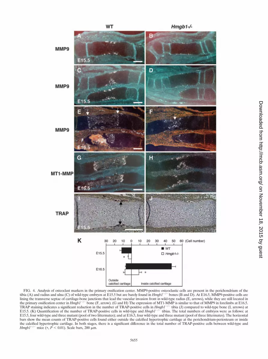
FIG. 4. Analysis of osteoclast markers in the primary ossification center. MMP9-positive osteoclastic cells are present in the perichondrium of thetibia (A) and radius and ulna (C) of wild-type embryos at E15.5 but are barely found in Hmgb1�/� bones (B and D). At E16.5, MMP9-positive cells arelining the transverse septae of cartilage-bone junctions that lead the vascular invasion front in wild-type radius (E, arrows), while they are still located inthe primary ossification center in Hmgb1�/� bone (F, arrow). (G and H) The expression of MT1-MMP is similar to that of MMP9 in forelimbs at E16.5.TRAP staining indicates a significant reduction in the number of TRAP-positive cells in Hmgb1�/� tibia (J) compared to wild-type bone (I, arrows) atE15.5. (K) Quantification of the number of TRAP-positive cells in wild-type and Hmgb1�/� tibias. The total numbers of embryos were as follows: atE15.5, four wild-type and three mutant (pool of two littermates); and at E16.5, four wild-type and three mutant (pool of three littermates). The horizontalbars show the mean counts of TRAP-positive cells found either outside the calcified hypertrophic cartilage at the perichondrium-periosteum or insidethe calcified hypertrophic cartilage. In both stages, there is a significant difference in the total number of TRAP-positive cells between wild-type andHmgb1�/� mice (�, P 0.01). Scale bars, 200 �m.
5655
on Novem
ber 18, 2015 by guesthttp://m
cb.asm.org/
Dow
nloaded from
(Fig. 2K), suggesting that HMGB1 was expressed just beforecell death.
Impaired invasion of osteoclasts in Hmgb1�/� mice. Next,we sought to identify which process during endochondral os-sification was disturbed by Hmgb1 gene deficiency. To examinethe rate of hypertrophic chondrocyte differentiation, we ob-served the expression of Indian hedgehog, a marker of prehy-pertrophic chondrocytes of cartilage elements (52), and foundthat it did not differ between wild-type (Fig. 3A) andHmgb1�/� (Fig. 3B) tibias. Col10a1 appeared in the region of
hypertrophic chondrocytes at E14.5 (Fig. 3C and D) and thendeclined in the most mature hypertrophic chondrocytes at thecenter of hypertrophic zones at E15.5 with a similar pattern inboth groups (Fig. 3E and F). These findings indicate thatHmgb1 gene deficiency does not affect the onset of cartilagematuration. In contrast, MMP9-positive osteoclastic cells (53)were distributed around the perichondrium in the tibias andradii of wild-type mice at E15.5 (Fig. 4A and C) but werebarely detectable in Hmgb1�/� bones (Fig. 4B and D). AtE16.5, the discrepancy became more remarkable. In the wild-
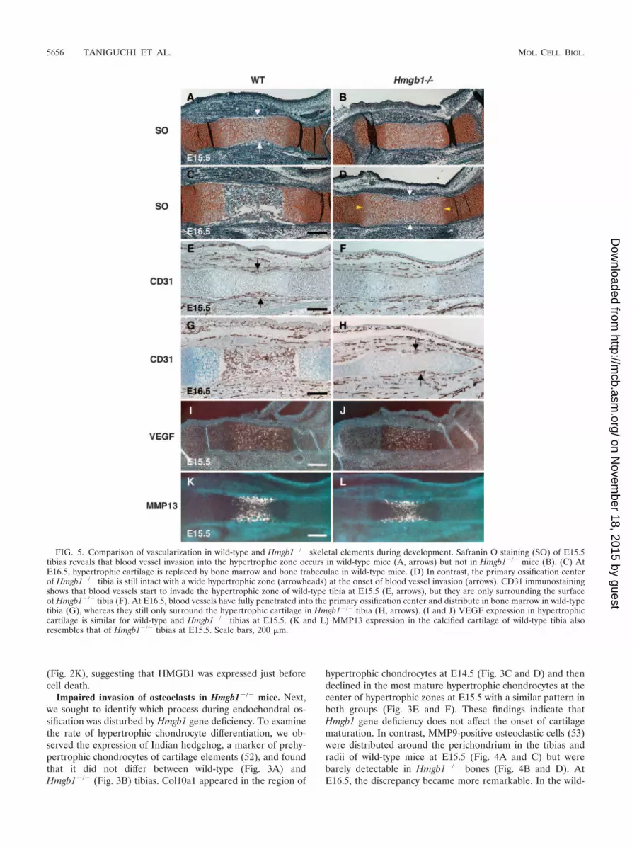
FIG. 5. Comparison of vascularization in wild-type and Hmgb1�/� skeletal elements during development. Safranin O staining (SO) of E15.5tibias reveals that blood vessel invasion into the hypertrophic zone occurs in wild-type mice (A, arrows) but not in Hmgb1�/� mice (B). (C) AtE16.5, hypertrophic cartilage is replaced by bone marrow and bone trabeculae in wild-type mice. (D) In contrast, the primary ossification centerof Hmgb1�/� tibia is still intact with a wide hypertrophic zone (arrowheads) at the onset of blood vessel invasion (arrows). CD31 immunostainingshows that blood vessels start to invade the hypertrophic zone of wild-type tibia at E15.5 (E, arrows), but they are only surrounding the surfaceof Hmgb1�/� tibia (F). At E16.5, blood vessels have fully penetrated into the primary ossification center and distribute in bone marrow in wild-typetibia (G), whereas they still only surround the hypertrophic cartilage in Hmgb1�/� tibia (H, arrows). (I and J) VEGF expression in hypertrophiccartilage is similar for wild-type and Hmgb1�/� tibias at E15.5. (K and L) MMP13 expression in the calcified cartilage of wild-type tibia alsoresembles that of Hmgb1�/� tibias at E15.5. Scale bars, 200 �m.
5656 TANIGUCHI ET AL. MOL. CELL. BIOL.
on Novem
ber 18, 2015 by guesthttp://m
cb.asm.org/
Dow
nloaded from
type radius, MMP9-positive cells were lining the transverseseptae of cartilage-bone junctions that lead the vascular inva-sion front (Fig. 4E), whereas they were still located in theprimary ossification center in Hmgb1�/� radius (Fig. 4F). Theexpression of MT1-MMP, which is highly expressed in osteo-clasts (44), was similar to that of MMP9 (Fig. 4G and H). Toconfirm the apparent reduction in osteoclast numbers, we
stained for TRAP and found significant reduction in the num-ber of TRAP-positive cells in Hmgb1�/� tibias at E15.5 (Fig. 4Iand J). Quantification of the number of these cells insideversus outside the calcified hypertrophic cartilage showed asignificant difference between wild-type and Hmgb1�/� tibias(Fig. 4K). These findings demonstrate that osteoclast recruit-ment was suppressed in the Hmgb1�/� bones.
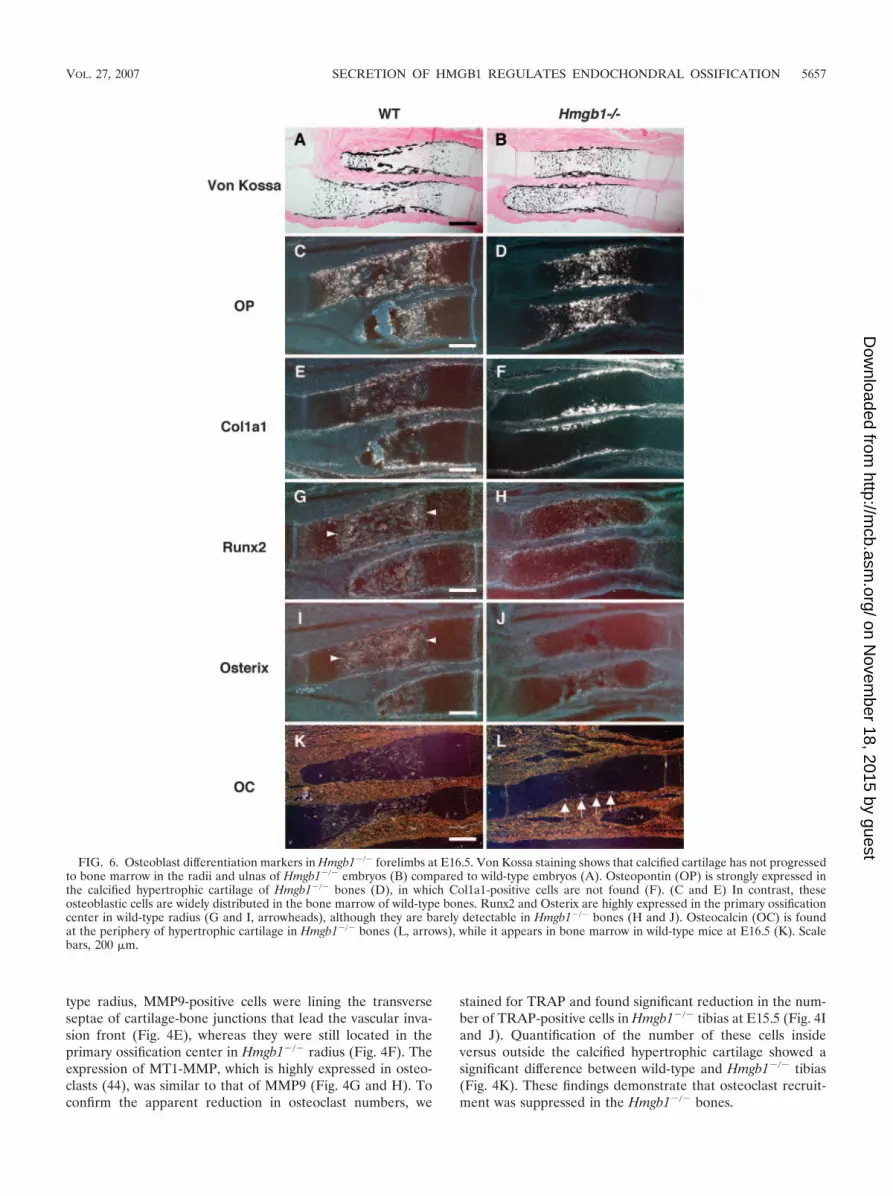
FIG. 6. Osteoblast differentiation markers in Hmgb1�/� forelimbs at E16.5. Von Kossa staining shows that calcified cartilage has not progressedto bone marrow in the radii and ulnas of Hmgb1�/� embryos (B) compared to wild-type embryos (A). Osteopontin (OP) is strongly expressed inthe calcified hypertrophic cartilage of Hmgb1�/� bones (D), in which Col1a1-positive cells are not found (F). (C and E) In contrast, theseosteoblastic cells are widely distributed in the bone marrow of wild-type bones. Runx2 and Osterix are highly expressed in the primary ossificationcenter in wild-type radius (G and I, arrowheads), although they are barely detectable in Hmgb1�/� bones (H and J). Osteocalcin (OC) is foundat the periphery of hypertrophic cartilage in Hmgb1�/� bones (L, arrows), while it appears in bone marrow in wild-type mice at E16.5 (K). Scalebars, 200 �m.
VOL. 27, 2007 SECRETION OF HMGB1 REGULATES ENDOCHONDRAL OSSIFICATION 5657
on Novem
ber 18, 2015 by guesthttp://m
cb.asm.org/
Dow
nloaded from
Altered vascularization of skeletal elements in Hmgb1�/�
mice during development. MMP9-positive cells enter the mes-enchyme surrounding the bone rudiments and migrate to-gether with endothelial cells through the nascent bone collar atthe primary ossification center (15). Thus, we examined thevascularization in skeletal elements of Hmgb1�/� mice. Safra-nin O staining revealed that blood vessel invasion into thehypertrophic zone occurred in wild-type tibias at E15.5 (Fig.5A) but not in Hmgb1�/� tibias (Fig. 5B). At E16.5, hypertro-phic cartilage was replaced by bone marrow and bone trabec-ulae in wild-type mice (Fig. 5C). In contrast, the primary os-sification center of Hmgb1�/� tibias was still intact with a widehypertrophic zone and only the onset of blood vessel invasion(Fig. 5D). Using CD31 (PECAM) antibody, which is a markerof endothelial cells, we performed immunostaining and foundthat blood vessels started to invade the hypertrophic zone ofwild-type tibia at E15.5 (Fig. 5E), but they were only on thesurface of Hmgb1�/� tibia (Fig. 5F). At E16.5, blood vesselshad fully penetrated into the primary ossification center and
distributed in bone marrow in wild-type tibia (Fig. 5G),whereas they were still only surrounding the hypertrophic car-tilage in Hmgb1�/� bone (Fig. 5H). At the growth plate, hy-pertrophic cartilage expresses VEGF, and inhibition of VEGFactivity blocks the recruitment of MMP9-positive and TRAP-positive cells, as well as endothelial cells (17). We found nodifference in VEGF expression in hypertrophic cartilage be-tween wild-type and Hmgb1�/� tibia at E15.5 (Fig. 5I and J).MMP13, which is expressed by both terminal hypertrophicchondrocytes and osteoblasts, is also important for the vascu-larization of hypertrophic cartilage because it degrades nativecollagen, a major component of the hypertrophic cartilage(47). MMP13 expression in the calcified cartilage of the wild-type tibia resembled that of the Hmgb1�/� tibia (Fig. 5K andL). Taken together, these findings suggest that the cell in-vasion into hypertrophic cartilage by endothelial cells wasdisrupted in Hmgb1�/� bones during the process of endo-chondral ossification, although VEGF and MMP13 expres-sion was unaltered.
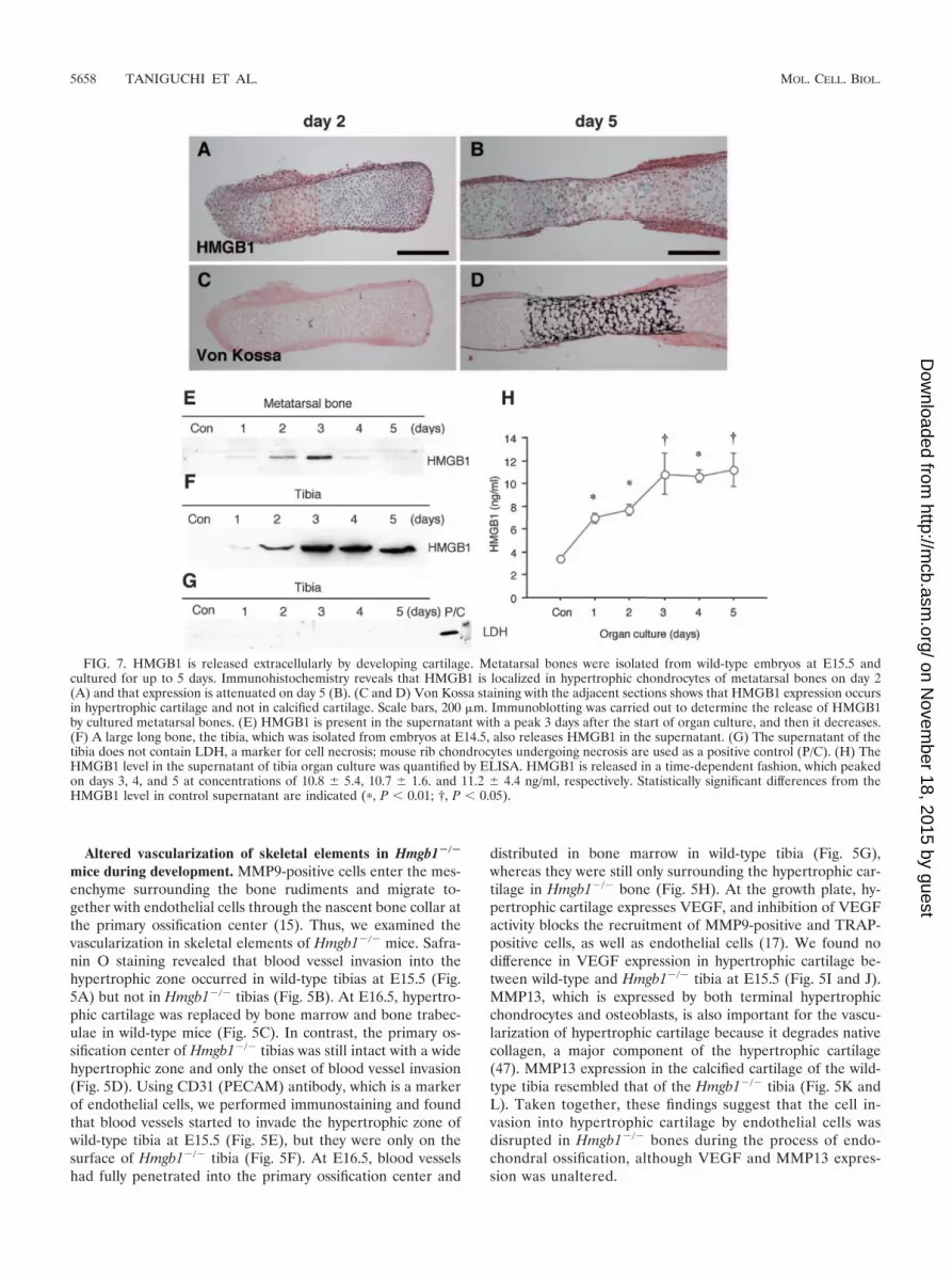
FIG. 7. HMGB1 is released extracellularly by developing cartilage. Metatarsal bones were isolated from wild-type embryos at E15.5 andcultured for up to 5 days. Immunohistochemistry reveals that HMGB1 is localized in hypertrophic chondrocytes of metatarsal bones on day 2(A) and that expression is attenuated on day 5 (B). (C and D) Von Kossa staining with the adjacent sections shows that HMGB1 expression occursin hypertrophic cartilage and not in calcified cartilage. Scale bars, 200 �m. Immunoblotting was carried out to determine the release of HMGB1by cultured metatarsal bones. (E) HMGB1 is present in the supernatant with a peak 3 days after the start of organ culture, and then it decreases.(F) A large long bone, the tibia, which was isolated from embryos at E14.5, also releases HMGB1 in the supernatant. (G) The supernatant of thetibia does not contain LDH, a marker for cell necrosis; mouse rib chondrocytes undergoing necrosis are used as a positive control (P/C). (H) TheHMGB1 level in the supernatant of tibia organ culture was quantified by ELISA. HMGB1 is released in a time-dependent fashion, which peakedon days 3, 4, and 5 at concentrations of 10.8 � 5.4, 10.7 � 1.6. and 11.2 � 4.4 ng/ml, respectively. Statistically significant differences from theHMGB1 level in control supernatant are indicated (�, P 0.01; †, P 0.05).
5658 TANIGUCHI ET AL. MOL. CELL. BIOL.
on Novem
ber 18, 2015 by guesthttp://m
cb.asm.org/
Dow
nloaded from

Osteogenesis in Hmgb1�/� mice. As shown in Fig. 1,Hmgb1�/� forelimbs appeared to be reduced in size and cal-cification and were abnormally bent or fractured. Since thesefindings suggest a reduction of bone mineralization, we inves-tigated osteoblast differentiation in Hmgb1�/� bones. Usingvon Kossa staining, we found that calcified cartilage had pro-gressed to bone marrow in the radii and ulnas of wild-typemice (Fig. 6A) but not in Hmgb1�/� mice (Fig. 6B). Osteopon-tin, a hypertrophic cartilage marker as well as an osteoblastmarker (35), was strongly expressed in the calcified hypertro-phic cartilage of Hmgb1�/� bones (Fig. 6D) in which Col1a1-positive cells, an early marker of osteoblast differentiation (18),were not found; these cells were accumulated at the collarsurrounding the growth plate (Fig. 6F). In contrast, Col1a1-positive cells were widely distributed in the bone marrow ofwild-type mice (Fig. 6E), suggesting that osteoblast invasion
was suppressed in Hmgb1�/� limbs. The essential transcriptionfactors for osteoblast differentiation, Runx2 (27) and Osterix(34), were highly expressed in the primary ossification center ofthe wild-type radius (Fig. 6G and I), whereas they were barelydetectable in the Hmgb1�/� bones (Fig. 6H and J). Osteocal-cin, which is thought to be a terminal marker for osteoblasticmaturation (29), was found at the periphery of hypertrophiccartilage in Hmgb1�/� bones at E16.5 (Fig. 6L); however, itappeared in the bone marrow at E17.5 (data not shown) ratherthan at E16.5 as in wild-type mice (Fig. 6K). Thus, the delay inprimary ossification of Hmgb1�/� hypertrophic cartilage wascoupled to a delay in recruitment of osteoblasts, suggestingthat subsequent osteoblastic differentiation progressed simi-larly in wild-type and Hmgb1�/� mice.
HMGB1 is released from differentiating cartilage in organculture. To examine the secretion of HMGB1 from chon-
FIG. 8. HMGB1 release from differentiating cultured rib chondrocytes. (A) Immunofluorescence assay shows that monolayer rib chondrocytesisolated from the ventral parts of mice rib cartilage express HMGB1 only in the nucleus, whereas in pelleted rib chondrocytes cultured for 2 daysHMGB1 is localized in the cytosol. The extracellular release of HMGB1 was verified with immunoblotting. (B) HMGB1 was determined in thesupernatant of pelleted mice rib chondrocytes (mRC) on days 1 and 2, whereas human articular chondrocytes (hAC) do not release HMGB1 inpellet culture. (C) Immunoblotting with LDH antibody shows that this secretion is independent of necrotic cell death. The positive control (P/C)is the same sample as shown in Fig. 7G. (D) During the culture of pelleted mRC for 18 days, quantitative PCR demonstrates that the mRNA levelof cartilage maturation markers such as Col10a1 and MMP13 increases significantly on day 18, although that of HMGB1 is unchanged. (E) Onlythe supernatant on day 3 contains HMGB1. Statistically significant differences from mRNA expression on day 3 are indicated, respectively (�,P 0.01).
VOL. 27, 2007 SECRETION OF HMGB1 REGULATES ENDOCHONDRAL OSSIFICATION 5659
on Novem
ber 18, 2015 by guesthttp://m
cb.asm.org/
Dow
nloaded from

drocytes, we used the cartilage organ culture system (20).Metatarsal bones were isolated from embryos at E15.5 andcultured in conditioned medium for up to 5 days. Immuno-histochemistry revealed that HMGB1 was localized in hy-pertrophic chondrocytes on day 2 (Fig. 7A) and that expres-sion was attenuated on day 5 (Fig. 7B). Von Kossa stainingof the adjacent sections indicated that this expression oc-curred in hypertrophic cartilage and not in calcified carti-lage (Fig. 7C and D). Using immunoblotting, we determinedthat HMGB1 was present in the supernatant, with a peak 3days after the start of organ culture, and then it decreased(Fig. 7E), showing that HMGB1 was released into the me-dium by hypertrophic chondrocytes. This result was repro-duced with a large long bone, the tibia, which was isolatedfrom the embryos at E14.5 (Fig. 7F). Immunoblotting withLDH antibody was negative, indicating that HMGB1 wasactively secreted and not released passively as a conse-quence of necrotic cell death (Fig. 7G). Using ELISA, wequantified the HMGB1 protein released into the medium oftibia organ culture and found that it peaked on days 3through 5 at concentrations of �10 ng/ml (Fig. 7H).
HMGB1 is released specifically from hypertrophic chondro-cytes. It has been previously demonstrated that HMGB1 isreleased from osteoclasts and osteoblast-like cells (12, 42). Toprove that the release of HMGB1 into the supernatant wasfrom chondrocytes in organ culture, we used pellet cultures ofrib growth plate chondrocytes, since this culture system mimicsin vivo cartilage differentiation (5). Monolayer chondrocytesisolated from the ventral parts of mouse rib cartilage expressedHMGB1 only in the nucleus; however, when cultured as dif-ferentiating cell pellets, HMGB1 was localized in the cytosol(Fig. 8A). Extracellular HMGB1 was detected in the superna-tant of pelleted rib chondrocytes on days 1 and 2; in contrast,articular chondrocytes, which do not differentiate to hypertro-phic cartilage under the three-dimensional condition such aspellet or alginate culture (6, 40), did not release HMGB1(Fig. 8B). Immunoblotting with LDH antibody showed thatHMGB1 release was not caused by necrotic cell death (Fig.8C). In addition, to examine HMGB1 expression in longer-term cultures, we maintained the rib chondrocyte pellets for 18days. Quantitative PCR demonstrated that mRNA of cartilagematuration markers such as Col10a1 and MMP13 increasedsignificantly, showing that chondrocyte differentiation had oc-curred (Fig. 8D). Only the medium from day 3 containedHMGB1 (Fig. 8E), although the mRNA level of HMGB1 didnot significantly change between day 3 and 18. These resultsindicate that HMGB1 is secreted during the early phase ofcartilage maturation.
HMGB1 is a chemoattractant for osteoclasts and osteo-blasts. As we showed in Fig. 4, 5, and 6, Hmgb1�/� embryos
FIG. 9. Chondrocyte-secreted HMGB1 is a chemoattractant forosteoclasts. The chemotactic effect of recombinant HMGB1 on osteo-clasts was examined by Boyden chambers with or without addition ofHMGB1 to either the lower chamber (LC) or the upper chamber (UC)as indicated. (A) HMGB1 recruits osteoclasts at 10 ng/ml, and efficacypeaks at 100 ng/ml. The addition of HMGB1 to the upper chamberdoes not significantly activate osteoclast invasion. (B) HMGB1 alsorecruits osteoblastic MC3T3-E1 cells with a tendency similar to thatdescribed above, although VEGF does not. Statistically significantdifferences from control migrations without added chemoattractantsare indicated, respectively (�, P 0.01; †, P 0.05). (C) Chemotaxis
assay using the supernatant of pelleted mice rib chondrocytes (mRC)and human articular chondrocytes (hAC) after 3 days culture. Thesupernatant of hAC does not recruit osteoclasts, whereas that of mRCattracts osteoclasts significantly, and this effect is abrogated by additionof anti-HMGB1 IgY. Cell migration is shown as mean � the standarddeviation of four replicates. Statistically significant differences fromcontrol migrations by the supernatant of hAC are indicated (�, P 0.01).
5660 TANIGUCHI ET AL. MOL. CELL. BIOL.
on Novem
ber 18, 2015 by guesthttp://m
cb.asm.org/
Dow
nloaded from
were defective in invasion by TRAP- and Col1a1-positive cells,as well as CD31-positive cells, at the primary ossification cen-ter. Since HMGB1 has chemotactic effects on endothelial cells(32, 46), we tested for similar effects on osteoclasts and osteo-blasts. Recombinant HMGB1 at 10 ng/ml recruited osteoclastsin Boyden chambers, and peak migration occurred at 100 ng/ml; this level of efficacy was similar to that of VEGF used as apositive control (22) (Fig. 9A). Addition of HMGB1 to theupper chamber did not significantly activate osteoclast migra-tion. HMGB1 also induced chemotaxis for MC3T3-E1 osteo-blast-like cells with a tendency similar to that described above(Fig. 9B), although VEGF did not (16). These findings suggestthat osteoclast and osteoblast invasion at the primary ossifica-tion center might be a direct effect of HMGB1-induced che-moattraction.
Finally, we investigated whether HMGB1 released by differ-entiating chondrocytes could promote osteoclast migration.Using the supernatant of pelleted rib chondrocytes and artic-ular chondrocytes cultured for 3 days (see Fig. S2 in the sup-plemental material), we compared the chemotactic effect forosteoclasts. The supernatant of articular chondrocytes did notrecruit osteoclasts; however, the supernatant of rib chondro-cytes attracted osteoclasts significantly, and the effect was ab-rogated by neutralizing anti-HMGB1 IgY (Fig. 9C). This resultsupports our hypothesis that differentiating chondrocytes couldregulate cell migration directly via HMGB1 secretion.
DISCUSSION
This study demonstrates that the stage-specific secretion ofHMGB1 in cartilage regulates endochondral ossification, atleast in part, by acting as a chemotactic factor for osteoclastsand osteoblasts, as well as endothelial cells. We examinedskeletal development in Hmgb1�/� embryos and found signif-
icant alterations in the bones formed by endochondral ossifi-cation, whereas calvariae, which are formed by intramembra-nous ossification, were somewhat misshapen, but the effect wasslight, and the cartilage formation was not affected. The anal-ysis of Hmgb1�/� limb sections revealed that the onset ofcartilage differentiation was similar in Hmgb1�/� and wild-typeembryos; however, the invasion of TRAP- and Col1a1-positivecells, as well as CD31-positive cells, into the primary ossifica-tion center was remarkably impaired in Hmgb1�/� limbs. Thus,the Hmgb1�/� growth plates are strikingly lengthened anddeficient in osteoblast and osteoclast invasion as well as vas-cularization, which may result in weak bones that can bend orfracture.
To examine the expression of HMGB1 in developing limbs,we used in situ hybridization: HMGB1 mRNA expression wasubiquitous in the cells of all zones of the growth plate fromE14.5 through E16.5 (data not shown). In contrast, HMGB1protein was present in the nuclei of prehypertrophic chondro-cytes in tibia at E14.5 and in the cytosol of hypertrophic chon-drocytes at E15.5 but was not detectable in resting and prolif-erating chondrocytes. The active secretion of HMGB1 fromchondrocytes was verified with organ culture and pellet culturesystems; we found that HMGB1 was translocated from thenucleus to the cytosol and actively secreted at the early phaseof chondrocyte differentiation, but the secretion ceased at thelate phase. Interestingly, secretion from pelleted rib chondro-cytes occurred actively without added any stimulatory factor,whereas articular chondrocytes did not release HMGB1 inpellet culture. Chondrocyte-secreted HMGB1 was sufficient tochemoattract osteoclasts and osteoblasts, as well as endothelialcells as previously shown by others (32, 46). These findingssuggest that HMGB1 released from hypertrophic chondrocytesmay regulate skeletal development by controlling cell invasion
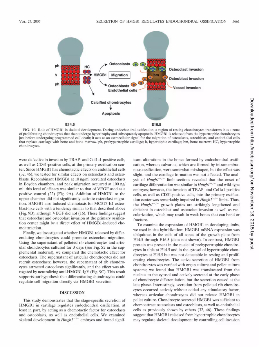
FIG. 10. Role of HMGB1 in skeletal development. During endochondral ossification, a region of resting chondrocytes transforms into a zoneof proliferating chondrocytes that then undergo hypertrophy and subsequently apoptosis. HMGB1 is released from the hypertrophic chondrocytesjust before undergoing programmed cell death; it acts as an extracellular signal for the migration of osteoclasts, osteoblasts, and endothelial cellsthat replace cartilage with bone and bone marrow. ph, prehypertrophic cartilage; h, hypertophic cartilage; bm, bone marrow; HC, hypertrophicchondrocytes.
VOL. 27, 2007 SECRETION OF HMGB1 REGULATES ENDOCHONDRAL OSSIFICATION 5661
on Novem
ber 18, 2015 by guesthttp://m
cb.asm.org/
Dow
nloaded from
into the growth plate. At present, however, a potential role ofHMGB1 as a nuclear factor, which is its other function, stillremains possible in the developing cartilage.
Secretion of HMGB1 during specific stages of cell differen-tiation is not unique to chondrocytes and has been reported fordendritic cells (14) and neonatal rat type I astrocytes (41),although the mechanism of HMGB1 secretion during cell dif-ferentiation has yet to be elucidated. Thus far, Bonaldi et al.have reported that HMGB1 contains two nuclear localizationsignals (NLSs), and the acetylation of both NLSs is involved inthe transport from the nucleus to the cytosol (8). Furthermore,HMGB1 can be phosphorylated, and the direction of transportis regulated by phosphorylation of both NLS regions (58).These findings suggest that HMGB1 release is independentfrom RNA expression and protein synthesis, which is compat-ible with our data showing that HMGB1 mRNA levels do notchange in chondrocyte pellet cultures, despite its secretion.
The inhibition of the interaction between HMGB1 and thereceptor for advanced glycation end products (RAGE), whichis a specific receptor for HMGB1, suppresses the tumor pro-liferation, metastatic invasion, and expression of MMPs (48).RAGE is expressed in osteoclasts, osteoblasts (12), and endo-thelial cells (9), suggesting that RAGE might be associatedwith cell invasion during endochondral ossification; however,an analysis of Rage�/� mice (1) showed no alteration in skel-etal development during embryogenesis (see Fig. S1B in thesupplemental material). Moreover, Rage�/� mice manifest in-creased bone mass and bone mineral density and decreasedbone resorptive activity due to a defect in osteoclast function(60). In our hands, however, MMP9 mRNA levels in calvariaeat E18.5 were similar between wild-type and Hmgb1�/� mice(see Fig. S3A in the supplemental material), and MMP9-pos-itive cells emerged in the bone marrow of developing limbs ofboth types of mice at E18.5 (see Fig. S3B in the supplementalmaterial). The evidence that HMGB1-RAGE interaction issufficient but not necessary for mesoangioblast migration (38)is a precedent for the idea that RAGE may not be the keyreceptor for HMGB1-induced cell recruitment at the primaryossification center. Additional HMGB1 receptors have beenidentified, including Toll-like receptors 2 and 4 (39), whichappear in osteoclasts, osteoblasts, and endothelial cells (26, 49,50), and syndecan (43), which is expressed in osteoblasts (24).
Our results indicate that HMGB1 might be important notonly for tissue repair after injury but also for the organizationof bone and cartilage development in the embryo. In endo-chondral ossification, a region of resting chondrocytes trans-forms into a zone of proliferating chondrocytes that then un-dergo hypertrophy and subsequently apoptosis (37). HMGB1release from the hypertrophic cartilage occurs just before pro-grammed cell death (Fig. 10), suggesting that HMGB1 may bean extracellular signal released from the tissue to be replaced(cartilage) toward the cells of new tissue to be formed (boneand bone marrow).
ACKNOWLEDGMENTS
We are grateful to Yasuhiko Kawakami and Thiennu Vu for helpfuldiscussions; Kim Henriksen, Noriyuki Namba, and Chisa Shukunamifor technical advice; Yasuhiko Yamamoto and Hiroshi Yamamoto forproviding Rage�/� mice; and Shingo Yamada for providing the neu-tralizing anti-HMGB1 IgY and ELISA kit. We especially thank LiloCreighton for her excellent immunohistochemical technique.
This study was supported by grants from the NIH (AR47360,AR50631, and AG07996), NIBI (ID05-24), Arthritis Foundation, JSTSORST, Genome Network Project (MEXT), and Grants-in Aid forScientific Research (MEXT) (H.A.) and a postdoctoral fellowship ofthe Japan Research Foundation for Clinical Pharmacology and Re-search Grant for Rheumatology Disease, Japan Rheumatism Founda-tion (N.T.).
REFERENCES
1. Abeyama, K., D. M. Stern, Y. Ito, K. Kawahara, Y. Yoshimoto, M. Tanaka,T. Uchimura, N. Ida, Y. Yamazaki, S. Yamada, Y. Yamamoto, H. Yamamoto,S. Iino, N. Taniguchi, and I. Maruyama. 2005. The N-terminal domain ofthrombomodulin sequesters high-mobility group-B1 protein, a novel antiin-flammatory mechanism. J. Clin. Investig. 115:1267–1274.
2. Agresti, A., and M. E. Bianchi. 2003. HMGB proteins and gene expression.Curr. Opin. Genet. Dev. 13:170–178.
3. Albrecht, U., G. Eichele, J. A. Helms, and H. Lu. 1997. Molecular andcellular methods in developmental toxicology. CRC Press, Boca Raton, FL.
4. Andersson, U., H. Wang, K. Palmblad, A. C. Aveberger, O. Bloom, H.Erlandsson-Harris, A. Janson, R. Kokkola, M. Zhang, H. Yang, and K. J.Tracey. 2000. High mobility group 1 protein (HMG-1) stimulates proinflam-matory cytokine synthesis in human monocytes. J. Exp. Med. 192:565–570.
5. Ballock, R. T., and A. H. Reddi. 1994. Thyroxine is the serum factor thatregulates morphogenesis of columnar cartilage from isolated chondrocytes inchemically defined medium. J. Cell Biol. 126:1311–1318.
6. Binette, F., D. P. McQuaid, D. R. Haudenschild, P. C. Yaeger, J. M. McPher-son, and R. Tubo. 1998. Expression of a stable articular cartilage phenotypewithout evidence of hypertrophy by adult human articular chondrocytes invitro. J. Orthop. Res. 16:207–216.
7. Blavier, L., and J. M. Delaisse. 1995. Matrix metalloproteinases are obliga-tory for the migration of preosteoclasts to the developing marrow cavity ofprimitive long bones. J. Cell Sci. 108(Pt. 12):3649–3659.
8. Bonaldi, T., F. Talamo, P. Scaffidi, D. Ferrera, A. Porto, A. Bachi, A. Rubar-telli, A. Agresti, and M. E. Bianchi. 2003. Monocytic cells hyperacetylatechromatin protein HMGB1 to redirect it toward secretion. EMBO J. 22:5551–5560.
9. Brett, J., A. M. Schmidt, S. D. Yan, Y. S. Zou, E. Weidman, D. Pinsky, R.Nowygrod, M. Neeper, C. Przysiecki, A. Shaw, et al. 1993. Survey of thedistribution of a newly characterized receptor for advanced glycation endproducts in tissues. Am. J. Pathol. 143:1699–1712.
10. Bustin, M. 1999. Regulation of DNA-dependent activities by the functionalmotifs of the high-mobility-group chromosomal proteins. Mol. Cell. Biol.19:5237–5246.
11. Calogero, S., F. Grassi, A. Aguzzi, T. Voigtlander, P. Ferrier, S. Ferrari, andM. E. Bianchi. 1999. The lack of chromosomal protein Hmg1 does notdisrupt cell growth but causes lethal hypoglycaemia in newborn mice. Nat.Genet. 22:276–280.
12. Charoonpatrapong, K., R. Shah, A. G. Robling, M. Alvarez, D. W. Clapp, S.Chen, R. P. Kopp, F. M. Pavalko, J. Yu, and J. P. Bidwell. 2006. HMGB1expression and release by bone cells. J. Cell Physiol. 207:480–490.
13. Degryse, B., T. Bonaldi, P. Scaffidi, S. Muller, M. Resnati, F. Sanvito, G.Arrigoni, and M. E. Bianchi. 2001. The high mobility group (HMG) boxes ofthe nuclear protein HMG1 induce chemotaxis and cytoskeleton reorganiza-tion in rat smooth muscle cells. J. Cell Biol. 152:1197–1206.
14. Dumitriu, I. E., P. Baruah, B. Valentinis, R. E. Voll, M. Herrmann, P. P.Nawroth, B. Arnold, M. E. Bianchi, A. A. Manfredi, and P. Rovere-Querini.2005. Release of high mobility group box 1 by dendritic cells controls T-cellactivation via the receptor for advanced glycation end products. J. Immunol.174:7506–7515.
15. Engsig, M. T., Q. J. Chen, T. H. Vu, A. C. Pedersen, B. Therkidsen, L. R.Lund, K. Henriksen, T. Lenhard, N. T. Foged, Z. Werb, and J. M. Delaisse.2000. Matrix metalloproteinase 9 and vascular endothelial growth factor areessential for osteoclast recruitment into developing long bones. J. Cell Biol.151:879–889.
16. Fukuyama, R., T. Fujita, Y. Azuma, A. Hirano, H. Nakamuta, M. Koida, andT. Komori. 2004. Statins inhibit osteoblast migration by inhibiting Rac-Aktsignaling. Biochem. Biophys. Res. Commun. 315:636–642.
17. Gerber, H. P., T. H. Vu, A. M. Ryan, J. Kowalski, Z. Werb, and N. Ferrara.1999. VEGF couples hypertrophic cartilage remodeling, ossification andangiogenesis during endochondral bone formation. Nat. Med. 5:623–628.
18. Gerstenfeld, L. C., S. D. Chipman, J. Glowacki, and J. B. Lian. 1987.Expression of differentiated function by mineralizing cultures of chickenosteoblasts. Dev. Biol. 122:49–60.
19. Guazzi, S., A. Strangio, A. T. Franzi, and M. E. Bianchi. 2003. HMGB1, anarchitectural chromatin protein and extracellular signalling factor, has aspatially and temporally restricted expression pattern in mouse brain. GeneExpr. Patterns 3:29–33.
20. Haaijman, A., R. N. D’Souza, A. L. Bronckers, S. W. Goei, and E. H. Burger.1997. OP-1 (BMP-7) affects mRNA expression of type I, II, X collagen, andmatrix Gla protein in ossifying long bones in vitro. J. Bone Miner. Res.12:1815–1823.
5662 TANIGUCHI ET AL. MOL. CELL. BIOL.
on Novem
ber 18, 2015 by guesthttp://m
cb.asm.org/
Dow
nloaded from

21. Hashimoto, S., R. L. Ochs, F. Rosen, J. Quach, G. McCabe, J. Solan, J. E.Seegmiller, R. Terkeltaub, and M. Lotz. 1998. Chondrocyte-derived apop-totic bodies and calcification of articular cartilage. Proc. Natl. Acad. Sci.USA 95:3094–3099.
22. Henriksen, K., M. Karsdal, J. M. Delaisse, and M. T. Engsig. 2003. RANKLand vascular endothelial growth factor (VEGF) induce osteoclast chemo-taxis through an ERK1/2-dependent mechanism. J. Biol. Chem. 278:48745–48753.
23. Holmbeck, K., P. Bianco, J. Caterina, S. Yamada, M. Kromer, S. A.Kuznetsov, M. Mankani, P. G. Robey, A. R. Poole, I. Pidoux, J. M. Ward, andH. Birkedal-Hansen. 1999. MT1-MMP-deficient mice develop dwarfism, os-teopenia, arthritis, and connective tissue disease due to inadequate collagenturnover. Cell 99:81–92.
24. Imai, S., M. Kaksonen, E. Raulo, T. Kinnunen, C. Fages, X. Meng, M. Lakso,and H. Rauvala. 1998. Osteoblast recruitment and bone formation enhancedby cell matrix-associated heparin-binding growth-associated molecule (HB-GAM). J. Cell Biol. 143:1113–1128.
25. Inada, M., Y. Wang, M. H. Byrne, M. U. Rahman, C. Miyaura, C. Lopez-Otin, and S. M. Krane. 2004. Critical roles for collagenase-3 (Mmp13) indevelopment of growth plate cartilage and in endochondral ossification.Proc. Natl. Acad. Sci. USA 101:17192–17197.
26. Kikuchi, T., T. Matsuguchi, N. Tsuboi, A. Mitani, S. Tanaka, M. Matsuoka,G. Yamamoto, T. Hishikawa, T. Noguchi, and Y. Yoshikai. 2001. Geneexpression of osteoclast differentiation factor is induced by lipopolysaccha-ride in mouse osteoblasts via Toll-like receptors. J. Immunol. 166:3574–3579.
27. Komori, T., H. Yagi, S. Nomura, A. Yamaguchi, K. Sasaki, K. Deguchi, Y.Shimizu, R. T. Bronson, Y. H. Gao, M. Inada, M. Sato, R. Okamoto, Y.Kitamura, S. Yoshiki, and T. Kishimoto. 1997. Targeted disruption of Cbfa1results in a complete lack of bone formation owing to maturational arrest ofosteoblasts. Cell 89:755–764.
28. Lefebvre, V., S. Garofalo, G. Zhou, M. Metsaranta, E. Vuorio, and B. DeCrombrugghe. 1994. Characterization of primary cultures of chondrocytesfrom type II collagen/beta-galactosidase transgenic mice. Matrix Biol. 14:329–335.
29. Lian, J. B., G. S. Stein, C. Stewart, E. Puchacz, S. Mackowiak, M. Aronow,M. Von Deck, and V. Shalhoub. 1989. Osteocalcin: characterization andregulated expression of the rat gene. Connect. Tissue Res. 21:61–69.
30. Liu, W., S. Toyosawa, T. Furuichi, N. Kanatani, C. Yoshida, Y. Liu, M.Himeno, S. Narai, A. Yamaguchi, and T. Komori. 2001. Overexpression ofCbfa1 in osteoblasts inhibits osteoblast maturation and causes osteopeniawith multiple fractures. J. Cell Biol. 155:157–166.
31. McLeod, M. J. 1980. Differential staining of cartilage and bone in wholemouse fetuses by alcian blue and alizarin red S. Teratology 22:299–301.
32. Mitola, S., M. Belleri, C. Urbinati, D. Coltrini, B. Sparatore, M. Pedrazzi, E.Melloni, and M. Presta. 2006. Cutting edge: extracellular high mobilitygroup box-1 protein is a proangiogenic cytokine. J. Immunol. 176:12–15.
33. Nakashima, K., and B. de Crombrugghe. 2003. Transcriptional mechanismsin osteoblast differentiation and bone formation. Trends Genet. 19:458–466.
34. Nakashima, K., X. Zhou, G. Kunkel, Z. Zhang, J. M. Deng, R. R. Behringer,and B. de Crombrugghe. 2002. The novel zinc finger-containing transcriptionfactor osterix is required for osteoblast differentiation and bone formation.Cell 108:17–29.
35. Nomura, S., A. J. Wills, D. R. Edwards, J. K. Heath, and B. L. Hogan. 1988.Developmental expression of 2ar (osteopontin) and SPARC (osteonectin)RNA as revealed by in situ hybridization. J. Cell Biol. 106:441–450.
36. Okubo, Y., and A. H. Reddi. 2003. Thyroxine downregulates Sox9 and pro-motes chondrocyte hypertrophy. Biochem. Biophys. Res. Commun. 306:186–190.
37. Ortega, N., D. J. Behonick, and Z. Werb. 2004. Matrix remodeling duringendochondral ossification. Trends Cell Biol. 14:86–93.
38. Palumbo, R., M. Sampaolesi, F. De Marchis, R. Tonlorenzi, S. Colombetti,A. Mondino, G. Cossu, and M. E. Bianchi. 2004. Extracellular HMGB1, asignal of tissue damage, induces mesoangioblast migration and proliferation.J. Cell Biol. 164:441–449.
39. Park, J. S., D. Svetkauskaite, Q. He, J. Y. Kim, D. Strassheim, A. Ishizaka,and E. Abraham. 2004. Involvement of Toll-like receptors 2 and 4 in cellularactivation by high mobility group box 1 protein. J. Biol. Chem. 279:7370–7377.
40. Park, K., B. H. Min, D. K. Han, and K. Hasty. 2007. Quantitative analysis oftemporal and spatial variations of chondrocyte behavior in engineered car-tilage during long-term culture. Ann. Biomed. Eng. 35:419–428.
41. Passalacqua, M., M. Patrone, G. B. Picotti, M. Del Rio, B. Sparatore, E.
Melloni, and S. Pontremoli. 1998. Stimulated astrocytes release high-mobil-ity group 1 protein, an inducer of LAN-5 neuroblastoma cell differentiation.Neuroscience 82:1021–1028.
42. Sakiyama, H., T. Nonaka, R. Masuda, N. Inoue, Y. Kuboki, M. Iijima, Y.Hirabayasi, M. Takahagi, K. Yoshida, K. Kuriiwa, M. Yoshida, and S.Imajoh-Ohmi. 2002. Characterization of mineral deposits formed in culturesof a hamster tartrate-resistant acid phosphatase (TRAP) and alkaline phos-phatase (ALP) double-positive cell line (CCP). Cell Tissue Res. 309:269–279.
43. Salmivirta, M., H. Rauvala, K. Elenius, and M. Jalkanen. 1992. Neuritegrowth-promoting protein (amphoterin, p30) binds syndecan. Exp. Cell Res.200:444–451.
44. Sato, T., M. del Carmen Ovejero, P. Hou, A. M. Heegaard, M. Kumegawa,N. T. Foged, and J. M. Delaisse. 1997. Identification of the membrane-typematrix metalloproteinase MT1-MMP in osteoclasts. J. Cell Sci. 110(Pt. 5):589–596.
45. Scaffidi, P., T. Misteli, and M. E. Bianchi. 2002. Release of chromatinprotein HMGB1 by necrotic cells triggers inflammation. Nature 418:191–195.
46. Schlueter, C., H. Weber, B. Meyer, P. Rogalla, K. Roser, S. Hauke, and J.Bullerdiek. 2005. Angiogenetic signaling through hypoxia: HMGB1: an an-giogenetic switch molecule. Am. J. Pathol. 166:1259–1263.
47. Stickens, D., D. J. Behonick, N. Ortega, B. Heyer, B. Hartenstein, Y. Yu, A. J.Fosang, M. Schorpp-Kistner, P. Angel, and Z. Werb. 2004. Altered endo-chondral bone development in matrix metalloproteinase 13-deficient mice.Development 131:5883–5895.
48. Taguchi, A., D. C. Blood, G. del Toro, A. Canet, D. C. Lee, W. Qu, N. Tanji,Y. Lu, E. Lalla, C. Fu, M. A. Hofmann, T. Kislinger, M. Ingram, A. Lu, H.Tanaka, O. Hori, S. Ogawa, D. M. Stern, and A. M. Schmidt. 2000. Blockadeof RAGE-amphoterin signalling suppresses tumour growth and metastases.Nature 405:354–360.
49. Takami, M., N. Kim, J. Rho, and Y. Choi. 2002. Stimulation by Toll-likereceptors inhibits osteoclast differentiation. J. Immunol. 169:1516–1523.
50. Talreja, J., M. H. Kabir, B. F. M., D. J. Stechschulte, and K. N. Dileepan.2004. Histamine induces Toll-like receptor 2 and 4 expression in endothelialcells and enhances sensitivity to gram-positive and gram-negative bacterialcell wall components. Immunology 113:224–233.
51. Taniguchi, N., K. Kawahara, K. Yone, T. Hashiguchi, M. Yamakuchi, M.Goto, K. Inoue, S. Yamada, K. Ijiri, S. Matsunaga, T. Nakajima, S. Komiya,and I. Maruyama. 2003. High mobility group box chromosomal protein 1plays a role in the pathogenesis of rheumatoid arthritis as a novel cytokine.Arthritis Rheum. 48:971–981.
52. Vortkamp, A., K. Lee, B. Lanske, G. V. Segre, H. M. Kronenberg, and C. J.Tabin. 1996. Regulation of rate of cartilage differentiation by Indian hedge-hog and PTH-related protein. Science 273:613–622.
53. Vu, T. H., and Z. Werb. 2000. Matrix metalloproteinases: effectors of devel-opment and normal physiology. Genes Dev. 14:2123–2133.
54. Wang, H., O. Bloom, M. Zhang, J. M. Vishnubhakat, M. Ombrellino, J. Che,A. Frazier, H. Yang, S. Ivanova, L. Borovikova, K. R. Manogue, E. Faist, E.Abraham, J. Andersson, U. Andersson, P. E. Molina, N. N. Abumrad, A.Sama, and K. J. Tracey. 1999. HMG-1 as a late mediator of endotoxinlethality in mice. Science 285:248–251.
55. Wang, H., H. Yang, and K. J. Tracey. 2004. Extracellular role of HMGB1 ininflammation and sepsis. J. Intern. Med. 255:320–331.
56. Wang, K., H. Yamamoto, J. R. Chin, Z. Werb, and T. H. Vu. 2004. Epidermalgrowth factor receptor-deficient mice have delayed primary endochondralossification because of defective osteoclast recruitment. J. Biol. Chem. 279:53848–53856.
57. Yamada, S., K. Inoue, K. Yakabe, H. Imaizumi, and I. Maruyama. 2003.High mobility group protein 1 (HMGB1) quantified by ELISA with a mono-clonal antibody that does not cross-react with HMGB2. Clin. Chem. 49:1535–1537.
58. Youn, J. H., and J. S. Shin. 2006. Nucleocytoplasmic shuttling of HMGB1 isregulated by phosphorylation that redirects it toward secretion. J. Immunol.177:7889–7897.
59. Zelzer, E., W. McLean, Y. S. Ng, N. Fukai, A. M. Reginato, S. Lovejoy, P. A.D’Amore, and B. R. Olsen. 2002. Skeletal defects in VEGF(120/120) micereveal multiple roles for VEGF in skeletogenesis. Development 129:1893–1904.
60. Zhou, Z., D. Immel, C. X. Xi, A. Bierhaus, X. Feng, L. Mei, P. Nawroth,D. M. Stern, and W. C. Xiong. 2006. Regulation of osteoclast function andbone mass by RAGE. J. Exp. Med. 203:1067–1080.
VOL. 27, 2007 SECRETION OF HMGB1 REGULATES ENDOCHONDRAL OSSIFICATION 5663
on Novem
ber 18, 2015 by guesthttp://m
cb.asm.org/
Dow
nloaded from


