Embed Size (px)
Citation preview

ARTICLE
Received 17 May 2013 | Accepted 2 Aug 2013 | Published 5 Sep 2013
HuR and miR-1192 regulate myogenesis bymodulating the translation of HMGB1 mRNAVirginie Dormoy-Raclet1,*, Anne Cammas1,*, Barbara Celona2, Xian Jin Lian1, Kate van der Giessen1,
Marija Zivojnovic1, Silvia Brunelli3,4, Francesca Riuzzi5, Guglielmo Sorci5, Brian T. Wilhelm6,
Sergio Di Marco1, Rosario Donato5, Marco E. Bianchi2 & Imed-Eddine Gallouzi1
Upon muscle injury, the high mobility group box 1 (HMGB1) protein is upregulated and
secreted to initiate reparative responses. Here we show that HMGB1 controls myogenesis
both in vitro and in vivo during development and after adult muscle injury. HMGB1 expression
in muscle cells is regulated at the translational level: the miRNA miR-1192 inhibits HMGB1
translation and the RNA-binding protein HuR promotes it. HuR binds to a cis-element, HuR
binding sites (HuRBS), located in the 30UTR of the HMGB1 transcript, and at the same time
miR-1192 is recruited to an adjacent seed element. The binding of HuR to the HuRBS prevents
the recruitment of Argonaute 2 (Ago2), overriding miR-1192-mediated translation inhibition.
Depleting HuR reduces myoblast fusion and silencing miR-1192 re-establishes the fusion
potential of HuR-depleted cells. We propose that HuR promotes the commitment of myo-
blasts to myogenesis by enhancing the translation of HMGB1 and suppressing the translation
inhibition mediated by miR-1192.
DOI: 10.1038/ncomms3388
1 McGill University, Biochemistry Department, Goodman Cancer Center, 3655 Promenade Sir William Osler, Montreal, Quebec, Canada H3G 1Y6. 2 SanRaffaele University and Scientific Institute, Division of Genetics and Cell Biology, via Olgettina 58, 20132 Milano, Italy. 3 Department of Health Sciences,University of Milan-Bicocca, 20090 Monza, Italy. 4 San Raffaele Scientific Institute, Division of Regenerative Medicine, Stem Cells and Gene Therapy, viaOlgettina 58, 20132 Milano, Italy. 5 Department of Experimental Medicine and Biochemical Sciences, University of Perugia, Via del Giochetto 06122, Perugia,Italy. 6 Institute for Research in Immunology and Cancer (IRIC), Universite de Montreal, 2950 Chemin Polytechnique, Montreal, Quebec, Canada H3T 1J4.* These authors equally contributed to this work. Correspondence and requests for materials should be addressed to I-E.G. (email: [email protected]).
NATURE COMMUNICATIONS | 4:2388 | DOI: 10.1038/ncomms3388 | www.nature.com/naturecommunications 1
& 2013 Macmillan Publishers Limited. All rights reserved.

The process leading to muscle fibre formation duringembryonic development, also known as myogenesis,involves the fusion of mononucleated myoblasts to form
multinucleated myofibers1. Likewise, upon injury, adult muscletissues are repaired by satellite cells, which are quiescentmononucleated cells that coexist with myofibers2. In responseto injuries, satellite cells are activated; they first proliferate andthen exit the cell cycle to fuse and form muscle fibre3–5. Duringboth embryonic and injury-induced myogenesis, a cohort ofintra- and extra-cellular factors act in concert.
HMGB1 (the high mobility group box 1) is a cytokine that issecreted by damaged muscle fibres and by infiltrating inflamma-tory cells after muscle injury. One of its main functions is topromote myogenesis by associating with the receptor foradvanced glycation end products, which is expressed on thesurface of myoblasts, resulting in the activation of a signaltransduction cascade that induces the expression of promyogenicfactors such as MyoD and myogenin6–12. It is also known thatwhile HMGB1 is highly expressed in myoblasts or satellite cells,its level in muscle fibres is significantly reduced3,9. This suggeststhat maintaining a high expression level of HMGB1 during theearly steps of myogenesis is required for the formation offunctional myotubes. However, the mechanism controllingHMGB1 levels during myogenesis have never been investigated.
It has been shown that the 30 untranslated region (30UTR) ofHMGB1 messenger RNA (mRNA) is very long and containselements that are uridyl-rich (U-rich)13. U-rich elements in the30UTR are known to modulate posttranscriptional events such asthe cellular movement, the turnover and the translation of manymRNAs14,15. The expression of mRNAs encoding MyoD andmyogenin is regulated posttranscriptionally. These mRNAsharbour AU-rich elements located in their 30UTRs that mediatetheir association with RNA-binding proteins such as HuR. Thisassociation is crucial for the stability and the expression of thesemessages during myogenesis16,17. As HuR binds to MyoD andmyogenin mRNAs only during the transition state frommyoblasts to myotubes but not at earlier stages17, we concludedthat HuR promotes myogenesis by stabilizing these mRNAsspecifically at this later step during the myogenic process.However, knocking down the expression of HuR inundifferentiated muscle cells prevented their entry into thedifferentiation process17. Thus, HuR-dependent promyogenicactivities could also involve modulating the expression of mRNAtargets during the early steps of myogenesis.
In this study, we show that HMGB1 is required for myogenesisand that its expression in muscle cells is controlled at thetranslational level. Both miR-1192 and HuR associate with aU-rich element in the 30UTR of the HMGB1 mRNA. miR-1192inhibits HMGB1 translation, but HuR promotes the translation ofHMGB1 mRNA by preventing the formation of Argonaute 2(Ago2)/miR-1192 complex. We propose that HuR promotes thecommitment of myoblasts to myogenesis by enhancing thetranslation of HMGB1 and suppressing the translation inhibitionmediated by miR-1192.
ResultsThe HuR-mediated expression of HMGB1 promotes myogen-esis. HuR modulates the expression of MyoD and myogeninmRNAs in an ARE-dependent manner during the transition statefrom myoblasts to myotubes, but not at the earlier stages16–18. Toidentify potential HuR mRNA targets during the early steps ofmyogenesis, we performed an immunoprecipitation (IP)experiment combined with cDNA microarray analysis on totalextracts from undifferentiated C2C12 cells, a well-establishedmurine myogenic cell line19.
C2C12 cell extracts were immunoprecipitated with an anti-HuRor –immunoglobulin G (IgG) antibody. The RNAs associated withHuR were isolated and hybridized to mouse arrays. We revealedthat HuR bound to 64 mRNAs in undifferentiated myoblasts(Supplementary Table S1). Among these messages, HMGB1 andthe b-actin mRNAs are known to encode proteins that directlyaffect muscle cell differentiation9,10,20. As HuR associates withMyoD and myogenin mRNAs only at later stages of the myogenicprocess17,21, these messages were not on this list. While b-actinmRNA expression is known to depend on HuR22, nothing isknown regarding the link between HMGB1 expression, itspromyogenic function and HuR protein. Using IP coupled withquantitative (q) reverse transcriptase–PCR (RT–qPCR), wevalidated the association between HuR and HMGB1 mRNA inthese cells (Supplementary Fig. S1a,b). Therefore, it is possible thatHuR regulates HMGB1 expression during the early steps ofmyogenesis.
Several studies have suggested that the high expression level ofHMGB1 in myoblasts is important for myogenesis3,9. Indeed, weobserved that while HMGB1 mRNA and protein are highlyexpressed during the early steps of muscle cell differentiation,their expression decreases at later steps (Supplementary Fig. S2).Despite this, the role of HMGB1 in the commitment of musclecells to myogenesis is still elusive. To address this question, wedepleted the expression of HMGB1 or HuR in C2C12 cells andthe efficiency of myogenesis was determined by assessing cellmorphology by phase contrast, the expression of the Myosinheavy chain (My-HC) by immunofluorescence (IF), and bydetermining the fusion index17 (Fig. 1a–e). We observed thatHMGB1 depletion reduced the efficiency of muscle fibreformation by 485% (Fig. 1c–e). As expected17, a similarreduction in myogenesis was also observed in C2C12 depletedof HuR. Western blot analysis showed that the levels of My-HCand to a lesser extent myoglobin were reduced due to theknockdown of HMGB1 or HuR (Fig. 1f). Interestingly, exposingC2C12 cells depleted of endogenous HMGB1 to 400 nM ofrecombinant HMGB1 (rHMGB1) re-established their ability toenter the myogenic process (Supplementary Fig. S2c,d). Theseobservations demonstrate that HMGB1 promotes the early stepsof myogenesis via its extracellular association with the receptorfor advanced glycation end products.
Next, we assessed the impact of HMGB1 on muscledevelopment and growth in vivo using Hmgb1 wild-type (wt)(þ /þ ), þ /� and � /� mice. During vertebrate embryogen-esis, skeletal muscle in the limb develops from progenitor cellsoriginating in the somites23. Mice where the Hmgb1 gene hasbeen deleted die perinatally24. To assess the role of HMGB1 inmouse myogenesis, we therefore crossed Hmgb1þ /� mice withhomozygous MLC1/3F-nlacZ transgenic mice (wt for Hmgb1)25
and collected the embryos at E10.5. As expected, halfof the embryos were Hmgb1þ /þ , whereas the other half wasHmgb1þ /� ; all of them carried the MLC1/3F-nlacZ transgeneand showed blue myonuclei when stained for X-gal (Fig. 2). Weobserved a significant reduction in the number of somites inHmgb1þ /� embryos when compared with wt controls (Fig. 2a).We then collected explants from wt, Hmgb1þ /� and Hmgb1� /�
embryos (E9.5), cultured them for 4 days26 and determined thedifferentiation efficiency of myoblasts. We observed thatHmgb1� /� and þ /� myoblasts formed significantly fewermyotubes than wt myoblasts (Fig. 2b). We next analysed 1-year-old Hmgb1þ /� mice to score for any reduction in muscle massand cellularity when compared with wt mice. Hmgb1þ /� miceshowed a significant reduction in both total body weight and themass of the tibialis anterior (TA) muscle (Fig. 2c). The myofibersin the TA muscle of Hmgb1þ /� mice had a smaller cross-sectional area (XSA) than those of control mice (Fig. 2d,e). We
ARTICLE NATURE COMMUNICATIONS | DOI: 10.1038/ncomms3388
2 NATURE COMMUNICATIONS | 4:2388 | DOI: 10.1038/ncomms3388 | www.nature.com/naturecommunications
& 2013 Macmillan Publishers Limited. All rights reserved.

also confirmed that mouse embryonic fibroblasts and muscletissue from Hmgb1þ /� mice express 50% less HMGB1 than theirHmgb1þ /þ counterparts (Fig. 2f). Additionally, a muscle injuryexperiment showed a marked delay in the Hmgb1þ /� mice in theregeneration process after injury as evidenced by the significantreduction in the XSA of regenerating fibres at both 7 and 14 daysafter injury (Fig. 3).
Together, these data demonstrate that maintaining a highexpression level of HMGB1 in myoblasts is required for embryonalmyogenesis and muscle regeneration after acute injury because ofits release from myoblasts and/or damaged myofibers10,12,27.
HuR promotes the translation of the HMGB1 mRNA. The HuRprotein is known to modulate the export, stability and/or the
translation of its target mRNAs14,28. The fact that HuR has a keyrole during the early steps of myogenesis17 and associates withHMGB1 mRNA in muscle cells (Supplementary Fig. S1) sug-gested that HuR promotes muscle differentiation by regulatingHMGB1 expression posttranscriptionally. Hence, we first deter-mined whether HuR is required for the expression of HMGB1 inC2C12 cells. We observed that knocking down HuR in myoblastsreduced the levels of the HMGB1 protein (by 465%), but not itsthree mRNA isoforms (Fig. 4a–d). Pulse-chase mRNA stabilityexperiments22, RNA fluorescence in situ hybridization29 andcellular fractionation indicated that HuR does not affect thecellular movement or the stability of HMGB1 messages(Supplementary Figs. S3 and 4). These observations thereforesuggest that HuR regulates the translation of the HMGB1 mRNA.
kDasiRNA : Ctr HMGB1 kDasiRNA : Ctr HuR
30HMGB132HuR
32HuR
50Tubulin50Tubulin
1 2 1 2
Day 3 of C2C12 differentiation
Differentiating C2C12 My-HC HMGB1 DAPI Merge
siCtr siHMGB1 siHuR
siCtr
Day 0
siHMGB1
Day 3
siHuR
Day 3 of C2C12 differentiation
kDasiRNA : Ctr HMGB1 HuR
220My-HC
20Myoglobin
Fus
ion
inde
x
50TubulinsiCtr
0.00
*** ***0.100.200.300.400.500.600.700.800.90
siHMGB1 siHuR
1 2 3
a
cd
e f
b
Figure 1 | Knocking down HMGB1 expression in myoblasts prevents their entry into the myogenic process. (a,b) HuR and HMGB1 knockdown
were performed in C2C12 cells and total cell lysates were prepared 48 h post-transfection. Western blotting was performed using antibodies against
HMGB1, HuR and a-tubulin as a loading control. (c) Phase contrast pictures showing the morphology of C2C12 cells transfected with control (siCtr),
siHMGB1 or siHuR, at the time of differentiation induction (day 0) and at day 3. Scale bars, 50mm (d) IF with anti-My-HC, anti-HMGB1 antibodies and
DAPI was performed to determine the differentiation status of the C2C12 cells treated with the indicated siRNAs. Scale bars, 20 mm. Representative
images from three independent experiments are shown. (e) The fusion index indicating the efficiency of C2C12 differentiation was determined by
calculating the number of nuclei in cells with more than 2 nuclei (myotubes) in relation to the total number of nuclei in each microscopic field. Data are
presented as þ /� s.e.m. of three independent experiments. ***Po0.0001 (t-test). (f) Total cell extracts were prepared at day 3 of differentiation.
Western blotting was performed using antibodies against My-HC, myoglobin, and a-tubulin as a loading control. Blots shown in a–b–f are representative of
three independent experiments.
NATURE COMMUNICATIONS | DOI: 10.1038/ncomms3388 ARTICLE
NATURE COMMUNICATIONS | 4:2388 | DOI: 10.1038/ncomms3388 | www.nature.com/naturecommunications 3
& 2013 Macmillan Publishers Limited. All rights reserved.

Sucrose fractionation experiments were performed to assess thispossibility. We observed that while the depletion of HuR had nosignificant effect on the general distribution profile of polysomes(Fig. 4e), knocking down HuR resulted in a shift in the dis-tribution of HMGB1 mRNA towards lighter polysome fractionswhen compared with siCtr-treated cells. These results indicatedthat the HMGB1 transcript is less translated in the absence ofHuR (Fig. 4f). As HuR-mediated effects on target mRNAs inmuscle cells have been linked to its ability to accumulate in the
cytoplasm30, we tested whether this could also be the case forHMGB1 mRNA. To do this, we treated undifferentiated C2C12cells with the HuR cleavage product 1 (HuR-CP1), whichpromotes the cytoplasmic accumulation of HuR duringmyogenesis30. We observed that HuR-CP1 increased HuR levelin the cytoplasm of muscle cells and promoted the expression ofthe HMGB1 protein (Fig. 4g,h). Taken together, these resultsshow that HuR maintains the high expression level of HMGB1 inmuscle cells by promoting the translation of HMGB1 mRNA.
Hmgb1+/+ Hmgb1+/–
Hmgb1–/–
Hmgb1–/–Hmgb1+/–
Hmgb1+/–
Hmgb1+/–60 wt wt
Hmgb1+/–wt
**30
20
10
Num
ber
of s
omite
s(E
10.5
)
0
50
***
40
30
Fus
ion
inde
x
20
10
0
Hmgb1+/–wt
Hmgb1+/–wt
Hmgb1+/–wt
wt
***
35
*3025
Wei
ght (
g)W
eigh
t (m
g)
20151050
Body
70
6050403020100
3,500
Ave
rage
XS
A (
μm2 ) 3,000
2,500
2,000
1,500
1,000
500
0
Tibialis Anterior
Tibialis Anterior
MEFs
100
% o
f HM
GB
1 le
vel
75
50
25
0+/+
+/++/+
HMGB1HMGB1
1 2
42
30
β-actin
MEFs
Tubulin
TA
kDakDa
30
50
1 2 +/++/–
+/–+/–
+/–
TA
*
a b
d e
f
c
Figure 2 | HMGB1 is required for proper muscle fibre formation in vivo. (a) Top panel: whole-mount X-Gal staining of control heterozygote
MLC1/3 F-nlacZ and double heterozygote MLC1/3 F-nlacZ-Hmgb1þ /� embryos collected at E10.5. Scale Bars, 1 mm. Bottom panel: Histogram showing
the average number of somites expressing MLC1/3 F-nlacZ in control and double heterozygotes embryos at E10.5. **Po0.01 indicates a significant
reduction in the number of transgene-expressing somites in Hmgb1þ /� embryos compared with Hmgb1þ /þ (Data are presented as þ /� s.d., t-test,
n¼ 6). (b) PSM explants were derived from wt, Hmgb1þ /� and Hmgb1� /� embryos (E9.5), cultured for 4 days and analysed by IF with a
specific antibody against My-HC (green) and DAPI counterstaining (blue). Scale bar, 20mm. Fusion index was calculated as the number of nuclei in My-HC
positive cells with more than 2 nuclei (myotubes) in relation to the total number of nuclei in each microscopic field. Data are presented as þ /� s.d. of
three independent experiments. *Po0.05 and **Po0.01 (t-test, n¼ 3). (c) Body and TA weights of 1-year-old wt and Hmgb1þ /� mice. Three
animals were analysed per group. Error bars represent s.d. *Po0.05 (t-test). (d) Histology of TA muscle. Representative images of H&E stained sections of
TA muscles of 3-month-old wt and Hmgb1þ /� mice. Scale bar, 100mm. (e) Mean cross-section area (XSA) of TA muscle fibres from 1-year-old wt
and Hmgb1þ /� mice. Error bars represent s.d. Nine hundred fibres were analysed for each group. ***Po0.001 versus wt (analysis of variance).
(f) Western blots for HMGB1 were performed on equal amounts of total extracts from wt (þ /þ ) and heterozygous (þ /� ) MEFs and adult TA. b-actin
and tubulin are shown as loading controls. The blots shown are representative of three independent experiments. Western blot signals were quantified
with ImageQuant software (GE Healthcare) and plotted þ /� s.d. from three independent experiments.
ARTICLE NATURE COMMUNICATIONS | DOI: 10.1038/ncomms3388
4 NATURE COMMUNICATIONS | 4:2388 | DOI: 10.1038/ncomms3388 | www.nature.com/naturecommunications
& 2013 Macmillan Publishers Limited. All rights reserved.

HuR prevents miR-1192-mediated repression of HMGB1translation. HuR affects the translation of target messages byassociating with HuRBS located at either the 50 or 30UTRs14.Sequence analysis revealed the presence of a U-rich element inthe 30UTR of the HMGB1 mRNA that is similar to a HuRBS inthe b-actin mRNA22 (Fig. 5a). To assess whether this and/orother elements could mediate HuR binding, we performed RNAelectromobility shift assays using total extracts from C2C12 cellsand thirteen radiolabelled cRNA probes that covered the entire 50
and 30 UTRs of HMGB1 mRNA (Fig. 5a). All the probes exceptP6 and P8 formed RNA-protein complexes when incubated withtotal cell extract (Fig. 5b). However, an anti-HuR antibody onlyshifted RNA-complexes (HuR-C) containing the P4 but not theother probes (Fig. 5b). A pull-down experiment confirmed thespecificity of HuR binding: biotinylated P4 associated with HuRbut not with CUGBP1, an RNA-binding protein known tomodulate muscle differentiation31 (Supplementary Fig. S5a).
To delineate the HuRBS within P4, we divided this elementinto radiolabelled cRNA probes P4-1, -2 and -3 (Fig. 5c). Asupershift containing the HuR-C was only generated with P4-1. AP4-1 mutant probe (mut-P4-1) in which every second U in theU-rich element (U15) was changed to a C (Fig. 5d) also failed toform a complex when incubated with extracts in the presence of
the anti-HuR antibody. P4-HuR complexes were graduallycompeted away in the presence of an excess of unlabelled P-4or P4-1 probes but not in the presence of an excess of unlabelledP4-3 or mut-P4-1 probes (Fig. 5e,f). These observations, togetherwith the fact that recombinant GST-HuR but not GST alone wasable to form an HuR-C (Supplementary Fig. S5b), demonstratethat the U15 element located between nucleotides (nt) 1218 and1233 comprises the HuRBS in the HMGB1-30UTR.
Previous studies have indicated that HuR either competes orcollaborates with miRNAs in order to regulate the translation ofsome of its mRNA targets32,33. We investigated whether thiscould also be the case for HMGB1 expression in muscle cells. As afirst step, we identified miRNAs that associate with HuR inmuscle cells. C2C12 cell extracts were immunoprecipitated withan anti-HuR antibody as described above and the miRNAsassociated with HuR were isolated and identified using miRNAarrays (Exiqon, USA). We identified 20 miRNAs that wereimmunoprecipitated twofolds or more with the anti-HuRantibody when compared with the IgG control (SupplementaryTable S2). Using the MSKCC (http://www.microrna.org/microrna/releaseNotes.do), the BiBiSer (http://bibiserv.techfak.uni-bielefeld.de/rnahybrid/) and the miRmap (http://mirmap.ezlab.org/app/) websites, we identified that among these 20 HuR-miRNAs
Wild type
3 days
wt1,500
1,000
2,500
2,000
1,500
1,000
500
0Tibialis Anterior
***
***500
0
Ave
rage
XS
A (
μm2 )
Ave
rage
XS
A (
μm2 )
Hmgb1+/–
7 days
14 days
Hmgb1+/–
Figure 3 | HMGB1 is required for normal muscle regeneration after injury. Representative sections of TA muscles from wt and Hmgb1þ /� mice
stained with H&E 3, 7 and 14 days after CTX injury. Scale bar, 100 mm. The cross-sectional area (XSA) of regenerating TA myofibers was decreased in
Hmgb1þ /� mice at both 7 and 14 days following injury. Nine hundred fibres were analysed for each group (three animals per group). Error bars
in the histograms represent s.d. ***Po0.001 versus wt (analysis of variance).
NATURE COMMUNICATIONS | DOI: 10.1038/ncomms3388 ARTICLE
NATURE COMMUNICATIONS | 4:2388 | DOI: 10.1038/ncomms3388 | www.nature.com/naturecommunications 5
& 2013 Macmillan Publishers Limited. All rights reserved.

only miR-1192 is predicted to target the HMGB1 mRNA. miR-1192 stood out, as the first seven Us (nt 1218-1225) of its ‘seed’sequence (miRNA binding site, miRBS) are part of the U-richelement containing the HuRBS (Fig. 6a,b). Additionally, ourscreening (RT–qPCR and sequencing) indicated that miR-1192 isexpressed in C2C12 cells as well as in muscle and heart tissues,but not in the lung, spleen, MEFs or HeLa cells (SupplementaryFig. S6 and Supplementary Table S3). Therefore, it was possiblethat this miRNA could contribute to the modulation of HMGB1expression in muscle cells.
We first determined the impact of the miRBS and HuRBS onthe translation of a reporter mRNA containing the HMGB130UTR. For these experiments, we used a Renilla luciferase (RLuc) reporter containing either a wt (R Luc-30HMGB1) or amutated 30UTR with mutations in the seed element of miR-1192
(R Luc-30HMGB1-mut-miRBS), or the HuRBS (R Luc-30HMGB1-mut-HuRBS) (Fig. 6c). Of note, as all the 15 Us ofHuRBS were mutated in R Luc-30HMGB1-mut-HuRBS, thisreporter should be unable to recruit both HuR and miR-1192 tothe HMGB1-30UTR. We transfected each one of the constructs inC2C12 cells depleted or not of HuR and measured luciferaseactivity34 in comparison to the baseline activity of R Luc alone(Fig. 6d). In cells expressing endogenous HuR, the luciferaseactivity of R Luc-30HMGB1 was 460% higher than R Luc alone(Fig. 6d). Mutating the HuRBS (R Luc-30HMGB1-mut-HuRBS)prevented this increase, confirming that the binding of HuR tothe HMGB1-30UTR actively promotes translation. Mutating theseed element of miR-1192 (R Luc-30HMGB1-mut-miRBS),however, did not prevent the translation increase mediated bythe HMGB1-30UTR. Indeed, REMSA experiments demonstrated
siRNA: Ctr kDa HuRCtrMocksiRNA: kb 120 qPCR
HMGB12.4 100
3080
HuR HM
GB
1
1.36032 0.8940
Tubulin 50 GAPDH 1.3 Rel
ativ
e H
MG
B1
mR
NA
leve
l
HM
GB
1 pr
otei
n le
vel
rela
tive
to tu
bulin
siCtr siHuR
**
siCtr siHuR
1 2 30
siCtr siHuR1 2
qPCR
% m
RN
A (
HM
GB
1/G
AP
DH
)
Non-polysomes (NP) Polysomes (P)
Nucleus Cytoplasm AP-HuR-CP1-GSTAP-HuR-CP1: –– ++ AP-GST kDa
AP-GST: + – + – kDa HuR-CP1-GST 61
End-HuRTubulin 50 32
AP-HuR-CP1-GST 61HMGB1 30
32End-HuRTubulin 50
1 2 3 41 2
HuR 120
100
80
60
40
20
020
4.0
Non-polysomes (NP)
60S40S
80S
10080
6040
20
Polysomes (P)
Non-polysomes (NP)
60S
40S80S
10080
6040
20
Polysomes (P)
3.5
3.0
2.5
2.0
1.5
1.0
0.5
0.00 2 4 6 8 10 12
siCtlsiHuR
14
a b c d
e f
hg
Figure 4 | HuR promotes the translation of HMGB1 in C2C12 cells. (a) Total extracts from C2C12 cells treated with siHuR or siCtr were used for
western blots with antibodies against HMGB1, HuR and a-tubulin as a loading control. (b) The HMGB1 protein level relative to a-tubulin for each treatment
was plotted as the percentage relative to siCtr-treated sample þ /� the s.e.m. of three independent experiments. **Po0.001 (t-test). (c) Northern
blot of total RNA from C2C12 cells treated with siHuR or siCtr. A representative blot of three independent experiments is shown. (d) Total RNAs from
siHuR- or siCtr-treated C2C12 cells were subjected to RT–qPCR analysis using specific primers for HMGB1 and GAPDH mRNAs and plotted þ /� the s.e.m
from three independent experiments. (e) Sucrose gradient (15–50%) polysome fractionation of extracts from exponentially growing C2C12 cells that
were treated with siHuR or siCtr. The profile of polysome distribution did not differ between C2C12 cells treated with siHuR or siCtr. (f) Quantitative
RT–PCR was performed on the sucrose fractions using specific primers for HMGB1 and GAPDH mRNAs. Error bars represent s.e.m. from four independent
experiments. (g,h) C2C12 cells were treated with HuR-CP1 as described 30. The nuclear and cytoplasmic fractions (g) or total extracts (h) were
prepared and used for western blot analysis with anti-HuR, -HMGB1 and a-tubulin antibodies. All blots shown in g and h are representative of three
independent experiments.
ARTICLE NATURE COMMUNICATIONS | DOI: 10.1038/ncomms3388
6 NATURE COMMUNICATIONS | 4:2388 | DOI: 10.1038/ncomms3388 | www.nature.com/naturecommunications
& 2013 Macmillan Publishers Limited. All rights reserved.

–– –
+TCE:
HMGB1 P4-1: GTT--- TTTT TTGTTTTgtt ttggTTTTTT TTTTTTTTTT ggc
GTTtggTTTT TTGTTTT--- ---- TTTTTT TTTTTCTTTT - - -β-actin HuR-BS:
HMGB1-5′UTR
b
cd
HMGB1-3′UTR
α-HuR:
TCE: – + +
+
++
+
+ +
+– – –
–
– – –
–
P4-1:
HuRBS
1,218 1,233
Cold RNA:
e f–
–
– –+ +
+
**
**
+
+
+
+
+
+
+
+
+
+
+
+
+
+–
–P4 P4-3
TCE:α-HuR:
HuR-C*
mut-P4-1:
α-HuR:
HuR-C*
Freeprobes
1 2 3 4 5 6 7 8 9
1 2 3 4 5 6 7 8 9
1 2
––– –
– –+ + + + +
+ + + +++ +
+ ++
++
++
++
++
++
3 4 5
P4 mut-P4-1 P4-1
6 7 8 9 10
10 11 12
Freeprobes
Freeprobes
Freeprobes
– –– –– –
+ +
P1 P2 P3 P4 P5 P6 P7 P8 P9 P10 P11 P12
+ + + – + + – + ++ – – + – – +
– + +–
**
– +– + +– – +
– + +– – +
– + +– – +
– + +– – +
– + +– – +
– + +– – +
– + +– – +++
1 2 3 4 5 6 7 8 9 10 11 12 13 14 15 16 17 18 19 20 21 22 23 24 25 26 27 28 29 30 31 32 33 34 35 36 37 38 39
5′UTR
648 NT
P4P4-1:
P4-1P4
P4 P4-1 mut-P4-1
–– –
+ ++
–– –
+ ++
**
*
–– –
+ ++
–– –
+ ++
P4-2 P4-3
P4-2:
P4-3:
1527 NT152 NT
HuRBS
PolyA I0.89 kb
PolyA II1.3 kb
PolyA III2.3 kb
3´UTRCoding region P1 P2 P3 P4 P5 P6 P8P9 P10P11P12
P7
5′
a
TCE:α-HuR:
HuR-C* HuR-C***
*
HuR-C*
1 2 3 4 5 6 7 8 9 10 11 12 13 14
α-HuR:TCE:
Cold RNA:
**
Freeprobes
GACAGTAGTTTGGTTTTTTGTTTTTTTTTTTTTTTCTTTT
GGTTTTCTTTTTGGGTTTTATTTTTTTCATCTTC
AGTTGTCTCTGATGCAGCTTATACGAAGATAA
GACAGTAGTTTGGTTTTTTGTTTTTTTTTTTTTTTCTTTTGACAGTAGTTTGGTTTTTTGTCTCTCTCTCTCTCTCTTTT
Figure 5 | HuR specifically binds to a U-rich element within the HMGB1 30UTR. (a) Upper panel: alignment of the U-rich HuRBS within the HMGB1
and b-actin 30UTRs. Lower panel: schematic representation of the HMGB1 mRNA sequence. The elements covering the 50 and the 30 (P1-P12) UTRs of HMGB1
used to generate radiolabelled RNA probes for RNA electromobility shift assays are indicated. The accession number in the NCBI database of the HMGB1
mRNA sequence used to generate these probes is NM_010439. (b–f) Representative gels of supershifts carried out by incubating C2C12 TCE with radiolabelled
cRNA probes and anti-HuR antibody. Supershifted complexes (HuR-C) contain HuR protein. (b) Gel-shift assay performed using radiolabelled 50UTR and
the 12 probes (P1 to P12) depicted in (a). (c) Nucleotide sequence of the probes P4-1, P4-2 and P4-3 that were generated to localize HuR binding site (upper
panel). These probes were used for gel-shift assays (lower panel). (d) Nucleotide sequences of probe P4-1 (HuRBS) and mut-P4-1, showing the T-4C changes
in mut-P4-1 (upper panel). The arrowhead shows HuR-C (lower panel). (e,f) Gel-shift competition was performed with radiolabelled probe P4 and 1� , 10�and 100� excess of the indicated unlabelled probes. All blots shown in b–f are representative of three independent experiments.
NATURE COMMUNICATIONS | DOI: 10.1038/ncomms3388 ARTICLE
NATURE COMMUNICATIONS | 4:2388 | DOI: 10.1038/ncomms3388 | www.nature.com/naturecommunications 7
& 2013 Macmillan Publishers Limited. All rights reserved.
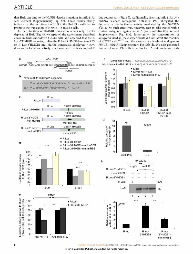
that HuR can bind to the HuRBS despite mutations in miR-1192seed element (Supplementary Fig. S7). These results clearlyindicate that the recruitment of HuR to the HuRBS is sufficient topromote the translation of HMGB1 in muscle cells.
As the inhibition of HMGB1 translation occurs only in cellsdepleted of HuR (Fig. 4), we repeated the experiments describedabove in HuR-knockdown C2C12 cells. We observed that the RLuc-30HMGB1 reporter, unlike the R Luc-30HMGB1-mut-miRBSor R Luc-30HMGB1-mut-HuRBS constructs, displayed B50%decrease in luciferase activity when compared with its control R
Luc counterpart (Fig. 6d). Additionally, silencing miR-1192 by amiRNA silencer (antagomir Anti-miR-1192) abrogated thedecrease in the luciferase activity mediated by the HMGB1-30UTR. No such effect was, however, seen in cells treated with acontrol antagomir against miR-16 (Anti-miR-16) (Fig. 6e andSupplementary Fig. S8a). Importantly, the concentration ofantagomir used in these experiments did not affect the viabilityof C2C12 cells35–37 and the steady state levels of endogenousHMGB1 mRNA (Supplementary Fig. S8b–d). We next generatedmimics of miR-1192 with or without an A-to-C mutation in its
200
***
R LucR Luc-3′HMGB1R Luc-3′HMGB1-mut-HuRBSR Luc-3′HMGB1-mut-miRBS
***
***
siCtr
12
10
8
6
4
2Rel
ativ
e am
ount
of
miR
NA
in H
uR IP
0miR-1192 U6
IP C2C12
R Luc-3′HMGB1-
mut-miR-BS:
R Luc-3′HMGB1:
R Luc:
HuR
–
– –
– –
–
1 3 4
kDa
32
2
–
–+
+
6 *** **
5
4
3
2
1
0
Rel
ativ
e am
ount
of
miR
-119
2 in
HuR
IP
R luc-3′HMGB1
R luc R luc-3′HMGB1-mut-miRBS
++
α-IgG α-HuR
***qPCR
qPCR
siHuR
**
siHuR
***R Luc
Mock*
Mimic miR-1192: 5′ –Mimic Mutant miR-1192: 5′ –
Mimic miR-1192
*
Mimic mutant miR-1192
1.6
1.4
1.2
1.0
0.8
0.6
0.4
0.2
0.0R Luc R Luc-3′-
HMGB1R Luc-3′-
HMGB1-mut-miRBS
R Luc-3′HMGB1
***
140
120
Luci
fera
se a
ctiv
ity r
elat
ive
to R
Luc
RN
A le
vel i
n si
HuR
-tre
ated
cel
ls
Luci
fera
se a
ctiv
ity r
elat
ive
toR
luc
RN
A le
vel
100
80
60
40
20
0Anti-miR-16 Anti-miR-1192
Luci
fera
se a
ctiv
ity r
elat
ive
to R
luc
RN
A le
vel
180160140120100
80604020
0
AAACAAACAAACAGACCAAAUU–
AAACCAACAAACAGACCAAAUU–
3′3′
miR-1192 BS
HMGB1:
HuRBS
1217
12251200CAGUAGUUUGGUUUUUUGUUUUUUUUUUUUUUUCUUUUGGUU
1243
a
R LucR Luc
R Luc-3′HMGB1
R Luc-3′HMGB1-mut-HuRBS
R Luc-3′HMGB1-mut-miRBS
3′UTR HMGB1
3′UTR HMGB1
3′UTR HMGB1
R Luc
HuRBS
mut-HuRBS
mut-miRBS
R Luc
R Luc
c
d
e
g
h
i
f
mmu-miR-1192/Hmgb1 alignmentb
uuAAACCAGACAAACAAACAAa
agUUUGGUUU–UUUGUUUUUUu
3′||||||:| ||||||| ||
5′
3′
mmu–miR–1192
Hmgb1305:5′
ARTICLE NATURE COMMUNICATIONS | DOI: 10.1038/ncomms3388
8 NATURE COMMUNICATIONS | 4:2388 | DOI: 10.1038/ncomms3388 | www.nature.com/naturecommunications
& 2013 Macmillan Publishers Limited. All rights reserved.

5th nucleotide (Fig. 6f). In HeLa cells, which do not expressendogenous miR-1192 (Supplementary Fig. S6), the mimic miR-1192 reduced the luciferase activity of R Luc-30HMGB1 byB50%. The mutant miR-1192 mimic, however, had no effect onthis activity (Fig. 6f). The luciferase activity of the R Luc-30HMGB1-mut-miRBS was not affected by the Mimic miR-1192,indicating that inhibition of R Luc-30HMGB1 translation occursvia a direct binding to the miRBS. Collectively, these resultsdemonstrate that in muscle cells, miR-1192 directly inhibits thetranslation of HMGB1 mRNA via the miRBS element, but thiseffect is overcome by the binding of HuR to its HuRBS.
Our IP combined to miRNA microarray data (SupplementaryTable S2), which were validated by performing IP/RT–qPCRexperiments (Fig. 6g) indicated that HuR and miR-1192 cancoexist in the same complex in C2C12 cells. To determinewhether this complex is indeed assembled on the HMGB1-30UTR,we performed the same IP/RT–qPCR experiment on C2C12 cellsexpressing either R Luc alone, R Luc-30HMGB1 or the R Luc-30HMGB1-mut-miRBS mRNAs. We observed that the level ofmiR-1192 in the HuR complex was fivefold greater in cellsexpressing the R Luc-30HMGB1 mRNA when compared with cellsexpressing mRNAs deficient in their ability to bind miR-1192 (RLuc or R Luc-30HMGB1-mut-miRBS) (Fig. 6h,i). Therefore, theseobservations clearly indicate that HuR and miR-1192 can bindsimultaneously the 30UTR of HMGB1 mRNA. We next verifiedmechanistically how HuR could negate the ability of miR-1192 torepress the translation of the HMGB1 mRNA. miRNAs areknown to translationally repress mRNAs via the recruitment ofthe Ago2, a component of the RNA-induced silencing complex(RISC)38. We thus verified if HuR prevents the miR-1192-mediated recruitment of Ago2 to the HMGB1-miRBS. Our dataindicate that although both HuR and miR-1192 simultaneouslybind to the HMGB1 mRNA 30UTR, the association of Ago2 toboth miR-1192 and the HMGB1 mRNA is increased when HuR isdepleted from C2C12 cells (Fig. 7). These experiments, therefore,indicate that HuR prevents the miR-1192-mediated translationalrepression of the HMGB1 mRNA by interfering with therecruitment of Ago2.
Our data show that HMGB1 is one of the mRNA targetsthrough which HuR promotes myogenesis. Specifically, theresults outlined in Fig. 6 suggest that HuR-depleted muscle cellsfail to enter myogenesis because in the absence of HuR, miR-1192inhibits HMGB1 translation. Hence, silencing miR-1192 shouldrescue HMGB1 expression in HuR-depleted muscle cells andshould also re-establish their myogenic potential. Our experi-ments showed that silencing miR-1192 but not miR-16 preventedthe inhibition of HMGB1 translation in HuR-depleted C2C12
cells (Fig. 8a,b and Supplementary Fig. S9a). In the presence ofendogenous HuR, however, Anti-miR-1192 had no effect onHMGB1 translation (Fig. 8a,b and Supplementary Fig. S9a). Thisresult was further confirmed by the fact that while the depletionof HuR shifted the distribution of HMGB1 mRNA towards lighterpolysome fractions (Fig. 4f), silencing miR-1192 reversed thiseffect (Supplementary Fig. S9b). Next, we assessed whether theAnti-miR-1192-mediated rescue of HMGB1 translation issufficient to promote the myogenic potential of HuR-knockdowncells. Indeed, silencing miR-1192 but not miR-16 re-establishedthe myogenic potential of HuR-depleted cells (Fig. 8c,d) and theexpression levels of My-HC, myoglobin and myogenin (Fig. 8e).
It was previously shown that miRNAs such as miR-519 andmiR-16 inhibit HuR expression in various cell lines39–41.However, as HuR levels does not significantly change duringthe early steps of myogenesis in vitro16,17,42 and increase duringmuscle regeneration in vivo30 (Supplementary Fig. S10), miRNA-mediated modulation of HuR expression in muscle cells seems tobe ineffective in these conditions. Our experiments show thatwhile the expression level of miR-16 increases during myogenesisboth in vitro and in vivo (Supplementary Fig. S11a,b), it is thedepletion but not the increased expression of miR-16 thatmodestly reduces the expression level of HuR, with limited effectson HMGB1 as well as My-HC and myoglobin (Fig. 8e). In fact, amarked reduction of HMGB1 expression and the inhibition ofmyogenesis is observed only when HuR level is further reducedby small interfering RNA (Fig. 8e). These observations indicatethat miR-1192 is active only when HuR level is below a criticalthreshold. All together, our data support a model whereby HuRhelps undifferentiated muscle cells to enter myogenesis bysupporting the translation of HMGB1 transcripts and preventingthe action of Ago2/miR-1192 (Fig. 8f).
DiscussionIn this study we show that HMGB1 is required for muscle fibreformation both in vitro and in vivo and uncover a novelmechanism by which muscle cells modulate HMGB1 expression.A general reduction in skeletal muscle tissues was previouslyobserved in Hmgb1� /� mice, among several other defects24.Here we show that a precise level of HMGB1 expression isrequired for proper muscle fibre formation, as a 50% reduction inHMGB1 expression in embryos significantly decreases theefficiency of myogenesis. Thus, a tight control of HMGB1expression levels appears essential for myogenesis.
More than a decade ago, it was discovered that HMGB1 mRNAharbours a long 30UTR with U-rich elements43–45 suggesting that
Figure 6 | HuR binding to a U8 element within HuRBS is sufficient to prevent the miR-1192-mediated inhibition of HMGB1 translation. (a) Schematic
representation of the HuRBS and the seed element of miR-1192 (miR-1192 BS). (b) Diagram illustrating the bioinformatic approach used to predict
miR-1192 as a putative miRNA targeting HMGB1 30UTR. Shown here are complementarities of miR-1192 with HMGB1 30UTR. (c) Schematic diagrams of
luciferase constructs. (d–g) Effects of HuR, miRNAs, antagomirs and mimics. Error bars represent s.e.m of three independent experiments. ***Po0.0001,
**Po0.001, *Po0.05 (Student’s t-test). The amount of reporter RNA expressed in cells was determined by qPCR and used to normalize R Luc activities for
each treatment. (d) Exponentially growing C2C12 cells were treated with siCtr or siHuR and 24 h later luciferase constructs were introduced. Luciferase
activity of R Luc construct was considered as 100%. (e) miR-1192 and miR-16 antagomirs were transfected in siHuR-treated C2C12 cells previously
transfected either with R luc alone or R luc-30HMGB1. (f) Upper panel: schematic representation of the Mimic miR-1192 and Mimic mutant miR-1192. The
asterisk (*) indicates the a–c mutation in the Mimic mutant miR-1192. Lower panel: HeLa cells were transfected with Mimic miR-1192 or Mimic
mutant miR-1192 and 24 h later luciferase constructs were transfected. The luciferase activity of the mock-treated cells was set as reference.
(g) Immunoprecipitation experiments were performed using a monoclonal HuR antibody, or IgG as a control, on total cell lysates from exponentially
growing C2C12 cells. RNA was isolated from the immunoprecipitate, and quantitative RT–PCR was performed using primers specific to miR-1192, miR-16
and U6 RNAs. The levels of miR-1192 and U6 RNAs in each IP were normalized against the level of miR-16. Error bars represent s.e.m of three independent
experiments. T-test was used for statistical analysis. (h,i) Immunoprecipitation experiments were performed as in (g) on total cell lysates from C2C12
cells transfected with either R luc alone, R luc-30HMGB1 or R luc-30HMGB1-mut-miRBS. (h) Western blot was performed using an HuR antibody. This blot is
a representation of three independent experiments. (i) The levels of miR-1192 in each IP were determined and are plotted with the s.e.m. of three
independent experiments. ***Po0.0001, **Po0.001 (Student’s t-test).
NATURE COMMUNICATIONS | DOI: 10.1038/ncomms3388 ARTICLE
NATURE COMMUNICATIONS | 4:2388 | DOI: 10.1038/ncomms3388 | www.nature.com/naturecommunications 9
& 2013 Macmillan Publishers Limited. All rights reserved.

posttranscriptional events could regulate HMGB1 expression.Surprisingly, this possibility was never explored. Our data clearlyestablish that a posttranscriptional mechanism, via HuR, has akey role in promoting HMGB1 expression in muscle cells. HuR,one of the well known posttranscriptional regulators, affects theexpression of its target mRNAs by binding to specific U-richelements in their 30UTRs14,15. We show that the 30UTR ofHMGB1 mRNA harbours such an element, the HuRBS, throughwhich HuR promotes the translation of HMGB1 mRNA. In theabsence of HuR, however, the microRNA miR-1192 is able toinhibit the expression of HMGB1. To our knowledge, this is thefirst report of the impact of a miRNA on HMGB1 expression.Incidentally, this is also the first report on the biological activityof miR-1192. Using several different techniques includingmiRNA microarray analysis, RT–qPCR with specific primersfor miR-1192, and the sequencing of the PCR product that wasgenerated by these primers, our data indicate that miR-1192 isexpressed in muscle cells. Previous reports using RNA sequencing(RNA-seq), however, have failed to detect miR-1192 in the wholemouse embryo and muscle tissues46,47. As demonstrated by
several recent studies48,49, technical biases during capture andpreparation of the small RNA library has an impact on detectionof small RNA molecules. Despite potential technical differences inmethods used to precisely quantify the expression level of miR-1192 in muscle cells, our data clearly show that this miRNA isexpressed in muscle cells and is functionally relevant inmodulating HMGB1 expression.
As HuR, HMGB1 and miR-1192 are highly expressed inmuscle cells and during muscle regeneration30 (SupplementaryFigs S10 and S11) and both HuR and miR-1192 can simulta-neously associate with the HMGB1-30UTR, our data support amodel whereby the binding of HuR to its HuRBS is sufficient toprevent miR-1192-mediated inhibition. Interestingly, the associa-tion of miR-1192 with Ago2, a key player in the RISCcomplex38,50, is dramatically enhanced in muscle cells depletedof HuR. This suggests that HuR prevents the action of miR-1192by interfering with the recruitment of Ago2 to the HMGB130UTR. These data are consistent with previous observationsshowing that HuR promotes the translation of target messages bypreventing the assembly of an active RISC complex on the Let-7
C2C12
siRNA: siRNA:
HuR
Tubulin
IP Ago24
3
2
1
0siCtr
Rel
ativ
e am
ount
of
HM
GB
1 m
RN
A
4
3
2
1
0
Rel
ativ
e am
ount
of
miR
-16
siHuR
siCtr siHuR
qPCRIP Ago2
IP Ago2
4
3
2
1
0siCtr
Rel
ativ
e am
ount
of
miR
-119
2
siHuR
qPCR
qPCR
Ctr CtrHuR HuRkDa
IP: IgG Ago2 IgG Ago2 kDa
50Ago2
1 2 3 4
32
50
21
a b
c d
e
Figure 7 | HuR binding to the HuRBS prevents the recruitment of Ago2 to the HMGB1 mRNA. (a) Western blot analysis was performed using
antibodies against HuR and a-tubulin as a loading control on total cell extracts obtained from exponentially growing C2C12 cells treated with a control (Ctr)
or HuR specific (siHuR) siRNA. (b) Immunoprecipitation experiments were performed using a monoclonal Ago2 antibody, or anti-IgG antibody as a
control, on the total cell lysates described in (a). The immunoprecipitation of Ago2 was then assessed by western blotting using an anti-Ago2 antibody.
(c–e) RNA was isolated from the immunoprecipitate described above, and quantitative RT–PCR was performed using primers specific to (c) HMGB1
(d) miR-1192 (e) miR-16. The levels of HMGB1 mRNA, miR-1192 and miR-16 in each IP, relative to those in the IgG IP, were respectively normalized against
the GAPDH mRNA and U6 levels. Error bars represent s.e.m of three independent experiments.
ARTICLE NATURE COMMUNICATIONS | DOI: 10.1038/ncomms3388
10 NATURE COMMUNICATIONS | 4:2388 | DOI: 10.1038/ncomms3388 | www.nature.com/naturecommunications
& 2013 Macmillan Publishers Limited. All rights reserved.

Ago2Ago2
Mock
HuRCtrHuRCtr
HuRCtr HuR kDaCtr
HMGB1
siRNA:
HuR
Tubulin
1 2 3 4 5 6
50
32
30
HuR kDaCtr
Anti-miR-1192
Anti-miR-16
Ant
i-miR
-16
Anti-miR-16Anti-miR-1192
Anti-miR16
Fus
ion
inde
x
Ant
i-miR
-119
2
Anti-miR-1192
My-HC DAPI Merge
Day 3 of C2C12 differentiation
siCtr
siCtr
siHuR
siHuR
1.2
0.8
0.6
0.4
0.2
0siCtr siRNA:siHuR siCtr siHuR siCtr siHuR
Mock
1
HM
GB
1 pr
otei
n le
vel
rel
ativ
e to
tubu
lin
0.9
0.8
0.7
0.6
0.5
0.4
0.3
0.2
0.1
0.0
siCtrsiHuR***
Anti-miR: 1192 16
32
30
20
36
50
4321
Tubulin
Myogenin
Myoglobin
MyHC
HMGB1
HuR
220
5′UTR 3′UTR
HMGB1 mRNAtranslation
MyogenesisHMGB1 CR
miR-1192
5′UTR 3′UTR
HMGB1 mRNAtranslation
MyogenesisHMGB1 CR
miR-1192
HuR
*** ***
a
b
c
d
e
f
Figure 8 | Silencing miR-1192 reestablishes HMGB1 translation and rescues the myogenic potential of HuR-depleted muscle cells. (a) Exponentially
growing C2C12 were treated with siCtr or siHuR and were transfected 5 h later with antagomirs to miR-1192 or miR-16. Extracts from these
cells were harvested and used for western blotting with anti-HMGB1, -HuR and -a-tubulin antibodies. (b) The expression level of HMGB1 protein relative to
a-tubulin. Error bars represent s.e.m. of three independent experiments. ***Po0.0001 (student’s t-test). (c) C2C12 treated as in (a) were grown to
confluency, induced to differentiate for three days and stained with anti-My-HC antibody and DAPI. Scale bars, 20mm. The images shown are
representative fields for each cell treatment from three independent experiments. (d) Fusion index of cells in (c). Error bars represent s.e.m. of three
independent experiments. ***Po0.0001 (student’s t-test). (e) Total extracts from cells treated as described above were blotted and probed with
antibodies against HuR, HMGB1, My-HC, myoglobin, myogenin and a-tubulin. A representative blot of two independent experiments is shown.
(f) Schematic model illustrating how HuR promotes the myogenesis in undifferentiated muscle cells. HuR recognizes an U8 element in the HMGB1-30UTR
adjacent to the seed element of miR-1192, and promotes HMGB1 translation and myogenesis. HuR mediates this effect even if miR-1192 is bound to
its seed element (upper panel). In the absence of HuR, however, miR-1192 recruits Ago2 thus inhibiting HMGB1 translation and preventing myogenesis
(lower panel).
NATURE COMMUNICATIONS | DOI: 10.1038/ncomms3388 ARTICLE
NATURE COMMUNICATIONS | 4:2388 | DOI: 10.1038/ncomms3388 | www.nature.com/naturecommunications 11
& 2013 Macmillan Publishers Limited. All rights reserved.

miRNA seed element51 even if its binding site is far away fromthe miRNA seed element. Therefore, these observations with ourdata suggest that HuR promotes the translation of target mRNAsby interfering with the formation of an active RISC complexregardless of the distance of its binding site from the seedelement. This mechanism of action is different from the onedescribed for the dead end 1 protein, another RBP, whichpromotes translation of the cyclin-kinase inhibitor p27cip bypreventing the recruitment of miR-221/222 to their seedelements52. Therefore, our findings not only add anotherexample to the few studies on the cross talk between RNA-binding proteins and miRNAs, but also uncover a novel way bywhich cells, via proteins such as HuR, control miRNA-mediatedeffects. Our data demonstrate that via such a mechanism HuRinduces the translation of HMGB1, which in turn promotes theentry of muscle cells into myogenesis.
By linking the interplay between HuR and miR-1192 to thetranslation modulation of HMGB1, we found a posttranscrip-tional mechanism that controls the expression of the main‘alarmin’ in the organism53,54. HMGB1 is a damage-associatedmolecular pattern molecule that upon severe injury activates theinnate immune system to recognize tissue damage and initiatereparative responses55,56. Typically, the increase in HMGB1 levelsin stressed or activated inflammatory cells precedes theupregulation of HMGB1 transcript levels by several hours56.Our results suggest that posttranscriptional events might beresponsible for this increase, as the HMGB1 mRNAs are bothabundant43,44 and stable (Supplementary Fig. S2). Therefore, ourstudy may open the door to new strategies to manipulate HMGB1levels under various conditions. Such a strategy could helppromote the beneficial effects of HMGB1 (for example, theactivation of muscle regeneration and wound healing) whilelimiting/preventing the deleterious outcomes of its overproduc-tion during life-threatening assaults (for example, ischaemia,burn, infection or sepsis).
MethodsPlasmid construction. The pCMV-SPORT6 plasmid containing the full-lengthHMGB1 cDNA (accession number: BC008565) was purchased from Open Bio-systems (catalogue number: MMM1013-64094). pRL-luc-30HMGB1-mut-miRBSwas generated by Norclone Biotech Laboratories, Kingston, ON, Canada. The full-length 30UTR of mouse HMGB1 was subcloned into a pRL-SV40 vector (Promega)by performing PCR amplification using the following primers: forward 50-TTGGTT CTA GCG CAG TTT TT-30 and reverse 50-TCA TCC AGG ACT CAT GTTCAG-30 . The pRL-SV40 vector was digested by Xba1 restriction enzyme (NewEngland Biolabs), followed by a treatment with the T4 DNA polymerase, and thendephosphorylated. The PCR insert was ligated into the plasmid using the QuickLigase enzyme (Promega) according to the manufacturer’s instructions. pRL-luc-30HMGB1-mutHuRBS was generated using a specific primer containing themutations and Quickchange site-directed mutagenesis kit (Stratagene). The MIS-SION short hairpin RNA (shRNA) plasmid (siHMGB1; Sigma-Aldrich) was usedto knock down HMGB1. MISSION pLKO.1-puro vector encoding scrambledshRNA (siCtr; SHC001) was used as a control.
Cell culture and transfection. C2C12 muscle cells (ATCC) were grown in mediacontaining 20% fetal bovine serum (Invitrogen) in DMEM (Dulbecco’s modifiedeagle medium from Invitrogen). In order to induce muscle cell differentiation, cellswere switched to a media containing DMEM, 2% horse serum, penicillin/strep-tomycin antibiotics (Invitrogen), and 50 mM HEPES, pH 7.4 (Invitrogen) whentheir confluency reached 100% (refs 17,42). Hmgb1þ /þ and Hmgb1� /� mouseembryonic fibroblasts (MEFs) (HMGBiotech, Milan, Italy) were cultured inDMEM supplemented with penicillin/streptomycin and 10% fetal bovine serum(Sigma-Aldrich). Transfections with small interfering RNAs specific for HuR orplasmids were performed using Lipofectamine and Plus reagents (Invitrogen) orjetPEI (Polyplus Transfection) according to the manufacturer’s instructions. Thesame transfection protocol was used to treat C2C12 cells with 200 nM miR-1192 ormiR-16 antagomirs (Dharmacon).
HMGB1 rescue experiments. Transient transfection of C2C12 cells was carriedout using jetPEI (Polyplus Transfection), as recommended by the manufacturer.Briefly, C2C12 cells were transfected with the MISSION shRNA plasmid (Sigma-
Aldrich) to knock down HMGB1 or with the MISSION pLKO.1-puro vectorencoding a scrambled shRNA (SHC001) as a control. Forty-eight hours post-transfection, the cells were switched to differentiation media and cultured foradditional 48 h in the absence or presence of recombinant HMGB1 (400 nM).
Preparation of cell extracts and immunoblotting. Total cell extracts were pre-pared by incubating undifferentiated or differentiated C2C12 cells on ice for 15 minwith lysis buffer (50 mM HEPES pH 7.0, 150 mM NaCl, 10% glycerol, 1% Triton,10 mM pyrophosphate sodium, 100 mM NaF, 1 mM EGTA, 1.5 mM MgCl2,1� protease inhibitor (Roche), 0.1 M orthovanadate, 0.2 M PMSF), then centrifugeat 12,000 g at 4 �C and the supernatant was kept17. The extracts were run on anSDS–PAGE and transferred to nitrocellulose membranes (Bio-Rad). The sampleswere analysed by western blotting57 with antibodies against HuR (3A2,1:10,000)57,HMGB1 (Abcam,1:1,000), My-HC (MY32;Sigma,1:1,000), Myogenin (F5D,Developmental studies Hybridoma Bank, 1:250), a-tubulin (Developmental studiesHybridoma Bank, 1:1,000), hnRNPA1 (Cell Signalling, 1:1,000)22, b-actin(Sigma,1:500), myoglobin (DAKO, 1:500) and Ago2 (Cell Signalling,1:1000). Fullblots are provided in Supplementary Fig. S12.
Immunofluorescence. IF was performed using undifferentiated or differentiatedC2C12 cells that were grown to sub-confluency in DMEM (Dulbecco’s modifiedeagle medium from Invitrogen)17. After the appropriate experimental treatments,cells were rinsed twice in phosphate-buffered saline (PBS), fixed in 3% phosphate-buffered paraformaldehyde (Sigma) and permeabilized in 0.5% PBS-goat serumwith Triton. After permeabilization, cells were incubated with primary antibodiesagainst My-HC (MF-20, developmental studies Hybridoma Bank, 1:1,000), HuR(3A2,1:1,000) or HMGB1 (Abcam,1:1,000)) for 1 h at room temperature and thenincubated with goat anti-mouse or anti-rabbit secondary antibodies conjugatedwith Rhodamine (red) or FITC (green) from Molecular Probes (Eugene, OR). Tovisualize the nucleus, cells were stained with DAPI (Molecular Probes).Microscopic analyses were performed using an AXIOVERT 200 M (Zeiss).
b-Galactosidase staining. MLC1/3F-nlacZ homozygous transgenic mice25 weremated with Hmgb1þ /� heterozygotes. Pregnant females were killed by cervicaldislocation at E10.5. Embryos were isolated in PBS, fixed for 2 h in 4%paraformaldehyde pH 7.4 at 4 �C, washed in PBS for 20 min and equilibrated in20% sucrose PBS. Fixed embryos were then stained for 1 h at 37 �C in 1 mg ml� 1
X-gal solution of PBS, also containing 5 mM K4Fe(CN)6, 5 mM K3Fe(CN)6, 2 mMMgCl2, 0.2% NP-40 PBS. Hmgb1þ /� embryos were identified by PCR analysis ofyolk sac DNA using the following primers: HMGB1for 50-GCA GGC TTC GTTGTT TTC ATA CAG-30 and HMGB1rev 50-TCA AAG AGT AAT ACT GCC ACCTTC-30 for the wt allele; NEOfor 50-TGG TTT GCA GTG TTC TGC CTA GC-30
and NEOrev 50-CCC AGT CAT AGC CGA ATA GCC-30 for the targeted allele.These animal studies were approved by the San Raffaele University Animal CareCommittee.
Histology and morphometric analysis. TA muscles were dissected from 1-year-old wild-type and Hmgb1þ /� mice and frozen in liquid nitrogen-cooledisopentane. Serial sections (8mm thick) were stained with hematoxylin and eosin.Morphometric analyses were performed on sections of TA using ImageJ todetermine the cross-sectional area of 900 fibres for each group.
Fusion index. Explants of presomitic mesoderm58 were dissected from wt,Hmgb1þ /� and Hmgb1� /� embryos at E9.5, plated on collagen-coated dishesand cultured for 4 days in DMEM supplemented with 20% FCS and 50 mg ml� 1
gentamycin. On day 4, cultures were processed for immunoflorescence with anantibody specific for My-HC (MF-20, Developmental Studies Hybridoma Bank,1:20 dilution). Fusion index was determined as the number of nuclei in sarcomericmyosin-expressing cells with more than two nuclei versus the total number ofnuclei.
Regeneration assay. Muscle injury was performed on the TA of 3-month-old wtand Hmgb1þ /� mice by injecting 50 ml of 10mM CTX (three animals per group).Mice were killed 3, 7 or 14 days after CTX injection, and the TA muscles weredissected and frozen in liquid N2–cooled isopentane. Ten-micrometre serial musclesections were stained with hematoxylin and eosin. Alternatively, injury of C57BL/6(Charles River) mouse muscles was performed by BaCl2 injection in the TA muscleof 8-week old mice, under zolazepam/tiletamine anaesthesia59. These animalstudies were approved by the San Raffaele University Animal Care Committee.
RNA electromobility shift assays. The HMGB1 cRNA probes were produced byin vitro transcription using a T7 RNA polymerase60. All HMGB1 probes (50UTRand P1 to P12) were generated by PCR amplification using a forward primer fusedto the T7 promoter as well as pCMV-HMGB1 expression vector as the template(see Supplementary Table S4). For smaller probes (P4-1 to P4-3), oligonucleotidesense and anti-sense were directly annealed and used for in vitro transcription. TheRNA-binding assays were performed22 by incubating either 10 mg total cell extracts
ARTICLE NATURE COMMUNICATIONS | DOI: 10.1038/ncomms3388
12 NATURE COMMUNICATIONS | 4:2388 | DOI: 10.1038/ncomms3388 | www.nature.com/naturecommunications
& 2013 Macmillan Publishers Limited. All rights reserved.

or 300 ng purified recombinant protein (GST or GST-HuR) with 50,000 c.p.m. of32P-labelled cRNAs in a total volume of 20 ml EBMK buffer (25 mM HEPES pH 7.6,1.5 mM KCl, 5 mM MgCl2, 75 mM NaCl, 6% sucrose and protease inhibitors) at RTfor 15 min. For competition assays, 0.01� , 0.1� , 1� , 10� and 100� excessunlabelled specific or unspecific transcripts were incubated with the TCE for15 min at RT before the 32P-labelled probes were added to the reaction. Twomicrolitres ml of a 50 mg ml� 1 heparin sulphate stock solution was then added tothe reaction for an additional 15 min at RT. In supershift experiments, 5 mg of apurified monoclonal anti-HuR antibody was then added to the reaction for anadditional 15 min at RT. Samples were then loaded on a 4% polyacrylamide gelcontaining 0.05% NP-40.
Quantitative RT–PCR. One microgram of total RNA was reverse transcribed usingthe M-MuLV Reverse Transcriptase (New England Biolabs) according to themanufacturer’s protocol. A total of 1/80 dilution of cDNA was used to detectHMGB1, GAPDH and R luc mRNAs using SsoFast EvaGreen Supermix (Bio-Rad).Expression of HMGB1 and R luc was standardized using GAPDH as a reference,and relative levels of expression were quantified by calculating 2�DDCT, whereDDCT is the difference in CT between target and reference.
For miRNA detection, 200 ng of total RNA was reverse transcribed using theUniversal cDNA synthesis kit (Exiqon) and the presence of miR-1192, miR-16 andU6 was assessed by qPCR using the SyBr Green Master Mix (Exiqon).
Northern blot analysis and Actinomycin D pulse-chase experiments. Theextraction of total RNA was performed using Trizol reagent (Invitrogen)22. For theisolation of miRNAs, twice as much isopropanol was used in the RNA purificationprotocol. Northern blot analysis was performed using 10 mg total RNA22. Aftertransferring to a Hybond-N membrane (Amersham) and UV-cross-linking, theblot was hybridized with probes specific for MyoD17, GAPDH17, 18S61, 5.8S34 andHMGB1 mRNAs were generated using the PCR Purification Kit (GE Healthcare)with primers described below. The probes were radiolabelled with a� 32P dCTPusing Ready-to-Go DNA labelling beads (GE Healthcare) according to themanufacturer’s instructions. The stability of HMGB1 mRNA was assessed by theaddition of the general transcriptional inhibitor actinomycin D (5 mg ml� 1)61 forthe indicated periods of time. Total RNA was isolated from the cells after 0, 4, 6, 8,10 and 12 h following ActD treatment using TRIzol reagent (Invitrogen), andanalysed by northern blotting. Full blots are provided in Supplementary Fig S12.
For miRNA detection, 25 mg of total RNA was separated on a 12% denaturingurea polyacrylamide gel. RNA was subsequently transferred to a Hybond-Nþ
membrane (Amersham Biosciences) and cross-linked to the membrane.Hybridization was carried out by using ULTRAHybOligo solution according to themanufacturer’s instructions (Ambion). The probe sequences were complementaryto the mature forms of miR-16 or U6 RNA and were labelled using StarFire system(Integrated DNA Technologies) according to the manufacturer’s protocol.
Polysome fractionation. Forty million myoblasts were grown and treated withsmall interfering RNAs as described above and polysome fractionation experimentswere performed. Briefly, the cytoplasmic extracts obtained from lysed myoblastcells were centrifuged at 130 000 g for 2 h on a sucrose gradient (10–50% w/v)62,63.Polysomal (P) or non-polysomal (NP) fractions were pooled and RNA wasextracted using Trizol LS (Invitrogen). RNA samples were then analysed on anagarose gel. The levels of HMGB1 and GAPDH mRNAs were determined usingquantitative RT–PCR.
Luciferase activity. The activity of R Luc was measured using a Renilla luciferaseassay system (Promega) with a luminometer following the manufacturer’sinstructions.
Fluorescence in situ hybridization. The fluorescence in situ hybridizationexperiments were performed29 using a DNA fragment of B500 bp correspondingto the coding region of mouse HMGB1. The fragment was amplified by PCR usingthe following primers fused to either a T7 or T3 minimal promoter sequence:HMGB1 forward, 50-AAA AAG CCG AGA GGC AAA AT-30, and HMGB1reverse, 50-CTT TTT CGC TGC ATC AGG TT-30 . The PCR product was used asthe template for in vitro transcription of the HMGB1 probe needed for fluorescencein situ hybridization. The anti-sense (T3) and sense (T7) probes were preparedusing digoxigenin-RNA labelling mix from Roche Diagnostics. The RNA probeswere quantified, denatured and incubated with permeabilized cells at 37 �Covernight in the hybridization buffer (50% formamide, 5� SSC, 50 mM phosphatebuffer, pH¼ 7.4, 5� Denhardt’s, 1 mM EDTA and 250 ng ml� 1 of salmon spermDNA). After the hybridization, the cells were used for IF to detect the HMGB1mRNA and HuR protein29. Finally, the cells were incubated with secondary goatanti-rabbit antibody and anti-DIG antibody for IF.
Preparation of mRNA complexes and analysis with RT–PCR. TCE wereprepared in lysis buffer (50 mM Tris, pH 8; 0.5% Triton X-100; 150 mM NaCl;complete protease inhibitor from Roche). Twenty-five microlitres of the anti-HuR
(3A2) or IgG (Jackson Immunoresearch Laboratories) antibodies were incubatedwith 200 ml of pre-swelled protein A-Sepharose beads for 4 h at 4 �C. Alternatively,C2C12 cells depleted or not of HuR were incubated with an anti-Ago2 (CellSignalling) or IgG control antibody. After three washes in lysis buffer, 1.5 mg of cellextract was added overnight at 4 �C. The final dilution of the antibodies during theincubation with lysates is 1/18. Beads were washed three times with cell lysis buffer,incubated with proteinase K and the RNA isolated by phenol/chloroform extrac-tion followed by precipitation overnight at � 20 �C with isopropanol17,64. PurifiedRNA was resuspended in 12 ml of water, and 2 ml was reverse transcribed using theM-MuLV Reverse Transcriptase (New England Biolabs) according to themanufacturer’s protocol. Association of HMGB1 and b-actin mRNAs with HuR aswell as HMGB1 with Ago2 was defined using quantitative RT–PCR. Ten micro-litres of purified RNA was reverse transcribed using the Universal cDNA synthesiskit (Exiqon) and the presence of miR-1192, miR-16 and U6 was assessed by qPCR.
cDNA array analysis. Microarray experiments were performed using mousearray, which contain 17,000 probe sets of known and unknown expressed sequencetags65. HuR was immunoprecipitated from exponentially growing C2C12 cellsusing an anti-HuR monoclonal antibody (3A2) and an anti-IgG monoclonalantibody as a negative control. The final dilution of the antibodies during theovernight incubation with lysates is 1/18. Then the samples were spun 5 min at-3,000 g and washed with TSE1 (0.1% SDS, 1% Triton X-100, 2 mM EDTA, 20 mMTris-HCl pH: 8.1, 150 mM NaCl), TSE2 (0.1% SDS, 1% Triton X-100, 2 mMEDTA, 20 mM Tris-HCl pH: 8.1, 500 mM NaCl) and TSE3 (0.25 M, LiCl, 1% NP-40, 1% deoxycholate, 1 mM EDTA, 10 mM Tris-HCl pH: 8.1) buffers. Theimmunoprecipitate were then incubated for 30 min at 55 �C in 100 ml NT2 buffer(50 mM Tris pH:7.4, 150 mM NaCl, 1 mM MgCl2, 0.05% NP-40) with0.1% SDSand 30 mg proteinase K. The associated RNAs were extracted by adding B15 ml of2 M NaOAc pH: 4.0, 150ml of water saturated phenol and B30ml chloroformfollowed by 15 min incubation on ice. After spinning (12,000 g for 20 min) theaqueous phase is mixed with glycogen and isopropanol and incubated overnight at� 20 �C. The isolated RNAs were then resuspended in water and hybridized oncDNA arrays. The data were processed using the Array Pro software (MediaCybernetics), then normalized by Z-score transformation66 and used to calculatedifferences in signal intensities. Significant values were tested using a two-tailed Z-test and a P of r0.01. The data were calculated from two independentexperiments.
Subcellular fractionation. Subcellular fractionation was performed using thePARIS kit (Ambion) (Austin, TX) according the manufacturer’s instructions. Anequal number of cells per sample were used.
Biotin pull-down assay. The P4 fragment of the 30UTR of mouse HMGB1 wassubcloned into a pGEM-Teasy vector (Promega) according to the manufacturer’sinstructions by performing PCR amplification using the following primers: forward50-GCC ACT AAC CTT GCC TGG TA-30 and reverse 50-TCG TAT AAG CTGCAT CAG AGA CA-30 . The transcript is transcribed using the T7RiboMAXExpress large scale RNA production system (Promega) according to themanufacturer’s instructions using the NotI digested PGEM-Teasy-P3P4 vector as atemplate. For preparing protein extracts, 30–40 millions of cells were washed withice-cold PBS and resuspended in 1 ml of EBMK buffer (25 mM Hepes pH 7,6;5 mM MgCl2; 1,5 mM KCl; 75 mM NaCl; 175 mM sucrose) containing 0,5% NP-40,complete protease inhibitor without EDTA (Roche). Lysates were sonicated for 10 s3 times at 200 W and cleared by centrifugation at 12,000g for 15 min. The proteinconcentration was determined (Bio-Rad microassay) and 1 mg were used for eachassay. Biotin pull-down assay is performed using Miltenyi Biotech mMACSstreptavidin kit according to the manufacturer’s instructions.
Primers used to prepare probes for northern blot analysis. mHMGB1: For: 50-GCA TCC TGG CTT ATC CAT TG-30 ; rev: 50-TGC TCT TTT CAG CCT TGACC-30
mGAPDH: For: 50-AAG GTC ATC CCA GAG CTG AA-30 ; rev: 50-AGG AGACAA CCT GGT CCT CA-30
Statistical analyses. The statistical analyses in this study were performed usingthe Graphpad Prism5 software to determine significance (two-tailed, Student’s t-test).
References1. Charge, S. B. & Rudnicki, M. A. Cellular and molecular regulation of muscle
regeneration. Physiol. Rev. 84, 209–238 (2004).2. Zhang, K., Sha, J. & Harter, M. L. Activation of Cdc6 by MyoD is associated
with the expansion of quiescent myogenic satellite cells. J. Cell Biol. 188, 39–48(2010).
3. De Mori, R. et al. Multiple effects of high mobility group box protein 1 inskeletal muscle regeneration. Arterioscler. Thromb. Vasc. Biol. 27, 2377–2383(2007).
NATURE COMMUNICATIONS | DOI: 10.1038/ncomms3388 ARTICLE
NATURE COMMUNICATIONS | 4:2388 | DOI: 10.1038/ncomms3388 | www.nature.com/naturecommunications 13
& 2013 Macmillan Publishers Limited. All rights reserved.

4. Filippin, L. I., Moreira, A. J., Marroni, N. P. & Xavier, R. M. Nitric oxide andrepair of skeletal muscle injury. Nitric Oxide 21, 157–163 (2009).
5. Palumbo, R. et al. Extracellular HMGB1, a signal of tissue damage, inducesmesoangioblast migration and proliferation. J. Cell Biol. 164, 441–449 (2004).
6. Takaesu, G. et al. Activation of p38alpha/beta MAPK in myogenesis viabinding of the scaffold protein JLP to the cell surface protein Cdo. J. Cell Biol.175, 383–388 (2006).
7. Serra, C. et al. Functional interdependence at the chromatin level between theMKK6/p38 and IGF1/PI3K/AKT pathways during muscle differentiation. Mol.Cell 28, 200–213 (2007).
8. Jones, N. C. et al. The p38alpha/beta MAPK functions as a molecular switch toactivate the quiescent satellite cell. J. Cell Biol. 169, 105–116 (2005).
9. Sorci, G., Riuzzi, F., Arcuri, C., Giambanco, I. & Donato, R. Amphoterinstimulates myogenesis and counteracts the antimyogenic factors basic fibroblastgrowth factor and S100B via RAGE binding. Mol. Cell Biol. 24, 4880–4894(2004).
10. Riuzzi, F., Sorci, G. & Donato, R. RAGE expression in rhabdomyosarcoma cellsresults in myogenic differentiation and reduced proliferation, migration,invasiveness, and tumor growth. Am. J. Pathol. 171, 947–961 (2007).
11. Riuzzi, F., Sorci, G. & Donato, R. S100B stimulates myoblast proliferation andinhibits myoblast differentiation by independently stimulating ERK1/2 andinhibiting p38 MAPK. J. Cell Physiol. 207, 461–470 (2006).
12. Riuzzi, F., Sorci, G., Sagheddu, R. & Donato, R. HMGB1-RAGE regulatesmuscle satellite cell homeostasis through p38-MAPK- and myogenin-dependent repression of Pax7 transcription. J. Cell Sci. 125, 1440–1454 (2012).
13. Begum, N., Pash, J. M. & Bhorjee, J. S. Expression and synthesis of highmobility group chromosomal proteins in different rat skeletal cell lines duringmyogenesis. J Biol Chem 265, 11936–11941 (1990).
14. Abdelmohsen, K., Kuwano, Y., Kim, H. H. & Gorospe, M. Posttranscriptionalgene regulation by RNA-binding proteins during oxidative stress: implicationsfor cellular senescence. Biol. Chem. 389, 243–255 (2008).
15. von Roretz, C. & Gallouzi, I. E. Decoding ARE-mediated decay: is microRNApart of the equation? J. Cell Biol. 181, 189–194 (2008).
16. Figueroa, A. et al. Role of HuR in skeletal myogenesis through coordinateregulation of muscle differentiation genes. Mol. Cell Biol. 23, 4991–5004 (2003).
17. van der Giessen, K., Di-Marco, S., Clair, E. & Gallouzi, I. E. RNAi-mediatedHuR depletion leads to the inhibition of muscle cell differentiation. J. Biol.Chem. 278, 47119–47128 (2003).
18. von Roretz, C., Beauchamp, P., Di Marco, S. & Gallouzi, I. E. HuR andmyogenesis: being in the right place at the right time. Biochim. Biophys. Acta.1813, 1663–1667 (2011).
19. Yaffe, D. & Saxel, O. Serial passaging and differentiation of myogenic cellsisolated from dystrophic mouse muscle. Nature 270, 725–727 (1977).
20. Conejo, R., de Alvaro, C., Benito, M., Cuadrado, A. & Lorenzo, M. Insulinrestores differentiation of Ras-transformed C2C12 myoblasts by inducing NF-kappaB through an AKT/P70S6K/p38-MAPK pathway. Oncogene 21,3739–3753 (2002).
21. Li, X. L., Andersen, J. B., Ezelle, H. J., Wilson, G. M. & Hassel, B. A. Post-transcriptional regulation of RNase-L expression is mediated by the 3’-untranslated region of its mRNA. J. Biol. Chem. 282, 7950–7960 (2007).
22. Dormoy-Raclet, V. et al. The RNA-binding protein HuR promotes cellmigration and cell invasion by stabilizing the beta-actin mRNA in a U-rich-element-dependent manner. Mol. Cell Biol. 27, 5365–5380 (2007).
23. Buckingham, M. et al. The formation of skeletal muscle: from somite to limb.J. Anat. 202, 59–68 (2003).
24. Calogero, S. et al. The lack of chromosomal protein Hmg1 does not disrupt cellgrowth but causes lethal hypoglycaemia in newborn mice. Nat. Genet. 22,276–280 (1999).
25. Kelly, R., Alonso, S., Tajbakhsh, S., Cossu, G. & Buckingham, M. Myosin lightchain 3F regulatory sequences confer regionalized cardiac and skeletal muscleexpression in transgenic mice. J. Cell Biol. 129, 383–396 (1995).
26. Cossu, G. et al. Activation of different myogenic pathways: myf-5 is induced bythe neural tube and MyoD by the dorsal ectoderm in mouse paraxialmesoderm. Development 122, 429–437 (1996).
27. Riuzzi, F., Sorci, G., Beccafico, S. & Donato, R. S100B engages RAGE or bFGF/FGFR1 in myoblasts depending on its own concentration and myoblast density.Implications for muscle regeneration. PloS One 7, e28700 (2012).
28. Gallouzi, I. E. & Steitz, J. A. Delineation of mRNA export pathways by the useof cell-permeable peptides. Science 294, 1895–1901 (2001).
29. Lian, X. J. & Gallouzi, I. E. Oxidative stress increases the number of stressgranules in senescent cells and triggers a rapid decrease in p21waf1/cip1translation. J. Biol Chem. 284, 8877–8887 (2009).
30. Beauchamp, P. et al. The cleavage of HuR interferes with its transportin-2-mediated nuclear import and promotes muscle fiber formation. Cell DeathDiffer. 17, 1588–1599 (2010).
31. Lee, J. E., Lee, J. Y., Wilusz, J., Tian, B. & Wilusz, C. J. Systematic analysis of cis-elements in unstable mRNAs demonstrates that CUGBP1 is a key regulator ofmRNA decay in muscle cells. PloS One 5, e11201 (2010).
32. Kim, H. H. et al. HuR recruits let-7/RISC to repress c-Myc expression. GenesDev. 23, 1743–1748 (2009).
33. Bhattacharyya, S. N., Habermacher, R., Martine, U., Closs, E. I. & Filipowicz,W. Relief of microRNA-mediated translational repression in human cellssubjected to stress. Cell 125, 1111–1124 (2006).
34. Dormoy-Raclet, V., Markovits, J., Jacquemin-Sablon, A. & Jacquemin-Sablon,H. Regulation of Unr expression by 5’- and 3’-untranslated regions of itsmRNA through modulation of stability and IRES mediated translation. RNA.Biol. 2, e27–e35 (2005).
35. Bordeleau, M. E. et al. RNA-mediated sequestration of the RNA helicase eIF4Aby Pateamine A inhibits translation initiation. Chem. Biol. 13, 1287–1295(2006).
36. Dang, Y. et al. Eukaryotic initiation factor 2alpha-independent pathway ofstress granule induction by the natural product pateamine A. J. Biol. Chem. 281,32870–32878 (2006).
37. Hood, K. A., West, L. M., Northcote, P. T., Berridge, M. V. & Miller, J. H.Induction of apoptosis by the marine sponge (Mycale) metabolites, mycalamideA and pateamine. Apoptosis. 6, 207–219 (2001).
38. Fabian, M. R., Sonenberg, N. & Filipowicz, W. Regulation of mRNA translationand stability by microRNAs. Annu. Rev. Biochem. 79, 351–379 (2010).
39. Xu, F. et al. Loss of repression of HuR translation by miR-16 may beresponsible for the elevation of HuR in human breast carcinoma. J. CellBiochem. 111, 727–734 (2010).
40. Abdelmohsen, K., Srikantan, S., Kuwano, Y. & Gorospe, M. miR-519 reducescell proliferation by lowering RNA-binding protein HuR levels. Proc. NatlAcad. Sci. USA 105, 20297–20302 (2008).
41. Marasa, B. S. et al. MicroRNA profiling in human diploid fibroblastsuncovers miR-519 role in replicative senescence. Aging (Albany NY) 2, 333–343(2010).
42. van der Giessen, K. & Gallouzi, I. E. Involvement of transportin 2-mediatedHuR import in muscle cell differentiation. Mol. Biol. Cell 18, 2619–2629(2007).
43. Ferrari, S., Finelli, P., Rocchi, M. & Bianchi, M. E. The active gene that encodeshuman high mobility group 1 protein (HMG1) contains introns and maps tochromosome 13. Genomics 35, 367–371 (1996).
44. Ferrari, S., Ronfani, L., Calogero, S. & Bianchi, M. E. The mouse gene coding forhigh mobility group 1 protein (HMG1). J. Biol. Chem. 269, 28803–28808(1994).
45. Bustin, M. Regulation of DNA-dependent activities by the functional motifs ofthe high-mobility-group chromosomal proteins. Mol. Cell Biol. 19, 5237–5246(1999).
46. Chiang, H. R. et al. Mammalian microRNAs: experimental evaluation of noveland previously annotated genes. Genes Dev 24, 992–1009 (2010).
47. Kuchen, S. et al. Regulation of microRNA expression and abundance duringlymphopoiesis. Immunity 32, 828–839 (2010).
48. Linsen, S. E. et al. Limitations and possibilities of small RNA digital geneexpression profiling. Nat. Methods 6, 474–476 (2009).
49. Ozsolak, F. & Milos, P. M. RNA sequencing: advances, challenges andopportunities. Nat. Rev. Genet. 12, 87–98 (2011).
50. Chi, S. W., Zang, J. B., Mele, A. & Darnell, R. B. Argonaute HITS-CLIP decodesmicroRNA-mRNA interaction maps. Nature 460, 479–486 (2009).
51. Kundu, P., Fabian, M. R., Sonenberg, N., Bhattacharyya, S. N. & Filipowicz, W.HuR protein attenuates miRNA-mediated repression by promoting miRISCdissociation from the target RNA. Nucleic Acid Res. 40, 5088–5100 (2012).
52. Kedde, M. et al. RNA-binding protein Dnd1 inhibits microRNA access to targetmRNA. Cell 131, 1273–1286 (2007).
53. Andersson, U. & Tracey, K. J. HMGB1 Is a Therapeutic Target for SterileInflammation and Infection. Annu. Rev. Immunol. 29, 139–162 (2010).
54. Bianchi, M. E. DAMPs, PAMPs and alarmins: all we need to know aboutdanger. J. Leukoc. Biol. 81, 1–5 (2007).
55. Scaffidi, P., Misteli, T. & Bianchi, M. E. Release of chromatin protein HMGB1by necrotic cells triggers inflammation. Nature 418, 191–195 (2002).
56. Bianchi, M. E. & Manfredi, A. A. High-mobility group box 1 (HMGB1) proteinat the crossroads between innate and adaptive immunity. Immunol. Rev. 220,35–46 (2007).
57. Gallouzi, I. E. et al. HuR binding to cytoplasmic mRNA is perturbed by heatshock. Proc. Natl Acad. Sci. USA 97, 3073–3078 (2000).
58. Cossu, G., Tajbakhsh, S. & Buckingham, M. How is myogenesis initiated in theembryo? Trends. Genet. 12, 218–223 (1996).
59. Caldwell, C. J., Mattey, D. L. & Weller, R. O. Role of the basement membrane inthe regeneration of skeletal muscle. Neuropathol. Appl. Neurobiol. 16, 225–238(1990).
60. Gallouzi, I. E. et al. A novel phosphorylation-dependent RNase activity ofGAP-SH3 binding protein: a potential link between signal transduction andRNA stability. Mol. Cell. Biol. 18, 3956–3965 (1998).
61. Di Marco, S. et al. NF-(kappa)B-mediated MyoD decay during muscle wastingrequires nitric oxide synthase mRNA stabilization, HuR protein, and nitricoxide release. Mol. Cell. Biol. 25, 6533–6545 (2005).
ARTICLE NATURE COMMUNICATIONS | DOI: 10.1038/ncomms3388
14 NATURE COMMUNICATIONS | 4:2388 | DOI: 10.1038/ncomms3388 | www.nature.com/naturecommunications
& 2013 Macmillan Publishers Limited. All rights reserved.

62. Cammas, A. et al. Cytoplasmic relocalization of heterogeneous nuclearribonucleoprotein A1 controls translation initiation of specific mRNAs. Mol.Biol. Cell. 18, 5048–5059 (2007).
63. Di Marco, S. et al. The translation inhibitor pateamine A prevents cachexia-induced muscle wasting in mice. Nat. Commun. 3, 896 (2012).
64. Tenenbaum, S. A., Lager, P. J., Carson, C. C. & Keene, J. D. Ribonomics:identifying mRNA subsets in mRNP complexes using antibodies to RNA-binding proteins and genomic arrays. Methods. 26, 191–198 (2002).
65. Cheadle, C., Vawter, M. P., Freed, W. J. & Becker, K. G. Analysis of microarraydata using Z score transformation. J. Mol. Diagn. 5, 73–81 (2003).
66. Lopez de Silanes, I. et al. Identification and functional outcome of mRNAsassociated with RNA-binding protein TIA-1. Mol. Cell Biol. 25, 9520–9531 (2005).
AcknowledgementsWe offer our sincere thanks to Dr C. von Roretz for critical review of this manuscript andinsightful discussion. We wish to thank Heikki Rauvala (Helsinki, Finland) for providingrecombinant HMGB1. VDR is a Research Fellow of the Terry Fox foundation through anaward from the Canadian Cancer Society (former National Cancer Institute of Canada).This work was supported by a CIHR (MOP-89798) operating grant and a NSERCdiscovery grant (RGPIN/371668-200) to I.G, a EU FP7 grant (Endostem) to M.E.B. andS.B. I.G. is a recipient of a TierII Canada Research Chair.
Author contributionsV.D. and A.C. designed, carried out, analysed and interpreted all of the in vitroexperiments in the manuscript. B.C., S.B. and M.B. designed and carried out the in vivo
experiments described in Figs 2 and 3. X.J.L., K.V. and M.Z. assisted with some of thein vitro experiments. F.R., G.S. and R.D. designed and carried out the experimentsdescribed in Supplementary Fig. S2c,d as well as Supplementary Fig. S10. R.D. helpedwriting part of the manuscript. B.W. provided technical expertise and helped in theanalysis of microarray and sequencing experiments. S.D. helped in the design of severalexperiments in the manuscript. M.B. participated in writing and editing the manuscript.I.E.G. conceived and helped design all experiments in the manuscript, aided in theanalysis of the data and wrote the manuscript.
Additional informationAccession codes: Microarray data have been deposited in Gene Expression Omnibusunder accession code GSE49857.
Supplementary Information accompanies this paper at http://www.nature.com/naturecommunications
Competing financial interests: M.E.B. is part owner of HMGBiotech, a company thatprovides reagents and services for HMGB1 research. The remaining authors declare nocompeting financial interests.
Reprints and permission information is available online at http://npg.nature.com/reprintsandpermissions/
How to cite this article: Dormoy-Raclet, V. et al. HuR and miR-1192 regulate myo-genesis by modulating the translation of HMGB1 mRNA. Nat. Commun. 4:2388 doi:10.1038/ncomms3388 (2013).
NATURE COMMUNICATIONS | DOI: 10.1038/ncomms3388 ARTICLE
NATURE COMMUNICATIONS | 4:2388 | DOI: 10.1038/ncomms3388 | www.nature.com/naturecommunications 15
& 2013 Macmillan Publishers Limited. All rights reserved.


