Embed Size (px)
Citation preview
DISEASES OF AQUATIC ORGANISMSDis Aquat Org
Vol. 102: 149–156, 2012doi: 10.3354/dao02538
Published December 27
INTRODUCTION
Southern right whales Eubalaena australis (SRW)are found in all oceans of the southern hemisphere,where they migrate between winter coastal nurserygrounds and, generally unknown, summer feedinggrounds (International Whaling Commission 2001).Nursery grounds are used primarily by adult femalesfor raising their calves during the first 3 mo of life(Payne 1986, Best 1994). Most SRW calving groundsare located in near-shore waters off the southern
coasts of Australia and South Africa and off the Ar-gentine coast of South America, primarily between 20and 45°S (International Whaling Commission 2001).
More right whales die and strand each year on thebeaches of Península Valdés in northeast Patagoniathan in any other region of the world, making this aunique area to evaluate the health of right whalesthrough post mortem examinations (Uhart et al.2008). The Southern Right Whale Health MonitoringProgram (SRWHMP) was established in 2003 withthe objective of monitoring the health and mortality
© Inter-Research 2012 · www.int-res.com*Email: [email protected]
Severe soft tissue ossification in a southern right whaleEubalaena australis
Luciano F. La Sala1,2,*, Luciana M. Pozzi1,3,4, Denise McAloose5, Frederick S. Kaplan6,7, Eileen M. Shore6,8, Erwin J. O. Kompanje9, Inga F. Sidor10,
Luciana Musmeci1,3,4, Marcela M. Uhart1,11
1Southern Right Whale Health Monitoring Program, CC No. 19, 9120 Puerto Madryn, Chubut, Argentina2Centro de Estudios Cuantitativos en Sanidad Animal, Universidad Nacional de Rosario, Facultad de Ciencias Veterinarias,
2170 Casilda, Sante Fe, and CONICET, Argentina3Fundación Patagonia Natural, Marcos A. Zar 760, 9120 Puerto Madryn, Chubut, Argentina
4Centro Nacional Patagónico (CENPAT-CONICET), Blvd. Brown 2915, 9120 Puerto Madryn, Chubut, Argentina5Wildlife Conservation Society, 2300 Southern Blvd., New York, New York 10460, USA
6Department of Orthopaedic Surgery, The Center for Research in FOP and Related Disorders, and 7Department of Medicine, Two Silverstein Pavilion, 3400 Spruce St., and 8Department of Genetics, 424 Stemmler Hall, 3450 Hamilton Walk,
The Perelman School of Medicine at The University of Pennsylvania, Philadelphia, Pennsylvania 19104, USA9Natural History Museum Rotterdam, Westzeedijk 345, AA Rotterdam, The Netherlands
10NH Veterinary Diagnostic Laboratory, University of New Hampshire, Kendall Hall, 129 Main Street, Durham, New Hampshire 03824, USA
11Global Health Program, Wildlife Conservation Society, CC No. 19, 9120 Puerto Madryn, Chubut, Argentina
ABSTRACT: The carcass of a stranded southern right whale Eubalaena australis, discovered onthe coast of Golfo Nuevo in Península Valdés, Argentina, exhibited extensive orthotopic and het-erotopic ossification, osteochondroma-like lesions, and early degenerative joint disease. Extensivesoft tissue ossification led to ankylosis of the axial skeleton in a pattern that, in many respects,appeared more similar to a disabling human genetic disorder, fibrodysplasia ossificans progres-siva (FOP), than to more common skeletal system diseases in cetaceans and other species. This isthe first reported case of a FOP-like condition in a marine mammal and raises important questionsabout conserved mechanisms of orthotopic and heterotopic ossification in this clade.
KEY WORDS: Baleen whale · Ankylosis · Joint disease · Orthotopic/heterotopic ossification ·Fibrodysplasia ossificans progressiva · FOP
Resale or republication not permitted without written consent of the publisher
Dis Aquat Org 102: 149–156, 2012
of SRW in Península Valdés. The SRWHMP conductssystematic beach surveys throughout the calvingperiod when right whales are abundant (June throughDecember) to locate and study beached whale car-casses. Between June 2003 and December 2011, theSRWHMP documented 489 SRW carcasses along thecoasts of Península Valdés (V. J. Rowntree et al.unpubl. data). Of these, 12% were adult whales.
Vertebral pathology has been widely documentedin captive (e.g. Morton 1978, Alexander et al. 1989)and wild cetaceans (e.g. Kompanje 1995a,b, 1999,Berghan & Visser 2000, Kompanje & Garcia Hart-mann 2001, Sweeny et al. 2005, Rothschild 2005a,b,Félix et al. 2007, Galatius et al. 2009, Groch et al.2012) from both baleen (Mysticeti) and toothedwhales (Odontoceti). All reported bone or joint dis-eases described to date have been localized to thevertebral column and were shown or presumed to bedegenerative, bacterial, or inflammatory in origin. Todate, widespread systemic bone disease has not beendescribed in marine mammals. This work representsthe first documented case of severe, generalized or-thotopic and heterotopic ossification in a right whale.
MATERIALS AND METHODS
The present study was conducted on a strandedadult male SRW Eubalaena australis found on 14October 2003 on the coast of Golfo Nuevo (42° 46’ S,64° 15’ W) in Península Valdés, Argentina. The whalewas not necropsied when initially found due to logisti-cal constraints. However, morphometric measure-ments were obtained at that time, and the whale wasleft to decompose. The skeletal remains of the animalwere relocated the following year. Due to the bulkand weight of the carcass, the nearly complete anky-losis of the vertebrae (see ‘Results’), and our inabilityto conduct a thorough pathological evaluation in situ,sections of affected vertebra were removed from thecarcass on the beach. A bow saw and electric drillwere used to obtain multiple bone samples from bothhealthy and diseased areas of each vertebra for fur-ther gross examination. Additionally, core bone sam-ples were stored in cryogenic tubes and kept frozen(−20°C) until assayed for the presence of Brucella spp.
Molecular testing for Brucella spp.
Brucella spp. infection is widespread in wild popu-lations of marine mammals, with serological/microbi-ological evidence being reported in the Northern
Hemisphere and Arctic since 1989, and in the Ant -arctic since 1998 (Retamal et al. 2000). Brucellae cancause primary disease in mammals, such as spondyli-tis (see ‘Discussion’). Under appropriate conditions(pH > 4, high humidity, low temperature and absenceof direct sunlight), Brucella spp. can survive in theenvironment for very long periods compared withmost other non-sporing pathogenic bacteria (JointFAO/WHO Expert Committee on Brucellosis 1986).Based on this background, Brucella spp. infectionwas investigated as the most likely infectious causeof the pathology observed.
Sections (1 cm in length) of 3 core bone samplesfrom 3 severely affected vertebrae were frozen in liq-uid nitrogen, pulverized, and finely ground. A sus-pension of 250 mg of powder in 400 µl ATL lysisbuffer and 40 µl Proteinase K from a commercialDNA ex traction kit (DNeasy, Qiagen) was preparedand incubated in a heat block at 55°C for 72 h. Abuffer (400 µl) was added to each sample, and theresulting suspension was incubated at 70°C for30 min and centrifuged at 14 000 rpm (18188 × g) for3 min. The supernatant (900 µl) was extractedaccording to kit protocol and eluted in 100 µl. Multi-plex real-time polymerase chain reaction (PCR) wasperformed as previously described (Sidor et al. inpress). Briefly, this multiplex targets the Brucellagenus− specific bcsp31 gene, with internal controlsfor amplification and mammalian host mitochondrialDNA integrity. Six replicates of DNA from each bonecore sample were assayed. Positive DNA (Brucellapinnipedialis, 1, 10, 100, 1000, and 10 000 fg) andnegative (no template) controls were included.
RESULTS
On 14 October 2003, a SRW Eubalaena australiscarcass was found during a routine beach survey(42° 46’ 11.3’’ S, 64° 15’ 12.0’’ W) for live stranded ordead whales. The whale was an adult male measur-ing 15.25 m (snout to fluke notch). Based on a prede-fined set of criteria used to score decompositionstages such as bloating, rancid stench, sloughing ofskin, and absence of whale lice (i.e. cyamids) on thehead callosities, the whale was thought to have beendead for at least 10 d when found (Geraci & Lounds-bury 1993). On external examination, there was noindication of human-related mortality factors, such asvessel collision or entanglement in fishing gear.
When relocated and re-examined approximately1 yr later, the carcass was in an advanced state ofdecomposition. The remains consisted only of the
150
La Sala et al.: Ossification in a southern right whale
skeleton and portions of dry leathery skin, which wasdraped over the skeletal remains and collapsed bodycavities. A fairly complete examination of the ani-mal’s skeletal system, which was markedly abnor-mal, was possible due to exposure of the skeleton.
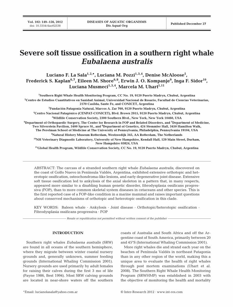
Predominant pathological findings included: (1)heterotopic bone (bone arising from soft tissue), (2)orthotopic bone (bone arising from the normotopicskeleton), (3) osteochondroma-like lesions, and (4)early degenerative joint disease. In general, cervical(1 to 7), thoracic (1 to 14), lumbar (1 to 15), and manyof the caudal (1 to 20) vertebrae were fused by rigidsynostoses composed of massive amounts of irregularsheets of heterotopic (Fig. 1A–C) and orthotopicbone (Fig. 2A,B). This bone was mature and cancel-lous along the lateral aspect of the affected thoracicand lumbar vertebrae, and cortical along the ventralside of the vertebral column. The thoracic and lum-bar vertebrae were the most severely affected.
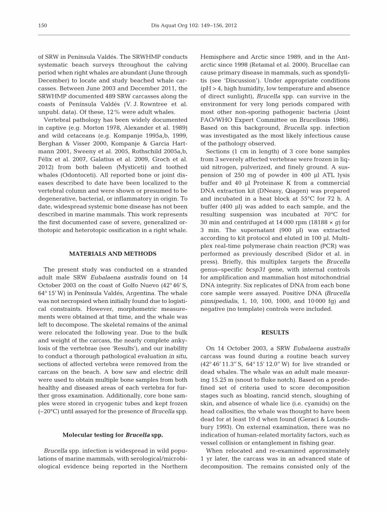
Orthotopic bone formed an approximately 11 cm indiameter, regular, smooth-surfaced groove along theventral aspect of the affected lumbar vertebrae thatwas traceable to Caudal Vertebrae 13. This groovecorresponded with the location of the abdominalaorta. Several, ca. 5 to 7 cm diameter, paired conduitsthat were embedded in dense orthotopic bone origi-nated in the aforementioned groove and extendeddorsolaterally between contiguous transverse pro-cesses on both sides of the spine (Fig. 2B). These con-duits corresponded with the location of lumbar arter-ies. In addition, orthotopic ossification also affectedthe pedicles of vertebral arches, transverse pro-cesses, and several zygapophyseal joints. Orthotopicossification was also present in the appendicularskeleton. The diaphysis and distal epiphyseal margin(near the humeroradioulnar joints) of both humeri,and the proximal epiphyses of both ulnae (elbowjoints) including the olecranon processes, wereaffected by abundant osteophytes (Fig. 2B,C). Otherbones of the appendicular system were missing andcould not be examined.
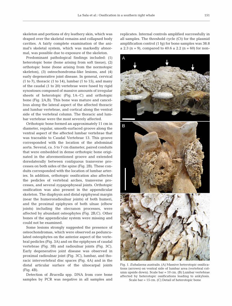
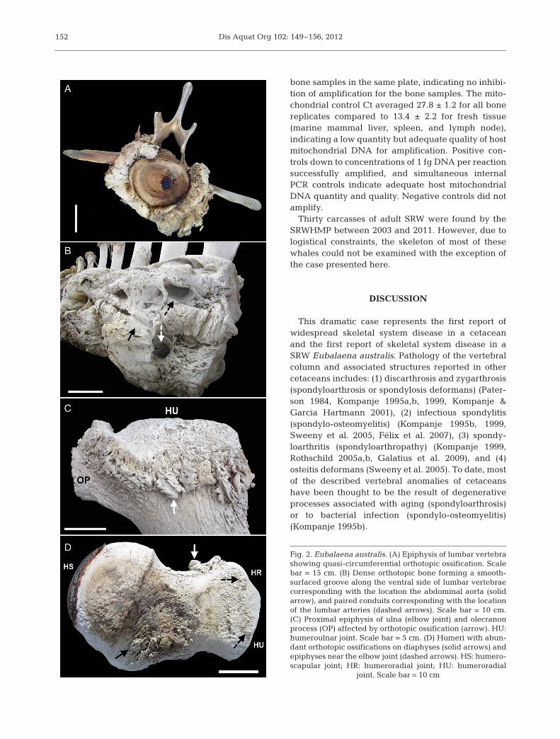
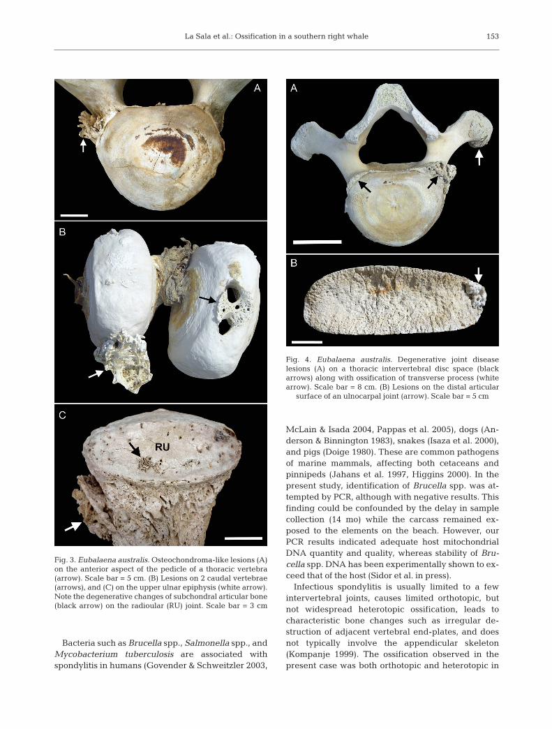
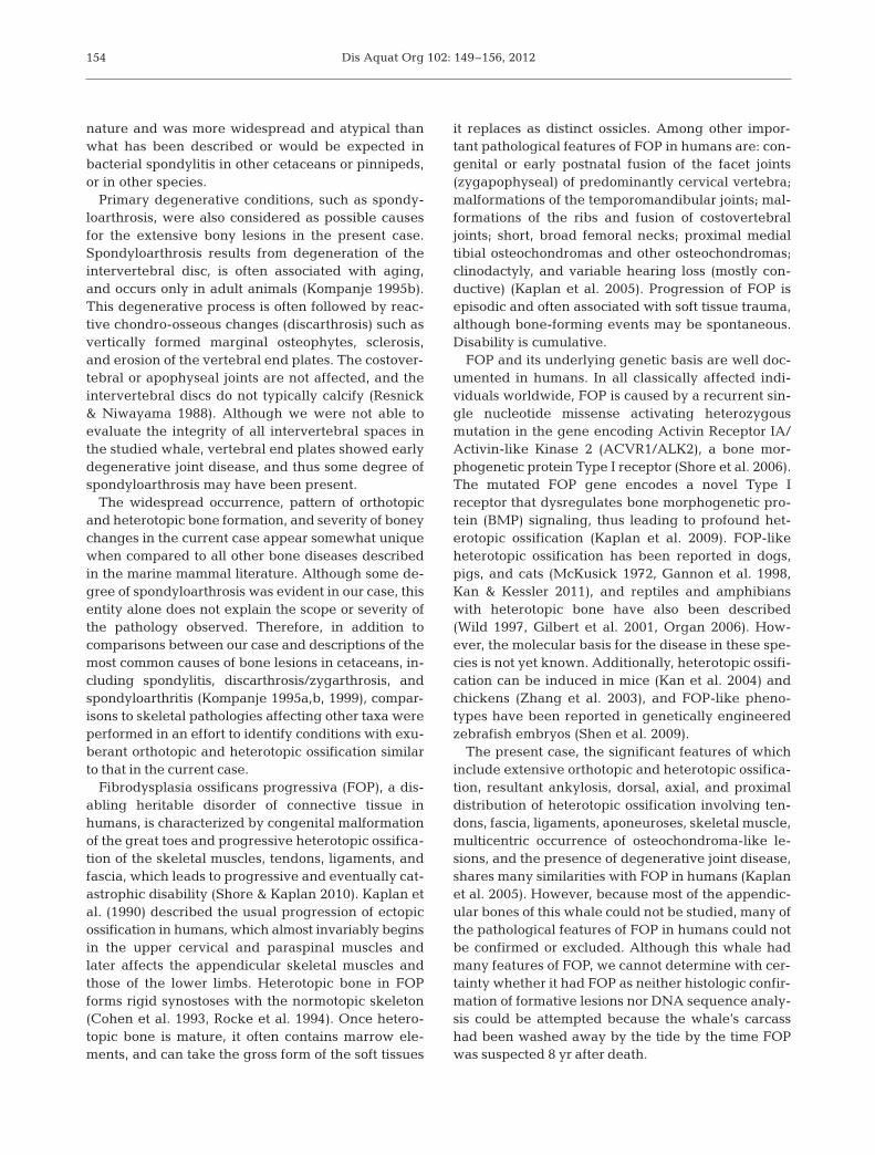
Some lesions strongly suggested the presence ofosteochondromas, which were observed as peduncu-lated osteophytes on the anterior aspect of the verte-bral pedicles (Fig. 3A) and on the epiphyses of caudalvertebrae (Fig. 3B) and radioulnar joints (Fig. 3C).Early degenerative joint disease was observed inproximal radioulnar joint (Fig. 3C), lumbar, and tho-racic intervertebral disc spaces (Fig. 4A) and in thedistal articular surface of the ulnocarpal joints(Fig. 4B).
Detection of Brucella spp. DNA from core bonesamples by PCR was negative in all samples and
replicates. Internal controls amplified successfully inall samples. The threshold cycle (Ct) for the plasmidamplification control (1 fg) for bone samples was 38.8± 2.3 (n = 9), compared to 40.6 ± 2.2 (n = 60) for non-
151
Fig. 1. Eubalaena australis. (A) Massive heterotopic ossifica-tions (arrows) on ventral side of lumbar area (vertebral col-umn upside down). Scale bar = 10 cm. (B) Lumbar vertebrae affected by heterotopic ossifications leading to ankylosis.
Scale bar = 15 cm. (C) Detail of heterotopic bone
Dis Aquat Org 102: 149–156, 2012
bone samples in the same plate, indicating no inhibi-tion of amplification for the bone samples. The mito-chondrial control Ct averaged 27.8 ± 1.2 for all bonereplicates compared to 13.4 ± 2.2 for fresh tissue(marine mammal liver, spleen, and lymph node),indicating a low quantity but adequate quality of hostmitochondrial DNA for amplification. Positive con-trols down to concentrations of 1 fg DNA per reactionsuccessfully amplified, and simultaneous internalPCR controls indicate adequate host mitochondrialDNA quantity and quality. Negative controls did notamplify.
Thirty carcasses of adult SRW were found by theSRWHMP between 2003 and 2011. However, due tologistical constraints, the skeleton of most of thesewhales could not be examined with the exception ofthe case presented here.
DISCUSSION
This dramatic case represents the first report ofwidespread skeletal system disease in a cetaceanand the first report of skeletal system disease in aSRW Eubalaena australis. Pathology of the vertebralcolumn and associated structures reported in othercetaceans includes: (1) discarthrosis and zygarthrosis(spondyloarthrosis or spondylosis deformans) (Pater-son 1984, Kompanje 1995a,b, 1999, Kompanje & Garcia Hartmann 2001), (2) infectious spondylitis(spondylo-osteomyelitis) (Kompanje 1995b, 1999,Sweeny et al. 2005, Félix et al. 2007), (3) spondy-loarthritis (spondyloarthropathy) (Kompanje 1999,Rothschild 2005a,b, Galatius et al. 2009), and (4)osteitis deformans (Sweeny et al. 2005). To date, mostof the described vertebral anomalies of cetaceanshave been thought to be the result of degenerativeprocesses associated with aging (spondyloarthrosis)or to bacterial infection (spondylo-osteomyelitis)(Kompanje 1995b).
152
Fig. 2. Eubalaena australis. (A) Epiphysis of lumbar vertebrashowing quasi-circumferential orthotopic ossification. Scalebar = 15 cm. (B) Dense orthotopic bone forming a smooth-surfaced groove along the ventral side of lumbar vertebraecorresponding with the location the abdominal aorta (solidarrow), and paired conduits corresponding with the locationof the lumbar arteries (dashed arrows). Scale bar = 10 cm.(C) Proximal epiphysis of ulna (elbow joint) and olecranonprocess (OP) affected by orthotopic ossification (arrow). HU:humeroulnar joint. Scale bar = 5 cm. (D) Humeri with abun-dant orthotopic ossifications on diaphyses (solid arrows) andepiphyses near the elbow joint (dashed arrows). HS: humero -scapular joint; HR: humeroradial joint; HU: humeroradial
joint. Scale bar = 10 cm
La Sala et al.: Ossification in a southern right whale
Bacteria such as Brucella spp., Salmonella spp., andMycobacterium tuberculosis are associated withspondylitis in humans (Govender & Schweitzler 2003,
McLain & Isada 2004, Pappas et al. 2005), dogs (An-derson & Binnington 1983), snakes (Isaza et al. 2000),and pigs (Doige 1980). These are common pathogensof marine mammals, affecting both ceta ceans andpinnipeds (Jahans et al. 1997, Higgins 2000). In thepresent study, identification of Brucella spp. was at-tempted by PCR, although with negative results. Thisfinding could be confounded by the delay in samplecollection (14 mo) while the carcass remained ex-posed to the elements on the beach. However, ourPCR results indicated adequate host mitochondrialDNA quantity and quality, whereas stability of Bru-cella spp. DNA has been experimentally shown to ex-ceed that of the host (Sidor et al. in press).
Infectious spondylitis is usually limited to a fewintervertebral joints, causes limited orthotopic, butnot widespread heterotopic ossification, leads tocharacteristic bone changes such as irregular de -struction of adjacent vertebral end-plates, and doesnot typically involve the appendicular skeleton(Kompanje 1999). The ossification observed in thepresent case was both orthotopic and heterotopic in
153
Fig. 3. Eubalaena australis. Osteochondroma-like lesions (A)on the anterior aspect of the pedicle of a thoracic vertebra(arrow). Scale bar = 5 cm. (B) Lesions on 2 caudal vertebrae(arrows), and (C) on the upper ulnar epiphysis (white arrow).Note the degenerative changes of subchondral articular bone(black arrow) on the radioular (RU) joint. Scale bar = 3 cm
Fig. 4. Eubalaena australis. Degenerative joint disease lesions (A) on a thoracic intervertebral disc space (black arrows) along with ossification of transverse process (whitearrow). Scale bar = 8 cm. (B) Lesions on the distal articular
surface of an ulnocarpal joint (arrow). Scale bar = 5 cm
Dis Aquat Org 102: 149–156, 2012
nature and was more widespread and atypical thanwhat has been described or would be expected inbacterial spondylitis in other cetaceans or pinnipeds,or in other species.
Primary degenerative conditions, such as spondy-loarthrosis, were also considered as possible causesfor the extensive bony lesions in the present case.Spondyloarthrosis results from degeneration of theintervertebral disc, is often associated with aging,and occurs only in adult animals (Kompanje 1995b).This degenerative process is often followed by reac-tive chondro-osseous changes (discarthrosis) such asvertically formed marginal osteophytes, sclerosis,and erosion of the vertebral end plates. The costover-tebral or apophyseal joints are not affected, and theintervertebral discs do not typically calcify (Resnick& Niwayama 1988). Although we were not able toevaluate the integrity of all intervertebral spaces inthe studied whale, vertebral end plates showed earlydegenerative joint disease, and thus some degree ofspondyloarthrosis may have been present.
The widespread occurrence, pattern of orthotopicand heterotopic bone formation, and severity of boneychanges in the current case appear somewhat uniquewhen compared to all other bone diseases describedin the marine mammal literature. Although some de-gree of spondyloarthrosis was evident in our case, thisentity alone does not explain the scope or severity ofthe pathology observed. Therefore, in addition tocomparisons between our case and de scriptions of themost common causes of bone lesions in cetaceans, in-cluding spondylitis, discarthrosis/ zygarthrosis, andspondyloarthritis (Kompanje 1995a,b, 1999), compar-isons to skeletal pathologies affecting other taxa wereperformed in an effort to identify conditions with exu-berant orthotopic and heterotopic ossification similarto that in the current case.
Fibrodysplasia ossificans progressiva (FOP), a dis-abling heritable disorder of connective tissue inhumans, is characterized by congenital malformationof the great toes and progressive heterotopic ossifica-tion of the skeletal muscles, tendons, ligaments, andfascia, which leads to progressive and eventually cat-astrophic disability (Shore & Kaplan 2010). Kaplan etal. (1990) described the usual progression of ectopicossification in humans, which almost invariably beginsin the upper cervical and paraspinal muscles andlater affects the appendicular skeletal muscles andthose of the lower limbs. Heterotopic bone in FOPforms rigid synostoses with the normotopic skeleton(Cohen et al. 1993, Rocke et al. 1994). Once hetero-topic bone is mature, it often contains marrow ele-ments, and can take the gross form of the soft tissues
it replaces as distinct ossicles. Among other impor-tant pathological features of FOP in humans are: con-genital or early postnatal fusion of the facet joints(zygapophyseal) of predominantly cervical vertebra;malformations of the temporomandibular joints; mal-formations of the ribs and fusion of costovertebraljoints; short, broad femoral necks; proximal medialtibial osteochondromas and other osteochondromas;clinodactyly, and variable hearing loss (mostly con-ductive) (Kaplan et al. 2005). Progression of FOP isepisodic and often associated with soft tissue trauma,although bone-forming events may be spontaneous.Disability is cumulative.
FOP and its underlying genetic basis are well doc-umented in humans. In all classically affected indi-viduals worldwide, FOP is caused by a recurrent sin-gle nucleotide missense activating heterozygousmutation in the gene encoding Activin Receptor IA/Activin-like Kinase 2 (ACVR1/ALK2), a bone mor-phogenetic protein Type I receptor (Shore et al. 2006).The mutated FOP gene encodes a novel Type Ireceptor that dysregulates bone morphogenetic pro-tein (BMP) signaling, thus leading to profound het-erotopic ossification (Kaplan et al. 2009). FOP-likeheterotopic ossification has been reported in dogs,pigs, and cats (McKusick 1972, Gannon et al. 1998,Kan & Kessler 2011), and reptiles and amphibianswith heterotopic bone have also been described(Wild 1997, Gilbert et al. 2001, Organ 2006). How-ever, the molecular basis for the disease in these spe-cies is not yet known. Additionally, heterotopic ossifi-cation can be induced in mice (Kan et al. 2004) andchickens (Zhang et al. 2003), and FOP-like pheno-types have been reported in genetically engineeredzebrafish embryos (Shen et al. 2009).
The present case, the significant features of whichinclude extensive orthotopic and heterotopic ossifica-tion, resultant ankylosis, dorsal, axial, and proximaldistribution of heterotopic ossification involving ten-dons, fascia, ligaments, aponeuroses, skeletal muscle,multicentric occurrence of osteochondroma-like le-sions, and the presence of degenerative joint disease,shares many similarities with FOP in humans (Kaplanet al. 2005). However, because most of the appendic-ular bones of this whale could not be studied, many ofthe pathological features of FOP in humans could notbe confirmed or excluded. Although this whale hadmany features of FOP, we cannot determine with cer-tainty whether it had FOP as neither histologic confir-mation of formative lesions nor DNA sequence analy-sis could be attempted because the whale’s carcasshad been washed away by the tide by the time FOPwas suspected 8 yr after death.
154
La Sala et al.: Ossification in a southern right whale
A genetic disorder of heterotopic ossification thatoccurs in humans and could be confidently excludedhere is progressive osseous heteroplasia (POH), arare condition characterized by early cutaneous ossi-fication with progression to deeper connective tis-sues, extensive deformity of the normotopic skeleton,and dramatic asymmetry of lesional distribution(Shore & Kaplan 2010). In our case, although we donot have early histological evidence to determine thepathway of bone formation, lesions did not involvethe skin, did not cause deformity of the normotopicskeleton, and were symmetrical.
The body axis (i.e. vertebral column, spinal mus-cles of the back, and tail flukes) represent the mainlocomotion organs of whales (Slijper 1979), and theintegrity of these structures allows flexibility forswimming, feeding, copulating, and other functions.It is very likely that the extensive pathology along theentire length of the vertebral column of this whalecompromised these activities. Extensive bone prolif-eration may also have contributed to compression ofspinal nerves, especially in the intervertebral spaces,and important blood vessels that form the central irri-gation system such as the aorta and vena cava.
A wide range of terrestrial and aquatic vertebratespossesses the genetic, cellular, and molecular path-ways to form heterotopic bone. The presence ofextensive heterotopic ossification in a marine mam-mal has important biological significance regardlessof the genetic or molecular basis of the lesions. Ourreport of a FOP-like condition in a southern rightwhale extends the observation of heterotopic ossifi-cation and tissue metamorphosis (Kaplan et al. 2009)to a marine mammal that occupies a unique ecologi-cal niche in the natural world.
Acknowledgements. We thank the Office of ProtectedResources of the U.S. National Marine Fisheries Service,National Ocean and Atmospheric Administration (OrdersDG133F-02-SE-0901, DG-133F-06-SE-5823, and DG133 F07SE 4651) for funding the Southern Right Whale Health Mon-itoring Program (SRWHMP) at Península Valdés during theyears 2003, 2004, and 2005. We also thank SRWHMP part-ner institutions, Instituto de Conservacion de Ballenas/Whale Conservation Institute, Wildlife Conservation Soci-ety, Fundacion Patagonia Natural and Fundacion Ecocentro,and the SRWHMP Stranding Network, Dirección de Fauna yFlora Silvestres, Dirección General de Conservación deÁreas Protegidas. The Administración del Área Natural Pro-tegida Península Valdés provided the necessary permits toconduct this study. We thank Soledad La Sala for digitalimage processing. This work was also supported in part bythe International Fibrodysplasia Ossificans ProgressivaAssociation, the Center for Research in FOP and RelatedDisorders, the Isaac & Rose Nassau Professorship of Ortho -paedic Molecular Medicine, and by grants from the U.S.National Institutes of Health (NIH R01-AR41916).
LITERATURE CITED
Alexander JW, Solangi MA, Riegel LS (1989) Vertebralosteo myelitis and suspected diskospondylitis in anAtlantic bottlenose dolphin (Tursiops truncatus). J WildlDis 25: 118−121
Anderson GI, Binnington AG (1983) Discospondylitis andorchitis associated with high Brucella titre in a dog. CanVet J 24: 249−252
Berghan J, Visser IN (2000) Vertebral column malformationsin New Zealand delphinids with review of cases world-wide. Aquat Mamm 26: 17−25
Best P (1994) Seasonality of reproduction and the length ofgestation in southern right whales Eubalaena australis. JZool (Lond) 232: 175−189
Cohen RB, Hahn GV, Tabas JA (1993) The natural history ofheterotopic ossification inpatients who have fibrodyspla-sia ossificans progressiva. A study of forty-four patients.J Bone Joint Surg Am 75: 215−219
Doige CE (1980) Discospondylitis in swine. Can J CompMed 44: 121−128
Félix F, Haase B, Aguirre WE (2007) Spondylitis in a hump-back whale (Megaptera novaeangliae) from the south-east Pacific. Dis Aquat Org 75: 259−264
Galatius A, Sonne C, Kinze CC, Dietz R, Beck Jensen JE(2009) Occurrence of vertebral osteophytes in a museumsample of white-beaked dolphins (Lagenorhynchusalbirostris) from Danish waters. J Wildl Dis 45: 19−28
Gannon FH, Valentine BA, Shore EM, Zasloff MA, KaplanFS (1998) Acute lymphocytic infiltration in an extremelyearly lesion of fibrodysplasia ossificans progressiva. ClinOrthop Relat Res 346: 19−25
Geraci JR, Loundsbury VJ (1993) Marine mammals ashore: a field guide for strandings. Texas A & M Sea Grant Pro-gram, Galveston, TX
Gilbert SF, Loredo GA, Brukman A, Burke AC (2001) Mor-phogenesis of the turtle shell: the development of a novelstructure in tetrapod evolution. Evol Dev 3: 47−58
Govender S, Schweitzler G (2003) Salmonella spondylitis ininflammatory disorders of the spine. In: Govender S (ed)Inflammatory diseases of the spine. TTG Asia Media Pty,Singapore
Groch KR, Marcondes MCC, Colosio AC, Catão-Dias JL(2012) Skeletal abnormalities in humpback whales Me ga -ptera novaeangliae stranded in the Brazilian breedingground. Dis Aquat Org 101:145–158
Higgins R (2000) Bacteria and fungi of marine mammals: areview. Can Vet J 41: 105−116
International Whaling Commission (2001) Report of theworkshop on the comprehensive assessment of rightwhales: a worldwide comparison. J Cetacean Res Manag2(Spec Issue): 1−60
Isaza R, Garner M, Jacobsen E (2000) Proliferative osteo -arthritis and osteoarthrosis in 15 snakes. J Zoo Wildl Med31: 20−27
Jahans KL, Foster G, Broughton ES (1997) The characteriza-tion of Brucella strains isolated from marine mammals.Vet Microbiol 57: 373−382
Joint FAO/WHO Expert Committee on Brucellosis (1986)Sixth report, Technical Report Series 740. WHO, Geneva
Kan L, Kessler JA (2011) Animal models of typical hetero-topic ossification. J Biomed Biotechnol 2011: 1−8
Kan L, Hu M, Gomes WA, Kessler JA (2004) Transgenicmice overexpressing BMP4 develop a fibrodysplasia os -si ficans progressiva (FOP)-like phenotype. Am J Pathol
155
Dis Aquat Org 102: 149–156, 2012
165: 1107−1115Kaplan FS, Tabas JA, Zasloff MA (1990) Fibrodysplasia ossi-
ficans progressiva: A clue from the fly? Calcif Tissue Int47: 117−125
Kaplan FS, Glaser DL, Shore EM, Deirmengian GK and oth-ers (2005) The phenotype of fibrodysplasia ossificansprogressiva. Clin Rev Bone Miner Metab 3: 183−188
Kaplan FS, Pignolo RJ, Shore EM (2009) The FOP metamor-phogene encodes a novel type I receptor that dysregu-lates BMP signaling. Cytokine Growth Factor Rev 20: 399−407
Kinze CC (1986) Note on the occurrence of spondylitisdeformans in a sample of harbor porpoises Phocoenaphocoena (L.) taken in Danish waters. Aquat Mamm 12: 25−27
Kompanje EJO (1995a) On the occurrence of spondylosisdeformans in white-beaked dolphins Lagenorhynchusalbirostris (Gray, 1846) stranded on the Dutch coast. ZoolMed Leiden 69: 231−250
Kompanje EJO (1995b) Differences between spondylo-osteomyelitis and spondylosis deformans in small odon-tocetes based on museum material. Aquat Mamm 21: 199−203
Kompanje EJO (1999) Considerations on the comparativepathology of the vertebrae in Mysticeti and Odontoceti;evidence for the occurrence of discarthrosis, zygarthro-sis, infectious spondylitis and spondyloarthritis. ZoolMed Leiden 73: 99−130
Kompanje EJO, Garcia Hartmann M (2001) Intraspongiousdisc herniation (Schmorl’s node) in Phocoena phocoenaand Lagenorhynchus albirostris (Mammalia: Cetacea,Odonticeti). Deinsea 8: 135−141
McKusick VA (1972) Other heritable and generalized disor-ders of connective tissue. In: Beighton (ed) Heritable dis-orders of connective tissue. CV Mosby Company, St.Louis, MO
McLain RF, Isada C (2004) Spinal tuberculosis deserved aplace on the radar screen. Cleve Clin J Med 71: 537−549
Morton B (1978) Osteomyelitis (pyogenic spondylitis) of thespine in a dolphin. J Am Vet Med Assoc 173: 1119−1120
Organ CL (2006) Thoracic epaxial muscles in livingarchosaurs and ornithopod dinosaurs. Anat Rec A DiscovMol Cell Evol Biol 288: 782−793
Pappas G, Akritidis N, Bosilkovski M, Tsianos E (2005) Bru-cellosis. N Engl J Med 352: 2325−2336
Paterson RA (1984) Spondylitis deformans in a Bryde’swhale (Balenoptera edeni Anderson) stranded on thesouthern coast of Queensland. J Wildl Dis 20: 250−252
Payne R (1986) Long term behavioral studies of the southernright whale (Eubalaena australis). Rep Int Whal Comm10(Spec Issue): 161−167
Resnick D, Niwayama G (1988) Degenerative diseases of thespine. In: Manke D (ed) Diagnosis of bone and joint dis-orders: articular diseases. W. B. Saunders, Philadelphia,PA
Retamal P, Blank O, Abalos P, Torres D (2000) Detection ofanti-Brucella antibodies in pinnipeds from the Antarcticterritory. Vet Rec 146: 166−167
Rocke DM, Zasloff M, Peeper J (1994) Age-and-joint specificrisk of initial heterotopic ossification in patients whohave fibrodysplasia ossificans progressiva. Clin OrthopRelat Res 301: 243−248
Rothschild BM (2005a) What causes lesions in sperm whalebones? Science 308: 631−632
Rothschild BM (2005b) Whale of a tale. Ann Rheum Dis 64: 1385−1386
Shen Q, Little SC, Xu M, Haupt J and others (2009) Thefibrodysplasia ossificans progressiva R206H ACVR1mutation activates BMP-independent chondrogenesisand zebrafish embryo ventralization. J Clin Invest 119: 3462−3472
Shore EM, Kaplan FS (2010) Inherited human diseases ofheterotopic ossification. Nature Rev Rheumatol 6: 518−527
Shore EM, Xu M, Feldman GJ, Fenstermacher DA and oth-ers (2006) A recurrent mutation in the BMP type I recep-tor ACVR1 causes inherited and sporadic fibrodysplasiaossificans progressiva. Nat Genet 38: 525−527
Sidor IS, Dunn JL, Tsongalis GJ, Carlson J, Frasca S (inpress) A multiplex real-time PCR assay with two internalcontrols for the detection of Brucella spp. in tissues,blood and feces from marine mammals. J Vet DiagnInvest 25(1)
Slijper EJ (1979) Whales. Cornell University Press, Ithaca,NY
Sweeny MM, Price JM, Jones GS, French TW, Early GA,Moore MJ (2005) Spondylitic changes in long-finnedpilot whales (Globicephala melas) stranded on CapeCod, Massachusetts, USA, between 1982 and 2000. JWildl Dis 41: 717−727
Uhart M, Rowntree VJ, Mohamed N, Pozzi L and others(2008) Strandings of southern right whales (Eubalaenaaustralis) at Península Valdés, Argentina from 2003−2007. International Whaling Commission, Cambridge
Wild ER (1997) Description of the adult skeleton and devel-opmental osteology of the hyperossified horned frog(Anura: Leptodactylidae). J Morphol 232: 169−206
Zhang D, Schwarz EM, Rosier RN, Zuscik MJ, Puzas JE,O’Keefe RJ (2003) ALK2 functions as a BMP type Ireceptor and induces Indian hedgehog in chondrocytesduring skeletal development. J Bone Miner Res 18: 1593−1604
156
Editorial responsibility: Michael Moore, Woods Hole, Massachusetts, USA
Submitted: May 15, 2012; Accepted: September 24, 2012Proofs received from author(s): December 12, 2012


