Embed Size (px)
Citation preview
The Auditory Anatomy of the MinkeWhale (Balaenoptera acutorostrata): A
Potential Fatty Sound ReceptionPathway in a Baleen Whale
MAYA YAMATO,1* DARLENE R. KETTEN,1,2 JULIE ARRUDA,1,3
SCOTT CRAMER,1 AND KATHLEEN MOORE4
1Woods Hole Oceanographic Institution Biology Department, Woods Hole, Massachusetts2Harvard Medical School Department of Otology and Laryngology, Boston, Massachusetts
3Massachusetts Eye and Ear Infirmary, Boston, Massachusetts4International Fund for Animal Welfare Marine Mammal Rescue and Research,
Yarmouth Port, Massachusetts
ABSTRACTCetaceans possess highly derived auditory systems adapted for under-
water hearing. Odontoceti (toothed whales) are thought to receive soundthrough specialized fat bodies that contact the tympanoperiotic complex, thebones housing the middle and inner ears. However, sound reception path-ways remain unknown in Mysticeti (baleen whales), which have very differ-ent cranial anatomies compared to odontocetes. Here, we report a potentialfatty sound reception pathway in the minke whale (Balaenoptera acutoros-trata), a mysticete of the balaenopterid family. The cephalic anatomy of sevenminke whales was investigated using computerized tomography and mag-netic resonance imaging, verified through dissections. Findings include alarge, well-formed fat body lateral, dorsal, and posterior to the mandibularramus and lateral to the tympanoperiotic complex. This fat body inserts intothe tympanoperiotic complex at the lateral aperture between the tympanicand periotic bones and is in contact with the ossicles. There is also a second,smaller body of fat found within the tympanic bone, which contacts theossicles as well. This is the first analysis of these fatty tissues’ associationwith the auditory structures in a mysticete, providing anatomical evidencethat fatty sound reception pathways may not be a unique feature of odonto-cete cetaceans. Anat Rec, 00:000–000, 2012. VC 2012 Wiley Periodicals, Inc.
Key words: cetacea;mysticete; hearing; ear; acoustic fat; imaging
The transition to aquatic life resulted in several modifi-cations to the auditory anatomy of cetaceans. Cetaceanslack external pinnae, and the external auditory canal hasbeen reduced to a very narrow channel. The middle and
inner ear migrated laterally out from the skull and areencased in the dense tympanoperiotic complex (Hunter,1787; Eschricht and Reinhardt, 1866; Kernan, 1919).Other characteristics of the auditory system are specific to
Re-use of this article is permitted in accordance with theTerms and Conditions set out at http://wileyonlinelibrary.com/onlineopen#OnlineOpen_Terms.Grant sponsors: National Science Foundation Graduate
Research Fellowship, Ocean Life Institute Graduate Fellowship,WHOI Summer Student Fellowship, WHOI Minority Fellowship,Princeton University Ecology and Evolutionary BiologyDepartment Senior Thesis Fund, Joint Industry Program, Officeof Naval Research Marine Mammal Program, Chief of NavalOperations Energy and Environmental Readiness Division.
*Correspondence to: Maya Yamato, Woods HoleOceanographic Institution, 266 Woods Hole Road MS #50,Woods Hole, MA 02543. Fax: 508-457-2041.E-mail: [email protected] 13 December 2011; Accepted 13 March 2012.
DOI 10.1002/ar.22459
Published online in Wiley Online Library(wileyonlinelibrary.com).
THE ANATOMICAL RECORD 000:000–000 (2012)
VVC 2012 WILEY PERIODICALS, INC.
each suborder. The gross auditory anatomy and hearingpathways in Odontoceti (toothed whales) have been rela-tively well described. In odontocetes, the external auditorycanal is considered vestigial (Reysenbach de Haan, 1957;Dudok Van Heel, 1962; Norris, 1968; McCormick et al.,1970). Bone conduction is thought to play a minor rolebecause there is no osseous connection between the tym-panoperiotic complex and the rest of the skull in mostodontocete species (Claudius, 1858, in Yamada, 1953; Ket-ten and Wartzok, 1990; Nummela et al., 2007). Inaddition, the air spaces around the tympanoperiotic com-plex are thought to provide acoustic insulation from therest of the skull, which may be important for directionalhearing (Reysenbach de Haan, 1957).
A more likely mechanism for sound reception in odon-tocetes is via perimandibular ‘‘acoustic’’ fat bodies thatare in direct contact with the ears, including both thetympanic and periotic bones (Norris, 1964; Ketten, 1994,1997; Ridgway, 1999; Cranford et al., 2010). Althoughodontocetes receive sounds across various locations onthe head (Bullock et al., 1968; Brill, 1988; Mohl et al.,1999; Mooney et al., 2008; Cranford et al., 2008a), thesebiochemically distinct fats are thought to act as a prefer-ential pathway of sound from the environment to theears (Norris, 1964; Bullock et al., 1968; Varanasi andMalins, 1971; Litchfield et al., 1975; Brill et al., 1988;Koopman et al., 2006; Zahorodny et al., 2009).
These odontocete ‘‘acoustic fats’’ are composed of multiplelobes, including the inner lobe filling the enlarged mandib-ular hiatus and the outer lobe covering the lateral andventral portions of the mandible (Norris, 1968; Ketten,1994, 1997; Ridgway, 1999). In addition to these two fatlobes, which are located anterior to the tympanoperioticcomplex, there is also increasing evidence for a third fatchannel located lateral to the tympanoperiotic complex. Inan electrophysiological study focused on striped dolphins(Stenella coeruleoalba), Bullock et al. (1968) found that thelateral area near the external auditory meatus openingwas sensitive to low-frequency sounds below 3 kHz.Renaud and Popper (1975) also found that the region nearthe external auditory meatus opening was more sensitiveto lower frequency sounds (below 20 kHz) in a behavioralstudy on bottlenose dolphins (Tursiops truncatus). Further-more, Ketten (1994) provided anatomical evidence for adistinct lateral fat channel by applying magnetic resonanceimaging (MRI) techniques to multiple odontocete species.Most recently, Popov et al. (2008) used auditory brainstemresponse latencies to advance the hypothesis that there aretwo acoustic windows in the bottlenose dolphin. The acous-tic window was calculated to be near the external auditorymeatus opening at frequencies below 22 kHz, while soundsabove 32 kHz were received through the lower jaws.
The pathways of sound reception are unknown in Mys-ticeti (baleen whales), and there have been no reports ofsound-conducting fats similar to those of odontocetes. Thesmall opening to the external auditory meatus is visibleon the surface, as in odontocetes. However, researchersdisagree on whether the auditory canal is continuousfrom the opening of the external auditory meatus to thetympanic membrane and whether it is a functional part ofthe auditory system (Carte and Macalister, 1868; Yamada,1953). At the end of the auditory canal is the ‘‘glove fin-ger,’’ an everted, extended, thickened tympanicmembrane, the function of which remains unclear (Lillie,1910; Fraser and Purves, 1960). This elongated glove
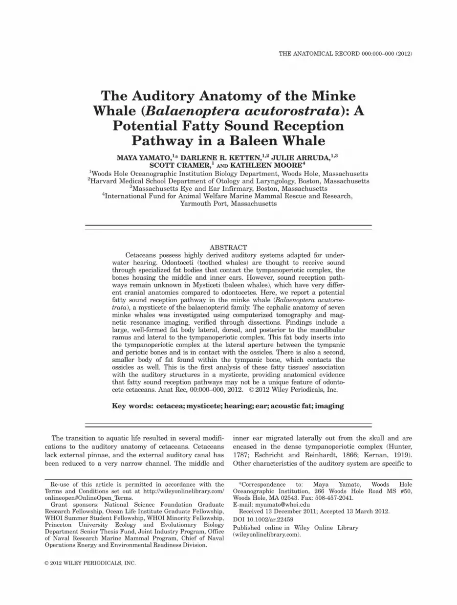
Fig. 1. Photograph of a minke whale skull (B-acu21; not part of ourstudy). (a) Ventral view of the skull, where the mandibles have beenremoved. The tympanic bone has been removed on the right side ofthe animal (left side of the photograph) to expose the periotic bone.(b) Enlarged view of the right ear showing the periotic bone, which isfirmly embedded in the skull. Abbreviations: T, tympanic; P, periotic; E,exoccipital; Sq, squamosal; Pal, palatine; Max, maxilla; PF, posteriorflange of the periotic.
2 YAMATO ET AL.
finger is not found in odontocetes or any other mammals.Another major difference between odontocete and mysti-cete ears is the connection of the tympanoperiotic complexwith the skull. In mysticetes, the posterior flange of theperiotic bone is wedged against the squamosal and theexoccipital bones (Yamada, 1948; Fig. 1). The anteriorflange of the periotic is also firmly embedded in the squa-mosal bone, reducing the acoustic isolation of thetympanoperiotic complex. Bone conduction has not beendismissed as a potential sound reception pathway in ba-leen whales (Ketten, 1992, 2000).
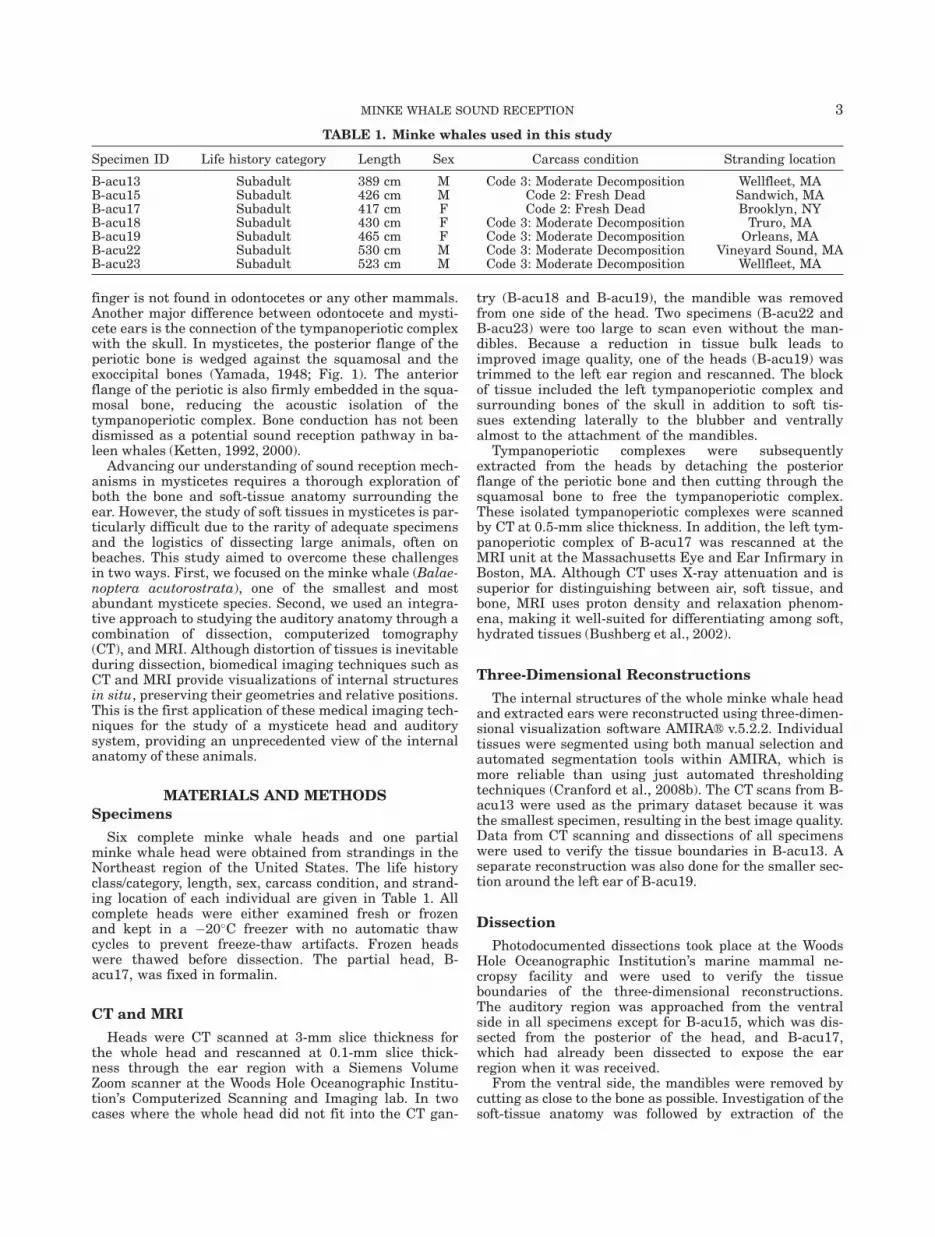
Advancing our understanding of sound reception mech-anisms in mysticetes requires a thorough exploration ofboth the bone and soft-tissue anatomy surrounding theear. However, the study of soft tissues in mysticetes is par-ticularly difficult due to the rarity of adequate specimensand the logistics of dissecting large animals, often onbeaches. This study aimed to overcome these challengesin two ways. First, we focused on the minke whale (Balae-noptera acutorostrata), one of the smallest and mostabundant mysticete species. Second, we used an integra-tive approach to studying the auditory anatomy through acombination of dissection, computerized tomography(CT), and MRI. Although distortion of tissues is inevitableduring dissection, biomedical imaging techniques such asCT and MRI provide visualizations of internal structuresin situ, preserving their geometries and relative positions.This is the first application of these medical imaging tech-niques for the study of a mysticete head and auditorysystem, providing an unprecedented view of the internalanatomy of these animals.
MATERIALS AND METHODS
Specimens
Six complete minke whale heads and one partialminke whale head were obtained from strandings in theNortheast region of the United States. The life historyclass/category, length, sex, carcass condition, and strand-ing location of each individual are given in Table 1. Allcomplete heads were either examined fresh or frozenand kept in a �20�C freezer with no automatic thawcycles to prevent freeze-thaw artifacts. Frozen headswere thawed before dissection. The partial head, B-acu17, was fixed in formalin.
CT and MRI
Heads were CT scanned at 3-mm slice thickness forthe whole head and rescanned at 0.1-mm slice thick-ness through the ear region with a Siemens VolumeZoom scanner at the Woods Hole Oceanographic Institu-tion’s Computerized Scanning and Imaging lab. In twocases where the whole head did not fit into the CT gan-
try (B-acu18 and B-acu19), the mandible was removedfrom one side of the head. Two specimens (B-acu22 andB-acu23) were too large to scan even without the man-dibles. Because a reduction in tissue bulk leads toimproved image quality, one of the heads (B-acu19) wastrimmed to the left ear region and rescanned. The blockof tissue included the left tympanoperiotic complex andsurrounding bones of the skull in addition to soft tis-sues extending laterally to the blubber and ventrallyalmost to the attachment of the mandibles.
Tympanoperiotic complexes were subsequentlyextracted from the heads by detaching the posteriorflange of the periotic bone and then cutting through thesquamosal bone to free the tympanoperiotic complex.These isolated tympanoperiotic complexes were scannedby CT at 0.5-mm slice thickness. In addition, the left tym-panoperiotic complex of B-acu17 was rescanned at theMRI unit at the Massachusetts Eye and Ear Infirmary inBoston, MA. Although CT uses X-ray attenuation and issuperior for distinguishing between air, soft tissue, andbone, MRI uses proton density and relaxation phenom-ena, making it well-suited for differentiating among soft,hydrated tissues (Bushberg et al., 2002).
Three-Dimensional Reconstructions
The internal structures of the whole minke whale headand extracted ears were reconstructed using three-dimen-sional visualization software AMIRAVR v.5.2.2. Individualtissues were segmented using both manual selection andautomated segmentation tools within AMIRA, which ismore reliable than using just automated thresholdingtechniques (Cranford et al., 2008b). The CT scans from B-acu13 were used as the primary dataset because it wasthe smallest specimen, resulting in the best image quality.Data from CT scanning and dissections of all specimenswere used to verify the tissue boundaries in B-acu13. Aseparate reconstruction was also done for the smaller sec-tion around the left ear of B-acu19.
Dissection
Photodocumented dissections took place at the WoodsHole Oceanographic Institution’s marine mammal ne-cropsy facility and were used to verify the tissueboundaries of the three-dimensional reconstructions.The auditory region was approached from the ventralside in all specimens except for B-acu15, which was dis-sected from the posterior of the head, and B-acu17,which had already been dissected to expose the earregion when it was received.
From the ventral side, the mandibles were removed bycutting as close to the bone as possible. Investigation of thesoft-tissue anatomy was followed by extraction of the
TABLE 1. Minke whales used in this study
Specimen ID Life history category Length Sex Carcass condition Stranding location
B-acu13 Subadult 389 cm M Code 3: Moderate Decomposition Wellfleet, MAB-acu15 Subadult 426 cm M Code 2: Fresh Dead Sandwich, MAB-acu17 Subadult 417 cm F Code 2: Fresh Dead Brooklyn, NYB-acu18 Subadult 430 cm F Code 3: Moderate Decomposition Truro, MAB-acu19 Subadult 465 cm F Code 3: Moderate Decomposition Orleans, MAB-acu22 Subadult 530 cm M Code 3: Moderate Decomposition Vineyard Sound, MAB-acu23 Subadult 523 cm M Code 3: Moderate Decomposition Wellfleet, MA
MINKE WHALE SOUND RECEPTION 3
tympanoperiotic complex, which is a technically challeng-ing procedure in mysticetes because the fragile connectionsbetween the periotic and tympanic bones are easily brokenduring attempts to dislodge the tympanoperiotic complexfrom the skull. Once all soft tissues were removed from thearea, the posterior flange was detached using an oscillatingautopsy saw. The anterior flange of the periotic was freedusing bone shears by incrementally chipping the thin sheetof squamosal bone lateral to the tympanic bone. Severingthe soft tissue connections from inside the braincase helpedto loosen the tympanoperiotic complex as well.
RESULTS
In all minke whales examined, there was a distinct,depigmented (white) line on the epidermis projectingposteriorly from the aperture of the external auditorymeatus. This marker is rarely, if ever, mentioned in theliterature but would be helpful in locating the minusculeexternal auditory meatus. The auditory canal appearedto be continuous from its external opening to the glovefinger, though winding and narrow.
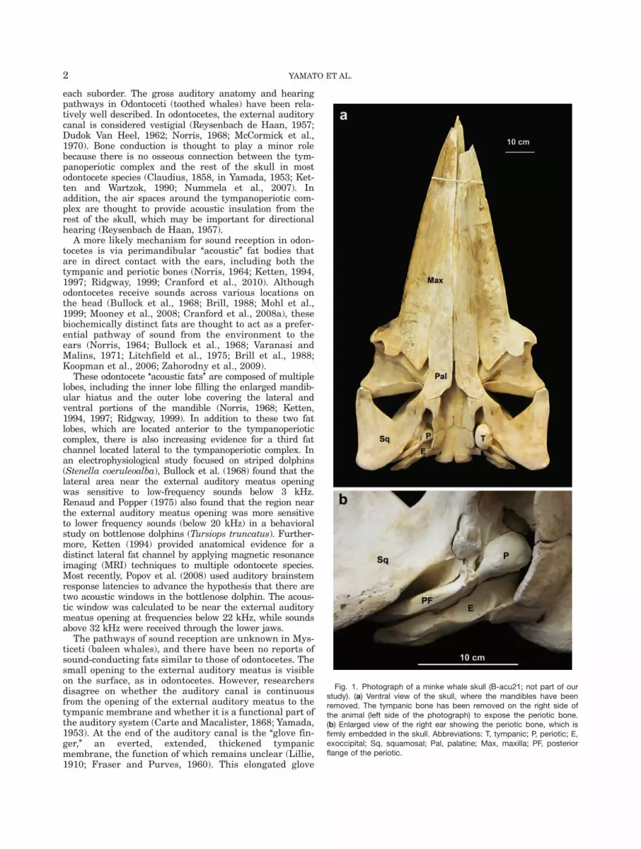
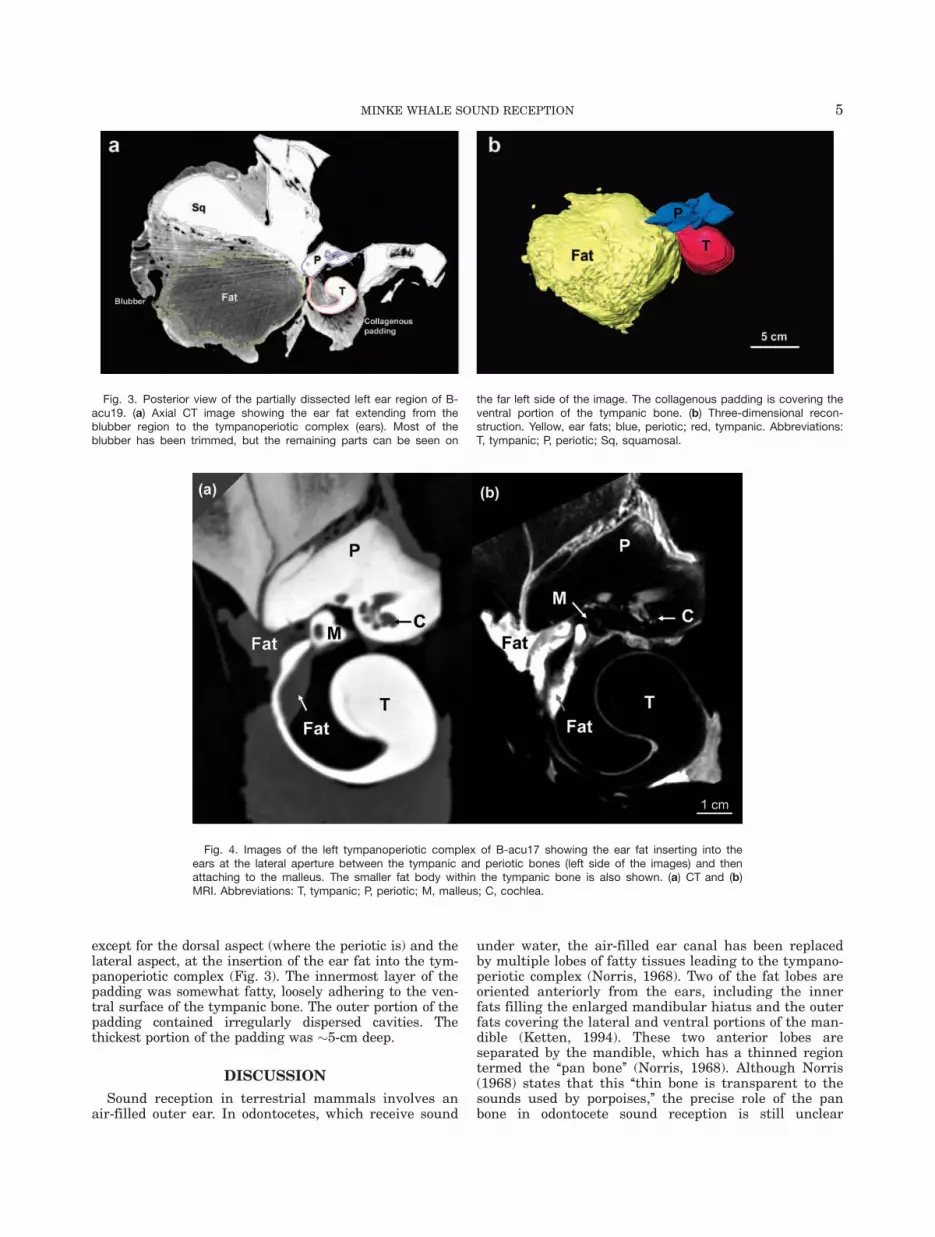
The CT images showed a large, well-formed fat body lat-eral, dorsal, and posterior to the mandibular ramus,ventral to the squamosal bone, and lateral to the tympano-periotic complex. This fat body will be referred to as ‘‘earfat’’ (Fig. 2). Preliminary results from lipid extractions onear fat tissues suggest that some regions are made up of>80% lipid by wet weight (Yamato et al., 2011). The CTimages and dissections indicated that the ear fat bundlebecame more fibrous ventrally and is integrated with thefibrous joint with the mandible. The posterior portion ofthe ear fat is also more fibrous, affording an attachment tothe posterior margin of the squamosal bone.
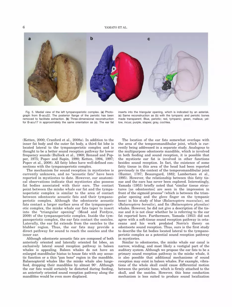
From the ventral perspective, the ear fat has a some-what triangular shape with the three prominencescontacting the blubber region (lateral), tympanoperioticcomplex (medial), and the mandible (anterior; Fig. 2).Thus, a portion of the ear fat extends from the blubberregion to the tympanoperiotic complex (Fig. 3). The ante-rior portion of the ear fat is well removed from theblubber layer and is adjacent to muscle. The ear fatattaches to the tympanoperiotic complex at the lateralaperture between the tympanic and periotic bones,inserting into the space that Mead and Fordyce (2009)term the ‘‘triangular opening’’ (Figs. 2–5). Althoughdirect contact with the glove finger could not be deter-mined, the ear fat is pressed against an area of thetympanoperiotic complex including the ventral portion ofthe glove finger. At the entry to the middle ear, the earfat contacts the malleus (Fig. 4).
Within the middle ear space, the malleus also contactsa smaller fat pad attached to the inner wall of the tym-panic bone, adjacent to the base of the glove finger (Fig.4). The CT and MRI of the tympanoperiotic complexshow these structures clearly, and they are readily visi-ble on careful dissection. The malleus was attached tothe inside of the glove finger by a strong ligamentousconnection, consistent with previous reports (Lillie,1910). Although the smaller fat pad attaches to the baseof the glove finger inside the tympanoperiotic complex,neither of the fat bodies extend into the distal regions ofthe internal surface of the glove finger.
The tympanic bone was covered in a thick, dense, whitepadding composed of collagenous tissues on all sides
Fig. 2. Three-dimensional reconstructions showing the contactbetween the ear fats and the tympano-periotic complex (ears) in theminke whale. The mandibles are still attached. (a) Ventral view. (b)Posterior view. Yellow, ear fats; purple, tympanoperiotic complex;white, other bones.
4 YAMATO ET AL.
except for the dorsal aspect (where the periotic is) and thelateral aspect, at the insertion of the ear fat into the tym-panoperiotic complex (Fig. 3). The innermost layer of thepadding was somewhat fatty, loosely adhering to the ven-tral surface of the tympanic bone. The outer portion of thepadding contained irregularly dispersed cavities. Thethickest portion of the padding was �5-cm deep.
DISCUSSION
Sound reception in terrestrial mammals involves anair-filled outer ear. In odontocetes, which receive sound
under water, the air-filled ear canal has been replacedby multiple lobes of fatty tissues leading to the tympano-periotic complex (Norris, 1968). Two of the fat lobes areoriented anteriorly from the ears, including the innerfats filling the enlarged mandibular hiatus and the outerfats covering the lateral and ventral portions of the man-dible (Ketten, 1994). These two anterior lobes areseparated by the mandible, which has a thinned regiontermed the ‘‘pan bone’’ (Norris, 1968). Although Norris(1968) states that this ‘‘thin bone is transparent to thesounds used by porpoises,’’ the precise role of the panbone in odontocete sound reception is still unclear
Fig. 3. Posterior view of the partially dissected left ear region of B-acu19. (a) Axial CT image showing the ear fat extending from theblubber region to the tympanoperiotic complex (ears). Most of theblubber has been trimmed, but the remaining parts can be seen on
the far left side of the image. The collagenous padding is covering theventral portion of the tympanic bone. (b) Three-dimensional recon-struction. Yellow, ear fats; blue, periotic; red, tympanic. Abbreviations:T, tympanic; P, periotic; Sq, squamosal.
Fig. 4. Images of the left tympanoperiotic complex of B-acu17 showing the ear fat inserting into theears at the lateral aperture between the tympanic and periotic bones (left side of the images) and thenattaching to the malleus. The smaller fat body within the tympanic bone is also shown. (a) CT and (b)MRI. Abbreviations: T, tympanic; P, periotic; M, malleus; C, cochlea.
MINKE WHALE SOUND RECEPTION 5
(Ketten, 2000; Cranford et al., 2008a). In addition to theinner fat body and the outer fat body, a third fat lobe islocated lateral to the tympanoperiotic complex and isthought to be a better sound reception pathway for lowerfrequency sounds (Bullock et al., 1968; Renaud and Pop-per, 1975; Popov and Supin, 1990; Ketten, 1994, 1997;Popov et al., 2008). All fatty lobes have well-defined con-nections with the tympanoperiotic complex.
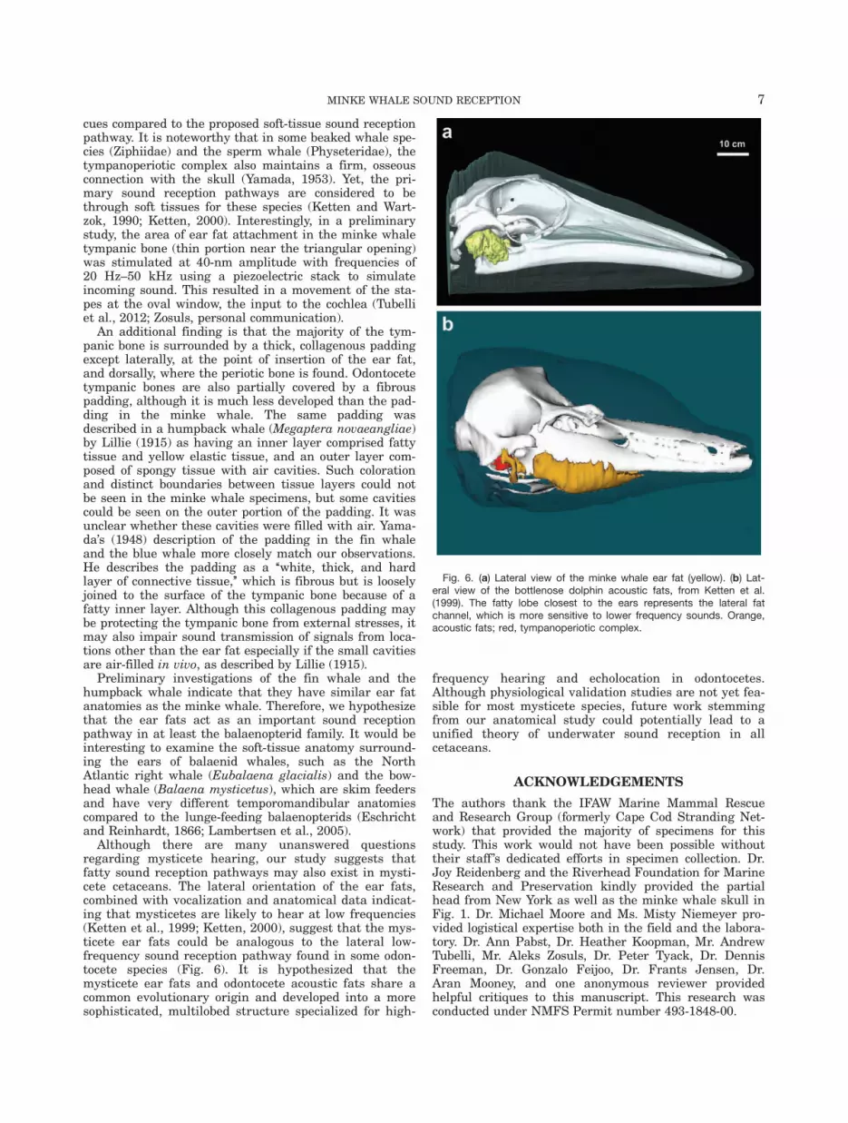
The mechanism for sound reception in mysticetes iscurrently unknown, and no ‘‘acoustic fats’’ have beenreported in mysticetes to date. However, our anatomi-cal observations indicate that mysticetes also possessfat bodies associated with their ears. The contactpoint between the minke whale ear fat and the tympa-noperiotic complex is similar to the area of contactbetween odontocete acoustic fats and their tympano-periotic complex. Although the odontocete acousticfats contact a larger surface area of the tympanoperi-otic complex, the minke whale ear fats taper to insertinto the ‘‘triangular opening’’ (Mead and Fordyce,2009) of the tympanoperiotic complex. Inside the tym-panoperiotic complex, the ear fats contact the ossicles.Laterally, the ear fat extends from the ossicles to theblubber region. Thus, the ear fats may provide adirect pathway for sound to reach the ossicles and theinner ear.
Although odontocete acoustic fats are composed of bothanteriorly oriented and laterally oriented fat lobes, anexclusively lateral sound reception pathway in baleenwhales is appealing. Baleen whales do not have anenlarged mandibular hiatus to house fats with any acous-tic function or a thin ‘‘pan bone’’ region in the mandible.Balaenopterid whales like the minke whale also lunge-feed, dropping their mandibles by almost 90�. Althoughthe ear fats would certainly be distorted during feeding,an anteriorly oriented sound reception pathway along themandibles would be even more displaced.
The location of the ear fats somewhat overlaps withthe area of the temporomandibular joint, which is cur-rently being addressed in a separate study. Analogous tothe multipurpose odontocete mandible, which is involvedin both feeding and sound reception, it is possible thatthe mysticete ear fat is involved in other functionsbesides sound reception. In fact, the existence of somefatty tissue in this area of the head had been reportedpreviously in the context of the temporomandibular joint(Hunter, 1787; Beauregard, 1882; Lambertsen et al.,1995). However, the relationship between this fatty tis-sue and the ears has never been explored. Interestingly,Yamada (1953) briefly noted that ‘‘similar tissue struc-tures [as odontocetes] are seen in the impression infront of the sigmoid process’’ (which is between the trian-gular opening and the glove finger on the tympanicbone) in his study of blue (Balaenoptera musculus), sei(Balaenoptera borealis), and fin (Balaenoptera physalus)whales. However, he did not give a description of the tis-sue and it is not clear whether he is referring to the earfat reported here. Furthermore, Yamada (1953) did notagree with a soft-tissue sound reception pathway in ceta-ceans and his work predated Norris’s theory onodontocete sound reception. Thus, ours is the first studyto describe the fat bodies located lateral to the tympano-periotic complex as a potential sound reception pathwayin mysticetes.
Similar to odontocetes, the minke whale ear canal isnarrow, winding, and most likely a vestigial part of theauditory system. Although we propose the ear fats to be aprimary sound reception pathway in the minke whale, itis also possible that additional mechanisms of soundreception may exist in baleen whales. For example, vibra-tions of the whole skull could cause differential motionbetween the periotic bone, which is firmly attached to theskull, and the ossicles. However, this bone conductionmechanism is less suited to produce sound localization
Fig. 5. Medial view of the left tympanoperiotic complex. (a) Photo-graph from B-acu22. The posterior flange of the periotic has beenremoved to facilitate extraction. (b) Three-dimensional reconstructionfor B-acu17 in approximately the same orientation as (a). The ear fat
inserts into the triangular opening, which is indicated by an asterisk.(c) Same reconstruction as (b) with the tympanic and periotic bonesmade transparent. Blue, periotic; red, tympanic; green, malleus; yel-low, incus; purple, stapes; gray, cochlea.
6 YAMATO ET AL.
cues compared to the proposed soft-tissue sound receptionpathway. It is noteworthy that in some beaked whale spe-cies (Ziphiidae) and the sperm whale (Physeteridae), thetympanoperiotic complex also maintains a firm, osseousconnection with the skull (Yamada, 1953). Yet, the pri-mary sound reception pathways are considered to bethrough soft tissues for these species (Ketten and Wart-zok, 1990; Ketten, 2000). Interestingly, in a preliminarystudy, the area of ear fat attachment in the minke whaletympanic bone (thin portion near the triangular opening)was stimulated at 40-nm amplitude with frequencies of20 Hz–50 kHz using a piezoelectric stack to simulateincoming sound. This resulted in a movement of the sta-pes at the oval window, the input to the cochlea (Tubelliet al., 2012; Zosuls, personal communication).
An additional finding is that the majority of the tym-panic bone is surrounded by a thick, collagenous paddingexcept laterally, at the point of insertion of the ear fat,and dorsally, where the periotic bone is found. Odontocetetympanic bones are also partially covered by a fibrouspadding, although it is much less developed than the pad-ding in the minke whale. The same padding wasdescribed in a humpback whale (Megaptera novaeangliae)by Lillie (1915) as having an inner layer comprised fattytissue and yellow elastic tissue, and an outer layer com-posed of spongy tissue with air cavities. Such colorationand distinct boundaries between tissue layers could notbe seen in the minke whale specimens, but some cavitiescould be seen on the outer portion of the padding. It wasunclear whether these cavities were filled with air. Yama-da’s (1948) description of the padding in the fin whaleand the blue whale more closely match our observations.He describes the padding as a ‘‘white, thick, and hardlayer of connective tissue,’’ which is fibrous but is looselyjoined to the surface of the tympanic bone because of afatty inner layer. Although this collagenous padding maybe protecting the tympanic bone from external stresses, itmay also impair sound transmission of signals from loca-tions other than the ear fat especially if the small cavitiesare air-filled in vivo, as described by Lillie (1915).
Preliminary investigations of the fin whale and thehumpback whale indicate that they have similar ear fatanatomies as the minke whale. Therefore, we hypothesizethat the ear fats act as an important sound receptionpathway in at least the balaenopterid family. It would beinteresting to examine the soft-tissue anatomy surround-ing the ears of balaenid whales, such as the NorthAtlantic right whale (Eubalaena glacialis) and the bow-head whale (Balaena mysticetus), which are skim feedersand have very different temporomandibular anatomiescompared to the lunge-feeding balaenopterids (Eschrichtand Reinhardt, 1866; Lambertsen et al., 2005).
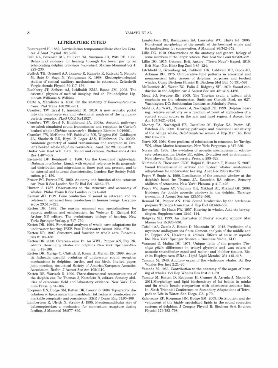
Although there are many unanswered questionsregarding mysticete hearing, our study suggests thatfatty sound reception pathways may also exist in mysti-cete cetaceans. The lateral orientation of the ear fats,combined with vocalization and anatomical data indicat-ing that mysticetes are likely to hear at low frequencies(Ketten et al., 1999; Ketten, 2000), suggest that the mys-ticete ear fats could be analogous to the lateral low-frequency sound reception pathway found in some odon-tocete species (Fig. 6). It is hypothesized that themysticete ear fats and odontocete acoustic fats share acommon evolutionary origin and developed into a moresophisticated, multilobed structure specialized for high-
frequency hearing and echolocation in odontocetes.Although physiological validation studies are not yet fea-sible for most mysticete species, future work stemmingfrom our anatomical study could potentially lead to aunified theory of underwater sound reception in allcetaceans.
ACKNOWLEDGEMENTS
The authors thank the IFAW Marine Mammal Rescueand Research Group (formerly Cape Cod Stranding Net-work) that provided the majority of specimens for thisstudy. This work would not have been possible withouttheir staff ’s dedicated efforts in specimen collection. Dr.Joy Reidenberg and the Riverhead Foundation for MarineResearch and Preservation kindly provided the partialhead from New York as well as the minke whale skull inFig. 1. Dr. Michael Moore and Ms. Misty Niemeyer pro-vided logistical expertise both in the field and the labora-tory. Dr. Ann Pabst, Dr. Heather Koopman, Mr. AndrewTubelli, Mr. Aleks Zosuls, Dr. Peter Tyack, Dr. DennisFreeman, Dr. Gonzalo Feijoo, Dr. Frants Jensen, Dr.Aran Mooney, and one anonymous reviewer providedhelpful critiques to this manuscript. This research wasconducted under NMFS Permit number 493-1848-00.
Fig. 6. (a) Lateral view of the minke whale ear fat (yellow). (b) Lat-eral view of the bottlenose dolphin acoustic fats, from Ketten et al.(1999). The fatty lobe closest to the ears represents the lateral fatchannel, which is more sensitive to lower frequency sounds. Orange,acoustic fats; red, tympanoperiotic complex.
MINKE WHALE SOUND RECEPTION 7
LITERATURE CITED
Beauregard H. 1882. L’articulation temporomaxillaire chez les Ceta-ces. J Anat Physiol 18:16–26.
Brill RL, Sevenich ML, Sullivan TJ, Sustman JD, Witt RE. 1988.Behavioral evidence for hearing through the lower jaw by anecholocating dolphin (Tursiops truncatus). Marine Mammal Sci 4:223–230.
Bullock TH, Grinnell AD, Ikezono E, Kameda K, Katsuki Y, NomotoM, Sato O, Suga N, Yanigasawa K. 1968. Electrophysiologicalstudies of central auditory mechanisms in cetaceans. ZeitschriftVergleichende Physiol 59:117–156.
Bushberg JT, Seibert AJ, Leidholdt EMJ, Boone JM. 2002. Theessential physics of medical imaging. 2nd ed. Philadelphia: Lip-pincott Williams & Wilkins.
Carte A, Macalister A. 1868. On the anatomy of Balaenoptera ros-trata. Phil Trans 158:201–261.
Cranford TW, Krysl P, Amundin M. 2010. A new acoustic portalinto the odontocete ear and vibrational analysis of the tympano-periotic complex. PLoS ONE 5:e11927.
Cranford TW, Krysl P, Hildebrand JA. 2008a. Acoustic pathwaysrevealed: simulated sound transmission and reception in Cuvier’sbeaked whale (Ziphius cavirostris). Bioinspir Biomim 3:016001.
Cranford TW, McKenna MF, Soldevilla MS, Wiggins SM, GoldbogenJA, Shadwick RE, Krysl P, Leger JAS, Hildebrand JA. 2008b.Anatomic geometry of sound transmission and reception in Cuv-ier’s beaked whale (Ziphius cavirostris). Anat Rec 291:353–378.
Dudok Van Heel WH. 1962. Sound and cetacea. Netherlands J SeaRes 1:407–507.
Eschricht DF, Reinhardt J. 1866. On the Greenland right-whale:(Balaena mysticetus. Linn.): with especial reference to its geograph-ical distribution and migrations in times past and present, and toits external and internal characteristics. London: Ray Society Publi-cation. p 1–150.
Fraser FC, Purves PE. 1960. Anatomy and function of the cetaceanear. Proc R Soc London Ser B: Biol Sci 152:62–77.
Hunter J. 1787. Observations on the structure and oeconomy ofwhales. Philos Trans R Soc London 77:371–450.
Kernan JD. 1919. Bone conduction of sound in cetacean and itsrelation to increased bone conduction in human beings. Laryngo-scope 29:510–521.
Ketten DR. 1992. The marine mammal ear: specializations foraquatic audition and echolocation. In: Webster D, Richard RF,Arthur NP, editors. The evolutionary biology of hearing. NewYork: Springer-Verlag. p 717–750.
Ketten DR. 1994. Functional analyses of whale ears: adaptations forunderwater hearing. IEEE Proc Underwater Acoust 1:264–270.
Ketten DR. 1997. Structure and function in whale ears. Bioacous-tics 8:103–136.
Ketten DR. 2000. Cetacean ears. In: Au WWL, Popper AN, Fay RR,editors. Hearing by whales and dolphins. New York: Springer-Ver-lag. p 43–108.
Ketten DR, Merigo C, Chittick E, Krum H, Melvin EF. 1999. Acous-tic fatheads: parallel evolution of underwater sound receptionmechanisms in dolphins, turtles, and sea birds. Invited paper,joint meeting, Acoustical Society of America/European AcousticsAssociation, Berlin. J Acoust Soc Am 105:1110.
Ketten DR, Wartzok D. 1990. Three-dimensional reconstructions ofthe dolphin ear. In: Thomas J, Kastelein R, editors. Sensory abil-ities of cetaceans: field and laboratory evidence. New York: Ple-num Press. p 81–105.
Koopman HN, Budge SM, Ketten DR, Iverson S. 2006. Topographic dis-tribution of lipids inside the mandibular fat bodies of odontocetes: re-markable complexity and consistency. IEEE J Ocean Eng 31:95–106.
Lambertsen R, Ulrich N, Straley J. 1995. Frontomandibular stay ofbalaenopteridae: a mechanism for momentum recapture duringfeeding. J Mammal 76:877–899.
Lambertsen RH, Rasmussen KJ, Lancaster WC, Hintz RJ. 2005.Functional morphology of the mouth of the bowhead whale andits implications for conservation. J Mammal 86:342–352.
Lillie DG. 1910. Observations on the anatomy and general biology ofsome members of the larger cetacea. Proc Zool Soc Lond 80:769–792.
Lillie DG. 1915. Cetacea. Brit. Antarc. (‘‘Terra Nova’’) Exped. 1910.Brit Mus (Nat Hist) Rep Zool 3:85–124.
Litchfield C, Greenberg AJ, Caldwell DK, Caldwell MC, Sipos JC,Ackman RG. 1975. Comparative lipid patterns in acoustical andnonacoustical fatty tissues of dolphins, porpoises and toothedwhales. Comp Biochem Physiol B: Biochem Mol Biol 50:591–597.
McCormick JG, Wever EG, Palin J, Ridgway SH. 1970. Sound con-duction in the dolphin ear. J Acoust Soc Am 48:1418–1428.
Mead JG, Fordyce RE. 2009. The Therian skull: a lexicon withemphasis on the odontocetes. Smithson Contrib Zool, no 627.Washington DC: Smithsonian Institution Scholarly Press.
Mohl B, Au WWL, Pawloski J, Nachtigall PE. 1999. Dolphin hear-ing: relative sensitivity as a function of point of application of acontact sound source in the jaw and head region. J Acoust SocAm 105:3421–3424.
Mooney TA, Nachtigall PE, Castellote M, Taylor KA, Pacini AF,Esteban JA. 2008. Hearing pathways and directional sensitivityof the beluga whale, Delphinapterus leucas. J Exp Mar Biol Ecol362:108–116.
Norris KS. 1964. Some problems of echolocation in cetaceans. In: TavolgaWN, editor. Marine bioacoustics. New York: Pergamon. p 317–336.
Norris KS. 1968. The evolution of acoustic mechanisms in odonto-cete cetaceans. In: Drake ET, editor. Evolution and environment.New Haven: Yale University Press. p 298–323.
Nummela S, Thewissen JGM, Bajpai S, Hussain T, Kumar K. 2007.Sound transmission in archaic and modern whales: anatomicaladaptations for underwater hearing. Anat Rec 290:716–733.
Popov V, Supin A. 1990. Localization of the acoustic window at thedolphin’s head. In: Thomas JA, Kastelein RA, editors. Sensoryabilities of cetaceans. New York: Plenum. p 417–426.
Popov VV, Supin AY, Vladimir OK, Mikhail BT, Mikhail GP. 2008.Evidence for double acoustic windows in the dolphin, Tursiopstruncatus. J Acoust Soc Am 123:552–560.
Renaud DL, Popper AN. 1975. Sound localization by the bottlenoseporpoise Tursiops truncatus. J Exp Biol 63:569–585.
Reysenbach De Haan FW. 1957. Hearing in whales. Acta oto-laryng-ologica. Supplementum 134:1–114.
Ridgway SH. 1999. An illustraton of Norris’ acoustic window. MarMamm Sci 15:926–930.
Tubelli AA, Zosuls A, Ketten D, Mountain DC. 2012. Prediction of amysticete audiogram via finite element analysis of the middle ear.In: Popper AN, Hawkins A, editors. Effects of noise on aquaticlife. New York: Springer Science þ Business Media, LLC.
Varanasi U, Malins DC. 1971. Unique lipids of the porpoise (Tur-siops gilli): differences in triacyl glycerols and wax esters ofacoustic (mandibular canal and melon) and blubber tissues. Bio-chim Biophys Acta (BBA)—Lipid Lipid Metabol 231:415–418.
Yamada M. 1948. Auditory organ of the whalebone whales. Sci RepWhales Res Inst 2:21–30.
Yamada M. 1953. Contribution to the anatomy of the organ of hear-ing of whales. Sci Rep Whales Res Inst 8:1–79
Yamato M, Ketten D, Koopman H, Cramer S, Arruda J, Moore K.2011.Morphology and lipid biochemistry of fat bodies in minkeand fin whale heads: comparison with odontocete acoustic fats.In: Sixth Triennial Conference on Secondary Adaptations of Tetra-pods to Life in Water. San Diego, CA. p 79.
Zahorodny ZP, Koopman HN, Budge SM. 2009. Distribution and de-velopment of the highly specialized lipids in the sound receptionsystems of dolphins. J Compar Physiol B: Biochem Syst EnvironPhysiol 179:783–798.
8 YAMATO ET AL.


