Embed Size (px)
Citation preview

HAL Id: hal-00535920https://hal.archives-ouvertes.fr/hal-00535920
Submitted on 14 Nov 2010
HAL is a multi-disciplinary open accessarchive for the deposit and dissemination of sci-entific research documents, whether they are pub-lished or not. The documents may come fromteaching and research institutions in France orabroad, or from public or private research centers.
L’archive ouverte pluridisciplinaire HAL, estdestinée au dépôt et à la diffusion de documentsscientifiques de niveau recherche, publiés ou non,émanant des établissements d’enseignement et derecherche français ou étrangers, des laboratoirespublics ou privés.
Specific antibodies induced by inactivated parapoxvirusovis potently enhance oxidative burst in canine blood
polymorphonuclear leukocytes and monocytesNicole Schütze, Rüdiger Raue, Mathias Büttner, Gabriele Köhler, Colin J.
Mcinnes, Gottfried Alber
To cite this version:Nicole Schütze, Rüdiger Raue, Mathias Büttner, Gabriele Köhler, Colin J. Mcinnes, et al.. Specificantibodies induced by inactivated parapoxvirus ovis potently enhance oxidative burst in canine bloodpolymorphonuclear leukocytes and monocytes. Veterinary Microbiology, Elsevier, 2009, 140 (1-2),pp.81. �10.1016/j.vetmic.2009.07.027�. �hal-00535920�
Accepted Manuscript
Title: Specific antibodies induced by inactivated parapoxvirusovis potently enhance oxidative burst in canine bloodpolymorphonuclear leukocytes and monocytes
Authors: Nicole Schutze, Rudiger Raue, Mathias Buttner,Gabriele Kohler, Colin J. McInnes, Gottfried Alber
PII: S0378-1135(09)00349-6DOI: doi:10.1016/j.vetmic.2009.07.027Reference: VETMIC 4519
To appear in: VETMIC
Received date: 19-3-2009Revised date: 8-7-2009Accepted date: 31-7-2009
Please cite this article as: Schutze, N., Raue, R., Buttner, M., Kohler, G., McInnes,C.J., Alber, G., Specific antibodies induced by inactivated parapoxvirus ovis potentlyenhance oxidative burst in canine blood polymorphonuclear leukocytes and monocytes,Veterinary Microbiology (2008), doi:10.1016/j.vetmic.2009.07.027
This is a PDF file of an unedited manuscript that has been accepted for publication.As a service to our customers we are providing this early version of the manuscript.The manuscript will undergo copyediting, typesetting, and review of the resulting proofbefore it is published in its final form. Please note that during the production processerrors may be discovered which could affect the content, and all legal disclaimers thatapply to the journal pertain.

Page 1 of 35
Accep
ted
Man
uscr
ipt
1
Specific antibodies induced by inactivated parapoxvirus ovis potently
enhance oxidative burst in canine blood polymorphonuclear leukocytes and
monocytes
Nicole Schütze1,2, Rüdiger Raue3, Mathias Büttner4, Gabriele Köhler5, Colin J. McInnes6, and 5
Gottfried Alber1
1Institute of Immunology, College of Veterinary Medicine, University of Leipzig, Germany
2present address: Helmholtz University Young Investigators Group LIPAD, Centre for
Enviromental Research, Leipzig and Department of Dermatology, Venerology and 10
Allergology, Leipzig University Medical Center, Leipzig, Germany
3Pfizer Animal Health, VMR&D, Biologicals Development, Sandwich, Kent, U.K.
4Bavarian Health and Food Safety Authority, Oberschleissheim, Germany
5Gerhard-Domagk-Institute of Pathology, University of Münster, Germany
6Moredun Research Institute, Edinburgh, U.K.15
Address correspondence and reprint requests to Dr. Gottfried Alber, Institute of
Immunology, College of Veterinary Medicine, University of Leipzig, An den Tierkliniken
11, 04103 Leipzig, Germany. E-mail address: [email protected]
phone number: +49-341-9738328, fax number: +49-341-9738147
25
Page 2 of 35
Accep
ted
Man
uscr
ipt
2
Abstract25
We have recently shown that inactivated parapoxvirus ovis (iPPVO) effectively stimulates
canine blood phagocytes. However, a potential link between innate and adaptive immunity
induced by iPPVO remained open. The objective of this study was to define the effects of
repeated iPPVO treatment of dogs to evaluate (i) iPPVO-specific antibody production, and
(ii) modulation of iPPVO-induced oxidative burst by anti-iPPVO antibodies. Serum analysis 30
of dogs treated repeatedly with iPPVO (Zylexis®) showed transient production of non-
neutralising iPPVO-specific IgG. There was a correlation between iPPVO-specific IgG levels
and enhanced oxidative burst rates in vitro upon transfer of immune sera. Even four years post
Zylexis® treatment considerably stronger oxidative burst rates in response to iPPVO were
observed in monocytes and PMN, whereas only moderate burst rates were detected in 35
monocytes, but not in PMN, from dogs treated with a placebo. Depletion of serum IgG by
protein A-sepharose or by parapoxvirus ovis coupled to sepharose abolished the increase of
oxidative burst responses and resulted in burst rates similar to blood leukocytes from control
dogs. However, uptake of viral particles was found to be independent of iPPVO-specific IgG
and restricted to cells with dendritic and monocytic morphology. These data demonstrate that 40
non-neutralising iPPVO-specific IgG is produced during treatment with Zylexis®. Moreover,
for the first time the interaction of iPPVO with antibodies is shown to enhance oxidative
burst.
Keywords: innate immunity, oxidative burst, antibody45

Page 3 of 35
Accep
ted
Man
uscr
ipt
3
Introduction
The family Poxviridae comprises the largest of all known human and animal DNA viruses.
Poxviruses are highly immunogenic, as has been demonstrated for several attenuated virus
strains such as modified vaccinia virus Ankara (MVA) (Gherardi and Esteban, 2005). The
genus Parapoxvirus (PPV) includes parapoxvirus ovis (PPVO) as the prototype species, a50
pathogen that is responsible for orf (contagious pustular dermatitis), as well as parapoxvirus
bovis 1 (bovine papular stomatitis) and parapoxvirus bovis 2 (pseudocowpoxvirus, milker´s
node) (Büttner and Rziha, 2002). Parapoxvirus ovis (PPVO) has a restricted host range for
sheep and goats. Manifestation of the disease is limited to the skin and, in contrast to vaccinia
virus, there is no systemic virus spread (McKeever et al., 1988). For an effective immune 55
response, both CD4+ T cells and a humoral immune response seem to be necessary, since high
levels of PPVO-specific antibodies (IgG) were found to correlate with fast healing of lesions.
The existence of neutralizing antibodies in sheep is reported, however these antibodies do not
mediate long-lasting protection and even fail to protect newborn lambs by colostrum and milk
transfer (Buddle and Pulford, 1984; Le et al., 1978). Additionally, duration of acquired 60
PPVO-specific immunity is transient, therefore re-infection occurs frequently (Haig and
Mercer, 1998; Lloyd et al., 2000; Yirrell et al., 1991). PPVO infection can be transmitted to
humans also leading to a local infection (milker’s nodule) and an antibody response (Yirrell et
al., 1994).
The known effects of poxviruses on the innate immune system, the host restriction, and the65
transient humoral immune response make PPVO attractive for application as an
immunostimulant for non-permissive hosts. The immunostimulatory capacities of PPVO have
been demonstrated in various mouse models (Büttner and Mayr, 1986; Mayr et al., 1978).
Effective innate immune responses generated against herpes viruses, amongst others, have
been demonstrated following administration of inactivated PPVO (Castrucci et al., 2000;70
Weber et al., 2003; Ziebell et al., 1997). The antiviral activity of iPPVO against hepatitis B

Page 4 of 35
Accep
ted
Man
uscr
ipt
4
virus and HSV1 was mediated by cytokine induction (IFN-, IL-12p40, IL-18 and TNF- ) as
demonstrated in mice (Weber et al., 2003). In dogs, another non-permissive species for
infection with PPVO, antibody production has been demonstrated after intramuscular
injection followed by intra-dermal administration of an attenuated PPVO strain (Büttner et al., 75
1995). Multiple application of immunostimulants in short term intervals is commonly used to
induce and enhance innate immune reactions in defence against a variety of infections (Mayr
et al., 1978). In contrast to PPVO infection, it is unknown whether treatment of dogs with
inactivated PPVO (iPPVO) is as well able to induce antibody production against
parapoxviruses. Moreover, it is of interest to explore if iPPVO-specific antibodies are able to 80
contribute to the known immunostimulatory properties of iPPVO. We have recently shown in
vitro stimulation of canine phagocytes by iPPVO (Schütze et al., 2009). Inactivated PPVO
potently enhanced phagocytosis of Listeria by monocytes and PMN. Moreover, oxidative
burst was found to be induced by iPPVO in canine monocytes. However, a potential link
between innate immune activation (e.g. oxidative burst) and adaptive immunity especially 85
after multiple treatment (e.g. antibody production induced by iPPVO treatment of dogs)
remained open.
Oxidative burst activity induced by certain parasites and Salmonella ssp. has been shown to
be enhanced in the presence of specific antibodies (Arbo et al., 2006; Laursen et al., 2003;
Uppington et al., 2006). Therefore, in this study the impact of iPPVO-specific antibodies from 90
dogs previously treated with Zylexis on the modulation of oxidative burst in phagocytes was
analysed. Our results demonstrate a link for iPPVO-induced innate and adaptive immune
responses: (i) We found that non-neutralising iPPVO-specific IgG is produced during
multiple treatment with iPPVO/Zylexis®, and (ii) interestingly, iPPVO-specific IgG was
found to significantly enhance oxidative burst of monocytes and induce oxidative burst in 95
PMN which were non-responsive in the absence of immune serum.
Page 5 of 35
Accep
ted
Man
uscr
ipt
5
Materials/ Methods
Virus/Reagents
PPVO was grown in bovine foetal kidney BK-KL-3A cells. Chemically inactivated PPVO, 100
strain D1701 in a pharmaceutical formulation, provided as Zylexis®, with 10E7.57
TCID50/dose PPVO (resuspended in 1 ml PBS), the stabiliser (polygeline) of Zylexis,
chosen as a control, and filter (5 m)-purified iPPVO, (10e7.2 TCID50/ml, without stabiliser)
were supplied by Pfizer Animal Health, Louvain-la- Neuve, Belgium. Sucrose gradient-
purified iPPVO prepared in Bavarian Health and Food Safety Authority, Oberschleissheim, 105
Germany as previously described (Mayr et al., 1989) was used as immunoblot antigen and for
sepharosis coating. Immunoblot results obtained using filtrated vs. sucrose gradient-purified
iPPVO were similar for most viral proteins except the >250 kDa band (data not shown). For
electron microscopy sucrose gradient-purified PPVO was used. Inactivation of PPVO was
performed using binary ethyleneimine (BEI) or -propiolactone (BPL), two agents commonly 110
used for virus inactivation. Inactivation efficacy was tested by three blind passages of
undiluted iPPVO in highly permissive primary bovine oesophagus cells. Lipopolysaccharide
(LPS) from Salmonella Abortus-equi (Alexis®, Gruenberg, Germany); phorbol 12-myristate
13-acetate (PMA, Alexis®, Gruenberg, Germany) and dihydrorhodamine (Invitrogen,
Karlsruhe, Germany) were purchased.115
Animals, blood and sera
Blood was obtained from healthy dogs by vein puncture of vena cephalica antebrachii into
heparinised vacutainer tubes (4 µg/ml Lithium-Heparine; Kalbe-Labortechnik, Nümbrecht-
Elsenroth). All dogs belong to the College of Veterinary Medicine, University of Leipzig. All 120
procedures were performed in accordance with local and animal welfare legislations.

Page 6 of 35
Accep
ted
Man
uscr
ipt
6
In the context with a previous study dealing with treatment of an experimental oral papilloma
virus infection six dogs (Labrador Retriever) were treated with Zylexis® and seven with a
placebo control (polygeline) at the Department of Small Animal Medicine, University of
Leipzig, Leipzig, Germany (März M., 2007; Raue M., 2008). Dogs received six doses 125
Zylexis® or placebo subcutaneously within 28 days, respectively. Serum samples were taken
before treatment, and 14 days and 2 month post treatment and stored at –80 °C.
Approximately four years after the last administration blood samples were taken again from
three Zylexis-treated and four placebo-treated individuals. For our study blood and serum of
further untreated beagles (n=3) and a Zylexis-treated Labrador Retriever (high responder after 130
6 doses Zylexis; PPVO-specific ELISA: 16.1 KELA units, Western Blot analysis see Figure 3
A, C) was used. Serum of the latter dog was used for serum-transfer and antibody depletion.
Oxidative burst assay
The oxidative burst activity of the leukocytes was assessed by addition of respiratory burst 135
sensitive dye dihydrorhodamine (DHR, 8.9 µg/ml) (Rothe et al., 1988) post stimulation of
leucocytes with Zylexis, stabiliser (polygeline), purified iPPVO or PBS; LPS (5 µg/ml) and
PMA (185 ng/ml) were used as positive controls (15 min, 37 °C). Erythrocyte lysis was
performed using lysis buffer (150 mM NH4Cl, 8 mM KHCO3, 1 mM EDTA; pH 7.0)
incubated for 10 min at room temperature. The lysis reaction was stopped by addition of PBS 140
and cells were washed twice. The samples were analyzed by flow cytometry (FACSCalibur,
BD) within 30 min. PMNs and monocytes were gated by FSC/SSC and analyzed separately.
The burst rate equates to the percentage of oxidative burst active cells within one cell
population (monocytes or PMN). The serum-transfer experiments were performed with pre-
filtered (0.45 µm pore) and heat-inactivated (30 min at 56 °C) sera to exclude cellular 145
components and complement.

Page 7 of 35
Accep
ted
Man
uscr
ipt
7
PPVO-specific ELISA
96-well maxisorb plates (Nunc, Wiesbaden, Germany) were coated with PPVO (preparation
of PPVO strain Orf11) or negative control antigen (preparation of cell line, that was used for 150
PPVO multiplication) in carbonate buffer (0.1 M, pH 9.6), kindly provided by Moredun
Scientific Ltd (Midlothian, Scotland, UK). The plates were blocked with PBS/2 % skimmed
milk powder/0.05 % Tween. Diluted sera (1:50 in PBS/2 % skimmed milk powder/0.05 %
Tween) were incubated. To detect PPVO-specific IgG a goat-anti-dog IgG-HRP conjugate
(1:4000; MP Biomedicals, Illkirch, France) which may also cross-react with IgM was used. 155
Samples were developed with the substrate TMB/H2O2 (KPL, purchased from Medac GmbH,
Medel, Germany) and measured kinetically (3 times, 45 s intervals) at 450 nm with ELISA
reader Spectra Max 340PC (Molecular Devices, Ismaning, Germany). Data were analysed
with Softmax 5.0 software by calculating KELA units as the slope of reaction rate as
previously described (Shin et al., 1993). For each serum sample the level of non-specifically 160
bound Ig was measured. Finally, non-specific signals from the negative controls (see above)
were subtracted from their respective PPVO-specific signals to determine the KELA units.
PPVO-specific virus neutralisation test (VNT)
All sera were initially diluted 1:5 in cell culture medium followed by 2-fold dilution steps 165
resulting in a dilution range from 1:5 to 1:640. An equal amount of PPVO strain D1701
(MSV +4; 110 TCID50/well as demonstrated by virus back-titration in the same assay) was
added to each well and incubated at 37 ºC for 1 h. Afterwards, 100 μl trypsinised BK-KL-3A
cells (passage 215) were added to each well and the assay was incubated at 37 ºC for 4 or 7
days. Data from 4-days and 7-days readings were similar (data not shown). Data derived from 170
4-days readings are presented since increasing numbers of dead cells after 7 days made the
reading of the test more difficult. An anti-PPVO sheep antiserum with a VN50=191 was used

Page 8 of 35
Accep
ted
Man
uscr
ipt
8
as a positive control in the VNT. All samples were tested in quadruplicates. The titre of the
sera was calculated using the Spearman-Karber method.
175
Immunoblot analysis of iPPVO-specific IgG
Purified iPPVO strain D1701 was fractionated on vertical 12 % SDS-PAGE after reduction
with mercaptoethanol and heat-denaturation (2 min, 95 °C). Viral proteins were immobilized
on nitrocellulose membrane (MembraPure, Bodenheim, Germany). Dog sera (dilution: 1:10 –
1:100) were incubated with the membrane. Virus-protein bound serum-IgG were detected 180
with goat-anti-canine IgG-HRP (1:1000; MP Biomedicals, Heidelberg, Germany) and
developed with Chloro-2-naphtol Opti-4-CN (SIGMA-Aldrich, Taufkirchen, Germany) and
H2O2. To detect PPVO F1L env protein (apparently ~37 kDa), either mAb 8D7 or 10E6
which recognize the F1L env protein (Housawi et al., 1998) followed by goat-anti-mouse
IgG-HRP (1:1000; R&D Systems, Wiesbaden, Germany) were used. 185
Antibody-depletion with Protein A-sepharose or iPPVO-loaded sepharose from immune
serum
0.25 g Protein A-sepharose purchased from SIGMA-Aldrich (Taufkirchen, Germany) were
washed with PBS for three times. 2 ml of diluted immune-serum (1:10 in PBS) was incubated 190
with washed protein A-sepharose for 2 h at room temperature by end-over-end rotation in a
batch approach. To completely remove protein A sepharose after the 2-h incubation, the
serum was centrifuged and the supernatant afterwards filtered by 0.45 µm-syringe filters
(Nalgene, VWR International, Darmstadt, Germany).
Filter-purified iPPVO with a protein content of 4 mg protein (according to Bradford assay) 195
was dialysed using coupling buffer (0.1 M Na2CO3, 0.5 NaCl pH 8.3) in a ZelluTrans 6.0
dialysis tube (molecular cut-off of 8.000 - 10.000 MWCO; Roth, Karlsruhe, Germany). CnBr-
activated Sepharose 4 Fast Flow (1 g, GE Healthcare, Munich, Germany) was equilibrated in

Page 9 of 35
Accep
ted
Man
uscr
ipt
9
5 ml of 1 mM HCl (4 °C) and washed with 10 volumes of 1 mM HCl (4 °C) twice. The
prepared sepharose was divided into 2 parts and residual HCl solution was removed. To one 200
part (0.5 g sepharose) the prepared 4 mg iPPVO antigen was added. The residual CnBr-
activated sepharose part was put into pure coupling buffer as a negative control for the
depletion. The aliquots were mixed by end-over-end rotation at 4 °C over night. After
removing the antigen solution the remaining active binding groups of CnBr-activated
sepharose were blocked with block buffer (0.5 M Ethanolamine, 0.5 M NaCl, pH 8.3) for 2 h 205
and afterwards washed with 3 cycles of alternating buffers (A: 0.1 M NaC2H3COO, pH 4.0
containing 0.5 M NaCl and B: 0.1 M Tris HCl, pH 8.0 containing 0.5 M NaCl). The iPPVO-
coupled or pure sepharose were divided in 3 equal parts, and 1 ml of diluted immune-serum
(1:10 in PBS) was incubated with one part of prepared sepharoses at 4 °C over night by end-
over-end rotation in batch approach. Serum was recovered by centrifugation and transferred to 210
the second part, incubated 2 h at room temperature by end-over-end rotation. The same
procedure was performed again using the third part of sepharose, respectively. After removal
of most of the sepharose by centrifugation, the remaining sepharose particles were removed
using a 0.45 µm-syringe filter (Nalgene, VWR International, Darmstadt).
215
Electron microscopy
Peripheral blood leukocytes (PBL) were obtained from heparinized blood samples by densitiy
centrifugation (400 g) in Histopaque® (d = 1.077 g/ml; Sigma-Aldrich, Taufkirchen,
Germany). 7 x 10e5 cells were seeded in 24-well plates and incubated with sucrose-gradient
purified iPPVO (preincubated with either immune serum or non-immune serum, final dilution 220
1:100) at a MOI of 10 for 4 h at 37 °C, 5 % CO2. Subsequently the cells were harvested with a
cell scraper (TPP, MIDSCI; St. Louis, MO, USA) and washed in PBS twice. Cell pellets were
embedded in cold fixation buffer (3% cacodylate-buffered glutaraldehyde, pH 7.35) and
stored at 4 °C until further processing for electron microscopy. For postfixation in cacodylate-

Page 10 of 35
Accep
ted
Man
uscr
ipt
10
buffered 1 % osmium tetroxide, the tissues were rinsed in buffer, dehydrated in graded 225
alcohols and processed into polymerized blocks of Epon resin. Sections for initial light
microscopy to determine tissue quality and architecture suitable for ultrastructure were cut at
1 µm and stained with toluidene blue. For electron microscopy, thin sections were cut with a
diamond knife, mounted on copper grids, stained with 0.2 % lead citrate and 5 % uranyl
acetate, and examined using a Philips EM 208 electron microscope (Philips, Eindhoven, 230
Netherlands) operating at 80 kV. Photography was performed with a Morada digital camera
(Olympus SIS, Münster, Germany).
Statistical analysis
Statistical analysis of parametric data (Gaussian distribution) was performed by ANOVA. 235
Bonferroni´s multiple comparison test was used for post test to compare groups of interest.
Non-parametric data were analysed by Kruskal-Wallis Test and Dunn´s multiple comparison
test was used for post test to compare groups of interest.
Page 11 of 35
Accep
ted
Man
uscr
ipt
11
Results
Detection and characterization of iPPVO-specific antibodies in sera of Zylexis-treated dogs240
To answer the question if administration of iPPVO in dogs would result in PPVO-specific
antibody production the humoral response following multiple iPPVO treatment (6 doses) of
dogs was analyzed. Sera from the six Zylexis and seven placebo administrated dogs were
taken from three different time points (prior to, two weeks and two month post Zylexis® or
placebo administration) and analysed by PPVO-specific Western blot and ELISA.245
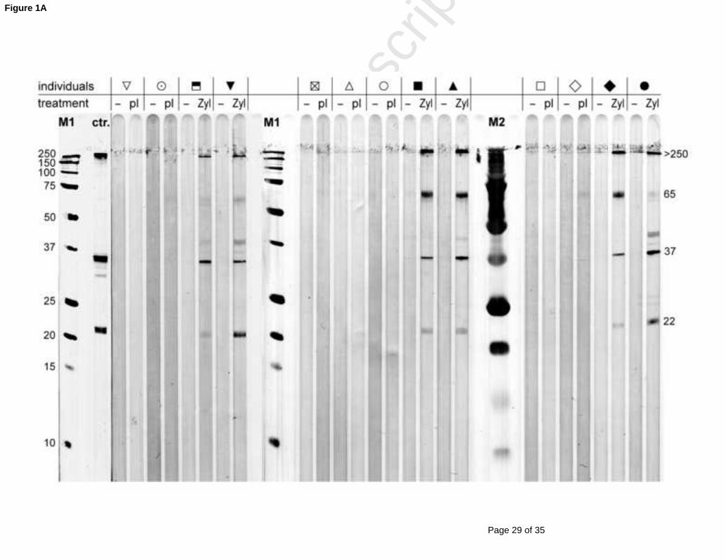
Western Blot analysis of all sera taken before and 2 weeks after treatment demonstrates a
iPPVO-specific humoral immune response in the Zylexis group in contrast to placebo-treated
dogs (Figure 1A). The most abundant PPVO immuno-reactive proteins have been previously
described with molecular masses of 65 kDa, 39 kDa and 22 kDa (Czerny et al., 1997). All
sera of Zylexis-treated dogs taken two weeks after the treatment show immuno-reactive 250
bands at >250, 65 and <39, which are not detectable in sera taken prior to the Zylexis
treatment (Fig. 1A). Zylexis-treated dogs show also a signal against 22 kDa protein with
different intensity. The protein close to 37 kDa could be identified with two monoclonal Abs
10E6 and 8D7 specific for the envelope protein which is also known as F1L/env protein of
PPVO (data not shown) with an apparent molecular mass of 39 kDa as previously described 255
(Housawi et al., 1998). Additionally we used a serum from a PPVO-immunized rabbit as a
positive control, that show also a signal at about 37 kDa. In contrast to the >250 kDa and
65 kDa protein bands, specific serum IgG for the 39 kDa and 22 kDa bands were not
detectable four years post treatment by immuno-staining. The signal against a 65 kDa protein
was, surprisingly, stronger four years post treatment, but was not detectable in pre-treatment 260
sera (data not shown).
For quantification of the iPPVO-specific antibodies sera (1:50 diluted) from these six
Zylexis and seven placebo administrated dogs were taken from three different time points
(prior to, two weeks and two month post Zylexis® or placebo administration) and were
Page 12 of 35
Accep
ted
Man
uscr
ipt
12
measured in PPVO-specific ELISA. Additionally, sera were investigated from three Zylexis-265
and four placebo-treated dogs four years after their last treatment. A statistically significant
increase of PPVO-specific IgG in sera of the Zylexis-treated dogs in contrast to the placebo-
treated individuals was observed two weeks and two month post treatment by PPVO-specific
ELISA (Fig. 1B). However, detectable serum levels of anti-PPVO IgG could not be found by
the ELISA approximately four years after the final iPPVO administration.270
We were also interested to analyze whether the iPPVO-specific antibodies were neutralizing.
Therefore, we performed a VNT with BK-KL-3A cells as target cells for PPVO. No
neutralizing antibodies could be detected in any of the sera (<1:5) from Zylexis-treated dogs
taken from different time points after administration. In contrast, using an anti-PPVO sheep
antiserum as positive control, neutralizing activity was detectable (VN50=191; data not 275
shown). Thus, iPPVO treatment of dogs induces a humoral response similar to natural PPVO
infection in permissive hosts and similar to viable PPVO application in dogs (Büttner et al.,
1995), characterized by a transient production of non-neutralizing serum antibodies against
already known immunogeneic PPVO proteins.
280
Enhanced iPPVO-induced oxidative burst rates in Zylexis-treated dogs is correlated to
iPPVO antibody titers
We have recently shown that iPPVO induces moderate oxidative burst in canine blood
monocytes but not in PMN (Schütze et al., 2009). In different infection models for parasites
and bacteria it was shown that the oxidative burst activity was enhanced by opsonizing 285
antibodies and complement (Arbo et al., 2006; Laursen et al., 2003; Uppington et al., 2006).
To investigate whether the PPVO-specific antibodies in serum of Zylexis treated dogs would
augment oxidative burst of peripheral blood leukocytes, an oxidative burst assay was
performed by supplementing serum of Zylexis-treated dogs (diluted 1:10) to blood cells of
an untreated dog. In this experiment it was demonstrated that the iPPVO-induced oxidative 290
Page 13 of 35
Accep
ted
Man
uscr
ipt
13
burst was enhanced by serum transfer of anti-iPPVO immune sera taken two weeks and two
months following Zylexis administration in comparison to sera sampled before treatment
(***P<0.001, *P<0.05, ANOVA and Bonferroni´s Multiple Comparison Post test; Fig. 1C).
In contrast, stimulation with PMA did not show any differences between sera from distinct
time points. Interestingly, in the presence of immune serum not only monocytes but also PMN 295
responded to iPPVO stimulation with oxidative burst, whereas samples supplemented with
serum before treatment showed iPPVO-induced burst activity exclusively in monocytes as
recently described (Schütze et al., 2009). Consistent with the time course of the antibody
levels (see Fig. 1B) transfer of sera taken two months after Zylexis treatment still enhanced
the oxidative burst rate, although to a lower extent as in the presence of serum taken after two300
weeks (Fig. 1C). Furthermore, the high responder dog identified by ELISA analogously
shows a high response in oxidative burst in monocytes after stimulation with iPPVO in
comparison to the other Zylexis-treated individuals. Thus, the iPPVO-specific IgG in immune
sera quantified by ELISA correlates well with the enhancement of burst activity in the serum
transfer experiment performed with cells from a naive donor dog. We also investigated serum 305
transfer from a placebo-treated dog to cells of an untreated dog. Here we found no
enhancement of oxidative burst in contrast to serum of a Zylexis-treated dog (data not
shown). Finally, we tested the serum of the cell donor itself. As expected we found a weak
reactivity of monocytes by iPPVO stimulation in contrast to PMN (data not shown).
Additionally we demonstrated by a dilution series of immune serum (derived from a high 310
responder) added to blood samples of an untreated dog, that high serum concentrations
correlate with an enhancement of iPPVO-induced burst activity (Fig. 1D).
Long-term enhancement of iPPVO-induced oxidative burst in Zylexis-treated dogs is
mediated by a transferable serum component315

Page 14 of 35
Accep
ted
Man
uscr
ipt
14
To analyze a potential long-term effect of Zylexis blood samples from Zylexis-treated dogs
were investigated (n = 3) >four years after administration of six Zylexis® doses. Blood
samples from placebo-treated dogs (n=4), administrated >four years ago, served as control
group. Surprisingly, the undiluted blood samples from the Zylexis®-treated individulas
showed increased burst rates after in vitro stimulation with iPPVO in both, monocytes and 320
PMNs, although the treatment had been stopped four years ago (Fig. 2). In contrast, whole
blood from placebo-treated dogs exhibited burst rates following iPPVO stimulation
comparable to those from untreated dogs (Schütze et al., 2009), characterized by a moderate
burst response to iPPVO exclusively in monocytes but not in PMNs. The enhanced burst rates
observed in whole blood from Zylexis®-treated dogs were found to be specific for iPPVO 325
stimulation. The burst responses observed for LPS or PMA did not differ between both
groups of dogs. Unfortunately, due to the small group sizes (Zylexis®-treated animals, n = 3;
placebo-treated dogs, n = 4), it was impossible to determine statistical significance.
Nevertheless, using all separately measured values of triplicates (3 measurements for every
stimulus/dog) for statistical analysis, the difference between whole blood measurement 330
between Zylexis and the placebo-treated group was found to be highly significant (p<0.001,
Bonferroni's Multiple Comparison Test, data not shown). Thus, even when serum anti-PPVO
IgG was not detectable any more in dogs four years after their last Zylexis® treatment, the
oxidative burst rates were enhanced in blood samples from these dogs (see Fig 2).
In order to verify that the enhancement of iPPVO-induced oxidative burst observed in blood335
samples from Zylexis®-treated dogs (>4 years post administration) depends on a serum
component, again serum transfer experiments were done. The undiluted serum of the
Zylexis®-treated dogs (n = 2, >4 years post administration) enhanced iPPVO-induced
oxidative burst in PMN and monocytes of a placebo-treated dog (data not shown). As a
negative control the homologous serum of the placebo-treated dogs themselves was utilized. 340
In contrast, cells supplemented with the different sera responded similarly upon stimulation

Page 15 of 35
Accep
ted
Man
uscr
ipt
15
with PMA (data not shown). Therefore, even very low and by ELISA undetectable anti-
iPPVO antibody levels appear to be sufficient for the iPPVO-induced enhancement of
oxidative burst.
345
Antibody depletion abrogates enhancement of oxidative burst by blood monocytes and PMN
stimulated with iPPVO
To prove the involvement of specific antibodies in the enhancement of iPPVO-induced
oxidative burst, Ig (mainly IgG and IgM) from the immune serum of a Zylexis-treated dog
was depleted by protein A-sepharose beads as previously described (Scott et al., 1997; Peng et 350
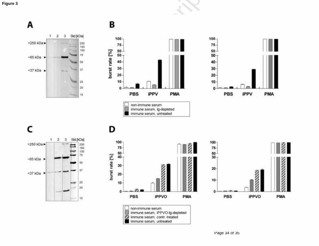
al., 1991). Following Ig depletion by protein A-sepharose treatment of immune serum, all
specific-PPVO Ig (with only a minor reactivity against the 65 kDa band left) were depleted as
demonstrated in an PPVO-specific immunoblot using protein A-depleted as compared with
non-depleted sera (Fig. 3A). This Ig-depleted serum was utilized in an oxidative burst
experiment in comparison to untreated immune serum. The homologous serum of the 355
untreated dog (cell donor) was used as an additional control. After stimulation with iPPVO,
the Ig-depleted immune-serum showed a strongly reduced oxidative burst activity similar to
the non-immune serum, in contrast to the untreated immune serum (Fig. 3B).
To selectively deplete PPVO-specific antibodies from the immune serum it was treated with
iPPVO-coupled sepharose beads. As a negative control, the immune serum was exposed to360
uncoupled sepharose. The anti-PPVO antibody level was strongly reduced as demonstrated in
immunoblot analysis (Fig. 3C), albeit complete antibody depletion was not reached with the
iPPVO-coupled sepharose beads. This is especially evident for the F1L/env protein at about
37 kDa. As expected, treatment with uncoupled sepharose did not change antibody levels
significantly (Fig 3C). Again depleted and control-treated sera were tested in oxidative burst 365
assay. The transfer of serum depleted of PPVO-specific antibodies to whole blood cells of a
naïve dog resulted in a reduction of oxidative burst activity in iPPVO-stimulated PMN and
Page 16 of 35
Accep
ted
Man
uscr
ipt
16
monocytes in contrast to untreated and mock-treated immune serum (Fig. 3D). This result is
consistent with the decreased iPPVO immunoreactivity detected by immunoblot analysis (Fig.
3C).370
Uptake of iPPVO particle exclusively in cells with dendritic and monocytic morphology but
not in PMN is independent of specific antibodies
From vaccinia virus it is known that the most abundant form of poxvirus particles are
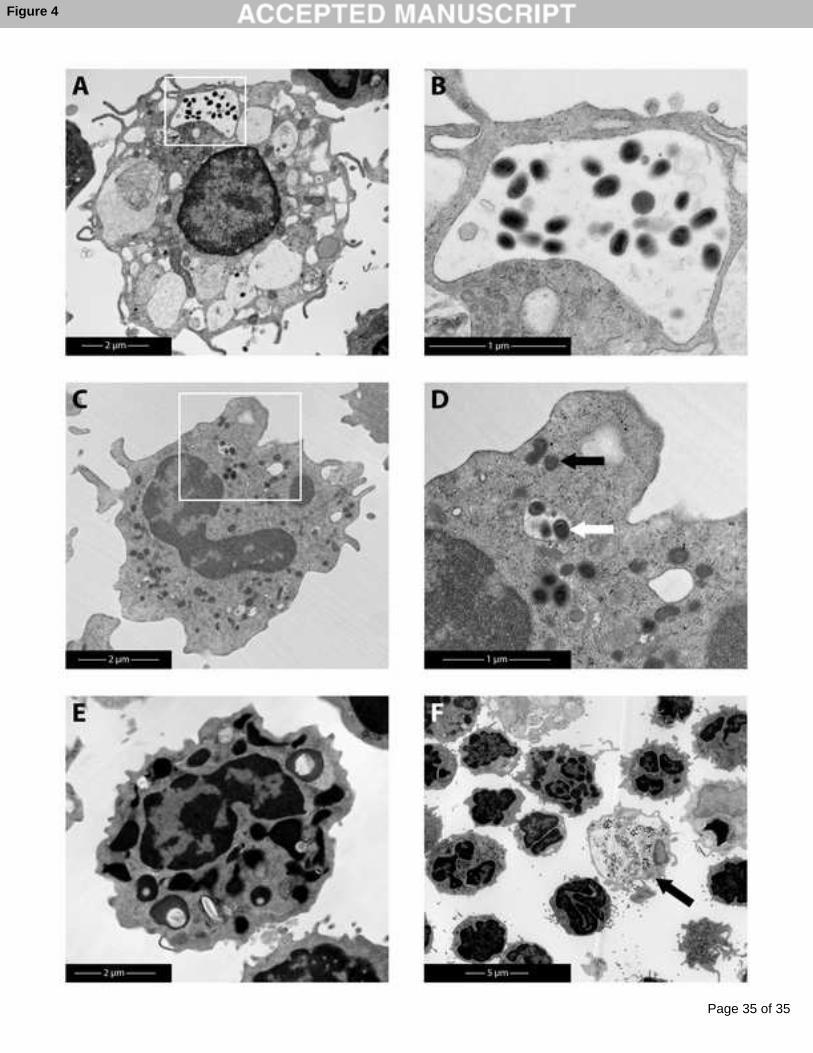
intracellular mature virus (IMV) derived from cell lysates (Smith et al., 2002). Analysis of 375
short-term iPPVO-exposed canine PBL (4h) by electron microscopy provide an insight into
the interaction of iPPVO with the immune cells. In the following experiment iPPVO was
added to PBL in the presence vs. absence of immune serum. To allow formation of immune
complexes, undiluted immune sera were incubated with iPPVO at 37 °C for 2 hours before
addition to cells. As control a non-immune serum was utilized. Analysis by electron 380
microscopy of these samples demonstrated that cells with dendritic and monocytic
morphology can take up viral particles (Fig. 4A,C), with the majority being found in the
lysosomal compartments (Fig. 4B), but iPPVO was also found in the cytoplasm (Fig. 4
A,C,D). Dendritic cells, in particular, appeared to contain a high virus load per cell, mostly in
lysosomes. In contrast, PMN appeared unable to take up iPPVO (Fig. 4E,F). Comparing viral 385
uptake using immune vs. non-immune serum samples by counting the viral particle load per
cell (n > 15 cells / per group) revealed no difference. This suggests that the uptake of iPPVO
particles is independent of the presence of specific antibodies. However, electron microscopic
analysis is limited in that it is unlikely to be fully representative or quantitative, because
iPPVO-positive cells were found only rarely (Fig. 4F).390

Page 17 of 35
Accep
ted
Man
uscr
ipt
17
Discussion
The present study demonstrates (i) that multiple Zylexis treatment of dogs results in the
formation of PPVO-specific serum antibodies, (ii) that the moderate iPPVO-induced
oxidative burst in monocytes can be enhanced, and additionally, PMN become sensitive to
iPPVO-induced oxidative burst in the presence of PPVO-specific serum IgG of Zylexis-395
treated dogs, (iii) , that the uptake of inactivated virus particles is restricted to dendritic-like
cells and monocytes, independently of the presence of PPVO-specific antibodies.
From infected hosts (man and ruminants) and immunization studies in mice it is known that
non-permissive hosts generate an antibody response to infection with live PPVO (Czerny et
al., 1997; Housawi et al., 1998; Yirrell et al., 1994). However, in this study animals were 400
treated with chemically inactivated virus. The quantification of PPVO-specific antibodies
demonstrates the development of specific IgG shortly after a series of six applications of
Zylexis. In the dogs accessible for this study the antibody level was found to drop below the
detection limit in ELISA four years post final treatment. This result is in agreement with data
from experimentally and naturally PPVO-infected sheep that also show high antibody levels 405
which increased after productive primary infection and decreased after recovery (Lloyd et al.,
2000; McKeever et al., 1987). Thus, iPPVO treatment of a non-permissive host induces a
transient humoral response similar to natural PPVO infection in permissive hosts and viable
PPVO administration in dogs. However, it remains open how many applications with iPPVO
are necessary to induce a humoral response. In any case the humoral response seems to 410
consist of non-neutralising antibodies with longevity and effect on oxidative burst of PMN
and monocytes.
In detail, antibodies against an immuno-dominant PPVO protein previously designated as a
39 kDa protein were detected (Housawi et al., 1998). Antibody reactivity against this protein
in the molecular weight range of ~40 kDa, which is enriched in viral envelope preparations, 415
has been described previously (Balassu and Robinson, 1987; Housawi et al., 1998). The

Page 18 of 35
Accep
ted
Man
uscr
ipt
18
coding gene F1L reveals an open reading frame of 1,002 bp with the potential to encode a
protein with a MW of 36.7 kDa (Housawi et al., 1998). The specific band observed by
immunoblot in this study was near 37 kDa, which was confirmed by different monoclonal
antibodies directed against the 39 kDa protein. The 39 kDa protein is homologous to the H3L 420
gene of vaccinia virus which has also been described as an immunogenic envelope protein
(Housawi et al., 1998). Besides the 39 kDa protein other immunoreactive proteins of PPVO
have been described with molecular weights of 22 and 65 kDa (Czerny et al., 1997). Like
others, a strong increase of anti-65 kDa-specific IgG in the Zylexis-treated dogs was found,
that did not decline over 4 years post administration. The identity of the 65 kDa protein 425
remains elusive. Czerny et al. suggest that the protein is a major core protein (Czerny et al.,
1997). Here some cross-reactivity with this 65 kDa protein was observed in serum of dogs
treated with placebo or untreated dogs. In addition, sera of all Zylexis-treated dogs showed
specific antibodies against a ~250 kDa protein and some of those also against the 22 kDa
protein.430
In our recent report differences between canine monocytes and PMN for the iPPVO-induced
oxidative burst were found (Schütze et al., 2009). In the absence of specific antibodies,
iPPVO-induced burst was restricted to canine monocytes, demonstrated in this study by blood
samples of placebo- or non-treated dogs. In contrast, Förster et al. demonstrated PPVO-
induced oxidative burst in human neutrophils, that was accompained by an up-regulation of 435
complement receptors CR1 and CR3 (Förster et al., 1994). One possible explanation for this
cell specificity of oxidative burst in the absence of specific antibodies is that only monocytes
express adequate pattern recognition receptors (PRR) that respond to iPPVO as a pathogen-
associated molecular pattern or danger signal, whereas canine PMN might not express such
receptors. Previously it was demonstrated that Toll-like receptors (TLR) mediate oxidative 440
burst (Hoarau et al., 2007). Until now, the potential PRR used by parapoxviruses are not
characterized. It is also conceivable that indirect activation of burst activity by secretion of

Page 19 of 35
Accep
ted
Man
uscr
ipt
19
mediators from other cells activate oxidative burst, e.g. by G-protein coupled receptors on
monocytes (Sheppard et al., 2005). The electron microscopic analysis of canine cells
incubated with iPPVO additionally demonstrated differences between monocytes/dendritic 445
cells and PMN, because only dendritic cells and monocytes were found to take up viral
particles.
By serum transfer experiments this study demonstrated that immune serum mediates
enhancement of the iPPVO/Zylexis-induced oxidative burst. Interestingly the enhancement
of oxidative burst was not only observed in monocytes but also in PMN, which showed no 450
oxidative burst in naïve animals. To prove wether PPVO-specific antibodies are actually
responsible for the enhanced burst activity antibody-depletion from serum containing PPVO-
specific IgGs was performed. By protein-A sepharose all specific IgGs (anti-39 kDa) were
depleted from immune serum, which completely abrogated the enhancement effect. A more
selective strategy of depletion eliminating only specific PPVO antibodies by iPPVO-coupled 455
sepharose did not completely abolish the presence of anti-39 kDa antibodies as demonstrated
by a reduced but still visible signal for the 39 kDa protein in the immunoblot (Fig. 3C).
However, the partial depletion resulted in a partial decrease of burst enhancement (Fig. 3D).
This may suggest that in particular the anti-39 kDa antibodies are mediators for the
enhancement of PPVO-induced oxidative burst. This finding fits well with those of other 460
groups studying innate immunity to parasites, fungi or bacteria. Induction of oxidative burst
by Giardia lamblia trophozoites was enhanced by opsonisation with anti-Giardia
hyperimmune serum and complement in PMN (Arbo et al., 2006). Laursen et al.
demonstrated that together with complement, lung surfactant D and mannan binding lectin the
specific IgG antibodies in contrast to IgM mediate enhanced burst activity in neutrophils in 465
response to Pneumocystis carinii (Laursen et al., 2003). Furthermore, the uptake of
Salmonella enterica serovar Typhimurium and oxidative burst induction was increased by
binding to high affinity IgG receptor FcRI mediated by immune serum (Uppington et al.,

Page 20 of 35
Accep
ted
Man
uscr
ipt
20
2006). For our model we suggest that the presence of PPVO-specific antibodies results in
formation of immune complexes that make the virus particles accessible to the low affinity 470
Fcreceptors, which are also expressed on PMN. Suh et al. demonstrated a FcRIIA-mediated
NADPH oxidase activation in human cells, that was considerably disrupted by
phosphatidylinositol-3 kinase (PI3) inhibitors, whereas phagocytosis was inhibited to a lower
extent (Suh et al., 2006).
Unexpectedly leukocytes of whole blood samples taken from dogs which were treated four 475
years previously with Zylexis showed enhanced iPPVO-induced oxidative burst, in contrast
to the placebo-treated group, although PPVO-specific antibodies were no longer detectable by
ELISA (using a pre-dilution of sera at 1:50, see Fig. 1B). This suggests that very low serum
levels of specific IgGs are sufficient to enhance iPPVO-induced oxidative burst.
The electron microscopic analysis showed that iPPVO particles were taken up even in the 480
presence of immune serum exclusively in dendritic cells and monocytes, but not in PMN,
which indicates that the antibody-mediated oxidative burst activity is independent from the
uptake of virus particles. However, further investigations are needed to confirm these
hypotheses. In addition, it remains to be evaluated whether macropinocytosis is the
mechanism for PPVO uptake in dendritic cells and monocytes as recently demonstrated for 485
VACV (Mercer and Helenius, 2008). Interestingly, the 39 kDa protein of PPVO was shown to
bind glucosamine heparane sulphate on host cells (Scagliarini et al., 2004) similar to its
homologue, the H3L vaccinia protein that is part of the virus entry machinery (Lin et al.,
2000).
490
Conclusion
In summary, iPPVO-specific antibodies enhancing oxidative burst present an interesting
example of collaboration between innate and adaptive immunity. In future studies the
importance of the antibody-dependent iPPVO-induced oxidative burst activity needs to be
Page 21 of 35
Accep
ted
Man
uscr
ipt
21
investigated in vivo and its relevance has to be identified in innate immunity against viral, 495
bacterial and fungal infections.
Acknowledgements
We are indebted to Dr. H. Müller (Institute of Virology, College of Veterinary Medicine,
University of Leipzig, Germany) for making sera from Zylexis/placebo-treated dogs 500
available to us. We also thank Juliane Richter for technical assistance and C. Westermann for
electron microscopy. The blood samples were kindly provided by Ina Hochheim (Institute of
Pharmacology) and Christian Boelzig (Department of Small Animal Medicine), both from the
College of Veterinary Medicine, University of Leipzig, Germany. The project was financially
supported by Pfizer Animal Health. We wish to thank Dr. Uwe Müller for careful reading of 505
this manuscript.

Page 22 of 35
Accep
ted
Man
uscr
ipt
22
Figure 1:
PPVO-specific IgG in serum of Zylexis-treated dogs results in enhanced iPPVO-induced
oxidative burst activity510
The presence of orf virus-specific antibodies was analysed in sera of Zylexis or placebo-
treated dogs (n = 6 and 7, respectively) from samples taken before, two weeks (2 wk), two
month (2 mo) and four years (4 yr) post application. A) PPVO-specific immunoblot using
vertically fractionated (12 % SDS-PAGE) and on nitrocellulose membrane immobilized
iPPVO and unstained (M1) or prestained (M2) protein standards. PPVO-specific IgGs from 515
dog sera (1:10) before (-) and 2 weeks after treatment with Zylexis (Zyl) or placebo (pl) was
detected by using anti-dog IgG-HRP. As control (ctr.) a rabbit serum specific for orf virus
(1:100 dilution) and anti-rabbit IgG-HRP was used. B) PPVO-specific ELISA: PPVO-antigen
and control-antigen (see Mat. & Methods) were coated to 96-well plates; virus-specific IgG
from the diluted sera (1:50) were detected by goat-anti-dog-HRP developed with TMB 520
substrate. KELA units of optical density at 650 nm are shown. Statistical Analysis was
performed with Kruskal-Wallis Test and Dunn´s multiple comparison test (***P < 0.001,
*P < 0.05); C) Sera (1:10) of 5 Zylexis-treated dogs (2 wk and 2 mo) in comparison to sera
before Zylexis-treatment were tested in serum transfer experiments for iPPVO-induced
oxidative burst activity. As control stimuli PBS and PMA (185 ng/ml) were used. All serum 525
transfer experiments were performed with blood cells from non-Zylexis-treated dogs
(laboratory animals). Statistical analysis was performed with ANOVA and Bonferroni´s
multiple comparison test (*P < 0.05, ***P < 0.001). D) Serial dilutions of immune sera were
added to blood cells of an untreated dog in the iPPVO-induced oxidative burst assay.
530

Page 23 of 35
Accep
ted
Man
uscr
ipt
23
Figure 2:
Long-lasting enhancement of oxidative burst induced by iPPVO in Zylexis-treated dogs
Dogs were treated on six occasions with Zylexis (n = 3) or placebo (n = 4) respectively, four
years previously. Blood samples from dogs were taken and oxidative burst activity of
Zylexis- vs placebo-treated dogs was analysed by in vitro-restimulation with pharmaceutical 535
formulation of iPPVO in contrast to stabiliser control. As further control stimuli PBS
(negative control), LPS (5 µg/ml) and PMA (185 ng/ml) were utilized. Measurement of
oxidative burst activity in canine monocytes and PMN was performed by flow cytometry.
Detection of ROIs was achieved by staining cells with DHR (8.9 µg/ml), that is oxidized by
ROIs to the green fluorescent dye rhodamine. Burst rates were calculated as percentage of 540
rhodamine-positive cells within the specified cell populations.
Page 24 of 35
Accep
ted
Man
uscr
ipt
24
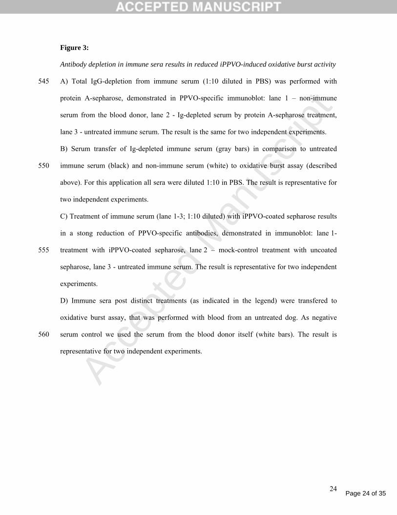
Figure 3:
Antibody depletion in immune sera results in reduced iPPVO-induced oxidative burst activity
A) Total IgG-depletion from immune serum (1:10 diluted in PBS) was performed with 545
protein A-sepharose, demonstrated in PPVO-specific immunoblot: lane 1 – non-immune
serum from the blood donor, lane 2 - Ig-depleted serum by protein A-sepharose treatment,
lane 3 - untreated immune serum. The result is the same for two independent experiments.
B) Serum transfer of Ig-depleted immune serum (gray bars) in comparison to untreated
immune serum (black) and non-immune serum (white) to oxidative burst assay (described 550
above). For this application all sera were diluted 1:10 in PBS. The result is representative for
two independent experiments.
C) Treatment of immune serum (lane 1-3; 1:10 diluted) with iPPVO-coated sepharose results
in a stong reduction of PPVO-specific antibodies, demonstrated in immunoblot: lane 1-
treatment with iPPVO-coated sepharose, lane 2 – mock-control treatment with uncoated 555
sepharose, lane 3 - untreated immune serum. The result is representative for two independent
experiments.
D) Immune sera post distinct treatments (as indicated in the legend) were transfered to
oxidative burst assay, that was performed with blood from an untreated dog. As negative
serum control we used the serum from the blood donor itself (white bars). The result is 560
representative for two independent experiments.

Page 25 of 35
Accep
ted
Man
uscr
ipt
25
Figure 4:
Dendritic cells and monocytes but not PMN show uptake of iPPVO particles
Isolated PBL were cultivated for 4h with iPPVO, incubated before with immune serum (to 565
allow formation of immune complexes) or non-immune serum. Analysis by electron
microscopy was performed as detailed in Mat & Methods. Since we could not find any
differences between samples incubated with immune or non-immune serum, only data from
incubation with preformed immune complexes (immune serum) are shown. Internalized
iPPVO particles in: A) a dendritic cell, B) in the lysosomal compartment (inset from dendritic 570
cell); or C) in monocytes D) in lysosomal compartment (white arrow) or cytoplasm (black
arrow), see inset from monocyte. E) Granulocytes did not internalize iPPVO particles F) as
demonstrated additionally in an overview picture in contrast to dendritic cells (black arrow).

Page 26 of 35
Accep
ted
Man
uscr
ipt
26
575Reference List
1. Arbo, A., Pavia-Ruz, N., Santos, J.I., 2006. Opsonic requirements for the respiratory burst of neutrophils against Giardia lamblia trophozoites. Arch. Med. Res. 37, 465-473.
2. Balassu, T.C. and Robinson, A.J., 1987. Orf virus replication in bovine testis cells: 580kinetics of viral DNA, polypeptide, and infectious virus production and analysis of virion polypeptides. Arch. Virol. 97, 267-281.
3. Buddle, B.M. and Pulford, H.D., 1984. Effect of passively-acquired antibodies and vaccination on the immune response to contagious ecthyma virus. Vet. Microbiol. 9, 515-522.585
4. Büttner, M., Czerny, C.P., Schumm, M., 1995. [Behavior of Orf virus in permissive and nonpermissive systems]. Tierarztl. Prax. 23, 179-184.
5. Büttner, M. and Mayr, A., 1986. Tests on protection against viral diseases. Comp Immunol. Microbiol. Infect. Dis. 9, 205-215.
6. Büttner, M. and Rziha, H.J., 2002. Parapoxviruses: from the lesion to the viral genome. 590J. Vet. Med. B Infect. Dis. Vet. Public Health 49, 7-16.
7. Castrucci, G., Osburn, B.I., Frigeri, F., Ferrari, M., Salvatori, D., Lo, D.M., Barreca, F., 2000. The use of immunomodulators in the control of infectious bovine rhinotracheitis. Comp Immunol. Microbiol. Infect. Dis. 23, 163-173.
8. Czerny, C.P., Waldmann, R., Scheubeck, T., 1997. Identification of three distinct 595antigenic sites in parapoxviruses. Arch. Virol. 142, 807-821.
9. Förster, R., Wolf, G., Mayr, A., 1994. Highly attenuated poxviruses induce functional priming of neutrophils in vitro. Arch. Virol. 136, 219-226.
10. Gherardi, M.M. and Esteban, M., 2005. Recombinant poxviruses as mucosal vaccine vectors. J. Gen. Virol. 86, 2925-2936.600
11. Haig, D.M. and Mercer, A.A., 1998. Ovine diseases. Orf. Vet. Res. 29, 311-326.
12. Hoarau, C., Gerard, B., Lescanne, E., Henry, D., Francois, S., Lacapere, J.J., El, B.J., Dang, P.M., Grandchamp, B., Lebranchu, Y., Gougerot-Pocidalo, M.A., Elbim, C., 2007. TLR9 activation induces normal neutrophil responses in a child with IRAK-4 deficiency: involvement of the direct PI3K pathway. J. Immunol. 179, 4754-4765.605
13. Housawi, F.M., Roberts, G.M., Gilray, J.A., Pow, I., Reid, H.W., Nettleton, P.F., Sumption, K.J., Hibma, M.H., Mercer, A.A., 1998. The reactivity of monoclonal antibodies against orf virus with other parapoxviruses and the identification of a 39 kDa immunodominant protein. Arch. Virol. 143, 2289-2303.
14. Laursen, A.L., Obel, N.S., Holmskov, U., Jensenius, J.C., Aliouat, e.M., Andersen, P.L., 6102003. Activation of the respiratory burst by Pneumocystis carinii. Efficiency of different

Page 27 of 35
Accep
ted
Man
uscr
ipt
27
antibody isotypes, complement, lung surfactant protein D, and mannan-binding lectin. APMIS 111, 405-415.
15. Le, J.C., L'Haridon, R., Madelaine, M.F., Cornu, C., Asso, J., 1978. Transfer of antibodies against the CPD virus through colostrum and milk. Ann. Rech. Vet. 9, 342-615346.
16. Lin, C.L., Chung, C.S., Heine, H.G., Chang, W., 2000. Vaccinia virus envelope H3L protein binds to cell surface heparan sulfate and is important for intracellular mature virion morphogenesis and virus infection in vitro and in vivo. J. Virol. 74, 3353-3365.
17. Lloyd, J.B., Gill, H.S., Haig, D.M., Husband, A.J., 2000. In vivo T-cell subset depletion 620suggests that CD4+ T-cells and a humoral immune response are important for the elimination of orf virus from the skin of sheep. Vet. Immunol. Immunopathol. 74, 249-262.
18. März M., 2007. Trial of Baypamune® in the Infectious Model of Canine Oral Papillomatosis in the dog. Thesis. Faculty of Veterinary Medicine, University of 625Leipzig, Germany.
19. Mayr, A., Buttner, M., Wolf, G., Meyer, H., Czerny, C., 1989. [Experimental detection of the paraspecific effects of purified and inactivated poxviruses]. Zentralbl Veterinarmed B 36, 81-99.
20. Mayr, A., Himmer, B., Baljer, G., and Sailer, J. Erregerunspezifische Prophylaxe und 630Therapie von Pseudomonas-aeruginosa-Wundinfektion mittels Paraimmunisierung im Mausmodell. Zentbl.Bakteriol.Hyg.Parasitenkd.Infektionskr. [176], 506-514. 1978. Ref Type: Magazine Article
21. McKeever, D.J., Jenkinson, D.M., Hutchison, G., Reid, H.W., 1988. Studies of the pathogenesis of orf virus infection in sheep. J. Comp Pathol. 99, 317-328.635
22. McKeever, D.J., Reid, H.W., Inglis, N.F., Herring, A.J., 1987. A qualitative and quantitative assessment of the humoral antibody response of the sheep to orf virus infection. Vet. Microbiol. 15, 229-241.
23. Mercer, J. and Helenius, A., 2008. Vaccinia virus uses macropinocytosis and apoptotic mimicry to enter host cells. Science 320, 531-535.640
24. Peng, Z.K., Simons, F.E., Becker, A.B., 1991. Differential binding properties of protein A and protein G for dog immunoglobulins. J Immunol. Methods 145, 255-258.
25. Raue M., 2008. Influence of an immunomodulator on the persistence of the COPV genome and on the immune response in dogs with clinical signs of canine oral papillomatosis. Thesis. Faculty of Veterinary Medicine, University of Leipzig, 645Germany.
26. Rothe, G., Oser, A., Valet, G., 1988. Dihydrorhodamine 123: a new flow cytometric indicator for respiratory burst activity in neutrophil granulocytes. Naturwissenschaften 75, 354-355.
27. Scagliarini, A., Gallina, L., Dal, P.F., Battilani, M., Ciulli, S., Prosperi, S., 2004. 650Heparin binding activity of orf virus F1L protein. Virus Res. 105, 107-112.
Page 28 of 35
Accep
ted
Man
uscr
ipt
28
28. Schütze, N., Raue, R., Buttner, M., Alber, G., 2009. Inactivated parapoxvirus ovis activates canine blood phagocytes and T lymphocytes. Vet Microbiol. 137, 260-267.
29. Scott, M.A., Davis, J.M., Schwartz, K.A., 1997. Staphylococcal protein A binding to canine IgG and IgM. Vet Immunol. Immunopathol. 59, 205-212.655
30. Sheppard, F.R., Kelher, M.R., Moore, E.E., McLaughlin, N.J., Banerjee, A., Silliman, C.C., 2005. Structural organization of the neutrophil NADPH oxidase: phosphorylation and translocation during priming and activation. J. Leukoc. Biol. 78, 1025-1042.
31. Shin, S.J., Chang, Y.F., Jacobson, R.H., Shaw, E., Lauderdale, T.L., Appel, M.J., Lein, D.H., 1993. Cross-reactivity between B. burgdorferi and other spirochetes affects 660specificity of serotests for detection of antibodies to the Lyme disease agent in dogs. Vet Microbiol. 36, 161-174.
32. Smith, G.L., Vanderplasschen, A., Law, M., 2002. The formation and function of extracellular enveloped vaccinia virus. J Gen. Virol. 83, 2915-2931.
33. Suh, C.I., Stull, N.D., Li, X.J., Tian, W., Price, M.O., Grinstein, S., Yaffe, M.B., 665Atkinson, S., Dinauer, M.C., 2006. The phosphoinositide-binding protein p40phox activates the NADPH oxidase during FcgammaIIA receptor-induced phagocytosis. J. Exp. Med. 203, 1915-1925.
34. Uppington, H., Menager, N., Boross, P., Wood, J., Sheppard, M., Verbeek, S., Mastroeni, P., 2006. Effect of immune serum and role of individual Fcgamma receptors 670on the intracellular distribution and survival of Salmonella enterica serovar Typhimurium in murine macrophages. Immunology 119, 147-158.
35. Weber, O., Siegling, A., Friebe, A., Limmer, A., Schlapp, T., Knolle, P., Mercer, A., Schaller, H., Volk, H.D., 2003. Inactivated parapoxvirus ovis (Orf virus) has antiviral activity against hepatitis B virus and herpes simplex virus. J. Gen. Virol. 84, 1843-1852.675
36. Yirrell, D.L., Reid, H.W., Norval, M., Entrican, G., Miller, H.R., 1991. Response of efferent lymph and popliteal lymph node to epidermal infection of sheep with orf virus. Vet. Immunol. Immunopathol. 28, 219-235.
37. Yirrell, D.L., Vestey, J.P., Norval, M., 1994. Immune responses of patients to orf virus infection. Br. J. Dermatol. 130, 438-443.680
38. Ziebell, K.L., Steinmann, H., Kretzdorn, D., Schlapp, T., Failing, K., Schmeer, N., 1997. The use of Baypamun N in crowding associated infectious respiratory disease: efficacy of Baypamun N (freeze dried product) in 4-10 month old horses. Zentralbl Veterinarmed B 44, 529-536.
685
Page 29 of 35
Accep
ted
Man
uscr
ipt
Figure 1A

Page 30 of 35
Accep
ted
Man
uscr
ipt
Figure 1B

Page 31 of 35
Accep
ted
Man
uscr
ipt
Figure 1C

Page 32 of 35
Accep
ted
Man
uscr
ipt
Figure 1D

Page 33 of 35
Accep
ted
Man
uscr
ipt
Figure 2
Page 34 of 35
Accep
ted
Man
uscr
ipt
Figure 3
Page 35 of 35
Accep
ted
Man
uscr
ipt
Figure 4