Embed Size (px)
Citation preview
HAL Id: hal-03335772https://hal.archives-ouvertes.fr/hal-03335772
Submitted on 20 Oct 2021
HAL is a multi-disciplinary open accessarchive for the deposit and dissemination of sci-entific research documents, whether they are pub-lished or not. The documents may come fromteaching and research institutions in France orabroad, or from public or private research centers.
L’archive ouverte pluridisciplinaire HAL, estdestinée au dépôt et à la diffusion de documentsscientifiques de niveau recherche, publiés ou non,émanant des établissements d’enseignement et derecherche français ou étrangers, des laboratoirespublics ou privés.
Soluble Guanylate Cyclase Inhibitors Discovered amongNatural Compounds
Olga Petrova, Isabelle Lamarre, Fabienne Fasani, Catherine Grillon, MichelNegrerie
To cite this version:Olga Petrova, Isabelle Lamarre, Fabienne Fasani, Catherine Grillon, Michel Negrerie. Soluble Guany-late Cyclase Inhibitors Discovered among Natural Compounds. Journal of Natural Products, AmericanChemical Society, 2020, 83 (12), pp.3642-3651. �10.1021/acs.jnatprod.0c00854�. �hal-03335772�
1
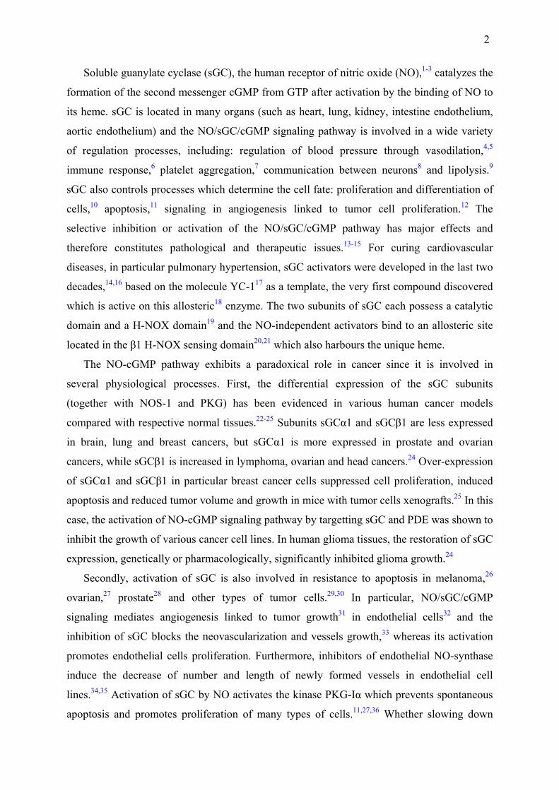
Soluble Guanylate Cyclase Inhibitors Discovered among Natural Compounds Olga N. Petrova, Isabelle Lamarre, Fabienne Fasani, Catherine Grillon and Michel Negrerie Corresponding author: [email protected] ABSTRACT: Soluble guanylate cyclase (sGC) is the human receptor of nitric oxide (NO) in numerous kinds of cells and produces the second messenger cGMP upon NO binding to its heme. sGC is involved in many cell signaling pathways both in healthy conditions and in pathological conditions, such as angiogenesis associated with tumor growth. Addressing the selective inhibition of the NO/cGMP pathway is a strategy worthwhile to be investigated for slowing down tumoral angiogenesis or for curing vasoplegia. However sGC inhibitors are lacking to investigation. We have explored a chemical library of various natural compounds and have discovered inhibitors of sGC. The selected compounds were evaluated for their inhibition of purified sGC in vitro and sGC in endothelial cells. Six natural compounds, from various organisms, have IC50 in the range 0.2 – 1.5 µM for inhibiting the NO-activated synthesis of cGMP by sGC and selected compounds exhibit a quantified anti-angiogenic activity using an endothelial cell line. These sGC inhibitors can be used directly as tools to investigate angiogenesis and cell signaling, or as templates for drug design.
Table of Contents/Abstract Graphic
2
Soluble guanylate cyclase (sGC), the human receptor of nitric oxide (NO),1-3 catalyzes the
formation of the second messenger cGMP from GTP after activation by the binding of NO to
its heme. sGC is located in many organs (such as heart, lung, kidney, intestine endothelium,
aortic endothelium) and the NO/sGC/cGMP signaling pathway is involved in a wide variety
of regulation processes, including: regulation of blood pressure through vasodilation,4,5
immune response,6 platelet aggregation,7 communication between neurons8 and lipolysis.9
sGC also controls processes which determine the cell fate: proliferation and differentiation of
cells,10 apoptosis,11 signaling in angiogenesis linked to tumor cell proliferation.12 The
selective inhibition or activation of the NO/sGC/cGMP pathway has major effects and
therefore constitutes pathological and therapeutic issues.13-15 For curing cardiovascular
diseases, in particular pulmonary hypertension, sGC activators were developed in the last two
decades,14,16 based on the molecule YC-117 as a template, the very first compound discovered
which is active on this allosteric18 enzyme. The two subunits of sGC each possess a catalytic
domain and a H-NOX domain19 and the NO-independent activators bind to an allosteric site
located in the β1 H-NOX sensing domain20,21 which also harbours the unique heme.
The NO-cGMP pathway exhibits a paradoxical role in cancer since it is involved in
several physiological processes. First, the differential expression of the sGC subunits
(together with NOS-1 and PKG) has been evidenced in various human cancer models
compared with respective normal tissues.22-25 Subunits sGCα1 and sGCβ1 are less expressed
in brain, lung and breast cancers, but sGCα1 is more expressed in prostate and ovarian
cancers, while sGCβ1 is increased in lymphoma, ovarian and head cancers.24 Over-expression
of sGCα1 and sGCβ1 in particular breast cancer cells suppressed cell proliferation, induced
apoptosis and reduced tumor volume and growth in mice with tumor cells xenografts.25 In this
case, the activation of NO-cGMP signaling pathway by targetting sGC and PDE was shown to
inhibit the growth of various cancer cell lines. In human glioma tissues, the restoration of sGC
expression, genetically or pharmacologically, significantly inhibited glioma growth.24
Secondly, activation of sGC is also involved in resistance to apoptosis in melanoma,26
ovarian,27 prostate28 and other types of tumor cells.29,30 In particular, NO/sGC/cGMP
signaling mediates angiogenesis linked to tumor growth31 in endothelial cells32 and the
inhibition of sGC blocks the neovascularization and vessels growth,33 whereas its activation
promotes endothelial cells proliferation. Furthermore, inhibitors of endothelial NO-synthase
induce the decrease of number and length of newly formed vessels in endothelial cell
lines.34,35 Activation of sGC by NO activates the kinase PKG-Iα which prevents spontaneous
apoptosis and promotes proliferation of many types of cells.11,27,36 Whether slowing down
3
tumor progression by targetting the NO-cGMP pathway must involve its activation or its
inhibition strongly depends on the target: diretcly tumor cells or endothelial cells to stop
associated angiogenesis. For slowing down tumoral angiogenesis, the selective inhibition of
the NO/cGMP pathway constitutes a possible strategy.
Beside cancer and apoptosis, sGC is also involved in other pathological states such as
vasoplegia37 and Parkinson's disease,38 pathologies which can gain benefits from sGC
inhibitors. Attempts to control the pathological states described above could be performed by
targeting sGC, but no efficient allosteric inhibitor exits so far. The most used inhibitors of
sGC are ODQ (1-H(1,2,4)oxadiazolo(4,3-a)quinoxalin-1-one)39,40 and its analogue NS-202841
which are non-competitive. They oxidize the heme and irreversibly damage the enzyme, are
non specific and also target NO-synthase.42 Calmidazolium43 and an artificial compound44
were reported to inhibit full-length sGC in vitro but with a rather high IC50, in the range 10 –
30 µM.
Here, we aimed at discovering sGC inhibitors by screening a library of very diverse
natural compounds. We also evaluated the inhibition of sGC by hypericin, a
naphthodianthrone pigment from the plant Hypericium perforatum (St John's Wort). We have
previously shown that hypericin decreases cellular cGMP level in cardiomyocytes45 and
assigned its activity to sGC inhibition, but it was never assayed in vitro on purified sGC so
far. All selected compounds were evaluated for their inhibition of sGC both in vitro and in
cells (HUVEC) and four tested compounds exhibit a quantified anti-angiogenic activity using
an endothelial cell line. They could either be used directly as tools or serve as templates for
drug design, as discussed below.
RESULTS
Searching sGC Inhibitors Among Natural Compounds. We have first screened a
library containing 320 natural compounds extracted from plants and fungi which was chosen
because of its broad diversity of chemicals (alkaloids, polyphenols, terpenoids and
macrocycles). All compounds were first tested at a concentration of 20 µM by measuring the
cGMP production from GTP in the presence of the NO-donor nitroprusside (Supporting
Figure S1). From this initial screening we found six compounds which fully inhibited purified
sGC at 20 µM. This number of hits was favored by the chemical diversity of preselected
compounds in the library. We did not find any compound which increases cGMP production
in these conditions, in extenso, which activates sGC in synergy with NO. These compounds
4
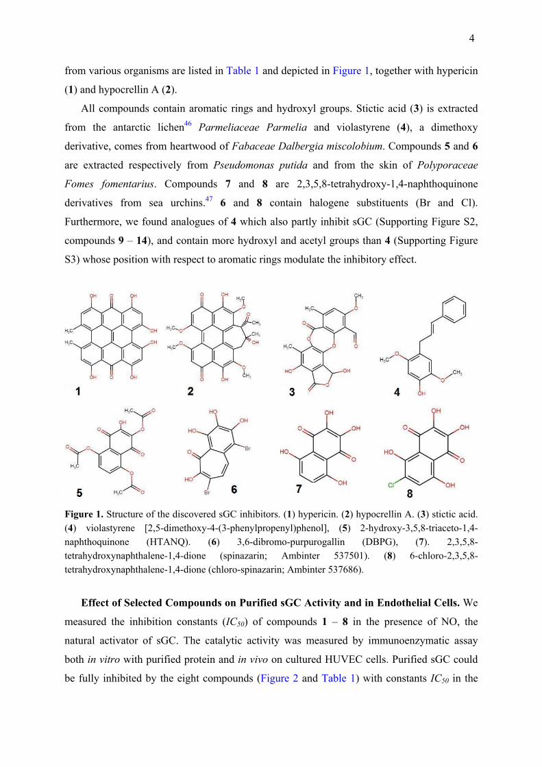
from various organisms are listed in Table 1 and depicted in Figure 1, together with hypericin
(1) and hypocrellin A (2).
All compounds contain aromatic rings and hydroxyl groups. Stictic acid (3) is extracted
from the antarctic lichen46 Parmeliaceae Parmelia and violastyrene (4), a dimethoxy
derivative, comes from heartwood of Fabaceae Dalbergia miscolobium. Compounds 5 and 6
are extracted respectively from Pseudomonas putida and from the skin of Polyporaceae
Fomes fomentarius. Compounds 7 and 8 are 2,3,5,8-tetrahydroxy-1,4-naphthoquinone
derivatives from sea urchins.47 6 and 8 contain halogene substituents (Br and Cl).
Furthermore, we found analogues of 4 which also partly inhibit sGC (Supporting Figure S2,
compounds 9 – 14), and contain more hydroxyl and acetyl groups than 4 (Supporting Figure
S3) whose position with respect to aromatic rings modulate the inhibitory effect.
Figure 1. Structure of the discovered sGC inhibitors. (1) hypericin. (2) hypocrellin A. (3) stictic acid. (4) violastyrene [2,5-dimethoxy-4-(3-phenylpropenyl)phenol], (5) 2-hydroxy-3,5,8-triaceto-1,4-naphthoquinone (HTANQ). (6) 3,6-dibromo-purpurogallin (DBPG), (7). 2,3,5,8-tetrahydroxynaphthalene-1,4-dione (spinazarin; Ambinter 537501). (8) 6-chloro-2,3,5,8-tetrahydroxynaphthalene-1,4-dione (chloro-spinazarin; Ambinter 537686).
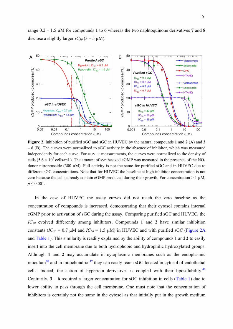
Effect of Selected Compounds on Purified sGC Activity and in Endothelial Cells. We
measured the inhibition constants (IC50) of compounds 1 – 8 in the presence of NO, the
natural activator of sGC. The catalytic activity was measured by immunoenzymatic assay
both in vitro with purified protein and in vivo on cultured HUVEC cells. Purified sGC could
be fully inhibited by the eight compounds (Figure 2 and Table 1) with constants IC50 in the
5
range 0.2 – 1.5 µM for compounds 1 to 6 whereas the two naphtoquinone derivatives 7 and 8
disclose a slightly larger IC50 (3 – 5 µM).
A B
0
10
20
30
40
50
0.001 0.01 0.1 1 10 100
cGM
P pr
oduc
ed (p
icom
oles
/mL)
Compounds concentration (µM)
Purified sGC
sGC in HUVEC
Hypericin: IC50 = 0.2 µMHypocrellin: IC50 = 1.5 µM
Hypericin: IC50 = 0.7 µMHypocrellin: IC50 = 1.5 µM
0
10
20
30
40
50
0.001 0.01 0.1 1 10 100
Violastyrene
Stictic acid
DPG
HTANQ
Violastyrene
Stictic acid
HTANQ
cGM
P pr
oduc
ed (p
icom
oles
/mL)
Compounds concentration (µM)
Purified sGC
sGC in HUVEC
IC50 = 47 µMIC50 = 28 µMIC50 = 16 µM
IC50 = 0.2 µMIC50 = 0.2 µMIC50 = 0.8 µMIC50 = 0.7 µM
Figure 2. Inhibition of purified sGC and sGC in HUVEC by the natural compounds 1 and 2 (A) and 3 – 6 (B). The curves were normalized to sGC activity in the absence of inhibitor, which was measured independently for each curve. For HUVEC measurements, the curves were normalized to the density of cells (5.6 × 10⁵ cells/mL). The amount of synthesized cGMP was measured in the presence of the NO-donor nitroprusside (300 µM). Full activity is not the same for purified sGC and in HUVEC due to different sGC concentrations. Note that for HUVEC the baseline at high inhibitor concentration is not zero because the cells already contain cGMP produced during their growth. For concentration > 1 µM, p ≤ 0.001.
In the case of HUVEC the assay curves did not reach the zero baseline as the
concentration of compounds is increased, demonstrating that their cytosol contains internal
cGMP prior to activation of sGC during the assay. Comparing purified sGC and HUVEC, the
IC50 evolved differently among inhibitors. Compounds 1 and 2 have similar inhibition
constants (IC50 = 0.7 µM and IC50 = 1.5 µM) in HUVEC and with purified sGC (Figure 2A
and Table 1). This similarity is readily explained by the ability of compounds 1 and 2 to easily
insert into the cell membrane due to both hydrophobic and hydrophilic hydroxylated groups.
Although 1 and 2 may accumulate in cytoplasmic membranes such as the endoplasmic
reticulum48 and in mitochondria,49 they can easily reach sGC located in cytosol of endothelial
cells. Indeed, the action of hypericin derivatives is coupled with their liposolubility.48
Contrarily, 3 – 6 required a larger concentration for sGC inhibition in cells (Table 1) due to
lower ability to pass through the cell membrane. One must note that the concentration of
inhibitors is certainly not the same in the cytosol as that initially put in the growth medium
6
and is necessarily lower. Compound 6 did not have effect on HUVEC sGC up to 100 µM. The
most plausible reason is a sequestration or adsorption of 6 on the cell membrane, precluding
its diffusion into the cytosol. Possibly, the presence of two Br atoms in 6 precludes its
insertion through the cell membrane.
Table 1. Inhibition constants for NO-activated sGC and origin of natural compounds.
IC50 (µM) Compound
Purified sGC HUVEC Organism
1 Hypericin 0.2 ± 0.03 0.7 ± 0.05 Plant Hypericium perforatum 2 Hypocrellin A 1.5 ± 0.20 1.5 ± 0.1 Fungus Hypocrella bambuase 3 Stictic acid 0.2 ± 0.05 47 ± 5 Lichen Parmeliaceae Parmelia 4 Violastyrene 0.8 ± 0.05 16 ± 3 Plant Dalbergia miscolobium 5 HTANQ 0.2 ± 0.05 28 ± 2 Bacteria Pseudomonas putida 6 DBPG 0.7 ± 0.04 > 200 Fungus Fomes fomentariu 7 Spinazarin 5 ± 0.5 10 ± 2 Echinodermata Scaphechinus mirabilis 8 Chloro-spinazarin 3 ± 0.5 13 ± 2 Echinodermata Sea urchins
For HUVEC the concentration is that put in the growth medium, not that in the cytosol.
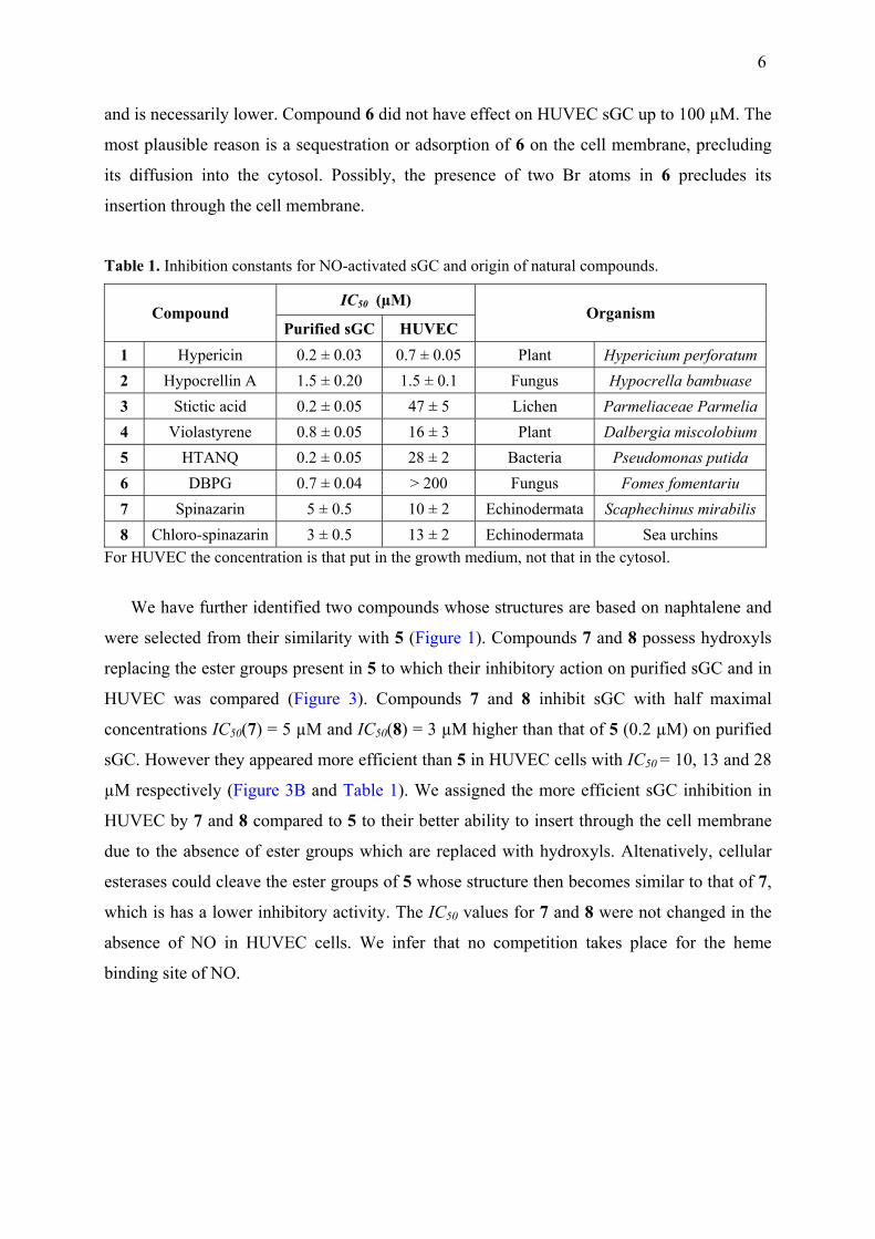
We have further identified two compounds whose structures are based on naphtalene and
were selected from their similarity with 5 (Figure 1). Compounds 7 and 8 possess hydroxyls
replacing the ester groups present in 5 to which their inhibitory action on purified sGC and in
HUVEC was compared (Figure 3). Compounds 7 and 8 inhibit sGC with half maximal
concentrations IC50(7) = 5 µM and IC50(8) = 3 µM higher than that of 5 (0.2 µM) on purified
sGC. However they appeared more efficient than 5 in HUVEC cells with IC50 = 10, 13 and 28
µM respectively (Figure 3B and Table 1). We assigned the more efficient sGC inhibition in
HUVEC by 7 and 8 compared to 5 to their better ability to insert through the cell membrane
due to the absence of ester groups which are replaced with hydroxyls. Altenatively, cellular
esterases could cleave the ester groups of 5 whose structure then becomes similar to that of 7,
which is has a lower inhibitory activity. The IC50 values for 7 and 8 were not changed in the
absence of NO in HUVEC cells. We infer that no competition takes place for the heme
binding site of NO.
7
A B
0.1 1 10 100
C7 + NO
C8 + NO
0
10
20
30
40
50
60
70
Inhibitors concentration (µM)
cGM
P pr
oduc
ed (
pico
mol
es/m
L)
Purified sGCwith NO
IC50 (C7) = 5 ± 0.5 µM
EC50 (C8) = 3 ± 0.5 µM
1 10 100
C7 + NO
C7 - NO
C8 + NO
C8 - NO
0
2
4
6
8
10
12
14
16
Inhibitors concentration (µM)
cGM
P pr
oduc
ed (
pico
mol
es/m
L)
sGC in HUVEC
IC50 = 12 ± 3 µM
without NO
with NO
Figure 3. Inhibition of purified sGC activity in vitro (A) and sGC in HUVEC (B) by compounds 7 and 8. In HUVEC the activity was measured in the presence and absence of the NO-donor nitroprusside (300 µM) during the assay. The density of cells was 4 × 10⁴ cells/mL. Volume of DMSO added 10 µL. The respective IC50 were calculated from the fit of data to a sigmoid curve. Incubation time of cells with inhibitors 7 and 8 was 48 h. Incubation with HUVEC performed at 37 °C with CO2 in the dark. The control for all experiment with cells was 1 % DMSO only in the growth medium, which did not inhibit the formation of cGMP. For concentration ≥ 5 µM in A and ≥ 10 µM in B, p ≤ 0.001.
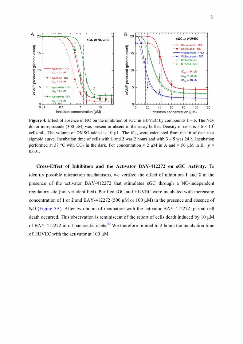
Cross-Effect of NO with Inhibitors on sGC Activity in HUVEC. We compared the
effect of the inhibitors in the presence and absence of NO on sGC activity in cells, verifying
whether they are NO-dependent or independent (Figure 3 and 4). We must note that purified
sGC cannot be activated in vitro without either NO or an artificial activator (such as BAY-
412272). Inhibitors 1 and 2 were slightly influenced by NO with a two-fold decrease of IC50
to 0.4 µM and 0.8 µM respectively in the absence of NO (Figure 4A), whereas 3 – 5 were not
influenced by the presence of NO (Figure 4B). For the five compounds, the basal level of
cGMP production (in the range 1 – 5 picomoles/mL) increased with increasing IC50. This
level represents cGMP produced during growth of cells, including the period of incubation
with inhibitors, but not that produced during the assay which provides external GTP and NO.
Inhibitors 1 – 5 do not compete with NO binding to the heme and do not interfere by attacking
NO, a behavior compatible with an allosteric inhibition, locking sGC in the inactivated state.
8
A B
0
5
10
15
20
0.01 0.1 1 10
hypericin + NO
hypericin - NO
hypocrellin + NO
hypocrellin - NO
cGM
P pr
oduc
ed (p
icom
oles
/ml)
Inhibitors concentration (µM)
sGC in HUVEC
IC50 = 0.7 µM
IC50 = 0.4 µM
IC50 = 1.5 µM
IC50 = 0.8 µM0
5
10
15
20
0 20 40 60 80 100 120
Stictic acid + NOStictic acid - NOViolastyrene + NOViolastyrene - NOHTANQ+NOHTANQ - NO
cGM
P pr
oduc
ed (p
icom
oles
/ml)
Inhibitors concentration (µM)
IC50 = 44 µMIC50 = 29 µMIC50 = 16 µM
sGC in HUVEC
Figure 4. Effect of absence of NO on the inhibition of sGC in HUVEC by compounds 1 – 5. The NO-donor nitroprusside (300 µM) was present or absent in the assay buffer. Density of cells is 5.6 × 10⁵ cells/mL. The volume of DMSO added is 10 µL. The IC50 were calculated from the fit of data to a sigmoid curve. Incubation time of cells with 1 and 2 was 2 hours and with 3 – 5 was 24 h. Incubation performed at 37 °C with CO2 in the dark. For concentration ≥ 2 µM in A and ≥ 50 µM in B, p ≤ 0.001.
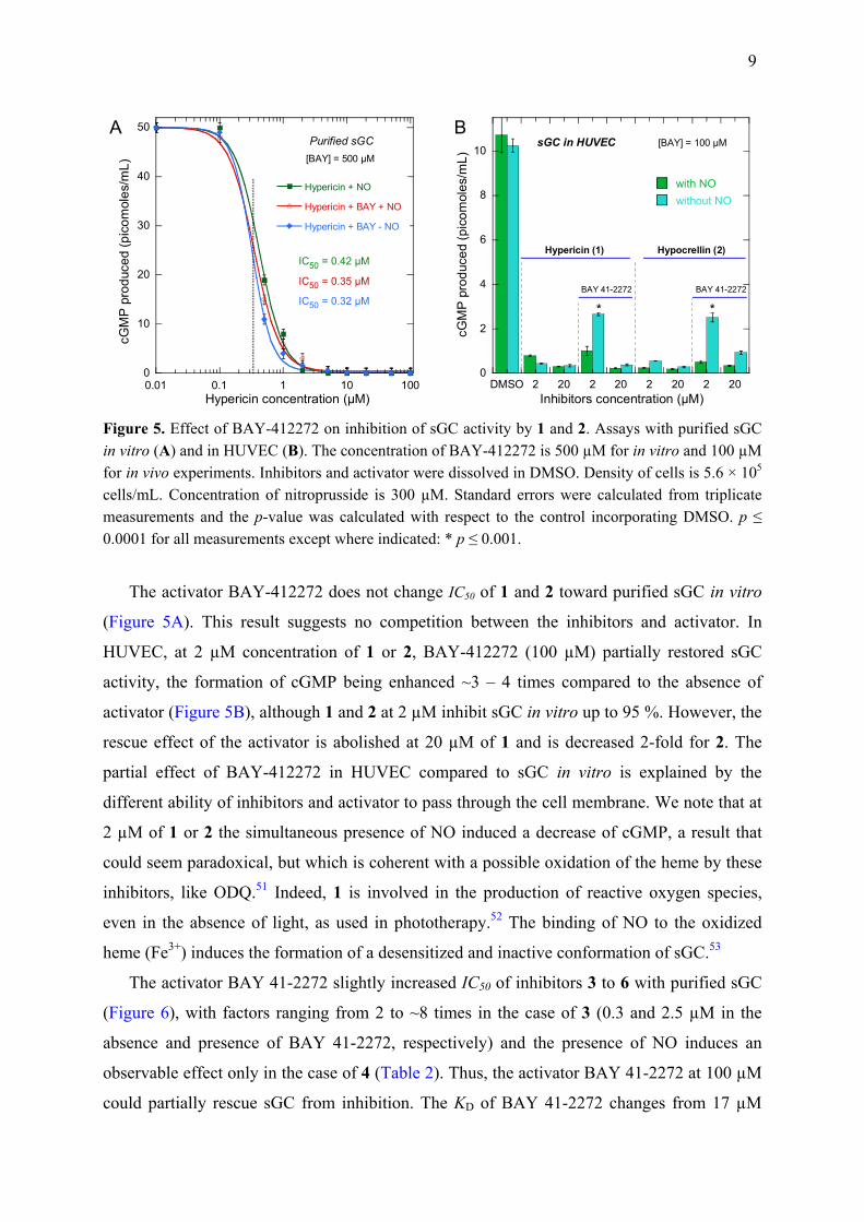
Cross-Effect of Inhibitors and the Activator BAY-412272 on sGC Activity. To
identify possible interaction mechanisms, we verified the effect of inhibitors 1 and 2 in the
presence of the activator BAY-412272 that stimulates sGC through a NO-independent
regulatory site (not yet identified). Purified sGC and HUVEC were incubated with increasing
concentration of 1 or 2 and BAY-412272 (500 µM or 100 µM) in the presence and absence of
NO (Figure 5A). After two hours of incubation with the activator BAY-412272, partial cell
death occurred. This observation is reminiscent of the report of cells death induced by 10 µM
of BAY-412272 in rat pancreatic islets.50 We therefore limited to 2 hours the incubation time
of HUVEC with the activator at 100 µM.
9
A B
0
10
20
30
40
50
0.01 0.1 1 10 100
Hypericin + NO
Hypericin + BAY + NO
Hypericin + BAY - NO
cGM
P p
rodu
ced
(pic
omol
es/m
L)
Hypericin concentration (µM)
IC50 = 0.42 µM
Purified sGC[BAY] = 500 µM
IC50 = 0.35 µM
IC50 = 0.32 µM
0
2
4
6
8
10
DMSO 2 20 2 20 2 20 2 20
with NOwithout NO
cGM
P pr
oduc
ed (p
icom
oles
/mL)
Inhibitors concentration (µM)
BAY 41-2272 BAY 41-2272
Hypericin (1) Hypocrellin (2)
sGC in HUVEC [BAY] = 100 µM
* *
Figure 5. Effect of BAY-412272 on inhibition of sGC activity by 1 and 2. Assays with purified sGC in vitro (A) and in HUVEC (B). The concentration of BAY-412272 is 500 µM for in vitro and 100 µM for in vivo experiments. Inhibitors and activator were dissolved in DMSO. Density of cells is 5.6 × 10⁵ cells/mL. Concentration of nitroprusside is 300 µM. Standard errors were calculated from triplicate measurements and the p-value was calculated with respect to the control incorporating DMSO. p ≤ 0.0001 for all measurements except where indicated: * p ≤ 0.001.
The activator BAY-412272 does not change IC50 of 1 and 2 toward purified sGC in vitro
(Figure 5A). This result suggests no competition between the inhibitors and activator. In
HUVEC, at 2 µM concentration of 1 or 2, BAY-412272 (100 µM) partially restored sGC
activity, the formation of cGMP being enhanced ~3 – 4 times compared to the absence of
activator (Figure 5B), although 1 and 2 at 2 µM inhibit sGC in vitro up to 95 %. However, the
rescue effect of the activator is abolished at 20 µM of 1 and is decreased 2-fold for 2. The
partial effect of BAY-412272 in HUVEC compared to sGC in vitro is explained by the
different ability of inhibitors and activator to pass through the cell membrane. We note that at
2 µM of 1 or 2 the simultaneous presence of NO induced a decrease of cGMP, a result that
could seem paradoxical, but which is coherent with a possible oxidation of the heme by these
inhibitors, like ODQ.51 Indeed, 1 is involved in the production of reactive oxygen species,
even in the absence of light, as used in phototherapy.52 The binding of NO to the oxidized
heme (Fe3+) induces the formation of a desensitized and inactive conformation of sGC.53
The activator BAY 41-2272 slightly increased IC50 of inhibitors 3 to 6 with purified sGC
(Figure 6), with factors ranging from 2 to ~8 times in the case of 3 (0.3 and 2.5 µM in the
absence and presence of BAY 41-2272, respectively) and the presence of NO induces an
observable effect only in the case of 4 (Table 2). Thus, the activator BAY 41-2272 at 100 µM
could partially rescue sGC from inhibition. The KD of BAY 41-2272 changes from 17 µM
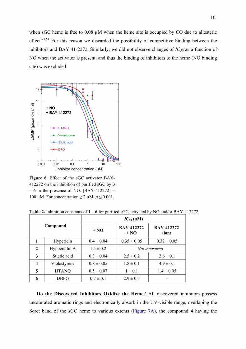
10
when sGC heme is free to 0.08 µM when the heme site is occupied by CO due to allosteric
effect.21,54 For this reason we discarded the possibility of competitive binding between the
inhibitors and BAY 41-2272. Similarly, we did not observe changes of IC50 as a function of
NO when the activator is present, and thus the binding of inhibitors to the heme (NO binding
site) was excluded.
0
2
4
6
8
10
12
0.001 0.01 0.1 1 10 100
HTANQ
Violastyrene
Stictic acid
DPG
cGM
P (p
icom
oles
/ml)
Inhibitor concentration (µM)
+ NO+ BAY-412272
Figure 6. Effect of the sGC activator BAY-412272 on the inhibition of purified sGC by 3 – 6 in the presence of NO. [BAY-412272] = 100 µM. For concentration ≥ 2 µM, p ≤ 0.001.
Table 2. Inhibition constants of 1 – 6 for purified sGC activated by NO and/or BAY-412272. IC50 (µM)
Compound + NO BAY-412272
+ NO BAY-412272
alone
1 Hypericin 0.4 ± 0.04 0.35 ± 0.05 0.32 ± 0.05
2 Hypocrellin A 1.5 ± 0.2 Not measured
3 Stictic acid 0.3 ± 0.04 2.5 ± 0.2 2.6 ± 0.1 4 Violastyrene 0.8 ± 0.05 1.8 ± 0.1 4.9 ± 0.1
5 HTANQ 0.5 ± 0.07 1 ± 0.1 1.4 ± 0.05
6 DBPG 0.7 ± 0.1 2.9 ± 0.5 –
Do the Discovered Inhibitors Oxidize the Heme? All discovered inhibitors possess
unsaturated aromatic rings and electronically absorb in the UV-visible range, overlaping the
Soret band of the sGC heme to various extents (Figure 7A), the compound 4 having the
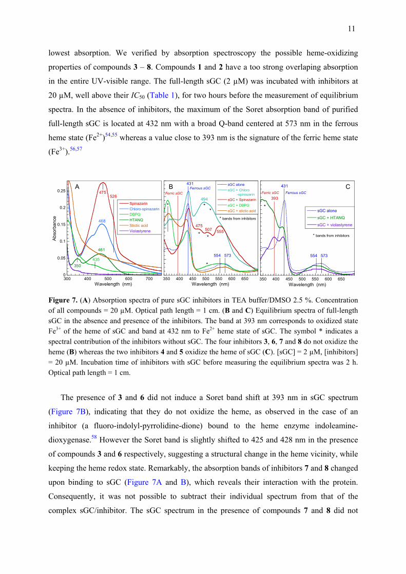
11
lowest absorption. We verified by absorption spectroscopy the possible heme-oxidizing
properties of compounds 3 – 8. Compounds 1 and 2 have a too strong overlaping absorption
in the entire UV-visible range. The full-length sGC (2 µM) was incubated with inhibitors at
20 µM, well above their IC50 (Table 1), for two hours before the measurement of equilibrium
spectra. In the absence of inhibitors, the maximum of the Soret absorption band of purified
full-length sGC is located at 432 nm with a broad Q-band centered at 573 nm in the ferrous
heme state (Fe2+)54,55 whereas a value close to 393 nm is the signature of the ferric heme state
(Fe3+).56,57
0
0.05
0.1
0.15
0.2
0.25
300 400 500 600 700
SpinazarinChloro-spinazarinDBPGHTANQStictic acidViolastyrene
Abs
orba
nce
Wavelength (nm)
475
468
461
350
526
435
A
350 400 450 500 550 600 650
sGC alonesGC + Chloro -spinazarinsGC + SpinazarinsGC + DBPGsGC + stictic acid
Wavelength (nm)
Ferric sGCFerrous sGC
* bands from inhibitors
*
**
*
*
475
494
507 555
554 573
*
431B
350 400 450 500 550 600 650
sGC alone
sGC + HTANQ
sGC + violastyrene
Wavelength (nm)
Ferric sGC Ferrous sGC
* bands from inhibitors
*
431
393
554 573
C
Figure 7. (A) Absorption spectra of pure sGC inhibitors in TEA buffer/DMSO 2.5 %. Concentration of all compounds = 20 µM. Optical path length = 1 cm. (B and C) Equilibrium spectra of full-length sGC in the absence and presence of the inhibitors. The band at 393 nm corresponds to oxidized state Fe3+ of the heme of sGC and band at 432 nm to Fe2+ heme state of sGC. The symbol * indicates a spectral contribution of the inhibitors without sGC. The four inhibitors 3, 6, 7 and 8 do not oxidize the heme (B) whereas the two inhibitors 4 and 5 oxidize the heme of sGC (C). [sGC] = 2 µM, [inhibitors] = 20 µM. Incubation time of inhibitors with sGC before measuring the equilibrium spectra was 2 h. Optical path length = 1 cm.
The presence of 3 and 6 did not induce a Soret band shift at 393 nm in sGC spectrum
(Figure 7B), indicating that they do not oxidize the heme, as observed in the case of an
inhibitor (a fluoro-indolyl-pyrrolidine-dione) bound to the heme enzyme indoleamine-
dioxygenase.58 However the Soret band is slightly shifted to 425 and 428 nm in the presence
of compounds 3 and 6 respectively, suggesting a structural change in the heme vicinity, while
keeping the heme redox state. Remarkably, the absorption bands of inhibitors 7 and 8 changed
upon binding to sGC (Figure 7A and B), which reveals their interaction with the protein.
Consequently, it was not possible to subtract their individual spectrum from that of the
complex sGC/inhibitor. The sGC spectrum in the presence of compounds 7 and 8 did not
12
either show a shifted Soret band to 393 nm (Figure 7B) indicating that the sGC heme is not
oxidized in their presence. The addition of 4 to sGC led to the appearance of a second Soret
peak at 393 nm and a shift of the Q-bands to 550 – 571 nm (Figure 7C), indicating about 50 %
of heme in the oxidized state (Fe3+). In the presence of sGC, a broad band in near-UV at 350
nm due to 5 increased, together with the disappearance of the inhibitor band located at 461 nm
when it is alone (Figure 7A). We assigned the shoulder located at 393 nm (Figure 7C) to the
oxidized heme because the amplitude of the Soret band at 431 nm decreased with respect to
sGC alone. The changes induced in the spectrum of 5 did not allow to calculate the spectral
difference of inhibitor-bound sGC with respect to free sGC. The Q-bands were also shifted
from ferrous to ferric heme state in the presence of 5. A well studied sGC inhibitor acting
through heme oxidation is ODQ51,56 and a mechanism was proposed to explain its oxidizing
action which involves transfer of an electron from heme to ODQ to generate an ODQ
radical.56 One must note that ODQ also oxidizes hemoglobin.56 Structures of ODQ, 4 and 5
are quite different (Supporting Figure S4) although ODQ and 5 share a ketone group coupled
to a ring, whereas 5 possesses ester and 4 only two methoxy groups. It is not possible to infer
the exact electron transfer mechanism involving the inhibitors 4 and 5 from their spectra, a
question beyond the scope of the present study.
Thus compounds 4 and 5 oxidize the heme of sGC which cannot be activated by diatomic
ligands in the ferric state59 and may be subject to heme loss,60 whereas the inhibition
mechanism of other compounds which do not oxidize the heme (1 – 3, 6 – 8) is still unknown
but is inferred to be allosteric.
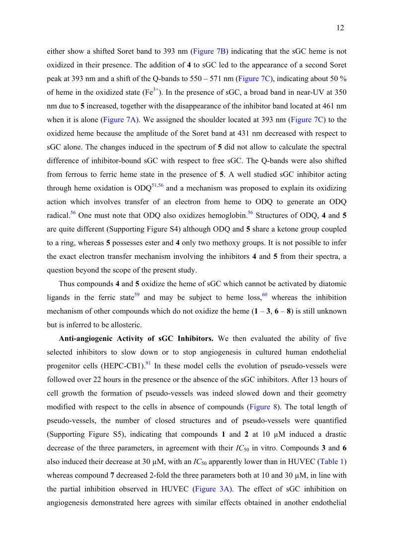
Anti-angiogenic Activity of sGC Inhibitors. We then evaluated the ability of five
selected inhibitors to slow down or to stop angiogenesis in cultured human endothelial
progenitor cells (HEPC-CB1).91 In these model cells the evolution of pseudo-vessels were
followed over 22 hours in the presence or the absence of the sGC inhibitors. After 13 hours of
cell growth the formation of pseudo-vessels was indeed slowed down and their geometry
modified with respect to the cells in absence of compounds (Figure 8). The total length of
pseudo-vessels, the number of closed structures and of pseudo-vessels were quantified
(Supporting Figure S5), indicating that compounds 1 and 2 at 10 µM induced a drastic
decrease of the three parameters, in agreement with their IC50 in vitro. Compounds 3 and 6
also induced their decrease at 30 µM, with an IC50 apparently lower than in HUVEC (Table 1)
whereas compound 7 decreased 2-fold the three parameters both at 10 and 30 µM, in line with
the partial inhibition observed in HUVEC (Figure 3A). The effect of sGC inhibition on
angiogenesis demonstrated here agrees with similar effects obtained in another endothelial
13
cell model by the inhibition of endothelial NO-synthase,34 the immediate upstream partner of
sGC in the NO/cGMP pathway.
Figure 8. Inhibition of endothelial cell angiogenesis by sGC inhibitors. The angiogenesis of HEPC.CB1 on MatrigelTM, in the presence of 10 and 30 μM of hypericin (1), hypocrellin (2), stictic acid (3), DBPG (6) or spinazarin (7) was monitored under a videomicroscope. Photos are presented after 13 h of culture on MatrigelTM. In all images the scale bar represents 200 µm. Controls have been performed without and with DMSO at same final concentration (0.5 %) used after introducing the inhibitors.

14
DISCUSSION
Activities of the Discovered sGC Inhibitors Toward Other Enzymes. We have
previously measured a decreased cellular cGMP level in the presence of 1 (at 4 µM) in
cardiomyocytes,45 together with an increased conductance of L-type Ca2+ channels, that was
assigned to sGC inhibition, a hypothesis fully confirmed by the present results. Beyond its
study for photodynamic therapy,61 1 was shown to have effects on various cell signaling
pathways.52 Numerous studies focussed on Hypericum perforatum extract (i.e. not purified
compounds), but some target proteins were identified for 1, including c-type kinases,62
proteins involved in breast cancer resistance,63 glutathione-S-transferases (with Ki in the
range 0.2 – 2.5 µM)64 and dopamine-β-hydroxylase with an IC50 of 21 μM.65 Importantly, 1
was shown to reduce angiogenesis in several ocular models66,67 by inhibiting phosphorylation
of MAP kinases.68 Both 1 and 2 can also bind to human serum albumin.68 Although its
physiological effects were much less investigated than 1, compound 2 was also tested for
photodynamic therapy and induces the decrease of endothelin-1 and VEGF in HUVEC,
probably through inhibition of PKC.69
Albeit few studies reported pharmacological activity of 3, it induces intracellular lactate
dehydrogenase release, linked to apoptosis of hepatocytes (13 µM < IC50 < 230 µM).70
Importantly, stictic acid has tumor-suppressing effects via reactivation of p53 protein mutants
in cancer cells.71 It was also shown to inhibit competitively two bacterial β-hydroxyacyl-acyl
carrier protein dehydratases (IC50 = 13 – 28 µM).72 No physiological action is known for
compounds 4, 5 and 6, whereas the only effect reported for 7 and its derivative 8 is the
scavenging of free radicals.73
Therapeutic Uses of the Discovered sGC Natural Inhibitors. As described in
Introduction, sGC is involved particularly in cancer, apoptosis and angiogenesis. It has also a
central role in vasoplegia (also called vasodilatory shock) whose causes can be multiple37,74
and is characterized by a drop of blood pressure, the treatment of which appears critical.37
Proposed vasoplegia treatments include the use of adrenergic vasopressors to restore blood
pressure, but blood vessels may not respond to vasopressors even at high concentration. A
more efficient treatment has been proposed by inhibiting directly sGC,75,76 essentially by
means of methylene blue76-78 whose action on coronary arteries has been recognized79 soon
after the discovery of the role of NO as a messenger. However, methylene blue is not specific,
15
since it inhibits constitutive NO-synthase80 and other enzymes,81 similarly with ODQ82 and
can hardly be used as a therapeuthic agent. More specific sGC inhibitors are needed here.
The NO/cGMP cell signaling has been recognized to be involved in regulating the
interactions between dopamine and glutamate neurotransmission in striatum.83 The
administration of the sGC inhibitor ODQ could reverse markers of ganglia dysfunction in a
rat model,38 so that sGC inhibition has been proposed as a treatment of parkinsonian
symptoms.38 The oxidant sGC inhibitors ODQ and methylene blue could reduce dyskinesia
induced by L-dihydroxyphenylalanine in rats.84 Related to this issue, the role of sGC has also
been shown in the neurodegeneration in Drosophila85 which was improved by treatment with
the NOS inhibitor L-NAME (nitro-L-arginine-methyl-ester), inhibiting the NO/sGC pathway.
sGC is involved in many cell signaling pathways which may have the ability of
counteracting each other in therapeutic action (e. g. apoptosis versus tumoral angiogensis). It
is obvious that, like most of drugs, the use of sGC activators or inhibitors may have side-
effects, yet, strong benefits from their use are expected. A promissing method of removing (or
at least attenuating) side-effects would be to associate inhibitors with a strategy to target
organs for drug delivery. In this view, the use of sGC inhibitors as drugs for angiogenesis will
benefit from advances of nanoparticles (especially bionanoparticles) for targeted drug
delivery,86,87 which is a very active field in chemical biology. Promising systems consist of
cell membrane-based drug carriers.88,89 Both fields of research must progress in parallel.
CONCLUSIONS
The natural compounds from plants and fungi described here are the first molecules
reported so far to inhibit sGC in vitro with IC50 in the range 0.2 µM − 1.5 µM, although some
of their properties remain to be evaluated. Further advances require to know the architecture
of the inhibitors binding site(s) and to co-crystallize the full-length sGC with the compounds
described here, but it appeared impossible to obtain crystals of sGC, certainly because of its
flexibility.19 However, a full three-dimensional structure model90 from a cryo-
electromicroscopy template19 and from X-ray crystallographic models of the different
domains (H-NOX, PAS and catalytic domains) can be used for calculations. The cross-
reactivity of compounds with NO and the sGC activator BAY 41-2272 suggests an allostric
mechanism. These discovered inhibitors can be used directly as tools for investigating the role
of sGC in signaling pathways. Importantly, they provide templates amenable to improvement
for drug design to target tumoral angiogenesis or vasoplegia, both involving sGC.
16
EXPERIMENTAL SECTION
Enzymes, Cells and Natural Compounds. We used either sGC from bovine lung
purified as previously described 55 or full-length recombinant human sGC purchased from
Alexis (ALX-201-177). Human umbilical vein endothelial cells (HUVEC) isolated from
normal vein were purchased from Gibco. Purified hypericin and hypocrellin (98 % purity)
were purchased from Invitrogen and the activator BAY-412272 (98 % purity) from Enzo. The
chemical library containing 320 natural compounds (GPNCL, Greenpharma) and some
isolated compounds from Ambinter (naphtoquinone derivatives Amb537501 and Amb537686
and violastyrene derivatives, 98 % purity). These compounds comprises mostly alkaloids,
phenol derivatives and terpenoids from plants and fungi. Each compounds from the library
was first tested at a concentration of 20 µM for its effect on catalytic activity of the purified
sGC (inhibition or activation). Then the effects of the more active compounds at 20 µM were
tested as a function of increasing concentration on both purified sGC and HUVEC.
Catalytic Activity of Purified sGC. Activity measurements were carried out on purified
sGC as previously described45 with minor modifications. All compounds from Greenpharma
library, hypericin, hypocrellin and the activator BAY-412272 were first diluted in pure
DMSO to get a stock solution, then in H2O at the desired working concentration. Purified sGC
was resuspended in TEA buffer: triethanolamine 25 mM, NaCl 10 mM, dithiothreitol (DTT) 1
mM, pH 7.4. Selected inhibitors at increasing concentration were mixed with sGC at 100 nM
and incubated in the dark, either 2 hours at 20 °C or overnight at 4 °C. When investigating the
cross-effect of BAY-412272, this activator was mixed together with inhibitors. The final
concentration of DMSO is 2.5 %. Then sGC (25 nM final concentration) was incubated in
assay buffer (100 µL final volume) for 10 minutes at 37 °C. The assay buffer composition
was: TEA 100 mM, pH 7.4, DTT 1 mM, MnCl2 3 mM, GTP 0.6 mM, creatine phosphate 5
mM, creatine phosphokinase 150 U/L, nitroprusside (NPS) 0.3 mM. The NO-donor NPS was
omitted for assays in absence of NO. The reaction was stopped by precipitation with Zn
acetate and Na2CO3 and 40 µL of supernatant are used for assaying the synthetized cGMP.
After this step, the immunoenzymatic assay was performed using commercial ELISA kits,
either from Enzo (ADI-901-013) or from GE-Healthcare (RPN 226), according to
manufacturer's instructions, with the acetylation protocole. A standard curve of known cGMP
concentrations was measured together each experiment allowing to calculate produced cGMP
from the absorbance.
17
Catalytic Activity of sGC in HUVEC. The multiplication of HUVEC cells was realized
with complete medium (Medium 200 or 200PRF), low serum growth supplement (LSGS)
X50 factor, antibiotic X100 factor, serum bovine albumin (10 % from all of volume). HUVEC
were grown in 12-well plates (individual surface 3.8 cm2). When the cells were at ~90 %
confluence, the medium was replaced (500 µL final volume) and inhibitors at varying
concentrations (10 µL; 2 % DMSO final concentration) were added in the wells, (or only
DMSO for control) then incubated for 24 hours in a CO2 incubator at 36 °C in the dark. Some
compounds necessitated up to 24 h incubation with HUVEC due to their low hydrophobicity
and ability to pass through cell membrane. The cells incubated with compounds were then
lyzed for 20 minutes with 50 µL of the following lysis buffer: TEA 500 mM,
isobutylmethylxanthine (IBMX) 10 mM, Triton X100 10 %, pH = 7.4. The lyzed cell
suspension was incubated directly in the wells of the culture plate with same assay buffer (50
µL; 10X) as used for purified sGC (in the presence or absence of NPS) for 20 minutes with
mild shaking at room temperature. Then, 500 µL of solution from each well were transfered
into polypropylene tubes for acetylation of the synthesized cGMP. After this step,
immunoenzymatic assay of cGMP was performed with the ELISA kit as performed for the
purified protein. Where applicable, statistic evaluation of the data was perform by calculating
the p-value with Anova test implemented in Kaleidagraph V3.6 (Synergy Software).
Assay of the Anti-angiogenic Activity of Natural Compounds. The human endothelial
progenitor cell line HEPC-CB1, which was established and characterized in our laboratory,91
was used to assess the anti-angiogenic properties of five compounds. Cells were briefly
treated with trypsin (Invitrogen), washed and were seeded at 1.5 × 10⁴ cells/well in serum-free
Opti-MEM medium, in the presence of two different concentrations of compounds on 96-well
plates coated with Matrigel™ (BD Biosciences, San José, CA). The final concentration of
DMSO was kept at 0.5 %. Cultures were performed at 37 °C with 5 % CO2 in the
videomicroscope incubation chamber. Cell rearrangement and pseudo-vessels formation were
visualized using an AXIO OBSERVER Z1 fluorescence inverted microscope (Zeiss, Le Pecq,
France) equipped with a high-resolution numeric camera linked to a computer driving the
acquisition software Zen (Zeiss). The direct real-time visualization of the formation of
pseudo-vessels was monitored during 22 h. Angiogenesis was quantified by the determination
of pseudo-vessel number, closed structures (meshes) number and by the measurement of
pseudo-vessel total length using Image J software with the Angiogenesis analyzer add-on.
Spectroscopy. The absorption spectra of sGC and inhibitors were measured with a
UV-1700 Shimadzu spectrophotometer in a 1 × 1 cm QX-quartz cell (Hellma) at 20 °C. The
18
concentrations used were [sGC] = 2 µM and [inhibitors] = 20 µM. The inhibitors were
incubated with sGC during 2 h before measuring the spectra at equilibrium.
ASSOCIATED CONTENT
Supporting Information is available.
AUTHOR INFORMATION Corresponding Author
Michel Negrerie – Laboratoire d'Optique et Biosciences, INSERM U1182, Ecole Polytechnique, Palaiseau, France. Email: [email protected] Phone: 133 69 33 50 52. ORCID: 0000-0001-9918-031X
Authors
Olga N. Petrova – Laboratoire d'Optique et Biosciences, INSERM U1182, Ecole Polytechnique, Palaiseau, France. Present address : Handicap Neuromusculaire: Physiologie, Biothérapie et Pharmacologie Appliquées, Inserm U1179, Université de Versailles Saint-Quentin-en-Yvelines, Montigny-le-Bretonneux, France. Isabelle Lamarre – Laboratoire d'Optique et Biosciences, INSERM U1182, Ecole Polytechnique, Palaiseau, France. Fabienne Fasani – Centre de Biophysique Moléculaire, UPR4301 CNRS, Orléans, France. Catherine Grillon – Centre de Biophysique Moléculaire, UPR4301 CNRS, Orléans, France.
Notes
The authors declare no competing financial interest. AKNOWLEDGMENTS
O. N. P. acknowledges an "Initiative Doctorale Interdisciplinaire" fellowship from Université Paris-Saclay. The authors thank David Gosset for his assistance in imaging angiogenesis on the P@CYFIC platform facility at the Center for Molecular Biophysics (Orléans, France). ABBREVATIONS
sGC, soluble guanylate cyclase; eNOS, endothelial NO-synthase; cGMP, 3',5'-cyclic guanosine monophosphate; GTP, guanosine 5'-triphosphate; PKG, cGMP-activated protein
19
kinase; IP3, inositol triphosphate; HTANQ, 2-hydroxy-3,5,8-triaceto-1,4-naphthoquinone; DMSO, dimethylsulfoxide; DPG, 3,6-dibromo-purpurogallin. ODQ, 1H-[1,2,4]-oxadiazolo[4,3-a]quinoxalin-1-one; HUVEC, human umbilical vein endothelial cells. VEGF, vascular endothelial growth factor; IBMX, isobutylmethylxanthin; NPS, sodium nitroprusside. HEPC, human endothelial progenitor cell line. REFERENCES
(1) Murad, F. Angew. Chem. Int. Ed. 1999, 38, 1857−1868. (2) Furchgott, R. F. Angew. Chem. Int. Ed. 1999, 38, 1870−1880. (3) Ignarro, L. J. Angew. Chem. Int. Ed. 1999, 38, 1882−1892. (4) Ignarro, L. J.; Cirino, G.; Casini, A.; Napoli, C. J. Cardiovasc. Pharmacol. 1999, 34,
879−886. (5) Buys, E.; Sips, P. Curr. Opin. Nephrol. Hyp. 2014, 23, 135−142. (6) Nagy, G.; Clark, J. M.; Buzás, E. I.; Gorman, C. L.; Cope, A. P. Immunol. Lett. 2007, 111,
1−5. (7) Zhang, G.; Xiang, B.; Dong, A.; Skoda, R. C.; Daugherty, A.; Smyth, S. S.; Du, X.; Li, Z.
Blood 2011, 118, 3670−3679. (8) Garthwaite, J. Br. J. Pharmacol. 2019, 176, 197−211. (9) Chin, C.-H.; Tsai, F.-C.; Chen, S.-P.; Wang, K.-C.; Chang, C.-C.; Pai, M.-H.; Fong, T.-H. Eur.
J. Pharmacol. 2012, 689, 1−7. (10) Bian, K.; Murad, F. Nitric Oxide 2014, 43, 3−7. (11) Fraser, M.; Chan, S. L.; Chan, S. S. L.; Fiscus, R. R.; Tsang, B. K. Oncogene 2006, 25, 2203–
2212. (12) Blaise, G. A.; Gauvin, D.; Gangal, M.; Authier, S. Toxicology 2005, 208, 177−192. (13) Bian, K.; Doursout, M.; Murad, F. J. Clin. Hypertens. 2008 10, 304−310. (14) Evgenov, O. V.; Pacher, P.; Schmidt, P. M.; Haskó, G.; Schmidt, H. H. H. W.; Stasch, J.-P.
Nat. Rev. Drug Disc. 2006, 5, 755−768. (15) Lundberg, J. O.; Gladwin, M. T.; Weitzberg, E. Nat. Rev. Drug. Discov. 2015, 14, 623−641. (16) Stasch, J. P.; Pacher, P.; Evgenov, O. V. Circulation 2011, 123, 2263−2273. (17) Ko, F.-N.; Wu, C.-C.; Kuo, S.-C.; Lee, F.-Y.; Teng, C.-M. Blood 1994, 84, 4226−4233. (18) Yoo, B. K.; Lamarre, I.; Martin, J. L.; Rappaport, F.; Negrerie, M. Proc. Natl. Acad. Sci. U. S.
A. 2015, 112, E1697−E1704. (19) Campbell, M. G.; Underbakke, E. S.; Potter, C. S.; Carragher, B.; Marletta, M. A. Proc. Natl.
Acad. Sci. U. S. A. 2014, 111, 2960−2965. (20) Yoo, B.-K.; Lamarre, I.; Rappaport, F.; Nioche, P.; Raman, C. S.; Martin, J. L.; Negrerie, M.
ACS Chem. Biol. 2012, 7, 2046−2054. (21) Purohit, R.; Fritz, B. G.; The, J.; Issaian, A.; Weichsel, A.; David, C. L.; Campbell, E.;
Hausrath, A. C.; Rassouli-Taylor, L.; Garcin, E. D.; Gage, M. J.; Montfort, W. R. Biochemistry 2013, 53, 101−114.
(22) Bian, K.; Sotolongo, A.; Lam, H.; Xiao, H.; Zhang, D.; Liu, J.; Murad, F. BMC Pharmacol. Toxicol. 2013, 14, P9.
(23) Mujoo, K.; Sharin, V. G.; Martin, E.; Choi, B.-K.; Sloan, C.; Nikonoff, L. E.; Kots, A. Y.; Murad, F. Nitric Oxide 2010, 22, 43−50.
(24) Zhu, H.; Li, J. T.; Zheng, F.; Martin, E.; Kots, A. Y.; Krumenacker, J. S.; Choi, B.-K.; McCutcheon, I. E.; Weisbrodt, N.; Bögler, O.; Murad, F.; Bian, K. Mol. Pharmacol. 2011, 80, 1076−1084.
20
(25) Wen, H.-C.; Chuu, C.-P.; Chen, C.-Y.; Shiah, S.-G.; Kung, H.-J.; King, K.-L.; Su, L.-C.; Chang, S.-C.; Chang, C.-H. PloS One 2015, 10, e0125518−e0125518.
(26) Grimm, E. A.; Ellerhorst, J.; Tang, C.-H.; Ekmekcioglu, S. Nitric Oxide 2008, 19, 133−137. (27) Leung, E. L.; Wong, J. C.; Johlfs, M. G.; Tsang, B. K.; Fiscus, R. R. Mol. Cancer Res. 2010,
8, 578−591. (28) Cai, C.; Chen, S. Y.; Zheng, Z.; Omwancha, J.; Lin, M. F.; Balk, S. P.; Shemshedini, L.
Oncogene 2006, 26, 1606−1615. (29) Menéndez, L.; Juárez, L.; García, V.; Hidalgo, A.; Baamonde, A. Neuropharmacology 2007,
53, 71−80. (30) Park, S.-W.; Lee, S.-G.; Song, S.-H.; Heo, D.-S.; Park, B.-J.; Lee, D.-W.; Kim, K.-H.; Sung,
M.-W. Int. J. Cancer 2003, 107, 729−738. (31) Isenberg, J. S.; Martin-Manso, G.; Maxhimer, J. B.; Robert, D. D. Nat. Rev. Cancer 2009, 9,
182−194. (32) Pyriochou, A.; Beis, D.; Koika, V.; Potytarchou, C.; Papadimitriou, E.; Zhou, Z.;
Papapetropoulos, A. J. Pharmacol. Exp. Ther. 2006, 319, 663−671. (33) Morbidelli, L.; Pyriochou, A.; Filippi, S.; Vasileiadis, I.; Roussos, C.; Zhou, Z.; Loutrari, H.;
Waltenberger, J.; Stoessel, A.; Giannis, A.; Ziche, M.; Papapetropoulos, A. Am. J. Physiol.-Regul. Integr. Comp. Physiol. 2010, 298, R824−R832.
(34) Carreau, A.; Kieda, C.; Grillon, C. Exp. Cell Res. 2011, 317, 29−41. (35) Collet, G.; Szade, K.; Nowak, W.; Klimkiewicz, K.; El Hafny-Rahbi, B.; Szczepanek, K.;
Sugiyama, D.; Weglarczyk, K.; Foucault-Collet, A.; Guichard, A.; Mazan, A.; Nadim, M.; Fasani, F.; Lamerant-Fayel, N.; Grillon, C.; Petoud, S.; Beloeil, J.-C.; Jozkowicz, A.; Dulak, J.; Kieda, C. Cancer Letters 2016, 370, 345−357.
(36) Wong, J. C.; Fiscus, R. R. Cardiovasc. Pathol. 2010, 19, e221−e231. (37) Kimmoun, A.; Ducrocq, N.; Levy, B. Curr. Vasc. Pharmacol. 2013, 11, 139−149. (38) Tseng, K. Y.; Caballero, A.; Dec, A.; Cass, D. K.; Simak, N.; Sunu, E.; Park, M. J.; Blume, S.
R.; Sammut, S.; Park, D. J.; West, A. R. PloS One 2011, 6, e27187. (39) Sousa, E. H. S.; de Franca Lopes, L. G.; Gonzalez, G.; Gilles-Gonzalez, M.-A. J. Inorg.
Biochem. 2017, 167, 12−20. (40) Haramis, G.; Zhou, Z.; Pyriochou, A.; Koutsilieris, M.; Roussos, C.; Papapetropoulos, A. Br.
J. Pharmacol. 2008, 6, 804−813. (41) Olesen, S.-P.; Drejer, J.; Axelsson, O.; Moldt, P.; Bang, L.; Nielsen-Kudsk, J. E.; Busse, R.;
Mülsch, A. Br. J. Pharmacol. 1998, 123, 299−309. (42) Luo, D.; Das, S.; Vincent, S. R. Eur. J. Pharmacol. 1995, 290, 247−251. (43) James, L. R.; Griffiths, C. H.; Garthwaite, J.; Bellamy, T. C. Br. J. Pharmacol. 2009, 158,
1454−1464. (44) Mota, F.; Gane, P.; Hampden-Smith, K.; Allerston, C. K.; Garthwaite, J.; Selwood, D. L.
Bioinorg. Med. Chem. 2015, 23, 5303−5310. (45) Sauviat, M.-P.; Colas, A.; Chauveau, M.-J.; Drapier, J.-C.; Negrerie, M. J. Nat. Prod. 2007,
70, 510−514. (46) Lohézic-Le Dévéhat, F.; Tomasi, S.; Elix, J. A.; Bernard, A.; Rouaud, I.; Uriac, P.; Boustie, J.
J. Nat. Prod. 2007, 70, 1218−1220. (47) Yakubovskaya, A. Y.; Pokhilo, N. D.; Mishchenko, N. P.; Anufriev, V. F. Russian Chem.
Bull. 2007, 56, 819−822. (48) Delaey, E. M.; Obermuëller, R.; Zupko, I.; De Vos, D.; Falk, H.; de Witte, P. A. M.
Photochem. Photobiol. 2001, 74, 164−171. (49) Theodossiou, T. A.; Noronha-Dutra, A.; Hothersall, J. S. Int. J. Biochem. Cell Biol. 2006, 38,
1946−1956. (50) Russell, M. A.; Morgan, N. G. Islets 2010, 2, 374−382.

21
(51) Schrammel, A.; Behrends, S.; Schmidt, K.; Koesling, D.; Mayer, B. Mol. Pharmacol. 1996, 50, 1−5.
(52) Velingkar, V. S.; Gupta, G. L.; Hegde, N. B. Phytochem. Rev. 2017, 16, 725−744. (53) Fernhoff, N.; Derbyshire, E.; Underbakke, E.; Marletta, M. J. Biol. Chem. 2012, 287,
43053−43062. (54) Yoo, B. K.; Lamarre, I.; Martin, J. L.; Negrerie, M. J. Biol. Chem. 2012, 287, 6851−6859. (55) Negrerie, M.; Bouzhir, L.; Martin, J.-L.; Liebl, U. J. Biol. Chem. 2001, 276, 46815−46821. (56) Zhao, Y. D.; Brandish, P. E.; Di Valentin, M.; Schelvis, J. P. M.; Babcock, G. T.; Marletta, M.
A. Biochemistry 2000, 39, 10848−10854. (57) Rahaman, M. M.; Nguyen, A. T.; Miller, M. P.; Hahn, S. A.; Sparacino-Watkins, C.; Jobbagy,
S.; Carew, N. T.; Cantu-Medellin, N.; Wood, K. C.; Baty, C. J.; Schopfer, F. J.; Kelley, E. E.; Gladwin, M. T.; Martin, E.; Straub, A. C. Circ. Res. 2017, 121, 137−148.
(58) Crosignani, S.; Bingham, P.; Bottemanne, P.; Cannelle, H.; Cauwenberghs, S.; Cordonnier, M.; Dalvie, D.; Deroose, F.; Feng, J. L.; Gomes, B.; Greasley, S.; Kaiser, S. E.; Kraus, M.; Négrerie, M.; Maegley, K.; Miller, N.; Murray, B. W.; Schneider, M.; Solowiej, J.; Stewart, A. E.; Tumang, J.; Torti, V.; Van den Eynde, B.; Wythes, M. J. Med. Chem. 2017, 60, 9617−9629.
(59) Stone, J. R.; Sands, R. H.; Dunham, W. R.; Marletta, M. A. Biochemistry 1996, 35, 3258−3262.
(60) Fritz, B. G.; Hu, X. H.; Brailey, J. L.; Berry, R. E.; Walker, F. A.; Montfort, W. R. Biochemistry 2011, 50, 5813−5815.
(61) Huntosova, V.; Stroffekova, K. Cancers 2016, 8, 93. (62) Galeotti, N.; Vivoli, E.; Bilia, A. R.; Vincieri, F. F.; Ghelardini, C. Biochem. Pharmacol.
2010, 79, 1327−1336. (63) Fan, X.; Bai, J.; Zhao, S.; Hu, M.; Sun, Y.; Wang, B.; Ji, M.; Jin, J.; Wang, X.; Hu, J.; Li, Y.
Toxicol. In Vitro 2019, 61, UNSP 104642. (64) Tuna, G.; Kulaksız-Erkmen, G.; Dalmizrak, O.; Dogan, A.; Ogus, I. H.; Ozer, N. Chem. Biol.
Interact. 2010, 188, 59−65. (65) Kleber, E.; Obry, T.; Hippeli, S.; Schneider, W.; Elstner, E. F. Drug Res. 1999, 49, 106−109. (66) Lavie, G.; Mandel, M.; Hazan, S.; Barliya, T.; Blank, M.; Grunbaum, A.; Meruelo, D.;
Solomon, A. Angiogenesis 2005, 8, 35−42. (67) Higuchi, A.; Yamada, H.; Yamada, E.; Jo, N.; Matsumura, M. Mol. Vis. 2008, 14, 249−254. (68) Das, K.; Smirnov, A. V.; Wen, J.; Miskovsky, P.; Petrich, J. W. Photochem. Photobiol. 1999,
69, 633−645. (69) Dang, L.; Seale, J. P.; Qu, X. Am. J. Chin. Med. 2007, 35, 713−723. (70) Correche, E. R.; Enriz, R. D.; Piovano, M.; Garbarino, J.; Gomez-Lechon, M. J. Altern. Lab.
Anim. 2004, 32, 605−615. (71) Wassman, C. D.; Baronio, R.; Demir, Ö.; Wallentine, B. D.; Chen, C.-K.; Hall, L. V.; Salehi,
F.; Lin, D.-W.; Chung, B. P.; Wesley Hatfield, G.; Richard Chamberlin, A.; Luecke, H.; Lathrop, R. H.; Kaiser, P.; Amaro, R. E. Nat. Commun. 2013, 4, 1407.
(72) McGillick, B. E.; Kumaran, D.; Vieni, C.; Swaminathan, S. Biochemistry 2016, 55, 1091−1099.
(73) Polonik, N. S.; Sabutskii, Y. E.; Polonik, S. G. Nat. Prod. Comm. 2018, 13, 1319−1322. (74) Landry, D. W.; Oliver, J. A. New Engl. J. Med. 2001, 345, 588−595. (75) Levy, B.; Fritz, C.; Tahon, E.; Jacquot, A.; Auchet, T.; Kimmoun, A. Crit. Care 2018, 22, 52. (76) Stawicki, S. P.; Sims, C.; Sarani, B.; Grossman, M. D.; Gracias, V. H. Mini Rev. Med. Chem.
2008, 8, 472−490. (77) Hosseinian, L.; Weiner, M.; Levin, M. A.; Fischer, G. W. Anesth. Analg. 2016, 122, 194−201. (78) Levin, R. L.; Degrange, M. A.; Bruno, G. F.; Del Mazo, C. D.; Taborda, D. J.; Griotti, J. J.;
Boullon, F. J. Ann. Thorac. Surg. 2004, 77, 496−499. (79) Gruetter, C. A.; Kadowitz, P. J.; Ignarro, L. J. Can. J. Physiol. Pharmacol. 1981, 59, 150−156.
22
(80) Mayer, B.; Brunner, F.; Schmidt, K. Biochem. Pharmacol. 1993, 45, 367−374. (81) Lou, D.; Das, S.; Vincent, S. R. Eur. J. Phamacol. 1995, 290, 247−251. (82) Feelisch, M.; Kotsonis, P.; Siebe, J.; Clement, B.; Schmidt, H. H. W. Mol. Pharmacol. 1999,
56, 243−253. (83) Sammut, S.; Threlfell, S.; West, A. R. Neuropharmacology 2010, 58, 624−631. (84) Bariotto-dos-Santos, K.; Padovan-Neto, F. E.; Bortolanza, M.; dos-Santos-Pereira, M.;
Raisman-Vozari, R.; Tumas, V.; Del Bel, E. Eur. J. Neurosci. 2019, 49, 869−882. (85) Kanao, T.; Sawada, T.; Davies, S.-A.; Ichinose, H.; Hasegawa, K.; Takahashi, R.; Hattori, N.;
Imai, Y. PLOS One 2012, 7, e30958. (86) Cai, Y.; Shen, H.; Zhan, J.; Lin, M.; Dai, L.; Ren, C.; Shi, Y.; Liu, J.; Gao, J.; Yang, Z. J. Am.
Chem. Soc. 2017, 139, 2876−2879. (87) Jain, A.; Jain, A.; Parajuli, P.; Mishra, V.; Ghoshal, G.; Singh, B.; Shivhare, U. S.; Katare, O.
P.; Kesharwani, P. Drug Disc.Today 2018, 23, 960−973. (88) Zhang, P.; Liu, G.; Chen, X. Nano Today 2017, 13, 7−9. (89) Blanco, E.; Shen, H.; Ferrari, M. Nat. Biotechnol. 2015, 33, 941−951. (90) Khalid, R. R.; Maryam, A.; Fadouloglou, V. E.; Siddiqi, A. R.; Zhang, Y. J. Mol. Graph.
Model. 2019, 90, 109−119. (91) Paprocka, M.; Krawczenko, A.; Dus, D.; Kantor, A.; Carreau, A.; Grillon C.; Kieda, C.
Cytometry 2011, 79A, 594−602.


