Embed Size (px)
Citation preview
A Critical Role for ATP in the Stimulation of Retinal GuanylylCyclase by Guanylyl Cyclase-activating Proteins*
Received for publication, April 9, 2003, and in revised form, May 27, 2003Published, JBC Papers in Press, June 10, 2003, DOI 10.1074/jbc.M303678200
Akio Yamazaki‡§¶�, Hao Yu‡§, Matsuyo Yamazaki‡§, Hanayo Honkawa‡§, Isao Matsuura‡§,Jiro Usukura**, and Russell K. Yamazaki¶
From the ‡Kresge Eye Institute and the Departments of §Ophthalmology and ¶Pharmacology, Wayne State University,School of Medicine, Detroit, Michigan 48201 and the **Department of Anatomy and Cell Biology, Nagoya University,School of Medicine, Nagoya 466, Japan
It has been believed that retinal guanylyl cyclase(retGC), a key enzyme in the cGMP recovery to the darkstate, is solely activated by guanylyl cyclase-activatingproteins (GCAPs) in a Ca2�-sensitive manner. However,a question has arisen as to whether the observed GCAPstimulation of retGC is sufficient to account for thecGMP recovery because the stimulated activity meas-ured in vitro is less than the light/GTP-activated cGMPphosphodiesterase activity. Here we report that theretGC activation by GCAPs is larger than previouslyreported and that a preincubation with adenine nucle-otide is essential for the large activation. Under certainconditions, ATP is two times more effective than adeny-lyl imidodiphosphate (AMP-PNP), a hydrolysis-resistantATP analog; however, this study mainly used AMP-PNPto focus on the role of adenine nucleotide binding toretGC. When photoreceptor outer segment homogenatesare preincubated with AMP-PNP (EC50 � 0.65 � 0.20mM), GCAP2 enhanced the retGC activity 10–13 timesover the control rate. Without AMP-PNP, GCAP2 stimu-lated the control activity only 3–4-fold as in previousreports. The large activation is due to a GCAP2-depend-ent increase in Vmax without an alteration of retGC af-finity for GCAP2 (EC50 � 47.9 � 2.7 nM). GCAP1 stimu-lated retGC activity in a similar fashion but with loweraffinity (EC50 � 308 nM). In the AMP-PNP preincubation,low Ca2� concentrations are not required, and retGCexists as a monomeric form. This large activation is ac-complished through enhanced action of GCAPs asshown by Ca2� inhibition of the activity (IC50 � 178 nM).We propose that retGC is activated by a two-step mech-anism: a conformational change by ATP binding to itskinase homology domain under high Ca2� concentra-tions that allows large enhancement of GCAP activationunder low Ca2� concentrations.
Photoexcitation of rhodopsin results in hydrolysis of cGMPby PDE1 in retinal photoreceptors. The decrease in cytoplasmiccGMP concentrations leads to closure of cGMP-gated channels,
blockade of Na� influx, and hyperpolarization of photoreceptorplasma membranes (1–3). The reduction of cGMP-gated chan-nel activity also blocks Ca2� influx and allows Na�/Ca2�, K�
exchangers to decrease cytoplasmic Ca2� concentrations from�500 nM to near 30 nM in photoreceptor OS (4–6). The decreasein free Ca2� concentrations acts as a trigger for cGMP synthe-sis (7, 8). Two membrane guanylyl cyclases, retGC-1 and -2(also referred to as ROS GC-1 and -2 or GC-E and GC-F,respectively), are involved in the cGMP synthesis in photore-ceptor OS (9–14). It has been shown that retGC-1 is localized inthe photoreceptor layer and that cone OS contains moreretGC-1 than rod OS (15, 16). In photoreceptor OS, retGC-1appears to be associated with the marginal region of diskmembranes and/or the plasma membranes (15). retGC-2 islocalized in photoreceptors (12); however, detailed localizationof retGC-2 in retina has not been reported.
RetGC shares an overall molecular configuration similar tothose of peptide-regulated GCs and contains an extracellulardomain, a transmembrane domain, a kinase homology domain(KHD), and a catalytic domain (17, 18). However, it has beenbelieved that regulation of retGC is different from those ofpeptide-regulated GCs. For example, GC-A, a typical peptide-regulated GC, is activated through binding of atrial natriureticpeptide to the extracellular domain (17, 18). In retGC, theextracellular domain appears not to be required for the stimu-lation, although its real function is unknown. Instead, theCa2�-sensitive stimulation of retGC is mediated by binding ofcalmodulin-like Ca2�-binding proteins termed GCAPs (19, 20)to the intracellular domains (21, 22). Three GCAPs have beenreported (19, 20, 23) and two GCAPs, 1 and 2, have beenextensively studied. The mechanism of retGC activation byGCAPs is not clear; however, dimerization (or oligomerization)of retGC appears to be involved (24–27). It is also known thatretGC is activated by S100 proteins, other Ca2�-binding pro-teins, at high Ca2� concentrations (28, 29). This regulation isbelieved to be involved in retinal cells other than photoreceptorOS. It should be noted that the relationship between GCAPlocalization and their functions appears to be controversial.Immunocytochemical analysis (30, 31), with genetic analysis ofcone degeneration (32, 33), suggests that GCAP1 is primarilyexpressed in cones, whereas GCAP2 is primarily detected inrods and, at a lower level, in cones. Very recent studies usingdouble knockout mice (GCAP�/�) showed, as expected, thatoverexpression of bovine GCAP2 in GCAP�/� rescued the Ca2�
sensitivity of retGC and the time for the rod recovery fromsaturating flashes (34, 35). However, the GCAP2 overexpres-sion could not restore the normal kinetics of response evoked by
* This work was supported in part by National Institutes of HealthGrant EY09631 and an unrestricted grant from Research to PreventBlindness. The costs of publication of this article were defrayed in partby the payment of page charges. This article must therefore be herebymarked “advertisement” in accordance with 18 U.S.C. Section 1734solely to indicate this fact.
� To whom correspondence should be addressed. Tel.: 313-577-2009;Fax: 313-577-0238; E-mail: [email protected].
1 The abbreviations used are: PDE, cGMP phosphodiesterase; retGC,retinal guanylyl cyclase; GC, membrane-bound guanylyl cyclase; KHD,kinase homology domain in GCs; OS, outer segments of retinal photo-receptors; ROS, rod outer segments; GCAPs, retGC-activating proteins;AMP-PNP, adenylyl imidodiphosphate; PMSF, phenylmethylsul-
fonyl fluoride; DTT, dithiothreitol; HPLC, high pressure liquidchromatography.
THE JOURNAL OF BIOLOGICAL CHEMISTRY Vol. 278, No. 35, Issue of August 29, pp. 33150–33160, 2003© 2003 by The American Society for Biochemistry and Molecular Biology, Inc. Printed in U.S.A.
This paper is available on line at http://www.jbc.org33150
by guest on June 30, 2016http://w
ww
.jbc.org/D
ownloaded from

subsaturating flashes (34). On the contrary, Howes et al. (36)reported that GCAP1 expression in GCAP�/� restored the wildtype properties of rod light response in the absence of GCAP2.They proposed that GCAP1 supports the generation of wildtype flash responses in rods. In these studies using doubleknockout mice, it is not clear whether the expression of bovineGCAPs in mouse rods completely restores the function of themissing mouse GCAPs and whether the overexpression ofGCAP2 disturbs normal functions. Moreover, it is not knownwhether all GCAPs expressed in GCAP�/� function properlyand whether GCAP�/� can restore all normal properties if bothGCAPs are expressed.
There is another crucial difference between peptide-regu-lated GCs and retGC. ATP is obligatory in the stimulation ofGC-A (17, 18, 37). However, ATP has not been reported to beessential for the GCAP stimulation of retGC. ATP has beenbelieved to only modify retGC activity; low ATP concentrations(less than �0.5 mM) slightly stimulate and high ATP concen-trations (more than �1 mM) significantly inhibit retGC activityin OS membranes (38–41). Several studies also reported thesynergetic effect of ATP and GCAPs on retGC activity (21,41–43), although the ATP concentrations used in these studies(less than 0.5 mM) were lower than the physiological ATPconcentrations in rods (3–4 mM) (44), and the activation wasnot large in OS membranes. It should be emphasized that thesignificance of the inhibition of retGC by physiological ATPconcentrations has been completely ignored in the previousstudies of retGC regulation.
There is another fundamental question ignored in the previ-ous retGC studies: whether the observed GCAP-stimulatedretGC activity is sufficient to account for the recovery of cGMPlevel to the dark state. Needless to say, the activity of purifiedretGC is less than that of light/GTP-stimulated PDE (45–48).Sitaramayya et al. (39) estimated, based on published dataobtained in vitro and their measurements of retGC activities invitro, that the maximal rate of cGMP hydrolysis in light/GTP-activated PDE is 7–200 times greater than the potential retGCactivity. In addition, the content of retGC in rods is not largerthan that of PDE, although these estimations varied (from 1:1to 1:10) (46–48). In addition, recent studies on GCAPs�/� miceshowed that the mean single photon response amplitude wasnearly five times larger than that of wild type rods (34, 35, 49).At all flash strengths examined, the light-evoked PDE activitymeasured as the rate of rise of the signal at early times was thesame in GCAP�/� and control rods. These observations suggestthat the level of GCAP-stimulated retGC activity in vivo maybe similar or higher than that of light/GTP-activated PDE.Other electrophysiological studies also estimated the retGCactivity in vivo as being higher than that measured in vitro (50,51). These studies imply that there may be another mechanismfor the retGC activation and/or that unknown components maybe involved in the further stimulation of retGC by GCAPs invivo.
In this study, we have attempted to reconcile the differencein retGC activities observed in vivo and in vitro. We show thatpretreatment of OS homogenates with adenine nucleotides(1–10 mM) enhances the level of retGC activity stimulated byGCAPs. Based on these observations, we propose a new mech-anism for retGC activation.
EXPERIMENTAL PROCEDURES
Materials—Frozen dark-adapted retinas were purchased from J. A.Lawson Co. (Lincoln, NE). Okadaic acid and AMP-PNP were purchasedfrom Sigma. TSKgel DEAE-2SW column (4.6 mm � 25 cm) was ob-tained from TosoHaas. [125I]Anti-rabbit IgG whole antibody from goatwas obtained from PerkinElmer Life Sciences. Sources of other mate-rials were described previously (25). GCAP1 and -2 were kind gifts from
Drs. Rameshwar K. Sharma (University of Medicine and Dentistry ofNew Jersey) and Alexander M. Dizhoor (Pennsylvania College of Op-tometry), respectively. Antibodies against retGC-1 and GCAP2 werekindly provided from Drs. Fumio Hayashi (Kobe University, Kobe,Japan) and Alexander M. Dizhoor, respectively.
Preparation of retGC Samples—OS preparations used in this studywere isolated from bovine retinas without separation of rod OS fromcone OS. Thus, the OS preparations were mixtures of rod and cone OS,although ROS is expected to present mainly in the preparations. Inaddition, retGC-1 was not separated from retGC-2 in the preparations.Therefore, the retGC activity described here is the activity measured asthe total activity of retGC-1 and –2, although the retGC-1 activity isexpected to be dominant due to the low contents of retGC-2 (12). BovineOS were prepared from dark-adapted retinas as described (52). GCAP-free membranes were prepared as described previously (25) with minormodifications. Briefly, OS isolated from 25 retinas were suspended in 5ml of Buffer A (5 mM HEPES, pH 7.5, 1 mM DTT, 5 mM MgCl2, 100 �M
CaCl2, 0.1 mM PMSF, 5 �M leupeptin, and 5 �M pepstatin A), homoge-nized by passing a needle with a 21-gauge diameter (seven times) andcentrifuged (100,000 � g, 15 min, 4 °C). The process was repeated seventimes. The membrane fraction was further washed in 5 ml of Buffer B(5 mM HEPES, pH 7.5, 1 mM DTT, 5 mM MgCl2, 2 mM EGTA, 0.1 mM
PMSF, 5 �M leupeptin, and 5 �M pepstatin A). The process was repeatedseven times. The membrane fraction (5 mg/ml) was suspended in BufferC (10 mM HEPES, pH 7.5, 1 mM DTT, 2 mM MgCl2, 0.1 mM PMSF, 5 �M
leupeptin, and 5 �M pepstatin A) and stored at �70 °C.Preincubation of retGC with Adenine Nucleotides—An OS prepara-
tion (150 �g) was homogenized in 300 �l of Buffer D (20 mM HEPES, pH7.5, 5 mM MgCl2, 0.1 mM PMSF, 5 �M leupeptin, and 5 �M pepstatin A)and incubated with 5 mM AMP-PNP. The preincubation was carried outon ice for 30 min unless otherwise noted. After centrifugation(100,000 � g, 10 min, 4 °C), the membrane fraction was washed in 750�l of buffer E (10 mM HEPES, pH 7.5, 1 mM DTT, 100 �M CaCl2, 0.1 mM
PMSF, 5 �M leupeptin, and 5 �M pepstatin A) containing 5 mM AMP-PNP (x3) and then 750 �l of Buffer F (10 mM HEPES (pH 7.5), 1 mM
DTT, 5 mM MgCl2, 2 mM EGTA, 0.1 mM PMSF, 5 �M leupeptin, and 5 �M
pepstatin A) to exclude AMP-PNP (two times). The membrane fractionwas suspended in 270 �l of Buffer F and used as retGC. Preincubationof OS homogenates with ATP (see Fig. 1) was carried out under slightlydifferent conditions. Details of these conditions are described in thefigure legend. We estimate that the residual concentration of AMP-PNPin the membrane fraction was less than 20 �M. This estimate is basedon experiments using the radioactive tracers [8H]cGMP and [125I]anti-rabbit IgG whole antibody from goat. This estimation was also con-firmed by HPLC using TSKgel DEAE-2SW column as described below.We also found that the AMP-PNP used was contaminated with �5%ADP. However, preincubation of OS homogenates with 5 mM ADP onlyincreased retGC activity �15% as compared with OS membranes incu-bated without ADP, suggesting that the ADP contamination in AMP-PNP was not involved in the activation.
Identification of Nucleotides—To estimate nucleotide concentrationsin samples, the nucleotides were separated by HPLC, and theiramounts were determined using the peak areas. The samples wereprepared by centrifugation (100,000 � g, 30 min, 4 °C). After appropri-ate dilution, a portion of the supernatant was applied to a TSKgelDEAE-2SW column that had been equilibrated with Solution A (CH3CNin 0.1 M phosphate, pH 3.0, 20/80). The column was washed with 5 mlof Solution A, and the nucleotides were eluted by a 30-min lineargradient from Solution A to B (CH3CN in 0.5 M phosphate, pH 3.0,20/80). The column chromatography was carried out with the flow rate1.0 ml/min, and the nucleotides were detected by using a UV detector at260 nm. The following are retention times of nucleotides measuredunder our conditions: cGMP, 7.6 min; ADP, 13.2 min; AMP-PNP, 18.8min; GDP, 20.2 min; ATP, 21.6 min; and GTP, 28.9 min.
Construction of Three-dimensional Models of KHD of retGC-1—Theamino acid sequence of retGC-1 (bovine guanylyl cyclase D; Swiss-Protein code P55203) was aligned using Clustal X (53) with the catalyticdomains of four protein kinases for which crystal structures have beendetermined. The KHD sequence of retGC-1 (residues 536–830 usingthe Swiss Protein sequence numbers) as predicted by the multiplesequence alignment was then modeled using the Swiss PdbViewer3.7b2 in conjunction with the Swiss Model program (54). For modelingof the unstimulated conformation of retGC1, the coordinates of thenonactivated insulin receptor kinase domain (Protein Data Bank code1IRK) were used as the template; fitting of the model to the templategave a root mean square deviation of 0.40 Å for 1080 carbon-� atoms.For the AMP-PNP-stimulated conformation, the coordinates for theactivated insulin receptor kinase domain with bound AMP-PNP (Pro-
A Role of ATP in Retinal Guanylyl Cyclase Activation 33151
by guest on June 30, 2016http://w
ww
.jbc.org/D
ownloaded from
tein Data Bank code 1IR3) were used; fitting gave a root mean squarevalue of 0.56 Å for 1030 carbon-� atoms.
Measurement of retGC Activity—The activity of retGC was measuredas described (25, 46, 55). Under the assay conditions, the hydrolysis ofcGMP formed was negligible, and a linear relationship exists betweenthe retGC activity measured and the protein amounts used. The linearrelationship was established even in the highly activated retGC (seeFig. 2B). All of the results about retGC activities were analyzed usingthe computer program Prism (GraphPad). We note that the concentra-tion of protein used for the enzyme assay is expressed as total proteinrather than the rhodopsin concentration frequently used for retGCactivity. We also note that the protein concentration measured beforethe adenine nucleotide preincubation was used to calculate the enzymeactivities. This calculation underestimates the actual specific activity ofmembrane-bound retGC because it includes soluble proteins that aresubsequently removed by membrane washes. Measurements of proteinconcentrations indicated that �82% of the total proteins in OS homo-genates were membrane-bound, implying that the actual enzyme activ-ity may be �20% higher than the activity shown.
Other Analytical Methods—Dimerization of retGC was monitoredusing a cross-linker, bis(sulfosuccinimidyl) suberate, and Western blot-ting with a retGC-1-specific antibody (25). SDS-PAGE was performedas described (56). Protein concentration was measured with bovineserum albumin as standard (57). Ca2�-EGTA buffers were prepared asdescribed (25). It should be emphasized that individual points obtainedin all experiments represent the average values of duplicate assays andthat all experiments were carried out more than two times, and theresults were similar. The data shown are representative of theseexperiments.
RESULTS
Pretreatment of OS Homogenates with Adenine NucleotidesEnhances Both Control and GCAP2-stimulated retGC Activi-ties—When OS homogenates were preincubated with 1 mM
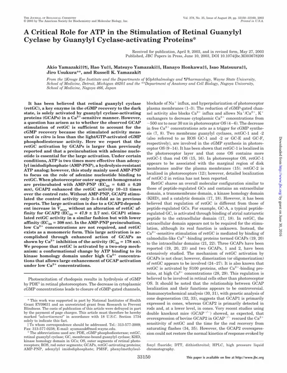
ATP (37 °C, 30 min) and soluble components were washed out,GCAP2-stimulated retGC activities in membrane fractionswere high (Fig. 1). This ATP activation was �10 times largerthan the GCAP-stimulated retGC activities in GCAP-freemembranes (47.6–56.2 as compared with 5.44 nmol cGMP/min/mg formed in the presence of 500 nM GCAP2). Such largeactivation of retGC was totally unexpected. We also found thatthe preincubation in the presence of high Ca2� concentrationswas more effective than in its low concentration (56.2–47.6nmol cGMP/min/mg formed in the presence of 500 nM GCAP2).It should be emphasized that the activity is expressed as spe-cific activity rather than the activation level compared withthat obtained without ATP and that GCAP2-stimulated retGCactivities in GCAP-free membranes were measured without thepreincubation. Thus, the large activation by ATP is not due tothe reduction of retGC control activity by instability of retGC/membranes incubated without ATP (58). Indeed, the GCAP2sensitivity of retGC in OS homogenates preincubated withoutATP was less than that in GCAP-free membranes, suggestingthe instability of retGC/membranes incubated without ATP.These observations strongly suggest that ATP is positivelyinvolved in the stimulation of retGC by GCAP2.
However, mechanisms underlying the ATP stimulation aredifficult to elucidate because under these conditions ATP mayaffect retGC activity in various ways. For example, ATP mayexpress its functions by binding to retGC. ATP may also be aphosphate donor in the phosphorylation of proteins involved inthe retGC regulation. We note that phosphatase inhibitorswere added to the ATP preincubation. We also found that OSwashed membranes contained high ATP hydrolytic activityunder retGC assay conditions (33 °C, 10 min, 5–10 �g of pro-tein/200 �l). For example, �60% of 5 mM ATP was hydrolyzedto ADP under our assay conditions, even with an ATP-regen-erating system present. This high ATP hydrolytic activitymakes data interpretation difficult. For example, 0.1–0.5 mM
ATP was added to the assay mixture in some previous studies;however, the final ATP concentrations may be substantially
lower. In addition, it is possible that ATP metabolites mayaffect retGC activity.
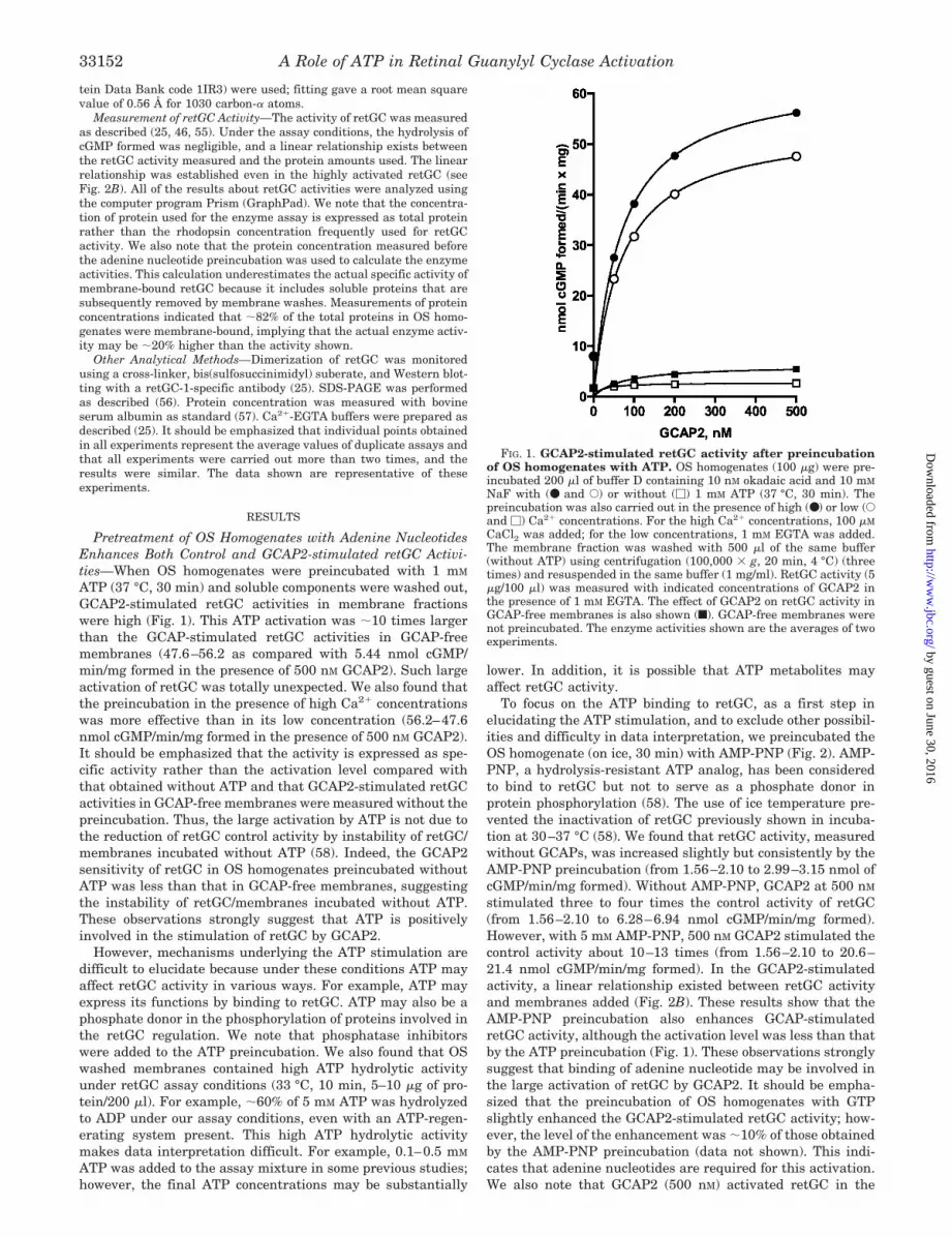
To focus on the ATP binding to retGC, as a first step inelucidating the ATP stimulation, and to exclude other possibil-ities and difficulty in data interpretation, we preincubated theOS homogenate (on ice, 30 min) with AMP-PNP (Fig. 2). AMP-PNP, a hydrolysis-resistant ATP analog, has been consideredto bind to retGC but not to serve as a phosphate donor inprotein phosphorylation (58). The use of ice temperature pre-vented the inactivation of retGC previously shown in incuba-tion at 30–37 °C (58). We found that retGC activity, measuredwithout GCAPs, was increased slightly but consistently by theAMP-PNP preincubation (from 1.56–2.10 to 2.99–3.15 nmol ofcGMP/min/mg formed). Without AMP-PNP, GCAP2 at 500 nM
stimulated three to four times the control activity of retGC(from 1.56–2.10 to 6.28–6.94 nmol cGMP/min/mg formed).However, with 5 mM AMP-PNP, 500 nM GCAP2 stimulated thecontrol activity about 10–13 times (from 1.56–2.10 to 20.6–21.4 nmol cGMP/min/mg formed). In the GCAP2-stimulatedactivity, a linear relationship existed between retGC activityand membranes added (Fig. 2B). These results show that theAMP-PNP preincubation also enhances GCAP-stimulatedretGC activity, although the activation level was less than thatby the ATP preincubation (Fig. 1). These observations stronglysuggest that binding of adenine nucleotide may be involved inthe large activation of retGC by GCAP2. It should be empha-sized that the preincubation of OS homogenates with GTPslightly enhanced the GCAP2-stimulated retGC activity; how-ever, the level of the enhancement was �10% of those obtainedby the AMP-PNP preincubation (data not shown). This indi-cates that adenine nucleotides are required for this activation.We also note that GCAP2 (500 nM) activated retGC in the
FIG. 1. GCAP2-stimulated retGC activity after preincubationof OS homogenates with ATP. OS homogenates (100 �g) were pre-incubated 200 �l of buffer D containing 10 nM okadaic acid and 10 mM
NaF with (● and E) or without (�) 1 mM ATP (37 °C, 30 min). Thepreincubation was also carried out in the presence of high (●) or low (Eand �) Ca2� concentrations. For the high Ca2� concentrations, 100 �M
CaCl2 was added; for the low concentrations, 1 mM EGTA was added.The membrane fraction was washed with 500 �l of the same buffer(without ATP) using centrifugation (100,000 � g, 20 min, 4 °C) (threetimes) and resuspended in the same buffer (1 mg/ml). RetGC activity (5�g/100 �l) was measured with indicated concentrations of GCAP2 inthe presence of 1 mM EGTA. The effect of GCAP2 on retGC activity inGCAP-free membranes is also shown (f). GCAP-free membranes werenot preincubated. The enzyme activities shown are the averages of twoexperiments.
A Role of ATP in Retinal Guanylyl Cyclase Activation33152
by guest on June 30, 2016http://w
ww
.jbc.org/D
ownloaded from
GCAP-free membranes approximately four times under ourassay conditions (from 0.877 to 3.83 nmol cGMP/min/mgformed) (Fig. 2A). Because the GCAP stimulation has beenmeasured using GCAP-free membranes without preincubationwith adenine nucleotides, our results indicate that most, if notall, activities of retGC/membranes described in previous stud-ies represent only a small portion of the potential retGC activ-ity in photoreceptors. We note that the control activity of retGCpreincubated without AMP-PNP is used to express the level ofretGC activation by the AMP-PNP preincubation. This compar-ison may be suitable to compare the activation of retGC prein-cubated with AMP-PNP with that preincubated without AMP-PNP and to compare the activity of retGC preincubated withAMP-PNP with the activities reported previously that weremeasured without preincubation with adenine nucleotides.
In AMP-PNP-pretreated OS homogenates, the retGC activ-ity was enhanced in a GCAP2 concentration-dependent man-ner (Fig. 2C). The GCAP2 concentration required for 50%enhancement appears to be similar at all AMP-PNP concentra-tions used (mean for five concentrations � 47.9 � 2.7 nM).
These results indicate that the AMP-PNP preincubation in-creases the Vmax of GCAP2-stimulated retGC activity but doesnot alter the affinity of retGC for GCAP2. We note that the realEC50 may be slightly higher than the average concentrationbecause the membranes already contain some endogenousGCAP2, as described below. Generally the EC50 obtained herewas similar to those reported previously (5, 59). The AMP-PNPconcentration required for the 50% enhancement is 0.65 � 0.20mM (Fig. 2D). The maximum stimulation was achieved by �5mM AMP-PNP, and the stimulation was not changed even afterthe preincubation with 10 mM AMP-PNP.
The effect of Ca2� on the AMP-PNP preincubation was alsoinvestigated (Fig. 2A). Without AMP-PNP, the activity ofretGC preincubated under high Ca2� concentrations was con-sistently lower than that preincubated under low Ca2� concen-trations (2 mM EGTA) (1.56–2.10 nmol cGMP/min/mg formed).With AMP-PNP, but without exogenous GCAP2, OS homoge-nates preincubated in low Ca2� concentrations also showslightly higher retGC activity than that in high Ca2� concen-trations (3.15–2.99 nmol cGMP/min/mg formed). However,
FIG. 2. GCAP2-stimulated retGC ac-tivity after preincubation of OS ho-mogenates with AMP-PNP. A, the ef-fect of AMP-PNP preincubation of OShomogenates on the GCAP2-stimulatedretGC activity. OS homogenates (150 �g/300 �l) were preincubated with(● and f) or without (E and �) 5 mM
AMP-PNP in the presence of high(● and E) or low (f and �) Ca2� concen-trations. For the high Ca2� concentra-tions, 100 �M CaCl2 was added; for thelow concentrations, 2 mM EGTA wasadded. After washing out the soluble frac-tion, retGC activities in the membranefraction (5.63 �g/200 �l) were assayedwith indicated GCAP2 concentrations inthe presence of 2 mM EGTA. As a refer-ence, the effect of GCAP2 on the retGCactivity in GCAP-free membranes (5 �g/200 �l) was also measured under thesame conditions but without preincuba-tion (*). B, the relationship betweenretGC activity and proteins in mem-branes after AMP-PNP preincubation. Af-ter incubation of OS homogenate (150 �g/300 �l) with (●) or without (E) 5 m M
AMP-PNP, retGC activities in the variousamounts of membrane fraction weremeasured with 200 nM GCAP2 in the as-say buffer (100 �l) containing 2 mM
EGTA. C, GCAP2 concentrations re-quired for the large activation of retGCafter preincubation of OS homogenates.After incubation of OS homogenates (150�g/300 �l) with indicated AMP-PNPconcentrations, the GCAP2-stimulatedactivities of retGC (5.63 �g/200 �l) weremeasured with indicated GCAP2 concen-trations in the presence of 2 mM EGTA.The following are the AMP-PNP concen-trations used: f, 0 mM; Œ, 1 mM; �, 2 mM;and �, 5 mM; and ●, 10 m M. D, AMP-PNPconcentrations in the preincubation of OShomogenates. The data shown in C werereplotted to determine the GCAP2 con-centration required for the large activa-tion of retGC. The following are theGCAP2 concentrations used: �, 0 nM; Œ,20 nM; �, 50 nM; �, 100 nM; ●, 200 n M;and f, 500 nM.
A Role of ATP in Retinal Guanylyl Cyclase Activation 33153
by guest on June 30, 2016http://w
ww
.jbc.org/D
ownloaded from
with GCAP2, the maximum activity of retGC preincubated inhigh Ca2� concentrations was slightly higher than that in lowCa2� concentrations (21.4–20.6 nmol cGMP/min/mg formed inthe presence of 500 nM GCAP2). These Ca2� effects were meas-ured five times, and the observations were consistent. Thehigher retGC activity with the preincubation in the presence ofhigh Ca2� concentrations was more clearly observed in theATP preincubation (Fig. 1). Although the physiological signif-icance of the slight increase in retGC activity by the incubationin the presence of high Ca2� concentrations is not clear now,these results indicate that the reduction of Ca2� concentra-tions, essential for the retGC activation by GCAPs, is notrequired during the AMP-PNP preincubation.
We also checked the effect of Mg2� concentrations in thepreincubation on the subsequent activation of retGC byGCAP2. The preincubation was also carried out with 5 mM
AMP-PNP. We found that the preincubation with 10 mM MgCl2most effectively enhanced the retGC activity. However, thedifferences with varying Mg2� concentrations were small. Forexample, the activity of retGC preincubated in 10 mM MgCl2was �110 and �105% of those preincubated in 5 and 15 mM
MgCl2, respectively. Thus, throughout this study we used 5 mM
MgCl2 as a source of Mg2� ion in the preincubation.RetGC Stimulation by GCAP2 after Preincubation of OS
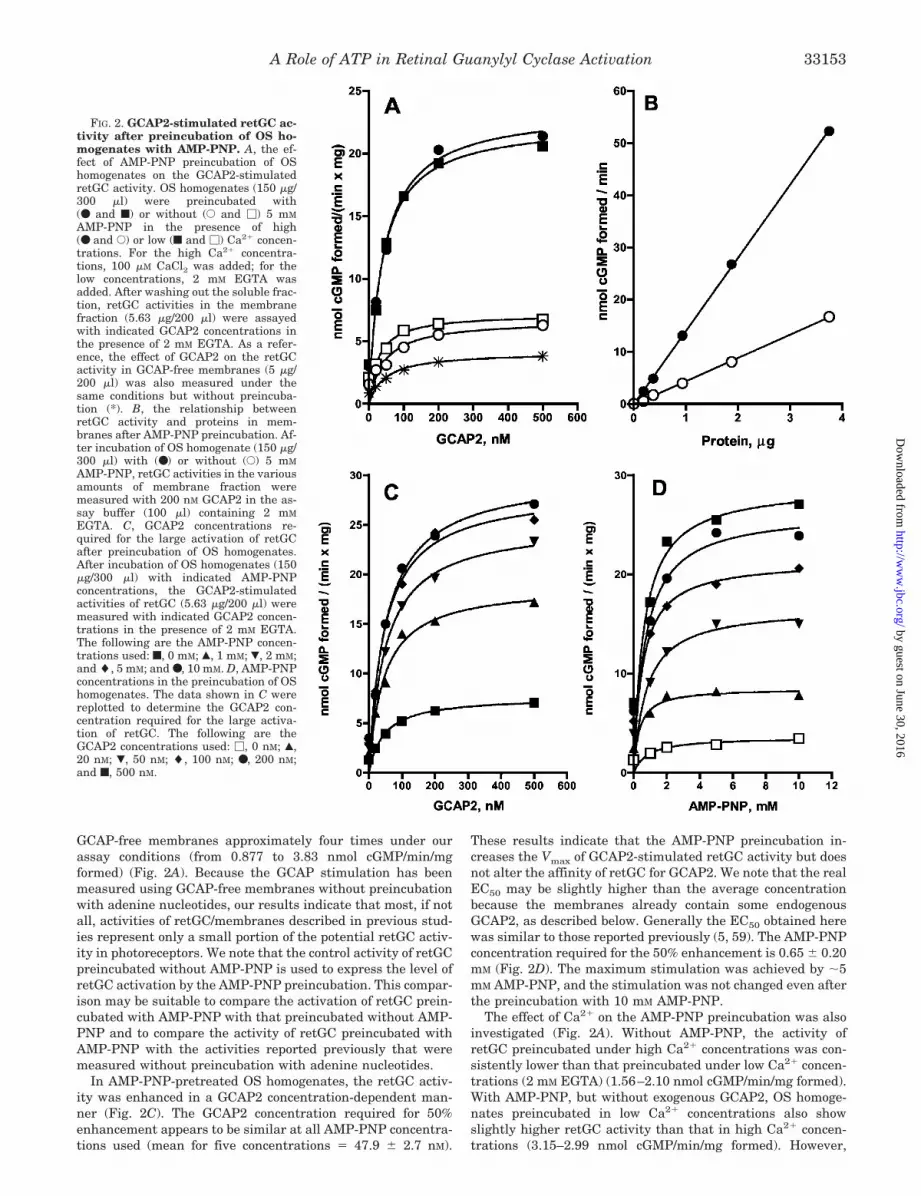
Homogenates with AMP-PNP Is Sensitive to High Ca2� Con-centrations—The activation of retGC by GCAP2 in OS homo-genates preincubated with AMP-PNP was abolished by highCa2� concentrations in the assay, and this Ca2� inhibition wasconsistently observed regardless of GCAP2 concentrations (Fig.3A). The sensitivity to high Ca2� concentrations was the sameas that observed in membranes preincubated without AMP-PNP (Fig. 3B). In the preincubation without AMP-PNP, theIC50 for calcium inhibition was 180 nM, and the Hill coefficientwas 1.42; with AMP-PNP, the IC50 was 178 nM, and the Hillcoefficient was 1.36. The sensitivity to high Ca2� concentra-tions was also the same as that observed in GCAP2-stimulatedretGC in GCAP-free membranes (data not shown). These re-sults indicate that the Ca2� sensitivity of GCAP-stimulatedretGC is preserved after OS homogenates were preincubatedwith AMP-PNP and that the highly activated retGC can beregulated by change in physiological Ca2� concentrations inOS.
FIG. 3. Sensitivity of GCAP2-stimu-lated retGC activity to high Ca2� con-centrations after preincubation ofOS homogenates with AMP-PNP. A,retGC activity measured with variousamounts of GCAP2 in the presence or ab-sence of high Ca2� concentrations. OS ho-mogenates (300 �g/600 �l) were preincu-bated with (f and ●) or without (� and E)5 mM AMP-PNP and membrane fractionswere washed as described. RetGC activityin the membrane fractions (5.63 �g/200�l) was measured with various amountsof GCAP2 in the presence of 2 mM EGTA(f and �) or 100 �M CaCl2 (● and E). B,GCAP2-activated retGC activities meas-ured with various Ca2� concentrations.OS homogenates (150 �g/300 �l) werepreincubated with (●) or without (E) 5mM AMP-PNP, and the membrane frac-tions were washed. retGC activity in themembrane fractions (5.63 �g/200 �l) wasmeasured with 200 nM GCAP2 in thepresence of various Ca2� concentrations.The Ca2� concentrations were adjustedusing Ca2�-EGTA buffers.
FIG. 4. Stimulation of retGC byGCAP1 or S100B after incubation ofOS homogenates with AMP-PNP. A,stimulation of retGC by GCAP1. OS ho-mogenates (150 �g/300 �l) were preincu-bated with (●) or without (E) 5 m M AMP-PNP, and the soluble fraction was washedout. RetGC activities in the membranefraction (2.82 �g/100 �l) were measuredwith indicated amounts of GCAP1 in thepresence of 2 mM EGTA. B, stimulation ofretGC by S100B. OS homogenates (150�g/300 �l) were pretreated with (● and f)or without (E and �) 5 mM AMP-PNP inthe presence of 100 �M CaCl2 (● and E) or2 mM EGTA (f and �), and the solublefraction was washed out. retGC activitiesof the membrane fraction (5.63 �g/200 �l)were measured with indicated amounts ofS100B in the presence of 200 �M CaCl2.
A Role of ATP in Retinal Guanylyl Cyclase Activation33154
by guest on June 30, 2016http://w
ww
.jbc.org/D
ownloaded from
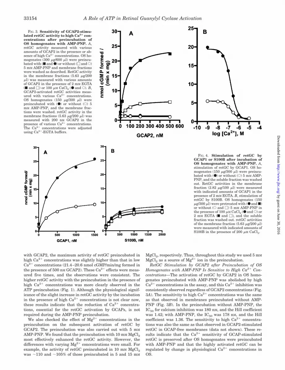
GCAP1, but Not S100B, Also Enhances retGC Activity afterPreincubation of OS Homogenates with AMP-PNP—After pre-incubation of OS homogenates with or without AMP-PNP,GCAP1-stimulated retGC activity was measured (Fig. 4A).Without AMP-PNP, the retGC activity was stimulated byGCAP1 (1.5 �M) approximately six times (from 1.69 to 9.57nmol cGMP/min/mg formed), whereas with AMP-PNP (5 mM),the GCAP1-stimulated retGC activity was about 15 times ofthe control activity (from 1.69 to 25.9 nmol cGMP/min/mgformed). The retGC control activity was also increased by theAMP-PNP preincubation (from 1.69 to 2.62 nmol cGMP/min/mg formed). As was the case with GCAP2, the concentra-tion for half-maximal stimulation by GCAP1 was similar in theabsence (EC50 � 298 nM) or presence (EC50 � 308 nM) ofAMP-PNP in the preincubation. The EC50 was similar to thatreported previously (43). However, when retGC in these mem-branes was activated by S100B, the AMP-PNP preincubationonly negligibly increased the retGC activity (Fig. 4B). In theexperiments, the AMP-PNP preincubation was carried out withhigh or low Ca2� concentrations (100 �M CaCl2 or 2 mM EGTA),indicating that the high activation does not occur regardless ofCa2� concentrations in the preincubation. Together, observa-tions shown here suggest that the large stimulation of retGC inOS homogenates preincubated with AMP-PNP occurs onlywhen retGC is stimulated by GCAPs.
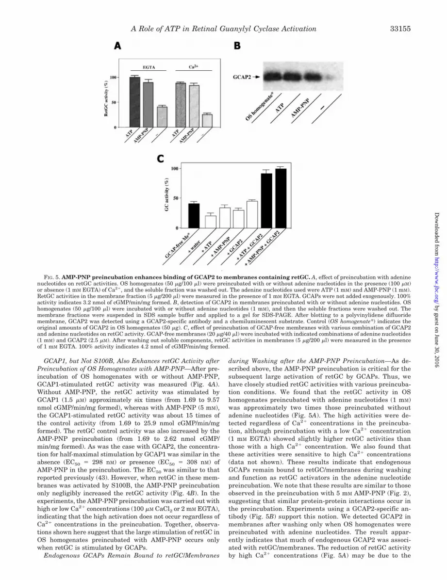
Endogenous GCAPs Remain Bound to retGC/Membranes
during Washing after the AMP-PNP Preincubation—As de-scribed above, the AMP-PNP preincubation is critical for thesubsequent large activation of retGC by GCAPs. Thus, wehave closely studied retGC activities with various preincuba-tion conditions. We found that the retGC activity in OShomogenates preincubated with adenine nucleotides (1 mM)was approximately two times those preincubated withoutadenine nucleotides (Fig. 5A). The high activities were de-tected regardless of Ca2� concentrations in the preincuba-tion, although preincubation with a low Ca2� concentration(1 mM EGTA) showed slightly higher retGC activities thanthose with a high Ca2� concentration. We also found thatthese activities were sensitive to high Ca2� concentrations(data not shown). These results indicate that endogenousGCAPs remain bound to retGC/membranes during washingand function as retGC activators in the adenine nucleotidepreincubation. We note that these results are similar to thoseobserved in the preincubation with 5 mM AMP-PNP (Fig. 2),suggesting that similar protein-protein interactions occur inthe preincubation. Experiments using a GCAP2-specific an-tibody (Fig. 5B) support this notion. We detected GCAP2 inmembranes after washing only when OS homogenates werepreincubated with adenine nucleotides. The result appar-ently indicates that much of endogenous GCAP2 was associ-ated with retGC/membranes. The reduction of retGC activityby high Ca2� concentrations (Fig. 5A) may be due to the
FIG. 5. AMP-PNP preincubation enhances binding of GCAP2 to membranes containing retGC. A, effect of preincubation with adeninenucleotides on retGC activities. OS homogenates (50 �g/100 �l) were preincubated with or without adenine nucleotides in the presence (100 �M)or absence (1 mM EGTA) of Ca2�, and the soluble fraction was washed out. The adenine nucleotides used were ATP (1 mM) and AMP-PNP (1 mM).RetGC activities in the membrane fraction (5 �g/200 �l) were measured in the presence of 1 mM EGTA. GCAPs were not added exogenously. 100%activity indicates 3.2 nmol of cGMP/min/mg formed. B, detection of GCAP2 in membranes preincubated with or without adenine nucleotides. OShomogenates (50 �g/100 �l) were incubated with or without adenine nucleotides (1 mM), and then the soluble fractions were washed out. Themembrane fractions were suspended in SDS sample buffer and applied to a gel for SDS-PAGE. After blotting to a polyvinylidene difluoridemembrane, GCAP2 was detected using a GCAP2-specific antibody and a chemiluminescent substrate. Control (OS homogenate*) indicates theoriginal amounts of GCAP2 in OS homogenates (50 �g). C, effect of preincubation of GCAP-free membranes with various combination of GCAP2and adenine nucleotides on retGC activity. GCAP-free membranes (20 �g/40 �l) were incubated with indicated combinations of adenine nucleotides(1 mM) and GCAP2 (2.5 �M). After washing out soluble components, retGC activities in membranes (5 �g/200 �l) were measured in the presenceof 1 mM EGTA. 100% activity indicates 4.2 nmol of cGMP/min/mg formed.
A Role of ATP in Retinal Guanylyl Cyclase Activation 33155
by guest on June 30, 2016http://w
ww
.jbc.org/D
ownloaded from
weaker binding of GCAP2 to membranes (and/or to retGC) inthe presence of high Ca2� (60).
To further test this notion, experiments were carried outusing GCAP-free membranes reconstituted with exogenousGCAP2 in the presence or absence of adenine nucleotides (Fig.5C). We found that only membranes pretreated with adeninenucleotides in combination with GCAP2 had higher retGC ac-tivities. A previous study showing that ATP (0.4 mM) increasedthe efficacy of GCAP1 to stimulate retGC-1 (43) suggests thatGCAP1 also shows the similar interaction with retGC in thepresence of adenine nucleotides. The simplest explanation ofthese phenomena is that GCAP-binding sites directly con-nected with retGC activation in membranes were increasedby the incubation with adenine nucleotide and more endoge-nous GCAPs bound to these sites. In the preincubation, wash-ing conditions used were not enough to exclude all GCAPsbound. Another explanation may be that after the incubationwith adenine nucleotides, endogenous GCAPs became tightlyassociated with retGC/membranes, remained bound to themembranes during washing, and functioned as a retGC acti-vator. However, the possibility of this explanation may not behigh because the retGC affinity to GCAP2 was not changedby the preincubation with or without adenine nucleotides(Fig. 2C).
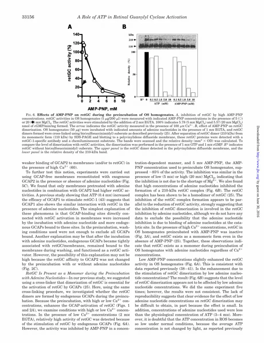
RetGC Is Present as a Monomer during the Preincubationwith Adenine Nucleotides—In our previous study, we suggestedusing a cross-linker that dimerization of retGC is essential forthe activation of retGC by GCAPs (25). Here, using the samecross-linking procedure, we investigated whether the retGCdimers are formed by endogenous GCAPs during the preincu-bation. Because the preincubation, with high or low Ca2� con-centrations, enhances the GCAP-activation of retGC (Figs. 1and 2A), we examine conditions with high or low Ca2� concen-trations. In the presence of low Ca2� concentrations (2 mM
EGTA), relatively high activity of retGC was detected becauseof the stimulation of retGC by endogenous GCAPs (Fig. 6A).However, the activity was inhibited by AMP-PNP in a concen-
tration-dependent manner, and 5 mM AMP-PNP, the AMP-PNP concentration used to preincubate OS homogenates, sup-pressed �85% of the activity. The inhibition was similar in thepresence of low (5 mM) or high (20 mM) MgCl2, indicating thatthe inhibition is not due to the shortage of Mg2�. We also foundthat high concentrations of adenine nucleotides inhibited theformation of a 210-kDa retGC complex (Fig. 6B). The retGCcomplex has been shown to be a homodimer of retGC (25). Theinhibition of the retGC complex formation appears to be par-allel to the reduction of retGC activity, strongly suggesting thatthe inhibition of retGC dimerization is involved in the retGCinhibition by adenine nucleotides, although we do not have anydata to exclude the possibility that the adenine nucleotideinhibition is due to binding of adenine nucleotides at the cata-lytic site. In the presence of high Ca2� concentrations, retGC inOS homogenates preincubated with AMP-PNP was inactive(Fig. 3A), and retGC exists as a monomeric form even in theabsence of AMP-PNP (25). Together, these observations indi-cate that retGC exists as a monomer during preincubation ofOS homogenates with adenine nucleotides regardless of Ca2�
concentrations.Low AMP-PNP concentrations slightly enhanced the retGC
activity in OS homogenates (Fig. 6A). This is consistent withdata reported previously (38–41). Is the enhancement due tothe stimulation of retGC dimerization by low adenine nucleo-tide concentrations? The result (Fig. 6B) suggests that the levelof retGC dimerization appears not to be affected by low adeninenucleotide concentrations. We did the same experiment fivetimes; however, the results were not consistent. The lack ofreproducibility suggests that clear evidence for the effect of lowadenine nucleotide concentrations on retGC dimerization maybe difficult to obtain, in part because the effect is small. Inaddition, concentrations of adenine nucleotides used were lessthan the physiological concentration of ATP (3–4 mM). More-over, it is doubtful that the ATP concentration in ROS becomesso low under normal conditions, because the average ATPconcentration is not changed by light, as reported previously
FIG. 6. Effects of AMP-PNP on retGC during the preincubation of OS homogenates. A, inhibition of retGC by high AMP-PNPconcentrations. retGC activities in OS homogenates (5 �g/200 �l) were measured with indicated AMP-PNP concentrations in the presence of 5 (E)or 20 (●) m M MgCl2. The retGC activities were stimulated by the addition of 2 mM EGTA. 100% indicates 5.78 (5 mM MgCl2) and 5.57 (20 mM MgCl2)nmol of cGMP/min/mg formed. The arrow indicates the retGC activity measured in the presence of 100 �M Ca2�. B, effect of AMP-PNP on retGCdimerization. OS homogenates (50 �g) were incubated with indicated amounts of adenine nucleotides in the presence of 1 mM EGTA, and retGCdimers formed were cross-linked using bis(sulfosuccinimidyl) suberate as described previously (25). After separation of retGC dimer (210 kDa) fromits monomeric form (110 kDa) by SDS-PAGE and blotting to a polyvinylidene difluoride membrane, these retGC proteins were detected with aretGC-1-specific antibody and a chemiluminescent substrate. The bands were scanned and the relative density (mm2 � OD) was calculated. Tocompare the level of dimerization with retGC activities, the dimerization was performed in the presence of 1 mM GTP and 1 mM cGMP. R* indicatesretGC without bis(sulfosuccinimidyl) suberate. The upper panel is the retGC dimer detected in the polyvinylidene difluoride membrane, and thelower panel is the relative density of the 210-kDa band.
A Role of ATP in Retinal Guanylyl Cyclase Activation33156
by guest on June 30, 2016http://w
ww
.jbc.org/D
ownloaded from
(61). Thus, the significance of the activation of retGC by lowconcentrations of adenine nucleotides is unclear.
DISCUSSION
This study has been carried out to obtain answers to afundamental but hitherto unexplored question regarding theregulation of retGC. The question is whether the GCAP stim-ulation of retGC previously observed can be further enhanced.As an answer, we found that retGC could be activated byGCAPs at least 10–13-fold over the control activity and thatinteraction with adenine nucleotides was essential for this highactivation of retGC. Significantly, recent studies using a doubleknockout mouse (GCAP�/�) showed that retGC is activated invivo by GCAPs �13-fold over the control activity, and the levelof stimulated retGC activity is similar or higher than that oflight-activated PDE (34, 35, 49). Our observations are in goodagreement with those in vivo studies. We feel that the largestimulation of retGC reported here represents a major portionof the mechanism involved in the retGC activation detected invivo.
In the previous model, the retGC activation by GCAPs is aone-step mechanism: activation by GCAPs when the Ca2� con-centration in OS is reduced. Low concentrations (less than 0.5mM) of adenine nucleotides in the assay mixture were shown to
stimulate retGC activity; however, the activation was generallysmall in OS membranes (less than 1.5 times) (Refs. 21 and38–43 and Fig. 6A). Thus, adenine nucleotides were believed tomodify the activity but not to be essential. Most aspects, if notall, of retGC including its physiological roles, its regulations,and its function-structure relationships appear to be inter-preted based on the one-step mechanism. Here, based on theresults described, we propose a two-step mechanism for theretGC activation: retGC first interacts with ATP to produce aconformational change and is then highly activated by GCAPswhen the Ca2� concentration is reduced. As described above,we estimated that less than 20 �M AMP-PNP were carried overfrom the preincubation mixture to the retGC preparation. Thisindicates that less than 1 �M AMP-PNP presented in the assaymixture because 10 �l of the retGC preparation was added toassay mixture (200 �l). This AMP-PNP concentration was toosmall to activate retGC in the assay mixture (Fig. 6A). Underthe conditions, GCAPs stimulated retGC (Fig. 2). Thus, in thismechanism, the step for the ATP interaction and the step forthe GCAP activation are separate and distinct, and both stepsare essential for the large stimulation. This adenine nucleotideinteraction requires concentrations in the millimolar range(EC50 � 0.65 mM for AMP-PNP) along with other factor(s)
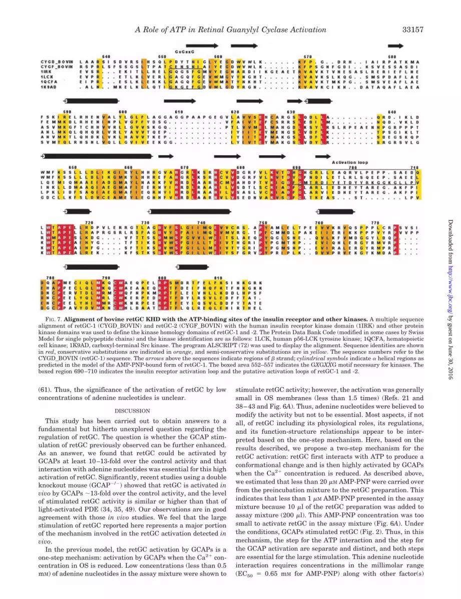
FIG. 7. Alignment of bovine retGC KHD with the ATP-binding sites of the insulin receptor and other kinases. A multiple sequencealignment of retGC-1 (CYGD_BOVIN) and retGC-2 (CYGF_BOVIN) with the human insulin receptor kinase domain (1IRK) and other proteinkinase domains was used to define the kinase homology domains of retGC-1 and -2. The Protein Data Bank Code (modified in some cases by SwissModel for single polypeptide chains) and the kinase identification are as follows: 1LCK, human p56-LCK tyrosine kinase; 1QCFA, hematopoieticcell kinase; 1K9AD, carboxyl-terminal Src kinase. The program ALSCRIPT (72) was used to display the alignment. Sequence identities are shownin red, conservative substitutions are indicated in orange, and semi-conservative substitutions are in yellow. The sequence numbers refer to theCYGD_BOVIN (retGC-1) sequence. The arrows above the sequences indicate regions of � strand; cylindrical symbols indicate � helical regions aspredicted in the model of the AMP-PNP-bound form of retGC-1. The boxed area 552–557 indicates the GXGXXG motif necessary for kinases. Theboxed region 690–710 indicates the insulin receptor activation loop and the putative activation loops of retGC-1 and -2.
A Role of ATP in Retinal Guanylyl Cyclase Activation 33157
by guest on June 30, 2016http://w
ww
.jbc.org/D
ownloaded from
(discussed below) and is indifferent to the calcium concentra-tion. On the other hand, the adenine nucleotide stimulation inthe assay mixtures requires adenine nucleotide concentrationsbelow the millimolar range because higher concentrations in-hibit retGC activity either directly or through inhibition ofdimerization. In addition, the stimulation in the assay mix-tures requires low calcium concentrations for the activation byGCAPs.
Is the mechanism for retGC activation by the adenine nucle-otide preincubation different from the activation by low con-centrations of adenine nucleotides in the assay mixture? TheretGC activity in AMP-PNP-preincubated OS membranes wasfurther activated by the low concentrations of AMP-PNP in theassay mixture.2 This implies that the AMP-PNP in the assaymixture further activates retGC already stimulated by theAMP-PNP preincubation and that the AMP-PNP activation inthe assay mixture does not require the unknown factor(s) (dis-cussed below) required for the activation by AMP-PNP prein-cubation. These implications are suggestive of the mechanisticdifference but not conclusive. On the other hand, in somestudies retGC activities similar to those stimulated by AMP-PNP preincubation were reported without the adenine nucleo-tide preincubation (58, 60). These activities were measured
with low concentrations (�0.5 mM) of ATP in the assay mix-ture. Although these high retGC activities were not constantlyobserved even in the same research groups and the ATP con-tribution to the high activities is unknown, these activities maysuggest that under certain conditions a mechanism similar tothat for the activation by adenine nucleotide preincubationmay function in the assay mixture.
Using AMP-PNP, we suggest that binding of ATP to retGCproduces the large activation by facilitating subsequent inter-action with GCAPs. However, it should be emphasized thatbinding of ATP to retGC is speculative based on the observationthat preincubation of OS homogenates with AMP-PNP wasrequired for high activation of retGC. However, this specula-tion is also supported by previous studies as follows. (a) RetGChas a molecular configuration similar to those of peptide-reg-ulated GCs (17, 18, 37), implying that the regulatory mecha-nism of retGC may be similar to those of peptide-regulatedGCs. In peptide-regulated GCs, ATP is an important factor fortheir regulation (17, 18, 37). It has been hypothesized in theseGCs that ATP binds to the KHD, serves as an intracellularallosteric regulator, and releases an inhibitory constraint of theKHD for the activation. (b) A previous study already suggestedthat AMP-PNP binds to retGC (58). Although the study did notdirectly show the AMP-PNP binding to retGC, the AMP-PNPbinding appears to be a reasonable explanation. c) As discussedabove, activation of retGC by low concentrations of ATP and itsanalogs has been reported in previous studies (38–41), al-though the concentration of adenine nucleotides is differentfrom those in this study. This stimulation has been speculatedto be due to the binding of ATP to the KHD in retGC.
To test the possibility that adenine nucleotides function asallosteric regulators by binding to the KHD, we used three-dimensional modeling. The availability of crystal structures forthe inactive and active conformations of the insulin receptorkinase domain (62) allowed construction of models for the KHDof retGC with and without bound AMP-PNP. For this purpose,an alignment of bovine retGC-1 with the ATP binding sites ofthe insulin receptor and other kinases was carried out to de-termine the boundaries of the KHD within the retGC-1 se-quence. Fig. 7 displays the alignment that indicates sequencesimilarity allowing three-dimensional modeling. Also indicatedis the lack of the GXGXXG motif (the boxed region 552–557)necessary for kinases in the retGC-1 sequence, a feature pre-viously noted (37, 63).
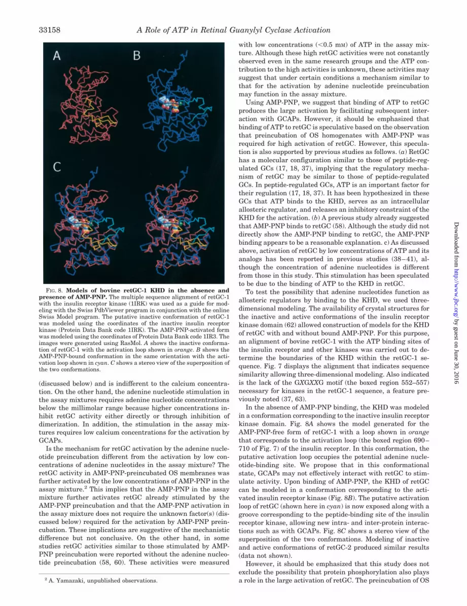
In the absence of AMP-PNP binding, the KHD was modeledin a conformation corresponding to the inactive insulin receptorkinase domain. Fig. 8A shows the model generated for theAMP-PNP-free form of retGC-1 with a loop shown in orangethat corresponds to the activation loop (the boxed region 690–710 of Fig. 7) of the insulin receptor. In this conformation, theputative activation loop occupies the potential adenine nucle-otide-binding site. We propose that in this conformationalstate, GCAPs may not effectively interact with retGC to stim-ulate activity. Upon binding of AMP-PNP, the KHD of retGCcan be modeled in a conformation corresponding to the acti-vated insulin receptor kinase (Fig. 8B). The putative activationloop of retGC (shown here in cyan) is now exposed along with agroove corresponding to the peptide-binding site of the insulinreceptor kinase, allowing new intra- and inter-protein interac-tions such as with GCAPs. Fig. 8C shows a stereo view of thesuperposition of the two conformations. Modeling of inactiveand active conformations of retGC-2 produced similar results(data not shown).
However, it should be emphasized that this study does notexclude the possibility that protein phosphorylation also playsa role in the large activation of retGC. The preincubation of OS2 A. Yamazaki, unpublished observations.
FIG. 8. Models of bovine retGC-1 KHD in the absence andpresence of AMP-PNP. The multiple sequence alignment of retGC-1with the insulin receptor kinase (1IRK) was used as a guide for mod-eling with the Swiss PdbViewer program in conjunction with the onlineSwiss Model program. The putative inactive conformation of retGC-1was modeled using the coordinates of the inactive insulin receptorkinase (Protein Data Bank code 1IRK). The AMP-PNP-activated formwas modeled using the coordinates of Protein Data Bank code 1IR3. Theimages were generated using RasMol. A shows the inactive conforma-tion of retGC-1 with the activation loop shown in orange. B shows theAMP-PNP-bound conformation in the same orientation with the acti-vation loop shown in cyan. C shows a stereo view of the superposition ofthe two conformations.
A Role of ATP in Retinal Guanylyl Cyclase Activation33158
by guest on June 30, 2016http://w
ww
.jbc.org/D
ownloaded from

homogenates with adenine nucleotides was initially carried outunder the conditions for protein phosphorylation. Comparisonof specific activities of retGC shown in Figs. 1 and 2 shows thatATP is approximately twice as effective as AMP-PNP for theretGC stimulation under our conditions. Moreover, phospha-tase inhibitors (10 nM okadaic acid and 10 mM NaF) furtherincreased the retGC activity.2 These results suggest that phos-phorylation of retGC and/or proteins involved in the retGCregulation may further enhance the GCAP-dependent retGCactivity that has already been stimulated by binding of adeninenucleotides. For retGC, we have already detected incorporationof the �-phosphate of ATP into the protein under the conditionsused for preincubation.3 Previous studies also reported thatretGC might be regulated by phosphorylation (64–66). More-over, it has been reported that phosphorylation, in addition toATP binding, is important for the activation of GC-A (18, 67).For proteins involved in the retGC regulation, GCAPs are theestablished candidates, although phosphorylation of an un-known protein regulator(s) (discussed below) may also be pos-sible. It has been reported that GCAP2 is phosphorylated bycyclic nucleotide-dependent protein kinases, but the phospho-rylation had little effect on retGC activation by GCAP2 (68).The study appears to have been conducted under conditions forthe single-step mechanism. It may not be surprising to detectthat GCAP phosphorylation is involved in the retGC regulationunder the two-step mechanism reported here. We also note thatthe GCAP2-stimulated activity of retGC preincubated in thepresence of high Ca2� concentrations was slightly but consis-tently higher than that in the presence of low Ca2� concentra-tions when AMP-PNP was added to the preincubation mixture(Fig. 2A). It is possible that the difference found in the AMP-PNP preincubation may be due to protein dephosphorylationbecause phosphatase inhibitors were not in the preincubationmixture. This Ca2� effect was detected more clearly when ATPwas used (37 °C) (Fig. 1). These observations imply that ifprotein phosphorylation is involved in the large activation, thephosphorylation level may be Ca2�-regulated.
Because this study is the first report for the two-step mech-anism, new questions have arisen that will require answers.We found that addition of GCAP2 to prewashed membranesfollowed by the incubation with AMP-PNP was not enough toobtain the large activation.2 These observations suggest thatthe simple binding of AMP-PNP to retGC with GCAP2 was notenough for the high activation. The possibility that AMP-PNPcannot bind to retGC in washed membranes may be excludedby a previous study suggesting that AMP-PNP appears tochange the retGC structure in washed membranes (58). Thesimplest explanation to our observations is that a factor(s)in OS homogenates may be required in the AMP-PNPpreincubation.
Another puzzling point is that the large activation was de-tected even after washing out of soluble components from OShomogenates. This indicates that the effect of AMP-PNP isretained on retGC after the washing, although EC50 of AMP-PNP for the large activation is relatively high (0.64 mM). Themechanism to retain the effect of AMP-PNP on retGC is un-clear. We speculate that after interaction with AMP-PNP,retGC changes its conformation, and the conformationalchange prevents AMP-PNP from being released from retGC.Alternatively, AMP-PNP may be released from retGC duringthe washing, but the conformational change in retGC is re-tained by a factor(s) in OS homogenates. Although there maybe other explanations, it is crucial to elucidate all of the com-ponents in OS homogenates involved for understanding of thelarge activation of retGC.
We have shown that endogenous GCAP2 is associated withretGC/membranes after preincubation of OS homogenates withadenine nucleotides (Fig. 5) and that the large activation of theretGC is accomplished by addition of exogenous GCAP2 (Fig.2). How many GCAP-binding sites are on the adenine nucleoti-de-treated retGC? As a model for one binding site, we presumetwo retGC conformations in equilibrium: an inactive form with-out enzyme activity and possessing no ability to be stimulatedby GCAP2 and an active form with enzyme activity that isdependent on GCAP2. Thus, the observed activity is dependentupon the proportion of the active form and the content ofGCAP2. Without adenine nucleotide treatment, only a smallfraction of retGC exists as the active form. The small increaseof the retGC activity by exogenous GCAP2 would be due to thatportion of the active form whose GCAP2 sites are not alreadyfilled by endogenous GCAP2, i.e. exogenous GCAP2 binds tothe residual GCAP-free active form to express retGC activity.However, because the fraction of total retGC present as activeform is small, the observed retGC activity is not large (Fig. 2).The requirement for exogenous GCAP2 may be due to lowcontent of GCAP2 compared with retGC in the preparationsused and/or the loss by washing. This explanation can also beapplied to the basal activity and GCAP-dependent activation ofretGC in previous studies. Treatment with adenine nucleotidesshifts the equilibrium from the inactive form toward the activeform. The large activation by adenine nucleotide preincubation(Fig. 2) is completed by binding of exogenous GCAP2 to theresulting larger fraction of retGC in the active form. This modelis supported by the observations that the affinity of retGC forGCAP2 is not changed by preincubation with or without AMP-PNP (Fig. 2C) and that the level of retGC activity observedwith saturating amounts of GCAP2 is determined by the con-centration of AMP-PNP in the preincubation (Fig. 2D).
Other explanations including multiple GCAP2-binding sitesare also possible. The GCAP2 added exogenously may bind to aretGC domain(s) different from the domain with which endog-enous GCAP2 is associated. It is possible that the new confor-mation of retGC, a product of adenine nucleotide binding toretGC, opens a new binding site(s) for GCAP2. We believe thatGCAP1 also associates with retGC under the same conditions,as described above. If so, which GCAP combinations can moreeffectively activate retGC? This question may lead not only toan explanation for the presence of two GCAPs, 1 and 2, in ROS(30, 31) but also to a new mechanism of retGC regulation.
It should be emphasized that a new preincubation of OShomogenates with adenine nucleotides is required for the largeactivation of retGC, although the adenine nucleotide effect isretained after washing of membranes. This implies that theadenine nucleotide effect is initiated and then terminated un-der certain conditions, i.e. there are mechanisms to turn on andturn off the ATP effect in OS. In particular, the mechanism toturn off the highly activated retGC may be as important asturning off light/GTP-activated PDE in the overall control ofcGMP metabolism in OS during normal function and adapta-tion. Several mechanisms for the inhibition of retGC have beenreported (69, 70), although these inhibitions cannot be easilyincorporated in the current model of retGC regulation: thesingle-step mechanism. Under the new mechanism of retGCregulation reported here, these retGC inhibitory mechanismsmay be incorporated more easily. We especially emphasize thatretGC inhibition by RGS9 (70, 71) is attractive because underthe large activation of retGC, the mutual regulation betweenthe retGC system and the PDE system is more important, andRGS9 has been proposed to function in both the PDE andretGC systems. Obviously, further studies are needed to estab-lish the mechanism suggested here and to answer puzzling3 H. Yu and A. Yamazaki, unpublished observations.
A Role of ATP in Retinal Guanylyl Cyclase Activation 33159
by guest on June 30, 2016http://w
ww
.jbc.org/D
ownloaded from
points. Moreover, it is important to reveal mechanisms to over-come the retGC inhibition by physiological ATP concentrationsin the system proposed here. It is clear, however, that thisstudy opens a new field of retGC regulation in photoreceptorOS.
Acknowledgments—We thank Drs. R. B. Needleman (Wayne StateUniversity) and A. Sitaramayya (Oakland University, Rochester,Michigan) for critical reading of the manuscript.
REFERENCES
1. Stryer, L. (1986) Annu. Rev. Neurosci. 9, 87–1192. Miller, W. H. (1990) Invest. Ophthalmol. Visual Sci. 31, 1664–16733. Pugh, Jr. E. N., and Lamb, T. D. (1993) Biochim. Biophys. Acta 1141, 111–1494. Yau, K.-W., and Baylor, D. A. (1989) Annu. Rev. Neurosci. 12, 289–3275. Dizhoor, A. M., and Hurley, J. B. (1999) Methods 19, 521–5316. Fain, G. L., Matthews, H. R., Cornwall, C., and Koutalos, Y. (2001) Physiol.
Rev. 81, 117–1517. Lolley, R. N., and Racz, E. (1982) Vision Res. 22, 1481–14868. Koch, K.-W., and Stryer, L. (1988) Nature 334, 64–669. Shyjan, A. W., de Sauvage, F. J., Gillett, N. A., Goeddel, D. V., and Lowe, D. G.
(1992) Neuron 9, 727–73710. Goraczniak, R. M., Duda, T., Sitaramayya, A., and Sharma, R. K. (1994)
Biochem. J. 302, 455–46111. Yang, R.-B., Foster, D. C., Garbers, D. L., and Fulle, H.-J. (1995) Proc. Natl.
Acad. Sci. U. S. A. 92, 602–60612. Lowe, D. G., Dizhoor, A. M., Liu, K., Gu, Q., Spencer, M., Laura, R., Lu, L., and
Hurley, J. B. (1995) Proc. Natl. Acad. Sci. U. S. A. 92, 5535–553913. Johnston, J. P., Farhangfar, F., Aparicio, J. G., Nam, S. H., and Applebury,
M. L. (1997) Gene (Amst.)193, 219–22714. Goraczniak, R. K., Duda, T., and Sharma, R. K. (1997) Biochem. Biophys. Res.
Commun. 234, 666–67015. Liu, X., Seno, K., Nishizawa, Y., Hayashi, F., Yamazaki, A., Matsumoto, H.,
Wakabayashi, T., and Usukura, J. (1994) Exp. Eye Res. 59, 761–76816. Cooper, N., Liu, L., Yoshida, A., Ponzdnyakov, N., Margulis, A., and
Sitaramayya, A. (1996) J. Mol. Neurosci. 6, 211–22217. Lucas, K., A., Pitari, G. M., Kazerounian, S., Ruiz-Stewart, I., Park, J., Schulz,
S., Chepenik, K. P., and Waldman, S. A. (2000) Pharmacol. Rev. 52,375–413
18. Potter, L. R., and Hunter, T. (2001) J. Biol. Chem. 276, 6057–606019. Gorczyca, W. A., Grey-Keller, M. P., Detwiler, P. B., and Palczewski, K. (1994)
Proc. Natl. Acad. Sci. U. S. A. 91, 4014–401820. Dizhoor, A. M., Lowe, D. G., Olshevskaya, E. V., Laura, R. P., and Hurley, J. B.
(1994) Neuron 12, 1345–135221. Laura, R. P., Dizhoor, A. M., and Hurley, J. B. (1996) J. Biol. Chem. 271,
11646–1165122. Duda, T., Goraczniak, R., Surgucheva, I., Rudnicka-Nawrot, M., Gorczyca,
W. A., Palczewski, K., Sitaramayya, A., Baehr, W., and Sharma, R. K.(1996) Biochemistry 35, 8478–8482
23. Haeseleer, F., Sokal, I., Li, N., Pettenati, M., Rao, N., Bronson, W., Wechter,R., Baehr, W., and Palczewski, K. (1999) J. Biol. Chem. 274, 6526–6535
24. Yang, R.-B., and Garbers, D. L. (1997) J. Biol. Chem. 272, 13738–1374225. Yu, H., Olshevskaya, E., Duda, T., Seno, K., Hayashi, F., Sharma, R. K.,
Dizhoor, A. M., and Yamazaki, A. (1999) J. Biol. Chem. 274, 15547–1555526. Tucker, C. L., Woodcock, S. C., Kelsell, R. E., Ramamurthy, V., Hunt, D. M.,
and Hurley, J. B. (1999) Proc. Natl. Acad. Sci. U. S. A. 96, 9039–904427. Duda, T., Venkataraman, V., Jankowska, A., Lange, C., Koch, K.-W., and
Sharma, R. K. (2000) Biochemistry 39, 12522–1253328. Pozdnyakov. N., Yoshida, A., Cooper, N. G. F., Margulis, A., Duda, T., Sharma,
R. K., and Sitaramayya, A. (1995) Biochemistry 34, 14279–1428329. Margulis, A., Pozdnyakov, N., and Sitaramayya, A. (1996) Biochem. Biophys.
Res. Commun. 218, 243–24730. Cuenca, N., Lopez, S., Howes, K., and Kolb, H. (1998) Invest. Ophthlmol.
Visual Sci. 39, 1243–125031. Kachi, S., Nishizawa, Y., Olshevskaya, E., Yamazaki, A., Miyake, Y.,
Wakabayashi, T., Dizhoor, A., and Usukura, J. (1999) Exp. Eye Res. 68,465–473
32. Payne, A. M., Downes, S. M., Bessant, D. A. R., Taylor, R., Holder, G. E.,Warren, M. J., Bird, A. C., and Bhattachraya, S. S. (1998) Hum. Mol. Genet.
7, 273–27733. Dizhoor, A. M., Boikov, S. G., and Olshevskaya, E. V. (1998) J. Biol. Chem.
273, 17311–1731434. Mendez, A., Burns, M. E., Sokal, I, Dizhoor, A. M., Baehr, W., Palczewski, K.,
Baylor, D. A., and Chen, J. (2001) Proc. Natl. Acad. Sci. U. S. A. 98,9948–9953
35. Burns, M., Mendez, A., Chen, J., and Baylor, D. A. (2002) Neuron 36, 81–9136. Howes, K. A., Pennesi, M. E., Sokal, I., Church-Kopish, J., Schmidt, B.,
Margolis, D., Frederick, J. M., Rieke, F., Palczewski, K., Wu, S. M.,Detwiler, P. D., and Baehr, W. (2002) EMBO J. 21, 1–10
37. Sharma, R. K., Yadav, P., and Duda, T. (2001) Can. J. Physiol. Pharmacol. 79,682–691
38. Krishnan, N., Fletcher, R. T., Chader, G. J., and Krishna, G. (1978) Biochim.Biophys. Acta 523, 506–515
39. Sitaramayya, A., Marala, R. B., Hakki, S., and Sharma, R. K. (1991) Biochem-istry 30, 6742–6747
40. Gorczyca, W. A., van Hooser, P., and Palczewski, (1994) Biochemistry 33,3217–3222
41. Sitaramayya, A., Duda, T., and Sharma, R. K. (1995) Mol. Cell. Biochem. 148,139–145
42. Frins, S., Bonigk, W., Muller, F., Kellner, R., Koch, K.-W. (1996) J. Biol. Chem.271, 8022–8027
43. Otto-Bruc, A., Buczylko, J., Surguchheva, I., Subbaraya, I., Rudnicka-Nawyot,M., Crabb, J. W., Arendt, A., Hargrave, P. A., Baehr, W., and Palczewski, K.(1997) Biochemistry 36, 4295–4302
44. Robinson, W. E., and Hagins, W. A. (1979) Nature 280, 398–40045. Hakki, S., and Sitaramayya, A. (1990) Biochemistry 29, 1088–109446. Hayashi, F., and Yamazaki, A. (1991) Proc. Natl. Acad. Sci. U. S. A. 88,
4746–475047. Koch, K.-W. (1991) J. Biol. Chem. 266, 8634–863748. Aparicio, J. G., and Applebury, M. L. (1995) Protein Expression Purif. 6,
501–51149. Detwiler, P. D. (2002) Neuron 36, 3–450. Cornwall, M. C., and Fain, G. L. (1994) J. Physiol. 480, 261–27951. Koutalos, Y., Nakatani, K., Tamura, T., and Yau, K.-W. (1995) J. Gen. Physiol.
106, 863–89052. Yamazaki, A., Tatsumi, M., and Bitensky, M. W. (1988) Methods Enzymol.
159, 702–71053. Thompson, J. D., Gibson, T. J., Plewniak, F., Jeanmougin, F., and Higgins,
D. J. (1997) Nucleic Acids Res. 25, 4876–488254. Guex, N., and Peitsch, M. C. (1997) Electrophoresis 18, 2714–272355. Hayashi, F., Hutson, L. D., Kishigami, A., Nagao, S., and Yamazaki, A. (1993)
Methods Neurosci. 15, 237–24756. Yamazaki, A., Tatsumi, M., Torney, D. C., and Bitensky, M. W. (1987) J. Biol.
Chem. 262, 9316–932357. Bradford, M. M. (1976) Anal. Biochem. 72, 248–25458. Tucker, C. L., Laura, R. P., and Hurley, J. B. (1997) Biochemistry 36,
11995–1200059. Laura, R. P., and Hurley, J. B. (1998) Biochemistry 37, 11264–1127160. Olshevskaya, E. V., Hughes, R. E., Hurley, J. B., and Dizhoor, A. M. (1997)
J. Biol. Chem. 272, 14327–1433361. Biernbaum. M. S., and Bownds, M. D. (1979) J. Gen. Physiol. 74, 649–66962. Hubbard, S. R. (1997) EMBO J. 16, 5573–558163. Duda, T., Goraczniak, R., and Sharma, R. K. (1996) Biochem. J. 319, 279–28364. Wolbring, G., and Schnetkamp, P. P. M. (1995) Biochemistry 34, 4689–469565. Wolbring, G., and Schnetkamp, P. P. M. (1996) Biochemistry 35, 11013–1101866. Aparicio, J. G., and Applebury, M. L. (1996) J. Biol. Chem. 271, 27083–2708967. Potter, L. R., and Hunter, T. (1998) Mol. Cell. Biol. 18, 2164–217268. Olshevskaya, E. V., and Dizhoor, A. M. (2002) Association for Research in
Vision and Ophthalmology Annual Meeting, Fort Lauderdale, FL, May5–10, 2002, Abstract 1413/B423, Association for Research in Vision andOphthalmology, Bethesda, MD
69. Li, N., Fariss, R. N., Zhang, K., Otto-Bruc, A., Haeseleer, F., Bronson, D., Qin,N., Yamazaki, A., Subbaraya, I., Milam, A. H., Palczewski, K., and Baehr,W. (1998) Eur. J. Biochem. 252, 591–599
70. Bondarenko, V. A., Yu, H., Yamazaki, R. K., and Yamazaki, A. (2002) Mol.Cell. Biochem. 230, 125–128
71. Seno, K., Kishigami, A., Ihara, S., Maeda, T., Bondarenko, V. A., Nishizawa,Y., Usukura, J., Yamazaki, A., and Hayashi, F. (1998) J. Biol. Chem. 273,22169–22172
72. Barton, G. J. (1993) Protein Eng. 6, 37–40
A Role of ATP in Retinal Guanylyl Cyclase Activation33160
by guest on June 30, 2016http://w
ww
.jbc.org/D
ownloaded from

Usukura and Russell K. YamazakiAkio Yamazaki, Hao Yu, Matsuyo Yamazaki, Hanayo Honkawa, Isao Matsuura, Jiro
Cyclase-activating ProteinsA Critical Role for ATP in the Stimulation of Retinal Guanylyl Cyclase by Guanylyl
doi: 10.1074/jbc.M303678200 originally published online June 10, 20032003, 278:33150-33160.J. Biol. Chem.
10.1074/jbc.M303678200Access the most updated version of this article at doi:
Alerts:
When a correction for this article is posted•
When this article is cited•
to choose from all of JBC's e-mail alertsClick here
http://www.jbc.org/content/278/35/33150.full.html#ref-list-1
This article cites 71 references, 29 of which can be accessed free at
by guest on June 30, 2016http://w
ww
.jbc.org/D
ownloaded from


