Embed Size (px)
Citation preview
Guanylate cyclases and associated activator proteins in retinaldisease
David M. Hunt • Prateek Buch • Michel Michaelides
Received: 17 June 2009 / Accepted: 4 November 2009 / Published online: 26 November 2009
� Springer Science+Business Media, LLC. 2009
Abstract Two isoforms of guanylate cyclase, GC1 and
GC2 encoded by GUCY2D and GUCY2F, are responsible
for the replenishment of cGMP in photoreceptors after
exposure to light. Both are required for the normal kinetics
of photoreceptor sensitivity and recovery, although disease
mutations are restricted to GUCY2D. Recessive mutations
in this gene cause the severe early-onset blinding disorder
Leber congenital amaurosis whereas dominant mutations
result in a later onset less severe cone–rod dystrophy.
Cyclase activity is regulated by Ca2? which binds to the
GC-associated proteins, GCAP1 and GCAP2 encoded by
GUCA1A and GUCA1B, respectively. No recessive muta-
tions in either of these genes have been reported. Dominant
missense mutations are largely confined to the Ca2?-
binding EF hands of the proteins. In a similar fashion to the
disease mechanism for the dominant GUCY2D mutations,
these mutations generally alter the sensitivity of the cyclase
to inhibition as Ca2? levels rise following a light flash.
Keywords Phototransduction � Retinal dystrophy
Retinal-specific guanylate cyclases (GCs) and their asso-
ciated activator proteins (GCAPs) are responsible for the
Ca2?-sensitive restoration of cGMP levels after the light
activation of the phototransduction cascade. These proteins
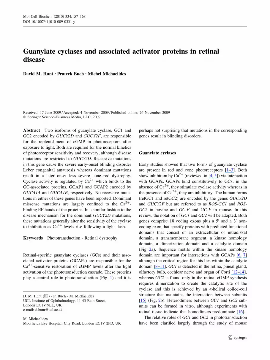
play a central role in phototransduction (Fig. 1) and it is
perhaps not surprising that mutations in the corresponding
genes result in blinding disorders.
Guanylate cyclases
Early studies showed that two forms of guanylate cyclase
are present in rod and cone photoreceptors [1–3]. Both
show inhibition by Ca2? (reviewed in [4, 5]) via interaction
with GCAPs. GCAPs bind constitutively to GCs; in the
absence of Ca2?, they stimulate cyclase activity whereas in
the presence of Ca2?, they are inhibitory. The human forms
(retGC1 and retGC2) are encoded by the genes GUCY2D
and GUCY2F but are referred to as ROS-GC1 and ROS-
GC2 in bovine and GC-E and GC-F in mouse. In this
review, the notation of GC1 and GC2 will be adopted. Both
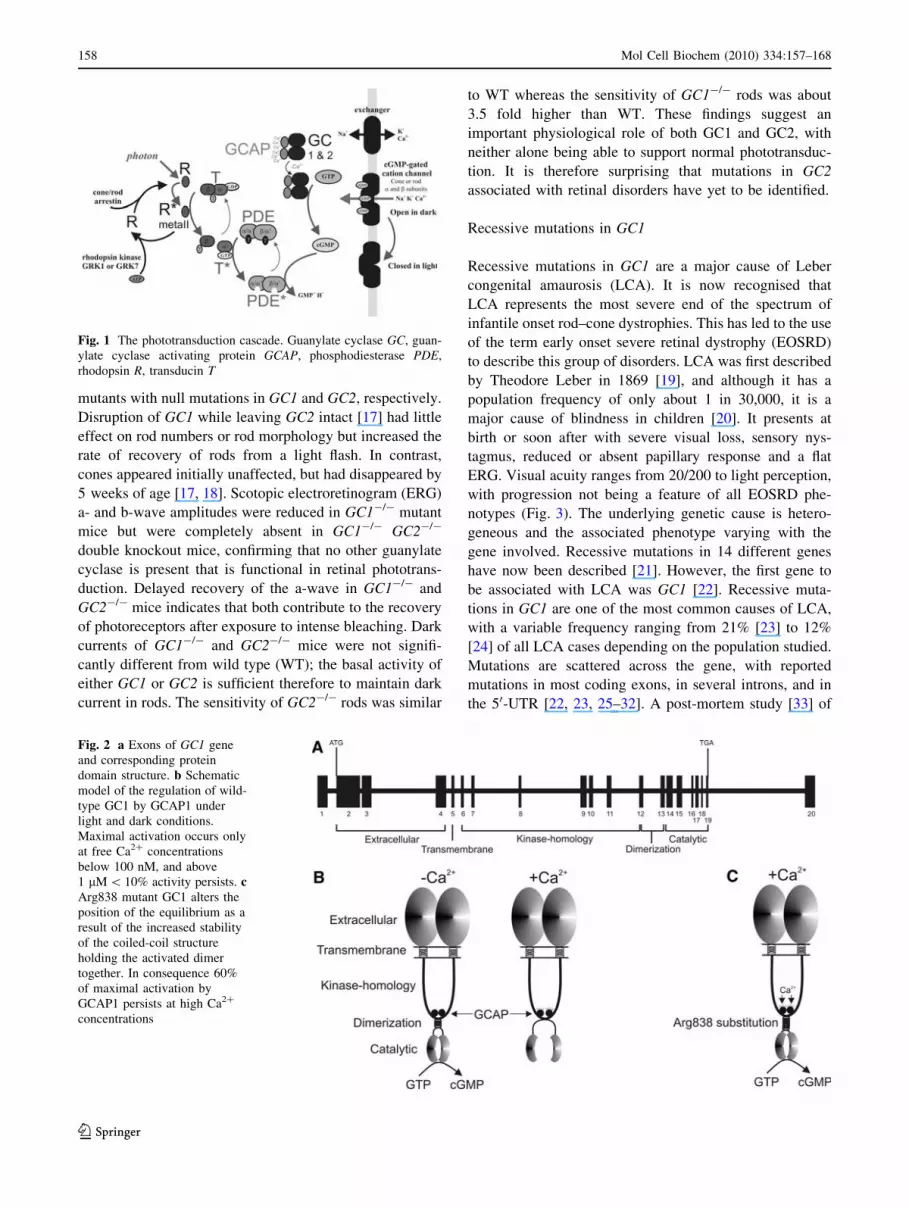
genes comprise 18 coding exons plus a 50 and a 30 non-
coding exon that specify proteins with predicted functional
domains that consist of an extracellular or intradiskal
domain, a transmembrane segment, a kinase homology
domain, a dimerization domain and a catalytic domain
(Fig. 2a). Sequence motifs within the kinase homology
domain are important for interactions with GCAPs [6, 7]
although the critical region for this lies within the catalytic
domain [8–11]. GC1 is detected in the retina, pineal gland,
olfactory bulb, cochlear nerve and organ of Corti [12–14],
whereas GC2 is found only in the retina. cGMP synthesis
requires dimerization to create the catalytic site of the
cyclase and this is achieved by an a-helical coiled-coil
structure that maintains the interaction between subunits
[15] (Fig. 2b). Heterodimers between GC1 and GC2 sub-
units can be formed in vitro, although experiments with
retinal tissue indicate that homodimers predominate [16].
The relative roles of GC1 and GC2 in phototransduction
have been clarified largely through the study of mouse
D. M. Hunt (&) � P. Buch � M. Michaelides
UCL Institute of Ophthalmology, 11-43 Bath Street,
London EC1V 9EL, UK
e-mail: [email protected]
M. Michaelides
Moorfields Eye Hospital, City Road, London EC1V 2PD, UK
123
Mol Cell Biochem (2010) 334:157–168
DOI 10.1007/s11010-009-0331-y
mutants with null mutations in GC1 and GC2, respectively.
Disruption of GC1 while leaving GC2 intact [17] had little
effect on rod numbers or rod morphology but increased the
rate of recovery of rods from a light flash. In contrast,
cones appeared initially unaffected, but had disappeared by
5 weeks of age [17, 18]. Scotopic electroretinogram (ERG)
a- and b-wave amplitudes were reduced in GC1-/- mutant
mice but were completely absent in GC1-/- GC2-/-
double knockout mice, confirming that no other guanylate
cyclase is present that is functional in retinal phototrans-
duction. Delayed recovery of the a-wave in GC1-/- and
GC2-/- mice indicates that both contribute to the recovery
of photoreceptors after exposure to intense bleaching. Dark
currents of GC1-/- and GC2-/- mice were not signifi-
cantly different from wild type (WT); the basal activity of
either GC1 or GC2 is sufficient therefore to maintain dark
current in rods. The sensitivity of GC2-/- rods was similar
to WT whereas the sensitivity of GC1-/- rods was about
3.5 fold higher than WT. These findings suggest an
important physiological role of both GC1 and GC2, with
neither alone being able to support normal phototransduc-
tion. It is therefore surprising that mutations in GC2
associated with retinal disorders have yet to be identified.
Recessive mutations in GC1
Recessive mutations in GC1 are a major cause of Leber
congenital amaurosis (LCA). It is now recognised that
LCA represents the most severe end of the spectrum of
infantile onset rod–cone dystrophies. This has led to the use
of the term early onset severe retinal dystrophy (EOSRD)
to describe this group of disorders. LCA was first described
by Theodore Leber in 1869 [19], and although it has a
population frequency of only about 1 in 30,000, it is a
major cause of blindness in children [20]. It presents at
birth or soon after with severe visual loss, sensory nys-
tagmus, reduced or absent papillary response and a flat
ERG. Visual acuity ranges from 20/200 to light perception,
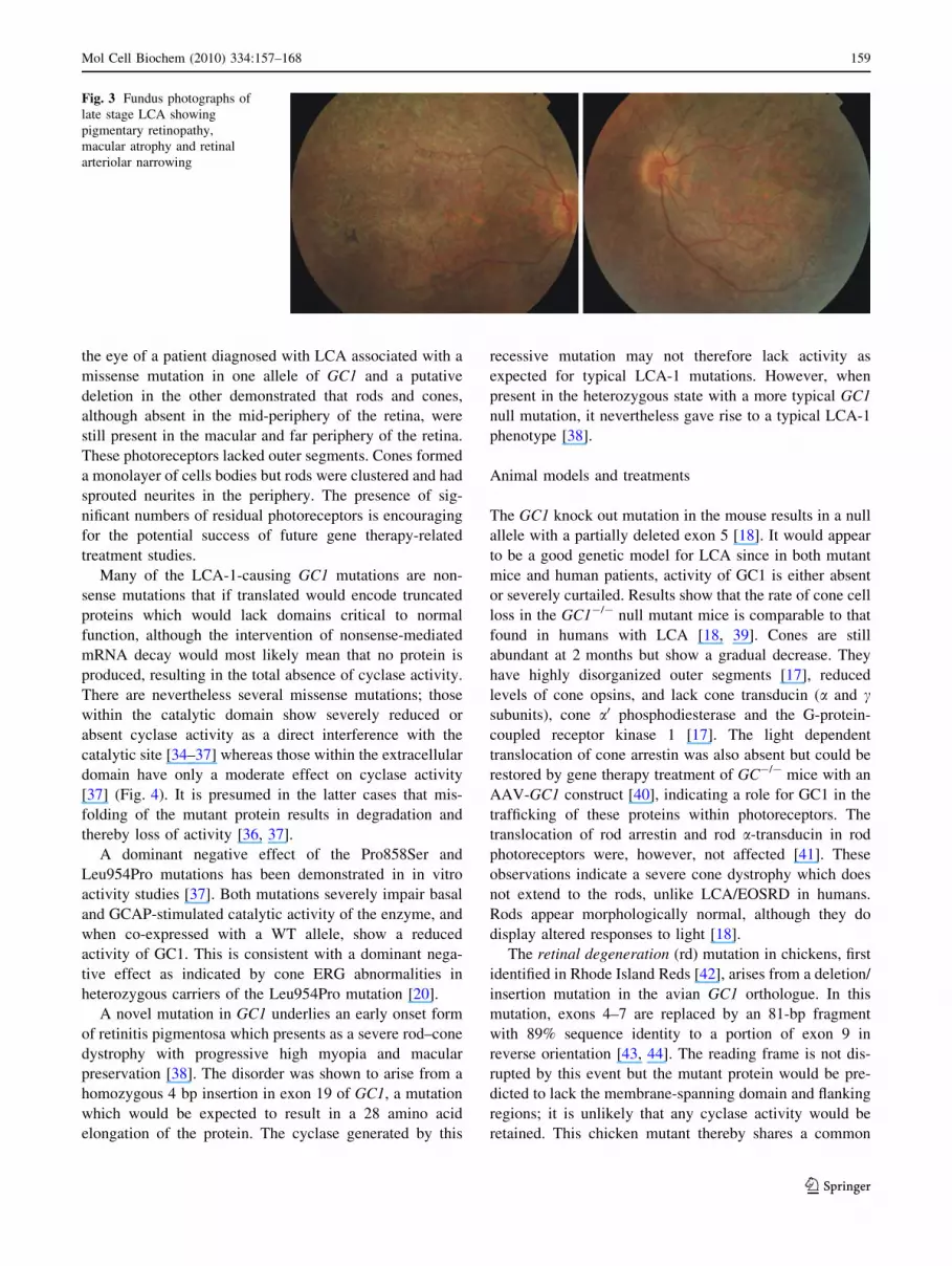
with progression not being a feature of all EOSRD phe-
notypes (Fig. 3). The underlying genetic cause is hetero-
geneous and the associated phenotype varying with the
gene involved. Recessive mutations in 14 different genes
have now been described [21]. However, the first gene to
be associated with LCA was GC1 [22]. Recessive muta-
tions in GC1 are one of the most common causes of LCA,
with a variable frequency ranging from 21% [23] to 12%
[24] of all LCA cases depending on the population studied.
Mutations are scattered across the gene, with reported
mutations in most coding exons, in several introns, and in
the 50-UTR [22, 23, 25–32]. A post-mortem study [33] of
Fig. 2 a Exons of GC1 gene
and corresponding protein
domain structure. b Schematic
model of the regulation of wild-
type GC1 by GCAP1 under
light and dark conditions.
Maximal activation occurs only
at free Ca2? concentrations
below 100 nM, and above
1 lM \ 10% activity persists. cArg838 mutant GC1 alters the
position of the equilibrium as a
result of the increased stability
of the coiled-coil structure
holding the activated dimer
together. In consequence 60%
of maximal activation by
GCAP1 persists at high Ca2?
concentrations
Fig. 1 The phototransduction cascade. Guanylate cyclase GC, guan-
ylate cyclase activating protein GCAP, phosphodiesterase PDE,
rhodopsin R, transducin T
158 Mol Cell Biochem (2010) 334:157–168
123
the eye of a patient diagnosed with LCA associated with a
missense mutation in one allele of GC1 and a putative
deletion in the other demonstrated that rods and cones,
although absent in the mid-periphery of the retina, were
still present in the macular and far periphery of the retina.
These photoreceptors lacked outer segments. Cones formed
a monolayer of cells bodies but rods were clustered and had
sprouted neurites in the periphery. The presence of sig-
nificant numbers of residual photoreceptors is encouraging
for the potential success of future gene therapy-related
treatment studies.
Many of the LCA-1-causing GC1 mutations are non-
sense mutations that if translated would encode truncated
proteins which would lack domains critical to normal
function, although the intervention of nonsense-mediated
mRNA decay would most likely mean that no protein is
produced, resulting in the total absence of cyclase activity.
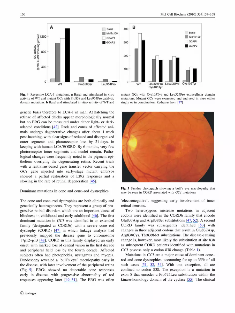
There are nevertheless several missense mutations; those
within the catalytic domain show severely reduced or
absent cyclase activity as a direct interference with the
catalytic site [34–37] whereas those within the extracellular
domain have only a moderate effect on cyclase activity
[37] (Fig. 4). It is presumed in the latter cases that mis-
folding of the mutant protein results in degradation and
thereby loss of activity [36, 37].
A dominant negative effect of the Pro858Ser and
Leu954Pro mutations has been demonstrated in in vitro
activity studies [37]. Both mutations severely impair basal
and GCAP-stimulated catalytic activity of the enzyme, and
when co-expressed with a WT allele, show a reduced
activity of GC1. This is consistent with a dominant nega-
tive effect as indicated by cone ERG abnormalities in
heterozygous carriers of the Leu954Pro mutation [20].
A novel mutation in GC1 underlies an early onset form
of retinitis pigmentosa which presents as a severe rod–cone
dystrophy with progressive high myopia and macular
preservation [38]. The disorder was shown to arise from a
homozygous 4 bp insertion in exon 19 of GC1, a mutation
which would be expected to result in a 28 amino acid
elongation of the protein. The cyclase generated by this
recessive mutation may not therefore lack activity as
expected for typical LCA-1 mutations. However, when
present in the heterozygous state with a more typical GC1
null mutation, it nevertheless gave rise to a typical LCA-1
phenotype [38].
Animal models and treatments
The GC1 knock out mutation in the mouse results in a null
allele with a partially deleted exon 5 [18]. It would appear
to be a good genetic model for LCA since in both mutant
mice and human patients, activity of GC1 is either absent
or severely curtailed. Results show that the rate of cone cell
loss in the GC1-/- null mutant mice is comparable to that
found in humans with LCA [18, 39]. Cones are still
abundant at 2 months but show a gradual decrease. They
have highly disorganized outer segments [17], reduced
levels of cone opsins, and lack cone transducin (a and csubunits), cone a0 phosphodiesterase and the G-protein-
coupled receptor kinase 1 [17]. The light dependent
translocation of cone arrestin was also absent but could be
restored by gene therapy treatment of GC-/- mice with an
AAV-GC1 construct [40], indicating a role for GC1 in the
trafficking of these proteins within photoreceptors. The
translocation of rod arrestin and rod a-transducin in rod
photoreceptors were, however, not affected [41]. These
observations indicate a severe cone dystrophy which does
not extend to the rods, unlike LCA/EOSRD in humans.
Rods appear morphologically normal, although they do
display altered responses to light [18].
The retinal degeneration (rd) mutation in chickens, first
identified in Rhode Island Reds [42], arises from a deletion/
insertion mutation in the avian GC1 orthologue. In this
mutation, exons 4–7 are replaced by an 81-bp fragment
with 89% sequence identity to a portion of exon 9 in
reverse orientation [43, 44]. The reading frame is not dis-
rupted by this event but the mutant protein would be pre-
dicted to lack the membrane-spanning domain and flanking
regions; it is unlikely that any cyclase activity would be
retained. This chicken mutant thereby shares a common
Fig. 3 Fundus photographs of
late stage LCA showing
pigmentary retinopathy,
macular atrophy and retinal
arteriolar narrowing
Mol Cell Biochem (2010) 334:157–168 159
123
genetic basis therefore to LCA-1 in man. At hatching the
retinae of affected chicks appear morphologically normal
but no ERG can be measured under either light- or dark-
adapted conditions [42]. Rods and cones of affected ani-
mals undergo degenerative changes after about 1 week
post-hatching, with clear signs of reduced and disorganized
outer segments and photoreceptor loss by 21 days, in
keeping with human LCA/EOSRD. By 6 months, very few
photoreceptor inner segments and nuclei remain. Patho-
logical changes were frequently noted in the pigment epi-
thelium overlying the degenerating retina. Recent trials
with a lentivirus-based gene transfer vector carrying the
GC1 gene injected into early-stage mutant embryos
showed a partial restoration of ERG responses and a
slowing in the rate of retinal degeneration [45].
Dominant mutations in cone and cone–rod dystrophies
The cone and cone–rod dystrophies are both clinically and
genetically heterogeneous. They represent a group of pro-
gressive retinal disorders which are an important cause of
blindness in childhood and early adulthood [46]. The first
dominant mutation in GC1 was identified in an extended
family (designated as CORD6) with a severe cone–rod
dystrophy (CORD) [47] in which linkage analysis had
previously mapped the disease gene to chromosome
17p12–p13 [48]. CORD in this family displayed an early
onset, with marked loss of central vision in the first decade
and peripheral field loss by the fourth decade. Affected
subjects often had photophobia, nystagmus and myopia.
Fundoscopy revealed a ‘bull’s eye’ maculopathy early in
the disease, with later involvement of the peripheral retina
(Fig. 5). ERGs showed no detectable cone responses
early in disease, with progressive abnormality of rod
responses appearing later [49–51]. The ERG was often
‘electronegative’, suggesting early involvement of inner
retinal neurons.
Two heterozygous missense mutations in adjacent
codons were identified in the CORD6 family that encode
Glu837Asp and Arg838Ser substitutions [47, 52]. A second
CORD family was subsequently identified [53] with
changes in three adjacent codons that result in Glu837Asp,
Arg838Cys, Thr839Met substitutions. The disease-causing
change is, however, most likely the substitution at site 838
as subsequent CORD patients identified with mutations in
GC1 possess only a codon 838 change (Table 1).
Mutations in GC1 are a major cause of dominant cone–
rod and cone dystrophies, accounting for up to 35% of all
such cases [51, 52, 54]. With one exception, all are
confined to codon 838. The exception is a mutation in
exon 8 that encodes a Pro575Leu substitution within the
kinase-homology domain of the cyclase [55]. The clinical
Fig. 4 Recessive LCA-1 mutations. a Basal and stimulated in vitro
activity of WT and mutant GCs with Pro858 and Leu954Pro catalytic
domain mutations. b Basal and stimulated in vitro activity of WT and
mutant GCs with Cys105Tyr and Leu325Pro extracellular domain
mutations. Mutant GCs were expressed and analysed in vitro either
singly or in combination. Redrawn from [37]
Fig. 5 Fundus photograph showing a bull’s eye maculopathy that
may be seen in CORD associated with GC1 mutations
160 Mol Cell Biochem (2010) 334:157–168
123
phenotype of this disorder is more accurately described as a
dominant cone dystrophy as scotopic (rod) function was
normal but photopic function was generally absent.
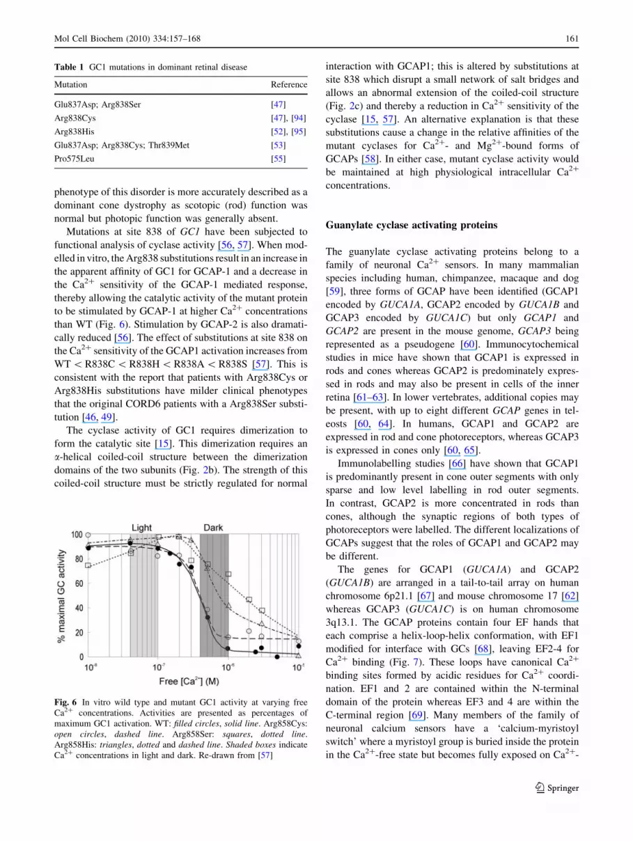
Mutations at site 838 of GC1 have been subjected to
functional analysis of cyclase activity [56, 57]. When mod-
elled in vitro, the Arg838 substitutions result in an increase in
the apparent affinity of GC1 for GCAP-1 and a decrease in
the Ca2? sensitivity of the GCAP-1 mediated response,
thereby allowing the catalytic activity of the mutant protein
to be stimulated by GCAP-1 at higher Ca2? concentrations
than WT (Fig. 6). Stimulation by GCAP-2 is also dramati-
cally reduced [56]. The effect of substitutions at site 838 on
the Ca2? sensitivity of the GCAP1 activation increases from
WT \ R838C \ R838H \ R838A \ R838S [57]. This is
consistent with the report that patients with Arg838Cys or
Arg838His substitutions have milder clinical phenotypes
that the original CORD6 patients with a Arg838Ser substi-
tution [46, 49].
The cyclase activity of GC1 requires dimerization to
form the catalytic site [15]. This dimerization requires an
a-helical coiled-coil structure between the dimerization
domains of the two subunits (Fig. 2b). The strength of this
coiled-coil structure must be strictly regulated for normal
interaction with GCAP1; this is altered by substitutions at
site 838 which disrupt a small network of salt bridges and
allows an abnormal extension of the coiled-coil structure
(Fig. 2c) and thereby a reduction in Ca2? sensitivity of the
cyclase [15, 57]. An alternative explanation is that these
substitutions cause a change in the relative affinities of the
mutant cyclases for Ca2?- and Mg2?-bound forms of
GCAPs [58]. In either case, mutant cyclase activity would
be maintained at high physiological intracellular Ca2?
concentrations.
Guanylate cyclase activating proteins
The guanylate cyclase activating proteins belong to a
family of neuronal Ca2? sensors. In many mammalian
species including human, chimpanzee, macaque and dog
[59], three forms of GCAP have been identified (GCAP1
encoded by GUCA1A, GCAP2 encoded by GUCA1B and
GCAP3 encoded by GUCA1C) but only GCAP1 and
GCAP2 are present in the mouse genome, GCAP3 being
represented as a pseudogene [60]. Immunocytochemical
studies in mice have shown that GCAP1 is expressed in
rods and cones whereas GCAP2 is predominately expres-
sed in rods and may also be present in cells of the inner
retina [61–63]. In lower vertebrates, additional copies may
be present, with up to eight different GCAP genes in tel-
eosts [60, 64]. In humans, GCAP1 and GCAP2 are
expressed in rod and cone photoreceptors, whereas GCAP3
is expressed in cones only [60, 65].
Immunolabelling studies [66] have shown that GCAP1
is predominantly present in cone outer segments with only
sparse and low level labelling in rod outer segments.
In contrast, GCAP2 is more concentrated in rods than
cones, although the synaptic regions of both types of
photoreceptors were labelled. The different localizations of
GCAPs suggest that the roles of GCAP1 and GCAP2 may
be different.
The genes for GCAP1 (GUCA1A) and GCAP2
(GUCA1B) are arranged in a tail-to-tail array on human
chromosome 6p21.1 [67] and mouse chromosome 17 [62]
whereas GCAP3 (GUCA1C) is on human chromosome
3q13.1. The GCAP proteins contain four EF hands that
each comprise a helix-loop-helix conformation, with EF1
modified for interface with GCs [68], leaving EF2-4 for
Ca2? binding (Fig. 7). These loops have canonical Ca2?
binding sites formed by acidic residues for Ca2? coordi-
nation. EF1 and 2 are contained within the N-terminal
domain of the protein whereas EF3 and 4 are within the
C-terminal region [69]. Many members of the family of
neuronal calcium sensors have a ‘calcium-myristoyl
switch’ where a myristoyl group is buried inside the protein
in the Ca2?-free state but becomes fully exposed on Ca2?-
Table 1 GC1 mutations in dominant retinal disease
Mutation Reference
Glu837Asp; Arg838Ser [47]
Arg838Cys [47], [94]
Arg838His [52], [95]
Glu837Asp; Arg838Cys; Thr839Met [53]
Pro575Leu [55]
Fig. 6 In vitro wild type and mutant GC1 activity at varying free
Ca2? concentrations. Activities are presented as percentages of
maximum GC1 activation. WT: filled circles, solid line. Arg858Cys:
open circles, dashed line. Arg858Ser: squares, dotted line.
Arg858His: triangles, dotted and dashed line. Shaded boxes indicate
Ca2? concentrations in light and dark. Re-drawn from [57]
Mol Cell Biochem (2010) 334:157–168 161
123
binding. However, GCAP2 has been shown to lack a Ca2?
-myristoyl switch [70, 71] and recent studies on the con-
formational structure of GCAP1 indicates that the myri-
stoyl group remains fully buried in both the Ca2?-free and
Ca2?-bound state [72].
The relative roles of GCAP1 and GCAP2 have been lar-
gely determined by studies with mouse knockout mutations.
In mice in which both genes have been disrupted, GC activity
lacks Ca2? dependence, confirming the role of GCAPs in the
Ca2? regulation [73]. In these mice, flash responses from
dark-adapted rods are larger and slower than WT, and
incremental flash sensitivity of rods fails to be maintained at
a WT type level. Restoration of GCAP2 production via a
transgene restores maximal light-induced GC activity but
does not restore normal kinetics to responses evoked by
saturating flashes [73]. In contrast, the provision of GCAP1
via a transgene shows that the degree of recovery of the rod a-
wave is correlated with the level of expression of GCAP1. In
single cell recordings, the majority of rods generated flash
responses identical to WT [74].
A more direct study of the role of GCAP2 on photo-
transduction has been carried out in mice with a disrupted
GCAP2 gene but retaining a fully functional GCAP1 gene
[75]. In these mice, rod viability and outer segment mor-
phology appeared normal, and there were no compensatory
changes in the levels of GC1, GC2 or GCAP1. However,
there was a two-fold drop in the maximal rate of cGMP
synthesis at low Ca2? concentrations, and an increase in
the Ca2? concentration at which the half-maximal rate of
cGMP synthesis was achieved. Flash responses recovered
more slowly than WT and rods were more sensitive to
flashes but tended to saturate at lower intensities. GCAP2 is
necessary therefore for normal recovery of rods and for
light adaptation; for the rapid activation and deactivation of
GC activity in response to light, both isoforms of GCAP
are required.
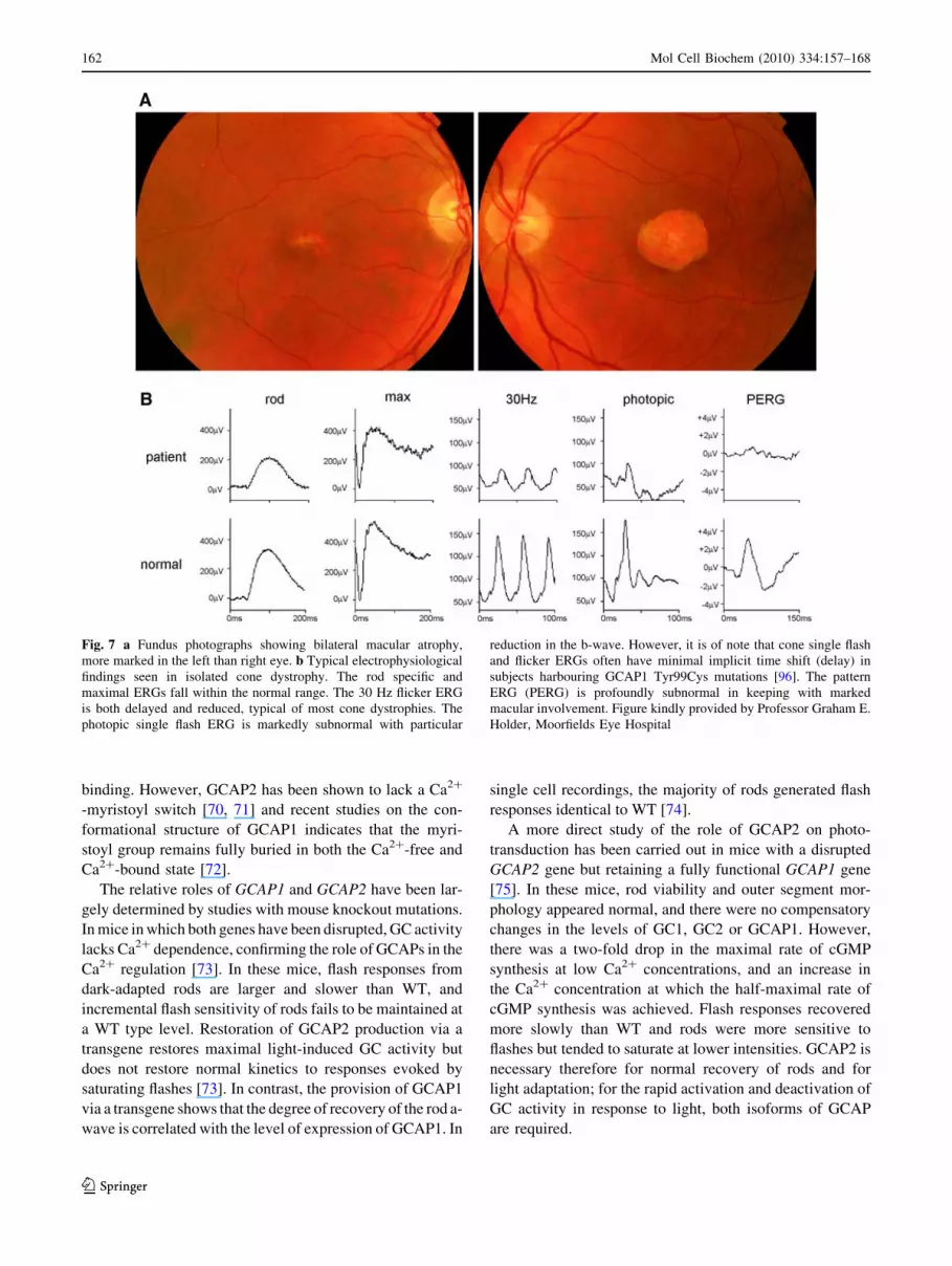
Fig. 7 a Fundus photographs showing bilateral macular atrophy,
more marked in the left than right eye. b Typical electrophysiological
findings seen in isolated cone dystrophy. The rod specific and
maximal ERGs fall within the normal range. The 30 Hz flicker ERG
is both delayed and reduced, typical of most cone dystrophies. The
photopic single flash ERG is markedly subnormal with particular
reduction in the b-wave. However, it is of note that cone single flash
and flicker ERGs often have minimal implicit time shift (delay) in
subjects harbouring GCAP1 Tyr99Cys mutations [96]. The pattern
ERG (PERG) is profoundly subnormal in keeping with marked
macular involvement. Figure kindly provided by Professor Graham E.
Holder, Moorfields Eye Hospital
162 Mol Cell Biochem (2010) 334:157–168
123
Cone responses are also affected by GCAP-knockout
mutations. Under cone isolation conditions, ERGs recorded
from mice lacking both GCAP1 and GCAP2 had normal
amplitudes of the saturated a-wave and b-wave [76].
However, the b-wave was widened, there was an increase in
the sensitivity of both M- and UV-cones, and the recoveries
of the cone-driven a-wave and b-wave were delayed. Res-
toration of GCAP1 via a transgene showed that recovery of
the cone-driven a-wave was restored to normal, although
the recovery of the cone-driven b-wave was slightly faster
than that observed in WT mice. These studies reveal that the
absence of GCAP1 and GCAP2 delays the recovery of light
responses, and that GCAP1 restores the recovery of cone
responses in the absence of GCAP2 [76], suggesting a more
important role for GCAP1 in cone phototransduction, con-
sistent with the proposal that GCAP1 is more highly
expressed in cones than in rods [66].
Mutations in GCAPs
No mutations in GCAP3 have so far been found in retinal
disease [77]. A dominant Gly157Arg missense mutation in
GCAP2 was identified in three independent families with
incomplete penetrance and a spectrum of disease pheno-
types that included retinitis pigmentosa (RP), RP with
macular involvement and macular degeneration [78]. The
first reported mutation in GCAP1 was a missense mutation
in codon 99 that results in the replacement of a highly
conserved Tyr with Cys (Tyr99Cys) [79]. Other reported
mutations are listed in Table 2. The clinical phenotype for
Tyr99Cys patients was initially reported as an isolated
progressive cone dystrophy [79] and this is a feature of
many of the other GCAP1 mutations [46], although a
recent clinical re-evaluation of the original family with the
Tyr99Cys mutation [80] found a range of phenotypes that
included reduced cone and rod responses (with cone loss
greater than rod, in keeping with CORD), and isolated
macular dysfunction (Fig. 7). This demonstrates the extent
of intrafamilial heterogeneity in retinal dysfunction that
may be present in persons with the same gene mutation. All
affected subjects complained of mild photophobia and
reduced central vision and colour vision. Onset is between
the third and fifth decade, with subsequent gradual deteri-
oration of visual acuity and colour vision.
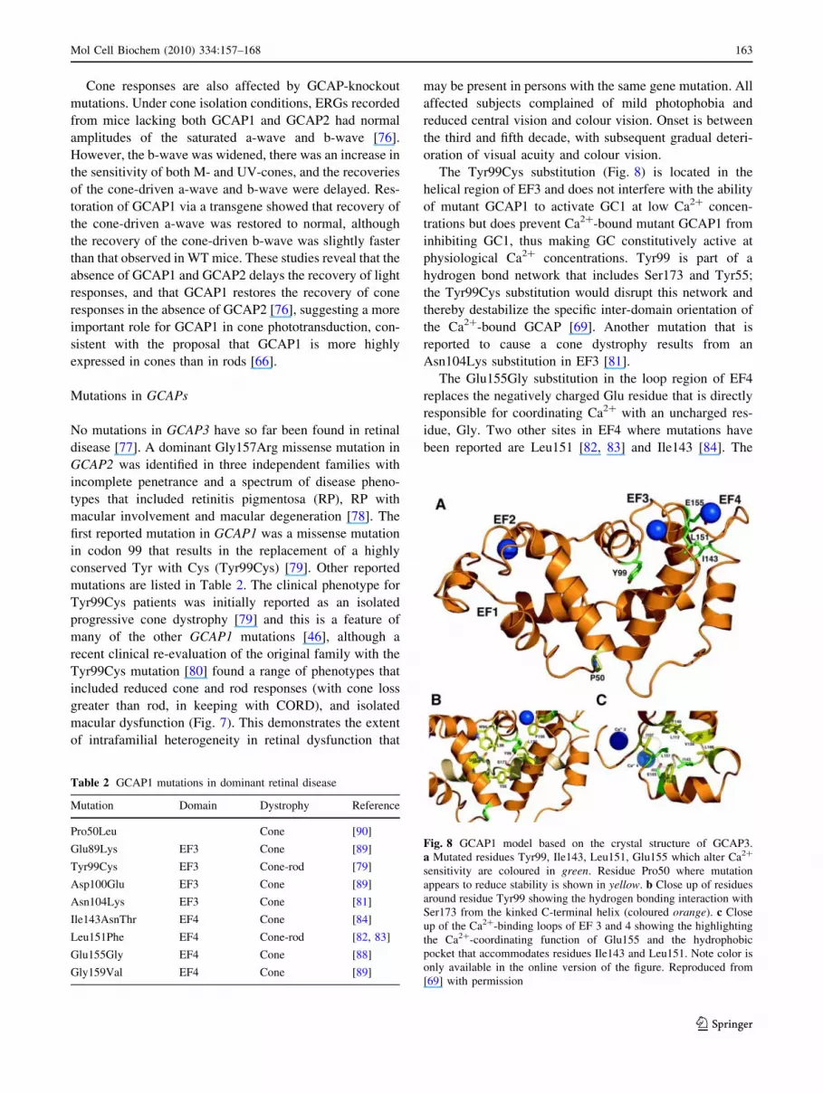
The Tyr99Cys substitution (Fig. 8) is located in the
helical region of EF3 and does not interfere with the ability
of mutant GCAP1 to activate GC1 at low Ca2? concen-
trations but does prevent Ca2?-bound mutant GCAP1 from
inhibiting GC1, thus making GC constitutively active at
physiological Ca2? concentrations. Tyr99 is part of a
hydrogen bond network that includes Ser173 and Tyr55;
the Tyr99Cys substitution would disrupt this network and
thereby destabilize the specific inter-domain orientation of
the Ca2?-bound GCAP [69]. Another mutation that is
reported to cause a cone dystrophy results from an
Asn104Lys substitution in EF3 [81].
The Glu155Gly substitution in the loop region of EF4
replaces the negatively charged Glu residue that is directly
responsible for coordinating Ca2? with an uncharged res-
idue, Gly. Two other sites in EF4 where mutations have
been reported are Leu151 [82, 83] and Ile143 [84]. The
Table 2 GCAP1 mutations in dominant retinal disease
Mutation Domain Dystrophy Reference
Pro50Leu Cone [90]
Glu89Lys EF3 Cone [89]
Tyr99Cys EF3 Cone-rod [79]
Asp100Glu EF3 Cone [89]
Asn104Lys EF3 Cone [81]
Ile143AsnThr EF4 Cone [84]
Leu151Phe EF4 Cone-rod [82, 83]
Glu155Gly EF4 Cone [88]
Gly159Val EF4 Cone [89]
Fig. 8 GCAP1 model based on the crystal structure of GCAP3.
a Mutated residues Tyr99, Ile143, Leu151, Glu155 which alter Ca2?
sensitivity are coloured in green. Residue Pro50 where mutation
appears to reduce stability is shown in yellow. b Close up of residues
around residue Tyr99 showing the hydrogen bonding interaction with
Ser173 from the kinked C-terminal helix (coloured orange). c Close
up of the Ca2?-binding loops of EF 3 and 4 showing the highlighting
the Ca2?-coordinating function of Glu155 and the hydrophobic
pocket that accommodates residues Ile143 and Leu151. Note color is
only available in the online version of the figure. Reproduced from
[69] with permission
Mol Cell Biochem (2010) 334:157–168 163
123
former is a simple point mutation generating a Leu151Phe
substitution, but the latter is a complex change involving a
single bp deletion in codon 143 followed by a 4 bp inser-
tion which results in the substitution of Ile by Asn and the
insertion of Thr (Ile143AsnThr) [84]. This mutation was
identified in two patients (father and son) with dominant
cone degeneration. A histopathological evaluation of the
father’s eyes at autopsy (age 75 years) showed no foveal
cones but a few, scattered cones remaining in the periph-
eral retina. The side-chains of Ile143 and Leu151 lie in the
hydrophobic pocket that stabilizes the conformational
structure of EF4 and the replacement by Asn/Thr and Phe,
respectively, may interfere with the hydrophobic core and
Ca2?-binding loop of EF4 [69].
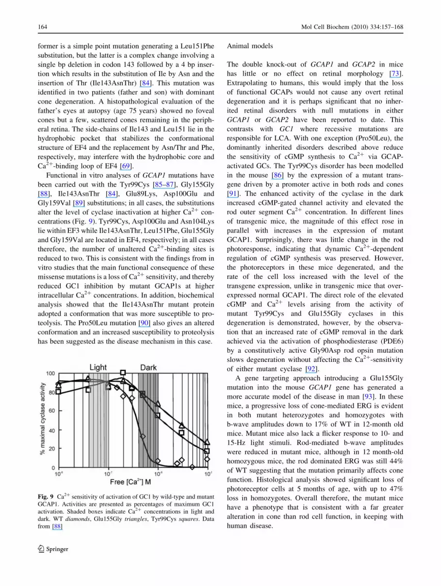
Functional in vitro analyses of GCAP1 mutations have
been carried out with the Tyr99Cys [85–87], Gly155Gly
[88], Ile143AsnThr [84], Glu89Lys, Asp100Glu and
Gly159Val [89] substitutions; in all cases, the substitutions
alter the level of cyclase inactivation at higher Ca2? con-
centrations (Fig. 9). Tyr99Cys, Asp100Glu and Asn104Lys
lie within EF3 while Ile143AsnThr, Leu151Phe, Glu155Gly
and Gly159Val are located in EF4, respectively; in all cases
therefore, the number of unaltered Ca2?-binding sites is
reduced to two. This is consistent with the findings from in
vitro studies that the main functional consequence of these
missense mutations is a loss of Ca2? sensitivity, and thereby
reduced GC1 inhibition by mutant GCAP1s at higher
intracellular Ca2? concentrations. In addition, biochemical
analysis showed that the Ile143AsnThr mutant protein
adopted a conformation that was more susceptible to pro-
teolysis. The Pro50Leu mutation [90] also gives an altered
conformation and an increased susceptibility to proteolysis
has been suggested as the disease mechanism in this case.
Animal models
The double knock-out of GCAP1 and GCAP2 in mice
has little or no effect on retinal morphology [73].
Extrapolating to humans, this would imply that the loss
of functional GCAPs would not cause any overt retinal
degeneration and it is perhaps significant that no inher-
ited retinal disorders with null mutations in either
GCAP1 or GCAP2 have been reported to date. This
contrasts with GC1 where recessive mutations are
responsible for LCA. With one exception (Pro50Leu), the
dominantly inherited disorders described above reduce
the sensitivity of cGMP synthesis to Ca2? via GCAP-
activated GCs. The Tyr99Cys disorder has been modelled
in the mouse [86] by the expression of a mutant trans-
gene driven by a promoter active in both rods and cones
[91]. The enhanced activity of the cyclase in the dark
increased cGMP-gated channel activity and elevated the
rod outer segment Ca2? concentration. In different lines
of transgenic mice, the magnitude of this effect rose in
parallel with increases in the expression of mutant
GCAP1. Surprisingly, there was little change in the rod
photoresponse, indicating that dynamic Ca2?-dependent
regulation of cGMP synthesis was preserved. However,
the photoreceptors in these mice degenerated, and the
rate of the cell loss increased with the level of the
transgene expression, unlike in transgenic mice that over-
expressed normal GCAP1. The direct role of the elevated
cGMP and Ca2? levels arising from the activity of
mutant Tyr99Cys and Glu155Gly cyclases in this
degeneration is demonstrated, however, by the observa-
tion that an increased rate of cGMP removal in the dark
achieved via the activation of phosphodiesterase (PDE6)
by a constitutively active Gly90Asp rod opsin mutation
slows degeneration without affecting the Ca2?-sensitivity
of either mutant cyclase [92].
A gene targeting approach introducing a Glu155Gly
mutation into the mouse GCAP1 gene has generated a
more accurate model of the disease in man [93]. In these
mice, a progressive loss of cone-mediated ERG is evident
in both mutant heterozygotes and homozygotes with
b-wave amplitudes down to 17% of WT in 12-month old
mice. Mutant mice also lack a flicker response to 10- and
15-Hz light stimuli. Rod-mediated b-wave amplitudes
were reduced in mutant mice, although in 12 month-old
homozygous mice, the rod dominated ERG was still 44%
of WT suggesting that the mutation primarily affects cone
function. Histological analysis showed significant loss of
photoreceptor cells at 5 months of age, with up to 47%
loss in homozygotes. Overall therefore, the mutant mice
have a phenotype that is consistent with a far greater
alteration in cone than rod cell function, in keeping with
human disease.
Fig. 9 Ca2? sensitivity of activation of GC1 by wild-type and mutant
GCAP1. Activities are presented as percentages of maximum GC1
activation. Shaded boxes indicate Ca2? concentrations in light and
dark. WT diamonds, Glu155Gly triangles, Tyr99Cys squares. Data
from [88]
164 Mol Cell Biochem (2010) 334:157–168
123
Conclusions
• Both GC1 and GC2 are required for normal photo-
transduction. Cone photoreceptors die in the GC1
knock-out mouse whereas there is no loss of rods. This
suggests a more critical role for GC1 in the protection
of cones than rods.
• No disease-associated mutations have been reported for
GC2.
• Recessive mutations in GC1 are a major cause of LCA
in humans. The mechanism of mutant gene action
would appear to arise from the loss of functional
cyclase.
• Dominant mutations in GC1 are a major cause of
inherited CORD. Mutations are clustered in codon 838
with replacement of Arg with either Cys, Ser or His.
The mutant cyclase remains functional but with a
reduced sensitivity to Ca2?-dependent inhibition.
• GCAP1 and GCAP2 are both required for normal
phototransduction. The pattern of expression differs,
with GCAP1 predominantly present in cones and
GCAP2 more concentrated in rods.
• In mouse models, the double deletion of GCAP1 and
GCAP2 does not result in photoreceptor loss. Consis-
tent with this, no recessive disease-associated mutations
for either gene have been reported in humans.
• A single dominant missense mutation in GCAP2 is
associated with incomplete penetrance and a spectrum
of disease phenotypes that includes RP with and without
macular involvement, and macular degeneration.
• Dominant mutations in GCAP1 result in either cone or
CORDs and this may reflect the higher levels of
GCAP1 in cones.
• Mutant GCAP1 proteins are functional but generally
have a reduced capacity to bind Ca2? as a result of
substitutions in the EF hands responsible for Ca2?
binding. As a consequence, cyclase activity is less
sensitive to inhibition at higher Ca2? concentrations.
• A novel knock-in mouse model of a dominant human
GCAP1 mutation shows significant cone loss with a
more limited effect on rod survival and function.
References
1. Goraczniak R, Duda T, Sharma RK (1997) Structural and func-
tional characterization of a second subfamily member of the
calcium-modulated bovine rod outer segment membrane guany-
late cyclase, ROS-GC2. Biochem Biophys Res Commun
234:666–670
2. Goraczniak RM, Duda T, Sitaramayya A, Sharma RK (1994)
Structural and functional characterization of the rod outer seg-
ment membrane guanylate cyclase. Biochem J 302:455–461
3. Yang RB, Foster DC, Garbers DL, Fulle HJ (1995) Two mem-
brane forms of guanylyl cyclase found in the eye. Proc Natl Acad
Sci USA 92:602–606
4. Pugh EN Jr, Duda T, Sitaramayya A, Sharma RK (1997) Pho-
toreceptor guanylate cyclases: a review. Biosci Rep 17:429–473
5. Dizhoor AM, Hurley JB (1999) Regulation of photoreceptor
membrane guanylyl cyclases by guanylyl cyclase activator pro-
teins. Methods 19:521–531
6. Lange C, Duda T, Beyermann M, Sharma RK, Koch KW (1999)
Regions in vertebrate photoreceptor guanylyl cyclase ROS-GC1
involved in Ca(2?)-dependent regulation by guanylyl cyclase-
activating protein GCAP-1. FEBS Lett 460:27–31
7. Krylov DM, Hurley JB (2001) Identification of proximate regions
in a complex of retinal guanylyl cyclase 1 and guanylyl cyclase-
activating protein-1 by a novel mass spectrometry-based method.
J Biol Chem 276:30648–30654
8. Sokal I, Haeseleer F, Arendt A, Adman ET, Hargrave PA, Pal-
czewski K (1999) Identification of a guanylyl cyclase-activating
protein-binding site within the catalytic domain of retinal gua-
nylyl cyclase 1. Biochemistry 38:1387–1393
9. Duda T, Goraczniak R, Surgucheva I, Rudnicka-Nawrot M,
Gorczyca WA, Palczewski K, Sitaramayya A, Baehr W, Sharma
RK (1996) Calcium modulation of bovine photoreceptor guany-
late cyclase. Biochemistry 35:8478–8482
10. Laura RP, Hurley JB (1998) The kinase homology domain of
retinal guanylyl cyclases 1 and 2 specifies the affinity and
cooperativity of interaction with guanylyl cyclase activating
protein-2. Biochemistry 37:11264–11271
11. Tucker CL, Laura RP, Hurley JB (1997) Domain-specific sta-
bilization of photoreceptor membrane guanylyl cyclase by ade-
nine nucleotides and guanylyl cyclase activating proteins
(GCAPs). Biochemistry 36:11995–12000
12. Duda T, Koch KW (2002) Retinal diseases linked with photo-
receptor guanylate cyclase. Mol Cell Biochem 230:129–138
13. Seebacher T, Beitz E, Kumagami H, Wild K, Ruppersberg JP,
Schultz JE (1999) Expression of membrane-bound and cytosolic
guanylyl cyclases in the rat inner ear. Hear Res 127:95–102
14. Venkataraman V, Nagele R, Duda T, Sharma RK (2000) Rod
outer segment membrane guanylate cyclase type 1-linked stim-
ulatory and inhibitory calcium signaling systems in the pineal
gland: biochemical, molecular, and immunohistochemical evi-
dence. Biochemistry 39:6042–6052
15. Ramamurthy V, Tucker C, Wilkie SE, Daggett V, Hunt DM,
Hurley JB (2001) Interactions within the coiled-coil domain of
RetGC-1 guanylyl cyclase are optimized for regulation rather
than for high affinity. J Biol Chem 276:26218–26229
16. Yang RB, Garbers DL (1997) Two eye guanylyl cyclases are
expressed in the same photoreceptor cells and form homomers in
preference to heteromers. J Biol Chem 272:13738–13742
17. Baehr W, Karan S, Maeda T, Luo DG, Li S, Bronson JD, Watt
CB, Yau KW, Frederick JM, Palczewski K (2007) The function
of guanylate cyclase 1 and guanylate cyclase 2 in rod and cone
photoreceptors. J Biol Chem 282:8837–8847
18. Yang RB, Robinson SW, Xiong WH, Yau KW, Birch DG,
Garbers DL (1999) Disruption of a retinal guanylyl cyclase gene
leads to cone-specific dystrophy and paradoxical rod behavior.
J Neurosci 19:5889–5897
19. Leber T (1869) Ueber Retinitis pigmentosa und angeborene
Amaurose. Albrecht von Graefes Arch Ophthal 15:1–25
20. Koenekoop RK (2004) An overview of Leber congenital amau-
rosis: a model to understand human retinal development. Surv
Ophthalmol 49:379–398
21. Kaplan J (2008) Leber congenital amaurosis: from darkness to
spotlight. Ophthalmic Genet 29:92–98
22. Perrault I, Rozet JM, Calvas P, Gerber S, Camuzat A, Dollfus H,
Chatelin S, Souied E, Ghazi I, Leowski C, Bonnemaison M, Le
Paslier D, Frezal J, Dufier JL, Pittler S, Munnich A, Kaplan J
(1996) Retinal-specific guanylate cyclase gene mutations in
Leber’s congenital amaurosis. Nat Genet 14:461–464
Mol Cell Biochem (2010) 334:157–168 165
123
23. Hanein S, Perrault I, Gerber S, Tanguy G, Barbet F, Ducroq D,
Calvas P, Dollfus H, Hamel C, Lopponen T, Munier F, Santos L,
Shalev S, Zafeiriou D, Dufier JL, Munnich A, Rozet JM, Kaplan J
(2004) Leber congenital amaurosis: comprehensive survey of the
genetic heterogeneity, refinement of the clinical definition, and
genotype-phenotype correlations as a strategy for molecular
diagnosis. Hum Mutat 23:306–317
24. den Hollander AI, Roepman R, Koenekoop RK, Cremers FP
(2008) Leber congenital amaurosis: genes, proteins and disease
mechanisms. Prog Retin Eye Res 27:391–419
25. Dharmaraj SR, Silva ER, Pina AL, Li YY, Yang JM, Carter CR,
Loyer MK, El-Hilali HK, Traboulsi EK, Sundin OK, Zhu DK,
Koenekoop RK, Maumenee IH (2000) Mutational analysis and
clinical correlation in Leber congenital amaurosis. Ophthalmic
Genet 21:135–150
26. Lotery AJ, Namperumalsamy P, Jacobson SG, Weleber RG,
Fishman GA, Musarella MA, Hoyt CS, Heon E, Levin A, Jan J,
Lam B, Carr RE, Franklin A, Radha S, Andorf JL, Sheffield VC,
Stone EM (2000) Mutation analysis of 3 genes in patients with
Leber congenital amaurosis. Arch Ophthalmol 118:538–543
27. Booij JC, Florijn RJ, ten Brink JB, Loves W, Meire F, van
Schooneveld MJ, de Jong PT, Bergen AA (2005) Identification of
mutations in the AIPL1, CRB1, GUCY2D, RPE65, and RPGRIP1
genes in patients with juvenile retinitis pigmentosa. J Med Genet
42:e67
28. Zernant J, Kulm M, Dharmaraj S, den Hollander AI, Perrault I,
Preising MN, Lorenz B, Kaplan J, Cremers FP, Maumenee I,
Koenekoop RK, Allikmets R (2005) Genotyping microarray
(disease chip) for Leber congenital amaurosis: detection of
modifier alleles. Invest Ophthalmol Vis Sci 46:3052–3059
29. Simonelli F, Ziviello C, Testa F, Rossi S, Fazzi E, Bianchi PE,
Fossarello M, Signorini S, Bertone C, Galantuomo S, Brancati F,
Valente EM, Ciccodicola A, Rinaldi E, Auricchio A, Banfi S
(2007) Clinical and molecular genetics of Leber’s congenital
amaurosis: a multicenter study of Italian patients. Invest Oph-
thalmol Vis Sci 48:4284–4290
30. Avila-Fernandez A, Vallespin E, Cantalapiedra D, Riveiro-
Alvarez R, Gimenez A, Trujillo-Tiebas MJ, Ayuso C (2007)
Novel human pathological mutations. Gene symbol: GUCY2D.
Disease: early onset retinitis pigmentosa. Hum Genet 121:
650–651
31. Vallespin E, Cantalapiedra D, Riveiro-Alvarez R, Wilke R,
Aguirre-Lamban J, Avila-Fernandez A, Lopez-Martinez MA,
Gimenez A, Trujillo-Tiebas MJ, Ramos C, Ayuso C (2007)
Mutation screening of 299 Spanish families with retinal dystro-
phies by Leber congenital amaurosis genotyping microarray.
Invest Ophthalmol Vis Sci 48:5653–5661
32. Perrault I, Rozet JM, Gerber S, Ghazi I, Ducroq D, Souied E,
Leowski C, Bonnemaison M, Dufier JL, Munnich A, Kaplan J
(2000) Spectrum of retGC1 mutations in Leber’s congenital
amaurosis. Eur J Hum Genet 8:578–582
33. Milam AH, Barakat MR, Gupta N, Rose L, Aleman TS, Pianta
MJ, Cideciyan AV, Sheffield VC, Stone EM, Jacobson SG (2003)
Clinicopathologic effects of mutant GUCY2D in Leber congen-
ital amaurosis. Ophthalmology 110:549–558
34. Duda T, Krishnan A, Venkataraman V, Lange C, Koch KW,
Sharma RK (1999) Mutations in the rod outer segment membrane
guanylate cyclase in a cone-rod dystrophy cause defects in cal-
cium signaling. Biochemistry 38:13912–13919
35. Duda T, Venkataraman V, Goraczniak R, Lange C, Koch KW,
Sharma RK (1999) Functional consequences of a rod outer seg-
ment membrane guanylate cyclase (ROS-GC1) gene mutation
linked with Leber’s congenital amaurosis. Biochemistry 38:509–
515
36. Rozet JM, Perrault I, Gerber S, Hanein S, Barbet F, Ducroq D,
Souied E, Munnich A, Kaplan J (2001) Complete abolition of the
retinal-specific guanylyl cyclase (retGC-1) catalytic ability
consistently leads to Leber congenital amaurosis (LCA). Invest
Ophthalmol Vis Sci 42:1190–1192
37. Tucker CL, Ramamurthy V, Pina AL, Loyer M, Dharmaraj S, Li
Y, Maumenee IH, Hurley JB, Koenekoop RK (2004) Functional
analyses of mutant recessive GUCY2D alleles identified in Leber
congenital amaurosis patients: protein domain comparisons and
dominant negative effects. Mol Vis 10:297–303
38. Perrault I, Hanein S, Gerber S, Lebail B, Vlajnik P, Barbet F,
Ducroq D, Dollfus H, Dufier JL, Munnich A, Kaplan J, Rozet JM
(2005) A novel mutation in the GUCY2D gene responsible for an
early onset severe RP different from the usual GUCY2D-LCA
phenotype. Hum Mutat 25:775
39. Coleman JE, Zhang Y, Brown GA, Semple-Rowland SL (2004)
Cone cell survival and downregulation of GCAP1 protein in the
retinas of GC1 knockout mice. Invest Ophthalmol Vis Sci
45:3397–3403
40. Haire SE, Pang J, Boye SL, Sokal I, Craft CM, Palczewski K,
Hauswirth WW, Semple-Rowland SL (2006) Light-driven cone
arrestin translocation in cones of postnatal guanylate cyclase-1
knockout mouse retina treated with AAV-GC1. Invest Ophthal-
mol Vis Sci 47:3745–3753
41. Coleman JE, Semple-Rowland SL (2005) GC1 deletion prevents
light-dependent arrestin translocation in mouse cone photore-
ceptor cells. Invest Ophthalmol Vis Sci 46:12–16
42. Ulshafer RJ, Allen C, Dawson WW, Wolf ED (1984) Hereditary
retinal degeneration in the Rhode Island Red chicken. I. Histol-
ogy and ERG. Exp Eye Res 39:125–135
43. Semple-Rowland SL, Lee NR, Van Hooser JP, Palczewski K,
Baehr W (1998) A null mutation in the photoreceptor guanylate
cyclase gene causes the retinal degeneration chicken phenotype.
Proc Natl Acad Sci USA 95:1271–1276
44. Semple-Rowland SL, Cheng KM (1999) rd and rc Chickens carry
the same GC1 null allele (GUCY1*). Exp Eye Res 69:579–581
45. Williams ML, Coleman JE, Haire SE, Aleman TS, Cideciyan
AV, Sokal I, Palczewski K, Jacobson SG, Semple-Rowland SL
(2006) Lentiviral expression of retinal guanylate cyclase-1
(RetGC1) restores vision in an avian model of childhood blind-
ness. PLoS Med 3:e201
46. Michaelides M, Hardcastle AJ, Hunt DM, Moore AT (2006)
Progressive cone and cone-rod dystrophies: phenotypes and
underlying molecular genetic basis. Surv Ophthalmol 51:232–258
47. Kelsell RE, Gregory-Evans K, Payne AM, Perrault I, Kaplan J,
Yang RB, Garbers DL, Bird AC, Moore AT, Hunt DM (1998)
Mutations in the retinal guanylate cyclase (RETGC-1) gene in
dominant cone-rod dystrophy. Hum Mol Genet 7:1179–1184
48. Kelsell RE, Gregory-Evans K, Gregory-Evans CY, Holder GE,
Jay MR, Weber BH, Moore AT, Bird AC, Hunt DM (1998)
Localization of a gene (CORD7) for a dominant cone-rod dys-
trophy to chromosome 6q. Am J Hum Genet 63:274–279
49. Downes SM, Payne AM, Kelsell RE, Fitzke FW, Holder GE,
Hunt DM, Moore AT, Bird AC (2001) Autosomal dominant
cone-rod dystrophy with mutations in the guanylate cyclase 2D
gene encoding retinal guanylate cyclase-1. Arch Ophthalmol
119:1667–1673
50. Gregory-Evans K, Kelsell RE, Gregory-Evans CY, Downes SM,
Fitzke FW, Holder GE, Simunovic M, Mollon JD, Taylor R, Hunt
DM, Bird AC, Moore AT (2000) Autosomal dominant cone-rod
retinal dystrophy (CORD6) from heterozygous mutation of
GUCY2D, which encodes retinal guanylate cyclase. Ophthal-
mology 107:55–61
51. Kitiratschky VB, Wilke R, Renner AB, Kellner U, Vadala M,
Birch DG, Wissinger B, Zrenner E, Kohl S (2008) Mutation
analysis identifies GUCY2D as the major gene responsible for
autosomal dominant progressive cone degeneration. Invest Oph-
thalmol Vis Sci 49:5015–5023
166 Mol Cell Biochem (2010) 334:157–168
123
52. Payne AM, Morris AG, Downes SM, Johnson S, Bird AC, Moore
AT, Bhattacharya SS, Hunt DM (2001) Clustering and frequency
of mutations in the retinal guanylate cyclase (GUCY2D) gene in
patients with dominant cone-rod dystrophies. J Med Genet
38:611–614
53. Perrault I, Rozet JM, Gerber S, Kelsell RE, Souied E, Cabot A,
Hunt DM, Munnich A, Kaplan J (1998) A retGC-1 mutation in
autosomal dominant cone-rod dystrophy. Am J Hum Genet
63:651–654
54. Ito S, Nakamura M, Ohnishi Y, Miyake Y (2004) Autosomal
dominant cone-rod dystrophy with R838H and R838C mutations
in the GUCY2D gene in Japanese patients. Jpn J Ophthalmol
48:228–235
55. Small KW, Silva-Garcia R, Udar N, Nguyen EV, Heckenlively
JR (2008) New mutation, P575L, in the GUCY2D gene in a
family with autosomal dominant progressive cone degeneration.
Arch Ophthalmol 126:397–403
56. Tucker CL, Woodcock SC, Kelsell RE, Ramamurthy V, Hunt
DM, Hurley JB (1999) Biochemical analysis of a dimerization
domain mutation in RetGC-1 associated with dominant cone-rod
dystrophy. Proc Natl Acad Sci USA 96:9039–9044
57. Wilkie SE, Newbold RJ, Deery E, Walker CE, Stinton I,
Ramamurthy V, Hurley JB, Bhattacharya SS, Warren MJ, Hunt
DM (2000) Functional characterization of missense mutations at
codon 838 in retinal guanylate cyclase correlates with disease
severity in patients with autosomal dominant cone-rod dystrophy.
Hum Mol Genet 9:3065–3073
58. Peshenko IV, Moiseyev GP, Olshevskaya EV, Dizhoor AM
(2004) Factors that determine Ca2? sensitivity of photoreceptor
guanylyl cyclase. Kinetic analysis of the interaction between the
Ca2?-bound and the Ca2?-free guanylyl cyclase activating
proteins (GCAPs) and recombinant photoreceptor guanylyl
cyclase 1 (RetGC-1). Biochemistry 43:13796–13804
59. Baehr W, Palczewski K (2007) Guanylate cyclase-activating
proteins and retina disease. In: Carafoli E, Brini M (eds) Calcium
signalling and disease. Springer, Netherlands, pp 71–91
60. Imanishi Y, Li N, Sokal I, Sowa ME, Lichtarge O, Wensel TG,
Saperstein DA, Baehr W, Palczewski K (2002) Characterization
of retinal guanylate cyclase-activating protein 3 (GCAP3) from
zebrafish to man. Eur J Neurosci 15:63–78
61. Cuenca N, Lopez S, Howes K, Kolb H (1998) The localization of
guanylyl cyclase-activating proteins in the mammalian retina.
Invest Ophthalmol Vis Sci 39:1243–1250
62. Howes K, Bronson JD, Dang YL, Li N, Zhang K, Ruiz C,
Helekar B, Lee M, Subbaraya I, Kolb H, Chen J, Baehr W (1998)
Gene array and expression of mouse retina guanylate cyclase
activating proteins 1 and 2. Invest Ophthalmol Vis Sci 39:
867–875
63. Otto-Bruc A, Fariss RN, Haeseleer F, Huang J, Buczylko J,
Surgucheva I, Baehr W, Milam AH, Palczewski K (1997)
Localization of guanylate cyclase-activating protein 2 in mam-
malian retinas. Proc Natl Acad Sci USA 94:4727–4732
64. Imanishi Y, Yang L, Sokal I, Filipek S, Palczewski K, Baehr W
(2004) Diversity of guanylate cyclase-activating proteins
(GCAPs) in teleost fish: characterization of three novel GCAPs
(GCAP4, GCAP5, GCAP7) from zebrafish (Danio rerio) and
prediction of eight GCAPs (GCAP1–8) in pufferfish (Fugu
rubripes). J Mol Evol 59:204–217
65. Palczewski K, Sokal I, Baehr W (2004) Guanylate cyclase-acti-
vating proteins: structure, function, and diversity. Biochem Bio-
phys Res Commun 322:1123–1130
66. Kachi S, Nishizawa Y, Olshevskaya E, Yamazaki A, Miyake Y,
Wakabayashi T, Dizhoor A, Usukura J (1999) Detailed locali-
zation of photoreceptor guanylate cyclase activating protein-1
and -2 in mammalian retinas using light and electron microscopy.
Exp Eye Res 68:465–473
67. Surguchov A, Bronson JD, Banerjee P, Knowles JA, Ruiz C,
Subbaraya I, Palczewski K, Baehr W (1997) The human GCAP1
and GCAP2 genes are arranged in a tail-to-tail array on the short
arm of chromosome 6 (p21.1). Genomics 39:312–322
68. Ermilov AN, Olshevskaya EV, Dizhoor AM (2001) Instead of
binding calcium, one of the EF-hand structures in guanylyl
cyclase activating protein-2 is required for targeting photore-
ceptor guanylyl cyclase. J Biol Chem 276:48143–48148
69. Stephen R, Palczewski K, Sousa MC (2006) The crystal structure
of GCAP3 suggests molecular mechanism of GCAP-linked cone
dystrophies. J Mol Biol 359:266–275
70. Hughes RE, Brzovic PS, Dizhoor AM, Klevit RE, Hurley JB
(1998) Ca2?-dependent conformational changes in bovine
GCAP-2. Protein Sci 7:2675–2680
71. Olshevskaya EV, Hughes RE, Hurley JB, Dizhoor AM (1997)
Calcium binding, but not a calcium-myristoyl switch, con-
trols the ability of guanylyl cyclase-activating protein GCAP-2
to regulate photoreceptor guanylyl cyclase. J Biol Chem 272:
14327–14333
72. Stephen R, Bereta G, Golczak M, Palczewski K, Sousa MC
(2007) Stabilizing function for myristoyl group revealed by the
crystal structure of a neuronal calcium sensor, guanylate cyclase-
activating protein 1. Structure 15:1392–1402
73. Mendez A, Burns ME, Sokal I, Dizhoor AM, Baehr W,
Palczewski K, Baylor DA, Chen J (2001) Role of guanylate
cyclase-activating proteins (GCAPs) in setting the flash sensitivity
of rod photoreceptors. Proc Natl Acad Sci USA 98:9948–9953
74. Howes KA, Pennesi ME, Sokal I, Church-Kopish J, Schmidt B,
Margolis D, Frederick JM, Rieke F, Palczewski K, Wu SM, Detwiler
PB, Baehr W (2002) GCAP1 rescues rod photoreceptor response in
GCAP1/GCAP2 knockout mice. EMBO J 21:1545–1554
75. Makino CL, Peshenko IV, Wen XH, Olshevskaya EV, Barrett R,
Dizhoor AM (2008) A role for GCAP2 in regulating the photo-
response. Guanylyl cyclase activation and rod electrophysiology
in GUCA1B knock-out mice. J Biol Chem 283:29135–29143
76. Pennesi ME, Howes KA, Baehr W, Wu SM (2003) Guanylate
cyclase-activating protein (GCAP) 1 rescues cone recovery
kinetics in GCAP1/GCAP2 knockout mice. Proc Natl Acad Sci
USA 100:6783–6788
77. Payne AM, Downes SM, Bessant DA, Plant C, Moore T, Bird
AC, Bhattacharya SS (1999) Genetic analysis of the guanylate
cyclase activator 1B (GUCA1B) gene in patients with autosomal
dominant retinal dystrophies. J Med Genet 36:691–693
78. Sato M, Nakazawa M, Usui T, Tanimoto N, Abe H, Ohguro H
(2005) Mutations in the gene coding for guanylate cyclase-acti-
vating protein 2 (GUCA1B gene) in patients with autosomal
dominant retinal dystrophies. Graefes Arch Clin Exp Ophthalmol
243:235–242
79. Payne AM, Downes SM, Bessant DA, Taylor R, Holder GE,
Warren MJ, Bird AC, Bhattacharya SS (1998) A mutation in
guanylate cyclase activator 1A (GUCA1A) in an autosomal
dominant cone dystrophy pedigree mapping to a new locus on
chromosome 6p21.1. Hum Mol Genet 7:273–277
80. Michaelides M, Wilkie SE, Jenkins S, Holder GE, Hunt DM,
Moore AT, Webster AR (2005) Mutation in the gene GUCA1A,
encoding guanylate cyclase-activating protein 1, causes cone,
cone-rod, and macular dystrophy. Ophthalmology 112:1442–
1447
81. Jiang L, Wheaton D, Bereta G, Zhang K, Palczewski K, Birch
DG, Baehr W (2008) A novel GCAP1(N104 K) mutation in EF-
hand 3 (EF3) linked to autosomal dominant cone dystrophy. Vis
Res 48:2425–2432
82. Jiang L, Katz BJ, Yang Z, Zhao Y, Faulkner N, Hu J, Baird J,
Baehr W, Creel DJ, Zhang K (2004) Autosomal dominant cone
dystrophy caused by a novel mutation in the GCAP1 gene
(GUCA1A). Mol Vis 11:143–151
Mol Cell Biochem (2010) 334:157–168 167
123
83. Sokal I, Dupps WJ, Grassi MA, Brown J Jr, Affatigato LM,
Roychowdhury N, Yang L, Filipek S, Palczewski K, Stone EM,
Baehr W (2005) A novel GCAP1 missense mutation (L151F) in a
large family with autosomal dominant cone-rod dystrophy
(adCORD). Invest Ophthalmol Vis Sci 46:1124–1132
84. Nishiguchi KM, Sokal I, Yang L, Roychowdhury N, Palczewski
K, Berson EL, Dryja TP, Baehr W (2004) A novel mutation
(I143NT) in guanylate cyclase-activating protein 1 (GCAP1)
associated with autosomal dominant cone degeneration. Invest
Ophthalmol Vis Sci 45:3863–3870
85. Dizhoor AM, Boikov SG, Olshevskaya EV (1998) Constitutive
activation of photoreceptor guanylate cyclase by Y99C mutant of
GCAP-1. Possible role in causing human autosomal dominant
cone degeneration. J Biol Chem 273:17311–17314
86. Olshevskaya EV, Calvert PD, Woodruff ML, Peshenko IV,
Savchenko AB, Makino CL, Ho YS, Fain GL, Dizhoor AM
(2004) The Y99C mutation in guanylyl cyclase-activating protein
1 increases intracellular Ca2? and causes photoreceptor degen-
eration in transgenic mice. J Neurosci 24:6078–6085
87. Sokal I, Li N, Surgucheva I, Warren MJ, Payne AM, Bhattach-
arya SS, Baehr W, Palczewski K (1998) GCAP1 (Y99C) mutant
is constitutively active in autosomal dominant cone dystrophy.
Mol Cell 2:129–133
88. Wilkie SE, Li Y, Deery EC, Newbold RJ, Garibaldi D, Bateman
JB, Zhang H, Lin W, Zack DJ, Bhattacharya SS, Warren MJ,
Hunt DM, Zhang K (2001) Identification and functional conse-
quences of a new mutation (E155G) in the gene for GCAP1 that
causes autosomal dominant cone dystrophy. Am J Hum Genet
69:471–480
89. Kitiratschky VB, Behnen P, Kellner U, Heckenlively JR, Zrenner
E, Jagle H, Kohl S, Wissinger B, Koch KW (2009) Mutations in
the GUCA1A gene involved in hereditary cone dystrophies
impair calcium-mediated regulation of guanylate cyclase. Hum
Mutat 30:E782–E796
90. Newbold RJ, Deery EC, Walker CE, Wilkie SE, Srinivasan N,
Hunt DM, Bhattacharya SS, Warren MJ (2001) The destabiliza-
tion of human GCAP1 by a proline to leucine mutation might
cause cone-rod dystrophy. Hum Mol Genet 10:47–54
91. Woodford BJ, Chen J, Simon MI (1994) Expression of rhodopsin
promoter transgene product in both rods and cones. Exp Eye Res
58:631–635
92. Woodruff ML, Olshevskaya EV, Savchenko AB, Peshenko IV,
Barrett R, Bush RA, Sieving PA, Fain GL, Dizhoor AM (2007)
Constitutive excitation by Gly90Asp rhodopsin rescues rods from
degeneration caused by elevated production of cGMP in the dark.
J Neurosci 27:8805–8815
93. Buch PK, Cottrill P, Wilkie SE, Pearson RA, Duran Y, West EL,
Bhattacharya SS, Dizhoor AM, Ali RR, Hunt DM (2008) A novel
‘knock-in’ mouse model for cone dystrophy: a point mutation in
guca1a causes a loss of cone-mediated retinal function and
photoreceptor degeneration. ARVO 2008 Presentation no. 4036
94. Van Ghelue M, Eriksen HL, Ponjavic V, Fagerheim T, Andre-
asson S, Forsman-Semb K, Sandgren O, Holmgren G, Tranebj-
aerg L (2000) Autosomal dominant cone-rod dystrophy due to a
missense mutation (R838C) in the guanylate cyclase 2D gene
(GUCY2D) with preserved rod function in one branch of the
family. Ophthalmic Genet 21:197–209
95. Weigell-Weber M, Fokstuen S, Torok B, Niemeyer G, Schinzel
A, Hergersberg M (2000) Codons 837 and 838 in the retinal
guanylate cyclase gene on chromosome 17p: hot spots for
mutations in autosomal dominant cone-rod dystrophy? Arch
Ophthalmol 118:300
96. Downes SM, Holder GE, Fitzke FW, Payne AM, Warren MJ,
Bhattacharya SS, Bird AC (2001) Autosomal dominant cone and
cone-rod dystrophy with mutations in the guanylate cyclase
activator 1A gene-encoding guanylate cyclase activating protein-
1. Arch Ophthalmol 119:96–105
168 Mol Cell Biochem (2010) 334:157–168
123


