Embed Size (px)
Citation preview
Singlet Molecular Oxygen Generation by Light-ActivatedDHN-Melanin of the Fungal Pathogen Mycosphaerellafijiensis in Black Sigatoka Disease of BananasMiguel J. Beltran-Garcıa1,2*, Fernanda M. Prado2, Marilene S. Oliveira2, David Ortiz-Mendoza1,3,
Alexsandra C. Scalfo2, Adalberto Pessoa Jr.4, Marisa H. G. Medeiros2, James F. White5, Paolo Di Mascio2*
1 Departamento de Quımica-ICET, Universidad Autonoma de Guadalajara, Zapopan Jalisco, Mexico, 2 Departamento de Bioquımica, Instituto de Quımica, Universidade de
Sao Paulo, Sao Paulo, SP, Brazil, 3 Instituto de Ingenierıa, Universidad Autonoma de Baja California, Mexicali Baja California, Mexico, 4 Faculdade de Ciencias Farmaceuticas,
Departamento de Tecnologia Bioquımico-Farmaceutica, Universidade de Sao Paulo, Sao Paulo, Brazil, 5 Department of Plant Biology and Pathology, School of
Environmental and Biological Sciences, Rutgers University, New Brunswick, New Jersey, United States of America
Abstract
In pathogenic fungi, melanin contributes to virulence, allowing tissue invasion and inactivation of the plant defence system,but has never been implicated as a factor for host cell death, or as a light-activated phytotoxin. Our research shows thatmelanin synthesized by the fungal banana pathogen Mycosphaerella fijiensis acts as a virulence factor through thephotogeneration of singlet molecular oxygen O2 (1Dg). Using analytical tools, including elemental analysis, ultraviolet/infrared absorption spectrophometry and MALDI-TOF mass spectrometry analysis, we characterized both pigment contentin mycelia and secreted to the culture media as 1,8-dihydroxynaphthalene (DHN)-melanin type compound. This is solemelanin-type in M. fijiensis. Isolated melanins irradiated with a Nd:YAG laser at 532 nm produced monomol light emission at1270 nm, confirming generation of O2 (1Dg), a highly reactive oxygen specie (ROS) that causes cellular death by reactingwith all cellular macromolecules. Intermediary polyketides accumulated in culture media by using tricyclazole andpyroquilon (two inhibitors of DHN-melanin synthesis) were identified by ESI-HPLC-MS/MS. Additionally, irradiation at532 nm of that mixture of compounds and whole melanized mycelium also generated O2 (1Dg). A pigmented-straingenerated more O2 (1Dg) than a strain with low melanin content. Banana leaves of cultivar Cavendish, naturally infected withdifferent stages of black Sigatoka disease, were collected from field. Direct staining of the naturally infected leaf tissuesshowed the presence of melanin that was positively correlated to the disease stage. We also found hydrogen peroxide(H2O2) but we cannot distinguish the source. Our results suggest that O2 (1Dg) photogenerated by DHN-melanin may beinvolved in the destructive effects of Mycosphaerella fijiensis on banana leaf tissues. Further studies are needed to fullyevaluate contributions of melanin-mediated ROS to microbial pathogenesis.
Citation: Beltran-Garcıa MJ, Prado FM, Oliveira MS, Ortiz-Mendoza D, Scalfo AC, et al. (2014) Singlet Molecular Oxygen Generation by Light-Activated DHN-Melanin of the Fungal Pathogen Mycosphaerella fijiensis in Black Sigatoka Disease of Bananas. PLoS ONE 9(3): e91616. doi:10.1371/journal.pone.0091616
Editor: Hiroyasu Nakano, Juntendo University School of Medicine, Japan
Received November 10, 2013; Accepted February 13, 2014; Published March 19, 2014
Copyright: � 2014 Beltran-Garcıa et al. This is an open-access article distributed under the terms of the Creative Commons Attribution License, which permitsunrestricted use, distribution, and reproduction in any medium, provided the original author and source are credited.
Funding: The authors acknowledge the Brazilian research funding institutions FAPESP (Fundacao de Amparo a Pesquisa do Estado de Sao Paulo; Proc. 2006/56530-4 and Pr. 2012/12663-1), CNPq (Conselho Nacional para o Desenvolvimento Cientıfico e Tecnologico), CAPES (Coordenacao de Aperfeicoamento de Pessoalde Nıvel Superior), PRONEX/FINEP (Programa de Apoio aos Nucleos de Excelencia), PRPUSP (Pro-Reitoria de Pesquisa da Universidade de Sao Paulo), Instituto doMilenio-Redoxoma (Proc. 420011/2005-6), INCT Redoxoma (FAPESP/CNPq/CAPES; Proc. 573530/2008-4), NAP Redoxoma (PRPUSP; Proc. 2011.1.9352.1.8), CEPIDRedoxoma (FAPESP; Proc. 2013/07937-8) and John Simon Guggenheim Memorial Foundation (PDM Fellowship). MJB-G thanks CONACYT for Grant SEP-CB-79626and National Council of Science and Technology (CONACYT) of Mexico and the Program of Estancias Sabaticas y Posdoctorales al Extranjero para la Consolidacionde Grupos de Investigacion awarded with the Fellowship number 186241. DO-M thanks CONACYT fellowship # 217649 for his PhD studies. JFW thanks fundingsupport from the John E. and Christina C. Craighead Foundation, United States Department of Agriculture-National Institute of Food and Agriculture (USDA-NIFA)Multistate Project W3147, and the New Jersey Agricultural Experiment Station. The funders had no role in study design, data collection and analysis, decision topublish, or preparation of the manuscript.
Competing Interests: The authors have declared that no competing interest exist.
* E-mail: [email protected] (MJB-G); [email protected] (PDM)
Introduction
The term ‘‘melanin’’ encompasses a heterogeneous group of
polymeric amorphous substances without a defined structure, that
share properties of being black or brown to red in colour, highly
insoluble in water and organic solvents, susceptible to bleaching by
oxidizing agents like hydrogen peroxide (H2O2), hypochlorite ion
(OCl-) and having a featureless absorption spectrum from the far
UV (ultraviolet) to the infrared (IR) region. Melanin is a unique
pigment with many functions in animals, plants, bacteria and
fungi. Three types of melanins occur naturally: eumelanins and
pheomelanins derived from DOPA (dihydroxyphenylalanine) and
allomelanins formed through oxidation and polymerization of 1,8-
dihydroxynaphthalene (DHN). Eumelanins contain nitrogen
atoms, pheomelanin contains nitrogen and sulphur atoms and
allomelanins contain neither. The association of melanin produc-
tion with protection against UV light is generally accepted [1], [2].
For microorganisms, melanin participates in energy transduction
and electron transfer processes [3], [4]. On the other hand
eumelanin was found to act as a photosensitizers under UV
radiation, thereby generating reactive oxygen species (ROS) such
as hydrogen peroxide (H2O2), hydroxyl radical (OH) [5] and
PLOS ONE | www.plosone.org 1 March 2014 | Volume 9 | Issue 3 | e91616
singlet molecular oxygen [O2 (1Dg)] [6], with some studies
implicating melanin photochemistry with the production of
DNA strand breaks [7]. However melanin also acts as a scavenger
of a variety of oxidizing and reducing radicals [8].
In the fungal kingdom, the ascomycetous fungi generally
produce 1,8-DHN-melanin-type, although Aspergillus produces
DOPA-melanin [9]. For basidiomycetous fungi, the pigment is
derived from phenolic precursors as glutaminyl-3,4-dihydroxy-
benzene (GDBH) or catechol. The pathogenic yeast Cryptococcus
neoformans produces DOPA-melanin when dihydroxyphenylalanine
compounds are present in the culture medium in which tyrosinases
and laccases hydroxylate tyrosine to DOPA to dopaquinone [10].
Melanins are typically localized in cell walls where they are likely
cross-linked to polysaccharides (mainly chitin), and sometimes
excreted into the medium as soluble extracellular polymers. The
1,8-DHN-melanin pigment is synthesized from acetyl-coA or
malonyl-CoA, and formation of 1,3,6,8-tetrahydroxynaphthalene
(1,3,6,8-THN) is catalysed by a polyketide synthase (PKS). After
reduction and dehydration reactions the intermediates scytalone,
1,3,8-trihydroxynaphthalene (1,3,8-THN), vermelone and finally
1,8-DHN are produced; and melanin forms by an oxidative
polymerization of 1,8-DHN catalysed by phenoloxidases [11].
These pigments are not considered essential for fungal growth and
development, but enhance fungal survival and competitive abilities
in extreme environments better than related non-pigmented
fungal strains. In vitro studies have shown that melanised fungi
resist extreme temperatures, desiccation, ionizing radiation, plant
defence mechanisms, hydrolytic enzymes, ROS, and heavy metal
toxicity. Melanin itself is a powerful cation chelator [12], [13]. For
fungal pathogens, melanin contributes to virulence in humans as
well as plants. Melanin provides protection from host defence
mechanisms involving oxidizing agents and protects sclerotia,
conidia or other melanized structures from lysis [12]. In the plant
pathogens Magnaphorthe grisea, Colletrotrichum species, Venturia in-
aequalis, and Diplocarpon rosae melanin is critical to host invasion.
These fungi produce appresoria, that require melanin to sustain
turgor pressure to penetrate host leaves [14], [15], [16]. Melanin
also impacts the overall porosity of the cell wall. The reduction in
pore size combined with the absorption properties of melanin are
suggested as a mechanism for acquired fungicide resistance [17].
The hemibiotrophic fungus Mycosphaerella fijiensis Morelet (sexual
phase) or Pseudocercospora fijiensis (Morelet) Deighton (asexual phase)
is a plant pathogen of banana and plantains, causing black leaf
streak also called black Sigatoka. This fungus is responsible for
more than 50% of the crop losses in productions areas. M. fijiensis
shows high levels of genetic diversity, aggressiveness and resistance
to fungicides and ROS [18], [19], [20]. This fungus accumulates
and secretes a dark-green pigment on the surface of the colony and
into potato-dextrose agar. When the fungus is grown in a liquid
medium, it forms dark mycelial pellets and the medium becomes
dark after 6 days of incubation. This increased dark coloration is
related age of the culture. Isogenic mutants of M. fijiensis that
display a pink pigmentation in mycelium and very low melanin
content are able to penetrate banana leaf tissue, but infection is
blocked at early stages and necrotic lesions that form on leaves are
suggested to result from hypersensitive defence response of the
host [21]. As a result of these observations, we hypothesized that
melanin itself was involved in the process that induced extensive
necrosis and cell death in plant tissues infected by the black
Sigatoka pathogen.
There are a growing number of publications on 1,8-DHN
fungal melanins, but these have been focused mainly on the
characterization of the genes involved in the synthesis pathway,
spectrophotometric characterization, ultra-structural localization,
measuring antioxidant capacity and the pathogenic behaviour of
melanin deficient strains on their hosts. To this last point it has
been proposed that fungal melanin acts as an antioxidant agent
against host defence mechanisms [22], [23]. Herein we studied the
melanin pigment of M. fijiensis accumulated at cell walls and
secreted into the culture medium, through applications of
spectrophotometric techniques like UV, IR and elemental analysis
and Matrix-Assisted Laser Desorption/Ionization-Time of Flight
(MALDI-TOF) mass spectrometry. We identified both secreted
and non-secreted pigments as 1,8-DHN-melanin. Moreover,
generation 1O2 (Dg) was investigated for M. fijiensis melanins,
and for intermediate products generated in the presence of
melanin biosynthesis inhibitors (tricyclazole and pyroquilon) by
flash photolysis with Nd:YAG laser at 532 nm. The identification
by High-Performance Liquid Chromatography coupled to mass
spectrometry in tandem with electrospray ionization source (ESI-
HPLC-MS/MS) of intermediate products in the liquid cultures
blocked by tricyclazole and pyroquilon confirming the generation
of O2 (1Dg). The presence of melanin in naturally infected leaf
tissue was positively related to the disease stage. Our results suggest
that the melanin produced by M. fijiensis generates O2 (1Dg) that
may function as a ‘‘photoactivated toxin’’ that triggers cell death in
infected leaves, resulting in the destructive symptoms of black
Sigatoka disease in bananas and plantains.
Results
The Green Black Pigment of M. fijiensis is a 1,8-DHNMelanin
Fungal strain Mf-1 of Mycosphaerella fijiensis grows slowly on
potato dextrose agar (PDA). The mycelium is dark green and a
secreted black pigment settles at the bottom of culture dishes. In
potato dextrose broth (PDB) cultures reach a logarithmic phase
after 7 days of growth at 27uC, after which black mycelial pellets
form and a green black pigment is secreted into the culture
medium. The phenotypic characteristics of fungal strains used in
this work are shown in table 1. We isolated pigments from
mycelium and from the culture medium (secreted). Both pigments
were insoluble in water, ethanol, acetone and chloroform. They
were dissolved in 1M NaOH and precipitated with 2M HCl. The
nature of the pigment was confirmed by its spectral properties.
The UV spectra of the melanin isolated were compared with
synthetic melanin (Sigma M8631) and exhibited a similar pattern
(Figure S1). Fungal pigments absorbed strongly in the UV region
and weakly at longest wavelength as previously reported [24]. The
absorption spectra showed bands in the UV regions (l= 230–
300 nm) and small shoulders at 280 nm that are characteristic in
all preparations studied. No peaks were present in the visible
region.
The infrared (IR) spectra of both pigments were also
characteristic of fungal melanin. For melanin extracted from
mycelium, the spectra displayed broad absorption bands at 3700–
3000 cm21 with a defined peak at 3433 cm21, which corresponds
to hydrogen bound groups OH and NH. The small peaks at
2953 cm21 and 2853 cm21 may result from aliphatic groups CH2
and CH3 stretching. Clear peaks appeared at 1711 cm21 and
1628 cm21 that correspond to the oscillations of C-O groups from
acids, esters, ketones. The peak at 1244 cm21 corresponds to C-
OH stretching or angular deformation of O-H. The absorption
peak at 1026 cm21 is attributed to the aromatic ring C-H. The IR
spectra did not show an absorption band at 1320–1390 cm–1 or
one peak at 1540 cm21 that would indicate nitrogen content (C-N
bending or N-H bending respectively) as DOPA melanins (Figure
S2). For secreted melanin, the IR spectrum display the same peaks
Singlet Molecular Oxygen Generated by DHN-Melanin
PLOS ONE | www.plosone.org 2 March 2014 | Volume 9 | Issue 3 | e91616
at 3433, 2920, 2852, 1707, 1626, 1384, 1241 and 1026 cm21 as
mycelium melanin, but a peak at 1582 cm21 corresponding to
oscillations of the C = C bonds in a condensed aromatic system
appeared distinctly in the spectra of two preparations. However,
this difference does not affect the interpretation that both are
considered melanin pigments lacking nitrogen in its structure.
To completely rule out the presence of nitrogen atom in the
structure of both pigments, an elemental composition analysis was
conducted. The elemental analysis provided C:H:N:S composition
percentage for mycelium melanin was 46.68% 4.96%, 2.33% and
0.09% and 43.88%: 10.27%: 0.34:% 0.08% for melanin secreted
(Table S1). The fungi that produce DOPA-melanin or fungal
humic acid-type melanins, have a nitrogen content from 5% to
10% (synthetic DOPA-melanin content around 6%) [9], [25],
[26]. The precursor and intermediates of 1,8-DHN synthesis:
1,3,6,8-THN, scytalone, 1,3,6, THN and vermelone does not
contain nitrogen [12]. The nitrogen that normally appears in the
analysis is from melanin complexed with cell wall proteins, and
that protein is the source of nitrogen. The nitrogen content for
secreted melanin was lower (0.34%) than melanin extracted from
cell wall (2.33%), this is a further proof that both melanin types
studied are derived from 1,8-DHN and that there are residues of
proteins in the pigment.
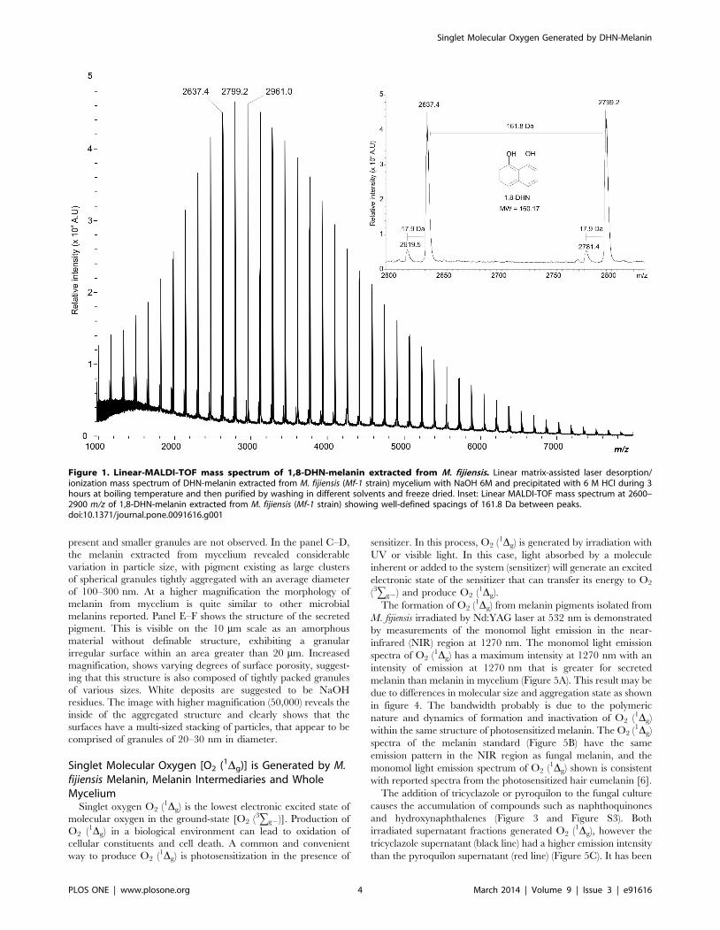
To evaluate whether melanin of M. fijiensis is a DHN type, we
made a structural analysis using Linear-MALDI-TOF mass
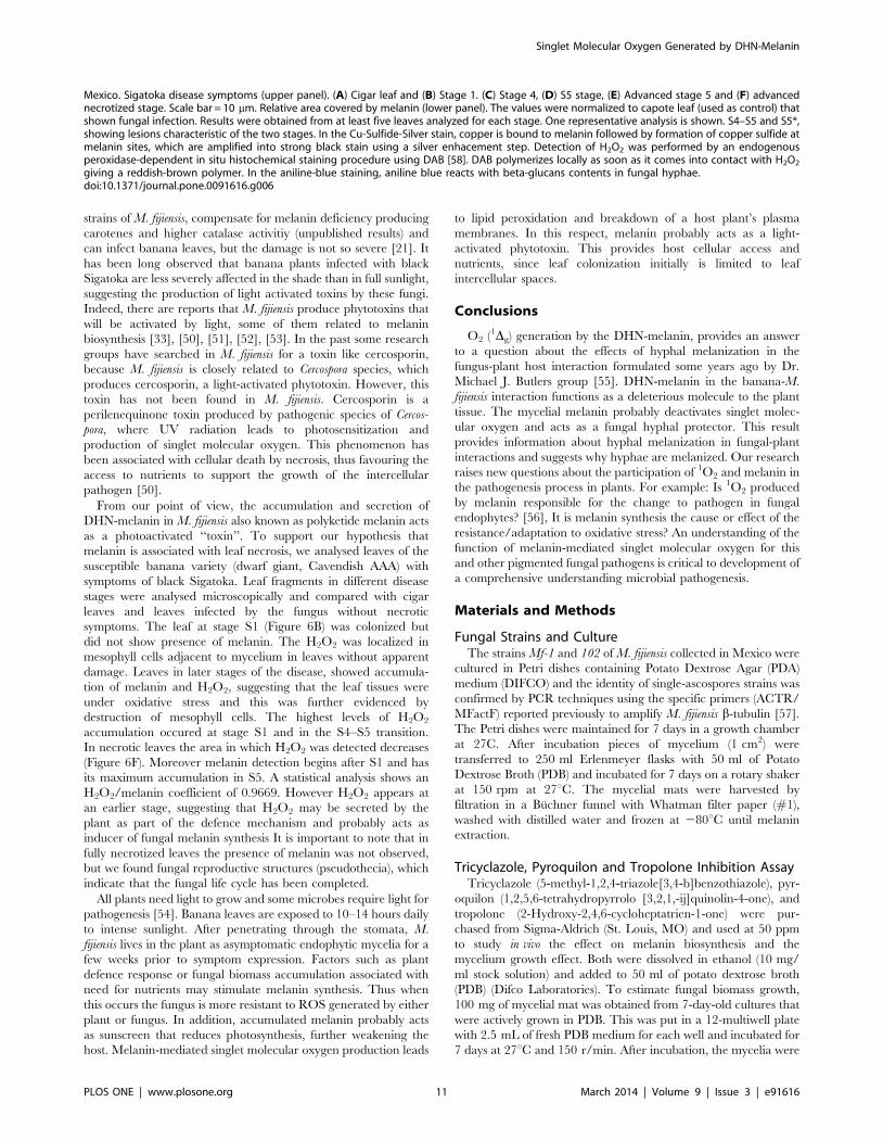
spectrometry. As shown in figure 1, melanin from mycelium had
molecular mass values not exceeding 8000 Da. It was further
noted that a series of peaks were well separated with well-defined
spacings of 161.8 Da, which is close to the theoretical mass of
DHN (160.17 MW). This mass difference (Dm) of 1.6 Da could be
due to MALDI-TOF low-resolution mass spectrum obtained.
According to the molecular mass observed we could calculate that
the pigment obtained after an extraction and purification process,
is comprised 50 units of 1,8-DHN. It is also noteworthy that other
minor peaks of 18 Da are present throughout the spectrum. These
probably result from the dehydration process of the melanin
polymer.
The Use of 1,8-DHN-melanin Inhibitors and Identificationof Related Pentaketides Confirms the Nature of thePigment of M. fijiensis
The 1,8-DHN biosynthesis pathway was elucidated by charac-
terizing autooxidation products, which accumulate in liquid
cultures blocked in melanin biosynthesis by 50 ppm of tricyclazole
and pyroquilon, two classic inhibitors that interfere with the
dehydrogenation of 1,3,6,8 tetrahydroxynaphthalene (1,3,6,8-
THN) to scytalone (S) and 1,3,8 trihydroxynaphthalene (1,3,8-
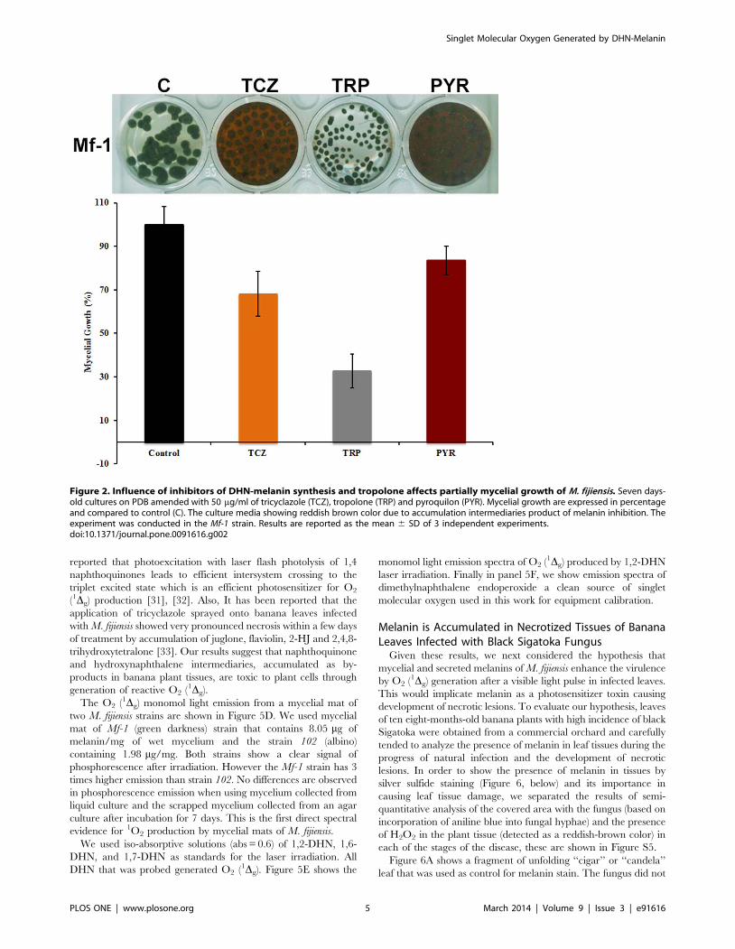
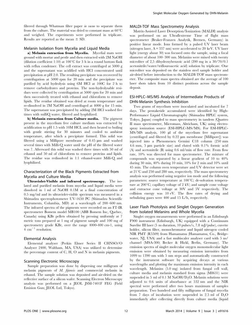
THN) to vermelone (V) [27], [28]. The mycelial mat changed
from black to reddish-brown colour and a typically reddish
pigment accumulated in the culture media after 7 days culture,
indicating that melanin biosynthesis was inhibited (Figure 2).
Tricyclazole inhibited mycelial growth almost double compared
with the pyroquilon (16.6% vs 31.8%). On the other hand, the
addition of 50 ppm of tropolone (a DOPA-melanin inhibitor) to
the culture medium did not cause any change in the coloration of
mycelia, and did not result in secretion of soluble pigments into the
culture medium as with pyroquilon and tricyclazole. Tropolone
inhibits the growth of the fungus up to 67% compared with the
control culture. These results support that melanin synthesized by
M. fijiensis is a DHN-melanin.
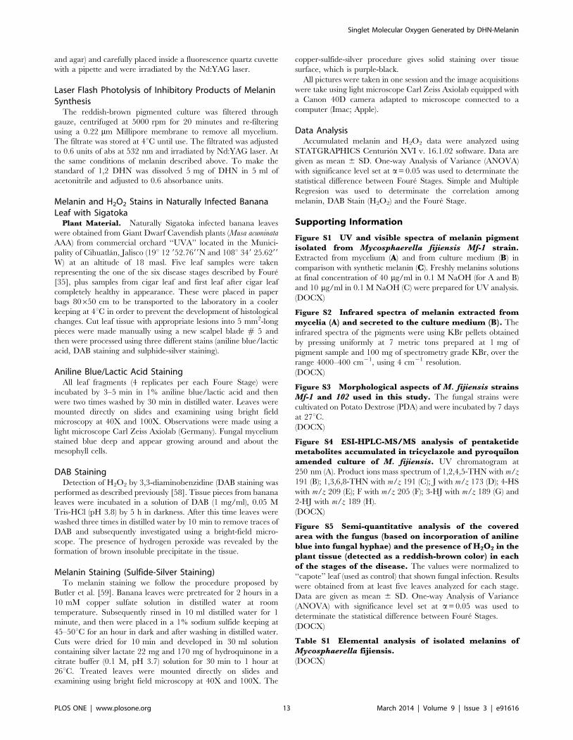
The ESI-HPLC-MS/MS analysis of 7 day-old PDB cultures
containing tricyclazole and pyroquilon showed a complex mixture
of compounds (Figure S4). In figure 3 we show the retention times,
m/z of precursor ions, and m/z of product ions of the metabolites
identified as: 1,3,6,8-THN (m/z 191), 1,2,4,5-tetrahydroxyna-
phathalene (1,2,4,5-THN, m/z 191) and typical metabolites
including flaviolin (F, m/z 205), juglone (J, m/z 173), 2-
hydroxyjuglone (2-HJ, m/z 189), 3-hydroxyjuglone (3-HJ, m/z
189) and 4 hydroxyscytalone (4-HS, m/z 209). This result confirms
that tricyclazole and pyroquilon blocked the 1,8-DHN-melanin
pathway. 1,3,8-trihydroxynaphthalene (1,3,8-THN) was not
found, suggesting that it is rapidly autoxidized to 2-HJ. Other
molecules were not identified, but were likely pentaketides since
fragmentation products have similar masses characteristics of those
compounds (data not show).
This is the first study where intermediary compounds of
melanin synthesis were identified directly in cell-free supernatants
without extracting with organic solvents. The analyses were
performed in three separate assays using different batches of Mf-1
cultures. Future studies will be focused on the elucidation of all
structures accumulated by melanin inhibitors in M. fijiensis.
Melanins Isolated from M. fijiensis Vary in Size andStructural Morphology
It has been reported that the structural morphology and specific
arrangements that melanin pigments adopt affect photoreactivity,
so that unaggregated oligomers have phototoxic effects and their
aggregation mitigates such processes [29]. This is the framework
for understanding contrasting antioxidant and pro-oxidant roles
exhibited by melanins. Any changes in structure could result in
increased oxidative stress [30]. We consider it important to know
their structures, as well as differences between both pigments
isolated from M. fijiensis. Both pigments were obtained using acid
precipitation, solvents extraction and freeze-drying. Figure 4
(panel A–B) shows the ultra-structural characteristics of synthetic
melanin. Uniform granule bodies within a narrow size range are
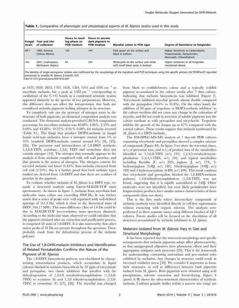
Table 1. Comparative of phenotypic and physiological aspects of M. fijiensis strains used in this study.
Fungalstrain
Year and siteof collection
Hours to reachlog phase onPDB medium
Hours to secretedark pigment toPDB medium Mycelial colour in PDA agar Degree of Resistance to fungicides
Mf-1 1999, ArmeriaColima, Mexico
120 144 Dark green on the surface andblack in bottom
Higher Sensitivity to Carbendazim,Propiconazole, Azoxystrobin,Mancozeb, Chlorothalonil.
102 2007, Coahuayana,Michoacan, Mexico
84 204 White-pink on the surface and whitewith small black spots in bottom
Higher resistance to all fungicidesmentioned above.
The identity of single-ascospores isolates was confirmed by: the morphology of the mycelium and PCR techniques using the specific primers (ACTR/MFactF) reportedpreviously to amplify M. fijiensis b-tubulin [57].doi:10.1371/journal.pone.0091616.t001
Singlet Molecular Oxygen Generated by DHN-Melanin
PLOS ONE | www.plosone.org 3 March 2014 | Volume 9 | Issue 3 | e91616
present and smaller granules are not observed. In the panel C–D,
the melanin extracted from mycelium revealed considerable
variation in particle size, with pigment existing as large clusters
of spherical granules tightly aggregated with an average diameter
of 100–300 nm. At a higher magnification the morphology of
melanin from mycelium is quite similar to other microbial
melanins reported. Panel E–F shows the structure of the secreted
pigment. This is visible on the 10 mm scale as an amorphous
material without definable structure, exhibiting a granular
irregular surface within an area greater than 20 mm. Increased
magnification, shows varying degrees of surface porosity, suggest-
ing that this structure is also composed of tightly packed granules
of various sizes. White deposits are suggested to be NaOH
residues. The image with higher magnification (50,000) reveals the
inside of the aggregated structure and clearly shows that the
surfaces have a multi-sized stacking of particles, that appear to be
comprised of granules of 20–30 nm in diameter.
Singlet Molecular Oxygen [O2 (1Dg)] is Generated by M.fijiensis Melanin, Melanin Intermediaries and WholeMycelium
Singlet oxygen O2 (1Dg) is the lowest electronic excited state of
molecular oxygen in the ground-state [O2 (3gg2)]. Production of
O2 (1Dg) in a biological environment can lead to oxidation of
cellular constituents and cell death. A common and convenient
way to produce O2 (1Dg) is photosensitization in the presence of
sensitizer. In this process, O2 (1Dg) is generated by irradiation with
UV or visible light. In this case, light absorbed by a molecule
inherent or added to the system (sensitizer) will generate an excited
electronic state of the sensitizer that can transfer its energy to O2
(3gg2) and produce O2 (1Dg).
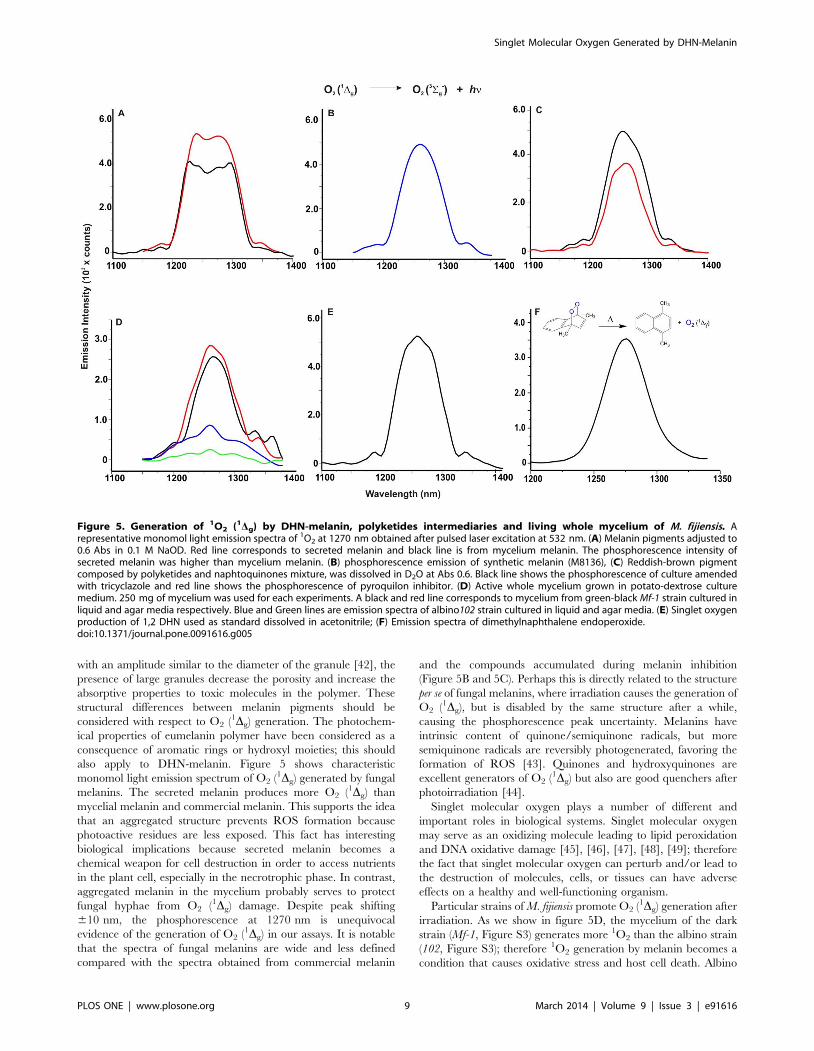
The formation of O2 (1Dg) from melanin pigments isolated from
M. fijiensis irradiated by Nd:YAG laser at 532 nm is demonstrated
by measurements of the monomol light emission in the near-
infrared (NIR) region at 1270 nm. The monomol light emission
spectra of O2 (1Dg) has a maximum intensity at 1270 nm with an
intensity of emission at 1270 nm that is greater for secreted
melanin than melanin in mycelium (Figure 5A). This result may be
due to differences in molecular size and aggregation state as shown
in figure 4. The bandwidth probably is due to the polymeric
nature and dynamics of formation and inactivation of O2 (1Dg)
within the same structure of photosensitized melanin. The O2 (1Dg)
spectra of the melanin standard (Figure 5B) have the same
emission pattern in the NIR region as fungal melanin, and the
monomol light emission spectrum of O2 (1Dg) shown is consistent
with reported spectra from the photosensitized hair eumelanin [6].
The addition of tricyclazole or pyroquilon to the fungal culture
causes the accumulation of compounds such as naphthoquinones
and hydroxynaphthalenes (Figure 3 and Figure S3). Both
irradiated supernatant fractions generated O2 (1Dg), however the
tricyclazole supernatant (black line) had a higher emission intensity
than the pyroquilon supernatant (red line) (Figure 5C). It has been
Figure 1. Linear-MALDI-TOF mass spectrum of 1,8-DHN-melanin extracted from M. fijiensis. Linear matrix-assisted laser desorption/ionization mass spectrum of DHN-melanin extracted from M. fijiensis (Mf-1 strain) mycelium with NaOH 6M and precipitated with 6 M HCl during 3hours at boiling temperature and then purified by washing in different solvents and freeze dried. Inset: Linear MALDI-TOF mass spectrum at 2600–2900 m/z of 1,8-DHN-melanin extracted from M. fijiensis (Mf-1 strain) showing well-defined spacings of 161.8 Da between peaks.doi:10.1371/journal.pone.0091616.g001
Singlet Molecular Oxygen Generated by DHN-Melanin
PLOS ONE | www.plosone.org 4 March 2014 | Volume 9 | Issue 3 | e91616
reported that photoexcitation with laser flash photolysis of 1,4
naphthoquinones leads to efficient intersystem crossing to the
triplet excited state which is an efficient photosensitizer for O2
(1Dg) production [31], [32]. Also, It has been reported that the
application of tricyclazole sprayed onto banana leaves infected
with M. fijiensis showed very pronounced necrosis within a few days
of treatment by accumulation of juglone, flaviolin, 2-HJ and 2,4,8-
trihydroxytetralone [33]. Our results suggest that naphthoquinone
and hydroxynaphthalene intermediaries, accumulated as by-
products in banana plant tissues, are toxic to plant cells through
generation of reactive O2 (1Dg).
The O2 (1Dg) monomol light emission from a mycelial mat of
two M. fijiensis strains are shown in Figure 5D. We used mycelial
mat of Mf-1 (green darkness) strain that contains 8.05 mg of
melanin/mg of wet mycelium and the strain 102 (albino)
containing 1.98 mg/mg. Both strains show a clear signal of
phosphorescence after irradiation. However the Mf-1 strain has 3
times higher emission than strain 102. No differences are observed
in phosphorescence emission when using mycelium collected from
liquid culture and the scrapped mycelium collected from an agar
culture after incubation for 7 days. This is the first direct spectral
evidence for 1O2 production by mycelial mats of M. fijiensis.
We used iso-absorptive solutions (abs = 0.6) of 1,2-DHN, 1,6-
DHN, and 1,7-DHN as standards for the laser irradiation. All
DHN that was probed generated O2 (1Dg). Figure 5E shows the
monomol light emission spectra of O2 (1Dg) produced by 1,2-DHN
laser irradiation. Finally in panel 5F, we show emission spectra of
dimethylnaphthalene endoperoxide a clean source of singlet
molecular oxygen used in this work for equipment calibration.
Melanin is Accumulated in Necrotized Tissues of BananaLeaves Infected with Black Sigatoka Fungus
Given these results, we next considered the hypothesis that
mycelial and secreted melanins of M. fijiensis enhance the virulence
by O2 (1Dg) generation after a visible light pulse in infected leaves.
This would implicate melanin as a photosensitizer toxin causing
development of necrotic lesions. To evaluate our hypothesis, leaves
of ten eight-months-old banana plants with high incidence of black
Sigatoka were obtained from a commercial orchard and carefully
tended to analyze the presence of melanin in leaf tissues during the
progress of natural infection and the development of necrotic
lesions. In order to show the presence of melanin in tissues by
silver sulfide staining (Figure 6, below) and its importance in
causing leaf tissue damage, we separated the results of semi-
quantitative analysis of the covered area with the fungus (based on
incorporation of aniline blue into fungal hyphae) and the presence
of H2O2 in the plant tissue (detected as a reddish-brown color) in
each of the stages of the disease, these are shown in Figure S5.
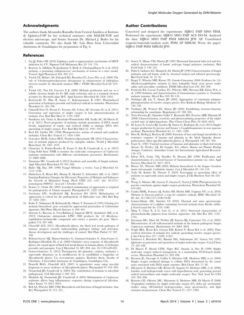
Figure 6A shows a fragment of unfolding ‘‘cigar’’ or ‘‘candela’’
leaf that was used as control for melanin stain. The fungus did not
Figure 2. Influence of inhibitors of DHN-melanin synthesis and tropolone affects partially mycelial growth of M. fijiensis. Seven days-old cultures on PDB amended with 50 mg/ml of tricyclazole (TCZ), tropolone (TRP) and pyroquilon (PYR). Mycelial growth are expressed in percentageand compared to control (C). The culture media showing reddish brown color due to accumulation intermediaries product of melanin inhibition. Theexperiment was conducted in the Mf-1 strain. Results are reported as the mean 6 SD of 3 independent experiments.doi:10.1371/journal.pone.0091616.g002
Singlet Molecular Oxygen Generated by DHN-Melanin
PLOS ONE | www.plosone.org 5 March 2014 | Volume 9 | Issue 3 | e91616
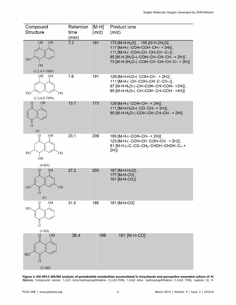
Figure 3. ESI-HPLC-MS/MS analysis of pentaketide metabolites accumulated in tricyclazole and pyroquilon amended culture of M.fijiensis. Compound names: 1,2,4,5 tetra-hydroxynaphthalene (1,2,4,5-THN), 1,3,6,8 tetra- hydroxynaphthalene (1,3,6,8 THN), Juglone (J), 4-
Singlet Molecular Oxygen Generated by DHN-Melanin
PLOS ONE | www.plosone.org 6 March 2014 | Volume 9 | Issue 3 | e91616
infect the cigar leaf and no melanin and H2O2 were detected in
tissues (Figure S5) Figure 6B shows a leaf fragment in infection
stage 1 (S1) according to the Foure scale [34]. The leaves at this
stage tend to lose the bright green to pale green color, without
manifestation of black Sigatoka lesions. At the light microscopic
level, the aniline blue stain shows a greater staining of fungal
hyphae (in deep blue) distributed among the cells of palisade layer
and intercellular spaces of leaf mesophyll; with hyphae covering
approximately 27% of leaf area analyzed (Figure S5). At this stage
of infection, melanin is not detected in leaf tissue or in areas where
hyphae are located, however H2O2 was detected in 7.4% of area
analyzed. In Figure 6C a banana leaf fragment has wider brown
streaks at the S4 stage (also called ‘‘the first spot stage’’) with
discoloration and loss of vigor. Microscopically, we observed
fungal structures, including asexual conidia, on mesophyll cells. No
significant difference was found in the areas covered by the fungus
compared to previous stages. In addition, melanin covered 20.43%
of the leaf area. However DAB staining shows 40% of the area
with H2O2 presence in the intercellular spaces and light brown
deposits in the cells of the palisades layer. It is difficult to
distinguish whether the fungus or the plant is responsible for
accumulation of hydrogen peroxide in this site. In the transition
from stage S4 to S5 no change in the area covered by the fungus
was observed, but the H2O2 level approached 50%. In the
transition from stage S4 to S5 melanin increased by 5%.
Symptoms of S5 or ‘‘second spot stage’’ in banana leaf are shown
in the Figure 6D. This stage is characterized by many black spots
with yellow halos. At this stage the fungus has penetrated the cells
causing irreversible structural changes in mesophyll cells. Intra-
cellular penetration damages plant cell membranes and increases
nutrient leakage into intercellular spaces. Although the fungal area
diminished 5%, the number of reproductive structures (pseu-
dothecia) increased. Melanin reaches the maximum value 28.6%
in this stage of disease progression. The amount of H2O2 at this
stage is greater than in previous infection stages. It is estimated
that this stage occurs after 50–60 days post-penetration of stomata.
This finding suggests that the amount of fungal melanin deposited
within foliar tissues is directly related to mesophyll cell death and
necrosis. Figure 6E shows a leaf in advanced S5* stage. Here is an
increase in chlorotic regions, and the black streaks are larger.
There are decreases in mycelium content of plant tissues and plant
cell morphologies are altered. A decrease in the melanin content
and H2O2 production were observed. In Stage 6 (Figure 6F),
leaves may be completely necrotized colorless and colorless with
gray halos and black dots (pseudothecia) within them. Microscop-
ically, we observed a loss of plant tissue structure and a decrease in
the mycelium content up to 5% was observed.
Discussion
In this study, we report that the pigments contained in the
mycelium and secreted into the culture medium of M. fijiensis are
melanins that absorb visible light and act as photosensitizers that
can generate O2 (1Dg). Based on these findings, our work suggests
that melanin should be further studied as a potentially important
contributor to progression of black Sigatoka disease of bananas
and plantains. In this research we identified a melanin pigment
from mycelium and secreted into culture media as DHN-melanin,
the sole melanin-type in M. fijiensis. The molecular mass obtained
by MALDI-TOF analysis is approximately 8000 Da and shows a
molar mass distribution with peak-to-peak increments of
161.8 Da. It is possible to see other peak distributions with less
extension than 8000 Da, but with the same molar mass
distribution. This may be due to the harsh chemical treatment
that is required to extract and purify melanin. The polymer must
be hydrolyzed to small oligomeric fragments. The polymerized
form would have a higher molecular mass. Fungal melanin
synthesized in vitro using laccase and 1,8-DHN has a molecular
mass greater than 60 KDa [35].
To ascertain the chemical nature of M. fijiensis melanin,
inhibitors may be used to identify the type of melanin synthesized
by a fungus. Compounds such as tricyclazole, pyroquilon, fthalide
and chlobenthiazone inhibit DHN-melanin synthesis but not
DOPA melanin [36]. We found by ESI-HPLC-MS/MS that the
use of pyroquilon and tricyclazole increased the accumulation of
typically naphthoquinone intermediates by inhibition of scytalone
dehydratase such as flaviolin, juglone, 4-Hidroxyscitalone, 3-HJ
and 2-HJ (Figure 3). The generation of flaviolin and 2-HJ in fungal
cultures treated with tricyclazole, is usually accepted as proof of
the presence of 1,3,6,8-THN and 1,3,8-THN that were involved
in the synthesis of 1,8-DHN. Also the unstable intermediate
1,2,4,5-THN and the precursor of the synthesis of 1,8-DHN, the
1,3,6,8-THN was identified. We interpret the accumulation of
these compounds in the culture media to be an indication of an
inhibitory effect on mycelial growth (Figure 2). This hypothesis is
supported by formation of intermediary compounds that may act
as photosensitizers and generate O2 (1Dg) (Figure 5). Decreases in
the melanin accumulated in the fungal cell walls probably increase
the cytotoxic effects of 1O2. This is because melanin can function
as a scavenger of this ROS, reducing oxidative damage to cell
membrane components, mainly lipids, as has been demonstrated
for eumelanin [37], [38]. Interestingly, tropolone strongly inhibits
mycelial growth compared to pyroquilon and tricyclazole.
Tropolone is classified as a DOPA melanin inhibitor, however it
is also used to inhibit laccase, an enzyme involved in the oxidative
synthesis of DHN-melanin. Therefore, the inhibition of this
enzyme could be a target for the design of new fungicides. This is
especially possible because DHN-melanin is primarily produced in
the fungal kingdom, particularly Ascomycota.
Melanin in biological systems is usually associated with
protection from UV radiation and as free radical trap [39].
However, melanins also produce ROS upon UV illumination.
This dichotomy of photoprotection and phototoxicity are linked to
the polymer backbone [29]. Melanins isolated from M. fijiensis are
structurally similar to the other microbial melanins [40], [41].
They consist of a spherical granular body arrangement in clusters
with different sizes and aggregations (Figure 4). Higher magnifi-
cations of secreted and hyphal melanin revealed a substructure of
spherical units that were variable in size. The size of granular
aggregates was higher in the melanin obtained from the cell walls
than melanin secreted into the culture medium (Figure 4). It has
been reported that melanins associated with cell walls consist of
large granules of 50–80 nm in diameter with multiple layers and
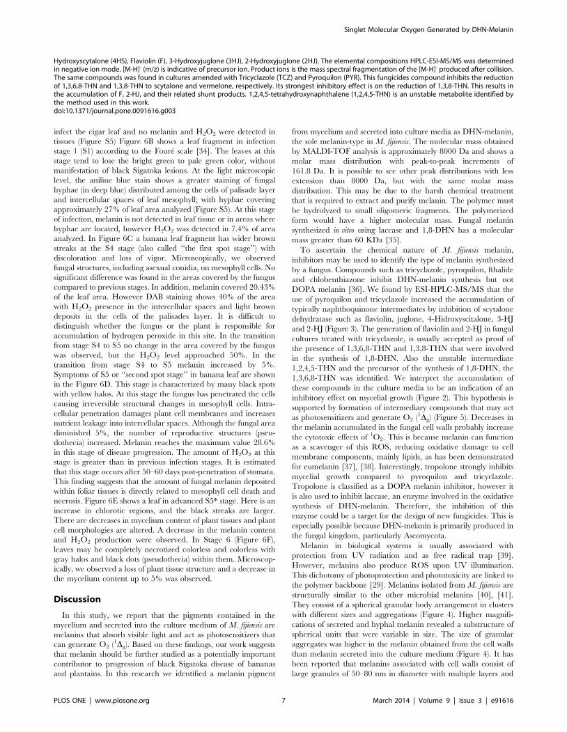
Hydroxyscytalone (4HS), Flaviolin (F), 3-Hydroxyjuglone (3HJ), 2-Hydroxyjuglone (2HJ). The elemental compositions HPLC-ESI-MS/MS was determinedin negative ion mode. [M-H]- (m/z) is indicative of precursor ion. Product ions is the mass spectral fragmentation of the [M-H]- produced after collision.The same compounds was found in cultures amended with Tricyclazole (TCZ) and Pyroquilon (PYR). This fungicides compound inhibits the reductionof 1,3,6,8-THN and 1,3,8-THN to scytalone and vermelone, respectively. Its strongest inhibitory effect is on the reduction of 1,3,8-THN. This results inthe accumulation of F, 2-HJ, and their related shunt products. 1,2,4,5-tetrahydroxynaphthalene (1,2,4,5-THN) is an unstable metabolite identified bythe method used in this work.doi:10.1371/journal.pone.0091616.g003
Singlet Molecular Oxygen Generated by DHN-Melanin
PLOS ONE | www.plosone.org 7 March 2014 | Volume 9 | Issue 3 | e91616
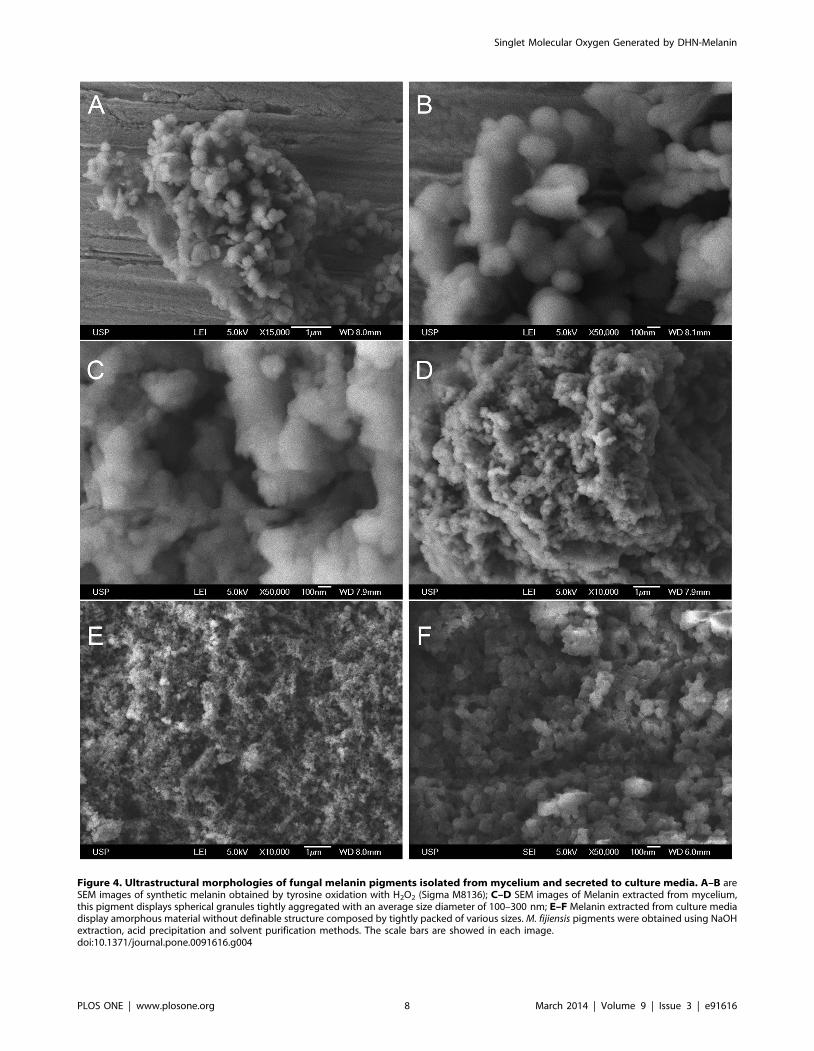
Figure 4. Ultrastructural morphologies of fungal melanin pigments isolated from mycelium and secreted to culture media. A–B areSEM images of synthetic melanin obtained by tyrosine oxidation with H2O2 (Sigma M8136); C–D SEM images of Melanin extracted from mycelium,this pigment displays spherical granules tightly aggregated with an average size diameter of 100–300 nm; E–F Melanin extracted from culture mediadisplay amorphous material without definable structure composed by tightly packed of various sizes. M. fijiensis pigments were obtained using NaOHextraction, acid precipitation and solvent purification methods. The scale bars are showed in each image.doi:10.1371/journal.pone.0091616.g004
Singlet Molecular Oxygen Generated by DHN-Melanin
PLOS ONE | www.plosone.org 8 March 2014 | Volume 9 | Issue 3 | e91616
with an amplitude similar to the diameter of the granule [42], the
presence of large granules decrease the porosity and increase the
absorptive properties to toxic molecules in the polymer. These
structural differences between melanin pigments should be
considered with respect to O2 (1Dg) generation. The photochem-
ical properties of eumelanin polymer have been considered as a
consequence of aromatic rings or hydroxyl moieties; this should
also apply to DHN-melanin. Figure 5 shows characteristic
monomol light emission spectrum of O2 (1Dg) generated by fungal
melanins. The secreted melanin produces more O2 (1Dg) than
mycelial melanin and commercial melanin. This supports the idea
that an aggregated structure prevents ROS formation because
photoactive residues are less exposed. This fact has interesting
biological implications because secreted melanin becomes a
chemical weapon for cell destruction in order to access nutrients
in the plant cell, especially in the necrotrophic phase. In contrast,
aggregated melanin in the mycelium probably serves to protect
fungal hyphae from O2 (1Dg) damage. Despite peak shifting
610 nm, the phosphorescence at 1270 nm is unequivocal
evidence of the generation of O2 (1Dg) in our assays. It is notable
that the spectra of fungal melanins are wide and less defined
compared with the spectra obtained from commercial melanin
and the compounds accumulated during melanin inhibition
(Figure 5B and 5C). Perhaps this is directly related to the structure
per se of fungal melanins, where irradiation causes the generation of
O2 (1Dg), but is disabled by the same structure after a while,
causing the phosphorescence peak uncertainty. Melanins have
intrinsic content of quinone/semiquinone radicals, but more
semiquinone radicals are reversibly photogenerated, favoring the
formation of ROS [43]. Quinones and hydroxyquinones are
excellent generators of O2 (1Dg) but also are good quenchers after
photoirradiation [44].
Singlet molecular oxygen plays a number of different and
important roles in biological systems. Singlet molecular oxygen
may serve as an oxidizing molecule leading to lipid peroxidation
and DNA oxidative damage [45], [46], [47], [48], [49]; therefore
the fact that singlet molecular oxygen can perturb and/or lead to
the destruction of molecules, cells, or tissues can have adverse
effects on a healthy and well-functioning organism.
Particular strains of M. fijiensis promote O2 (1Dg) generation after
irradiation. As we show in figure 5D, the mycelium of the dark
strain (Mf-1, Figure S3) generates more 1O2 than the albino strain
(102, Figure S3); therefore 1O2 generation by melanin becomes a
condition that causes oxidative stress and host cell death. Albino
Figure 5. Generation of 1O2 (1Dg) by DHN-melanin, polyketides intermediaries and living whole mycelium of M. fijiensis. Arepresentative monomol light emission spectra of 1O2 at 1270 nm obtained after pulsed laser excitation at 532 nm. (A) Melanin pigments adjusted to0.6 Abs in 0.1 M NaOD. Red line corresponds to secreted melanin and black line is from mycelium melanin. The phosphorescence intensity ofsecreted melanin was higher than mycelium melanin. (B) phosphorescence emission of synthetic melanin (M8136), (C) Reddish-brown pigmentcomposed by polyketides and naphtoquinones mixture, was dissolved in D2O at Abs 0.6. Black line shows the phosphorescence of culture amendedwith tricyclazole and red line shows the phosphorescence of pyroquilon inhibitor. (D) Active whole mycelium grown in potato-dextrose culturemedium. 250 mg of mycelium was used for each experiments. A black and red line corresponds to mycelium from green-black Mf-1 strain cultured inliquid and agar media respectively. Blue and Green lines are emission spectra of albino102 strain cultured in liquid and agar media. (E) Singlet oxygenproduction of 1,2 DHN used as standard dissolved in acetonitrile; (F) Emission spectra of dimethylnaphthalene endoperoxide.doi:10.1371/journal.pone.0091616.g005
Singlet Molecular Oxygen Generated by DHN-Melanin
PLOS ONE | www.plosone.org 9 March 2014 | Volume 9 | Issue 3 | e91616
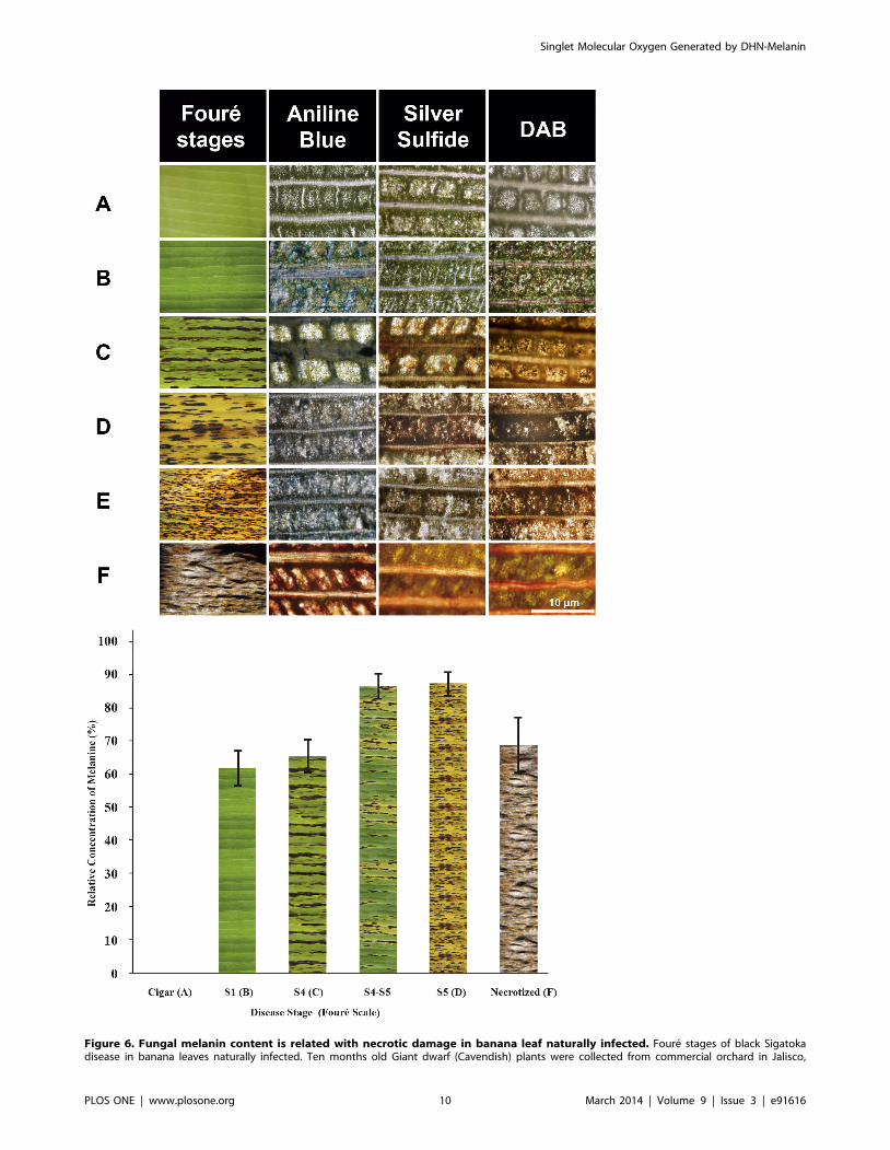
Figure 6. Fungal melanin content is related with necrotic damage in banana leaf naturally infected. Foure stages of black Sigatokadisease in banana leaves naturally infected. Ten months old Giant dwarf (Cavendish) plants were collected from commercial orchard in Jalisco,
Singlet Molecular Oxygen Generated by DHN-Melanin
PLOS ONE | www.plosone.org 10 March 2014 | Volume 9 | Issue 3 | e91616
strains of M. fijiensis, compensate for melanin deficiency producing
carotenes and higher catalase activitiy (unpublished results) and
can infect banana leaves, but the damage is not so severe [21]. It
has been long observed that banana plants infected with black
Sigatoka are less severely affected in the shade than in full sunlight,
suggesting the production of light activated toxins by these fungi.
Indeed, there are reports that M. fijiensis produce phytotoxins that
will be activated by light, some of them related to melanin
biosynthesis [33], [50], [51], [52], [53]. In the past some research
groups have searched in M. fijiensis for a toxin like cercosporin,
because M. fijiensis is closely related to Cercospora species, which
produces cercosporin, a light-activated phytotoxin. However, this
toxin has not been found in M. fijiensis. Cercosporin is a
perilenequinone toxin produced by pathogenic species of Cercos-
pora, where UV radiation leads to photosensitization and
production of singlet molecular oxygen. This phenomenon has
been associated with cellular death by necrosis, thus favouring the
access to nutrients to support the growth of the intercellular
pathogen [50].
From our point of view, the accumulation and secretion of
DHN-melanin in M. fijiensis also known as polyketide melanin acts
as a photoactivated ‘‘toxin’’. To support our hypothesis that
melanin is associated with leaf necrosis, we analysed leaves of the
susceptible banana variety (dwarf giant, Cavendish AAA) with
symptoms of black Sigatoka. Leaf fragments in different disease
stages were analysed microscopically and compared with cigar
leaves and leaves infected by the fungus without necrotic
symptoms. The leaf at stage S1 (Figure 6B) was colonized but
did not show presence of melanin. The H2O2 was localized in
mesophyll cells adjacent to mycelium in leaves without apparent
damage. Leaves in later stages of the disease, showed accumula-
tion of melanin and H2O2, suggesting that the leaf tissues were
under oxidative stress and this was further evidenced by
destruction of mesophyll cells. The highest levels of H2O2
accumulation occured at stage S1 and in the S4–S5 transition.
In necrotic leaves the area in which H2O2 was detected decreases
(Figure 6F). Moreover melanin detection begins after S1 and has
its maximum accumulation in S5. A statistical analysis shows an
H2O2/melanin coefficient of 0.9669. However H2O2 appears at
an earlier stage, suggesting that H2O2 may be secreted by the
plant as part of the defence mechanism and probably acts as
inducer of fungal melanin synthesis It is important to note that in
fully necrotized leaves the presence of melanin was not observed,
but we found fungal reproductive structures (pseudothecia), which
indicate that the fungal life cycle has been completed.
All plants need light to grow and some microbes require light for
pathogenesis [54]. Banana leaves are exposed to 10–14 hours daily
to intense sunlight. After penetrating through the stomata, M.
fijiensis lives in the plant as asymptomatic endophytic mycelia for a
few weeks prior to symptom expression. Factors such as plant
defence response or fungal biomass accumulation associated with
need for nutrients may stimulate melanin synthesis. Thus when
this occurs the fungus is more resistant to ROS generated by either
plant or fungus. In addition, accumulated melanin probably acts
as sunscreen that reduces photosynthesis, further weakening the
host. Melanin-mediated singlet molecular oxygen production leads
to lipid peroxidation and breakdown of a host plant’s plasma
membranes. In this respect, melanin probably acts as a light-
activated phytotoxin. This provides host cellular access and
nutrients, since leaf colonization initially is limited to leaf
intercellular spaces.
Conclusions
O2 (1Dg) generation by the DHN-melanin, provides an answer
to a question about the effects of hyphal melanization in the
fungus-plant host interaction formulated some years ago by Dr.
Michael J. Butlers group [55]. DHN-melanin in the banana-M.
fijiensis interaction functions as a deleterious molecule to the plant
tissue. The mycelial melanin probably deactivates singlet molec-
ular oxygen and acts as a fungal hyphal protector. This result
provides information about hyphal melanization in fungal-plant
interactions and suggests why hyphae are melanized. Our research
raises new questions about the participation of 1O2 and melanin in
the pathogenesis process in plants. For example: Is 1O2 produced
by melanin responsible for the change to pathogen in fungal
endophytes? [56], It is melanin synthesis the cause or effect of the
resistance/adaptation to oxidative stress? An understanding of the
function of melanin-mediated singlet molecular oxygen for this
and other pigmented fungal pathogens is critical to development of
a comprehensive understanding microbial pathogenesis.
Materials and Methods
Fungal Strains and CultureThe strains Mf-1 and 102 of M. fijiensis collected in Mexico were
cultured in Petri dishes containing Potato Dextrose Agar (PDA)
medium (DIFCO) and the identity of single-ascospores strains was
confirmed by PCR techniques using the specific primers (ACTR/
MFactF) reported previously to amplify M. fijiensis b-tubulin [57].
The Petri dishes were maintained for 7 days in a growth chamber
at 27C. After incubation pieces of mycelium (1 cm2) were
transferred to 250 ml Erlenmeyer flasks with 50 ml of Potato
Dextrose Broth (PDB) and incubated for 7 days on a rotary shaker
at 150 rpm at 27uC. The mycelial mats were harvested by
filtration in a Buchner funnel with Whatman filter paper (#1),
washed with distilled water and frozen at 280uC until melanin
extraction.
Tricyclazole, Pyroquilon and Tropolone Inhibition AssayTricyclazole (5-methyl-1,2,4-triazole[3,4-b]benzothiazole), pyr-
oquilon (1,2,5,6-tetrahydropyrrolo [3,2,1,-ij]quinolin-4-one), and
tropolone (2-Hydroxy-2,4,6-cycloheptatrien-1-one) were pur-
chased from Sigma-Aldrich (St. Louis, MO) and used at 50 ppm
to study in vivo the effect on melanin biosynthesis and the
mycelium growth effect. Both were dissolved in ethanol (10 mg/
ml stock solution) and added to 50 ml of potato dextrose broth
(PDB) (Difco Laboratories). To estimate fungal biomass growth,
100 mg of mycelial mat was obtained from 7-day-old cultures that
were actively grown in PDB. This was put in a 12-multiwell plate
with 2.5 mL of fresh PDB medium for each well and incubated for
7 days at 27uC and 150 r/min. After incubation, the mycelia were
Mexico. Sigatoka disease symptoms (upper panel). (A) Cigar leaf and (B) Stage 1. (C) Stage 4, (D) S5 stage, (E) Advanced stage 5 and (F) advancednecrotized stage. Scale bar = 10 mm. Relative area covered by melanin (lower panel). The values were normalized to capote leaf (used as control) thatshown fungal infection. Results were obtained from at least five leaves analyzed for each stage. One representative analysis is shown. S4–S5 and S5*,showing lesions characteristic of the two stages. In the Cu-Sulfide-Silver stain, copper is bound to melanin followed by formation of copper sulfide atmelanin sites, which are amplified into strong black stain using a silver enhacement step. Detection of H2O2 was performed by an endogenousperoxidase-dependent in situ histochemical staining procedure using DAB [58]. DAB polymerizes locally as soon as it comes into contact with H2O2
giving a reddish-brown polymer. In the aniline-blue staining, aniline blue reacts with beta-glucans contents in fungal hyphae.doi:10.1371/journal.pone.0091616.g006
Singlet Molecular Oxygen Generated by DHN-Melanin
PLOS ONE | www.plosone.org 11 March 2014 | Volume 9 | Issue 3 | e91616
filtered through Whatman filter paper in vacuo to separate them
from the culture. The material was dried to constant mass at 60uCand weighed. The experiments were performed in triplicate.
Results are reported as the mean 6 SD.
Melanin Isolation from Mycelia and Liquid Mediaa) Melanin extraction from Mycelia. Mycelial mats were
ground with mortar and pestle and then extracted with 2M NaOH
(dilution coefficient 1:10) at 100uC for 3 h in a round bottom flask
with reflux condenser. The cell extract was centrifuged at 5000 g
and the supernatant was acidified with HCl concentrated until
precipitation at pH 2.0. The resulting precipitate was recovered by
centrifugation at 5000 rpm for 20 min and the precipitate was
purified by acid hydrolysis using 6M HCl at 100C for 2 h to
remove carbohydrates and proteins. The non-hydrolysable resi-
dues were collected by centrifugation at 5000 rpm for 20 min and
then successively treated with ethanol and chloroform to remove
lipids. The residue obtained was dried at room temperature and
re-dissolved in 2M NaOH and centrifuged at 4000 g for 15 min.
The supernatant was precipitated by adding 2M HCl washed five
times with milliQ water, filtered and lyophilized.
b) Melanin extraction from Culture media. The pigment
present in the mycelium free culture medium was extracted by
acidification to pH 1.5 with 10M HCL and then heated to boiling
with gentle stirring for 30 minutes and cooled to ambient
temperature, after which a precipitate formed. This solid was
filtered using a Millipore membrane of 0.45 mm and washed
several times with Milli-Q water until the pH of the filtered water
was 7. Afterward this solid was washed three times with 50 ml of
ethanol and 50 ml of chloroform to remove proteins and lipids.
The residue was redissolved in 1:1 ethanol-water Milli-Q and
lyophilized.
Characterization of the Black Pigments Extracted fromMycelia and Culture Media
Ultraviolet-Visible and infrared spectroscopy. The iso-
lated and purified melanin from mycelia and liquid media were
dissolved in 1 ml of NaOH 0.1M at a final concentration of
0.5 mg/ml and its ultraviolet-visible spectrum was measured in a
Shimadzu spectrophotometer UV-1650 PC (Shimadzu Scientific
Instruments, Columbia, MD) at a wavelength of 200–600 nm.
The infrared spectra of the pigments were recorded on an FT-IR
spectrometer Bomem model MB100 (ABB Bomem Inc, Quebec,
Canada) using KBr pellets obtained by pressing uniformly at 7
metric tons prepared at 1 mg of pigment sample and 100 mg of
spectrometry grade KBr, over the range 4000-400 cm-1, using
4 cm21 resolution.
Elemental AnalysisElemental analyser (Perkin Elmer Series II CHNSO/O
Analyser 2400, Waltham, MA, USA) was utilized to determine
the percentage content of C, H, O and N in melanin pigments.
Scanning Electronic MicroscopySample preparation was done by dispersing one milligram of
melanin pigments of M. fijiensis and commercial melanin in
ethanol. The sample solution was deposited and air-dried on the
reflective surface of a silicon wafer. Scanning Electron Microscopy
analysis was performed on a JEOL JSM-7401F FEG (Field
Emision Gun; JEOL Ltd, Tokyo).
MALDI-TOF Mass Spectrometry AnalysisMatrix-Assisted Laser Desorption/Ionization (MALDI) analysis
was performed on an Ultraflextreme Time of flight mass
spectrometer (Bruker-Franzen Analytik, Bremen, Germany) in
positive linear mode. Ions formed by a pulsed UV laser beam
(nitrogen laser, A = 337 nm) were accelerated to 20 keV. UV laser
light (energy about 30) was focused onto the sample, using a focal
diameter of about 100–300 nm. Melanins were mixed with twenty
microliter of 2,5 dihydroxybenzoic acid (200 mg in a 30/70/0.1
acetonitrile/water/triflouroacetic acid) solution by triplicate. One
microliter was deposited on the stainless steel sample holder and
air-dried before introduction to the MALDI-TOF mass spectrom-
eter. The composite mass spectra obtained are the average of 100
laser shots taken from 10 distinct positions across the sample
deposit.
ESI-HPLC–MS/MS Analysis of Intermediate Products ofDHN-Melanin Synthesis Inhibition
Two grams of mycelium were inoculated and incubated for 7
days. The pentaketide accumulated were identified by High-
Performance Liquid Chromatography (Shimadzu HPLC system,
Tokyo, Japan) coupled to mass spectrometry in tandem (Quattro
II mass spectrometer, Micromass, Altricham, UK) using electro-
spray ionization source (ESI-HPLC-MS/MS). For ESI-HPLC-
MS/MS analysis, 100 ml of the mycelium free supernatant
(centrifuged and filtered by 0.22 mm membrane [Millipore]) was
injected into a Phenomenex Gemini C18 column (250 mm
4.6 mm, 5 mm particle size) and eluted with 0.1% formic acid
(A) and acetonitrile (B) using 0.6 ml/min of flow rate. From flow
rate, 10% was directed for mass spectrometer. The mixture of
compounds was separated by a linear gradient of 10 to 40%
during 30 min, 40% during 10 min, 10% for 2 min and 10% until
45 min. The column oven temperature and UV detector were set
at 21uC and 250 and 280 nm, respectively. The mass spectrometry
analysis was performed using negative ion mode and the following
parameters: source temperature at 100uC; de-solvation tempera-
ture at 200uC; capillary voltage of 2 kV; and sample cone voltage
and extractor cone voltage at 30V and 5V respectively. The
collision energy was 10 eV. The flow rates of drying and
nebulizing gases were 400 and 15 L/h, respectively.
Laser Flash Photolysis and Singlet Oxygen Generationfrom Isolated Melanins and Whole Mycelia
Singlet oxygen measurements were performed in an Edinburgh
F900 instrument (Edinburgh, UK) equipped with a Continuum
Surelite III laser (5 ns duration, 10 pulses/s, 7.8 mJ/pulse), cuvette
holder, silicon filter, monochromator and liquid nitrogen–cooled
NIR PMT (R5509) from Hamamatsu (Hamamatsu, Co., Bridge-
water, NJ, USA) and a fast multiscaler analyser card with 5 ns/
channel (MSA-300; Becker & Hickl, Berlin, Germany). The
emission spectra of singlet molecular oxygen monomolecular light
emission were obtained by measuring emission intensities from
1099 to 1390 nm with 5 nm steps and automatically constructed
by the instrument software by acquiring decays at various
wavelengths and plotting the maximum emission intensity in each
wavelength. Melanins (5.0 mg) isolated from fungal cell wall,
culture media and melanin standard from sigma (M8631) were
suspended in 5 ml of 0.1 M NaOH/D2O. Melanin solutions were
adjusted to 0.6 units of absorbance at 532 nm and the NIR
spectral were performed after two hours maximum of samples
preparation. Two hundred and fifty milligrams of fungal mycelia
from 7 days of incubation were suspended in 2.5 ml of D2O
immediately after collecting directly from culture media (liquid
Singlet Molecular Oxygen Generated by DHN-Melanin
PLOS ONE | www.plosone.org 12 March 2014 | Volume 9 | Issue 3 | e91616
and agar) and carefully placed inside a fluorescence quartz cuvette
with a pipette and were irradiated by the Nd:YAG laser.
Laser Flash Photolysis of Inhibitory Products of MelaninSynthesis
The reddish-brown pigmented culture was filtered through
gauze, centrifuged at 5000 rpm for 20 minutes and re-filtering
using a 0.22 mm Millipore membrane to remove all mycelium.
The filtrate was stored at 4uC until use. The filtrated was adjusted
to 0.6 units of abs at 532 nm and irradiated by Nd:YAG laser. At
the same conditions of melanin described above. To make the
standard of 1,2 DHN was dissolved 5 mg of DHN in 5 ml of
acetonitrile and adjusted to 0.6 absorbance units.
Melanin and H2O2 Stains in Naturally Infected BananaLeaf with Sigatoka
Plant Material. Naturally Sigatoka infected banana leaves
were obtained from Giant Dwarf Cavendish plants (Musa acuminata
AAA) from commercial orchard ‘‘UVA’’ located in the Munici-
pality of Cihuatlan, Jalisco (19u 12 952.7699N and 108u 349 25.6299
W) at an altitude of 18 masl. Five leaf samples were taken
representing the one of the six disease stages described by Foure
[35], plus samples from cigar leaf and first leaf after cigar leaf
completely healthy in appearance. These were placed in paper
bags 80650 cm to be transported to the laboratory in a cooler
keeping at 4uC in order to prevent the development of histological
changes. Cut leaf tissue with appropriate lesions into 5 mm2-long
pieces were made manually using a new scalpel blade # 5 and
then were processed using three different stains (aniline blue/lactic
acid, DAB staining and sulphide-silver staining).
Aniline Blue/Lactic Acid StainingAll leaf fragments (4 replicates per each Foure Stage) were
incubated by 3–5 min in 1% aniline blue/lactic acid and then
were two times washed by 30 min in distilled water. Leaves were
mounted directly on slides and examining using bright field
microscopy at 40X and 100X. Observations were made using a
light microscope Carl Zeiss Axiolab (Germany). Fungal mycelium
stained blue deep and appear growing around and about the
mesophyll cells.
DAB StainingDetection of H2O2 by 3,3-diaminobenzidine (DAB staining was
performed as described previously [58]. Tissue pieces from banana
leaves were incubated in a solution of DAB (1 mg/ml), 0.05 M
Tris-HCl (pH 3.8) by 5 h in darkness. After this time leaves were
washed three times in distilled water by 10 min to remove traces of
DAB and subsequently investigated using a bright-field micro-
scope. The presence of hydrogen peroxide was revealed by the
formation of brown insoluble precipitate in the tissue.
Melanin Staining (Sulfide-Silver Staining)To melanin staining we follow the procedure proposed by
Butler et al. [59]. Banana leaves were pretreated for 2 hours in a
10 mM copper sulfate solution in distilled water at room
temperature. Subsequently rinsed in 10 ml distilled water for 1
minute, and then were placed in a 1% sodium sulfide keeping at
45–50uC for an hour in dark and after washing in distilled water.
Cuts were dried for 10 min and developed in 30 ml solution
containing silver lactate 22 mg and 170 mg of hydroquinone in a
citrate buffer (0.1 M, pH 3.7) solution for 30 min to 1 hour at
26uC. Treated leaves were mounted directly on slides and
examining using bright field microscopy at 40X and 100X. The
copper-sulfide-silver procedure gives solid staining over tissue
surface, which is purple-black.
All pictures were taken in one session and the image acquisitions
were take using light microscope Carl Zeiss Axiolab equipped with
a Canon 40D camera adapted to microscope connected to a
computer (Imac; Apple).
Data AnalysisAccumulated melanin and H2O2 data were analyzed using
STATGRAPHICS Centurion XVI v. 16.1.02 software. Data are
given as mean 6 SD. One-way Analysis of Variance (ANOVA)
with significance level set at a= 0.05 was used to determinate the
statistical difference between Foure Stages. Simple and Multiple
Regresion was used to determinate the correlation among
melanin, DAB Stain (H2O2) and the Foure Stage.
Supporting Information
Figure S1 UV and visible spectra of melanin pigmentisolated from Mycosphaerella fijiensis Mf-1 strain.Extracted from mycelium (A) and from culture medium (B) in
comparison with synthetic melanin (C). Freshly melanins solutions
at final concentration of 40 mg/ml in 0.1 M NaOH (for A and B)
and 10 mg/ml in 0.1 M NaOH (C) were prepared for UV analysis.
(DOCX)
Figure S2 Infrared spectra of melanin extracted frommycelia (A) and secreted to the culture medium (B). The
infrared spectra of the pigments were using KBr pellets obtained
by pressing uniformly at 7 metric tons prepared at 1 mg of
pigment sample and 100 mg of spectrometry grade KBr, over the
range 4000–400 cm21, using 4 cm21 resolution.
(DOCX)
Figure S3 Morphological aspects of M. fijiensis strainsMf-1 and 102 used in this study. The fungal strains were
cultivated on Potato Dextrose (PDA) and were incubated by 7 days
at 27uC.
(DOCX)
Figure S4 ESI-HPLC-MS/MS analysis of pentaketidemetabolites accumulated in tricyclazole and pyroquilonamended culture of M. fijiensis. UV chromatogram at
250 nm (A). Product ions mass spectrum of 1,2,4,5-THN with m/z
191 (B); 1,3,6,8-THN with m/z 191 (C); J with m/z 173 (D); 4-HS
with m/z 209 (E); F with m/z 205 (F); 3-HJ with m/z 189 (G) and
2-HJ with m/z 189 (H).
(DOCX)
Figure S5 Semi-quantitative analysis of the coveredarea with the fungus (based on incorporation of anilineblue into fungal hyphae) and the presence of H2O2 in theplant tissue (detected as a reddish-brown color) in eachof the stages of the disease. The values were normalized to
‘‘capote’’ leaf (used as control) that shown fungal infection. Results
were obtained from at least five leaves analyzed for each stage.
Data are given as mean 6 SD. One-way Analysis of Variance
(ANOVA) with significance level set at a= 0.05 was used to
determinate the statistical difference between Foure Stages.
(DOCX)
Table S1 Elemental analysis of isolated melanins ofMycosphaerella fijiensis.
(DOCX)
Singlet Molecular Oxygen Generated by DHN-Melanin
PLOS ONE | www.plosone.org 13 March 2014 | Volume 9 | Issue 3 | e91616
Acknowledgments
The authors thank Alessandra Ramalho from Central Analitica at Instituto
de Quımica-USP for her technical assistance with MALDI-TOF and
electron microscope, and Senior Scientist Dr. Michael J. Butler for his
valuable comments. We also thank Mr. Luis Ham from Universidad
Autonoma de Guadalajara for preparation of Fig. 6.
Author Contributions
Conceived and designed the experiments: MJB-G FMP MSO PDM.
Performed the experiments: MJB-G MSO FMP ACS DO-M. Analyzed
the data: MJB-G MSO FMP PDM MHGM JFW AP. Contributed
reagents/materials/analysis tools: PDM AP MHGM. Wrote the paper:
MJB-G FMP PDM MHGM JFW.
References
1. Liu JJ, Fisher DE (2010) Lighting a path to pigmentation: mechanism of MITF
induction by UV. Pigment Cell Melanoma Res 23: 741–774.
2. Kunwar A, Adhikary B, Jayakumar S, Barik A, Chattopadhayay S, et al. (2012)melanin, a promising radioprotector: mechanism of actions in a mice model.
Toxicol Appl Pharmacol 264: 202–211.
3. Turick CE, Beliaev AS, Zakrajsek BA, Reardon CL, Lowy DA, et al. (2009) The
role of 4-hydroxyphenylpyruvate dioxygenase in enhancement of solid-phase
electron transfer by Shewanella oneidensis MR-1. FEMS Microbiol Ecol 68: 223–225.
4. Turick CE, Tisa LS, Caccavo F Jr (2002) Melanin production and use as a
soluble electron shuttle for Fe (III) oxide reduction and as a terminal electronacceptor by Shewanella algae BrY. Appl Environ Microbiol 68: 2436–2444.
5. Korytowski W, Pilas B, Sarna T, Kalyanaraman B (1987) Photoinducedgeneration of hydrogen peroxide and hydroxyl radicals in melanins. Photochem
Photobiol 45: 185–190.
6. Chiarelli-Neto O, Pavani C, Ferreira AS, Uchoa AF, Severino D, et al. (2011)Generation and supression of singlet oxygen in hair photosensitization of
melanin. Free Rad Biol Med 51: 1195–1202.
7. Suzukawa AA, Vieira A, Brochado-Winnischofer SM, Scalfo AC, Di Mascio P,et al. (2011) Novel properties of melanins include promotion of DNA strand
breaks, impairment of repair, and reduced ability to damage DNA afterquenching of singlet oxygen. Free Rad Biol Med 52: 1945–1952.
8. Krol ES, Liebler DC (1998) Photoprotective actions of natural and synthetic
melanins. Chem Res Toxicol 11: 1434–1440.
9. Goncalves RCR, Lisboa HCF, Pombeiro-Sponchiado SR (2012) Characteriza-tion of melanin pigment produced by Aspergillus nidulans. World J Microbiol
Biotechnol. 28: 1467–1474.
10. Chatterjee S, Prados-Rosales R, Frases S, Itin B, Casadevall A, et al. (2012)
Using Solid State NMR to monitor the molecular consequences of Cryptoccocus
neoformans melanization with different catecholamine precursors. Biochemistry51: 6080–6088.
11. Eisenman HC, Casadevall A (2012) Synthesis and assembly of fungal melanin.
Appl Microbiol Biotechnol 93: 931–940.
12. Butler MJ, Day AW (1998) Fungal Melanin: a review. Can J Microbiol 44:
1115–1136.
13. Dadachova E, Bryan RA, Huang X, Moadel T, Schweitzer AD, et al. (2007)Ionizing Radiation Changes the Electronic Properties of Melanin and Enhances
the Growth of Melanized Fungi. PLoS ONE 2(5): e457. doi: 10.1371/journal.pone.0000457. Accesed 1 October 2013.
14. Steiner U, Oerke EC (2007) Localized melanization of appressoria is required
for pathogenicity of Venturia inaequalis. Phytopathol 97: 1222–1230.
15. Gachomo EW, Seufferheld MJ, Kotchoni SO (2010) Melanization of
appressoria is critical for the pathogenicity of Diplocarpon rosae. Mol Biol Rep
37: 3583–3591.
16. Kubo Y, Nakamura H, Kobayashi K, Okuno T, Furusawa I (1991) Cloning of a
melanin biosynthetic gene essential for appressorial penetration of Colletotrichum
lagenarium. Mol Plant Microb Interact 4: 440–445.
17. Llorente C, Barcena A, Vera-Bahima J, Saparrat MCN, Arambarri AM, et al.
(2012) Cladosporium cladosporioides LPSC 1088 produces the 1,8 dihydroxy-naphthalene-melanin-like compound and carries a putative pks gene. Myco-
pathol 174: 397–408.
18. Churchill ACL (2011) Mycosphaerella fijiensis, the black leaf streak pathogen ofbanana: progress towards understanding pathogen biology and detection,
disease development and the challenges of control. Mol Plant Pathol 12: 307–
328.
19. Beltran-Garcia MJ, Manzo-Sanchez G, Guzman-Gonzalez S, Arias-Castro C,
Rodriguez-Mendiola M, et al. (2009) Oxidative stress response of Mycosphaerella
fijiensis, the causal agent of black leaf streak disease in banana plants, to hydrogen
peroxide and paraquat. Can J Microbiol 55: 887–894. doi: 10.1139/w09-023.
20. Gomez-Gutierrez A. (2012) Participacion del glutation, catalasa, melanina ysuperoxido dismutasa en la modificacion de la sensibilidad a fungicidas en
Mycosphaerella fijiensis: Un acercamiento quimico. Bachelor thesis, Faculty of
Chemistry, Universidad Autonoma de Guadalajara, Guadalajara City.
21. Donzelli BGG, Churchill ACL (2007) A quantitative assay using mycelial
fragments to assess virulence of Mycosphaerella fijiensis. Phytopathol 97: 916–929.
22. Nosanchuk JD, Casadevall A. (2003) The contribution of melanin to microbialpathogenesis. Cell Microbiol 5: 203–223.
23. Mednick AJ, Nosanchuk JD, Casadevall A (2005) Melanization of Cryptococcus
neoformans affects lung inflammatory responses during cryptococcal infection.Infect Immu 73: 2012–2019.
24. Bell AA, Wheeler MH (1986) Biosynthesis and functions of fungal melanins. AnnRev Phytopathol 24: 411–451.
25. Senesi N, Miano TM, Martin JP (1987) Elemental functional infra-red and free
radical characterization of humic acid-type fungal polymers (melanins). BiolFertil Soils 5: 120–125.
26. Palm S, Linhares LF, Mangrich AS, Martin JP (1990) Characterization of fungal
melanins and soil humic acids by chemical analysis and infrared spectroscopy.Biol Fertil Soils 10: 72–76.
27. Kogej T, Wheeler MH, Rizner TL, Gunde-Cimerman (2004) Evidence for 1,8-
dihydroxynaphthalene melanin in three halophilic black yeast grown undersaline and non-saline conditions. FEMS Microbiol Lett 232: 203–209.
28. Frederick BA, Caesar-Tonthat TC, Wheeler MH, Sheenan KB, Edens WA, et
al. (1999) Isolation and characterization of Gaeumannomyces graminis vr. Graminismelanin mutants. Mycol Res 103: 99–110.
29. Nofsinger JB, Liu Y, Simon JD (2002) Aggregation of eumelanin mitigates
photogeneration of reactive oxygen species. Free Radicals Biology Medicine 32:720–730.
30. Nosfinger JB, Weinert EE, Simon JD (2002) Establishing structure-functionrelationship for eumelanin. Biopolymers 67: 302–305.
31. Netto-Ferreira JC, Lhiaubet-Vallet V, Bernardes BO, Ferreira ABB, Miranda M
(2008) Characterization, reactivity and photosensitizing properties of the tripletexcited state of alpha-lapachone. Phys Chem Chem Phys 10: 6645–6652.
32. De Lucas N, Correa RJ, Garden SJ, Santos G, Rodrigues R, et al. (2012) Singlet
oxygen production by pyrano and furano 1,4-naphthoquinones in non aqueousmedium. Photochem Photobiol Sci 11: 1201–1209.
33. Hoss R, Helbig J, Bochow H (2000) Function of host and fungal metabolites in
resistance response of banana and plantain in the black Sigatoka diseasepathosystem (Musa spp-Mycosphaerella fijiensis). J. Phytopathol 148: 387–394.
34. Foure E. (1987) Varietal reactions of bananas and plantains to black leaf streak
disease. In: Persley, GJ, De Langhe, EA, editors. Banana and Plantain Breeding
Strategies, Canberra: Australian Centre for International Agricultural Research.
110–113.
35. Edens WA, Goins TQ, Doodley D, Henson JM (1999) Purification andcharacterization of a secreted laccase of Gaeumannomyces graminis var. tritici. Appl
Environ Microbiol 65: 3071–3074.
36. Rizner TL, Wheeler MH (2003) Melanin biosynthesis in the fungus Curvularia
lunata (teleomorph: Cochliobolus lunatus). Can J Microbiol 49: 110–119.
37. Tada M, Kohno M, Niwano Y (2010) Scavenging or quenching effect ofmelanin on superoxide anion and singlet oxygen. J Clin Biochem Nutr 46: 224–
228.
38. Wang A, Marino AR, Gasyna Z, Gasyna E, Norris J (2008) Photoprotection byporcine eumelanin against singlet oxygen production. Photochem Photobiol 84:
679–682.
39. Cunha MML, Franzen AJ, Seabra SH, Herbst MH, Vugman NV, et al. (2010)Melanin in Fonsecaea pedrosoi: a trap for oxidative radicals. BMC Microbiol 10:
80. doi: 10.1186/1471-2180-10-80.
40. Gomez-Marin AM, Sanchez CI (2010) Thermal and mass spectroscopiccharacterization of a sulphur containing bacterial melanin from Bacillus subtilis.
J Non-Crystal Sol 56: 1576–1580.
41. Ming Y, Chen X, li G, Guo G, et al. (2011) Structural characteristics ofpheomelanin-like pigment from Lachnum singenarium. Adv Mat Res 284: 1742–
1745.
42. Eisenman HC, Mues M, Webber JB, Emerso RJ, Camesano TA, et al. (2005)Microstructure of cell wall-associated melanin in the human pathogenic fungus
Cryptococcus neoformans. Biochemistry 44: 3683–3693.43. Seagle BLL, Rezai KA, Gasyna EM, Kobori Y, Rezai KA et al. (2005) Time
resolved detection of melanin free radicals quenching reactive oxygen species.
J Am Chem Soci 127: 11220–11221.44. Gutierrez I, Bertolotti SG, Biasutti MA, Soltermann AT, Garcia NA (1997)
Quinones as generators and quenchers of singlet molecular oxygen. Can J Chem
75: 423–428.45. Di Mascio P, Menck CFM, Nigro RG, Sarasin A, Sies H (1990) Singlet
molecular oxygen induced mutagenicity in a mammalian SV40-based shuttle
vector. Photochem Photobiol 51: 293–298.46. Ravanat JL, Sauvagio S, Caillat S, Martinez GR, Medeiros MH, et al. (2004)
Singlet oxygen mediated damage to cellular DNA determined by the cometassay associated with DNA repair enzymes. Biol Chem 385: 17–20.
47. Miyamoto S, Martinez GR, Rettori D, Augusto O, Medeiros MH, et al. (2006)
Linoleic acid hydroperoxide reacts with hypochlorous acid, generating peroxylradical intermediates and singlet molecular oxygen. Proc Natl Acad Sci USA
103: 293–298.
48. Ronsein GE, Oliveira MC, Miyamoto S, Medeiros MH, Di Mascio P (2008)Tryptophan oxidation by singlet molecular oxygen [O2 (delta g)]: mechanistic
studies using 18O-labeled hydroperoxides, mass spectrometry, and lightemission measurements. Chem Res Toxicol 21: 1271–1283.
Singlet Molecular Oxygen Generated by DHN-Melanin
PLOS ONE | www.plosone.org 14 March 2014 | Volume 9 | Issue 3 | e91616
49. Cadet J, Ravanat JL, Martinez GR, Medeiros MH, Di Mascio P (2006) Singlet
oxygen oxidation of isolated and celular DNA: product formation andmechanistic insights. Photochem Photobiol 82: 1219–1225.
50. Daub ME, Ehrenshaft M (2000) The photoactivated Cercospora toxin cercosporin:
contributions to plant disease and fundamental biology. Ann Rev Phytopathol38: 461–490.
51. Stierle AA, Upadhyay R, Hershenhorn J, Strobel GA, Molina G (1991) Thephytotoxins of Mycosphaerella fijiensis, the causative agent of black Sigatoka disease
of bananas and plantains. Experientia 47: 853–859.
52. El Hadrami A, Kone D, Lepoivre P (2005) Effect of juglone on active oxygenspecies and antioxidant enzymes in susceptible and partially resistant banana
cultivars to Black Leaf Streak Disease. Eur J Plant Pathol 113: 241–254.53. Busogoro JP, Etame JJ, Harelimana G, Longnay G, Messiaen J, et al. (2004)
Experimental evidence for the action of M. fijiensis toxins on bananaphotosynthetic apparatus. In: Mohan-Jain S, Swennen R, editors. Banana
Improvement: cellular, Molecular biology and induced mutations. Science
Publishers, Inc., Enfield, NH, USA 161–170.
54. Idnurm A, Crosson S (2009) The Photobiology of Microbial Pathogenesis. PLoS
Pathog 5(11): e1000470. doi:10.1371/journal.ppat.1000470.55. Henson JM, Butler MJ, Day AW (1999) The dark side of the mycelium:
melanins of phytopathogenic fungi. Ann Rev Phytopathol 37: 447–471.
56. Alvarez-Loayza P, White JF Jr, Torres MS, Balslev H, Kristiansen T, et al.(2011) Light Converts Endosymbiotic Fungus to Pathogen, Influencing Seedling
Survival and Niche-Space Filling of a Common Tropical Tree, Iriartea deltoidea.PLoS ONE 6(1): e16386 doi:10.1371/journal.pone.0016386.
57. Arzanlou M, Abeln ECA, Kema GHJ, Waalwijk C, Carlier J, et al. (2007)
Molecular diagnostic for the Sigatoka disease complex of banana. Phytopathol-ogy 97: 1112–1118.
58. Thordal-Christensen H, Zhang ZG, Wei YD, Collinge DB (1997) Subcellularlocalization of H2O2 in plants. H2O2 accumulation in papillae and hypersen-
sitive response during the barley-powdery mildew interaction. Plant J 11: 1187–1194.
59. Butler MJ, Gardiner RB, Day AW (2005) Fungal melanin detection by the use of
copper sulfide-silver. Mycologia, 97: 312–319.
Singlet Molecular Oxygen Generated by DHN-Melanin
PLOS ONE | www.plosone.org 15 March 2014 | Volume 9 | Issue 3 | e91616