Embed Size (px)
Citation preview
Severity of cardiomyopathy associated with adeninenucleotide translocator-1 deficiency correlateswith mtDNA haplogroupKevin A. Straussa,b,c, Lauren DuBinerb, Mariella Simond, Michael Zaragozad, Partho P. Senguptae, Peng Lif,Navneet Narulag, Sandra Dreiked,h, Julia Plattd,i, Vincent Procacciod,j, Xilma R. Ortiz-Gonzálezk, Erik G. Puffenbergera,b,Richard I. Kelleyl, D. Holmes Mortona,b,c, Jagat Narulae, and Douglas C. Wallaced,k,1
aClinic for Special Children, Strasburg, PA 17579; bDepartment of Biology, Franklin and Marshall College, Lancaster, PA 17603; cLancaster General Hospital,Lancaster, PA 17602; dDepartments of Pediatrics and Biological Chemistry and Center for Molecular and Mitochondrial Medicine and Genetics, University ofCalifornia, Irvine, CA 92697; eMount Sinai Medical Center, New York, NY 10029; fDepartment of Medicine, University of California, Irvine, CA 92697;gDepartment of Pathology, Weill Cornell Medical College, New York, NY 10019; hKapiolani Medical Center for Women and Children, Honolulu, HI 96826;iDepartment of Pediatrics, Stanford University School of Medicine, Palo Alto, CA 94305; jBiochemistry and Genetics Department, National Center forNeurodegenerative and Mitochondrial Diseases, Centre Hospitalier Universitaire d’ Angers, 49933 Angers, France; kCenter for Mitochondrial and EpigenomicMedicine, Children’s Hospital of Philadelphia and Department of Pathology and Laboratory Medicine, University of Pennsylvania, Philadelphia, PA 19104;and lKennedy Krieger Institute, The Johns Hopkins University School of Medicine, Baltimore, MD 21205
Contributed by Douglas C. Wallace, January 15, 2013 (sent for review December 29, 2012)
Mutations of both nuclear and mitochondrial DNA (mtDNA)–encoded mitochondrial proteins can cause cardiomyopathy associ-ated with mitochondrial dysfunction. Hence, the cardiac phenotypeof nuclear DNA mitochondrial mutations might be modulated bymtDNA variation. We studied a 13-generation Mennonite pedigreewith autosomal recessive myopathy and cardiomyopathy due to anSLC25A4 frameshift null mutation (c.523delC, p.Q175RfsX38), whichcodes for the heart-muscle isoform of the adenine nucleotide trans-locator–1. Ten homozygous null (adenine nucleotide translocator–1−/−) patients monitored over a median of 6 years had a phenotypeof progressive myocardial thickening, hyperalaninemia, lactic acido-sis, exercise intolerance, and persistent adrenergic activation. Elec-trocardiography and echocardiography with velocity vector imagingrevealed abnormal contractile mechanics, myocardial repolarizationabnormalities, and impaired left ventricular relaxation. End-stageheart disease was characterized by massive, symmetric, concentriccardiac hypertrophy; widespread cardiomyocyte degeneration;overabundant and structurally abnormal mitochondria; extensivesubendocardial interstitial fibrosis; and marked hypertrophy of ar-teriolar smooth muscle. Substantial variability in the progressionand severity of heart disease segregated with maternal lineage,and sequencing of mtDNA from five maternal lineages revealedtwo major European haplogroups, U and H. Patients with the hap-logroupUmtDNAs hadmore rapid and severe cardiomyopathy thanthose with haplogroup H.
ANT1 | oxidative stress | mitochondrial disease | variable penetrance |oxidative phosphorylation
The heart relies on brisk mitochondrial oxidative phosphoryla-tion (OXPHOS) and can preferentially be affected by dis-
orders of mitochondrial energy production (1). Cardiomyopathyassociated with OXPHOS dysfunction typically manifests as con-centric cardiac enlargement, sometimes beginning in early infancy,and often accompanied by lactic acidosis and progressive multi-system disease (2, 3). Mutations in a number of nuclear DNA(nDNA)–encoded mitochondrial proteins impair OXPHOS andcause cardiomyopathy (4). Recently, two case reports demon-strated homozygous solute carrier family 25, member 4 (SCL25A4)(adenine nucleotide translocator–1, ANT1) mutations (A123D;c.111+1G > A) (5, 6) in patients who had cardiomyopathy andmitochondrial myopathy without the chronic progressive externalophthalmoplegia (CPEO) characteristic of certain autosomaldominant ANT1 missense mutations (L98P, A90D, D104G,A114P, and V289M) (7–10).There are four ANT isoforms in humans; ANT1 is the pre-
dominant isoform in heart and skeletal muscle (1, 11). Beforethe report of ANT1-deficient cardiomyopathy in humans, we
inactivated Slc25a4 to eliminate Ant1 function in a mouse model.This resulted in impaired mitochondrial ADP–ATP exchange, de-creased ADP-stimulated tissue respiration, and increased mito-chondrial reactive oxygen species (ROS) production in associationwith cardiomyopathy, mitochondrial myopathy, and lactic acidosis(12, 13). Longitudinal study of Ant1−/− mice indicated that theircardiomyopathy could progress to dilation and heart failure (14).Although mouse extraocular muscles showed mitochondrial pa-thology, we found no detectable evidence of ophthalmoplegia (15).Mutations in mtDNA have also been linked to cardiomyopathy
(4, 16, 17) and mtDNA mutations have been observed in somepatients who have nDNA-encoded sarcomere protein cardiomy-opathies (i.e., sarcomeropathies) (18). Both recent deleterious andancient evolutionarily adaptive mtDNA variants can affect humanclinical phenotypes; the latter are associated with region-specificclusters of related mtDNA haplotypes, termed haplogroups (19–21). Haplogroups can differ substantially in their mitochondrialbiochemistry, as shown by comparison of cybrids harboring Eu-ropean mtDNA haplogroups H and Uk (22).In the same year that the first homozygous SLC25A4 missense
mutation was reported (5), we encountered ANT1 deficiencyamong three Mennonite cousins with cardiomyopathy. We thenidentified seven additional affected individuals who were part ofa larger pedigree segregating an SLC25A4 frameshift mutation(c.523delC, p.Q175RfsX38), rendering these patients ANT1 null(ANT1−/−). Despite shared autozygous (i.e., identical-by-de-scent) SLC25A4 mutations and similar environmental exposuresamong 10 Mennonite patients, the pace and severity of cardio-myopathy were variable and segregated with maternal lineageand mtDNA haplogroup (U versus H).
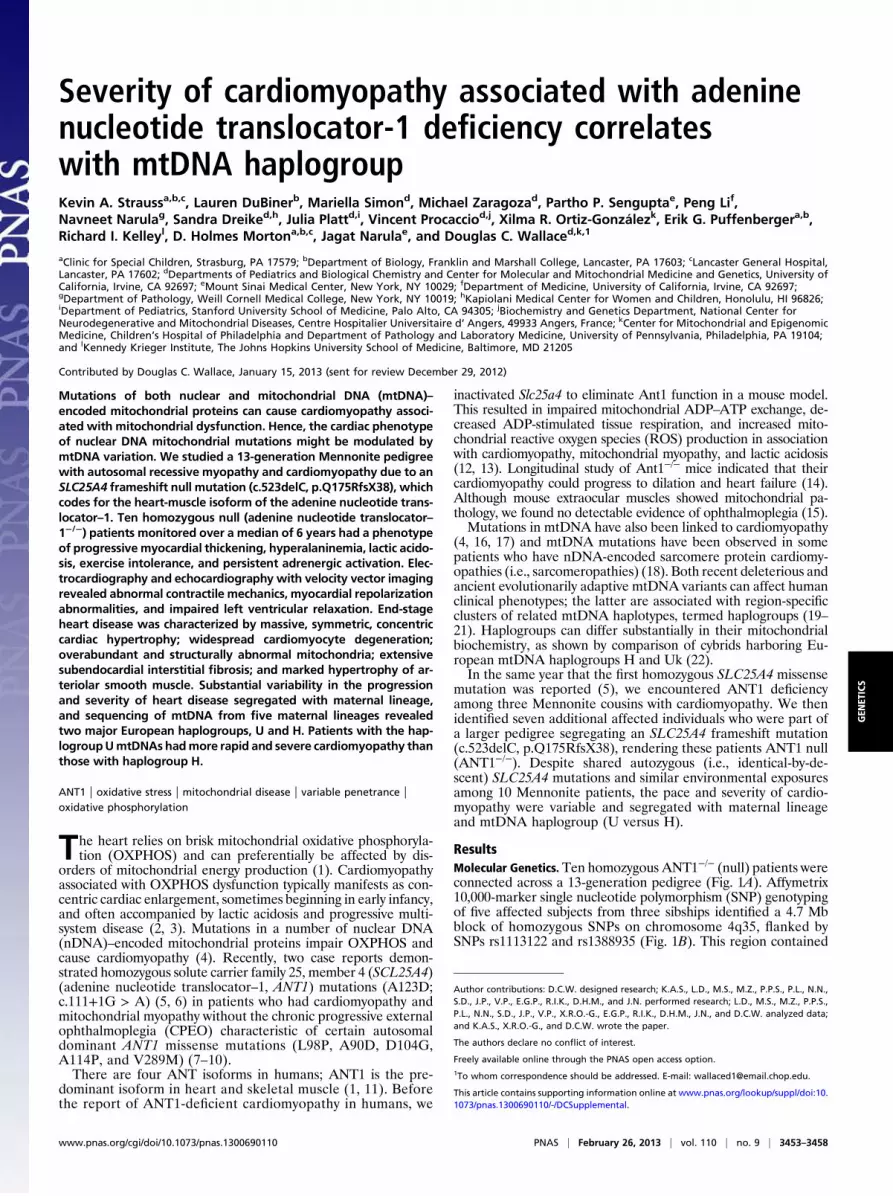
ResultsMolecular Genetics. Ten homozygous ANT1−/− (null) patients wereconnected across a 13-generation pedigree (Fig. 1A). Affymetrix10,000-marker single nucleotide polymorphism (SNP) genotypingof five affected subjects from three sibships identified a 4.7 Mbblock of homozygous SNPs on chromosome 4q35, flanked bySNPs rs1113122 and rs1388935 (Fig. 1B). This region contained
Author contributions: D.C.W. designed research; K.A.S., L.D., M.S., M.Z., P.P.S., P.L., N.N.,S.D., J.P., V.P., E.G.P., R.I.K., D.H.M., and J.N. performed research; L.D., M.S., M.Z., P.P.S.,P.L., N.N., S.D., J.P., V.P., X.R.O.-G., E.G.P., R.I.K., D.H.M., J.N., and D.C.W. analyzed data;and K.A.S., X.R.O.-G., and D.C.W. wrote the paper.
The authors declare no conflict of interest.
Freely available online through the PNAS open access option.1To whom correspondence should be addressed. E-mail: [email protected].
This article contains supporting information online at www.pnas.org/lookup/suppl/doi:10.1073/pnas.1300690110/-/DCSupplemental.
www.pnas.org/cgi/doi/10.1073/pnas.1300690110 PNAS | February 26, 2013 | vol. 110 | no. 9 | 3453–3458
GEN
ETICS

48 RefSeq genes including SLC25A4. Sanger sequencing ofSLC25A4 showed a homozygous single base pair deletion in exon2 (c.523delC) shared among all affected patients (Fig. 1C). Themutation changes codon 175 from glutamine to arginine, pre-maturely terminates translation at codon 212 (p.Q175RfsX38)(Fig.1C and Fig. S1A) and removes over a third of the C terminusof the ANT1 polypeptide, which contains several highly conservedamino acids (R234, R235, R236, and E264) critical to the for-mation of the solute channel (23).Mitochondrial DNAs from five maternal sibships revealed two
European haplogroups, H and U. Haplogroup H mtDNAs com-prised subhaplogroups H1, H5, and H6, and haplogroup UmtDNAs belonged to subhaplogroupU2 (Fig. 1A and Fig. S1B–G).
Clinical Course. Most ANT1−/− infants achieved motor milestoneson schedule but were subjectively weaker than their unaffectedsiblings. Exercise intolerance was first noted during recess activitiesin the early school years. By later childhood, patients regulatedactivities to avoid heavy lifting and strenuous exercise. Amongadults, even modest exertion (e.g., sweeping the floor) could pro-voke weakness, dyspnea, and palpitations. Illnesses were oftenfollowed by protracted fatigue lasting several days. Cognitivefunction, academic performance, vision, and hearing were reportednormal. However, insomnia and inattention were common, andthree of four adult patients suffered from depression and anxiety.There was no uniform treatment strategy. Eight patients rep-
resenting both H and U haplogroups were treated with beta-blockers (nadolol, atenolol, metoprolol, or carvedilol), in twocases coupled to an angiotensin II receptor antagonist and in onecase coupled to a calcium channel blocker. Two sisters (H1)
remained on sustained vitamin-antioxidant therapy (vitamin E,vitamin C, B-complex, coenzyme Q10, and L-carnitine) and re-ceived no cardiac medications. Among H haplogroup patients,data were insufficient to determine if mode of treatment (medi-cation versus vitamin-antioxidant therapy) affected disease pro-gression. Among U haplogroup patients, 5 y (range, 2.4–13.3 y) ofbeta-blocker therapy did not arrest cardiac enlargement (Fig. S2).
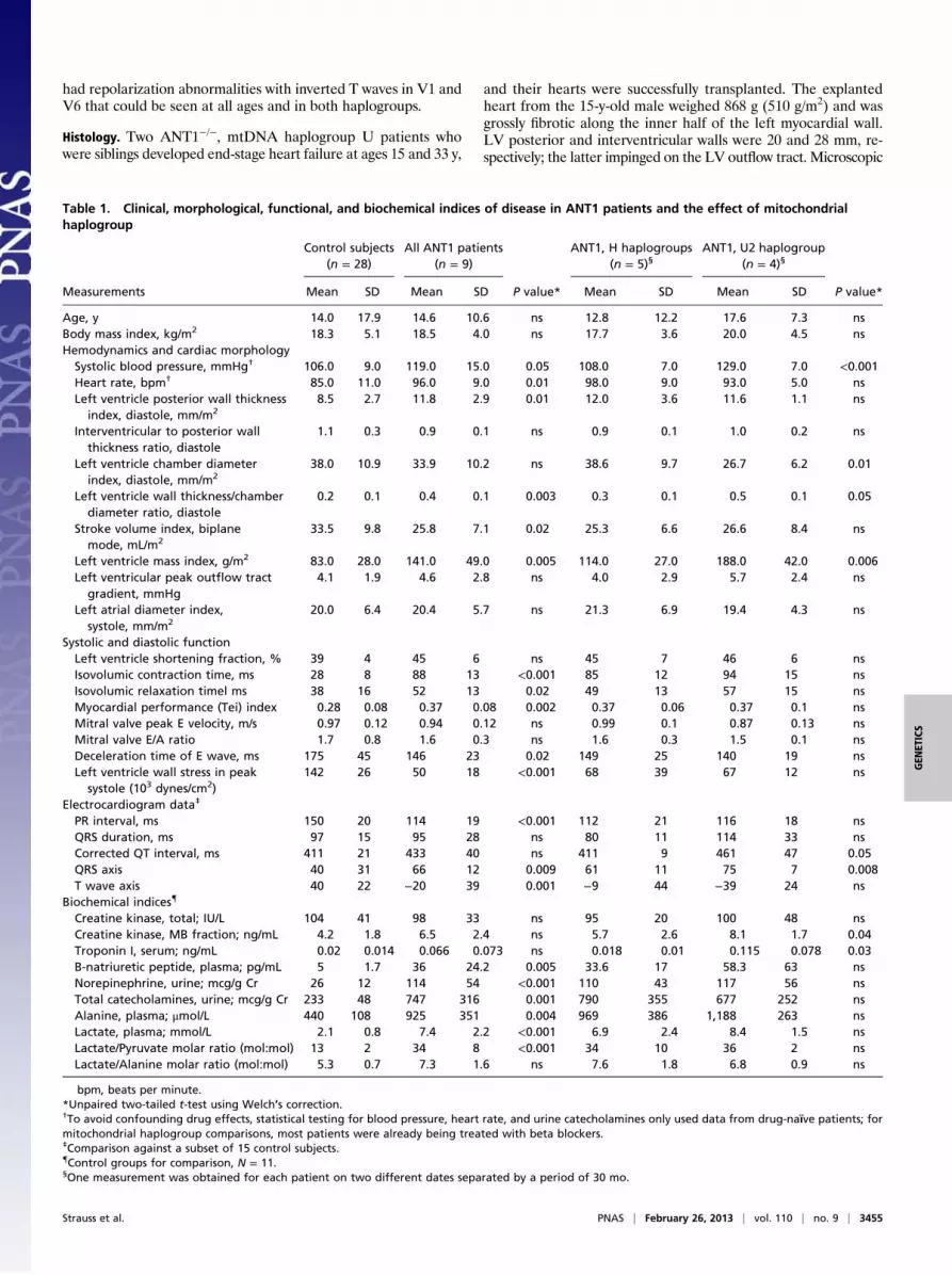
Heart Morphology and Performance. ANT1−/− patients were mildlytachycardic and hypertensive (Table 1). Shortening fraction, leftventricular (LV) outflow gradient, and left atrial diameter werenormal. Progressive concentric myocardial enlargement beganafter 3 y of age. All homozygous null patients had longer iso-volumic contraction and relaxation times, 40% higher myocardialperformance indices (indicating lower myocardial performance),lower estimated cardiac output (mean 2.48 L/m2 min versus 2.85L/m2 min), and shorter mitral valve peak early (E) wave flow ve-locity deceleration times than normal controls. E velocity and itsratio to the atrial (A) wave flow velocities were normal. Only Evelocity correlated with LV mass index among all ANT1−/−
patients (rs = –0.56, P = 0.020).Velocity vector imaging (VVI) echocardiography (14) was
performed on nine patients; eight patients had radial strains lessthan 40% of normal, and four of these patients had attenuatedlongitudinal and circumferential strains (Fig. 2A). Mean PRintervals were below average in ANT1 null patients irrespective ofage or mitochondrial haplogroup but still within the referencerange (Table 1 and Fig. 2 B and C) (24). As expected, all patientshad large R and S wave voltages in precordial leads. Seven of nine
Fig. 1. Extended Mennonite cardiomyopathy pedigree, homozygosity mapping, ANT1 (SLC25A4) frameshift mutant identification, and mtDNA haplogroupdetermination. (A) Genealogical analysis permitted connection of all 10 ANT1−/− patients across 13 generations. Among seven affected sibships, there weretwo haplogroups (H and U) encompassing four different mtDNA subhaplotypes: U2 (red), H5 (violet), H6 (light blue), and H1 (dark blue). (B) To map thechromosomal mutant locus, five affected individuals were screened for regions of shared homozygosity using the Affymetrix 10,000-marker SNP genotypingarray. Chromosome blocks are separated by downward ticks along the horizontal axis, with chromosome 4 indicated. The vertical axis indicates the number ofserial homozygous SNPs shared by all five patients (yellow) or the location score (violet), a calculated value that incorporates population-specific allelefrequencies to determine the likelihood that shared blocks are autozygous. A single shared 4.7 Mb region on chromosome 4q35, flanked by SNPs rs1113122and rs1388935, had the highest location score and contained 48 RefSeq genes, including SLC25A4. (C) Regional sequence of the mutant ANT1 (SLC25A4) geneshowing the c.523delC single base deletion that results in a frame shift (p.Q175RfsX38) that destroys the enzyme (Fig. S1A).
3454 | www.pnas.org/cgi/doi/10.1073/pnas.1300690110 Strauss et al.
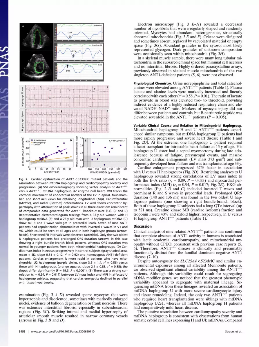
had repolarization abnormalities with inverted T waves in V1 andV6 that could be seen at all ages and in both haplogroups.
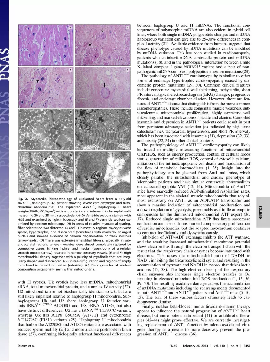
Histology. Two ANT1−/−, mtDNA haplogroup U patients whowere siblings developed end-stage heart failure at ages 15 and 33 y,
and their hearts were successfully transplanted. The explantedheart from the 15-y-old male weighed 868 g (510 g/m2) and wasgrossly fibrotic along the inner half of the left myocardial wall.LV posterior and interventricular walls were 20 and 28 mm, re-spectively; the latter impinged on the LV outflow tract. Microscopic
Table 1. Clinical, morphological, functional, and biochemical indices of disease in ANT1 patients and the effect of mitochondrialhaplogroup
Control subjects(n = 28)
All ANT1 patients(n = 9)
ANT1, H haplogroups(n = 5)§
ANT1, U2 haplogroup(n = 4)§
Measurements Mean SD Mean SD P value* Mean SD Mean SD P value*
Age, y 14.0 17.9 14.6 10.6 ns 12.8 12.2 17.6 7.3 nsBody mass index, kg/m2 18.3 5.1 18.5 4.0 ns 17.7 3.6 20.0 4.5 nsHemodynamics and cardiac morphologySystolic blood pressure, mmHg† 106.0 9.0 119.0 15.0 0.05 108.0 7.0 129.0 7.0 <0.001Heart rate, bpm† 85.0 11.0 96.0 9.0 0.01 98.0 9.0 93.0 5.0 nsLeft ventricle posterior wall thickness
index, diastole, mm/m28.5 2.7 11.8 2.9 0.01 12.0 3.6 11.6 1.1 ns
Interventricular to posterior wallthickness ratio, diastole
1.1 0.3 0.9 0.1 ns 0.9 0.1 1.0 0.2 ns
Left ventricle chamber diameterindex, diastole, mm/m2
38.0 10.9 33.9 10.2 ns 38.6 9.7 26.7 6.2 0.01
Left ventricle wall thickness/chamberdiameter ratio, diastole
0.2 0.1 0.4 0.1 0.003 0.3 0.1 0.5 0.1 0.05
Stroke volume index, biplanemode, mL/m2
33.5 9.8 25.8 7.1 0.02 25.3 6.6 26.6 8.4 ns
Left ventricle mass index, g/m2 83.0 28.0 141.0 49.0 0.005 114.0 27.0 188.0 42.0 0.006Left ventricular peak outflow tract
gradient, mmHg4.1 1.9 4.6 2.8 ns 4.0 2.9 5.7 2.4 ns
Left atrial diameter index,systole, mm/m2
20.0 6.4 20.4 5.7 ns 21.3 6.9 19.4 4.3 ns
Systolic and diastolic functionLeft ventricle shortening fraction, % 39 4 45 6 ns 45 7 46 6 nsIsovolumic contraction time, ms 28 8 88 13 <0.001 85 12 94 15 nsIsovolumic relaxation timel ms 38 16 52 13 0.02 49 13 57 15 nsMyocardial performance (Tei) index 0.28 0.08 0.37 0.08 0.002 0.37 0.06 0.37 0.1 nsMitral valve peak E velocity, m/s 0.97 0.12 0.94 0.12 ns 0.99 0.1 0.87 0.13 nsMitral valve E/A ratio 1.7 0.8 1.6 0.3 ns 1.6 0.3 1.5 0.1 nsDeceleration time of E wave, ms 175 45 146 23 0.02 149 25 140 19 nsLeft ventricle wall stress in peak
systole (103 dynes/cm2)142 26 50 18 <0.001 68 39 67 12 ns
Electrocardiogram data‡
PR interval, ms 150 20 114 19 <0.001 112 21 116 18 nsQRS duration, ms 97 15 95 28 ns 80 11 114 33 nsCorrected QT interval, ms 411 21 433 40 ns 411 9 461 47 0.05QRS axis 40 31 66 12 0.009 61 11 75 7 0.008T wave axis 40 22 −20 39 0.001 −9 44 −39 24 ns
Biochemical indices{
Creatine kinase, total; IU/L 104 41 98 33 ns 95 20 100 48 nsCreatine kinase, MB fraction; ng/mL 4.2 1.8 6.5 2.4 ns 5.7 2.6 8.1 1.7 0.04Troponin I, serum; ng/mL 0.02 0.014 0.066 0.073 ns 0.018 0.01 0.115 0.078 0.03B-natriuretic peptide, plasma; pg/mL 5 1.7 36 24.2 0.005 33.6 17 58.3 63 nsNorepinephrine, urine; mcg/g Cr 26 12 114 54 <0.001 110 43 117 56 nsTotal catecholamines, urine; mcg/g Cr 233 48 747 316 0.001 790 355 677 252 nsAlanine, plasma; μmol/L 440 108 925 351 0.004 969 386 1,188 263 nsLactate, plasma; mmol/L 2.1 0.8 7.4 2.2 <0.001 6.9 2.4 8.4 1.5 nsLactate/Pyruvate molar ratio (mol:mol) 13 2 34 8 <0.001 34 10 36 2 nsLactate/Alanine molar ratio (mol:mol) 5.3 0.7 7.3 1.6 ns 7.6 1.8 6.8 0.9 ns
bpm, beats per minute.*Unpaired two-tailed t-test using Welch’s correction.†To avoid confounding drug effects, statistical testing for blood pressure, heart rate, and urine catecholamines only used data from drug-naïve patients; formitochondrial haplogroup comparisons, most patients were already being treated with beta blockers.‡Comparison against a subset of 15 control subjects.{Control groups for comparison, N = 11.§One measurement was obtained for each patient on two different dates separated by a period of 30 mo.
Strauss et al. PNAS | February 26, 2013 | vol. 110 | no. 9 | 3455
GEN
ETICS
examination (Fig. 3 A–D) revealed sparse myocytes that werehypertrophic and disoriented, sometimes with markedly enlargednuclei, evidence of balloon degeneration or frank necrosis. Therewas extensive interstitial fibrosis, especially in subendocardialregions (Fig. 3C). Striking intimal and medial hypertrophy ofarteriolar smooth muscle resulted in narrow coronary vessels(arrows in Fig. 3 B and D).
Electron microscopy (Fig. 3 E–H) revealed a decreasednumber of myofibrils that were irregularly shaped and randomlyoriented. Myocytes had abundant, heterogeneous, structurallyabnormal mitochondria (Fig. 3 E and F). Cristae were disfiguredand sometimes absent, replaced by vacuolated material or emptyspace (Fig. 3G). Abundant granules in the cytosol most likelyrepresented glycogen. Dark granules of unknown compositionwere occasionally seen within mitochondria (Fig. 3H).In a skeletal muscle sample, there were many long tubular mi-
tochondria in the subsarcolemmal space but minimal cell necrosisand no interstitial fibrosis. Highly ordered paracrystalline arrays,previously observed in skeletal muscle mitochondria of the twosingleton ANT1-deficient patients (5, 6), were not observed.
Physiological Chemistry. Urine norepinephrine and total catechol-amines were elevated among ANT1−/− patients (Table 1). Plasmalactate and alanine levels were markedly increased and linearlycorrelated with each other (r2= 0.58, P= 0.01). The ratio of lactateto pyruvate in blood was elevated two- to threefold, providingindirect evidence of a highly reduced respiratory chain and ele-vated NADH–NAD+ ratio. Markers of myocyte injury did notdiffer between patients and controls, but B-natriuretic peptide waselevated sevenfold in the ANT1−/− patients (P = 0.005).
Variable Clinical Course and Relation to Mitochondrial Haplogroup.Mitochondrial haplogroup H and U ANT1−/− patients experi-enced similar symptoms, but mtDNA haplogroup U patients hadmore rapidly progressive and severe heart disease (Table 1 andFig. 2D). At the extreme, one haplogroup U patient requireda heart transplant for intractable heart failure at 15 y of age. Hishaplogroup U sister had a septal myomectomy during late ado-lescence because of fatigue, presyncopal events, and massiveconcentric cardiac enlargement (LV mass 373 g/m2) and sub-sequently developed heart failure and was transplanted at age 33 y.Cardiac enlargement progressed 67% faster in association
with U versus H haplogroups (Fig. 2D). Restricting analyses to Uhaplogroup revealed strong correlations of LV mass index toboth the E–A ratio (rs = 0.89, P = 0.033) and myocardial per-formance index (MPI) (rs = 0.94, P = 0.017; Fig. 2E). EKG ab-normalities (Fig. 2 B and C) included inverted T waves andprominent R and S waves in precordial leads. Prolonged QRSduration (148 and 136 ms) was found in the two oldest U hap-logroup patients (one showing a right bundle-branch block).Both of these haplogroup U subjects had a long QTc interval (upto 521 ms). Creatine kinase MB (cardiac isoform) fraction andtroponin I were 40% and sixfold higher, respectively, in U versusH haplogroup ANT1−/− patients (Table 1).
DiscussionClinical analysis of nine related ANT1−/− patients has confirmedthat complete absence of ANT1 activity in humans is associatedwith lactic academia, cardiomyopathy, and mitochondrial my-opathy without CPEO, consistent with previous case reports (5,6). Therefore, ANT1−/− disease is clinically and presumablyfunctionally distinct from the familial dominant negative ANT1disease (7–10).Despite autozygosity for SLC25A4 c.523delC and similar en-
vironmental exposures among all affected Mennonite patients,we observed significant clinical variability among the ANT1−/−
patients. Although this variability could result for segregatingnDNA modifier genes, we noticed that the greatest phenotypicvariability appeared to segregate with maternal lineage. Se-quencing mtDNA from these lineages revealed an association ofmtDNA haplogroup U with more severe cardiomyocyte injuryand tissue remodeling. Indeed, the only two ANT1−/− patientswho required heart transplantation were siblings with mtDNAhaplogroup U2e1, whereas all mtDNA haplogroup H patientshad comparatively mild heart disease.The putative association between cardiomyopathy severity and
mtDNA haplogroup is consistent with observations from humansomatic cybrid cell lines expressingH andUkmtDNAs. Compared
Fig. 2. Cardiac dysfunction of ANT1 c.523delC mutant patients and theassociation between mtDNA haplogroup and cardiomyopathy severity andprogression. (A) VVI echocardiography showing vector analysis of ANT1+/+
versus ANT1−/−, mtDNA haplogroup U2 enzyme null heart. VVI tracks thevectorial movement of endocardial borders of the LV in apical, four-cham-ber, and short axis views for obtaining longitudinal (Top), circumferential(Middle), and radial (Bottom) deformations. LV wall shows concentric hy-pertrophy with attenuation of peak strains in all three directions reminiscentof comparable data generated for Ant1−/− knockout mice (14). (B and C)Representative electrocardiogram tracings from a 33-y-old woman with Hhaplogroup mtDNA (B) and a 25-y-old man with U haplogroup mtDNA (C)show tall R and S wave voltages in precordial leads. Seven of nine ANT1patients had repolarization abnormalities with inverted T waves in V1 andV6, which could be seen at all ages and in both haplotype groups (arrow-heads). Shortened PR intervals were observed (asterisks). Only the two oldestU haplogroup patients had prolonged QRS duration (arrow), in this caseshowing a right bundle-branch block pattern, whereas QRS duration wasnormal in younger patients from both mitochondrial haplogroups. (D) Car-diac mass index increases progressively in control subjects (gray shaded area,mean ± SD, slope 0.81 ± 0.12, r2 = 0.92) and homozygous ANT1-deficientpatients. Cardiac enlargement is more rapid in patients who have mito-chondrial U2 haplogroup (purple circles, slope 3.5 ± 1.4, r2 = 0.56) versusthose with H haplogroups (orange squares, slope 2.1 ± 0.88, r2 = 0.88); theslopes differ significantly (F = 19.5, P < 0.0001). (E) There was a strong cor-relation (rs = 0.94, P = 0.017) between LV mass index and MPI in affected Uhaplogroup subjects, suggesting that cardiac energetics declined in parallelwith tissue hypertrophy.
3456 | www.pnas.org/cgi/doi/10.1073/pnas.1300690110 Strauss et al.
with H cybrids, Uk cybrids have less mtDNA, mitochondrialrRNA, total mitochondrial protein, and complex IV activity (22).U2 mitochondria are not biochemically identical to Uk, but arestill likely impaired relative to haplogroup H mitochondria. Sub-haplogroups Uk and U2 share haplogroup U founder vari-ants tRNALeu(CUN) A12308G and 16S rRNA A118G, but alsohave distinct differences: U2 has a tRNAThr T15907C variant,whereas Uk has ATP6 G9055A (A177T) and cytochromeb T14798C (F18L) variants (25). Haplogroup U mitochondriathat harbor the A12308G and A118G variants are associated withreduced sperm motility (26) and more alkaline postmortem braintissue (27), confirming biologically relevant functional differences
between haplogroup U and H mtDNAs. The functional con-sequences of polymorphic mtDNA are also evident in cybrid celllines, where both single mtDNA polypeptide changes and mtDNAhaplogroup variation can give rise to 25–30% differences in com-plex I activity (21). Available evidence from humans suggests thatdisease phenotype caused by nDNA mutations can be modifiedby mtDNA variation. This has been studied in cardiomyopathypatients who co-inherit nDNA contractile protein and mtDNAmutations (18), and in the pathological interaction between a mildX-linked complex I gene NDUFA1 variant and a pair of non-pathogenic mtDNA complex I polypeptidemissense mutations (28).The pathology of ANT1−/− cardiomyopathy is similar to other
forms of end-stage hypertrophic cardiomyopathy caused by sar-comeric protein mutations (29, 30). Common clinical featuresinclude concentric myocardial wall thickening, tachycardia, shortPR interval, typical electrocardiogram (EKG) changes, progressivefibrosis, and end-stage chamber dilation. However, there are fea-tures ofANT1−/− disease that distinguish it from themore commonsarcomeropathies. These include congenital muscle weakness, sub-sarcolemmal mitochondrial proliferation, highly symmetric wallthickening, and marked elevations of lactate and alanine. Comorbidinsomnia and depression in ANT1−/− patients could result in partfrom persistent adrenergic activation (as indicated by high urinecatecholamines, tachycardia, hypertension, and short PR interval),which has been associated with insomnia (31), depression (32, 33),and anxiety (32, 34) in other clinical contexts.The pathophysiology of ANT1−/− cardiomyopathy can likely
be traced to multiple interacting functions of mitochondrialOXPHOS, such as energy production, control of cellular redoxstatus, generation of cellular ROS, control of cytosolic calcium,initiation of the intrinsic apoptotic cell death, and modulation ofthe flow of metabolic intermediates (1, 35). Insight into thepathophysiology can be gleaned from Ant1 null mice, whichclosely parallel the mitochondrial and cardiac phenotype ofMennonite patients and have similar contractile abnormalitieson echocardiographic VVI (12, 14). Mitochondria of Ant1−/−
mice have markedly reduced ADP-stimulated respiration rates,most apparent in the skeletal muscle mitochondria that rely al-most exclusively on ANT1 as an ADP/ATP translocator andshow a massive induction of mitochondrial proliferation anda down-regulation of glycolysis, presumably as a futile attempt tocompensate for the diminished mitochondrial ATP export (36,37). Reduced single mitochondrion ATP flux limits sarcomerecontraction and also entrains marked compensatory proliferationof cardiac mitochondria, but the adapted myocardium continuesto contract inefficiently and dyssynchronously.Inhibition of ATP–ADP exchange inhibits the ATP synthase,
and the resulting increased mitochondrial membrane potentialslows electron flux through the electron transport chain with theresult that the respiratory chain enzymes become saturated withelectrons. This raises the mitochondrial ratio of NADH toNAD+, inhibiting the tricarboxylic acid cycle, and resulting in theaccumulation of pyruvate and NADH in cytosol that drives lacticacidosis (12, 38). The high electron density of the respiratorychain enzymes also increases single electron transfer to O2,resulting in elevated mitochondrial ROS production (1, 13, 36,39, 40). The resulting oxidative damage causes the accumulationof mtDNA mutations including the rearrangements documentedin both ANT1−/− and ANT1+/− patients and Ant1−/− mice (5–10,13). The sum of these various factors ultimately leads to car-diomyocyte demise.Finally, neither beta-blocker nor antioxidant-vitamin therapy
appear to influence the natural progression of ANT1−/− heartdisease, but more potent antioxidant (41) or antifibrotic thera-pies (42–44) might prove effective. We are currently investigat-ing replacement of ANT1 function by adeno-associated virusgene therapy as a means to more decisively prevent the pro-gression of ANT1−/− disease (36).
Fig. 3. Myocardial histopathology of explanted heart from a 15-y-oldANT1−/−, haplogroup U2, patient showing severe cardiomyocyte and mito-chondrial abnormalities. The explanted ANT1−/−, haplogroup U heartweighed 868 g (510 g/m2) with left posterior and interventricular septal wallsmeasuring 20 and 28 mm, respectively. (A–D) Ventricle sections stained withH&E and examined by light microscopy and (E and F) ventricle sections ex-amined by electron microscopy. (A) In areas of relative myocardial sparing,fiber orientation was distorted. (B and C) In most LV regions, myocytes weresparse, hypertrophic, and disoriented (sometimes with markedly enlargednuclei) and showed evidence of balloon degeneration or frank necrosis(arrowheads). (D) There was extensive interstitial fibrosis, especially in sub-endocardial regions, where myocytes were almost completely replaced byconnective tissue. Striking intimal and medial hypertrophy of arteriolarsmooth muscle (arrow) resulted in narrow coronary vessels. (E and F) Highmitochondrial density together with a paucity of myofibrils that are irreg-ularly shaped and disoriented. (G) Cristae disfiguration and regions of emptymitochondria devoid of cristae (asterisks). (H) Dark granules of unclearcomposition occasionally seen within mitochondria.
Strauss et al. PNAS | February 26, 2013 | vol. 110 | no. 9 | 3457
GEN
ETICS
Patients and MethodsThis study was approved by the Institutional Review Boards of LancasterGeneral Hospital and the University of California, Irvine. Adult patientsconsented in writing to participate and parents consented for their children.Ten patients of Northeastern Mennonite ancestry were studied. One hada cardiac transplant before molecular diagnosis; thus, nine (age, 14.6 ± 10.6 y;range, 1–36 y; seven female) were available for detailed longitudinal studies.Standard histological sections and electron micrographs were examinedfrom the explanted heart and a single skeletal muscle biopsy.
We used five affected children from four sibships to scan 10K-marker SNPgenotypes (Affymetrix) for regions of autozygosity (identity-by-descent) (45)and sequenced target genes by the Sanger method (46). mtDNA sequencing
and haplogroup assignment were performed as previously described (25, 47)and reported as differences from the revised Cambridge Reference Se-quence (48). Clinical data were collected during routine office visits overa median of 6 (range, 4–16) y, but we restricted statistical analyses for Table1 to values obtained on two separate days (2008 and 2011) spaced 30 moapart. Clinical data collection by ultrasound, electrocardiographic, andphysiological chemical methods are detailed in SI Patients and Methods.
ACKNOWLEDGMENTS. The authors deeply appreciate the assistance of Ms.Marie Lott for preparation of this manuscript. This work was supported byPostdoctoral Fellowship 5T32NS007413-14 (to X.R.O.-G.) and National Insti-tutes of Health R01 Grants NS41850, NS21328, and DK73691 (to D.C.W.).
1. Wallace DC (2005) A mitochondrial paradigm of metabolic and degenerative diseases,aging, and cancer: A dawn for evolutionary medicine. Annu Rev Genet 39:359–407.
2. Nishizawa M, et al. (1987) A mitochondrial encephalomyopathy with cardiomyopa-thy. A case revealing a defect of complex I in the respiratory chain. J Neurol Sci 78(2):189–201.
3. Guenthard J, Wyler F, Fowler B, Baumgartner R (1995) Cardiomyopathy in respiratorychain disorders. Arch Dis Child 72(3):223–226.
4. Wallace DC, Lott MT, Procaccio V (2013) Emery and Rimoin’s Principles and Practice ofMedical Genetics, Mitochondrial Medicine: The Mitochondrial Biology and Geneticsof Metabolic and Degenerative Diseases, Cancer, and Aging, Chapter 13, eds RimoinDL, Pyeritz RE, Korf BR (Churchill Livingstone Elsevier, Philadelphia), 6th Ed, Vol 1.
5. Palmieri L, et al. (2005) Complete loss-of-function of the heart/muscle-specific adeninenucleotide translocator is associated with mitochondrial myopathy and cardiomyop-athy. Hum Mol Genet 14(20):3079–3088.
6. Echaniz-Laguna A, et al. (2012) Complete loss of expression of the ANT1 gene causingcardiomyopathy and myopathy. J Med Genet 49(2):146–150.
7. Kaukonen J, et al. (2000) Role of adenine nucleotide translocator 1 in mtDNAmaintenance. Science 289(5480):782–785.
8. Chen XJ (2002) Induction of an unregulated channel by mutations in adenine nu-cleotide translocase suggests an explanation for human ophthalmoplegia. Hum MolGenet 11(16):1835–1843.
9. De Marcos Lousa C, et al. (2005) Valine 181 is critical for the nucleotide exchangeactivity of human mitochondrial ADP/ATP carriers in yeast. Biochemistry 44(11):4342–4348.
10. De Marcos Lousa C, Trézéguet V, Dianoux AC, Brandolin G, Lauquin GJ (2002) Thehuman mitochondrial ADP/ATP carriers: Kinetic properties and biogenesis of wild-type and mutant proteins in the yeast S. cerevisiae. Biochemistry 41(48):14412–14420.
11. Stepien G, Torroni A, Chung AB, Hodge JA, Wallace DC (1992) Differential expressionof adenine nucleotide translocator isoforms in mammalian tissues and during musclecell differentiation. J Biol Chem 267(21):14592–14597.
12. Graham BH, et al. (1997) A mouse model for mitochondrial myopathy and cardio-myopathy resulting from a deficiency in the heart/muscle isoform of the adeninenucleotide translocator. Nat Genet 16(3):226–234.
13. Esposito LA, Melov S, Panov A, Cottrell BA, Wallace DC (1999) Mitochondrial diseasein mouse results in increased oxidative stress. Proc Natl Acad Sci USA 96(9):4820–4825.
14. Narula N, et al. (2011) Adenine nucleotide translocase 1 deficiency results in dilatedcardiomyopathy with defects in myocardial mechanics, histopathological alterations,and activation of apoptosis. JACC Cardiovasc Imaging 4(1):1–10.
15. Yin H, et al. (2005) Eliminating the Ant1 isoform produces a mouse with CPEO pa-thology but normal ocular motility. Invest Ophthalmol Vis Sci 46(12):4555–4562.
16. Arbustini E, et al. (1998) Mitochondrial DNA mutations and mitochondrial abnor-malities in dilated cardiomyopathy. Am J Pathol 153(5):1501–1510.
17. Zaragoza MV, Brandon MC, Diegoli M, Arbustini E, Wallace DC (2011) Mitochondrialcardiomyopathies: How to identify candidate pathogenic mutations by mitochondrialDNA sequencing, MITOMASTER and phylogeny. Eur J Hum Genet 19(2):200–207.
18. Arbustini E, et al. (1998) Coexistence of mitochondrial DNA and beta myosin heavychain mutations in hypertrophic cardiomyopathy with late congestive heart failure.Heart 80(6):548–558.
19. Ruiz-Pesini E, Mishmar D, Brandon M, Procaccio V, Wallace DC (2004) Effects of pu-rifying and adaptive selection on regional variation in human mtDNA. Science303(5655):223–226.
20. Ruiz-Pesini E, Wallace DC (2006) Evidence for adaptive selection acting on the tRNAand rRNA genes of human mitochondrial DNA. Hum Mutat 27(11):1072–1081.
21. Ji F, et al. (2012) Mitochondrial DNA variant associated with Leber hereditary opticneuropathy and high-altitude Tibetans. Proc Natl Acad Sci USA 109(19):7391–7396.
22. Gómez-Durán A, et al. (2010) Unmasking the causes of multifactorial disorders: OX-PHOS differences between mitochondrial haplogroups. Hum Mol Genet 19(17):3343–3353.
23. Pebay-Peyroula E, et al. (2003) Structure of mitochondrial ADP/ATP carrier in complexwith carboxyatractyloside. Nature 426(6962):39–44.
24. Sharieff GQ, Rao SO (2006) The pediatric ECG. Emerg Med Clin North Am 24(1):195–vii–208viii.
25. Ruiz-Pesini E, et al. (2007) An enhanced MITOMAP with a global mtDNA mutationalphylogeny. Nucleic Acids Res 35(Database issue):D823–D828.
26. Montiel-Sosa F, et al. (2006) Differences of sperm motility in mitochondrial DNAhaplogroup U sublineages. Gene 368:21–27.
27. Rollins B, et al. (2009) Mitochondrial variants in schizophrenia, bipolar disorder, andmajor depressive disorder. PLoS ONE 4(3):e4913.
28. Potluri P, et al. (2009) A novel NDUFA1 mutation leads to a progressive mitochondrialcomplex I-specific neurodegenerative disease. Mol Genet Metab 96(4):189–195.
29. Elliott P, McKenna WJ (2004) Hypertrophic cardiomyopathy. Lancet 363(9424):1881–1891.
30. Maron BJ (2002) Hypertrophic cardiomyopathy: A systematic review. JAMA 287(10):1308–1320.
31. Mitchell HA, Weinshenker D (2010) Good night and good luck: Norepinephrine insleep pharmacology. Biochem Pharmacol 79(6):801–809.
32. Boulenger JP, Uhde TW (1982) Biological peripheral correlates of anxiety. Encephale8(2):119–130.
33. Oei TP, Dingle GA, McCarthy M (2010) Urinary catecholamine levels and responseto group cognitive behaviour therapy in depression. Behav Cogn Psychother 38(4):479–483.
34. Pervanidou P, Chrousos GP (2010) Neuroendocrinology of post-traumatic stress dis-order. Prog Brain Res 182:149–160.
35. Wallace DC (2012) Mitochondria and cancer. Nat Rev Cancer 12(10):685–698.36. Murdock DG, Boone BE, Esposito LA, Wallace DC (1999) Up-regulation of nuclear and
mitochondrial genes in the skeletal muscle of mice lacking the heart/muscle isoformof the adenine nucleotide translocator. J Biol Chem 274(20):14429–14433.
37. Subramaniam V, et al. (2008) MITOCHIP assessment of differential gene expression inthe skeletal muscle of Ant1 knockout mice: Coordinate regulation of OXPHOS, an-tioxidant, and apoptotic genes. Biochim Biophys Acta 1777(7-8):666–675.
38. Flierl A, Chen Y, Coskun PE, Samulski RJ, Wallace DC (2005) Adeno-associated virus-mediated gene transfer of the heart/muscle adenine nucleotide translocator (ANT) inmouse. Gene Ther 12(7):570–578.
39. Pryde KR, Hirst J (2011) Superoxide is produced by the reduced flavin in mitochondrialcomplex I: A single, unified mechanism that applies during both forward and reverseelectron transfer. J Biol Chem 286(20):18056–18065.
40. Lombardi R, et al. (2009) Resolution of established cardiac hypertrophy and fibrosisand prevention of systolic dysfunction in a transgenic rabbit model of human car-diomyopathy through thiol-sensitive mechanisms. Circulation 119(10):1398–1407.
41. Wallace DC, Fan W, Procaccio V (2010) Mitochondrial energetics and therapeutics.Annu Rev Pathol 5:297–348.
42. Cacciapuoti F (2011) Molecular mechanisms of left ventricular hypertrophy (LVH) insystemic hypertension (SH)-possible therapeutic perspectives. J Am Soc Hypertens5(6):449–455.
43. Kuster GM, et al. (2005) Alpha-adrenergic receptor-stimulated hypertrophy in adultrat ventricular myocytes is mediated via thioredoxin-1-sensitive oxidative modifica-tion of thiols on Ras. Circulation 111(9):1192–1198.
44. Teekakirikul P, et al. (2010) Cardiac fibrosis in mice with hypertrophic cardiomyopathyis mediated by non-myocyte proliferation and requires Tgf-β. J Clin Invest 120(10):3520–3529.
45. Strauss KA, Puffenberger EG (2009) Genetics, medicine, and the Plain people. AnnuRev Genomics Hum Genet 10:513–536.
46. Puffenberger EG, et al. (2004) Mapping of sudden infant death with dysgenesis of thetestes syndrome (SIDDT) by a SNP genome scan and identification of TSPYL loss offunction. Proc Natl Acad Sci USA 101(32):11689–11694.
47. Poole JC, Procaccio V, Brandon MC, Merrick G, Wallace DC (2010) Multiplex analysisof mitochondrial DNA pathogenic and polymorphic sequence variants. Biol Chem391(10):1115–1130.
48. Andrews RM, et al. (1999) Reanalysis and revision of the Cambridge reference se-quence for human mitochondrial DNA. Nat Genet 23(2):147.
3458 | www.pnas.org/cgi/doi/10.1073/pnas.1300690110 Strauss et al.


