Embed Size (px)
Citation preview
Self-Regulation of Candida albicansPopulation Size during GI ColonizationSarah Jane White
1¤a, Ari Rosenbach
1, Paul Lephart
1¤b, Diem Nguyen
1, Alana Benjamin
1¤c, Saul Tzipori
2,
Malcolm Whiteway3
, Joan Mecsas1
, Carol A. Kumamoto1*
1 Department of Molecular Biology and Microbiology, Tufts University, Boston, Massachusetts, United States of America, 2 Division of Infectious Diseases, Tufts University,
Grafton, Massachusetts, United States of America, 3 Genetics Group, Biotechnology Research Institute, National Research Council, Montreal, Quebec, Canada
Interactions between colonizing commensal microorganisms and their hosts play important roles in health anddisease. The opportunistic fungal pathogen Candida albicans is a common component of human intestinal flora. Togain insight into C. albicans colonization, genes expressed by fungi grown within a host were studied. The EFH1 gene,encoding a putative transcription factor, was highly expressed during growth of C. albicans in the intestinal tract.Counterintuitively, an efh1 null mutant exhibited increased colonization of the murine intestinal tract, a model ofcommensal colonization, whereas an EFH1 overexpressing strain exhibited reduced colonization of the intestinal tractand of the oral cavity of athymic mice, the latter situation modeling human mucosal candidiasis. When inoculated intothe bloodstream of mice, both efh1 null and EFH1 overexpressing strains caused lethal infections. In contrast, othermutants are attenuated in virulence following intravenous inoculation but exhibited normal levels of intestinalcolonization. Finally, although expression of several genes is dependent on transcription factor Efg1p duringlaboratory growth, Efg1p-independent expression of these genes was observed during growth within the murineintestinal tract. These results show that expression of EFH1 regulated the level of colonizing fungi, favoringcommensalism as opposed to candidiasis. Also, different genes are required in different host niches and the pathway(s)that regulates gene expression during host colonization can differ from well-characterized pathways used duringlaboratory growth.
Citation: White SJ, Rosenbach A, Lephart P, Nguyen D, Benjamin A, et al. (2007) Self-regulation of Candida albicans population size during GI colonization. PLoS Pathog 3(12):e184. doi:10.1371/journal.ppat.0030184
Introduction
The opportunistic fungal pathogen Candida albicans colo-nizes its host long before disease arises. In humans, C. albicanscolonization of the oral cavity is detected in most infants bythe age of one month [1]. The majority of adults aredetectably colonized in the intestinal tract by C. albicans [2].Colonization is believed to persist for long periods of time [2],and in this situation, C. albicans is primarily nonpathogenic.However, if the host becomes immunocompromised, diseasecaused by dissemination of commensal organisms from theintestinal tract can occur. For example, disseminatedcandidiasis occurs in neutropenic patients, and Candida sppare among the most common organisms isolated from theblood of hospitalized patients [3]. In AIDS patients, orophar-yngeal candidiasis (OPC) is a common opportunistic in-fection [4]. Despite the importance of commensal organismsas the source of infection, little is known about C. albicansfactors that influence intestinal colonization.
A growing body of literature indicates that the interplaybetween commensal organisms and the host GI tract ischaracterized by reciprocal regulatory interactions. In addi-tion to a role in nutrition, normal flora are required forproper development of the intestinal capillary network aswell as Peyer’s patches and other components of the intestinalimmune system [5]. Normal flora stimulate host toll-likereceptors, and these interactions are important for regu-lation of the inflammatory response. In the absence of toll-like receptors, dysregulation of the inflammatory responsewith concomitant damage to the GI tract occurs [6]. Thus, theflora make important contributions to the health of the host.
The host also influences its flora. Bulk movement ofmaterial through the GI tract regulates populations ofcommensal organisms [7], and host secretions and immuneeffectors play key roles in regulating colonization anddetermining the composition of the flora [8]. Therefore,reciprocal interactions between the commensal flora and thehost maintain the balance between overexuberant inflamma-tion and uncontrolled growth of microorganisms.To gain insight into the activities of C. albicans that are
important for host colonization or disease, studies of C.albicans gene expression during interaction with a host or host
Editor: Brendan P. Cormack, Johns Hopkins University School of Medicine, UnitedStates of America
Received April 16, 2007; Accepted October 22, 2007; Published December 7, 2007
Copyright: � 2007 White et al. This is an open-access article distributed under theterms of the Creative Commons Attribution License, which permits unrestricteduse, distribution, and reproduction in any medium, provided the original authorand source are credited.
Abbreviations: CFU, colony forming unit; CI, competitive index; IGB, immunosup-pressed gnotobiotic; OPC, oropharyngeal candidiasis; qRT-PCR, quantitative real-time reverse transcriptase PCR; qPCR, quantitative PCR; WT, wild type; yEGFP, yeast-enhanced green fluorescent protein
* To whom correspondence should be addressed. E-mail: [email protected]
¤a Current address: Department of Chemistry, Massachusetts Institute ofTechnology, Cambridge, Massachusetts, United States of America
¤b Current address: Clinical Microbiology/Microbiology and Immunology, Univer-sity of Rochester School of Medicine and Dentistry, Rochester, New York, UnitedStates of America
¤c Current address: Santa Rosa Family Medicine Program, Santa Rosa, California,United States of America
PLoS Pathogens | www.plospathogens.org December 2007 | Volume 3 | Issue 12 | e1841866
cells have been pursued. Microarray studies analyzinginteractions between C. albicans and cultured immune cellshave detected dramatic changes in C. albicans metabolism andstress responses [9–11]. For example, genes encoding enzymesof the glyoxylate cycle are highly expressed in phagocytosedfungal cells, and isocitrate lyase is important for systemicvirulence [9]. A study of antigens expressed during oralinfection revealed that components of a MAP kinase signaltransduction pathway were expressed [12]. In addition, theNot5 protein is expressed during oral infection and isrequired for systemic virulence. Recent analysis of C. albicanscells invading host parenchymal tissue revealed changes inexpression of numerous genes, demonstrating that invadingcells experience metabolic changes and initiate responses tostresses such as iron limitation [13].
The goals of this study were to identify C. albicans genes thatwere expressed during growth within a host and to comparethe genetic requirements for infection and intestinal colo-nization. Therefore, genes expressed in C. albicans cellsassociated with oral infection or in cells growing in theintestinal tract were identified, and the ability of mutantslacking some of the genes to colonize the murine intestinaltract or to produce disease was characterized. Mutantslacking the EFH1 gene, encoding a putative transcriptionfactor [14], colonized the intestinal tract at higher levels thanwild-type (WT) organisms. Strains engineered to overexpressEFH1 exhibited reduced levels of intestinal colonization andreduced colonization of the oral cavity of immunodeficient(athymic) mice. Therefore, EFH1 is important for determin-ing the population size of C. albicans during colonization.Despite its importance during intestinal colonization, EFH1did not affect virulence in the disseminated infection modelor cellular physiology under laboratory conditions. Incontrast, RBT1, encoding a putative cell wall protein, andRBT4, encoding a possible secreted protein, are required fornormal virulence in the disseminated infection model [15]but are not required for normal intestinal colonization,
despite their high expression during growth in the intestinaltract. Therefore, genes that influence commensal coloniza-tion can be distinct from genes that are required forvirulence.
Results
C. albicans Gene Expression during Growth within anImmunosuppressed Mammalian HostTo initiate studies on C. albicans genes required for
infection or colonization, preliminary microarray analysisof gene expression in C. albicans cells recovered from sites oforal infection was performed. WT C. albicans cells were orallyinoculated into germ-free newborn piglets as described [16].Following immunosuppression with cyclosporine A andmethylprednisolone for 7–10 d, the animals developeddramatic thrush and esosphagitis, and C. albicans plaqueswere visible in the esophagus at necropsy. C. albicans cells werescraped from the tongues of two immunosuppressed, gnoto-biotic (IGB) piglets and from the esophagus of one of themand were used for microarray analysis as described inMaterials and Methods. Because the majority of spotsexhibited poor hybridization with samples from the oralinfections, a comprehensive analysis of differential geneexpression could not be performed. However, there wereseveral candidate genes more highly expressed in samplestaken from infected piglets than in reference laboratory-grown cells.To confirm that candidate genes from the microarray
studies exhibited increased expression in mammalian hostscompared to log phase laboratory-grown cells, quantitativereal-time reverse transcriptase PCR (qRT-PCR) was per-formed. For the analysis, RNA was prepared from C. albicanscells scraped from the tongue, esophagus, or roof of themouth of two IGB piglets or recovered from the contents ofthe large intestine of one IGB piglet. cDNA prepared fromthe RNA was used as the template for qRT-PCR amplificationas described in Materials and Methods. Results werenormalized using C. albicans ACT1 (encoding actin) and areshown relative to the reference sample of log phase,laboratory-grown cells that were used for the microarrays.Seven genes that exhibited relatively high expression in C.
albicans cells recovered from the oral cavity or intestinal tractwere identified, including EFH1, YHB5, and ECE1 (Table 1),which became the subjects of this study. EFH1 encodes aparalog of the well-studied transcription factor Efg1p [17].Efg1p regulates several morphological transitions as well as
Table 1. Gene Designations
Gene Name ORF19 Designation CYGDa Designation
EFH1 Orf19.5498 CA2672
YHB5 Orf19.3710 CA3139
SOD3 Orf19.7111.1 CA5587
ECE1 Orf19.3374 CA1402
RBT1 Orf19.1327 CA2830
RBT4 Orf19.6202 CA0104
TEF1 Orf19.1435 CA0362
aComprehensive Yeast Genome Database.doi:10.1371/journal.ppat.0030184.t001
PLoS Pathogens | www.plospathogens.org December 2007 | Volume 3 | Issue 12 | e1841867
Self-Regulation of Colonization
Author Summary
Although the fungus Candida albicans commonly colonizes thehuman gastrointestinal tract as a commensal, the organism is alsoan opportunistic pathogen, responsible for a wide range ofinfections in immunocompromised persons. While numerousstudies of infection have been conducted, few studies have analyzedthe commensal state. The studies described here analyze C. albicanscells colonizing the intestinal tract of immunocompetent mice in theabsence of disease, a model for commensalism. Results showed thatexpression of the putative transcription factor Efh1p by cellscolonizing the intestinal tract was relatively high, but paradoxically,expression of Efh1p was associated with lower colonization. Efh1phad no detectable effect on the ability of C. albicans to cause lethaldisseminated infection in mice. In contrast, Rbt1p and Rbt4p, twoproteins of poorly defined function required for normal dissemi-nated infection, were not required for intestinal colonization. Theseresults argue that the commensal state is distinct from thepathogenic state and that different factors are important in differentstates. Also, the regulation of expression of genes RBT1, RBT4, andECE1 during intestinal colonization differed from their well-characterized regulation during laboratory growth. Further studiesof commensal colonization are needed to understand this importantstage of C. albicans biology.
the expression of hyphal and virulence genes [14,18–21].However, the absence of Efh1p does not lead to a pronouncedphenotype during laboratory growth [14], and thus, itsfunction is not understood. YHB5 (orf19.3710) encodes aflavohemoglobin; its paralog, YHB1, is induced by growth inthe presence of nitric oxide and is important for resistance tonitrosative stress [22,23]. YHB5, by contrast, is not inducedduring laboratory growth in nitric oxide [22]. ECE1, a genethat influences adhesion [24], but whose function is poorlyunderstood, is highly expressed by cells that are growing inthe hyphal form (filamentous cells lacking constrictions attheir septa) [21,24,25]. ECE1 is also highly expressed duringinvasion of host tissue [13].
In addition, other genes expressed under laboratoryconditions that resulted in increased ECE1 or EFH1 expres-sion (hyphal growth and post-exponential phase, respectively)were analyzed. RBT1 and RBT4, genes of poorly understoodfunction that are expressed in hyphal cells [15], and SOD3(orf19.7111.1), encoding a manganese-containing superoxidedismutase that is expressed in post-exponential phase [26],were studied. Finally, a control housekeeping gene, TEF1(encoding translation elongation factor 1-alpha), was ana-lyzed in some experiments.
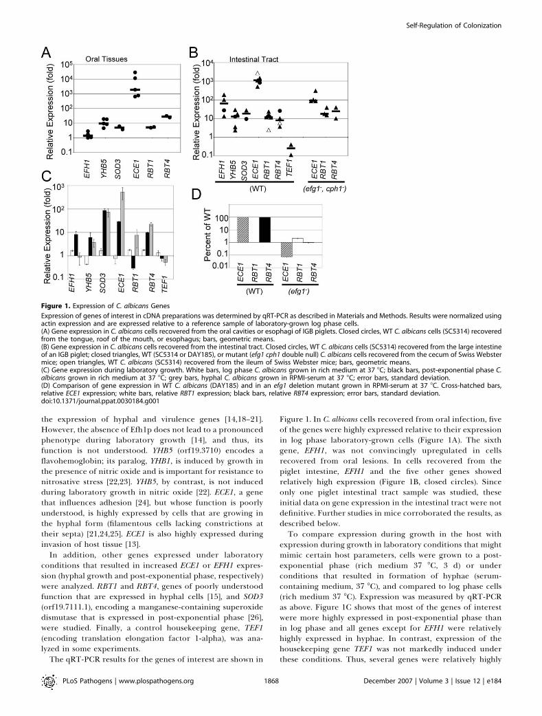
The qRT-PCR results for the genes of interest are shown in
Figure 1. In C. albicans cells recovered from oral infection, fiveof the genes were highly expressed relative to their expressionin log phase laboratory-grown cells (Figure 1A). The sixthgene, EFH1, was not convincingly upregulated in cellsrecovered from oral lesions. In cells recovered from thepiglet intestine, EFH1 and the five other genes showedrelatively high expression (Figure 1B, closed circles). Sinceonly one piglet intestinal tract sample was studied, theseinitial data on gene expression in the intestinal tract were notdefinitive. Further studies in mice corroborated the results, asdescribed below.To compare expression during growth in the host with
expression during growth in laboratory conditions that mightmimic certain host parameters, cells were grown to a post-exponential phase (rich medium 37 8C, 3 d) or underconditions that resulted in formation of hyphae (serum-containing medium, 37 8C), and compared to log phase cells(rich medium 37 8C). Expression was measured by qRT-PCRas above. Figure 1C shows that most of the genes of interestwere more highly expressed in post-exponential phase thanin log phase and all genes except for EFH1 were relativelyhighly expressed in hyphae. In contrast, expression of thehousekeeping gene TEF1 was not markedly induced underthese conditions. Thus, several genes were relatively highly
Figure 1. Expression of C. albicans Genes
Expression of genes of interest in cDNA preparations was determined by qRT-PCR as described in Materials and Methods. Results were normalized usingactin expression and are expressed relative to a reference sample of laboratory-grown log phase cells.(A) Gene expression in C. albicans cells recovered from the oral cavities or esophagi of IGB piglets. Closed circles, WT C. albicans cells (SC5314) recoveredfrom the tongue, roof of the mouth, or esophagus; bars, geometric means.(B) Gene expression in C. albicans cells recovered from the intestinal tract. Closed circles, WT C. albicans cells (SC5314) recovered from the large intestineof an IGB piglet; closed triangles, WT (SC5314 or DAY185), or mutant (efg1 cph1 double null) C. albicans cells recovered from the cecum of Swiss Webstermice; open triangles, WT C. albicans (SC5314) recovered from the ileum of Swiss Webster mice; bars, geometric means.(C) Gene expression during laboratory growth. White bars, log phase C. albicans grown in rich medium at 37 8C; black bars, post-exponential phase C.albicans grown in rich medium at 37 8C; grey bars, hyphal C. albicans grown in RPMI-serum at 37 8C; error bars, standard deviation.(D) Comparison of gene expression in WT C. albicans (DAY185) and in an efg1 deletion mutant grown in RPMI-serum at 37 8C. Cross-hatched bars,relative ECE1 expression; white bars, relative RBT1 expression; black bars, relative RBT4 expression; error bars, standard deviation.doi:10.1371/journal.ppat.0030184.g001
PLoS Pathogens | www.plospathogens.org December 2007 | Volume 3 | Issue 12 | e1841868
Self-Regulation of Colonization
expressed during growth within multiple tissues in the hostand in either post-exponential phase or hyphal growth in thelaboratory.
Gene Expression during Intestinal Colonization Is Not
Dependent on Host ImmunosuppressionFor further studies of the interaction between C. albicans
and a mammalian host, we analyzed gene expression duringcolonization of immunocompetent Swiss Webster mice.Following oral inoculation of antibiotic-treated Swiss Web-ster mice, commensal colonization persisted for several weeksas previously described, e.g., [27–29]. No symptoms of diseasewere observed in the colonized mice.
To determine whether the genes of interest were expressedduring commensal intestinal colonization, gene expressionwas measured by qRT-PCR using RNA prepared from C.albicans cells recovered from the cecum or ileum of colonizedmice. Expression normalized to ACT1 is expressed relative tothe same reference sample as above. As shown in Figure 1B(closed triangles), all six genes of interest were expressed atrelatively high levels in C. albicans cells recovered from themouse cecum in contrast to the housekeeping gene TEF1.Expression was very similar to the expression observed in C.albicans cells recovered from the IGB piglet intestinal tract.Therefore, the expression of these genes was not dependenton immunosuppression of the host. In addition, to compareexpression in a different part of the intestinal tract,expression of three genes was characterized in cells recoveredfrom the ileum. ECE1 and RBT4 were relatively highly
expressed and RBT1 was slightly increased. In summary, fiveof the six genes were relatively highly expressed duringgrowth within multiple tissues of immunosuppressed andimmunocompetent hosts. EFH1 exhibited a distinct patternof expression as it was relatively highly expressed in C. albicanscells recovered from the intestinal tract but not from sites oforal infection (esophagus or tongue lesions).
Uncharacterized Regulatory Pathways Control GeneExpression during Growth in the Intestinal TractBecause most of the genes of interest were highly expressed
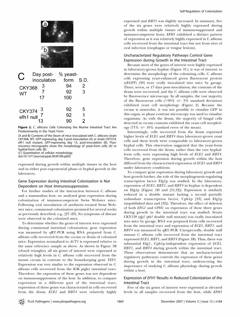
in laboratory-grown hyphae (Figure 1C), it was of interest todetermine the morphology of the colonizing cells. C. albicanscells expressing yeast-enhanced green fluorescent protein(yEGFP) [30] were orally inoculated into mice by gavage.Three, seven, or 17 days post-inoculation, the contents of theileum were recovered, and the C. albicans cells were observedby fluorescence microscopy. In all samples, the vast majorityof the fluorescent cells (.90% þ/� 3% standard deviation)exhibited yeast cell morphology (Figure 2). Because thececum is anaerobic, it was not possible to visualize GFP inthis organ, so phase contrast microscopy was used to visualizeorganisms. As with the ileum, the majority of fungal cellsdetected in cecum contents exhibited the yeast-cell morphol-ogy (71% þ/� 10% standard error of the mean).Interestingly, cells recovered from the ileum expressed
higher levels of ECE1 and RBT4 than laboratory-grown yeastcells and these levels were comparable to laboratory-grownhyphal cells. This observation suggested that the yeast-formcells recovered from the ileum, rather than the rare hyphal-form cells, were expressing high levels of ECE1 and RBT4.Therefore, gene expression during growth within the hostdiffered from the characterized expression of ECE1 and RBT4under laboratory conditions.To compare gene expression during laboratory growth and
host growth further, the role of the morphogenesis-regulatingtranscription factor Efg1p was studied. In the laboratory,expression of ECE1, RBT1, and RBT4 in hyphae is dependenton Efg1p (Figure 1D and [31,32]). Expression is similarlyreduced in a double mutant lacking a second partiallyredundant transcription factor, Cph1p [33] and Efg1p(unpublished data and [32]). Therefore, the effect of deletionof both EFG1 and CPH1 on expression of these three genesduring growth in the intestinal tract was studied. StrainCKY138 (efg1 cph1 double null mutant) was orally inoculatedinto mice by gavage. RNA was prepared from cells recoveredfrom the intestinal tract and expression of ECE1, RBT1, andRBT4 was measured by qRT-PCR. Unexpectedly, double nullmutant C. albicans cells recovered from the intestinal tractexpressed ECE1, RBT1, and RBT4 (Figure 1B). Thus, there wassubstantial Efg1-, Cph1p-independent expression of ECE1,RBT1, and RBT4 during growth within the intestinal tract.These observations demonstrate that an uncharacterizedregulatory pathway(s) controls the expression of these genesduring growth in the intestinal tract, underscoring theimportance of studying C. albicans physiology during growthwithin a host.
Expression of EFH1 Results in Reduced Colonization of theIntestinal TractFive of the six genes of interest were expressed at elevated
levels in all samples recovered from the host, while EFH1
Figure 2. C. albicans Cells Colonizing the Murine Intestinal Tract Are
Predominantly in the Yeast Form
(A and B) Contents of the ileum of mice inoculated with C. albicans strainCKY368, WT, GFP-expressing, day 3 post-inoculation (A) or strain CKY374,efh1 null mutant, GFP-expressing day 13, post-inoculation (B). Fluo-rescence micrographs show the morphology of yeast-form cells (A) orhyphal-form cells (B).(C) Quantitation of cellular morphology.doi:10.1371/journal.ppat.0030184.g002
PLoS Pathogens | www.plospathogens.org December 2007 | Volume 3 | Issue 12 | e1841869
Self-Regulation of Colonization
expression was elevated in cells recovered from the intestinaltract but not from the tongue or esophagus. To determinewhether EFH1 performs a function during colonization of theintestinal tract, efh1 null mutants and EFH1 reconstitutedstrains were constructed.
As previously reported [14], deletion of EFH1 did not resultin defects in growth under laboratory conditions. Hyphalformation by the mutant strain was also normal and the efh1null mutant was indistinguishable from WT in post-expo-nential phase stress resistance (heat shock at 55 8C ormenadione treatment; unpublished data). The mutant straingrew well anaerobically in defined medium [34] (I. Solteroand C. A. Kumamoto, unpublished data). Therefore, as notedpreviously [14], the efh1 deletion appeared to have minimaleffects on the physiology of laboratory-grown cells.
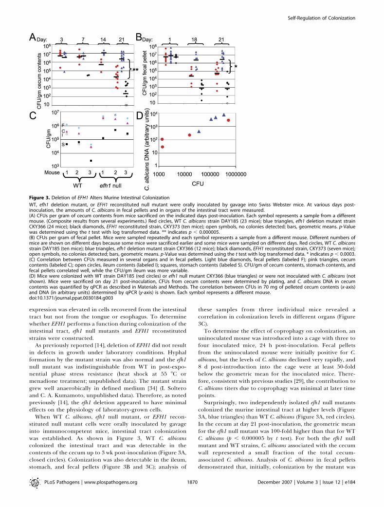
When WT C. albicans, efh1 null mutant, or EFH1 recon-stituted null mutant cells were orally inoculated by gavageinto immunocompetent mice, intestinal tract colonizationwas established. As shown in Figure 3, WT C. albicanscolonized the intestinal tract and was detectable in thecontents of the cecum up to 3 wk post-inoculation (Figure 3A,closed circles). Colonization was also detectable in the ileum,stomach, and fecal pellets (Figure 3B and 3C); analysis of
these samples from three individual mice revealed acorrelation in colonization levels in different organs (Figure3C).To determine the effect of coprophagy on colonization, an
uninoculated mouse was introduced into a cage with three tofour inoculated mice, 24 h post-inoculation. Fecal pelletsfrom the uninoculated mouse were initially positive for C.albicans, but the levels of C. albicans declined very rapidly, and8 d post-introduction into the cage were at least 50-foldbelow the geometric mean for the inoculated mice. There-fore, consistent with previous studies [29], the contribution toC. albicans titers due to coprophagy was minimal at later timepoints.Surprisingly, two independently isolated efh1 null mutants
colonized the murine intestinal tract at higher levels (Figure3A, blue triangles) than WT C. albicans (Figure 3A, red circles).In the cecum at day 21 post-inoculation, the geometric meanfor the efh1 null mutant was 100-fold higher than that for WTC. albicans (p , 0.000005 by t test). For both the efh1 nullmutant and WT strains, C. albicans associated with the cecumwall represented a small fraction of the total cecum-associated C. albicans. Analysis of C. albicans in fecal pelletsdemonstrated that, initially, colonization by the mutant was
Figure 3. Deletion of EFH1 Alters Murine Intestinal Colonization
WT, efh1 deletion mutant, or EFH1 reconstituted null mutant were orally inoculated by gavage into Swiss Webster mice. At various days post-inoculation, the amounts of C. albicans in fecal pellets and in organs of the intestinal tract were measured.(A) CFUs per gram of cecum contents from mice sacrificed on the indicated days post-inoculation. Each symbol represents a sample from a differentmouse. (Composite results from several experiments.) Red circles, WT C. albicans strain DAY185 (23 mice); blue triangles, efh1 deletion mutant strainCKY366 (24 mice); black diamonds, EFH1 reconstituted strain, CKY373 (ten mice); open symbols, no colonies detected; bars, geometric means. p-Valuewas determined using the t test with log transformed data. ** indicates p , 0.000005.(B) CFUs per gram of fecal pellet. Mice were sampled repeatedly and each symbol represents a sample from a different mouse. Different numbers ofmice are shown on different days because some mice were sacrificed earlier and some mice were sampled on different days. Red circles, WT C. albicansstrain DAY185 (ten mice); blue triangles, efh1 deletion mutant strain CKY366 (12 mice); black diamonds, EFH1 reconstituted strain, CKY373 (seven mice);open symbols, no colonies detected; bars, geometric means. p-Value was determined using the t test with log transformed data. * indicates p , 0.0003.(C) Correlation between CFUs measured in several organs and in fecal pellets. Light blue diamonds, fecal pellets (labeled F); pink triangles, cecumcontents (labeled C); open circles, ileum contents (labeled I); squares, stomach contents (labeled S). CFU/gm of cecum contents, stomach contents, andfecal pellets correlated well, while the CFU/gm ileum was more variable.(D) Mice were colonized with WT strain DAY185 (red circles) or efh1 null mutant CKY366 (blue triangles) or were not inoculated with C. albicans (notshown). Mice were sacrificed on day 21 post-inoculation, CFUs from cecum contents were determined by plating, and C. albicans DNA in cecumcontents was quantified by qPCR as described in Materials and Methods. The correlation between CFUs in 70 mg of pelleted cecum contents (x-axis)and DNA (in arbitrary units) determined by qPCR (y-axis) is shown. Each symbol represents a different mouse.doi:10.1371/journal.ppat.0030184.g003
PLoS Pathogens | www.plospathogens.org December 2007 | Volume 3 | Issue 12 | e1841870
Self-Regulation of Colonization
similar to colonization by WT C. albicans (Figure 3B) but atlater time points, e.g., day 21 post-inoculation, colonizationby the efh1 null mutant persisted at higher levels than the WTstrain (Figure 3B, p , 0.0003 by t test). Enhanced colonizationwas also observed in the ileum and stomach (Figure 3C andunpublished data).
As a control to show that the differences in colony formingunits (CFUs) recovered from WT or efh1 mutant–containingceca truly reflected differences in the numbers of fungal cellsrather than preferential recovery of mutant cells, analternative method of quantification was used. DNA wasextracted from cecum contents and the amount of C. albicansgenomic DNA was determined by quantitative PCR (qPCR).The results showed that the amounts of C. albicans DNArecovered from cecum contents containing WT or efh1 nullmutant cells (in arbitrary units) correlated with the CFUsdetermined by plating (Figure 3D), and the average ratio ofthe amount of DNA/CFU was the same for the WT and efh1null mutant strains. Therefore, differences in CFUs reflecteddifferences in the numbers of colonizing C. albicans.
To demonstrate that the enhanced colonization reflectedthe absence of Efh1p, EFH1 was added back to the deletionmutant under control of a strong promoter to ensureexpression of the reintroduced gene. Introduction of theectopically controlled EFH1 to the mutant strain resulted inreduced colonization (Figure 3A, black diamonds), indicatingthat the level of EFH1 in the strain determined colonizationlevels. For unknown reasons, introduction of EFH1 at itsnative locus did not result in full complementation (unpub-lished data), as has been observed by others for other genes,e.g., [15]. These studies demonstrate that in the absence ofEfh1p, colonization of the intestinal tract was enhanced.Thus, paradoxically, expression of C. albicans EFH1 duringcommensal colonization of the intestinal tract resulted inreduced colonization.
To observe the morphology of efh1 null mutant cells, GFP-expressing efh1 null mutants were orally inoculated into miceby gavage. On day 3 or day 13 post-inoculation, ileumcontents were collected and organisms were visualized byobserving green fluorescence. As observed for WT C. albicans,the vast majority (.91% þ/� 1% standard deviation) of cellsexhibited yeast-form morphology (Figure 2).
To detect dissemination from the intestinal tract in mice,the kidneys, liver, and spleen were homogenized andcultured. With both efh1 null mutant and WT, all samples
were either negative or contained very few organisms (Table2). Therefore, there was no evidence of high-level coloniza-tion of deep organs, indicating that C. albicans was notescaping from the intestinal tract.Colonization of the tongue of mice was analyzed to
determine whether a significant level of oral candidiasis wasoccurring. Colonization was generally not detectable,although an occasional mouse exhibited below 103 CFU/gmtongue tissue (Table 2). No consistent differences incolonization by WT and mutant organisms were observed.Therefore, there was no evidence of either mucosal orsystemic disease in these immunocompetent mice.To determine the levels of residual bacteria remaining
after antibiotic treatment, ileum and cecum homogenateswere cultured under aerobic or anaerobic conditions on richmedia. The ranges of bacteria levels were very similar foruninoculated mice and for mice inoculated with all of the C.albicans strains described above (unpublished data).
EFH1 Overexpression Reduces Intestinal ColonizationSince reduction of Efh1p by deletion resulted in enhanced
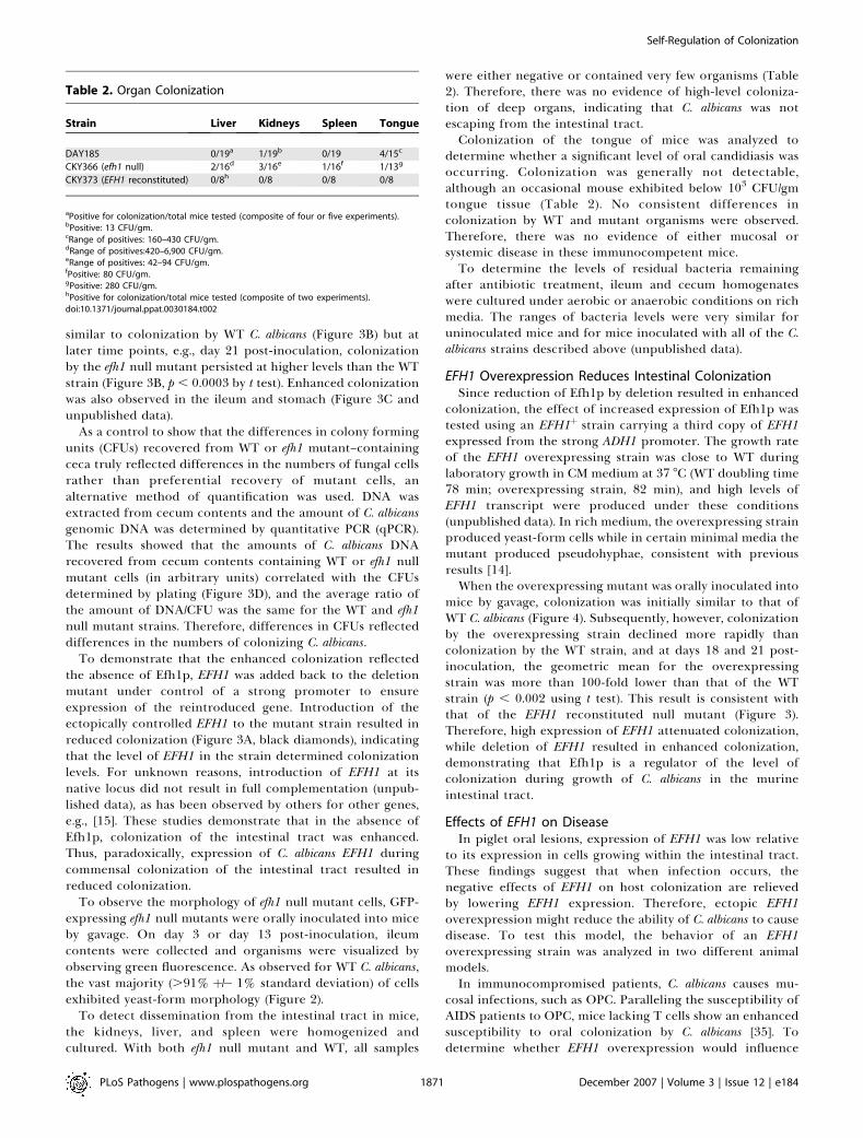
colonization, the effect of increased expression of Efh1p wastested using an EFH1þ strain carrying a third copy of EFH1expressed from the strong ADH1 promoter. The growth rateof the EFH1 overexpressing strain was close to WT duringlaboratory growth in CM medium at 37 8C (WT doubling time78 min; overexpressing strain, 82 min), and high levels ofEFH1 transcript were produced under these conditions(unpublished data). In rich medium, the overexpressing strainproduced yeast-form cells while in certain minimal media themutant produced pseudohyphae, consistent with previousresults [14].When the overexpressing mutant was orally inoculated into
mice by gavage, colonization was initially similar to that ofWT C. albicans (Figure 4). Subsequently, however, colonizationby the overexpressing strain declined more rapidly thancolonization by the WT strain, and at days 18 and 21 post-inoculation, the geometric mean for the overexpressingstrain was more than 100-fold lower than that of the WTstrain (p , 0.002 using t test). This result is consistent withthat of the EFH1 reconstituted null mutant (Figure 3).Therefore, high expression of EFH1 attenuated colonization,while deletion of EFH1 resulted in enhanced colonization,demonstrating that Efh1p is a regulator of the level ofcolonization during growth of C. albicans in the murineintestinal tract.
Effects of EFH1 on DiseaseIn piglet oral lesions, expression of EFH1 was low relative
to its expression in cells growing within the intestinal tract.These findings suggest that when infection occurs, thenegative effects of EFH1 on host colonization are relievedby lowering EFH1 expression. Therefore, ectopic EFH1overexpression might reduce the ability of C. albicans to causedisease. To test this model, the behavior of an EFH1overexpressing strain was analyzed in two different animalmodels.In immunocompromised patients, C. albicans causes mu-
cosal infections, such as OPC. Paralleling the susceptibility ofAIDS patients to OPC, mice lacking T cells show an enhancedsusceptibility to oral colonization by C. albicans [35]. Todetermine whether EFH1 overexpression would influence
Table 2. Organ Colonization
Strain Liver Kidneys Spleen Tongue
DAY185 0/19a 1/19b 0/19 4/15c
CKY366 (efh1 null) 2/16d 3/16e 1/16f 1/13g
CKY373 (EFH1 reconstituted) 0/8h 0/8 0/8 0/8
aPositive for colonization/total mice tested (composite of four or five experiments).bPositive: 13 CFU/gm.cRange of positives: 160–430 CFU/gm.dRange of positives:420–6,900 CFU/gm.eRange of positives: 42–94 CFU/gm.fPositive: 80 CFU/gm.gPositive: 280 CFU/gm.hPositive for colonization/total mice tested (composite of two experiments).doi:10.1371/journal.ppat.0030184.t002
PLoS Pathogens | www.plospathogens.org December 2007 | Volume 3 | Issue 12 | e1841871
Self-Regulation of Colonization
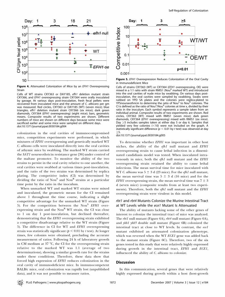
colonization in the oral cavities of immunocompromisedmice, competition experiments were performed, in whichmixtures of EFH1 overexpressing and genetically marked WTC. albicans cells were inoculated directly into the oral cavitiesof athymic mice by swabbing. The marked WT strain carriedthe SAT1 nourseothricin resistance gene [36] under control ofthe maltase promoter. To monitor the ability of the twostrains to persist in the oral cavity relative to one another, theoral cavities were swabbed at various times post-inoculation,and the ratio of the two strains was determined by replicaplating. The competitive index (CI) was determined bydividing the ratio of NouS and NouR strains at a particulartime point by the ratio in the inoculum.
When unmarked WT and marked WT strains were mixedand inoculated, the geometric means for the CI remainedabove 1 throughout the time course, indicating a slightcompetitive advantage for the unmarked WT strain (Figure5). For the competition between the NouS EFH1 over-expressing strain and the NouR WT strain, the CI was closeto 1 on day 1 post-inoculation, but declined thereafter,demonstrating that the EFH1 overexpressing strain exhibiteda competitive disadvantage relative to the WT strain (Figure5). The difference in CI for WT and EFH1 overexpressingstrain was statistically significant (p , 0.01 by t test). At longertimes, few colonies were obtained, precluding the accuratemeasurement of ratios. Following 24 h of laboratory growthin CM medium at 37 8C, the CI for the overexpressing strainrelative to the marked WT was 1.1 (average of twodeterminations), showing a similar growth rate for the strainsunder these conditions. Therefore, these data show thatforced high expression of EFH1 reduces colonization in theoral cavity of immunodeficient mice. In immunocompetentBALB/c mice, oral colonization was rapidly lost (unpublisheddata), and it was not possible to measure ratios.
To determine whether EFH1 was important in other hostniches, the ability of the efh1 null mutant and EFH1overexpressing strain to cause lethal infection in a dissemi-nated candidiasis model was tested. When inoculated intra-venously in mice, both the efh1 null mutant and the EFH1overexpressing strain retained the ability to cause lethalinfections. The mean survival time for mice inoculated withWT C. albicans was 5 6 3 d (23 mice). For the efh1 null mutant,the mean survival time was 5 6 3 d (16 mice) and for theEFH1 overexpressing strain, the mean survival time was 7 6 3d (seven mice) (composite results from at least two experi-ments). Therefore, both the efh1 null mutant and the EFH1overexpressing strain were virulent in this model.
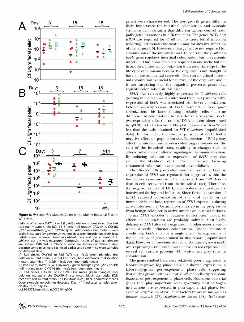
rbt1 and rbt4 Mutants Colonize the Murine Intestinal Tractat WT Levels while the ece1 Mutant Is AttenuatedThe ability of mutants lacking some of the other genes of
interest to colonize the intestinal tract of mice was analyzed.The rbt1 null mutant (Figure 6A), rbt4 null mutant (Figure 6A),and yhb1 yhb5 double null mutant (Figure 6B) colonized theintestinal tract at close to WT levels. In contrast, the ece1mutant exhibited an attenuated colonization phenotype,which was reversed when the WT ECE1 gene was added backto the mutant strain (Figure 6C). Therefore, two of the sixgenes tested in this study that were relatively highly expressedduring growth in the intestinal tract, EFH1 and ECE1,influenced the ability of C. albicans to colonize.
Discussion
In this communication, several genes that were relativelyhighly expressed during growth within a host (host-growth
Figure 4. Attenuated Colonization of Mice by an EFH1 Overexpressing
Strain
Cells of WT strains CKY363 or DAY185, efh1 deletion mutant strainCKY366, and EFH1 overexpressing strain CKY364 were orally inoculatedby gavage. At various days post-inoculation, fresh fecal pellets wererecovered from inoculated mice and the amount of C. albicans per gmwas measured. Red circles, CKY363 or DAY185 (WT) (seven mice); bluetriangles, efh1 deletion mutant strain CKY366 (six mice); dark greendiamonds, CKY364 (EFH1 overexpressing) (eight mice); bars, geometricmeans. Composite results of two experiments are shown. Differentnumbers of mice are shown on different days because some mice weresacrificed earlier and some mice were sampled on different days.doi:10.1371/journal.ppat.0030184.g004
Figure 5. EFH1 Overexpression Reduces Colonization of the Oral Cavity
in Immunodeficient Mice
Cells of strains CKY363 (WT) or CKY364 (EFH1 overexpressing, OE) weremixed in a 1:1 ratio with strain RMIS1 (NouR marked WT) and introducedinto the oral cavities of nude mice by swabbing. On various days post-inoculation, the oral cavities were sampled by swabbing. Swabs wererubbed on YPD SA plates and the colonies were replica-plated toYPSnourseothricin to determine the ratio of NouS to NouR colonies. TheCI is defined as the ratio of NouS/NouR colonies at time x, divided by theirratio in the inoculum. Each symbol represents a sample taken from anindividual animal. Composite results of two experiments are shown. Redcircles, CKY363 (WT) mixed with RMIS1 (seven mice); dark greendiamonds, CKY364 (EFH1 overexpressing) mixed with RMIS1 (six mice).Day �5 includes samples taken at either day 5 or day 6. Samples thatyielded very few colonies (,10) were not included on the graph. Astatistically significant difference (p , 0.01 by t test) was observed at day�5.doi:10.1371/journal.ppat.0030184.g005
PLoS Pathogens | www.plospathogens.org December 2007 | Volume 3 | Issue 12 | e1841872
Self-Regulation of Colonization
genes) were characterized. The host-growth genes differ intheir importance for intestinal colonization and systemicvirulence demonstrating that different factors control host-pathogen interactions in different sites. The genes RBT1 andRBT4 are required for C. albicans to cause lethal infectionfollowing intravenous inoculation and for invasive infectionof the cornea [15]. However, these genes are not required forcolonization of the intestinal tract. In contrast, the C. albicansEFH1 gene regulates intestinal colonization but not systemicinfection. Thus, some genes are required in one niche but notin another. Intestinal colonization is an essential stage in thelife cycle of C. albicans because the organism is not thought tohave an environmental reservoir. Therefore, optimal intesti-nal colonization is crucial for survival of the organism, and itis not surprising that the organism possesses genes thatregulate colonization in this niche.EFH1 was relatively highly expressed by C. albicans cells
growing in the mammalian intestinal tract, but paradoxically,expression of EFH1 was associated with lower colonization.Ectopic overexpression of EFH1 resulted in very poorcolonization; this latter finding probably reflects a truedifference in colonization, because for in vitro–grown EFH1overexpressing cells, the ratio of DNA content (determinedby qPCR) to CFUs (measured by plating) was less than 2-foldless than the ratio obtained for WT C. albicans (unpublisheddata). In this niche, therefore, expression of EFH1 had anegative effect on population size. Expression of Efh1p mayaffect the interactions between colonizing C. albicans and thecells of the intestinal tract, resulting in changes such asaltered adherence or altered signaling to the immune system.By reducing colonization, expression of EFH1 may alsoreduce the likelihood of C. albicans infection, favoringcommensal colonization as opposed to candidiasis.The effects of Efh1p on colonization are reversible, because
expression of EFH1 was regulated during growth within thehost (lower expression in cells recovered from OPC lesionsthan in cells recovered from the intestinal tract). Therefore,the negative effects of Efh1p that reduce colonization areinactivated during oral infection. Since forced expression ofEFH1 reduced colonization of the oral cavity in animmunodeficient host, repression of EFH1 expression duringactive infection may be an important step in the progressionfrom benign colonizer to active invader on mucosal surfaces.Since EFH1 encodes a putative transcription factor, its
effects on colonization are probably indirect. Most likely,deletion of EFH1 alters the expression of other genes, some ofwhich directly influence colonization. Under laboratoryconditions, EFH1 did not strongly affect the expression ofthe collection of genes studied in this report (unpublisheddata). However, in previous studies, a laboratory-grown EFH1overexpressing strain was shown to have altered expression ofseveral cell surface proteins [14], which may play roles incolonization.The genes studied here were relatively poorly expressed in
laboratory-grown log phase cells but showed expression inlaboratory-grown post-exponential phase cells, suggestingthat during growth within a host, C. albicans cells express somefeatures of post-exponential phase cells. Numerous bacterialgenes that play important roles governing host-pathogeninteractions are expressed in post-exponential phase. Forexample, expression of virulence factors by organisms such asBacillus anthracis [37], Staphylococcus aureus [38], Helicobacter
Figure 6. rbt1 and rbt4 Mutants Colonize the Murine Intestinal Tract at
WT Levels
Cells of WT strains DAY185 or F2U, rbt1 deletion mutant strain BCa 7–4,rbt4 null mutant strain BCa 11–3, ece1 null mutant CAW19–1, CKY362(ECE1 reconstituted), and CKY376 (yhb1 yhb5 double null mutant) wereorally inoculated by gavage. At various days post-inoculation, fresh fecalpellets were recovered from inoculated mice and the amount of C.albicans per gm was measured. Composite results of two experimentsare shown. Different numbers of mice are shown on different daysbecause some mice were sacrificed earlier and some mice were sampledon different days.(A) Red circles, DAY185 or F2U (WT) (six mice); green triangles, rbt1deletion mutant strain BCa 7–4 (six mice); blue diamonds, rbt4 deletionmutant strain Bca 11–3 (six mice); bars, geometric means.(B) Red circles, DAY185 (WT) (six mice); green triangles, yhb1 yhb5 doublenull mutant strain CKY376 (six mice); bars, geometric means.(C) Red circles, DAY185 or F2U (WT) (six mice); green triangles, ece1deletion mutant strain CAW19–1 (six mice); black diamonds, ECE1reconstituted mutant strain CKY362 (four mice); bars, geometric means.Open symbols, no colonies detected. Day � 14 indicates samples takenon day 14 or day 15.doi:10.1371/journal.ppat.0030184.g006
PLoS Pathogens | www.plospathogens.org December 2007 | Volume 3 | Issue 12 | e1841873
Self-Regulation of Colonization
pylori [39], Legionella pneumophila [40], or Salmonella [41] occursin post-exponential phase. Therefore, expression of post-exponential phase genes during host interaction is a commontheme for many bacterial pathogens and for the fungus C.albicans.
In the laboratory, expression of ECE1, RBT1, and RBT4 islinked to hyphal morphogenesis. However, in cells growingwithin the intestinal tract, expression of these genes does notdepend on Efg1p and most likely occurs in yeast cells. ECE1,RBT1, and RBT4 were also found to be expressed in efh1 nullmutants recovered from the cecum of mice (unpublisheddata). Therefore, the pathway(s) responsible for the expres-sion of these genes during growth within the host isuncharacterized. Since the genes show relatively highexpression during post-exponential phase in the laboratory,there may be a post-exponential phase regulator(s) that isresponsible for their expression in the host. Several regu-lators required for normal viability in post-exponential phasehave been recently described [42]. Deeper understanding ofthe biology of post-exponential phase cells may reveal C.albicans activities that are important for colonization anddisease.
During colonization, the population size of a microorgan-ism reflects a balance between external forces that limit thepopulation, such as the effects of the host immune system,and intrinsic factors, such as the ability of the organism toincrease in number. The ability of commensal C. albicans toregulate its own population size through reversible expres-sion of a negative regulator of colonization adds anotherlayer of regulation to the interactions that take place betweenhost and colonizer. The combined effects of these regulatory
interactions maintain the balance between healthy coloniza-tion and disease.
Materials and Methods
Strains. C. albicans strains are listed in Table 3. All C. albicans strainswere derived from the WT clinical strain SC5314 [43], using thefollowing genetically marked derivatives: CAI-4 [43], BWP17 [44],RM1000#2 [45], or SN100 [45].
Strain CKY366, the efh1 null mutant strain, was constructed bytransformation of BWP17 with constructs encoding HIS1 or ARG4flanked by 600 bp of sequence upstream of the EFH1 ORF and 542 bpof sequence downstream of the EFH1 ORF. The fragment was excisedfrom the plasmid backbone by digestion with BslI and BsmFI forefh1D::ARG4 or with NotI sites that were introduced at the ends of theEFH1 sequences for efh1D::HIS1.
The efh1 null mutant was transformed with pRC3915 [46] to restoreURA3 prototrophy or with pCK73 to introduce EFH1 under controlof the ADH1 promoter.
To construct an EFH1 overexpressing strain, the EFH1þ strainSN100 (his1/his1) was transformed with pCK74 encoding EFH1 undercontrol of the ADH1 promoter and carrying selectable marker HIS1.
yEGFP- [30] expressing EFH1þ and efh1 null mutant strains wereconstructed by transforming RM1000 Hisþ or the efh1 null mutantwith pDRG-GFPS6, encoding yEGFP under control of the DRG1promoter [47].
Strain CKY375, the yhb5 null mutant strain, was constructed bytransformation of BWP17 with constructs encoding HIS1 or ARG4flanked by 496 bp of sequence upstream of the YHB5 ORF and 355 bpof sequence downstream of the YHB5 ORF. The desired fragmentswere excised from their plasmid backbones by digestion with TseIand BslI for yhb5D::ARG4 or with NotI for yhb5D::HIS1. StrainCKY376, the yhb1 yhb5 double null mutant strain, was constructedby transformation of the yhb5 deletion mutant with a constructencoding the SAT flipper [36] with 750 bp of sequence upstream ofthe YHB1ORF and 490 bp of sequence downstream of the YHB1ORF.Nourseothricin-resistant (NouR) transformants were screened ini-tially by PCR. Positive candidates were plated on sucrose-containingmedium to induce expression of the FLP recombinase and replica-plated to nourseothricin medium. NouS colonies were purified and
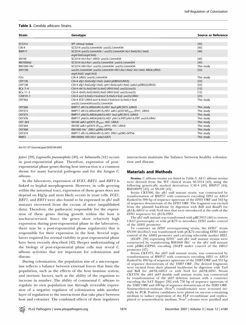
Table 3. Candida albicans Strains
Strain Genotype Source or Reference
SC5314 WT clinical isolate [43]
CAI-4 SC5314 ura3D::kimm434/ ura3D::kimm434 [43]
BWP17 SC5314 ura3D::kimm434 / ura3D::kimm434 his1::hisG/his1::hisG
arg4::hisG/arg4::hisG
[44]
SN100 SC5314 his1/his1 URA3/ ura3D::kimm434 [45]
RM1000#2 SC5314 his1/his1 ura3D::kimm434/ ura3D::kimm434 [45]
RM1000 Hisþ SC5314 HIS1/his1 ura3D::kimm434/ ura3D::kimm434 This study
DAY185 ura3D::kimm434/ ura3D::kimm434 HIS1::his1::hisG/ his1::hisG ARG4::URA3::
arg4::hisG/ arg4::hisG
[52]
F2U CAI-4 URA3/ ura3D::kimm434 This study
CKY136 CAI-4 efg1::hisG/efg1::hisG (ade2::pDBI52(URA3)) [53]
CKY138 CAI-4 efg1::hisG/efg1::hisG cph1::hisG/cph1::hisG (ade2::pDBI52(URA3)) [53]
BCa 7–4 CAI-4 rbt1D::hisG/rbt1D::hisG-URA3-hisG ura3D/ura3D [15]
BCa 11–3 CAI-4 rbt4D::hisG/rbt4D::hisG-URA3-hisG ura3D/ura3D [15]
CAW19–1 CAI-4 ece1D::hisG::I-SceI/ece1D::hisG::I-SceI ura3D/URA3 [25]
CKY362 CAI-4 ECE1-URA3::ece1D::hisG::I-SceI/ece1D::hisG::I-SceI
ura3D::kimm434/ura3D::kimm434
This study
CKY366 BWP17 efh1D::ARG4/efh1D::HIS1 leu2::pRC3915 (URA3) This study
CKY373 BWP17 efh1D::ARG4/efh1D::HIS1 adh1::pCK73(PADH1-EFH1, URA3) This study
CKY375 BWP17 yhb5D::ARG4/yhb5D::HIS1 leu2::pRC3915 (URA3) This study
CKY376 BWP17 yhb5D::ARG4/yhb5D::HIS1 yhb1D::FRT/yhb1D::FRT ura3D/URA3 This study
CKY 363 SN100 adh1::pCK75 (PADH1, HIS1 URA3) This study
CKY364 SN100 adh1::pCK74 (PADH1-EFH1, HIS1 URA3) This study
CKY368 RM1000 Hisþ DRG1::pDRG-GFPS6 This study
CKY374 BWP17 efh1D::ARG4/efh1D::HIS1 DRG1::pDRG-GFPS6 This study
RMIS1 RM1000#2 HIS1/his1 (ade2::iSAT) This study
doi:10.1371/journal.ppat.0030184.t003
PLoS Pathogens | www.plospathogens.org December 2007 | Volume 3 | Issue 12 | e1841874
Self-Regulation of Colonization
subjected to a second round of transformation as above and nullmutants were identified by Southern blot analysis. To generate URA3prototrophs, the strains were transformed with a URA3þ fragmentfrom plasmid pET16 [48].
The ece1 null mutant CAW19–1 was kindly provided by B. Fonzi(Georgetown University) and the rbt1 and rbt4 null mutants (BCa 7–4and Bca 11–3, respectively) were kindly provided by S. Johnson(UCSF).
To construct a genetically marked WT strain, plasmid iSAT wasdigested with BsgI and integratively transformed into strainRM1000#2, Hisþ. The resultant strain (RM1000 iSAT) exhibitsresistance to nourseothricin when grown on plates containingsucrose but poorer resistance on plates containing glucose. TheiSAT construct was used because expression from the maltasepromoter was expected to be low during growth within a host,minimizing possible deleterious effects due to expression of theheterologous SAT1 gene.
Media and growth conditions. Standard rich media were YPD (1%yeast extract, 2% peptone, 2% glucose) or YPS (1% yeast extract, 2%peptone, 2% sucrose). Minimal dropout media (lacking uracil,histidine, arginine, or combinations) were as described previously[49]. RPMI 1640 (Sigma) with 10% bovine serum was used to promotehyphal morphogenesis. Nourseothricin (200 lg/ml) was used asdescribed [36]. For plating contents of the intestinal tract, YPD agarmedium supplemented with 50 lg/ml ampicillin and 100 lg/mlstreptomycin (YPD SA) was used. For competition experiments,colonies on YPD SA were replica-plated to YPsucrose supplementedwith nourseothricin.
For culturing bacteria from the intestinal tract, BHIS medium wasused (3.7% Difco brain heart infusion broth, 0.5% yeast extract,0.0015% hemin, 0.2% agar), and plates were incubated aerobically oranaerobically at 37 8C.
For gene expression studies, reference cells were grown in YPSliquid medium at 34 8C in log phase. For the experiments shown inFigure 1C and 1D, cells were grown in YPD liquid medium at 37 8C inlog phase, or in YPD liquid medium at 37 8C for 3 d (post-exponentialphase), and, to stimulate hyphal morphogenesis, in RPMI-10% bovineserum medium for 4–6 h at 37 8C.
Plasmids. Plasmid pCK73 was constructed by amplifying the EFH1ORF with primers VEC53F and VEC53R. The resulting fragment wasdigested with BglII and XhoI and cloned onto BglII, XhoI-digestedpYB-ADH1pt [50]. Plasmid pCK74 and pCK75 were constructed byamplifying the C. albicans HIS1 gene with primer AHISF3 andAHISR3, digesting the resultant fragment with AhdI and NheIcloning onto XcmI,XbaI-digested pCK73 or pYB-ADH1pt, respec-tively.
For gene deletions, efh1D::ARG4, efh1D::HIS1, yhb5D::HIS1,yhb5D::ARG4, and yhb1D::SAT flipper constructs were produced inseveral steps. First, EFH1 sequences were amplified using primerspairs 53A and 53B2 or 53C2 and 53D2 followed by overlap PCR. The1.1-kb fragment with EFH1 upstream and downstream sequencesflanking a unique PacI site with NotI sites on the ends was TOPO-cloned onto pYES2.1/V5-His-TOPO (Invitrogen) using manufac-turer’s protocols. The YHB5 locus fragment was amplified withprimers 44KOF1 and 44KOR1 and the YHB1 locus fragment wasamplified with primers YHB1F1 and yhb1R1; the fragments wereTOPO-cloned as above.
To generate gapped or linearized plasmids, the efh1 plasmid wasdigested with PacI, the YHB5 plasmid with PacI, and the YHB1plasmid with HindIII. HIS1 and ARG4 markers were amplified frompGEM-HIS or pRS-ARG4DSpeI [44] with primer pair 53MF1 and53MR1 (for EFH1 knockout constructs) or 44MF1 and 44MR1 (forYHB5 knockout constructs). The SAT1 flipper [36] was amplified withprimers HB1NF1 and HB1NR1 for the YHB1 knockout construct.Digested plasmids were cotransformed with appropriate PCRproducts into Saccharomyces cerevisiae strain EGY40 (MATa ura3–1his3–11 trp1–1 leu2–3,112) [51]. Homologous recombination gener-ated the desired constructs.
To construct pDRG-GFPS6, the DRG1 promoter region [47] wasamplified with primers XC3 and XC4 and cloned onto plasmidpXC31, a vector containing BamHI, NsiI, SalI, and EcoRV sitesupstream of a promoterless yEGFP gene that was fused to the 39 UTRof ACT1 in pNUB1 (pNEB193 from New England Biolabs, with C.albicans URA3 [47]).
To construct plasmid iSAT (encoding SAT1 under control of themaltase promoter), the SAT1 ORF was amplified with primers SATECand SATR1 from pSFS2 [36]. The 39 UTR of ACT1 was amplified withprimers SATAFU and ACT3x and fused to the SAT1 fragment byoverlap PCR. The resultant fragment was cloned onto vector pDBI52,generating a plasmid carrying SAT1 under control of PMAL with a
fragment of C. albicans ADE2 for integration and the C. albicans URA3gene as a selectable marker.
Animal models. (1) Two germ-free piglets were inoculated orallywith 109 CFUs of C. albicans strain SC5314 and treated with 25 mg/kgbody weight methylprednisolone and 15 mg/kg body weight cyclo-sporine daily as described previously [16]. Seven or 10 d post-inoculation, a piglet was sacrificed and organs were dissected andfrozen in RNALater (Ambion) at �80 8C.
(2) Female Swiss Webster mice (18–20 g) were treated withtetracycline (1 mg/ml); streptomycin (2 mg/ml) and gentamycin (0.1mg/ml) added to their drinking water throughout the experimentbeginning 4 d prior to inoculation. C. albicans cells were grown for 24h at 37 8C in YPD liquid medium, washed twice with PBS, counted,and adjusted to 2.53 108 cells/ml. All strains were prototrophic. Micewere inoculated by gavage with 53 107 C. albicans cells in 0.2 ml usinga feeding needle. Colonization was monitored by collecting fecalpellets (produced within 10 min prior to collection) at various dayspost-inoculation and measuring C. albicans concentrations in thepellets by plating homogenates on YPD SA plates. Mice weresacrificed on various days post-inoculation and C. albicans concen-trations in cecum contents, stomach contents, and homogenates ofileum, kidneys, liver, spleen, and tongue were measured by plating onYPD SA plates. Composite results from at least two experiments areshown.
(3) Intravenous inoculation of female CF1 mice (18–20 g; CharlesRiver Laboratories) with 13106 C. albicans cells via the lateral tail veinwas conducted as previously described [47]. Mice were observed twicedaily after infection with C. albicans and were sacrificed whenmoribund.
(4) Competition experiments: To generate a marked WT strain,strain RM1000#2 Hisþ was transformed with plasmid iSAT, resultingin a prototrophic strain showing inducible resistance to nourseo-thricin. This genetic marker was used because it was expected that themaltase promoter would only weakly express the heterologous SAT1gene during growth of C. albicans in the mouse, thereby minimizingany potential deleterious effects due to SAT1 expression.
For direct inoculation of the oral cavity by swabbing, theprocedure was based on the previous work of Farah et al. [35] withminor modifications. Briefly, female BALB/c nude mice (5–7 wk, NCI)were given antibiotic water as described above. For inoculation, 1 3108 cells of C. albicans (mixture of two strains at a ratio of 1:1) wereapplied to a swab and introduced directly into the oral cavity.Colonization was monitored by swabbing the oral cavity and bycollecting fecal pellets. Swabs were rubbed on YPD SA plates to assesscolonization; fecal pellets were homogenized and plated. Intestinalcolonization levels were comparable to those obtained by gavage.
To determine the ratios of the two different strains, the YPD plateswere replica-plated to YPSnourseothricin and the ratio of nourseo-thricin-sensitive colonies (NouS) to nourseothricin-resistant colonies(NouR) was determined. At the later time points, some samplesyielded few colonies (,10) and these samples were not included onthe graph. The CI was obtained by dividing the ratio of NouS/NouR
colonies at time x by their ratio in the inoculum.RNA extraction. Tissues from WT strain SC5314-infected IGB
piglets, 7 or 10 d post-inoculation, were frozen in RNALater(Ambion) at �80 8C. For tongues and esophagus, the fungal cell–containing layer was cut from the tissue. WT C. albicans (SC5314 orDAY185) or mutant CKY138 (efg1 cph1 double null) were inoculatedinto Swiss Webster mice by gavage. Three days post-inoculation, thecontents of the cecum or ileum were recovered and frozen inRNALater at�80 8C. RNA was extracted from these samples using theQiagen MIDI procedures with mechanical disruption using glass-zirconium beads, protease digestion, and on-column DNase treat-ment. In some experiments, samples were extracted with TRIzol,applied to a Qiagen column, and treated with DNase. For controlpiglet RNA, tissue was cut from the central portion of the tongue ofan infected piglet and was not expected to contain significantnumbers of C. albicans cells. RNA was extracted from this material asdescribed above.
Reference RNA was extracted from cells grown in YPS medium at34 8C in log phase, using Qiagen procedures with mechanicaldisruption and on-column DNase digestion. This RNA preparationwas used as the reference sample for microarrays and for qRT-PCR.
qRT-PCR. 10 lg of total RNA was converted to cDNA byincubation with Superscript II Reverse Transcriptase (Invitrogen)using an oligo dT primer. After incubation for 1 h at 42 8C, RNA washydrolyzed and the reaction was stopped by addition of NaOH andEDTA to 0.16 N NaOH, 0.08 M EDTA, final concentrations. Followingneutralization, cDNA was purified using Qiaquick columns (Qiagen)as described by the manufacturer, except that sodium acetate (pH 5.2)
PLoS Pathogens | www.plospathogens.org December 2007 | Volume 3 | Issue 12 | e1841875
Self-Regulation of Colonization
was added to the PB buffer to ensure an acidic pH. cDNA wasquantitated by absorbance. Purified cDNA was stored frozen.
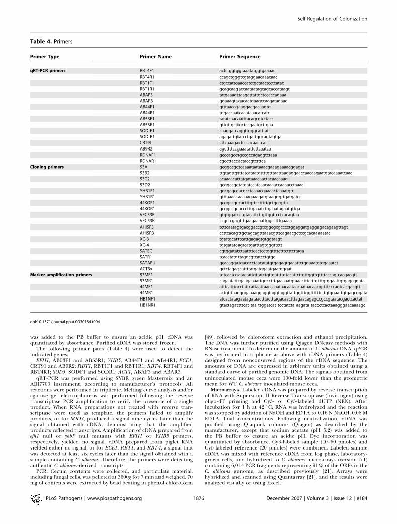
The following primer pairs (Table 4) were used to detect theindicated genes:
EFH1, AB53F1 and AB53R1; YHB5, AB44F1 and AB44R1; ECE1,CRT91 and AB9R2; RBT1, RBT1F1 and RBT1R1; RBT4, RBT4F1 andRBT4R1; SOD3, SODF1 and SODR1; ACT1, ABAF3 and ABAR3.
qRT-PCR was performed using SYBR green Mastermix and anABI7700 instrument, according to manufacturer’s protocols. Allreactions were performed in triplicate. Melting curve analysis and/oragarose gel electrophoresis was performed following the reversetranscriptase PCR amplification to verify the presence of a singleproduct. When RNA preparations not treated with reverse tran-scriptase were used as template, the primers failed to amplifyproducts, or for SOD3, produced a signal nine cycles later than thesignal obtained with cDNA, demonstrating that the amplifiedproducts reflected transcripts. Amplification of cDNA prepared fromefh1 null or yhb5 null mutants with EFH1 or YHB5 primers,respectively, yielded no signal. cDNA prepared from piglet RNAyielded either no signal, or for ECE1, RBT1, and RBT4, a signal thatwas detected at least six cycles later than the signal obtained with asample containing C. albicans. Therefore, the primers were detectingauthentic C. albicans-derived transcripts.
PCR: Cecum contents were collected, and particulate material,including fungal cells, was pelleted at 3600g for 7 min and weighed. 70mg of contents were extracted by bead beating in phenol-chloroform
[49], followed by chloroform extraction and ethanol precipitation.The DNA was further purified using Qiagen DNeasy methods withRNase treatment. To determine the amount of C. albicans DNA, qPCRwas performed in triplicate as above with rDNA primers (Table 4)designed from nonconserved regions of the rDNA sequence. Theamounts of DNA are expressed in arbitrary units obtained using astandard curve of purified genomic DNA. The signals obtained fromuninoculated mouse ceca were 100-fold lower than the geometricmean for WT C. albicans inoculated mouse ceca.
Microarrays. Labeled cDNA was prepared by reverse transcriptionof RNA with Superscript II Reverse Transcriptase (Invitrogen) usingoligo-dT priming and Cy3- or Cy5-labeled dUTP (NEN). Afterincubation for 1 h at 42 8C, RNA was hydrolyzed and the reactionwas stopped by addition of NaOH and EDTA to 0.16 N NaOH, 0.08 MEDTA, final concentrations. Following neutralization, cDNA waspurified using Qiaquick columns (Qiagen) as described by themanufacturer, except that sodium acetate (pH 5.2) was added tothe PB buffer to ensure an acidic pH. Dye incorporation wasquantitated by absorbance. Cy3-labeled sample (40–60 pmoles) andCy5-labeled reference (20 pmoles) were combined. Labeled samplecDNA was mixed with reference cDNA from log phase, laboratory-grown cells, and hybridized to C. albicans microarrays (version 5.1)containing 6,014 PCR fragments representing 91% of the ORFs in theC. albicans genome, as described previously [21]. Arrays werehybridized and scanned using Quantarray [21], and the results wereanalyzed visually or using Excel.
Table 4. Primers
Primer Type Primer Name Primer Sequence
qRT-PCR primers RBT4F1 actctggtggtaaatatggtgaaaac
RBT4R1 ccagctgggtcgtaggaacaaacaac
RBT1F1 ctgccattcaaccatctgctaactcctcatac
RBT1R1 gcagcaagaccaataatagcagcaccataagt
ABAF3 tatgaaagttaagattattgctccaccagaaa
ABAR3 ggaaagtagacaatgaagccaagatagaac
AB44F1 gtttaaccgaaggaagacaagtg
AB44R1 tggaccaatcaaataaacatcatc
AB53F1 tatatcaacaatttacagcgtcttacc
AB53R1 gttgttgcttgctccgaatgcttgaa
SOD F1 caaggatcaggttgggcatttat
SOD R1 agagattgtatcctgattggcagtagtga
CRT9I cttcaaagactcccacaactcat
AB9R2 agcttttccgaaatattcttcaatca
RDNAF1 gcccagcctgccgccagaggtctaaa
RDNAR1 cgccttaccactaccgtctttca
Cloning primers 53A gcggccgctcaaaataataaacgaaagaaaacggagat
53B2 ttgtagttgtttatcataatgttttgtttaattaagaggaaccaacaagaatgtacaaaatcaac
53C2 acaaaacattatgataaacaactacaacaaag
53D2 gcggccgctatgatccatcaacaaaaccaaaacctaaac
YHB1F1 ggcgcgccacgcctcaaacgaaaactaaaatgtc
YHB1R1 gtttaaaccaaaaagaaagatgtaagggttgatgatg
44KOF1 gcggccgccactttgttcctttttgctgctgtta
44KOR1 gcggccgcaccctttgaaatcttgaaatagaatgttga
VEC53F gtgtggatcctgtacattcttgttggttcctcacagtaa
VEC53R ccgctcgagtttgaagaaaattggcctttgaaaa
AHISF3 tcttcaatagtgacggaccgtcggcgcgcccctggaggatgaggagacagaagttagt
AHISR3 ccttcacagttgctagcagtttaaacgtttcagaacgctccgcacaaaaatac
XC-3 tgtatgcatttcattgagagtgtggtaagt
XC-4 tgtgatatcagtcatgatttagtgggttctt
SATEC cgtggatatctaattttcactcctggttttctttctttcttaga
SATR1 tcacatatgttaggcgtcatcctgtgc
SATAFU gcacaggatgacgcctaacatatgtgagagtgaaattctggaaatctggaaatct
ACT3x gctctagagcattttatgatggaatgaatgggat
Marker amplification primers 53MF1 tgtcactcgatactattgttatctgttgattttgtacattcttgttggttgttttcccagtcacgacgtt
53MR1 cagaatatttgaagaaaattggcctttgaaaaatgtaaactttctttgtttgtggaattgtgagcggata
44MF1 atttcatttccctattcattaattaaccaaataacaataacaataacaaggttttcccagtcacgacgtt
44MR1 actgtttaacgggaaaaggaggtaggtaggttattggtttggttttttcttgtggaattgtgagcggata
HB1NF1 atcactatagaatagataactttactttagacaactttagaacagagccgccgtaatacgactcactat
HB1NR1 gtactagattttcat taa ttggatcat tcctatcta aagata taccctcactaaagggaacaaaagc
doi:10.1371/journal.ppat.0030184.t004
PLoS Pathogens | www.plospathogens.org December 2007 | Volume 3 | Issue 12 | e1841876
Self-Regulation of Colonization
Fluorescence microscopy. Swiss Webster mice were inoculated withyEGFP-expressing C. albicans by gavage as above. At various days post-inoculation, mice were sacrificed and the contents of the ileum wererecovered. Samples were filtered through 35-lm nylon mesh (SmallParts) and the filtrate was concentrated by centrifugation in anEppendorf centrifuge for 1 min at maximum speed. Samples wereobserved using an Olympus BX60 microscope with GFP (excitation460–490 nm, emission 515–700 nm) and YFP filters (excitation 500/20nm, emission 535/30 nm) and photographed with the 603 objective.
Acknowledgments
We thank Daniel Dignard, Donna Akiyoshi, Lauren Logsden, JeniferCoburn, Tom Volkert, Jessica Pierce, Eric Rubin, Peter Cheslock,Perry Riggle, Igor Bruzual, Paola Zucchi, Marcelo Vinces, and Ralph
Isberg for stimulating discussions or help with techniques and BillFonzi and Sandy Johnson for kind gifts of strains. We are also gratefulto Andrew Camilli for careful review of the manuscript. This is NRCpublication #49532.
Author contributions. CAK conceived and designed the experi-ments. SJW, AR, PL, DN, AB, ST, and CAK performed the experi-ments. SJW, AR, PL, DN, AB, JM, and CAK analyzed the data. MW andJM contributed reagents/materials/analysis tools. CAK wrote thepaper.
Funding. This research was supported by National Institutes ofHealth grant AI038591 from the National Institute of Allergy andInfectious Diseases to CAK.
Competing interests. The authors have declared that no competinginterests exist.
References1. Russell C, Lay KM (1973) Natural history of Candida species and yeasts in the
oral cavities of infants. Arch Oral Biol 18: 957–962.2. Odds FC (1987) Candida infections: an overview. Crit Rev Microbiol 15: 1–5.3. Calderone RA (2002) Introduction and historical perspectives. In:
Calderone RA, editor. Candida and candidiasis. Washington (D.C.): ASMPress. pp. 3–13.
4. Ampel NM (1996) Emerging disease issues and fungal pathogens associatedwith HIV infection. Emerg Infect Dis 2: 109–116.
5. Hentschel U, Dobrindt U, Steinert M (2003) Commensal bacteria make adifference. Trends Microbiol 11: 148–150.
6. Rakoff-Nahoum S, Paglino J, Eslami-Varzaneh F, Edberg S, Medzhitov R(2004) Recognition of commensal microflora by toll-like receptors isrequired for intestinal homeostasis. Cell 118: 229–241.
7. Barbara G, Stanghellini V, Brandi G, Cremon C, Di Nardo G, et al. (2005)Interactions between commensal bacteria and gut sensorimotor functionin health and disease. Am J Gastroenterol 100: 2560–2568.
8. Gordon JI, Stappenbeck TS, Hooper LV (2003) Response from Jeffrey I.Gordon et al.: Commensal bacteria make a difference. Trends Microbiol 11:150–151.
9. Lorenz MC, Fink GR (2001) The glyoxylate cycle is required for fungalvirulence. Nature 412: 83–86.
10. Lorenz MC, Bender JA, Fink GR (2004) Transcriptional response of Candidaalbicans upon internalization by macrophages. Eukaryot Cell 3: 1076–1087.
11. Fradin C, De Groot P, MacCallum D, Schaller M, Klis F, et al. (2005)Granulocytes govern the transcriptional response, morphology, andproliferation of Candida albicans in human blood. Mol Microbiol 56: 397–415.
12. Cheng S, Clancy CJ, Checkley MA, Handfield M, Hillman JD, et al. (2003)Identification of Candida albicans genes induced during thrush offers insightinto pathogenesis. Mol Microbiol 48: 1275–1288.
13. Thewes S, Kretschmar M, Park H, Schaller M, Filler SG, et al. (2007) In vivoand ex vivo comparative transcriptional profiling of invasive and non-invasive Candida albicans isolates identifies gene associated with tissueinvasion. Mol Microbiol 63: 1606–1628.
14. Doedt T, Krishnamurthy S, Bockmuhl DP, Tebarth B, Stempel C, et al.(2004) APSES proteins regulate morphogenesis and metabolism in Candidaalbicans. Mol Biol Cell 15: 3167–3180.
15. Braun BR, Head WS, Wang MX, Johnson AD (2000) Identification andcharacterization of TUP1-regulated genes in Candida albicans. Genetics 156:31–44.
16. Andrutis KA, Riggle PJ, Kumamoto CA, Tzipori S (2000) Intestinal lesionsassociated with disseminated candidiasis in an experimental animal model.J Clin Microbiol 38: 2317–2323.
17. Stoldt VR, Sonnenborn A, Leuker CE, Ernst JF (1997) Efg1p, an essentialregulator of morphogenesis of the human pathogen Candida albicans is amember of a conserved class of bHLH proteins regulating morphogeneticprocesses in fungi. EMBO J 16: 1982–1991.
18. Sohn K, Urban C, Brunner H, Rupp S (2003) EFG1 is a major regulator ofcell wall dynamics in Candida albicans as revealed by DNA microarrays. MolMicrobiol 47: 89–102.
19. Staib P, Kretschmar M, Nichterlein T, Hof H, Morschhauser J (2002)Transcriptional regulators Cph1p and Efg1p mediate activation of theCandida albicans virulence gene SAP5 during infection. Infect Immun 70:921–927.
20. Lane S, Birse C, Zhou S, Matson R, Liu H (2001) DNA array studiesdemonstrate convergent regulation of virulence factors by Cph1, Cph2,and Efg1 in Candida albicans. J Biol Chem 276: 48988–48996.
21. Nantel A, Dignard D, Bachewich C, Harcus D, Marcil A, et al. (2002)Transcription profiling of Candida albicans cells undergoing the yeast-to-hyphal transition. Mol Biol Cell 13: 3452–3465.
22. Ullmann BD, Myers H, Chiranand W, Lazzell AL, Zhao Q, et al. (2004)Inducible defense mechanism against nitric oxide in Candida albicans.Eukaryot Cell 3: 715–723.
23. Hromatka BS, Noble SM, Johnson AD (2005) Transcriptional response of
Candida albicans to nitric oxide and the role of the YHB1 gene in nitrosativestress and virulence. Mol Biol Cell 16: 4814–4826.
24. Nobile CJ, Andes DR, Nett JE, Smith FJ, Yue F, et al. (2006) Critical role ofBcr1-dependent adhesins in C. albicans biofilm formation in vitro and invivo. PLoS Pathog 2: e63. doi:10.1371/journal.ppat.0020063
25. Birse CE, Irwin MY, Fonzi WA, Sypherd PS (1993) Cloning and character-ization of ECE1, a gene expressed in association with cell elongation of thedimorphic pathogen Candida albicans. Infect Immun 61: 3648–3655.
26. Lamarre C, LeMay JD, Deslauriers N, Bourbonnais Y (2001) Candida albicansexpresses an unusual cytoplasmic manganese-containing superoxidedismutase (SOD3 gene product) upon the entry and during the stationaryphase. J Biol Chem 276: 43784–43791.
27. Mellado E, Cuenca-Estrella M, Regadera J, Gonzalez M, Diaz-Guerra TM, etal. (2000) Sustained gastrointestinal colonization and systemic dissem-ination by Candida albicans, Candida tropicalis, and Candida parapsilosis in adultmice. Diagn Microbiol Infect Dis 38: 21–28.
28. Wiesner SM, Jechorek RP, Garni RM, Bendel CM, Wells CL (2001)Gastrointestinal colonization by Candida albicans mutant strains in anti-biotic-treated mice. Clin Diagn Lab Immunol 8: 192–195.
29. Ekenna O, Sherertz RJ (1987) Factors affecting colonization and dissem-ination of Candida albicans from the gastrointestinal tract of mice. InfectImmun 55: 1558–1563.
30. Cormack BP, Bertram G, Egerton M, Gow NA, Falkow S, et al. (1997) Yeast-enhanced green fluorescent protein (yEGFP) a reporter of gene expressionin Candida albicans. Microbiology 143 (Pt 2): 303–311.
31. Sharkey LL, McNemar MD, Saporito-Irwin SM, Sypherd PS, Fonzi WA(1999) HWP1 functions in the morphological development of Candidaalbicans downstream of EFG1, TUP1, and RBF1. J Bacteriol 181: 5273–5279.
32. Braun BR, Johnson AD (2000) TUP1, CPH1, and EFG1 make independentcontributions to filamentation in Candida albicans. Genetics 155: 57–67.
33. Liu H, Kohler J, Fink GR (1994) Suppression of hyphal formation in Candidaalbicans by mutation of a STE12 homolog. Science 266: 1723–1726.
34. Dumitru R, Hornby JM, Nickerson KW (2004) Defined anaerobic growthmedium for studying Candida albicans basic biology and resistance to eightantifungal drugs. Antimicrob Agents Chemother 48: 2350–2354.
35. Farah CS, Elahi S, Drysdale K, Pang G, Gotjamanos T, et al. (2002) Primaryrole for CD4(þ) T lymphocytes in recovery from oropharyngeal candidiasis.Infect Immun 70: 724–731.
36. Reuss O, Vik A, Kolter R, Morschhauser J (2004) The SAT1 flipper, anoptimized tool for gene disruption in Candida albicans. Gene 341: 119–127.
37. Saile E, Koehler TM (2002) Control of anthrax toxin gene expression by thetransition state regulator abrB. J Bacteriol 184: 370–380.
38. Recsei P, Kreiswirth B, O’Reilly M, Schlievert P, Gruss A, et al. (1986)Regulation of exoprotein gene expression in Staphylococcus aureus by agr.Mol Gen Genet 202: 58–61.
39. Thompson LJ, Merrell DS, Neilan BA, Mitchell H, Lee A, et al. (2003) Geneexpression profiling of Helicobacter pylori reveals a growth-phase-dependentswitch in virulence gene expression. Infect Immun 71: 2643–2655.
40. Molofsky AB, Swanson MS (2004) Differentiate to thrive: lessons from theLegionella pneumophila life cycle. Mol Microbiol 53: 29–40.
41. Guiney DG, Libby S, Fang FC, Krause M, Fierer J (1995) Growth-phaseregulation of plasmid virulence genes in Salmonella. Trends Microbiol 3:275–279.
42. Uppuluri P, Chaffin WL (2007) Defining Candida albicans stationary phase bycellular and DNA replication, gene expression, and regulation. MolMicrobiol 64: 1572–1586.
43. Fonzi WA, Irwin MY (1993) Isogenic strain construction and gene mappingin Candida albicans. Genetics 134: 717–728.
44. Wilson RB, Davis D, Mitchell AP (1999) Rapid hypothesis testing withCandida albicans through gene disruption with short homology regions. JBacteriol 181: 1868–1874.
45. Noble SM, Johnson AD (2005) Strains and strategies for large-scale genedeletion studies of the diploid human fungal pathogen Candida albicans.Eukaryot Cell 4: 298–309.
46. Cannon RD, Jenkinson HF, Shepherd MG (1990) Isolation and nucleotidesequence of an autonomously replicating sequence (ARS) element func-
PLoS Pathogens | www.plospathogens.org December 2007 | Volume 3 | Issue 12 | e1841877
Self-Regulation of Colonization
tional in Candida albicans and Saccharomyces cerevisiae. Mol Gen Genet 221:210–218.
47. Chen X, Kumamoto CA (2006) A conserved G protein (Drg1p) plays a rolein regulation of invasive filamentation in Candida albicans. Microbiology152: 3691–3700.
48. Gillum AM, Tsay EY, Kirsch DR (1984) Isolation of the Candida albicans genefor orotidine-59-phosphate decarboxylase by complementation of S.cerevisiae ura3 and E. coli pyrF mutations. Mol Gen Genet 198: 179–182.
49. Ausubel F, Brent R, Kingston R, Moore D, Seidman J, et al. (1989) Currentprotocols in molecular biology. New York: J. Wiley and Sons, Incorporated.
50. Leberer E, Harcus D, Broadbent ID, Clark KL, Dignard D, et al. (1996)Signal transduction through homologs of the Ste20p and Ste7p protein
kinases can trigger hyphal formation in the pathogenic fungus Candidaalbicans. Proc Natl Acad Sci U S A 93: 13217–13222.
51. Golemis EA, Khazak V (1997) Alternative yeast two-hybrid systems. Theinteraction trap and interaction mating. Methods Mol Biol 63: 197–218.
52. Davis D, Edwards JE Jr, Mitchell AP, Ibrahim AS (2000) Candida albicansRIM101 pH response pathway is required for host-pathogen interactions.Infect Immun 68: 5953–5959.
53. Riggle PJ, Andrutis KA, Chen X, Tzipori SR, Kumamoto CA (1999) Invasivelesions containing filamentous forms produced by a Candida albicans mutantthat is defective in filamentous growth in culture. Infect Immun 67: 3649–3652.
PLoS Pathogens | www.plospathogens.org December 2007 | Volume 3 | Issue 12 | e1841878
Self-Regulation of Colonization


