Embed Size (px)
Citation preview

Stage Specific Assessment of Candida albicansPhagocytosis by Macrophages Identifies Cell WallComposition and Morphogenesis as Key DeterminantsLeanne E. Lewis1, Judith M. Bain1, Christina Lowes1, Collette Gillespie1, Fiona M. Rudkin1,2, Neil A. R.
Gow2, Lars-Peter Erwig1,2*
1 Division of Applied Medicine, University of Aberdeen, Aberdeen, United Kingdom, 2 Aberdeen Fungal Group, University of Aberdeen, Aberdeen, United Kingdom
Abstract
Candida albicans is a major life-threatening human fungal pathogen. Host defence against systemic Candida infection reliesmainly on phagocytosis of fungal cells by cells of the innate immune system. In this study, we have employed videomicroscopy, coupled with sophisticated image analysis tools, to assess the contribution of distinct C. albicans cell wallcomponents and yeast-hypha morphogenesis to specific stages of phagocytosis by macrophages. We show thatmacrophage migration towards C. albicans was dependent on the glycosylation status of the fungal cell wall, but not cellviability or morphogenic switching from yeast to hyphal forms. This was not a consequence of differences in maximalmacrophage track velocity, but stems from a greater percentage of macrophages pursuing glycosylation deficient C.albicans during the first hour of the phagocytosis assay. The rate of engulfment of C. albicans attached to the macrophagesurface was significantly delayed for glycosylation and yeast-locked morphogenetic mutant strains, but enhanced for non-viable cells. Hyphal cells were engulfed at a slower rate than yeast cells, especially those with hyphae in excess of 20 mm, butthere was no correlation between hyphal length and the rate of engulfment below this threshold. We show that spatialorientation of the hypha and whether hyphal C. albicans attached to the macrophage via the yeast or hyphal end were alsoimportant determinants of the rate of engulfment. Breaking down the overall phagocytic process into its individualcomponents revealed novel insights into what determines the speed and effectiveness of C. albicans phagocytosis bymacrophages.
Citation: Lewis LE, Bain JM, Lowes C, Gillespie C, Rudkin FM, et al. (2012) Stage Specific Assessment of Candida albicans Phagocytosis by Macrophages IdentifiesCell Wall Composition and Morphogenesis as Key Determinants. PLoS Pathog 8(3): e1002578. doi:10.1371/journal.ppat.1002578
Editor: Stuart M. Levitz, University of Massachusetts Medical School, United States of America
Received October 14, 2011; Accepted January 26, 2012; Published March 15, 2012
Copyright: � 2012 Lewis et al. This is an open-access article distributed under the terms of the Creative Commons Attribution License, which permitsunrestricted use, distribution, and reproduction in any medium, provided the original author and source are credited.
Funding: The funders had no role in study design, data collection and analysis, decision to publish, or preparation of the manuscript. NARG was funded by aWellcome Trust Programme grant (080088) and an equipment grant (075470) (for DeltaVision), and by a FP7-2007–2013 grant (HEALTH-F2-2010-260338-ALLFUN).LPE is a Scottish Senior Clinical Fellow and acknowledges the support of the Chief Scientist Office (SCD/03). This work was funded by Wellcome Trust Project Grantto LPE 089930.
Competing Interests: The authors have declared that no competing interests exist.
* E-mail: [email protected]
Introduction
Invasive C. albicans infection can present a serious clinical
complication, especially in patients with an impaired immune
system. Host defence against systemic candidiasis relies mainly on
the ingestion and elimination of fungal cells by cells of the innate
immune system, especially neutrophils and macrophages [1–3].
Despite the clinical importance of phagocytosis, this process
remains poorly understood at a mechanistic level.
The fungal cell wall is the first point of contact with the innate
immune system and plays an important role in recognition and
phagocytosis by host immune cells [2]. It is a dynamic, highly
organized organelle that determines both the shape of the fungus
and its viability. The core structure of the C. albicans fungal cell
wall is composed of a skeleton of polysaccharide fibrils composed
of b-(1,3)-glucan that is covalently linked to b-(1,6)-glucan and
chitin (a b-(1,4)-linked polymer of N-acetylglucosamine), and is
designed to function as a robust exoskeleton and a scaffold for the
external glycoprotein layer [4]. This outer layer consists of highly
glycosylated mannoproteins that are modified by N-linked and O-
linked mannosylation and phosphomannosylation [5,6].
Another important feature of C. albicans biology thought to play
a major role in host recognition is the fungus’ ability to undergo
reversible morphological changes between yeast, pseudohyphal,
and hyphal forms in response to environmental signals [7,8]. Its
morphological plasticity is considered to be the most important
virulence attribute of C. albicans [9] and plays a major role in the
fungus’ capacity to successfully infect many different anatomical
sites of the human host. Hyphae have invasive properties that can
promote tissue penetration and escape from immune cells [10],
whereas yeasts are suited to dissemination in the bloodstream [9].
Phagocytic clearance of fungal pathogens may be considered to
consist of four distinct stages;(i) accumulation of phagocytes at the
site where fungal cells are located; (ii) recognition of fungal
pathogen-associated molecular patterns (PAMPs) through pattern
recognition receptors (PRRs) (reviewed in [11]; (iii) engulfment of
fungal cells bound to the phagocyte cell membrane, and (iv)
processing of engulfed cells within phagocytes by fusion with
lysosomal vesicles to form the phagolysosome [12].
There is very limited information on how alterations in C.
albicans morphogenesis or cell wall composition affect phagocyte
migration towards the fungus. In contrast, a significant body of
PLoS Pathogens | www.plospathogens.org 1 March 2012 | Volume 8 | Issue 3 | e1002578

literature has identified an increasing number of PRRs and
downstream signalling pathways that contribute to the recognition
of fungal cells by macrophages [11,13]. These pathways have
described recognition of N-linked mannans by the mannose
receptor (MR), O-linked mannans by Toll-like receptor 4 (TLR4),
b-glucans by dectin-1/TLR2, and a-mannosides by galectin-3/
TLR2 complexes [14]. More recently, additional PRRs have been
shown to contribute to C. albicans recognition, including the
scavenger receptors CD36 and SCARF1 [15], TLR9 recognition
of nucleic acids [16], dectin-2 [17] and the C-type lectin mincle
[18].
Comparatively little is known about the engulfment process
once the fungus is tethered to the phagocyte cell membrane.
However, a series of studies have shed some light on how the
overall phagocytic uptake process is affected by alterations in C.
albicans cell wall composition, morphogenesis and macrophage
activation state [10,19]. For example, we have recently shown that
the glycosylation status of the C. albicans cell wall profoundly
affected the rate of macrophage phagocytosis. Distinct patterns
emerged in that phosphomannan deficient strains (mnn4D, pmr1D,
and mnt3D mnt5D) were taken up at a lower rate than the wildtype
or reintegrant controls, and that O-linked and N-linked mannan
deficient strains are taken up at higher rates (mns1D and
mnt1Dmnt2D) [10]. A study by Keppler-Ross et al. conducting
competitive phagocytosis experiments suggested that macrophages
displayed strong preferences for phagocytosis based on genus,
species and morphology. Candida glabrata and Saccharomyces cerevisiae
were taken up by J774 macrophage cells more rapidly than C.
albicans, and C. albicans yeast cells were favoured over hyphal cells
[20].
These studies are informative but are limited in that they assess
phagocytosis in its entirety and do not break down any observed
differences into individual stages of the process, such as migration,
recognition or engulfment, which may be affected differentially.
Furthermore, such studies assess uptake at selected time points,
rather than as a continuous dynamic process, with the inherent
disadvantage of ignoring temporal differences in individual stages
of the phagocytosis process, which are likely to play a major role in
determining the overall outcome of pathogen-host interactions in
vivo.
Here we have conducted a comprehensive analysis of C. albicans
phagocytosis by primary macrophages and macrophage cell lines,
employing video microscopy, coupled with sophisticated image
analysis tools. To assess the contribution of C. albicans cell wall
glycosylation and the ability to switch from yeast to hyphal forms,
we have taken advantage of a large collection of genetically and
phenotypically characterized isogenic mutants of C. albicans,
depleted in specific cell wall components or impaired in
morphogenic switching. We show here for the first time a detailed
minute by minute account of the specific effects of C. albicans
viability, cell wall composition, morphogenesis and spatial
orientation on two distinct stages (macrophage migration and
engulfment of bound C. albicans) of the phagocytosis process. These
analyses revealed that macrophage migration towards C. albicans
was dependent on the glycosylation status of the fungal cell wall,
but not cell viability or morphogenic switching from yeast to
hyphal forms. Macrophages rapidly engulfed viable and UV-killed
C. albicans, but engulfment was protracted for all glycosylation and
morphogenetic mutants examined. Engulfment of hyphal C.
albicans was determined by multiple components including hyphal
length and spatial orientation.
Results
Macrophage migration towards C. albicans is affected byfungal cell wall glycosylation but not morphogenesis
C. albicans phagocytosis by macrophages is dependent on the C.
albicans cell wall glycosylation status [10], but the question remains
whether differences observed in overall uptake are a consequence
of changes in migration of macrophages towards C. albicans or
alterations in the engulfment process itself. Live cell video
microscopy enabled examination of the individual stages of the
uptake process. Representative videos are available to view in
Supporting Videos S1 and S2. First we addressed the question of
whether alterations in C. albicans cell wall glycosylation and
morphogenesis affect migration of macrophages towards C.
albicans. Primary macrophages and macrophage cell lines were
challenged with glycosylation and morphogenesis defective strains
of C. albicans. The strains used in this study are shown in Table 1.
Briefly, the mnt1Dmnt2Dstrain is deficient in O-glycosylation [21]
and has only a single O-linked mannose sugar. The mns1D strain
has an N-glycosylation defect due to curtailed a1, 2-mannosidase
activity in the endoplasmic reticulum [22] and the mnn4D strain
has a complete loss of phosphomannan [23]. Morphogenesis
defective strains included the hgc1D strain, a G1 cyclin mutant that
is unable to form true hyphae, and efg1D that lacks a specific
transcription factor that regulates yeast-hypha morphogenesis
pathways [24,25].
Migration of macrophages was assessed by live cell video
microscopy using our standard phagocytosis assay [10,26], with
track measurements taken at 1 min intervals over a 6 h period.
Figures 1A, B and C show images derived from video microscopy
depicting the track of a single macrophage migrating towards and
engulfing live C. albicans (wildtype strain). Initially, the macro-
phage’s movement appeared to be random (Figure 1A). However,
dynamic analysis suggested that macrophages sensed C. albicans,
accelerated and homed in on their target (Figure 1B), leading to
cell-cell contact and engulfment (Figure 1C). The corresponding
video is available to view in Supporting Video S3. Visual
inspection of the videos suggested enhanced macrophage migra-
tion towards C. albicans glycosylation mutants, in particular the
mnt1Dmnt2Dstrain, compared to wildtype control.
The suggestion that migration was enhanced in macrophages
exposed to the mnt1Dmnt2D mutant strain was further supported
by macrophage tracking diagrams (Figures 1D and 1E). Tracking
diagrams (Figures 1D and E) illustrate the distances travelled,
Author Summary
Host defence against systemic candidiasis relies mainly onthe ingestion and elimination of fungal cells by cells of theinnate immune system, especially neutrophils and macro-phages. Here we have used live cell video microscopycoupled with sophisticated image analysis to generate atemporal and spatial analysis in unprecedented detail ofthe specific effects of C. albicans viability, cell wallcomposition, morphogenesis and spatial orientation ontwo distinct stages (macrophage migration and engulf-ment of bound C. albicans) of the phagocytosis process.The novel methods employed here to study phagocytosisof C. albicans could be applied to study other pathogensand uptake of dying host cells. Thus, our studies havedirect implications for a much broader community andprovide a blueprint for future studies with other phago-cytes/microorganisms that would significantly enhanceour understanding of the mechanisms that governeffective phagocytosis and ultimately the innate immuneresponse to infection.
C. albicans Phagocytosis by Macrophages
PLoS Pathogens | www.plospathogens.org 2 March 2012 | Volume 8 | Issue 3 | e1002578

directionality and velocity of macrophages cultured with live
wildtype and mnt1Dmnt2D, respectively. Due to the large number
of macrophages tracked per video, the data were filtered to show
only macrophages with a mean track velocity greater than that of
inactive macrophages not pursuing fungal cells (1.80 mm/min).
Tracks represent the movement of individual macrophages
relative to their starting position, symbols indicate the location of
macrophages at 1 min intervals and arrows represent direction-
ality. These diagrams illustrate that although macrophages can
migrate rapidly and for long distances when cultured with both
live wildtype and the mnt1Dmnt2D mutant, when incubated with
live mnt1Dmnt2D a higher number of macrophages have a mean
track velocity of greater than 1.80 mm/min (Figure 1E).
Quantitative analysis of average macrophage track velocity for
the entire length of the observation period (6 h) showed no
significant differences between wildtype (1.8 mm/min 6 0.02 SE)
and yeast-locked morphogenetic mutants, but confirmed en-
hanced migration with UV-killed wildtype C. albicans (1.9460.02
SE, p,0.05) and the glycosylation mutants mnt1Dmnt2D (2.160.02
SE, p,0.001), mns1D (2.0960.03 SE, p,0.001) and mnn4D(1.9660.03 SE, p,0.01) (Figure 2B). The macrophage average
track velocity was highest for the first 30 min of the phagocytosis
assay (Figure 2B). The data for this period again showed increased
average track velocity for the glycosylation mutant strains
mnt1Dmnt2D (2.6860.04, p,0.001), mns1D (2.4760.05, p,0.05)
and mnn4D (2.5260.07, p,0.01) when compared with wildtype
(2.1960.07) (Figure 2A). In contrast, there was no significant
difference in the mean track velocity of macrophages when
incubated with morphogenesis defective mutants and UV-killed
wildtype. Overall track lengths were measured for the first 30 min
and 6 h, and not surprisingly the data reflected the average track
velocity for the wildtype and mutant strains tested (data not
shown). Enhanced macrophage migration in phagocytosis assays
with C. albicans glycosylation mutants was not a consequence of
alterations in maximal macrophage velocity (average max velocity
in mm/min: wildtype, 3.760.2; mnt1Dmnt2D, 3.660.1; mnn4D,
3.660.2; mns1D (3.160.2) but rather a reflection of increased
macrophage activity, particularly during the first hour of the
interaction assay.
Experiments with primary murine peritoneal macrophages
showed a similar pattern. We observed no differences between
the yeast-locked mutant strain hgc1D and wildtype but signifi-
cantly increased average track velocity for the glycosylation
mutant strain mnt1Dmnt2D (p,0.001) for the first 30 min and 6 h
of the phagocytosis assay (Table 2). Overall the mean track
velocity of peritoneal macrophages was found to be significantly
lower than for J774.1 macrophages (p,0.001). However, the
maximum velocity of peritoneal macrophages (3.960.2 mm/min)
was comparable to J774.1 macrophages (3.760.2 mm/min). We
hypothesised that the difference in mean track velocity between
peritoneal and J774.1 macrophages is due in part to the
peritoneal macrophages being more spread out and covering a
larger surface area, therefore, reducing the need to migrate to
achieve close proximity with fungal cells in the phagocytosis
assay. However, experiments using the same macrophage:C.
albicans ratios but lower macrophage densities confirmed that this
was not the case, as mean track velocities were unchanged
(Table 2). Thus, changes in C. albicans cell wall composition but
not hyphal morphogenesis markedly influenced macrophage
migration in vitro.
Macrophages rapidly engulfed viable and UV-killed C.albicans but engulfment was delayed for glycosylationand yeast-locked mutants
Effective migration of macrophages towards C. albicans is
necessary to establish cell-cell contact, which is a prerequisite for
initiation of the engulfment process. Next we addressed the
question of whether alterations in C. albicans cell wall glycosylation
and morphogenesis affected the ability and speed by which
macrophages engulfed C. albicans after cell-cell contact was
established. Live cell video microscopy coupled with image
analysis generated a detailed minute by minute account of the
engulfment process (Figures 3A, B and C). Wildtype C. albicans was
shown to be rapidly engulfed by macrophages once cell-cell
contact was established (Figure 3D). A three dimensional
projection image confirming C. albicans phagocytosis is available
in Supporting VideoS4.The average time taken for engulfment of
wildtype C. albicans is 6.760.3 min, and the vast majority (95%) of
fungal cells were engulfed within 15 min. UV-killed C. albicans
yeast cells were engulfed even more swiftly, with all cells taken up
within 15 min and engulfment taking an average of 4.260.1 min
(Figure 3E).
Interestingly, the rate of engulfment of all glycosylation mutant
strains (Figures 4B–D) was significantly slower than that of wildtype C.
Table 1. C. albicans strains used during this study.
Description of strain Strain Defect Reference
Reference control
CAI4+CIp10 NGY152 [45]
Glycosylation mutants
mnt1Dmnt2D NGY111 O-mannosylation [21]
mnt1Dmnt2D:MNT1 NGY335 [21]
mns1D HMY5 a1,2 mannosidase, terminal stage N-mannosylation [22]
mns1D::MNS1 HMY6 [22]
mnn4D CDH15 Phosphomannan synthesis [23]
mnn4D:MNN4 CDH13 [23]
Morphogenesis mutants
hgc1D WYZ12.2 G1 cyclin/yeast-hypha morphogenes [24]
efg1D CA79 cAMP pathway/yeast-hypha morphogenes [25]
doi:10.1371/journal.ppat.1002578.t001
C. albicans Phagocytosis by Macrophages
PLoS Pathogens | www.plospathogens.org 3 March 2012 | Volume 8 | Issue 3 | e1002578

Figure 1. Macrophage migration towards C. albicans. Differences in macrophage velocity were observed when macrophages were culturedwith different C. albicans glycosylation mutants. Tracking software was used to conduct a detailed dissection of macrophage migration dynamics.Figures 1A, B and C are snapshots from a tracking movie showing an individual macrophage (red, *) in pursuit of live wildtype C. albicans (green).Initially, the macrophage’s movement appeared to be random (1A). After the macrophage had sensed C. albicans, it accelerated and homed in on itstarget (1B) leading to engulfment (1C). Figures 1D and E show tracking diagrams illustrating the distances travelled, directionality and velocity ofJ774.1 macrophages cultured with live wildtype and mnt1Dmnt2D glycosylation mutant, respectively. Due to the large number of macrophagestracked per video, the data was filtered to show only active macrophages with a mean track velocity greater than 1.80 mm/min. Tracks represent themovement of individual macrophages relative to their starting position, symbols indicate the location of macrophages at 1 min intervals and arrowsrepresent directionality.doi:10.1371/journal.ppat.1002578.g001
C. albicans Phagocytosis by Macrophages
PLoS Pathogens | www.plospathogens.org 4 March 2012 | Volume 8 | Issue 3 | e1002578

albicans (mnt1Dmnt2D) (p,0.001); mns1D (p,0.01; mnn4D (p,0.001)
(Figure 4A). The delayed engulfment was most marked for the
mnt1Dmnt2D (13.560.7 min) and mnn4D (14.460.9 min) mutant
strains (Figures 4B and D), as macrophages on average took twice as
long to engulf these mutants than wildtype C. albicans (Figure 4A).
Control strains mnt1Dmnt2D::MNT1 and mnn4D::MNN4, containing a
single reintegrated copy of the corresponding deleted genes, partially
restored the ability of macrophages to swiftly engulf C. albicans (data
not shown). Experiments using primary thioglycollate elicited murine
peritoneal macrophages and human monocyte derived macrophages
also showed a significant delay for the engulfment of the glycosylation
deficient mutant mnt1Dmnt2D and this was partially restored in the
corresponding reintegrant control mnt1Dmnt2D::MNT1(Table 3).
Engulfment of yeast-locked morphogenetic mutant strains was
delayed and ultimately impaired, relative to wildtype controls in
J774 macrophages (p,0.001). Firstly, the average time taken for
engulfment of the hgc1D (Figure 5A and B) and efg1D (Figure 5A and
C) mutant strains was significantly greater than for the wildtype
control (16.261.4 min, 21.962.4 min and 6.760.3 min, respective-
ly). Engulfment of approximately 1.5% of wildtype C. albicans took
longer than 30 min, compared with approximately 11% and 17% for
hgc1D and efg1D, respectively.
Secondly, we observed that approximately 2% of hgc1D and
41% of efg1D mutants that established contact with macrophages
Figure 2. Macrophage migration towards wildtype and mutant C. albicans. Figures 2A and B show the mean track velocity+SE (mm/min) ofJ774.1 macrophages when cultured with C. albicans glycosylation and hyphal deficient mutants at 360 and 30 min time points, respectively. ns, notsignificant; *, p,0.05; **, p,0.01; ***, p,0.001.doi:10.1371/journal.ppat.1002578.g002
Table 2. Mean track velocity of peritoneal macrophages exposed to C. Albicans.
C. albicans strain Mean track velocity (mm/min) at 30 min Mean track velocity (mm/min) at 360 min
CAI4+CIp10 1.8860.012 1.3960.009
Sparce CAI4+CIp10 1.8260.014 1.2460.009
hgc1D 2.0260.012 1.5960.010
mnt1Dmnt2D 2.5360.013* 1.9760.011*
mnt1Dmnt2D::MNT1 2.0160.013 1.4460.009
Table 2 shows the mean track velocity+SE (mm/min) of peritoneal macrophages cultured with C. albicans glycosylation and hyphal deficient mutants at 30 and 360 mintime points, respectively.*,p,0.001.doi:10.1371/journal.ppat.1002578.t002
C. albicans Phagocytosis by Macrophages
PLoS Pathogens | www.plospathogens.org 5 March 2012 | Volume 8 | Issue 3 | e1002578

were not internalised, even after prolonged cell-cell contact. In
contrast, all wildtype C. albicans yeasts were successfully engulfed
following recognition. However, 66% of the efg1D mutant cells
were eventually engulfed by neighbouring phagocytes after
detachment from the macrophage they were originally in contact
with. Experiments using peritoneal macrophages showed a non
significant delay in the engulfment of the yeast locked mutant
strain hgc1D (Table 3) and little evidence of detachment once cell-
cell contact was established.
Thus, macrophages rapidly engulfed viable and UV-killed C.
albicans after cell-cell contact was established, but engulfment was
markedly slower for all glycosylation (in all macrophage subsets
Figure 3. Macrophage engulfment of live and UV-killed wildtype C. albicans. Figures 3A, B, and C are snapshots taken from live cell videomicroscopy capturing the engulfment process. Figure 3A shows a macrophage (*) and C. albicans prior to cell-cell contact, Figure 3B shows the samecells during contact and Figure 3C shows C. albicans within the macrophage post engulfment. Scale bar, 10 mm. Figures 3D and 3E show the timetaken for J774.1 macrophages to ingest live and UV-killed wildtype C. albicans following cell-cell contact verses the percentage of uptake events,respectively (n = 6). The majority of live C. albicans were engulfed rapidly by macrophages once cell-cell contact was established (Figure 3D).However, UV-killed C. albicans were engulfed more swiftly, with all cells internalised within 15 min (Figure 3E).doi:10.1371/journal.ppat.1002578.g003
C. albicans Phagocytosis by Macrophages
PLoS Pathogens | www.plospathogens.org 6 March 2012 | Volume 8 | Issue 3 | e1002578

C. albicans Phagocytosis by Macrophages
PLoS Pathogens | www.plospathogens.org 7 March 2012 | Volume 8 | Issue 3 | e1002578

studied) and yeast-locked (solely in the J774 macrophage cell line)
morphogenetic mutants examined.
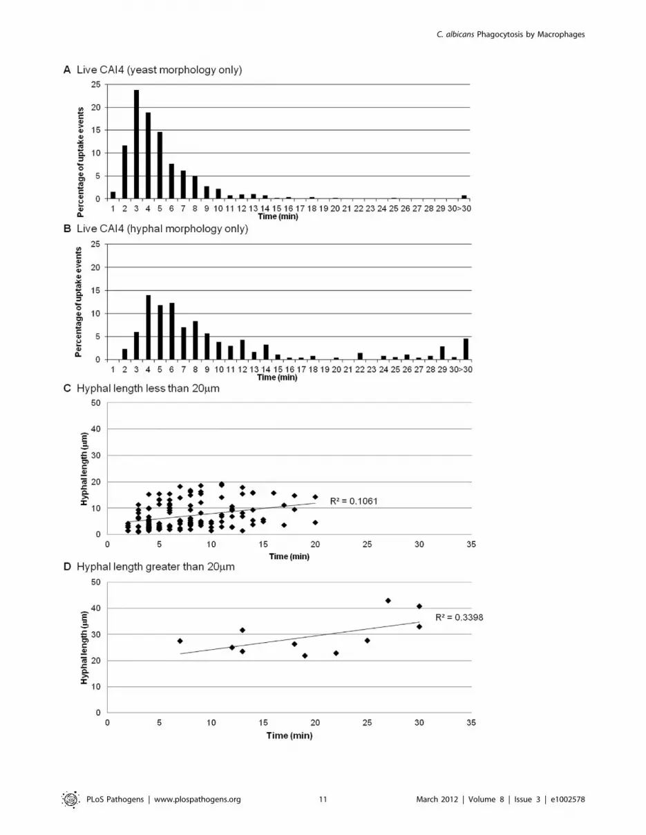
Macrophages are more effective at engulfing yeast ratherthan hyphal C. albicans and engulfment is influenced byhyphal length
The data above showed that UV-killed yeast cells were engulfed
more rapidly than live wildtype cells that were able to form
hyphae. The accelerated engulfment of UV-killed cells raised
questions about how cell morphology affects engulfment of C.
albicans by macrophages. We examined the engulfment of wildtype
C. albicans cells that had established cell-cell contact with
macrophages in either yeast or hyphal morphology. Hyphal C.
albicans cells were engulfed at a slower rate than C. albicans yeast
cells (10.860.9 min and 5.660.3 min, respectively). Furthermore,
the vast majority (98%) of yeast cells of C. albicans were taken up
within 15 min (Figure 6A), whereas there was greater variability
for hyphal cells of C. albicans, with 21% taking longer than 15 min
to become engulfed (Figure 6B).
Next we examined whether hyphal length influenced the speed of
engulfment, perhaps explaining the variations observed for hyphal
C. albicans engulfment. Macrophages were capable of ingesting C.
albicans with hyphae of more than twice the average diameter of
macrophages (the maximum observed length of ingested hyphae
was 42.9 mm), but the mean hyphal length at time of engulfment
was 8.760.7 mm. Intriguingly, and contrary to expectations, we
found no correlation between hyphal length and speed of
engulfment for hyphal cells of C. albicans of less than 20 mm length
(Figure 6C). However, when hyphal length exceeded 20 mm there
was as significant impact on the macrophage’s ability to engulf C.
albicans (Figure 6D). Although macrophages engulfed C. albicans with
hyphae larger than 20 mm, uptake was markedly slower with 64% of
uptake events requiring more than 15 min. It is worth noting that
despite having difficulty engulfing large hyphae macrophages were
nonetheless persistent in their attempt to do so. Thus, macrophages
were more effective at engulfing yeast cells rather than hyphal cells
of C. albicans and engulfment of hyphal cells was influenced in part
by hyphal length, with a cut off of 20 mm, above which macrophage
engulfment was markedly impaired.
The rate of engulfment of hyphal cells of C. albicans wasinfluenced by spatial orientation
Finally, we took advantage of the large quantity of data amassed
from live cell video microscopy phagocytosis assays to address
previously unanswered questions relating to how spatial orienta-
tion of C. albicans may affect the efficiency of engulfment by
macrophages. First, we established that hyphal cells of C. albicans
could be taken up by macrophages independent of their spatial
orientation (Figure 7A). C. albicans germ tubes could be engulfed
yeast-end on and germ tube apex-end on (Figures 7B and C), side-
on (Figure 7D) and at an angle (Figure 7E). However, it is
noteworthy that although cell-cell contact could be initiated in any
orientation, the rate of engulfment was affected; C. albicans that
made contact in an end-on orientation were taken up more rapidly
than those engulfed at an angle or where cell-cell contact was
initiated side-on (5.560.7 min, 8.860.5 min and 9.562.3 min,
respectively). The large SE observed when C. albicans makes
contact side-on can be explained by the fact that macrophages had
particular difficulty ingesting large hyphae (.20 mm) in the side-
on orientation. The end initially encountered by the macrophage
appeared to be random. Approximately equal numbers of
encounters occurred that were yeast end-on or hyphal end-on,
but there was a propensity for C. albicans to be taken up more
rapidly yeast-end on (8.060.6 min) than hyphal end-on
(9.861.0 min).
Initial C. albicans orientation when establishing cell-cell contact
with macrophages influenced the rate of engulfment with end-on
contact of the hyphal end resulting in the most rapid engulfment.
Thus, engulfment of hyphal cells of C. albicans was influenced by
multiple factors including hyphal length and spatial orientation,
and whether the initial encounter was by the yeast or hyphal end.
Discussion
C. albicans is a major life-threatening human fungal pathogen.
Host defence against systemic Candida infection relies mainly on
phagocytosis of fungal cells by cells of the innate immune system.
In this study, we analysed the contribution of distinct C. albicans
cell wall components and yeast-hypha morphogenesis to specific
stages of phagocytosis by macrophages.
We show that macrophage migration towards C. albicans was
dependent on the glycosylation status of the fungal cell wall, but
not cell viability or morphogenic switching from yeast to hyphal
forms. This finding was not a consequence of differences in
maximal macrophage track velocity, but stems from a greater
percentage of macrophages pursuing glycosylation deficient C.
albicans cells during the first hour of the phagocytosis assay. The
rate of engulfment of C. albicans by macrophages was significantly
slower for glycosylation and morphogenesis deficient mutant
strains, but enhanced for non-viable cells. Hyphal cells were
engulfed at a slower rate than yeast cells, especially those with
hyphae in excess of 20 mm, but there was no correlation between
hyphal length and the rate of engulfment below this threshold. We
show that spatial orientation of the hypha and whether hyphal C.
albicans attached to the macrophage via the yeast or hyphal end
were also important determinants of the rate of engulfment.
This is the first study, to our knowledge, to show that individual
stages of C. albicans phagocytosis by macrophages are differentially
affected by changes in C. albicans cell wall composition. Our
previous work, using assays that globally assess phagocytosis, have
shown increased phagocytosis of O-linked and N-linked mannan
deficient strains (mns1D and mnt1D mnt2D) [10]. Intriguingly, we
show here that changes in cell wall glycosylation enhance
macrophage migration towards C. albicans, but delay engulfment
once cell-cell contact is established. This illustrates that standard
assays do not differentiate between the individual stages of the
phagocytosis process and are unable to detect significant temporal
differences in migration or engulfment. For example, we show
here that phosphomannan deficient cells of C. albicans were
engulfed less efficiently. This effect was much less obvious in
previous studies that simply evaluated phagocytosis efficiency by
single time point measurements [27]. Macrophage migration was
enhanced in all glycosylation mutants but most markedly in the
mnt1Dmnt2D O-glycosylation mutant. This translated into much
higher overall uptake compared to the phosphomannan deficient
Figure 4. Macrophage engulfment of wildtype and glycosylation mutants of C. albicans. Figures 4A, B, C and D show the time taken forJ774.1 macrophages to ingest live wildtype (n = 6), mnt1Dmnt2D (n = 6), mns1D (n = 3) and mnn4D (n = 3) strains following initial cell-cell contactverses the percentage of uptake events. The rate of engulfment of all glycosylation mutant strains (Figures 4B–D) was significantly slower than that ofwildtype C. albicans (Figure 4A).doi:10.1371/journal.ppat.1002578.g004
C. albicans Phagocytosis by Macrophages
PLoS Pathogens | www.plospathogens.org 8 March 2012 | Volume 8 | Issue 3 | e1002578

mmn4D mutant and is in keeping with our previous published
results [10]. It is conceivable that enhanced macrophage migration
in response to the absence of O-linked (mnt1Dmnt2D) or N-linked
mannans (mns1D) is a consequence of unmasking underlying b-
glucans [28,29] or electrostatic signals as a consequence of
alterations in surface charge following loss of phosphomannan
(mnn4D) [30,31]. Observation of individual macrophage migration
patterns indicated that macrophage movement was slow and
random initially, but became directional towards a specific C.
albicans cell, associated with a marked increase in macrophage
velocity. Macrophage acceleration towards C. albicans occurred at
distances in excess of 15 mm and, therefore, suggests the presence
of a chemotactic signal. Key candidates are a number of
glycolipids that are known to be shed by C. albicans and are
Figure 5. Macrophage engulfment of wildtype and yeast-locked morphogenetic mutant strains of C. albicans. Figures 5A, B and C showthe time taken for J774.1 macrophages to ingest live wildtype (n = 6), hgc1D(n = 6) and efg1D (n = 3) following initial cell-cell contact plotted versusthe percentage of uptake events. The average time taken for engulfment of the hgc1D (Figure 5B) and efg1D (Figure 5C) mutant strains wassignificantly greater than for the wildtype control (Figure 5A).doi:10.1371/journal.ppat.1002578.g005
C. albicans Phagocytosis by Macrophages
PLoS Pathogens | www.plospathogens.org 9 March 2012 | Volume 8 | Issue 3 | e1002578

potent inducers of macrophage cytokine synthesis in vitro and in vivo
[32]. We are currently conducting detailed mathematical
modelling of the macrophage tracking patterns to further elucidate
the hypothesis that macrophage migration towards C. albicans is
affected by differences in shedding of glycolipids between wildtype
and glycosylation deficient strains.
We have shown that macrophage uptake of C. albicans is a multi-
step process, involving recognition and subsequent engulfment of
C. albicans. C. albicans cell wall mannosylation is a key determinant
in the rate of engulfment; the absence of specific PAMPs in the
glycosylation mutant strains delays engulfment once cell-cell
contact has been established, and this may be a consequence of
differential activation of macrophage PRRs. This is in keeping
with experiments in which C. albicans cell wall mutants were
combined with specific macrophage receptor blocking methods
that have been used to define the PAMP-PRR interactions
required for cytokine induction [14,33]. These in vitro findings are
relevant to C. albicans infections in vivo, since C. albicans mutants
with defects in cell wall mannosyl residues are also less virulent in
experimental models of disseminated candidiasis [21,22,34–36].
Morphological plasticity is one of the hallmarks of the human
fungal pathogen C. albicans [37], and its ability to switch between
yeast and hyphal forms is thought to contribute to pathogenesis
[24,25,38,39]. C. albicans mutants that are unable to form filaments
are less virulent [40], although conversely, mutants that are unable
to grow as yeast are also less virulent [41]. There are conflicting
reports in the literature regarding the efficiency of macrophage
phagocytosis for C. albicans yeast and hyphal forms [20,42,43].
Here we show definitively data supporting the notion that
macrophages are more effective at engulfing C. albicans yeasts.
Furthermore, the use of video microscopy coupled with thorough
analysis of large numbers of individual macrophage-C. albicans
interactions provides a minute-by-minute account of the engulf-
ment process, which offers detail that has not been previously
available. A prime example is our observation that yeast-locked C.
albicans cells were engulfed less efficiently than wildtype C. albicans.
Not only was this not obvious in previous studies that simply
evaluated phagocytosis efficiency by single time point measure-
ments [10], but in addition, we observed here that delayed
engulfment can result in detachment of the fungal cell and
engulfment by a neighbouring macrophage. One may speculate
that in vivo where phagocyte numbers are limited this may have a
significant impact on pathogen clearance and that yeast locked
mutant C. albicans cells have properties other than the induced
phenotype that differ from wildtype yeast C. albicans cells.
However, the observed delay in engulfment for yeast-locked
mutant C. albicans in the macrophage cell line was almost
completely abrogated in experiments using primary macrophages,
underlining the importance of studying host-pathogen interactions
in multiple phagocyte subsets.
The approach taken here further enabled us to dissect the
complexity of engulfment of hyphal C. albicans by macrophages.
We show that macrophages are capable of engulfing hyphal C.
albicans in excess of 40 mm (approximately twice the diameter of
macrophages) - in keeping with reports that macrophages are
capable of ingesting apoptotic epithelial cells in the involuting
mammary gland of similar or even larger size [44]. Hyphal length,
however, does play a major role in the engulfment process of C.
albicans, in that engulfment of C. albicans with hyphae in excess of
20 mm took significantly more time and phagocytosis was
frequently frustrated. Interestingly, we show that below a 20 mm
hyphal length threshold there was no correlation between hyphal
length and the rate of engulfment. These observations are most
likely related to difficulties associated with macrophages attempt-
ing to engulf very large particles. In addition to hyphal length, we
have identified two other factors that influenced engulfment of
hyphal C. albicans. We showed that the rate of engulfment was
determined by the orientation in which C. albicans was encoun-
tered, with end-on being favourable to side-on orientation,
suggesting that steric hindrance affects engulfment. We also
showed that yeast end-on engulfment was more efficient than
hyphal end-on encounters. This in turn may reflect differences in
the wall chemistry of the hyphal tip compared to the mother cell,
or be due to the efficiency of the assembly of proteins of the
phagocytic cup for objects of different sizes and shapes [45].
Here we have conducted the most detailed analysis of the
contribution of C. albicans viability, cell wall glycosylation and
morphogenesis to phagocytosis by macrophages to date, to our
knowledge. Our approach of combining live cell video microscopy
with image analysis tools for the migration analysis, and minute-
by-minute analysis of thousands of individual macrophage-C.
albicans interactions, provides unique insight into the complexity of
C. albicans phagocytosis by macrophages. The novel methods
employed here to study phagocytosis of C. albicans could be applied
to study other pathogens and uptake of dying host cells. Such
studies would significantly enhance our understanding of the
mechanisms that govern effective phagocytosis and ultimately the
innate immune response to infection.
Materials and Methods
Ethics statementAll animal experiments have been conducted in strict
accordance with UK Home Office guidelines. The appropriate
Table 3. Engulfment of wildtype and mutant of C. albicans by primary macrophages.
C. albicans strainAverage time for engulfment (min)by peritoneal macrophages
Average time for engulfment (min) by humanmonocyte derived macrophages
CAI4+CIp10 8.5662.56 6.960.32
UV killed CAI4+CIp10 7.3960.24 -
hgc1D 9.8563.14 -
mnt1Dmnt2D 11.961.1* 8.560.28*
mnt1Dmnt2D::MNT1 7.4160.67 -
Table 3 shows the average time taken+SD (min) for peritoneal macrophages and human monocyte derived macrophages to engulf wildtype and mutant C. albicans.N = 3,*,p,0.01.doi:10.1371/journal.ppat.1002578.t003
C. albicans Phagocytosis by Macrophages
PLoS Pathogens | www.plospathogens.org 10 March 2012 | Volume 8 | Issue 3 | e1002578
C. albicans Phagocytosis by Macrophages
PLoS Pathogens | www.plospathogens.org 11 March 2012 | Volume 8 | Issue 3 | e1002578

project and personal licenses are in place PIL 60/6194 and
approved by the UK Home office.
C. albicans strains and growth conditionsC. albicans serotype A strain CAI4+CIp10, hitherto referred to as
the parental wildtype, was used as a control and its parent strain,
CAI4, was used to construct mutants using targeted gene
disruption [46]. The mutants used are listed in Table 1. C. albicans
strains containing a single reintegrated copy of the corresponding
deleted genes to regenerate the heterozygous genotype acted as
controls. Most of the C. albicans strains used were created in house
and have been described previously [21–25]. C. albicans strains
were obtained from glycerol stocks stored at 280uC, and plated on
SC-Ura plates (except hgc1Dand efg1D). SC-Ura plates consist of
6.9 g yeast nitrogen base without amino acids (Formedium,
Norfolk, UK), 1 ml 1 M NaOH (BDH Chemicals, VWR
International, Leicestershire, UK), 10 ml 1% (w/v) adenine
hemisulphate salt (Sigma, Dorset, UK), 50 ml 40% D-glucose
(Fisher Scientific, Leistershire, UK), 50 ml 4% SC-Ura dropout
(Formedium, Norfolk, UK) and 2% (w/v) technical agar (Oxoid,
Cambridge, UK) made up to 1000 ml in distilled H2O. The C.
albicans morphogenetic mutants hgc1D and efg1D were grown on
YPD plates consisting of 1% yeast extract (Duchefa Biochemie,
Haarlem, Holland), 2% mycopeptone (Oxoid, Cambridge, UK),
2% D-glucose and 2% technical agar in distilled H2O. All plates
were incubated at 30uC until colonies formed, and were then
stored at 5uC.
Preparation of thioglycollate-induced peritoneal mousemacrophages
Intraperitoneal injections of 1 ml Brewer’s thioglycollate broth
(BD, New Jersey, USA) were administered to 8 week old female
BALB/c mice. After 4 days, the peritoneal cavity of sacrificed mice
was lavaged with 5 mM EDTA in 16 PBS, to harvest thioglycol-
late-induced macrophages. These Thio-macrophages were washed
3 times with RPMI medium 1640 (Sigma, Dorset, UK) supple-
mented with 10% (v/v) foetal calf serum (FCS) (Biosera, Ringmer,
UK), 200 U/ml penicillin/streptomycin antibiotics (Invitrogen Ltd,
Paisley, UK), 10 mM HEPES (Invitrogen Ltd, Paisley, UK) and
2 mM L-glutamine (Invitrogen, Paisley, UK). For phagocytosis
assays, 16106 thio-macrophages in 2 ml supplemented RPMI
medium were seeded onto glass bottomed Iwaki dishes (VWR,
Leistershire, UK) and cultured overnight at 37uC with 5% CO2.
Immediately prior to experiments, RPMI medium was replaced
with 2 ml pre-warmed supplemented CO2-independent medium
(Gibco, Invitrogen, Paisley, UK) containing 1 mM LysoTracker Red
DND-99 (Invitrogen, Paisley, UK). LysoTracker Red DND-99 is a
red fluorescent dye that stains macrophage acidic organelles,
enabling macrophage paths to be tracked using Volocity 5.0
software (Improvision, PerkinElmer, Coventry, UK).
Preparation of J774.1 mouse macrophage cell line andHMDM
J774.1 macrophages (ECACC, HPA, Salisbury, UK) were
maintained in tissue culture flasks in DMEM medium (Lonza,
Slough, UK), supplemented with 10% (v/v) FCS (Biosera,
Ringmer, UK), 200 U/ml penicillin/streptomycin antibiotics
(Invitrogen, Paisley, UK) and 2 mM L-glutamine (Invitrogen,
Paisley, UK) at 37uC with 5% CO2. Human monocyte derived
macrophages were prepared as previously described in detail [26].
For phagocytosis assays, 16106 J774.1 macrophages in 2 ml
supplemented DMEM medium were seeded onto glass based
Iwaki dishes (VWR, Leistershire, UK) and cultured overnight at
37uC with 5% CO2. Immediately prior to experiments, DMEM
medium was replaced with 2 ml pre-warmed supplemented CO2-
independent medium (Gibco, Invitrogen, Paisley, UK) containing
1 mM LysoTracker Red DND-99 (Invitrogen, Paisley, UK).
C. albicans preparation and staining with fluoresceinisothiocyanate (FITC)
Single C. albicans colonies from plates stored at 5uC were
cultured in 5 ml SC-Ura/YPD medium (recipes as above,
excluding technical agar) and incubated overnight at 30uC,
200 rpm. In order to determine the impact of C. albicans viability
on macrophage migration and engulfment, wildtype C. albicans
were killed by UV-irradiation; 1006106 fungal cells in 1 ml 16PBS were exposed to 20 doses of UV irradiation at 100 mJ/cm2 in
6 well plates. To aid visualisation of C. albicans during phagocytosis
assays, 1006106 live or UV-killed C. albicans were stained using
1 mg/ml FITC (Sigma, Dorset, UK) in 0.05 M carbonate-
bicarbonate buffer (pH 9.6) (BDH Chemicals, VWR Internation-
al, Leicestershire, UK) for 10 min at room temperature in the
dark. Fungal cells were washed 3 times in PBS to remove unbound
FITC and resuspended in 16 PBS.
Live cell video microscopy phagocytosis assaysOur standard phagocytosis assays were performed as previously
described [10]. In brief, 36106 FITC-stained C. albicans were
added to 16106 macrophages in glass based Iwaki dishes (VWR,
Leistershire, UK) immediately prior to imaging. Video microscopy
experiments were performed using a DeltaVision Core microscope
(Applied Precision, Washington, USA) with an environmental
control chamber set at 37uC. Images were captured at 1 min
intervals for 6 h using an EMCCD camera. At least two
independent experiments were conducted for each C. albicans
strain, and at least 3 movies were analysed from each experiment.
One hundred macrophages were randomly selected from each
movie and their phagocytic activity determined (as below).
Analysis of live cell video microscopy moviesVolocity 5.0 imaging analysis software was used to track
macrophage migration at 1 min intervals throughout the 6 h
phagocytosis assay. The software enabled high throughput analysis
of macrophage migration, providing detailed information on the
distances travelled, directionality and velocity of thousands of
individual macrophages. Data were subsequently displayed in
tracking diagrams and used to calculate the mean track velocity
and track length of macrophages cultured with C. albicans. These
analyses enabled assessment of the affects of C. albicans viability,
glycosylation status and morphology on migration.
Figure 6. Role of hyphal morphogenesis in the engulfment of C. albicans by macrophages. Figures 6A and B show the time taken forJ774.1 macrophages to ingest live C. albicans in yeast and hyphal morphology plotted against the percentage of uptake events (n = 6). Hyphal C.albicans was engulfed at a significantly slower rate than the yeast form of C. albicans. Figures 6C and D show the time taken for macrophages toengulf C. albicans with hyphae less than and greater than 20 mm, respectively (n = 6). There was no correlation between hyphal length and speed ofengulfment for hyphal C. albicans of less than 20 mm length (Figure 6C). However, when hyphal length exceeded 20 mm there was significant impacton the macrophage’s ability to engulf C. albicans (Figure 6D).doi:10.1371/journal.ppat.1002578.g006
C. albicans Phagocytosis by Macrophages
PLoS Pathogens | www.plospathogens.org 12 March 2012 | Volume 8 | Issue 3 | e1002578

Figure 7. The role of C. albicans spatial orientation in engulfment by J774.1 macrophages. Figure 7A plots the time taken for engulfmentof individual hyphal wildtype C. albicans in relation to the exact angles at which cell-cell contact was established (0u, side-on; 90u, end-on). C. albicanscan be engulfed in any spatial orientation, including end-on, side-on and at an angle (Figure 7A). Figures 7B–E show snapshots from live cell videomicroscopy movies showing C. albicans being taken up in a variety of orientations, including yeast end on, (Figure 7B), germ tube apex end-on(Figure 7C), side-on (7D) and at an angle (7E). Scale bar, 10 mm; macrophage of interest, *.doi:10.1371/journal.ppat.1002578.g007
C. albicans Phagocytosis by Macrophages
PLoS Pathogens | www.plospathogens.org 13 March 2012 | Volume 8 | Issue 3 | e1002578

One hundred macrophages from each movie were analysed
individually at 1 min intervals throughout the 6 h phagocytosis
assay. Measurements taken include the time points at which initial
cell-cell contact occurred and at which C. albicans was fully
enclosed, the number of C. albicans taken up and their morphology,
the orientation of hyphal C. albicans relative to the macrophage
and hyphal length. The rate of engulfment of live and UV-killed
wildtype C. albicans, and glycosylation and yeast-locked morpho-
genetic mutant C. albicans was calculated by subtracting the time
point at which initial cell-cell contact occurred from the time point
at which the fungus was fully phagocytosed. This enabled accurate
assessment of the affects of C. albicans viability, glycosylation status
and morphology on the speed of engulfment. C. albicans spatial
orientation, morphology, hyphal length and the end of hyphal C.
albicans recognised were determined to assess whether these factors
impact on the rate of engulfment. This strategy enabled in depth
analysis of individual C. albicans-macrophage interactions in real
time.
Statistical analysisMean values and standard errors were calculated. One-way
analysis of variance (ANOVA) and Tukey-Kramer Multiple
Analysis Comparison Tests were used to determine statistical
significance.
Supporting Information
Video S1 Phagocytosis of live C. albicans by J774.1macrophages. Shows a representative 6 hour live video
microscopy of live C. albicans being ingested by macrophages. It
further illustrates hyphal growth within macrophages and
macrophage killing by hyphal C. albicans.
(MP4)
Video S2 Phagocytosis of live C. albicans by J774.1macrophages. Shows a high magnification (680) representative
6 hour live video microscopy of live C. albicans being ingested by
macrophages. It further illustrates hyphal growth within macro-
phages and macrophage killing by hyphal C. albicans.
(MP4)
Video S3 Macrophage tracking of C. albicans. Shows an
example of a J774.1 macrophage tracking a UV-killed C.albicans
yeast cell. It illustrates random macrophage movement followed by
directional tracking of the fungal cell and ultimately uptake of the
pathogen.
(MP4)
Video S4 Projection movie of C. albicans phagocytosisby macrophages. Shows a 3D projection of C. albicans ingested
by macrophages. It also confirms co-localisation of green FITC
stained fungal cells with lysotracker red in macrophage phago-
somes.
(MOV)
Acknowledgments
We would like to thank Hector M. Mora-Montes and Gordon D. Brown
for advice and useful discussions. We would also like to thank the
University of Aberdeen imaging facility, in particular Kevin MacKenzie,
for helpful support and advice.
Author Contributions
Conceived and designed the experiments: LEL NARG LPE. Performed
the experiments: LEL JMB CG CL FMR. Analyzed the data: LEL NARG
LPE. Contributed reagents/materials/analysis tools: NARG. Wrote the
paper: LEL JMB NARG LPE.
References
1. Bistoni F, Vecchiarelli A, Cenci E, Puccetti P, Marconi P, et al. (1986) Evidence
for macrophage-mediated protection against lethal Candida albicans infection.
Infect Immun 51: 668–674.
2. Netea MG, Brown GD, Kullberg BJ, Gow NA (2008) An integrated model of
the recognition of Candida albicans by the innate immune system. Nat Rev
Microbiol 6: 67–78.
3. Sheth CC, Hall R, Lewis L, Brown AJ, Odds FC, et al. (2011) Glycosylation
status of the C. albicans cell wall affects the efficiency of neutrophil phagocytosis
and killing but not cytokine signaling. Med Mycol 49: 513–524.
4. Kapteyn JC, Hoyer LL, Hecht JE, Muller WH, Andel A, et al. (2000) The cell
wall architecture of Candida albicans wild-type cells and cell wall-defective
mutants. Mol Microbiol 35: 601–611.
5. Cutler JE (2001) N-glycosylation of yeast, with emphasis on Candida albicans. Med
Mycol 39 Suppl 1: 75–86.
6. Ernst JF, Prill SK (2001) O-glycosylation. Med Mycol 39 Suppl 1: 67–74.
7. Moyes DL, Runglall M, Murciano C, Shen C, Nayar D, et al. (2010) A biphasic
innate immune MAPK response discriminates between the yeast and hyphal
forms of Candida albicans in epithelial cells. Cell Host Microbe 8: 225–235.
8. Cheng SC, van de Veerdonk FL, Lenardon M, Stoffels M, Plantinga T, et al.
(2011) The dectin-1/inflammasome pathway is responsible for the induction of
protective T-helper 17 responses that discriminate between yeasts and hyphae of
Candida albicans. J Leukoc Biol 90: 357–66.
9. Kumamoto CA, Vinces MD (2005) Contributions of hyphae and hypha-co-
regulated genes to Candida albicans virulence. Cell Microbiol 7: 1546–1554.
10. McKenzie CG, Koser U, Lewis LE, Bain JM, Mora-Montes HM, et al. (2010)
Contribution of Candida albicans cell wall components to recognition by and
escape from murine macrophages. Infect Immun 78: 1650–1658.
11. Brown GD (2011) Innate antifungal immunity: The key role of phagocytes.
Annu Rev Immunol 29: 1–21.
12. Kaposzta R, Marodi L, Hollinshead M, Gordon S, da Silva RP (1999) Rapid
recruitment of late endosomes and lysosomes in mouse macrophages ingesting
Candida albicans. J Cell Sci 112: 3237–3248.
13. Netea MG, Marodi L (2010) Innate immune mechanisms for recognition and
uptake of Candida species. Trends Immunol 31: 346–353.
14. Netea MG, Gow NA, Munro CA, Bates S, Collins C, et al. (2006) Immune
sensing of Candida albicans requires cooperative recognition of mannans and
glucans by lectin and toll-like receptors. J Clin Invest 116: 1642–1650.
15. Means TK, Mylonakis E, Tampakakis E, Colvin RA, Seung E, et al. (2009)
Evolutionarily conserved recognition and innate immunity to fungal patho-
gens by the scavenger receptors SCARF1 and CD36. J Exp Med 206: 637–
653.
16. Miyazato A, Nakamura K, Yamamoto N, Mora-Montes HM, Tanaka M, et al.
(2009) Toll-like receptor 9-dependent activation of myeloid dendritic cells by
deoxynucleic acids from Candida albicans. Infect Immun 77: 3056–3064.
17. McGreal EP, Rosas M, Brown GD, Zamze S, Wong SY, et al. (2006) The
carbohydrate-recognition domain of dectin-2 is a C-type lectin with specificity
for high mannose. Glycobiology 16: 422–430.
18. Wells CA, Salvage-Jones JA, Li X, Hitchens K, Butcher S, et al. (2008) The
macrophage-inducible C-type lectin, mincle, is an essential component of the
innate immune response to Candida albicans. J Immunol 180: 7404–7413.
19. Mora-Montes HM, Netea MG, Ferwerda G, Lenardon MD, Brown GD, et al.
(2011) Recognition and blocking of innate immunity cells by Candida albicans
chitin. Infect Immun 79: 1961–1970.
20. Keppler-Ross S, Douglas L, Konopka JB, Dean N (2010) Recognition of yeast
by murine macrophages requires mannan but not glucan. Eukaryot Cell 9:
1776–1787.
21. Munro CA, Bates S, Buurman ET, Hughes HB, Maccallum DM, et al. (2005)
Mnt1p and Mnt2p of Candida albicans are partially redundant alpha-1,2-
mannosyltransferases that participate in O-linked mannosylation and are
required for adhesion and virulence. J Biol Chem 280: 1051–1060.
22. Mora-Montes HM, Bates S, Netea MG, Diaz-Jimenez DF, Lopez-Romero E,
et al. (2007) Endoplasmic reticulum alpha-glycosidases of Candida albicans are
required for N glycosylation, cell wall integrity, and normal host-fungus
interaction. Eukaryot Cell 6: 2184–2193.
23. Hobson RP, Munro CA, Bates S, MacCallum DM, Cutler JE, et al. (2004) Loss
of cell wall mannosylphosphate in Candida albicans does not influence
macrophage recognition. J Biol Chem 279: 39628–39635.
24. Zheng X, Wang Y, Wang Y (2004) Hgc1, a novel hypha-specific G1 cyclin-
related protein regulates Candida albicans hyphal morphogenesis. EMBO J 23:
1845–1856.
25. Lo HJ, Kohler JR, DiDomenico B, Loebenberg D, Cacciapuoti A, et al. (1997)
Nonfilamentous C. albicans mutants are avirulent. Cell 90: 939–949.
26. McPhillips KA, Erwig LP (2009) Assessment of apoptotic cell phagocytosis by
macrophages. Methods Mol Biol 559: 247–256.
C. albicans Phagocytosis by Macrophages
PLoS Pathogens | www.plospathogens.org 14 March 2012 | Volume 8 | Issue 3 | e1002578

27. Hobson RP, Munro CA, Bates S, MacCallum DM, Cutler JE, et al. (2004) Loss
of cell wall mannosylphosphate in Candida albicans does not influence
macrophage recognition. J Biol Chem 279: 39628–39635.
28. Klippel N, Cui S, Groebe L, Bilitewski U (2010) Deletion of the Candida albicans
histidine kinase gene CHK1 improves recognition by phagocytes through an
increased exposure of cell wall beta-1,3-glucans. Microbiology 156: 3432–3444.
29. Mora-Montes HM, Bates S, Netea MG, Castillo L, Brand A, et al. (2010) A
multifunctional mannosyltransferase family in Candida albicans determines cell
wall mannan structure and host-fungus interactions. J Biol Chem 285:
12087–12095.
30. Erwig LP, McPhilips KA, Wynes MW, Ivetic A, Ridley AJ, et al. (2006)
Differential regulation of phagosome maturation in macrophages and dendritic
cells mediated by rho GTPases and ezrin-radixin-moesin (ERM) proteins. Proc
Natl Acad Sci U S A 103: 12825–12830.
31. McKenzie CG, Koser U, Lewis LE, Bain JM, Mora-Montes HM, et al. (2010)
Contribution of Candida albicans cell wall components to recognition by and
escape from murine macrophages. Infect Immun 78: 1650–1658.
32. Jouault T, Fradin C, Trinel PA, Bernigaud A, Poulain D (1998) Early signal
transduction induced by Candida albicans in macrophages through shedding of a
glycolipid. J Infect Dis 178: 792–802.
33. Mora-Montes HM, Bates S, Netea MG, Castillo L, Brand A, et al. (2010) A
multifunctional mannosyltransferase family in Candida albicans determines cell
wall mannan structure and host-fungus interactions. J Biol Chem 285:
12087–12095.
34. Buurman ET, Westwater C, Hube B, Brown AJ, Odds FC, et al. (1998)
Molecular analysis of CaMnt1p, a mannosyl transferase important for adhesion
and virulence of Candida albicans. Proc Natl Acad Sci U S A 95: 7670–7675.
35. Bates S, MacCallum DM, Bertram G, Munro CA, Hughes HB, et al. (2005)
Candida albicans Pmr1p, a secretory pathway P-type Ca2+/Mn2+-ATPase, is
required for glycosylation and virulence. J Biol Chem 280: 23408–23415.
36. Bates S, Hughes HB, Munro CA, Thomas WP, MacCallum DM, et al. (2006)
Outer chain N-glycans are required for cell wall integrity and virulence ofCandida albicans. J Biol Chem 281: 90–98.
37. Lu Y, Su C, Wang A, Liu H (2011) Hyphal development in Candida albicans
requires two temporally linked changes in promoter chromatin for initiation andmaintenance. PLoS Biol 9: e1001105.
38. Gow NA, Brown AJ, Odds FC (2002) Fungal morphogenesis and host invasion.Curr Opin Microbiol 5: 366–371.
39. Sudbery PE (2011) Growth of Candida albicans hyphae. Nat Rev Microbiol 9:
737–748.40. Kadosh D, Johnson AD (2005) Induction of the Candida albicans filamentous
growth program by relief of transcriptional repression: A genome-wide analysis.Mol Biol Cell 16: 2903–2912.
41. Saville SP, Lazzell AL, Monteagudo C, Lopez-Ribot JL (2003) Engineeredcontrol of cell morphology in vivo reveals distinct roles for yeast and filamentous
forms of Candida albicans during infection. Eukaryot Cell 2: 1053–1060.
42. Marcil A, Gadoury C, Ash J, Zhang J, Nantel A, et al. (2008) Analysis of PRA1and its relationship to Candida albicans- macrophage interactions. Infect Immun
76: 4345–4358.43. d’Ostiani CF, Del Sero G, Bacci A, Montagnoli C, Spreca A, et al. (2000)
Dendritic cells discriminate between yeasts and hyphae of the fungus Candida
albicans. Implications for initiation of T helper cell immunity in vitro and in vivo.J Exp Med 191: 1661–1674.
44. Monks J, Henson PM (2009) Differentiation of the mammary epithelial cellduring involution: Implications for breast cancer. J Mammary Gland Biol
Neoplasia 14: 159–170.45. Champion JA, Mitragotri S (2006) Role of target geometry in phagocytosis. Proc
Natl Acad Sci U S A 103: 4930–4934.
46. Murad AM, Lee PR, Broadbent ID, Barelle CJ, Brown AJ (2000) CIp10, anefficient and convenient integrating vector for Candida albicans. Yeast 16:
325–327.
C. albicans Phagocytosis by Macrophages
PLoS Pathogens | www.plospathogens.org 15 March 2012 | Volume 8 | Issue 3 | e1002578


