Embed Size (px)
Citation preview

ACADEMIA ROMÂNĂ
Revue Roumaine de Chimie
http://web.icf.ro/rrch/
Rev. Roum. Chim.,
2015, 60(5-6), 427-445
MINI-REVIEW
RECENT ANALYTICAL APPLICATIONS ON ANTIHYPERTENSIVE DRUGS
Cansu YAKAR, Burcin BOZAL-PALABIYIK, Burcu DOGAN-TOPAL, Sibel A. OZKAN and Bengi USLU*
Ankara University, Faculty of Pharmacy, Department of Analytical Chemistry, 06100 Tandogan, Ankara, Turkey
Received November 17, 2014
Antihypertensive drugs primarily affect systemic vascular resistance through producing vasodilation. This vasodilatory effect is achieved either by interference with sympathetic adrenergic vascular tone or by the blocking of the formation of angiotensin II or its vascular receptors. There are other antihypertensive drugs which have direct arterial dilatory or mixed arterial and venous dilatory effect. A survey of the literature published in analytical chemistry journals has been conducted and analytical methods which were developed and used for the determination of the antihypertensive drugs in their dosage forms and in biological samples have been reviewed in this paper. This review covers the time period from 2011 to present, during which over 125 recent analytical procedures including liquid chromatographic, spectrophotometric and voltammetric techniques were reported. The available information such as supporting electrolyte, pH, column, mobile phase, measuring or detection potential, sensitivity, selectivity etc. is given in the Tables.
INTRODUCTION*
The choice of an appropriate technique for the determination of a drug or a mixture of drug compounds mainly depends on the sample matrix complexity, the analyte concentration and the sample treatment time. The ultimate selection of an analytical technique is related to the intrinsic structure and physicochemical properties of the studied analyte. The extensive development of the pharmaceutical field requires more rigorous analytical methods for the control of antihypertensive drugs. Nowadays, these compounds are extensively used in clinical practice and therapy. Therefore, there is need for simple, sensitive, accurate, time saving and stability indicating methods for the *
determination of drugs in pure solutions, body fluids, and pharmaceutical dosage forms.
The aim of this review is to give some examples and information about the use of recent analytical applications on antihypertensive drugs in their dosage forms and in biological samples. The selected pharmaceutical active compounds, which have been determined using different electroanalyti-cal, LC, spectrophotometric techniques, are reported in tables between 1 and 3 together in detail. The available information on supporting electrolyte, pH, measuring or detection potential, column, mobile phase, extraction procedure, internal standard, sensitivity, selectivity, precision, accuracy etc. is presented in the tables shown below. Chemical names or structural formulae are given to help identification of electroactive moieties.
* Corresponding author: [email protected], tel:+903122033178, fax: +90 312 2031081

428 Cansu Yakar et al.
Antihypertensive drugs and classifications Antihypertensives are used to treat high blood
pressure, namely hypertension. The main goal of antihypertensive therapy is to prevent the complications of high blood pressure, including stroke or myocardial infarction. The reduction of the blood pressure is known to decrease the risk of stroke and ischaemic heart disease; moreover it reduces the probability of dementia, heart failure, and mortality from cardiovascular disease as well. Among different groups of antihypertensives, the most important and most widely used are the thiazide diuretics, the ACE inhibitors, the calcium channel blockers, the beta blockers, and the angiotensin II receptor antagonists or ARBs.1,2 In the treatment of hypertension, diuretics are essential for getting rid of unneeded water and salt through the urine, which lead to lower blood pressure and facilitate the circulation of blood in veins. Diuretics are widely used to treat a number of heart-related conditions, including high blood pressure, heart failure, kidney and liver problems, and glaucoma.1,2 The diuretic drugs are listed below:
Loop diuretics: bumetanide, ethacrynic acid, furosemide, torsemide
Thiazide diuretics: epitizide, hydrochlorothiazide, chlorothiazide, bendroflumethiazide Thiazide-like diuretics: indapamide, chlorthalidone, metolazone Potassium-sparing diuretics: amiloride, triamterene, spironolactone Among these different groups of diuretics, the thiazide and thiazide-like diuretics have a significant effect for lowering hypertension, and therefore, they are generally chosen as the first choice for hypertension treatment because of their vasodilating properties. Hypertension is mainly a condition caused by the sympathetic nervous system and it is mediated by α and β receptors. There are two types of α receptor, α1 and α2; this distinction is based on the response given to epinephrine and norepinephrine. Through selective blockade of the α1-adrenergic receptors, drugs such as prazosin, doxazosin, and terazosin act to reduce blood pressure. These agents contribute to the correction of elevated total peripheral resistance, which is the fundamental hemodynamic abnormality causing essential hypertension. α1-adrenergic blockers also have positive implications on plasma lipoproteins,
which facilitate the decrease in the levels of triglycerides and cholesterol and the increase in the levels of high-density lipoprotein (HDL) cholesterol and the HDL cholesterol/total cholesterol ratio. β-adrenergic blockers, such as propranolol and atenolol, tend to increase levels of triglycerides and decrease HDL cholesterol. Adrenergic modulation of lipoprotein lipase and the triglyceride secretion rate also contribute to these effects.3 The beta and alpha blockers are listed below: Beta blockers: atenolol, metoprolol, nadolol, nebivolol, oxprenolol, pindolol, propranolol, timolol Alpha blockers: doxazosin, phentolamine, indoramin, phenoxybenzamine, prazosin, terazosin, tolazoline Mixed alpha and beta blockers: bucindolol, carvedilol, labetalol Another hypertensive drug group is the benzodiazepines. These drugs are effective on the central nervous system through their selective action on gamma-aminobutyric acid-A (GABA-A) receptors in the brain. They strengthen the response to the inhibitory neurotransmitter GABA. This enhancement is achieved by opening GABA-activated chloride channels and allowing chloride ions to penetrate into the neuron. With this penetration the neuron is negatively charged and becomes resistant to excitation. In addition to their antihypertensive use, benzodiazepines are also used as sedatives, hypnotics, anxiolytics, anticonvulsants and muscle relaxants.1,2 Calcium channel blockers (CCBs) are attached to the L-type calcium channels on the vascular smooth muscle, cardiac myocytes, and cardiac nodal tissue. Since these channels regulate the influx of calcium into muscle cells and stimulate smooth muscle as well as cardiac myocyte contraction, by blocking calcium entry into the cell, CCBs contribute to vasodilation, lower myocardial force generation, decrease heart rate as well as conduction velocity particularly at the atrioventricular node.1,2 The calcium channel blockers are listed below: dihydropyridines: amlodipine, cilnidipine, felodipine, isradipine, lercanidipine, levamlodipine, nicardipine, nifedipine, nimodipine, nitrendipine non-dihydropyridines: diltiazem, verapamil Another hypertensive drug group, direct renin inhibitors, hampers the renin enzyme from triggering a process regulating the blood pressure. By this intervention, blood vessels relax and widen, facilitating the flow of blood and thus

Analytical applications on antihypertensive drugs 429
lowering blood pressure. Renin inhibitors are a new class of agents, and there are not sufficient clinical data regarding their ability. Aliskiren (Scheme 1) is the only renin inhibitor in the market, which has been approved by the U.S. FDA for the treatment of hypertension. In comparison with earlier renin inhibitors, aliskiren has favorable physiochemical properties with high aqueous solubility and lower lipophilicity, making it more resistant to degradation. This leads to improved bioavailability after oral administration.4
Scheme 1 – Chemical structure of Aliskiren.
ACE inhibitors slow down the activity of the
ACE enzyme, which decreases the production of angiotensin II, and thereby results in the enlargement or dilation of the blood vessels and reduction in the blood pressure. Angiotensin II is an efficient chemical produced by the body that causes the muscles surrounding blood vessels to contract. This contraction narrows the vessels and increases the blood pressure. Angiotensin II is formed from angiotensin I in the blood through an enzyme called angiotensin converting enzyme (ACE).1,2 The ACE inhibitors are listed below: captopril, enalapril, fosinopril, lisinopril, perindopril, quinapril, ramipril, trandolapril, benazepril. Angiotensin II receptor blockers have a similar impact and they inhibit a substance that causes blood vessels to narrow. As a result, blood vessels relax and widen which facilitates the flow of blood and reduces blood pressure. These medicines also increase the release of water and salt (sodium) to the urine, which in turn lowers blood pressure as well. Angiotensin II receptor blockers also act directly on the hormones that regulate sodium and water balance.1,2
The Angiotensin II receptor blockers are: candesartan, eprosartan, irbesartan, losartan, olmesartan, telmisartan, valsartan. Aldosterone receptor antagonists are a group of diuretics that help the body get rid of extra water.
By doing so, these medicines help retaining potassium by inhibiting the action of the hormone aldosterone.1,2 The molecules of that group are given as eplerenone and spironolactone. Vazodilator medicines lower high blood pressure by opening up the bloodvessels. This allows blood to flow more easily, which lowers blood pressure. Sodium nitroprusside, a very effective, short-acting vasodilator, is most commonly used for the quick, temporary reduction of blood pressure in emergencies (such as malignant hypertension or aortic dissection). Unlike this drug, hydralazine and its derivatives are generally avoided in emergencies due to severe side effects and safety concerns; still, hydralazine remains a drug of choice in gestational hypertension.1,2 The adrenoceptors (or adrenergic receptors) are rhodopsin-like G protein-coupled receptors that are targets of the catecholamines, such as norepinephrine (noradrenaline) and epinephrine (adrenaline). The clinical uses of adrenergic compounds are vast, particularly in the treatment of various diseases, such as hypertension, angina pectoris, congestive heart failure, asthma, depression, benign prostatic hypertrophy, and glaucoma. These drugs are also useful in several other therapeutic situations including shock, premature labour and opioid withdrawal, and as adjuncts to general anaesthetics.1,2 That group are listed below: clonidine, guanabenz, guanfacine, methyldopa, moxonidine. Bosentan (Scheme 2) belongs to a new class of drug and works by blocking the receptors of the hormone endothelin. Endothelin-1 (ET-1) is a 21 amino acid peptide that is produced by the vascular endothelium. As an extremely potent vasoconstrictor, it binds to smooth muscle endothelin receptors, being the ETA and ETB
receptors. These receptors are coupled to a Gq-protein and receptor activation leads to the formation of IP3. This formation causes the release of calcium by the sarcoplasmic reticulum (SR) and increases smooth muscle contraction and vasoconstriction. ET-1 receptors in the heart are also linked to the Gq-protein and IP3 signal transduction pathway. Therefore, ET-1 in the heart causes SR release of calcium, which increases contractility. ET-1 also increases heart rate. Bosentan is specifically indicated only for the treatment of pulmonary artery hypertension in patients with moderate to severe heart failure.5

430 Cansu Yakar et al.
Scheme 2 – Chemical structure of Bosentan.
Assay of antihypertensive drugs
The analysis of pharmaceuticals is an integral and increasingly important part of an overall drug development process. Technological and scientific progress has led to the development of numerous synthetic drugs. It is therefore imperative to develop analytical methods to determine these drugs both in the quality control manufacturing phase of the pharmaceutical formulations their determination in the human body. Several techniques like liquid chromatography (LC), spectrophotometry, voltammetry, etc have been used for the analysis of antihypertensive drugs (Table 1-3). Requirement for such methods is often accompanied by an increasing number of biological samples needing fast quantitative analysis, together with a decrease in the desired quantitation levels, as the bioavailability of many drugs is at a low level and thus target concentrations are very low. Consequently, appropriately designed, fast, effective and sensitive bio-analytical methods are needed. In general, a reliable bio-analytical method, which is convenient for an intended purpose, should fulfill the requirements of validation guidelines, including accuracy, precision, selectivity, sensitivity, reproducibility and stability.6
LC methods
Currently the most extensively used techniques in pharmaceutical analysis are chromatographical methods. They enable separation, identification and determination of huge amount of biologically active compounds. Among chromatographical techniques the special focus should be given to liquid chromatography (LC), especially high-performance liquid chromatography (HPLC), the technique of choice in the analysis of drugs and emerging ultra performance liquid chromatography (UPLC).
HPLC still remains a method of choice, as it is able to separate quite complicated mixtures of low and high molecular weight compounds, as well as different polarities and acid–base properties in various matrices. HPLC is a well-known and widely used analytical technique which is prevalent through-out the pharmaceutical industry as a research tool for estimation of impurities in drug substances and drug products. Despite its prominence, HPLC possesses some disadvantages, most notably longer analysis time and large consumption of organic solvents. UPLC is a relatively new technique which offers better separation capabilities when compared to HPLC with added benefits of shorter run time and lower solvent consumption. One of the key developments which facilitate the new UPLC technology is sub 2-µm particles used as column packing material. These particles withstand higher operating pressures and decreased flow rates to drastically increase the resolution, sensitivity and speed of analysis. Owing to its speed and sensitivity, this technique is gaining considerable attention in recent years for the analysis of pharmaceutical and biomedical compounds of interest.7-11 Because of their complexity and protein content, samples derived from biological materials are generally not directly compatible with HPLC analyses. In other words, these samples are problematic due to the irreversible adsorption of proteins in the stationary phase, which resulted in a considerable loss of column efficiency and an increase in back-pressure. Sample preparation has conventionally been performed using protein precipitation (PPT), liquid–liquid extraction (LLE), or solid phase extraction (SPE). The manual operations associated with these processes are very labor-intensive and time-consuming, consisting of many steps. Besides wide-spread conventional and automatic SPE, LLE and PP technique, newly developed sample preparation techniques include solid phase microextraction (SPME), liquid–liquid microextraction (LLME), pressurized liquid extraction (PLE), extraction using restricted access material (RAM), microextraction by packed sorbent (MEPS), molecularly imprinted polymer (MIP), mono-lith spin extraction, turbulent chromatography (TFC), salting-out liquid–liquid extraction (SALLE), stir bar sorptive extraction (SBSE) and others. Recent investigations have focused on the development of methods to reduce the sample volume required, the analytical time,

Analytical applications on antihypertensive drugs 431
the cost and the solvent consumption and even the elimination of chlorinated solvents. Further, miniaturization and automation using on-line coupling of analytical methods is a very important development as well. A complete review of the current status and recent advances in sample preparation techniques was conducted by Chen et al.,12 in which the detailed classification of all sample preparation techniques is outlined and selected techniques are discussed in detail. It is worth to focus on different detection possibilities, types of columns and variety of mobile phases in chromatographic techniques. There are three types of detectors used: UV (ultra-violet) sometimes in form of DAD (diode array detector), MS (mass spectrometry) and fluorometric as seen in Table 1. UV detection is the most popular detection method for liquid chromatography in the pharmaceutical industry, due to its high sensitivity, broad linear range, ease of operation and other advantages, as well as its compatibility with most mobile phase solvents. However, it requires a tedious and time-consuming derivatization procedure unless pharmaceutical compounds possess a UV-absorbing chromophore. Techniques such as refractive index (RI) detection or mass spectrometry (MS) detection have been employed for the detection of UV-undetectable compounds. But RI detection has the disadvantages of low sensitivity and incompatibility with gradient elution; MS detection, moreover, is expensive for routine use, and its requirement of specially trained operators limits its applicability further. Evaporative light scattering detection (ELSD) and, charged aerosol detection (CAD), introduced more recently, are additional alternatives to UV detection. Consequently, the response generated by CAD and ELSD are independent of the chemical structures of the compounds.13
Spectrophotometry
UV–vis spectrophotometry represents a suitable method for the routine analysis of active ingredients in raw materials, since it is fast, easy to perform and does not require expensive instruments. These methods show very simple and accurate way for the analysis of this binary mixture without the need of sophisticated instruments, HPTLC expensive solvents or large number of samples. In the last decades, Derivative Spectrophotometry (DS) has rapidly gained application in the field of pharmaceutical analysis to overcome the problem
of interference, due to the substances other than analytes, commonly present in pharmaceutical formulations or for combination of two or more drug substances. DS has been successfully used as a quality control tool in pharmaceutical analysis for the simultaneous determination of drugs in multi-component formulations. This technique, accessible to most laboratories, offers an alternative means of enhancing the sensitivity and specificity in mixture analysis. The procedure is simple, rapid and does not require any preliminary separations or treatment of the samples. The analytical characteristics of DS methods for individual drugs determination are described in Table 2.
Electroanalytical techniques
Applications of electrochemical techniques to redox-active drug development studies are one of the current interests in drug discovery. Electrochemical mechanisms are important to all redox chemistry including biological systems relating to electron transport chains. The performance of the electroanalytical procedures is strongly influenced by the working electrode materials. Voltammetry represents a class of electroanalytical methods in which the current at the solid working electrode (polarized) is measured as a function of the potential applied to that electrode; the recording of current versus potential is termed a voltammogram. The choice of an electrode material depends on a great extent on the useful potential range of the electrode in the particular solvent employed and the qualities and purity of the material. The working electrode selection depends on mainly two factors: the redox behavior of the investigated analyte and the background current over the potential region required for the measurements. Various types of carbon can be used as working electrode materials. The best known carbon electrode materials are those involving carbon paste, glassy carbon, graphite, diamond, carbon fiber, carbon blacks, screen-printed carbon strips, pyrolytic graphite, carbon films, fullerenes, wax impregnated graphite, carbon nanotube, kelgraf, reticulated vitreous carbon, whiskers etc. These types of carbon based electrodes have more or less a graphite structure but differ in their agglomeration, particle size, size distribution, degree of graphitization, and therefore in their physical and chemical properties. All these type of carbon materials are the result of specific synthesis routes.

432 Cansu Yakar et al.
Currently, various solid electrodes are available and their bulk or surface modification to improve their sensitivity and selectivity constitutes a principal issue for the majority of electroanalysts worldwide. The application of nanomaterials in various fields of science and technology has been extensively developed due to the unique properties of these materials. Nanoparticles of a variety of shapes, sizes and compositions are changing the bioanalytical measurement nowadays as seen in Table 3. The recent literature includes many electroanalyti-cal methods using voltammetric and amperometric techniques with modified electrodes. Among them, cyclic voltammetry is generally used for characterization, while differential pulse voltammetry (DPV) or square-wave voltammetry (SWV) and amperometry (AMP) are usually used for quantification of the analytes as seen in Table 3.
Examples of Analytical Applications
LC methods for antihypertensive drugs Several papers have been published dealing with the determination by LC of the active ingredients of antihypertensive drugs formulations. For example; a simple, sensitive, and specific method for furosemide (FUR) analysis by reverse-phase-HPLC was developed by employing a Spherisorb C18 ODS 2 column by Youm and Youan.14 They carried out a chromatographic analysis based on a mobile phase consisting of acetonitrile and 10 mM potassium phosphate buffer solution: 70:30 (v/v) at pH 3.85, at a flow rate of 1 mL/min. The UV-detection method was carried out at 233 nm at room temperature. The LOD and LOQ were found to be 5.2 and 15.8 ng/mL, respectively. Their method was found to be accurate (RSD less than 2%), precise, and specific with an intraday and interday RSD range of 1.233–1.509 and 1.615 to 1.963% and it can be used for the quantitative analysis of FUR from nanocarriers, USP tablets and release media related to hearing research. Mass spectrometric detection is mainly used in the bioanalysis of antihypertensive drugs. By Qi et al., a sensitive and rapid ultra performance liquid chromatography tandem mass spectrometry (UPLC–MS/MS) method was attempted to be developed for simultaneous determination of olmesartan and amlodipine levels in human plasma and urine.15 The Acquity UPLC BEH C18 column was used for chromatographic separation. Mass spectrometric analysis was carried out using a QTrap5500 mass spectrometer coupled with an
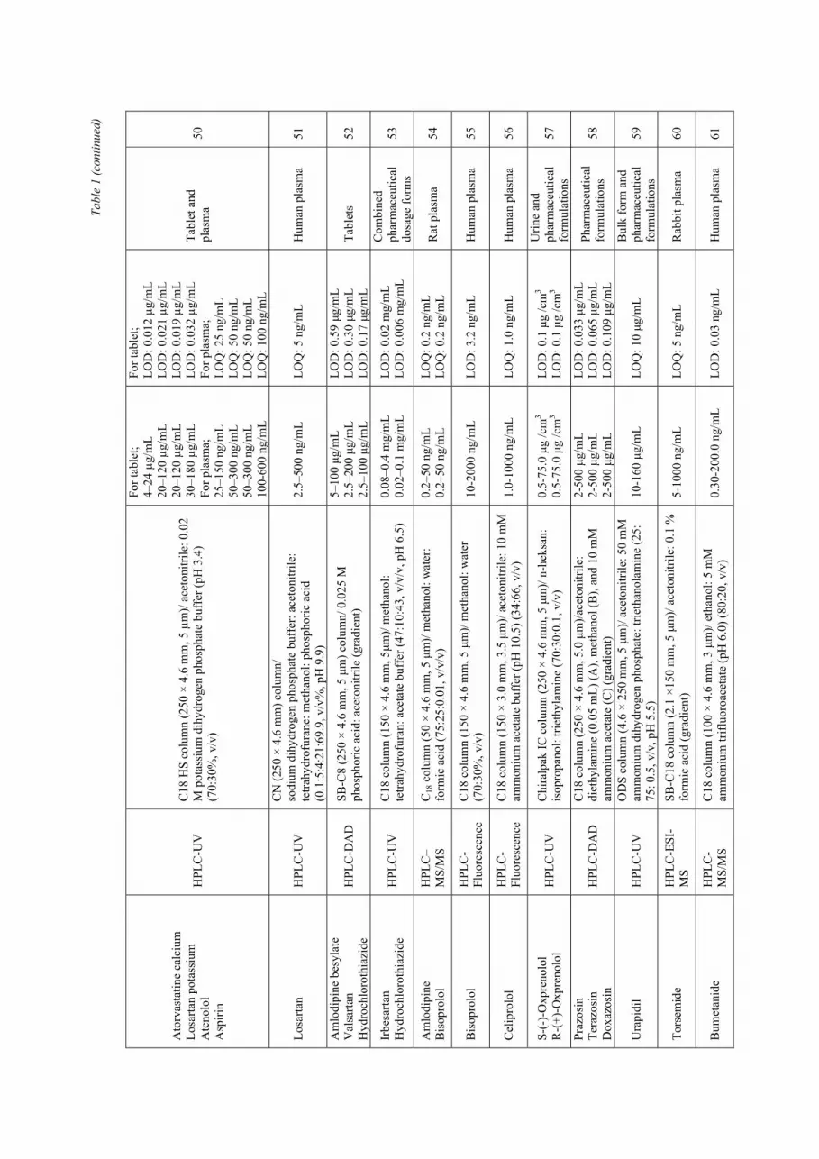
electro-spray ionization (ESI). The MRM transitions of m/z 447 → 207 and 409 → 238 were used to quantify olmesartan and amlodipine, respectively. The linearity of this method was found to be within the concentration range of 2–1000 ng/mL and 0.1–50 ng/mL for amlodipine in human urine and plasma and 4–5000 ng/mL and 0.2–500 ng/mL for olmesartan in human urine and plasma. The analysis was finished within only 2 min. Other selected examples of assays of dosage forms and biological samples are listed in Table 1.
Spectrophotometric methods
for antihypertensive drugs
Recent advances in pharmaceutical analysis with derivative spectrophotometry (DS) have rapidly gained application in the field of pharmaceutical analysis to overcome the problem of interference for combination of two or more drug substances. For example, Stolarczyk et al. attempted to develop a spectrophotometric method for determination of losartan potassium, quinapril hydrochloride and hydrochlorothiazide in pharmaceutical preparations.94 Spectrophotometric method involved derivative spectrophotometry and zero order spectrophotometry. The measurements were carried out at λ = 224.0 nm for quinapril, λ = 261.0 nm for hydrochlorothiazide and λ = 270.0 nm for losartan when the derivative spectrophotometry was applied and λ = 317.0 nm when zero order spectrophotometry was applied for the determination of hydrochlorothiazide. Sensitivity of developed method is high, for spectrophotometric method LOD was in the range from 0.68 to 2.01 µg/mL and LOQ from 2.06 to 6.08µg/mL.
By Gizawy et al., the first derivative of the ratio spectra method was developed for simultaneous determination of amlodipine besylate (AML) and Perindopril Erbumine (PER) without previous separation.95 This method was dependent on the by measuring the amplitudes at 348 nm for amlodipine using 50 µg/mL of perindopril as a divisor and at 227 nm for perindopril using 30 µg/mL of amlodipine as a divisor. The proposed method is applicable over a concentration range of 10–60 µg/mL and 20–80 µg/mL for AMLand PER, respectively. The validation of the proposed methods was made in accordance with the ICH guidelines and LOD and LOQ were accordingly calculated. These methods were successfully applied for the determination of the cited drugs in bulk powder and commercial tablets.

A
naly
tical
app
licat
ions
on
antih
yper
tens
ive
drug
s 43
3
Ta
ble
1
Som
e se
lect
ed L
iqui
d C
hrom
atog
raph
ic a
pplic
atio
ns o
n an
tihyp
erte
nsiv
e dr
ugs
Dru
g M
etho
d C
olum
n/m
obile
pha
se
Lin
ear
dyna
mic
ra
nge
LO
D/L
OQ
A
pplic
atio
n R
ef
Hyd
roch
loro
thia
zide
Ir
besa
rtan
Losa
rtan
pota
ssiu
m
Telm
isar
tan
Val
sarta
n
HPL
C-D
AD
C
18 c
olum
n (2
50 ×
4.6
mm
, 5µm
)/ 0.
025
M p
otas
sium
di
hydr
ogen
pho
spha
te (p
H 6
.0):
acet
onitr
ile (8
0:20
%,
v/v)
2.5–
15 µ
g/m
L 30
–180
µg/
mL
10–6
0 µg
/mL
16–9
6 µg
/mL
16–9
6 µg
/mL
LOD
: 0.0
4 µg
/mL
LOD
: 0.1
4 µg
/mL
LOD
: 0.0
8 µg
/mL
LOD
: 0.0
3 µg
/mL
LOD
: 0.0
4 µg
/mL
Phar
mac
eutic
al
form
ulat
ions
16
Cap
topr
il In
dapa
mid
e H
PLC
-DA
D
RP8
col
umn
(250
× 4
.6 m
m, 5
µm)/
30 m
M p
otas
sium
di
hydr
ogen
pho
spha
te (p
H 2
.8):
met
hano
l: ac
eton
itrile
(6
:2:2
, v/v
/v)
0.25
-150
µg/
mL
0.2-
100
µg/m
L LO
D: 0
.08
µg/m
L LO
D: 0
.07
µg/m
L Ta
blet
s 17
Lisi
nopr
il In
dapa
mid
e H
PLC
-UV
C
18 c
olum
n/m
etha
nol:w
ater
(50:
50, v
/v, p
H: 3
.1)
16-2
4 µg
/mL
8-12
µg/
mL
LOD
: 0.4
µg/
mL
LOD
: 0.5
µg/
mL
Har
d ge
latin
e ca
psul
e 18
Bis
opro
lol
Ram
ipril
at
Prop
rano
lol
Mid
azol
am
HPL
C-
MS/
MS
C18
colu
mn
(50
× 2.
1 m
m, 2
.6 µ
m)/
0.1%
form
ic a
cid
in
Mill
i-Q w
ater
(A) a
nd 1
00%
ace
toni
trile
(B) (
grad
ient
)
5-25
0 µg
/L
5-25
0 µg
/L
5-25
0 µg
/L
5-25
0 µg
/L
LOQ
: 5 µ
g/L
LOQ
: 5 µ
g/L
LOQ
: 5 µ
g/L
LOQ
: 5 µ
g/L
Rat
blo
od
19
Irbe
sarta
n H
ydro
chlo
roth
iazi
de
U-H
PLC
-M
S/M
S
C18
col
umn
(2.1
× 5
0 m
m, 1
.7 µ
m)/
solv
ent A
(0.1
%
form
ic a
cid
in w
ater
) and
solv
ent B
(ace
toni
trile
) (g
radi
ent)
5–30
00 n
g/m
L 0.
5–30
0 ng
/mL
LOQ
: 5.0
ng/
mL
LOQ
: 0.
5 ng
/mL
Hum
an p
lasm
a 20
S-(-
)-Pr
opan
olol
R
-(+)
- Pro
pano
lol
HPL
C-
Fluo
resc
ence
C
hira
lpak
IB c
olum
n (2
50 ×
4.6
mm
, 5 µ
m)/
n-he
xane
-et
hano
l-trie
thyl
amin
e (9
5:5:
0.4%
, v/v
/v)
10-4
00 n
g/m
L 10
-400
ng/
mL
LOD
: 3 n
g/m
L LO
D: 3
ng/
mL
Rat
seru
m
21
Cin
epaz
ide
mal
eate
H
PLC
-DA
D
C18
col
umn
(150
× 4
.6 m
m, 5
µm
)/ 10
mM
pot
assi
um
dihy
drog
en p
hosp
hate
(pH
4.5
): m
etha
nol (
40:6
0, v
/v)
0.12
-120
µg/
mL
LOD
: 0.0
6 µg
/mL
Rat
pla
sma
22
Ram
ipril
Te
lmis
arta
n A
mlo
dipi
ne b
esyl
ate
Ato
rvas
tatin
cal
cium
HPL
C-D
AD
C
18 c
olum
n (2
50 ×
4.6
mm
, 5 µ
m)/
0.02
5 M
pot
assi
um
dihy
drog
en p
hosp
hate
(pH
6.0
): ac
eton
itrile
(60:
40%
, v/
v)
10-6
0 µg
/mL
16-9
6 µg
/mL
10-6
0 µg
/mL
10-6
0 µg
/mL
LOD
: 0.5
8 µg
/mL
LOD
: 0.1
6 µg
/mL
LOD
: 0.7
2 µg
/mL
LOD
: 0.3
µg/
mL
Tabl
ets
23
Olm
esar
tan
Hyd
roch
loro
thia
zide
H
PLC
-ESI
-M
S/M
S
RP-
18A
col
umn
(4.6
× 1
50 m
m, 4
µm
)/ 0.
2% fo
rmic
ac
id: a
ceto
nitri
le (3
0:70
, v/v
)
4.05
1-25
00.9
12
ng/m
L 0.
506-
304.
109
ng/m
L
LOQ
: 4.0
51 n
g/m
L LO
Q: 0
.506
ng/
mL
Hum
an p
lasm
a 24
Telm
isar
tan
Hyd
roch
loro
thia
zide
U
PLC
-M
S/M
S
C18
col
umn
(50
× 2.
1 m
m, 1
.7 µ
m)/
acet
onitr
ile -
met
hano
l-10
mM
am
mon
ium
ace
tate
-for
mic
aci
d (5
0:30
:20:
0.1%
v/v
/v)
1-50
0 ng
/mL
1-50
0 ng
/mL
LOQ
: 1 n
g/m
L LO
Q: 1
ng/
mL
Hum
an p
lasm
a 25
Am
lodi
pine
B
enaz
epril
e B
enaz
epril
at
LC-
HES
I/MS/
MS
C18
col
umn
(100
× 4
.6 m
m, 5
µm
)/ m
etha
nol-
acet
onitr
ile
-5 m
M a
mm
oniu
m a
ceta
te-f
orm
ic a
cid
(30:
30:4
0:0.
1)
0.02
-6.0
0 ng
/mL
0.2-
1,50
0 ng
/mL
0.2-
1,50
0 ng
/mL
LOQ
: 0.0
2 ng
/mL
LOQ
: 0.2
ng/
mL
LOQ
: 0.2
ng/
mL
Hum
an p
lasm
a 26

434
Can
su Y
akar
et a
l. Ta
ble
1 (c
ontin
ued)
Met
olaz
one
Losa
rtan
pota
ssiu
m
HPL
C-
Fluo
resc
ence
C
18 c
olum
n (2
50×4
.6 m
m, 5
µm
)/ m
etha
nol:
0.02
M
phos
phat
e bu
ffer
(65:
35, v
/v, p
H 3
.0)
2.0–
40.0
ng/
mL
40.0
–800
.0 n
g/m
L LO
Q: 0
.22
ng/m
L LO
Q: 4
.52
ng/m
L
Com
bine
d ta
blet
s an
d hu
man
pl
asm
a 27
Car
vedi
lol
Hyd
roch
loro
thia
zide
H
PLC
-DA
D
SB-C
8 co
lum
n (4
.6 ×
250
mm
, 5 µ
m)/
0.02
5 M
ph
osph
oric
aci
d: a
ceto
nitri
le (g
radi
ent)
5-30
0 µg
/mL
5-20
0 µg
/mL
LOD
: 0.3
3 µg
/mL
LOD
: 0.3
0 µg
/mL
Com
bine
d ta
blet
s 28
Met
form
in
Lisi
nopr
il En
alap
ril
Cap
topr
il
HPL
C-U
V
RP1
8 co
lum
n (2
50 ×
4.6
mm
)/ m
etha
nol:w
ater
(50:
50,
v/v)
(pH
3.2
)
5.0-
50 µ
g/m
L 2.
5-25
0 µg
/mL
2.5-
250
µg/m
L
2.5-
250
µg/m
L
2.5-
250
µg/m
L
LOD
: 0.0
28 µ
g/m
L LO
D: 0
.044
µg/
mL
LOD
: 0.2
0 µg
/mL
LOD
: 0.1
45 µ
g/m
L
Hum
an se
rum
an
d ph
arm
aceu
tical
do
sage
form
s
29
Perin
dopr
il A
mlo
dipi
ne
HPL
C-D
AD
(I
on-P
air)
OD
S (4
.6 m
m
150
mm
, 5 µ
m) c
olum
n/ p
otas
sium
dih
ydro
gen
phos
phat
e bu
ffer
(0.0
5 M
, pH
3.0
±
0.02
): ac
eton
itrile
(30:
70, v
/v)
5-12
0 µg
/mL
5-20
0 µg
/mL
LOD
: 1.3
8 µg
/mL
LOD
: 4.6
2 µg
/mL
Bul
k fo
rm a
nd
tabl
ets
30
Qui
napr
il H
ydro
chlo
roth
iazi
de
HPL
C-U
V
C18
col
umn
(250
× 4
.6 m
m, 1
0 µm
)/ ac
eton
itrile
: po
tass
ium
dih
ydro
gen
phos
phat
e (p
H 2
.5; 0
.067
M,
40:6
0, v
/v)
2-30
µg/
mL
1.25
-18.
75 µ
g/m
L LO
D: 0
.019
5 µg
/mL
LOD
: 0.0
030
µg/m
L Ph
arm
aceu
tical
do
sage
form
s 31
Am
lodi
pine
bes
ylat
e V
alsa
rtan
Hyd
roch
loro
thia
zide
H
PLC
-DA
D
SB-C
8 co
lum
n (4
.6 ×
250
mm
, 5 µ
m) /
0.0
25 M
ph
osph
oric
aci
d an
d ac
eton
itrile
(gra
dien
t)
5-20
0 µg
/mL
5-20
0 µg
/mL
10-2
00 µ
g/m
L
LOD
: 0.2
6 µg
/mL
LOD
: 0.2
4 µg
/mL
LOD
: 0.1
2 µg
/mL
Tabl
ets
32
Am
lodi
pine
bes
ylat
e O
lmes
arta
n m
edox
omil
Val
sarta
n H
ydro
chlo
roth
iazi
de
HPL
C -U
V
RP-
CN
col
umn
(4.6
× 2
00 m
m, 5
µm
) / a
ceto
nitri
le:
met
hano
l: 10
mM
orth
opho
spho
ric a
cid
(pH
2.5
, 7: 1
3:
80, v
/v/v
)
0.1-
18.5
µg/
mL
0.4-
25.6
µg/
mL
0.3-
15.5
µg/
mL
0.3-
22 µ
g/m
L
LOQ
: 0.1
µg/
mL
LOQ
: 0.4
µg/
mL
LOQ
: 0.3
µg/
mL
LOQ
: 0.3
µg/
mL
Hum
an p
lasm
a 33
Olm
esar
tan
Am
lodi
pine
U
PLC
-M
S/M
S B
EH C
18 c
olum
n (2
.0 ×
50
mm
, 1.7
µm
)/ ac
eton
itrile
: w
ater
con
tain
ing
1% fo
rmic
aci
d (g
radi
ent)
Hum
an p
lasm
a;
0.2-
500
ng/m
L 0.
1-50
ng/
mL
Hum
an u
rine;
4-
5000
ng/
mL
2-10
00 n
g/m
L
Hum
an p
lasm
a;
TAS:
0.2
ng/
mL
TAS:
0.1
ng/
mL
Hum
an u
rine;
TA
S: 4
ng/
mL
TAS:
2 n
g/m
L
Hum
an p
lasm
a an
d ur
ine
15
Met
opro
lol
α-H
ydro
xym
etop
rolo
l O
-des
met
hylm
etop
rolo
l
HPL
C-
Fluo
resc
ence
X
DB
-C18
col
umn
(150
× 4
.6 m
m, 5
µm
)/ ac
eton
itrile
: H
2O: 0
.1%
Trif
luor
oace
tic a
cid
(gra
dien
t)
5–60
0 ng
/mL
2.5–
300
ng/m
L 2.
5–30
0 ng
/mL
LOQ
: 5 n
g/m
L LO
Q: 2
.5 n
g/m
L LO
Q: 2
.5 n
g/m
L
Hum
an p
lasm
a an
d ur
ine
34
Neb
ivol
ol
HPL
C-
MS/
MS
C18
col
umn
(4.6
× 1
50 m
m, 5
µm
)/ 0.
01%
form
ic a
cid:
ac
eton
itrile
(40:
60, v
/v)
50–5
000
pg/m
L LO
Q: 3
0 pg
/mL
Hum
an p
lasm
a 35
Am
lodi
pine
A
torv
asta
tin
Ato
rvas
tatin
met
abol
ites
HPL
C-E
SI-
MS/
MS
CR
1:4
col
umn
(150
× 2
.0 m
m, 5
µm
) / a
ceto
nitri
le:
amm
oniu
m a
ceta
te b
uffe
r (20
mM
, con
tain
ing
0.3%
fo
rmic
aci
d) (5
0:50
, v/v
)
35-1
0,00
0 pg
/mL
35-2
5,00
0 pg
/mL
20-1
0,00
0 pg
/mL
15-7
500
pg/m
L
LOQ
: 35
pg/m
L LO
Q: 3
5 pg
/mL
LOQ
: 20
pg/m
L LO
Q: 1
5 pg
/mL
Hum
an p
lasm
a 36

A
naly
tical
app
licat
ions
on
antih
yper
tens
ive
drug
s 43
5 Ta
ble
1 (c
ontin
ued)
Inda
pam
ide
HPL
C-
MS/
MS
C18
col
umn
(100
× 2
.1 m
m, 1
.7 µ
m)/
acet
onitr
ile:
amm
oniu
m fo
rmat
e (9
0:10
, v/v
) 1-
50 n
g/m
L LO
Q: 1
ng/
mL
Hum
an b
lood
37
Bis
opro
lol
Ram
ipril
Si
mva
stat
in
LC–H
RM
S C
18 H
D c
olum
n (1
00 ×
2.1
mm
, 1.8
µm)/w
ater
con
tain
ing
0.2%
form
ic a
cid
(A):
acet
onitr
ile c
onta
inin
g 0.
2% fo
rmic
ac
id (B
) (gr
adie
nt)
0.1-
100
ng/m
L 0.
5-10
0 ng
/mL
1-10
0 ng
/mL
- - - B
lood
38
Am
lodi
pine
A
torv
asta
tine
Ato
rvas
tatin
e m
etab
olite
s
HPL
C-
MS/
MS
RP8
0A c
olum
n (1
50 ×
4.6
mm
, 4 µ
m)/
wat
er: m
etha
nol
(pH
3.2
, 14:
86%
, v/v
)
0.2-
20 n
g/m
L 1.
5-15
0 ng
/mL
1.0-
100
ng/m
L 0.
2-20
ng/
mL
LOQ
: 0.2
ng/
mL
LOQ
: 1.5
ng/
mL
LOQ
: 1.0
ng/
mL
LOQ
: 0.2
ng/
mL
Hum
an p
lasm
a 39
Ura
pidi
l A
ripip
razo
le
HPL
C-
MS/
MS
C18
kol
onu
(4.6
× 5
0 m
m, 5
µm
)/ 0.
1% fo
rmic
aci
d:
acet
onitr
ile (1
0:90
, v/v
) 2.
0-25
03.9
5 ng
/mL
1.0-
500.
19 n
g/m
L LO
D: 2
.0 n
g/m
L LO
D: 1
.0 n
g/m
L H
uman
pla
sma
40
Nife
dipi
ne
Ate
nolo
l H
PLC
-M
S/M
S C
18 c
olum
n (4
.6 ×
50
mm
, 5 µ
m)/
5 m
M a
mm
oniu
m a
ceta
te: a
ceto
nitri
le (1
5:85
%, v
/v)
1.02
-101
ng/
mL
5.05
-503
ng/
mL
LOQ
: 1.0
2 ng
/mL
LOQ
: 5.0
5 ng
/mL
Hum
an p
lasm
a 41
Laci
dipi
ne
HPL
C-
MS/
MS
SB C
18 co
lum
n (5
0 ×
4.6
mm
, 5 µ
m)/
5 m
M a
mm
oniu
m
acet
ate
buff
er: a
ceto
nitri
le (1
5:85
, v/v
) 50
-15,
000
pg/m
L LO
Q: 5
0 pg
/mL
Hum
an p
lasm
a 42
Nic
ardi
pine
H
PLC
-ESI
-M
S SB
-C18
colu
mn
(2.1
× 1
50 m
m, 5
µm
)/ ac
eton
itrile
-0.1
%
form
ic a
cid
(gra
dien
t) 5-
1000
ng/
mL
LOQ
: 5 n
g/m
L R
at p
lasm
a 43
Laci
dipi
ne
HPL
C-D
AD
C
-18
colu
mn
(150
× 4
.6 m
m, 5
µm
)/ am
mon
ium
ace
tate
: ac
eton
itrile
(gra
dien
t) 50
- 25
0 µg
/mL
LOD
: 1.0
µg/
mL
Phar
mac
eutic
al
dosa
ge fo
rms
44
Cap
topr
il H
PLC
-UV
C
4 co
lum
n /s
odiu
m a
zide
solu
tion
(4%
, w/v
, pH
:5.8
): ac
eton
itrile
: wat
er (5
0:5:
45, v
/v/v
) 0.
06-2
.25
µmol
/mL
LOD
: 0.0
3 µm
ol/m
L U
rine
sam
ples
45
Lisi
nopr
il H
PLC
-M
S/M
S O
DS-
3 co
lum
n (2
.1 ×
50
mm
, 3 µ
m)/
met
hano
l: w
ater
(c
onta
inin
g 0.
2% fo
rmic
aci
d) (5
5:45
, v/v
) 1.
03-2
06 n
g/m
L LO
Q: 1
.03
ng/m
L H
uman
pla
sma
46
Cel
ipro
lol H
Cl
Chl
orth
alid
one
HPL
C-U
V
C8
colu
mn
(250
× 4
.6 m
m, 5
µm
)/ m
etha
nol:
0.04
M
phos
phat
e bu
ffer
(35:
65, v
/v, p
H 7
.0)
0.2-
20 µ
g/m
L 0.
2-10
µg/
mL
LOD
: 0.0
6 µg
/mL
LOD
: 0.0
4 µg
/mL
Tabl
ets a
nd
biol
ogic
al fl
uids
47
Ver
apam
il Tr
ando
lapr
il H
PLC
-DA
D
C18
col
umn
(250
× 4
.6 m
m, 5
µm
)/ 15
mM
met
hano
l-w
ater
(55:
45%
, v/v
, pH
2.7
) 0.
50-1
8.00
µg/
mL
0.05
-1.0
0 µg
/mL
LOD
: 0.0
08 µ
g/m
L LO
D: 0
.018
µg/
mL
Phar
mac
eutic
al
form
ulat
ions
48
Losa
rtan
Car
vedi
lol
HPL
C-U
V
C18
OD
S-3
colu
mn
(250
× 4
.6 m
m, 5
µm)/
15 m
M
sodi
um d
ihyd
roge
n ph
osph
ate
buff
er (p
H 4
.0):
acet
onitr
ile: 2
-pro
pano
l (70
/27.
5/2.
5, v
/v/v
)
For h
uman
pla
sma;
0.
1-1.
0 µg
/mL
0.05
-0.7
5 µg
/mL
For u
rine;
0.
05-1
.0 µ
g/m
L 0.
02-1
.0 µ
g/m
L
For h
uman
pla
sma;
TS
: 0.0
11 µ
g/m
L TS
: 0.0
14 µ
g/m
L Fo
r urin
e;
TS: 0
.007
µg/
mL
TS: 0
.004
µg/
mL
Hum
an p
lasm
a an
d ur
ine
sam
ples
49
436
Can
su Y
akar
et a
l. Ta
ble
1 (c
ontin
ued)
Ato
rvas
tatin
e ca
lciu
m
Losa
rtan
pota
ssiu
m
Ate
nolo
l A
spiri
n
HPL
C-U
V
C18
HS
colu
mn
(250
× 4
.6 m
m, 5
µm
)/ ac
eton
itrile
: 0.0
2 M
pot
assi
um d
ihyd
roge
n ph
osph
ate
buff
er (p
H 3
.4)
(70:
30%
, v/v
)
For t
able
t; 4–
24 µ
g/m
L 20
–120
µg/
mL
20–1
20 µ
g/m
L 30
–180
µg/
mL
For p
lasm
a;
25–1
50 n
g/m
L 50
–300
ng/
mL
50–3
00 n
g/m
L 10
0-60
0 ng
/mL
For t
able
t; LO
D: 0
.012
µg/
mL
LOD
: 0.0
21 µ
g/m
L LO
D: 0
.019
µg/
mL
LOD
: 0.0
32 µ
g/m
L Fo
r pla
sma;
LO
Q: 2
5 ng
/mL
LOQ
: 50
ng/m
L LO
Q: 5
0 ng
/mL
LOQ
: 100
ng/
mL
Tabl
et a
nd
plas
ma
50
Losa
rtan
HPL
C-U
V
CN
(250
× 4
.6 m
m) c
olum
n/
sodi
um d
ihyd
roge
n ph
osph
ate
buff
er: a
ceto
nitri
le:
tetra
hydr
ofur
ane:
met
hano
l: ph
osph
oric
aci
d (0
.1:5
:4:2
1:69
.9, v
/v%
, pH
9.9
)
2.5–
500
ng/m
L LO
Q: 5
ng/
mL
Hum
an p
lasm
a 51
Am
lodi
pine
bes
ylat
e V
alsa
rtan
Hyd
roch
loro
thia
zide
H
PLC
-DA
D
SB-C
8 (2
50 ×
4.6
mm
, 5 µ
m) c
olum
n/ 0
.025
M
phos
phor
ic a
cid:
ace
toni
trile
(gra
dien
t)
5–10
0 µg
/mL
2.5–
200
µg/m
L 2.
5–10
0 µg
/mL
LOD
: 0.5
9 µg
/mL
LOD
: 0.3
0 µg
/mL
LOD
: 0.1
7 µg
/mL
Tabl
ets
52
Irbe
sarta
n H
ydro
chlo
roth
iazi
de
HPL
C-U
V
C18
col
umn
(150
× 4
.6 m
m, 5
µm)/
met
hano
l: te
trahy
drof
uran
: ace
tate
buf
fer (
47:1
0:43
, v/v
/v, p
H 6
.5)
0.08
–0.4
mg/
mL
0.02
–0.1
mg/
mL
LOD
: 0.0
2 m
g/m
L LO
D: 0
.006
mg/
mL
Com
bine
d ph
arm
aceu
tical
do
sage
form
s 53
Am
lodi
pine
B
isop
rolo
l H
PLC
–M
S/M
S C
18 c
olum
n (5
0 ×
4.6
mm
, 5 µ
m)/
met
hano
l: w
ater
: fo
rmic
aci
d (7
5:25
:0.0
1, v
/v/v
) 0.
2–50
ng/
mL
0.2–
50 n
g/m
L LO
Q: 0
.2 n
g/m
L LO
Q: 0
.2 n
g/m
L R
at p
lasm
a 54
Bis
opro
lol
HPL
C-
Fluo
resc
ence
C
18 c
olum
n (1
50 ×
4.6
mm
, 5 µ
m)/
met
hano
l: w
ater
(7
0:30
%, v
/v)
10-2
000
ng/m
L LO
D: 3
.2 n
g/m
L H
uman
pla
sma
55
Cel
ipro
lol
HPL
C-
Fluo
resc
ence
C
18 c
olum
n (1
50 ×
3.0
mm
, 3.5
µm
)/ ac
eton
itrile
: 10
mM
am
mon
ium
ace
tate
buf
fer (
pH 1
0.5)
(34:
66, v
/v)
1.0-
1000
ng/
mL
LOQ
: 1.0
ng/
mL
Hum
an p
lasm
a 56
S-(-
)-O
xpre
nolo
l R
-(+)
-Oxp
reno
lol
HPL
C-U
V
Chi
ralp
ak IC
col
umn
(250
× 4
.6 m
m, 5
µm
)/ n-
heks
an:
isop
ropa
nol:
triet
hyla
min
e (7
0:30
:0.1
, v/v
) 0.
5-75
.0 µ
g /c
m3
0.5-
75.0
µg
/cm
3 LO
D: 0
.1 µ
g /c
m3
LOD
: 0.1
µg
/cm
3
Urin
e an
d ph
arm
aceu
tical
fo
rmul
atio
ns
57
Praz
osin
Te
razo
sin
Dox
azos
in
HPL
C-D
AD
C
18 c
olum
n (2
50 ×
4.6
mm
, 5.0
µm
)/ace
toni
trile
: di
ethy
lam
ine
(0.0
5 m
L) (A
), m
etha
nol (
B),
and
10 m
M
amm
oniu
m a
ceta
te (C
) (gr
adie
nt)
2-50
0 µg
/mL
2-50
0 µg
/mL
2-50
0 µg
/mL
LOD
: 0.0
33 µ
g/m
L LO
D: 0
.065
µg/
mL
LOD
: 0.1
09 µ
g/m
L
Phar
mac
eutic
al
form
ulat
ions
58
Ura
pidi
l H
PLC
-UV
O
DS
colu
mn
(4.6
× 2
50 m
m, 5
µm
)/ ac
eton
itrile
: 50
mM
am
mon
ium
dih
ydro
gen
phos
phat
e: tr
ieth
anol
amin
e (2
5:
75: 0
.5, v
/v, p
H 5
.5)
10-1
60 µ
g/m
L LO
Q: 1
0 µg
/mL
Bul
k fo
rm a
nd
phar
mac
eutic
al
form
ulat
ions
59
Tors
emid
e H
PLC
-ESI
-M
S
SB-C
18 c
olum
n (2
.1 ×
150
mm
, 5 µ
m)/
acet
onitr
ile: 0
.1 %
fo
rmic
aci
d (g
radi
ent)
5-10
00 n
g/m
L LO
Q: 5
ng/
mL
Rab
bit p
lasm
a 60
Bum
etan
ide
HPL
C-
MS/
MS
C18
col
umn
(100
× 4
.6 m
m, 3
µm
)/ et
hano
l: 5
mM
am
mon
ium
trifl
uoro
acet
ate
(pH
6.0
) (80
:20,
v/v
) 0.
30-2
00.0
ng/
mL
LOD
: 0.0
3 ng
/mL
Hum
an p
lasm
a 61

A
naly
tical
app
licat
ions
on
antih
yper
tens
ive
drug
s 43
7 Ta
ble
1 (c
ontin
ued)
Am
ilorid
e H
ydro
chlo
roth
iazi
de
HPL
C-M
S-M
S H
ypur
ity A
dvan
ce c
olum
n (1
00×4
.6 m
m, 5
µm)/
2 m
M
amm
oniu
m a
ceta
te (p
H 3
.0):
acet
onitr
ile (3
0:70
, v/v
) 0.
1-10
ng/
mL
5.0-
500.
0 ng
/mL
LOQ
: 0.1
ng/
mL
LOQ
: 5.0
ng/
mL
Hum
an p
lasm
a 62
Land
iolo
l H
PLC
-M
S/M
S
TC-C
18 c
olum
n (1
50 ×
4.6
mm
, 5 µ
m)/
met
hano
l: 10
mM
am
mon
ium
ace
tate
con
tain
ing
1% fo
rmic
aci
d (6
5:35
, v/
v)
0.5-
500
ng/m
L LO
Q: 0
.5 n
g/m
L H
uman
pla
sma
63
Losa
rtan
Losa
rtan
carb
oxyl
ic a
cid
HPL
C-
MS/
MS
RP1
8 co
lum
n (2
50 ×
4.6
mm
, 5 µ
m)/
acet
onitr
ile: 1
0 m
M
aque
ous a
mm
oniu
m a
ceta
te (4
0:60
, v/v
) 1-
200
ng/m
L 5-
1000
ng/
mL
LOQ
: 1.0
ng/
mL
LOQ
: 5.0
ng/
mL
Blo
od
64
Can
desa
rtan
HPL
C-
MS/
MS
C18
col
umn
(2.1
× 1
00 m
m, 5
µm
)/ 0.
1% fo
rmic
aci
d (A
) an
d m
etha
nol (
B) (
grad
ient
) 2-
200
ng/m
L LO
Q: 2
ng/
mL
Hum
an p
lasm
a 65
Olm
esar
tan
Hyd
roch
loro
thia
zide
H
PLC
-M
S/M
S R
P18
colu
mn
(4.6
× 1
50 m
m, 5
µm
)/ 2
mM
am
mon
ium
fo
rmat
e bu
ffer
(pH
3.5
): ac
eton
itrile
(30:
70, v
/v)
1.1-
1060
ng/
mL
1.0-
320
ng/m
L LO
Q: 1
.1 ng
/mL
LOQ
: 1.0
ng/m
L H
uman
pla
sma
66
Nife
dipi
ne
UPL
C-
MS/
MS
UPL
C B
EH C
18 c
olum
n (5
0 ×
2.1
mm
, 1.7
µm
)/ 4
mM
am
mon
ium
ace
tate
: ace
toni
trile
(15:
85, v
/v)
0.05
0-15
0 ng
/mL
LOQ
: 0.0
5 ng
/mL
Hum
an p
lasm
a 67
Laci
dipi
ne
HPL
C-
MS/
MS
X
DB
-Phe
nyl c
olum
n (7
5 ×
4.6
mm
, 3.5
µm
)/ ac
eton
itrile
: 5m
M a
mm
oniu
m a
ceta
te b
uffe
r (80
:20,
v/v
) 0.
05-1
2.5
ng/m
L LO
Q: 0
.05
ng/m
L H
uman
pla
sma
68
Moe
xipr
il H
PLC
-M
S/M
S C
18 c
olum
n (5
0 ×
4.6
mm
, 3.5
µm
)/ m
etha
nol:
0.1%
fo
rmic
aci
d bu
ffer
(85:
15, v
/v)
0.2-
204
ng/m
L LO
Q: 0
.2 n
g/m
L H
uman
pla
sma
69
Lerc
anid
ipin
e B
enaz
epril
e B
enaz
epril
ate
HPL
C-
MS/
MS
C18
col
umn
(150
× 4
.6 m
m, 5
µm
)/ 0.
1% a
cetic
aci
d:
acet
onitr
ile (5
0:50
, v/v
)
1-20
00 n
g/m
L 1-
2000
ng/
mL
1-16
00 n
g/m
L
LOQ
: 1 n
g/m
L LO
Q: 1
ng/
mL
LOQ
: 1 n
g/m
L H
uman
pla
sma
70
Felo
dipi
n H
PLC
-M
S/M
S X
DB
-C18
col
umn
(3.0
× 7
5 m
m, 3
.5 µ
m)/
acet
onitr
ile:
0.1%
form
ic a
cid
(75:
25, v
/v)
0.1-
20
ng/m
L LO
Q: 0
.1 n
g/m
L H
uman
pla
sma
71
Cap
topr
il H
PLC
-EC
D
C18
col
umn
(15
cm ×
4.1
mm
, 5 µ
m)/
phos
phat
e bu
ffer
(p
H 3
.0):
acet
onitr
ile (7
0:30
, v/v
) 2–
70 µ
g/m
L LO
D: 0
.6 µ
g/m
L Ta
blet
s 72
Hyd
roch
loro
thia
zide
V
alsa
rtan
Am
ilorid
e C
apto
pril
HPL
C-U
V
Plat
inum
col
umn
(100
× 4
.6 m
m, 3
µm
) / m
etha
nol:
0.02
M
pho
spha
te b
uffe
r (p
H 3
.0) (
45:5
5, v
/v)
1.5–
100
µg/m
L 0.
5–50
µg/
mL
0.5–
50 µ
g/m
L 5–
100
µg/m
L
LOQ
: 0.4
5 µg
/mL
LOQ
: 0.2
1 µg
/mL
LOQ
: 0.1
3 µg
/mL
LOQ
: 1.2
µg/
mL
Bul
k fo
rm a
nd
phar
mac
eutic
al
dosa
ge fo
rms
73
Ben
azep
ril H
Cl
Enal
april
mal
eate
En
alap
rilat
e Fo
sino
pril
sodi
um
Lisi
nopr
il R
amip
ril
Cap
topr
il di
sulfi
de
Hyd
roch
loro
thia
zide
HPL
C-U
V
RP-
C18
col
umn
(4.6
× 2
50 m
m, 2
5 µm
)/ 25
mM
am
mon
ia b
uffe
r (pH
:9):
acet
onitr
ile (g
radi
ent)
0.09
-8.0
0 µg
/mL
0.14
-8.0
0 µg
/mL
0.12
-8.0
0 µg
/mL
0.15
-8.0
0 µg
/mL
0.12
-8.0
0 µg
/mL
0.16
-8.0
0 µg
/mL
0.25
-8.0
0 µg
/mL
0.60
-8.0
0 µg
/mL
LOD
: 25
ng/m
L LO
D: 4
2 ng
/mL
LOD
: 34
µg/m
L LO
D: 4
4 ng
/mL
LOD
: 36
ng/m
L LO
D: 4
8 ng
/mL
LOD
: 64
ng/m
L LO
D: 1
7 ng
/mL
Phar
mac
eutic
al
dosa
ge fo
rms,
hum
an p
lasm
a an
d ur
ine
74

438
Can
su Y
akar
et a
l. Ta
ble
1 (c
ontin
ued)
Car
vedi
lol
Losa
rtan
Dilt
iaze
m
Furo
sem
ide
Prop
rano
lol
HPL
C-U
V
MZ-
anal
ytic
al c
olum
n (1
5 ×
4.6
mm
, 5 µ
m)/
acet
onitr
ile:
2-pr
opan
ol: 1
5 m
M p
hosp
hate
buf
fer (
pH 2
) (32
.5: 2
.5:
65, v
/v/v
)
0.02
5-0.
800
µg/m
L 0.
050-
0.80
0 µg
/mL
0.05
0-0.
800
µg/m
L 0.
025-
0.80
0 µg
/mL
0.02
5-0.
800
µg/m
L
LOQ
: 0.0
25 µ
g/m
L LO
Q: 0
.050
µg/
mL
LOQ
: 0.0
50 µ
g/m
L LO
Q: 0
.025
µg/
mL
LOQ
: 0.0
25 µ
g/m
L
Hum
an p
lasm
a 75
Val
sarta
n A
mlo
dipi
ne
HPL
C-
Fluo
resc
ence
Phen
yl 1
20A
col
umn
(250
× 4
.6 m
m, 5
µm
)/ ph
osph
ate
buff
er (p
H4.
0±0.
1): a
ceto
nitri
le: m
etha
nol (
60:3
0:10
, v/
v/v)
1-10
0 ng
/mL
10-1
000
ng/m
L LO
D: 0
.3 n
g/m
L LO
D: 1
.6 n
g/m
L H
uman
pla
sma
76
Hyd
roch
loro
thia
zide
Ir
besa
rtan
HPL
C-D
AD
C
4 co
lum
n (1
50 m
m ×
4.6
mm
, 5 µ
m)/
acet
onitr
ile:
phos
phat
e bu
ffer
(pH
3.6
) (gr
adie
nt)
2.5-
500
ng/m
L 20
-4,0
00 n
g/m
L LO
Q: 2
.5 n
g/m
L LO
Q: 2
0 ng
/mL
Hum
an p
lasm
a 77
Val
sarta
n H
PLC
-UV
C
18 S
elec
t B c
olum
n (2
50 ×
4.0
mm
, 5 µ
m) 2
0 m
M
pota
ssiu
m d
ihyd
roge
n or
thop
hosp
hate
buf
fer (
pH 2
.7 ±
0.
05):
acet
onitr
ile (6
0:40
, v/v
) 21
7.7-
6118
.4 n
g/m
L LO
Q: 2
17.7
ng/
mL
Hum
an p
lasm
a 78
Car
vedi
lol
HPL
C-U
V
C18
colu
mn
(250
× 4
.6 m
m, 5
µm
) /10
mM
pot
assi
um
dihy
drog
en p
hosp
hate
buf
fer (
pH 3
.5):
acet
onitr
ile
(60:
40, v
/v)
4-60
ng/
mL
LOQ
: 4 n
g/m
L H
uman
pla
sma
79
Alp
reno
lol
HPL
C-
Fluo
resc
ence
Si60
col
umn
(250
× 4
mm
, 5 µ
m)/
acet
onitr
ile: w
ater
(9
0:10
, v/v
) (co
ntai
ning
0.0
2% tr
ieth
ylam
ine
and
0.02
%
acet
ic a
cid)
2.
00-3
00 n
g/m
L LO
D: 0
.74
ng/m
L R
at p
lasm
a 80
Tim
olol
mal
eat
Ros
uvas
tatin
cal
cium
D
iclo
fena
c so
dium
H
PLC
-UV
C
18 c
olum
n (2
50 ×
4.6
mm
, 5 µ
m)/
0.2%
trie
thyl
amin
e:
acet
onitr
ile (4
0:60
, v/v
) (pH
2.7
5)
0.05
–2 µ
g/m
L 0.
05–2
µg/
mL
0.05
–2 µ
g/m
L
LOD
: 0.8
00 n
g/m
L LO
D: 0
.500
ng/
mL
LOD
: 0.2
50 n
g/m
L
Hum
an p
lasm
a an
d bo
vine
aq
ueou
s hum
or
81
Ram
ipril
R
amip
rilat
e Te
lmis
arta
n
UPL
C-
MS/
MS
C18
col
umn
(50
× 4.
6 m
m, 5
µm
)/ 2
mM
am
mon
ium
ac
etat
e: a
ceto
nitri
le (2
0:80
, v/v
)
0.1-
25 n
g/m
L 0.
1-25
ng/
mL
2 -4
00 n
g/m
L
LOQ
: 0.1
ng/
mL
LOQ
: 0.1
ng/
mL
LOQ
: 2 n
g/m
L H
uman
pla
sma
82
Bum
etan
ide
UPL
C-
MS/
MS
C18
col
umn
(100
× 2
.1 m
m, 3
µm
)/ m
etha
nol:
wat
er
(gra
dien
t) 1.
0–12
50 n
g/m
L LO
Q: 1
.0 n
g/m
L H
uman
pla
sma
83
Ato
rvas
tatin
A
mlo
dipi
ne
Ram
ipril
B
enaz
epril
HPL
C/M
S/
MS
C18
col
umn
(50
× 4.
6 m
m, 5
µm
)/ 0.
1% fo
rmic
aci
d:
acet
onitr
ile (1
5:85
, v/v
)
0.26
- 210
ng/
mL
0.05
-20.
5 ng
/mL
0.25
-208
ng/
mL
0.74
-607
ng/
mL
LOQ
: 0.2
6 ng
/mL
LOQ
: 0.0
5 ng
/mL
LOQ
: 0.2
6 ng
/mL
LOQ
: 0.7
6 ng
/mL
Hum
an p
lasm
a 84
Zofe
nopr
il Zo
feno
prila
te
HPL
C–
MS/
MS
Phen
yl-h
exyl
col
umn
(250
× 4
.6 m
m, 5
µm
)/ m
etha
nol:
wat
er (9
5:5,
v/v
) con
tain
ing
0.1%
form
ic a
cid
0.10
52- 1
052
ng/m
L 0.
2508
- 250
8 ng
/mL
LOQ
: 0.1
052
ng/m
L LO
Q: 0
.250
8 ng
/mL
Hum
an p
lasm
a 85
Hyd
rala
zine
H
PLC
-MS-
MS
SB-C
18 c
olum
n (1
50 ×
2.1
mm
)/ m
etha
nol:
0.01
M
amm
oniu
m a
ceta
te (6
0:40
, v/v
) 10
-200
ng/
mL
10-2
00 n
g/m
L
LOD
: 0.4
9 ng
/mL
(rat
pl
asm
a)
LOD
: 1.0
5 ng
/mL
(rat
br
ain)
Mou
se p
lasm
a an
d br
ain
86
Ura
pidi
l H
PLC
-M
S/M
S
XD
B‐
C18
colu
mn
(4.6
× 7
5 m
m, 3
.5 µ
m)/
2 m
M
amm
oniu
m a
ceta
te b
uffe
r (pH
3.0
): ac
eton
itrile
(8:9
2%,
v/v)
0.
1-50
0 ng
/mL
LOQ
: 0.1
ng/
mL
Rat
pla
sma
87

A
naly
tical
app
licat
ions
on
antih
yper
tens
ive
drug
s 43
9 Ta
ble
1 (c
ontin
ued)
Ura
pidi
l hyd
roch
lorid
e H
PLC
-MS-
MS
SB-C
18 c
olum
n (2
.1 ×
50
mm
, 3.5
µm
)/ ac
eton
itrile
: w
ater
(gra
dien
t) 5-
1000
ng/
mL
LOQ
: 5 n
g/m
L R
abbi
t pla
sma
88
Vin
cris
tine
Ver
apam
il U
PLC
-M
S/M
S
BEH
C18
colu
mn
(50
× 2.
1 m
m, 1
.7 µ
m)/
met
hano
l (B
) an
d 10
mM
am
mon
ium
ace
tate
con
tain
ing
0.1%
ace
tic
acid
(A) (
grad
ient
)
0.5-
500
ng/m
L 0.
1-10
0.0
ng/m
L LO
Q: 0
.5 n
g/m
L LO
Q: 0
.1 n
g/m
L R
at p
lasm
a 89
Nitr
endi
pine
H
PLC
-ESI
-M
S3 G
B-C
18 c
olum
n (1
00 ×
2.1
mm
, 3 µ
m)/
0.05
% fo
rmic
ac
id in
ace
toni
trile
(v/v
) 0.
05–5
0.0
ng/m
L LO
Q: 0
.05
ng/m
L H
uman
pla
sma
90
Nis
oldi
pine
H
PLC
-M
S/M
S C
18 an
alyt
ical
col
umn
(50
× 2.
1 m
m, 5
µm
)/ ac
eton
itrile
: w
ater
(66:
34, v
/v)
0.1-
30 n
g/m
L LO
Q: 0
.1 n
g/m
L H
uman
pla
sma
91
Nife
dipi
ne
UPL
C-
MS/
MS
BEH
C18
col
umn
(50
mm
× 2
.1 m
m, 1
.7 µ
m /
acet
onitr
ile:
10 m
M a
mm
oniu
m a
ceta
te (7
5:25
, v/v
) 0.
104-
52.0
ng/
mL
LOQ
: 0.1
04 n
g/m
L H
uman
pla
sma
92
Neb
ivol
ol
HPL
C-
MS/
MS
C18
col
umn
(150
× 2
.0 m
m, 4
.6 µ
m)/
acet
onitr
ile: w
ater
(c
onta
inin
g 0.
05%
form
ic a
cid)
(45:
55, v
/v)
0.02
5-25
ng/
mL
LOD
: 0.0
08 n
g/m
L H
uman
pla
sma
93
Ta
ble
2
Som
e se
lect
ed sp
ectro
phot
omet
ric a
pplic
atio
ns o
n an
tihyp
erte
nsiv
e dr
ugs
D
rug
Wav
elen
gth
(nm
) M
ediu
m
Lin
ear
dyna
mic
ran
ge
LO
D/L
OQ
A
pplic
atio
n R
ef
Am
lodi
pine
bes
ylat
e H
ydro
chlo
roth
iazi
de
Val
sarta
n
237.
6 27
0.2
249.
2 M
etha
nol
2-20
µg/
mL
5-25
µg/
mL
10-5
0 µg
/mL
LOD
: 0.0
3 µg
/mL
LOD
: 0.0
2 µg
/mL
LOD
: 0.0
3 µg
/mL
Tabl
ets
96
Dox
azos
in m
esyl
ate
547
Dis
tille
d w
ater
2-
14 µ
g/m
L LO
D: 0
.393
µg/
mL
Tabl
ets
97
Am
lodi
pine
bes
ylat
e B
enaz
epril
e H
Cl
366
237
0.01
N H
Cl
2–24
µg/
mL
2–24
µg/
mL
LOD
: 0.2
91 µ
g/m
L LO
D: 0
.070
µg/
mL
Bul
k fo
rm
98
Enal
april
41
5 Tr
opeo
lin 0
0 an
d H
Cl
(pH
2.0
) 10
-100
µg/
mL
LOQ
: 6.1
6 µg
/mL
Tabl
ets
99
Lisi
nopr
il di
hydr
ate
567
0.1
M p
otas
sium
hy
drox
ide
10-3
0 µg
/mL
LOQ
: 3.5
2 µg
/mL
Bul
k fo
rm a
nd
phar
mac
eutic
al
form
ulat
ions
10
0
Olm
esar
tan
med
oxom
il A
mlo
dipi
ne b
esyl
ate
Hyd
roch
loro
thia
zide
- 359
315
Ace
toni
trile
A
ceto
nitri
le
Ace
toni
trile
2.5-
40 µ
g/m
L 5–
40 µ
g/m
L 5–
40 µ
g/m
L
LOD
: 0.7
29 µ
g/m
L LO
D: 1
.278
µg/
mL
LOD
: 0.8
19 µ
g/m
L Ta
blet
s 10
1
Losa
rtan
pota
ssiu
m
Qui
napr
il H
Cl
Hyd
roch
loro
thia
zide
270.
0 22
4.0
261.
0 M
etha
nol
4.83
-28.
95 µ
g/m
L 4.
90-2
9.40
µg/
mL
3.10
-18.
60 µ
g/m
L
LOD
: 1.4
8 µg
/mL
LOD
: 1.9
5 µg
/mL
LOD
: 0.6
8 µg
/mL
Tabl
ets
94

440
Can
su Y
akar
et a
l.
Tabl
e 2
(con
tinue
d)
Perin
dopr
il A
mlo
dipi
ne
227
348
Met
hano
l 20
-80
µg/m
L 10
-60
µg/m
L LO
D: 2
.20
µg/m
L LO
D: 1
.19
µg/m
L B
ulk
form
and
tabl
ets
95
Can
desa
rtan
cile
xetil
22
5 25
0 M
etha
nol
1-12
µg/
mL
2-12
µg/
mL
LOD
: 0.2
8 µg
/mL
LOD
: 0.4
2 µg
/mL
Tabl
ets
102
Cap
topr
il 43
6 5
M H
Cl
0.2-
4.50
µg/
mL
LOD
: 0.0
6 µg
/mL
Bul
k fo
rm
103
Hyd
roch
loro
thia
zide
Pi
ndol
ol
337.
2 29
4.4
Met
hano
l 5-
40 µ
g/m
L
2-32
µg/
mL
LOD
: 0.7
6 µg
/mL
LOD
: 0.2
1 µg
/mL
Phar
mac
eutic
al
form
ulat
ions
10
4
Lisi
nopr
il 63
5 pH
9.3
bor
ate
buff
er
0.8-
6.4
µg/m
L LO
D: 0
.158
µg/
mL
Tabl
ets
105
Epro
sarta
n m
esyl
ate
Hyd
roch
loro
thia
zide
24
6 27
9 M
etha
nol
3.0-
14.0
µg/
mL
1.0-
10.0
µg/
mL
LOD
: 0.3
45 µ
g/m
L LO
D: 0
.174
µg/
mL
Tabl
ets
106
Olm
esar
tan
med
oxom
il 49
0 M
etha
nol
1-20
0 µg
/mL
LOD
: 0.3
µg/
mL
Tabl
ets
107
Bis
opro
lol
427
Ace
tic a
cid
and
met
hyl
oran
ge
0.5-
16 µ
g/m
L LO
D: 0
.20
µg/m
L Ta
blet
s 10
8
Ver
apam
il H
Cl
515
and
546
Eosi
n Y
0.
6036
-4.0
µg/
mL
LOD
: 0.1
8 µg
/mL
Tabl
ets a
nd h
uman
pl
asm
a 10
9
Ta
ble
3
Som
e se
lect
ed e
lect
roan
alyt
ical
app
licat
ions
on
antih
yper
tens
ive
drug
s
Dru
g U
sing
tech
niqu
e E
lect
rode
Su
ppor
ting
elec
trol
yte
Lin
ear
dyna
mic
ran
ge
LO
D/L
OQ
A
pplic
atio
n R
ef
Cap
topr
il D
PV
CPE
pH
7.0
buf
fer
solu
tion
0.2-
200
µM
0.08
µM
H
uman
urin
e an
d ta
blet
s 11
2
Zofe
nopr
il SW
CA
SV
HM
DE
pH 5
.5 B
R b
uffe
r 0.
03-1
.05
µM
0.01
µM
Ta
blet
s and
hum
an
seru
m
113
Val
sarta
n A
mlo
dipi
n be
sila
t D
PV
GC
E pH
5.0
BR
buf
fer
1.5
µM-3
2.0
µM
1.0
µM- 3
5.0
µM
0.36
µM
0.
31 µ
M
Hum
an se
rum
and
ph
arm
aceu
tical
do
sage
form
s 11
4
Met
hyld
opa
DPV
Ti
O2N
P/FM
TNC
PE
pH 7
.0 p
hosp
hate
bu
ffer
2.
0×10
- 7-1
.0×1
0- 4 M
8.
0×10
- 8 M
Ta
blet
s 11
5
Cap
topr
il H
ydro
chlo
roth
iazi
de
DPV
G
R/F
c/C
PE
pH 7
.0 b
uffe
r so
lutio
n 1.
0-43
0 µM
0.
5-39
0 µM
0.
87 µ
M
0.38
µM
Ph
arm
aceu
tical
fo
rmul
atio
ns
110

A
naly
tical
app
licat
ions
on
antih
yper
tens
ive
drug
s 44
1 C
apto
pril
LSV
M
odifi
ed C
PE
pH 6
.0 p
hosp
hate
bu
ffer
3.
0×10
-7–3
00×1
0−4 M
9.
0×10
−8 M
Ta
blet
s and
urin
e 11
6
Hyd
roch
loro
thia
zide
Lo
sarta
n D
PV
BD
DE
pH 9
.5 B
R b
uffe
r 3.
0×10
-6-7
.4×1
0-5 M
1.
2×10
-6 M
9.
5×10
-7 M
Ph
arm
aceu
tical
fo
rmul
atio
ns
117
Met
opro
lol
CV
, AdS
DPV
N
AF/
CN
T/G
CE
pH 7
.0 p
hosp
hate
bu
ffer
7.
02×1
0-8-9
.0×1
0-6 M
3.
51×1
0-8 M
Ph
arm
aceu
tical
fo
rmul
atio
ns, h
uman
se
rum
and
urin
e 11
1
Val
sarta
n SW
AA
dSV
D
PAA
dSV
G
CE
pH 3
.0 B
R b
uffe
r 0.
5-5.
0 µM
0.
8-7.
0 µM
0.
2932
µM
0.
1824
µM
Ta
blet
s and
urin
e 11
8

442 Cansu Yakar et al.
Other selected examples of assays of dosage forms and biological samples are listed in Table 2.
Electroanalytical methods for antihypertensive drugs
The surface modification of various solid electrodes to improve their sensitivity and selectivity are available in the recent literature. A voltammetric method has been developed for the simultaneous determination of captopril (CPT) and hydrochlorothiazide (HCT) in pharmaceutical combinations and clinical samples using a graphene/ferrocene composite carbon paste (GR/Fc/CP) electrode by Gholivand and Khodadadian.110 By cyclic voltammetry, both CPT and HCT showed well-behaved oxidation peaks at the surface of Fc/GR/CP electrode with their main oxidation steps at about 375 and 1018 mV, respectively. The presence of Fc/Fc+ couple together with graphene in the matrix of composite electrode makes it possible to detect CPT and HCT simultaneously. In differential pulse voltammetric (DPV) mode and under optimized experimental conditions, CPT and HCT gave linear responses over the concentration ranges 1.0–430 mM and 0.5–390 mM, respectively. The prepared electrode could be used for simultaneous determination of CPT and HCT in some real samples. By Desai and Srivastava, an electrochemical method employing a Nafion-carbon nanotube-nano-composite film modified glassy carbon electrode (NAF-CNT-GCE) has been developed for the determination of metoprolol (MET).111 Cyclic voltammetry and adsorptive stripping differential pulse voltammetry were employed to study the electrochemical behavior of MET. Studies reveal that the irreversible oxidation of MET was extremely facile on NAF-CNT-GCE. The optimized linear working range and detection limit (S/N = 3) are 7.02 × 10⁻⁸ to 9.0 × 10⁻⁶ M and 3.51 × 10⁻⁸ M, respectively. The analytical application of the modified GCE is demonstrated by determining MET in pharmaceutical formulations and biological fluids (serum and urine). Other selected examples of assays of dosage forms and biological samples are listed in Table 3.
CONCLUSIONS
The improvement of quality of life has stimulated considerable research in drug design
bioavailability and safety. Thus, in order to achieve these targets, highly sensitive and specific methods of analysis are necessary. An analytical method dedicated to the analysis of a drug compound in a given matrix should be developed in a critical manner, considering different available instrumentations and by knowing their performances in terms of selectivity, sensitivity, ease of use, speed of analysis etc. Several applications on these drugs described in this review are of limited relatively scope, but they are all of some significance in their own particular field and contribute to the general importance of analytical assay in modern pharmaceutical analysis. This is a rapidly developing subject and it can be anticipated that many new applications of analytical methods on antihypertensive drugs may be offered in the near future.
Abbreviations: 5AEB: 5-amino-2’-ethyl biphenyl-2-ol AdSDPV: Adsorptive stripping differential pulse voltammetry ASV: Adsorptive stripping voltammetry AuE: Gold electrode AuNPs: Gold nanoparticles BDDE: Boron doped diamond electrode BFCNPE: Benzoyl ferrocene modified carbon nanotubr paste electrode BR: Britton-Robinson CA: Chronoamperometry CNPE: Carbon nanotube paste electrode CNT: Carbon nanotube CPE: Carbon paste electrode CV: Cyclic voltammetry DAD: Diode array detector DME: Dropping mercury electrode DPAAdSV: Differential pulse anodic adsorptive stripping voltammetry DPP: Differential pulse polarography DPV: Differential pulse voltammetry ECD: Electrochemical detection ESI: Electrospray ionisation Fc: Ferrocene FMTNCPE: TiO2 nanoparticles carbon paste electrode modified with ferrocene monocarboxylic acid GC: Gas chromatography GCE: Glassy carbon electrode GPE: Graphite paste electrode GR: Graphene HESI: Heated electrospray ionization

Analytical applications on antihypertensive drugs 443
HMDE: Hanging mercury drop electrode HPLC: High performance liquid chromatography HPTLC: High performance thin layer chromatogra-phy HRMS: High resolution mass spectrometry IL: Ionic liquid LOD: Limit of detection LOQ: Limit of quantification LSV: Linear sweep voltammetry MgONPs: Magnesium oxide nanoparticles MS: Mass spectrometry MSTFA: N-methyl-N-(triethylsilyl) trifluoroacetamide MWCNT: Multi-walled carbon nanotube MWCNTPE: Multi-walled carbon nanotube paste electrode NAF: Nafion NPs: Nanoparticles o-MWCNT: Oxidized multi-walled carbon nanotube OPPY: Oxidized polypyrrole p-CAMCNTPE: Carbon nanotubes paste electrode modified with p-chloranil SFC: Supercritical fluid chromatography SWAAdSV: Square wave anodic adsorptive stripping voltammetry SWAdSV: Square wave adsorptive stripping voltam-metry SWASV: Square wave anodic stripping voltammetry SWCASV: Square wave cathodic adsorptive stripping voltammetry SWV: Square wave voltammetry TY: Titan yellow UPLC: Ultra performance liquid chromatography VFMCNTPE: Vinylferrocene modified multiwalled carbon nanotubes paste electrode
REFERENCES
1. L.L. Brunton, Goodman and Gilman’s “The
pharmacological basis of therapeutics” 11th Ed. McGraw Hill Press, New York, 2006.
2. B.G. Katzung, S.B. Masters and A.J. Trevor (Eds.) “Basic and Clinical Pharmacology” 11th Ed, The McGraw-Hill Companies Inc, 2009.
3. D.T. Nash, Clin. Cardiol., 1990, 13, 764-772. 4. A.H. Gradman and R. Kad, J. Am. Coll. Cardiol., 2008,
51, 519-528. 5. http://www.cvpharmacology.com/vasodilator/ETblockers.ht
m 6. International Conference on Harmonization (ICH) Q2
(R1) “Text on Validation of Analytical Procedures” US FDA Federal Register, 2005.
7. R. Russo, D. Guillarme, T-TD. Nguyen, C. Bicchi, S. Rudaz and J.L. Veuthey, J. Chromatogr. Sci., 2008, 46, 199-208.
8. D.T. Nguyen, D. Guillarme, S. Rudaz and J.L. Veuthey, J. Sep. Sci., 2006, 29, 1836-1848.
9. J.R. Mazzeo, U.V. Neue, K. Marianna and R.S. Plumb, Anal. Chem., 2005, 77, 460A-467A.
10. A.D. Villiers, F. Lestremau, R. Szucs, S. Gelebart, F. David and P. Sandra, J. Chromatogr. A, 2006, 1127, 60-69.
11. S.A.C. Wren and P. Tchelitcheff, J. Chromatogr. A, 2006, 1119, 140-146.
12. Y. Chen, Z. Guo, X. Wang and C. Qiu, J. Chromatogr. A, 2008, 1184, 191-219.
13. N. Vervoort, D. Daemen and G. Török, J. Chromatogr. A, 2008, 1189, 92-100.
14. I. Youm and B-BC. Youan, J. Anal. Methods. Chem., 2013, Article ID 207028, 1-9.
15. W. Qi, Q. Zhao, J. Jiang and P. Hu, J. Chromatogr. B, 2013, 938, 27-34.
16. A.A. Elshanawane, L.M. Abdelaziz, M.M. Kamal and H.M. Hafez, J. Liq. Chromatogr. Related. Technol., 2014, 37, 171-186.
17. A. El-Gindy, M.W. Nassar, KA-S. Attia, H.H. Abu-Seada and M. El-Ghandour, J. Liq. Chromatogr. Related. Technol., 2014, 37, 696-712.
18. A. Fernández- Carballido, E. Barcia, D. Córdoba-Díaz, M. Córdoba-Díaz and S. Negro, Curr. Pharm. Anal., 2014, 10, 10-19.
19. K.C. Trobec, J. Trontel, J. Springer, M. Lainscak and M.K. Kos, J. Chromatogr. B., 2014, 958, 29-35.
20. X. Qiu, Z. Wang, B. Wang, H. Zhan, X. Pan and R-A. Xu, J. Chromatogr. B., 2014, 957, 110-115.
21. A.M. Alanazi, M.M. Hefnaw, A.A. Al-Majed, A.K. Al-Suwailem, M.G. Kassem, G.A. Mostafa, S.M. Attia and M.M. Khedr, Chirality, 2014, 26, 194-199.
22. J. Zhao, Y. Song, H. Wang, Y. Sun, M. Liu, C. Lu, Y. Li, S. Wang, X. Zhu, W. Hai, A. Wen and Y. Jia, J. Chromatogr. B, 2014, 957, 105-109.
23. A.A. Elshanawane, L.M. Abdelaziz, M.M. Kamal and H.M. Hafez, J. Liq. Chromatogr. Related Technol., 2014, 37, 195-206.
24. A. Kumar, P.R. Prasad Verma, T. Monif, A.H. Khuroo and S.S. Iyer, Clin. Res. Regul. Aff., 2014, 31, 6-23.
25. S. Zargar and T.A. Wani, Lat. Am. J. Pharm., 2014, 33, 432-440.
26. H.-L. Pan, L.-S. Lin, J.-F. Ding, X.-Y. Chen and D.-F. Zhong, Yaoxue Xuebao, 2014, 49, 95-100.
27. M.I. Walash, N.M. El-Enany, M.I. Eid and M.E. Fathy, Luminescence, 2013, DOI: 10.1002/bio.2570.
28. T.S. Belal, R.A. Shaalan, F.A. El Yazbi and S.M. Elonsy, Chromatographia, 2013, 76, 1707-1720.
29. M.S. Arayne, N. Sultana, M.H. Zuberi, F.A. Siddiqui and U. Haroon, Med. Chem. Res., 2013, 22, 5717-5722.
30. S.M. Gizawy, L.I. Bebawy, O.H. Abdelmageed, M.A. Omar, S.M. Deryea and A.M. Abdel-Megied, J. Liq. Chromatogr. Related Technol., 2013, 36, 1323-1339.
31. S. Altunsoy, B. Bozal-Palabıyık and B. Uslu, Turkish J. Pharm. Sci., 2013, 10, 255-262.
32. R.A. Shaalan, T.S. Belal, F.A. El Yazbi and S.M. Elonsy, Arabian J. Chem., 2013, DOI: 10.1016/j.arabjc.2013.04.012.
33. S.E. Kepekci Tekkeli, J. Anal. Methods Chem., 2013, Article ID 179627 (10 pages).
34. T. Xu, S. Bao, P. Geng, J. Luo, L. Yu, P. Pan, Y. Chen and G. Hu, J. Chromatogr. B., 2013, 937, 60-66.
35. J. Nandania, S.J. Rajput, P. Contractor, P. Vasava, B. Solanki and M. Vohra, J. Chromatogr. B., 2013, 923-924, 110-119.

444 Cansu Yakar et al.
36. Y. Zhou, J. Li, X. He, M. Jia, M. Liu, H. Li, Z. Xiong, Y. Fan and W. Li, J. Pharm. Biomed. Anal., 2013, 83, 101-107.
37. N. Nakov, K. Mladenovska, N. Labacevski, A. Dimovski, R. Petkovska, A. Dimitrovska and Z. Kavrakovski, Biomed. Chromatogr., 2013, 27, 1540-1546.
38. G. Lawson, E. Cocks and S. Tanna, J. Pharm. Biomed. Anal., 2013, 81-82, 99-107.
39. M. Yacoub, A.A. Awwad, M. Alawi and T. Arafat, J. Chromatogr. B., 2013, 917-918, 36-47.
40. V.B.R. Ambavaram, V. Nandigam, M. Vemula, G.R. Kalluru and M. Gajulapalle, Biomed. Chromatogr., 2013, 27, 916-923.
41. R.R. Kallem, J.K. Inamadugu, M. Ramesh and J.V.L.N. Seshagirirao, Biomed. Chromatogr., 2013, 27, 349-355.
42. P.K. Chatki, K.K. Hotka, P.R.R. Kolagatla, D.V. Bharathi and V. Venkateswarulu, Biomed. Chromatogr., 2013, 27, 838-845.
43. M. Deng, Q. Zhang and Y. Zheng, Latin Am. J. Pharm., 2013, 32, 774-778.
44. M. Vinodh, M. Vinayak, K. Rahul and P. Pankaj, Malays J. Anal. Sci., 2012, 16, 213-219.
45. A. Łuczak, R. Zakrzewski and M. Dobrogowski, Bioanalysis, 2012, 4, 1481-1489.
46. F. Qin, D. Wang, S. Yang, L. Jing, Z. Xong and F. Li, Biomed. Chromatogr., 2012, 26, 691-696.
47. F. Belal, A. El-Brashy, N. El-Enany and M. Tolba, Acta Chromatogr., 2012, 24, 185-206.
48. M. Gumustas, S. Sanlı, N. Sanlı and S.A. Ozkan, J. Food Drug Anal., 2012, 20, 588-596.
49. S. Soltani, A.M. Ramezani, N. Soltani and A. Jouyban, Bioanalysis, 2012, 4, 2805-2821.
50. N.M. Bhatia, S.B. Gurav, S.D. Jadhav and M.S. Bhatia, J. Liq. Chromatogr. Related Technol., 2012, 35, 428-443.
51. F. Shokraneh, A. Dabirsiaghi and N. Adib, Orient J. Chem., 2012, 28, 237-241.
52. R.A. Shaalan and T.S. Belal, J. Liq. Chromatogr. Related Technol., 2012, 35, 215-230.
53. Z. Vujić, N. Mulavdić, M. Smajić, J. Brborić and P. Stankovic, Molecules, 2012, 17, 3461-3474.
54. H. Chang, J. Li, J. Li, X. Guan, F. Sun, Z. Qian, K. Bi and G. Fan, J. Pharm. Biomed. Anal., 2012, 71, 104-110.
55. S.T. Ulu and Z. Aydoğmuş, J. Chromatogr. Sci., 2012, 50, 615-619.
56. A. Itohda, K. Tsutsumi, H. Imai, M. Iwao, T. Kotegawa and K. Ohashi, J. Chromatogr. B., 2012, 904, 88-92.
57. M.A. Abounassif, M.M. Hefnawy and G.A.E. Mostafa, Monatsh Chem., 2012, 143, 365-371.
58. A. Shrivastava and V.B. Gupta, Sci. Pharm., 2012, 80, 619-631.
59. D. Cai and Q. Zhang, Asian J. Chem., 2012, 24, 315-318. 60. Z. Wang, Z. Wang, J. Wang, C. Chen, X. Chen and L.
Hu, Latin Am. J. Pharm., 2012, 31, 162-166. 61. D.S. Patel, N. Sharma, M.C. Patel, B.N. Patel, P.S.
Shrivastav and M. Sanyal, J. Pharm. Biomed. Anal., 2012, 66, 365-370.
62. A.G. Jangid, R.H. Tale and V.V. Vaidya, Biomed. Chromatogr., 2012, 26, 95-100.
63. Q. He, M. Shi, X. Liu, Y. Sun, L. Hu, Y. Yang, J.P. Fawcett, J. Gu and L. Zhao, J. Chromatogr. B., 2012, 891-892, 7-11.
64. R.N. Rao, S.S. Raju, R.M. Vali and G.G. Sankar, J. Chromatogr. B., 2012, 902, 47-54.
65. H.-G. Lou, Z.-R. Ruan and B. Jiang, J. Liq. Chromatogr. Related Technol., 2012, 35, 1027-1037.
66. A. Kumar, P.R.P. Verma, T. Monif, A.H. Khuroo, S.S. Iyer and A.K. Singh, J. Liq. Chromatogr. Related Technol., 2012, 35, 59-78.
67. D.P. Patel, P. Sharma, M. Sanyal, P. Singhal and P.S. Shrivastas, Biomed. Chromatogr., 2012, 26, 1509-1518.
68. V.K. Karra, N.R. Pilli, S.R. Polagani, J.K. Inamadugu and J.V.L.N.S. Rao, J. Pharm. Biomed. Anal., 2012, 66, 211-217.
69. V.K. Karra, R. Mullangi, N.R. Pilli, J.K. Inamadugu, V.B. Ravi and J.V.L.N.S. Rao, Biomed. Chromatogr., 2012, 26, 1552-1558.
70. K. Chen, J. Zhang, S. Liu, D. Zhang, Y. Teng, C. Wei, B. Wang, X. Liu, G. Yuan, R. Zhang, W. Zhao and R. Guo, J. Chromatogr. B., 2012, 899, 1-7.
71. S.-H. Kim and H.J. Lee, J. Pharm. Invest., 2012, 42, 177-184.
72. S.M. Khamanga and R.B. Walker, Talanta, 2011, 83, 1037-1049.
73. S. Ahmed, N.N. Atia and N.A. Mohamed, Talanta, 2011, 84, 666-672.
74. F. Elsebaei and Y. Zhu, Talanta, 2011, 85, 123-129. 75. S. Soltani and A. Jouyban, Asian J. Chem., 2011, 23,
1728-1734. 76. N.Y. Khalil, T.A. Wani, M.A. Abunassif and I.A.
Darwish, J. Liq. Chromatogr. Related Technol., 2011, 34, 2583-2595.
77. F. Coudoré, L. Harvard, S. Lefeuvre, E.M. Billaud, P. Beaune, G. Bobrie, M. Azizi, P. Prognon and S. Laurent, Chromatographia, 2011, 74, 559-565.
78. M. Iqbal and K.A. Al-Rashood, Clin. Res. Regul. Aff., 2011, 28, 7-13.
79. B. Chatterjee, A. Das and T.K. Pal, Asian J. Chem., 2011, 23, 1334-1336.
80. N. Kishikawa, M. Ohkuma, M. Wada, K. Ohyama, R. Ikeda, K. Nakashima and N. Kuroda, J. Chromatogr. A, 2011, 1218, 3002-3006.
81. F. Nasir, Z. Iqbal, A. Khan, L. Ahmad, Y. Shah, A.Z. Khan, J.A. Khan and S. Khan, J. Chromatogr. B., 2011, 879, 3434-3443.
82. V.K. Gupta, R. Jain, O. Lukram, S. Agarwal and A. Dwivedi, Talanta, 2011, 83, 709-716.
83. Y. Li, R. Cleary, M. Kellogg, J.S. Soul, G.T. Berry and F.E. Jensen, J. Chromatogr. B., 2011, 879, 998-1002.
84. N.R. Pilli, J.K. Inamadugu, R. Mullangi, V.K. Karra, J.R. Vaidya and J.V.L.N.S. Rao, Biomed. Chromatogr., 2011, 25, 439-449.
85. Y. Jiang, F. Yan, B. Di, F. Feng, L. You, L. Huang and J. Lu, J. Pharm. Biomed. Anal., 2011, 55, 527-532.
86. Y. Liu, H. Li, H. Luo, Z. Lin and W. Luo, Chromatographia, 2011, 73, 1183-1188.
87. R. Nirogi, V. Kandikere, P. Komarneni, R. Aleti, R. Boggavarapu and P. Madala, Biomed. Chromatogr., 2011, 25, 1319-1326.
88. J. Ma, G. Lin, X. Wang, J. Li, X. Wang and L. Hu, J. Liq. Chromatogr. Related Technol., 2011, 34, 307-316.
89. G. Ling, P. Zhang, J. Sun, W. Zhang, Q. Fu, T. Zhang, Y. Deng and Z. He, Biomed. Chromatogr., 2011, 25, 963-969.
90. X. Shi, M. Liu, M. Sun, Q. Wang, W. Liu and D. Cao, J. Pharm. Biomed. Anal., 2011, 56, 1101-1105.
91. R. Liu, Z. Teng, C. Yuan, M. Huan, X. Liu and S. Zhou, J. Liq. Chromatogr. Related Technol., 2011, 34, 1800-1810.
92. D. Wang, K. Jiang, S. Yang, F. Qin, X. Lu and F. Li, J. Chromatogr. B., 2011, 879, 1827-1832.

Analytical applications on antihypertensive drugs 445
93. Q. Si, Y.-C. Chen, L.-H. Huang, Y. Cheng, H. He and X.-Q. Liu, J. China Pharm. Univ., 2011, 42, 136-140.
94. M. Stolarczyk, A. Maślanka and A. Apola, J. Krzek, Acta Pol. Pharm., 2013, 70, 967-976.
95. S.M. Gizawy, L.I. Bebawy, O.H. Abdelmageed, M.A. Omar, S.M. Deryea and A.M. Abdel-Megied, J. Liq. Chromatogr. Related Technol., 2013, 36, 1323-1339.
96. M. Sharma, C. Kothari, O. Sherikar and P. Mehta, J. Chromatogr. Sci., 2014, 52 (1), 27-35.
97. R.A. Ammar, A.M. El-Brashy and T.N. Al-Sahly, Asian J. Chem., 2014, 26, 57-62.
98. A. Kavathia and M. Misra, Arabian J. Chem., 2013, DOI: 10.1016/j.arabjc.2013.11.043.
99. S. Gherman, D. Zavastin, A. Spac and V. Dorneanu, Rev. Chim., 2013, 64, 1224-1228.
100. L. Sbarcea, L. Udrescu, L. Dragan, C. Trandafirescu, Z. Szabadai and M. Bojita, Farmacia, 2013, 62 , 107-118.
101. H.W. Darwish, Chem. Cent. J., 2013, 7, Article number 22.
102. N. Al-Arfaj, W. Al-Onazi and A. Al-Brashy, Asian J. Chem., 2013, 25, 719-725.
103. Z. Moldovan, Rev. Roum. Chim., 2012, 57, 721-727. 104. F. Belal, A.M. El-Brash, N. El-Enany and M.M. Tolba, J.
Liq. Chromatogr. Related Technol., 2012, 35, 1779-1797. 105. M. Čakar and G. Popović, J. Serb. Chem. Soc., 2012, 77,
1437-1442. 106. F. Hacioglu and A. Önal, J. Chromatogr. Sci., 2012, 50,
688-693. 107. I.A. Darwish, T.A. Wani, N.Y. Khalil, A.-A. Al-Shaikh
and N. Al-Morshadi, Chem. Cent. J., 2012, 6, Article number 1.
108. A.D. Gudruman, A. Murarąu and V. Dorneanu, Farmacia, 2012, 60, 634-641.
109. C. Li, S. Liu, Z. Liu and X. Hu, J. Fluoresc., 2011, 21, 723-732.
110. M.B. Gholivand and M. Khodadadian, Electroanalysis, 2013, 25, 1263-1270.
111. P.B. Desai and A.K. Srivastava, Sens. Actuators B., 2013, 176, 632-638.
112. H. Karimi-Maleh, S. Mehdipour-Ataei, M. Hatami and M.A. Khalilzadeh, J. Anal. Chem., 2014, 69, 162-168.
113. I.H. Taşdemir and E. Kılıç, Int. J. Electrochem. Sci., 2014, 9, 1758-1770.
114. P.E. Erden, I.H. Taşdemir, C. Kaçar and E. Kılıç, Int. J. Electrochem. Sci., 2014, 9, 2208-2220.
115. E. Molaakbari, A. Mostafavi and H. Beitollahi, Mater. Sci. Eng. C., 2014, 36, 168-172.
116. D. Habibi, A.R. Faraji and A. Gil, Sens. Actuators B., 2013, 182, 80-86.
117. M.C.G. Santos, C.R.T. Tarley, L.H. Dallantonia and E.R. Sartori, Sens. Actuators B., 2013, 188, 263-270.
118. N. Gürler, D.E. Bayraktepe, Z. Durmus and E. Dınç, Rev. Chim., 2013, 64, 1211-1217.
119. M Çelebier, I Süslü and S Altınöz, Anal. Methods, 2013, 5, 1301-1306.
120. N.A. Alarfaj, J. Anal. Chem., 2013, 68, 335-340. 121. G.G. Oliveira, D.C. Azzi, F.C. Vicentini, E.R. Sartori and O.
Fatibello-Filho, J. Electroanal. Chem., 2013, 708, 73-79. 122. H. Beitollahi and F. Ghorbani, Ionics, 2013, 19, 1673-
1679. 123. M.C.G. Santos, C.R.T. Tarley, L.H. Dallantonia and E.R.
Sartori, Sens. Actuators B., 2013, 188, 263-270. 124. S. Tajik, M.A. Taher and H. Beitollahi, J. Electroanal.
Chem., 2013, 704, 137-144. 125. J. Vahedi, H. Karimi-Maleh, M. Baghayeri, A.L. Sanati,
M.A. Khalilzadeh and M. Bahrami, Ionics, 2013, 19, 1907-1914.
126. B. Pérez-Mella and A. Álvarez-Lueje, Electroanalysis, 2013, 25, 2193-2199.
127. M. Fouladgar and H. Karimi-Maleh, Ionics, 2013, 19, 1163-1170.
128. M.B. Gholivand and M. Amiri, J. Electroanal. Chem., 2013, 694, 56-60.
129. A Mohammadi, AB Moghaddam, K Eilkhanizadeh, E Alikhani, S Mozaffari and T Yavari Micro Nano Lett., 2013, 8, 413-417.
130. A.A. Ensafi, M. Monsef, B. Rezaei and H. Karimi-Maleh, Anal. Methods, 2012, 4, 1332-1338.
131. I.H. Taşdemir, A. Ece and E. Kiliç, Curr. Pharm. Anal., 2012, 8, 339-348.
132. Z.Z. Stoiljković, M.L. Avramov Ivić, S.D. Petrović, D.Z. Mijin, S.I. Stevanović, U.C. Lačnjevac and A.D. Marinković, Int. J. Electrochem. Sci., 2012, 7, 2288-2303.
133. S.J. Malode, J.C. Abbar, N.P. Shetti and S.T. Nandibewoor, Electrochim. Acta, 2012, 60, 95-101.
134. A. Salmanipour, M.A. Taher and H. Beitollahi, Anal. Methods, 2012, 4, 2982-2988
135. H. Karimi-Maleh, M.A. Khalilzadeh, Z. Ranjbarha, H. Beitollahi, A.A. Ensafi and D. Zareyee, Anal. Methods, 2012, 4, 2088-2094.
136. J.B. Raoof, R. Ojani and M. Baghayeri, Chin. J. Catal., 2011, 32, 1685-1692.
137. M. Fouladgar, Int. J. Electrochem. Sci., 2011, 6, 705-716. 138. V.K. Gupta, R. Jain, S. Agarwal, R. Mishra and A.
Dwivedi, Anal. Biochem., 2011, 410, 266-271. 139. S. Shahrokhian and S. Rastgar, Electrochim. Acta, 2011,
58, 125-133. 140. M.B. Gholivand and M.H. Parvin, J. Electroanal. Chem.,
2011, 660, 163-168. 141. H.S. El-Desoky, M.M. Ghoneim and A.D. Habazy, J.
Braz. Chem. Soc., 2011, 22, 239-247.

446 Cansu Yakar et al.