Embed Size (px)
Citation preview
Citation: Ibarz-Blanch, N.; Morales,
D.; Calvo, E.; Ros-Medina, L.;
Muguerza, B.; Bravo, F.I.; Suárez, M.
Role of Chrononutrition in the
Antihypertensive Effects of Natural
Bioactive Compounds. Nutrients
2022, 14, 1920. https://doi.org/
10.3390/nu14091920
Academic Editor: Lindsay Brown
Received: 12 April 2022
Accepted: 2 May 2022
Published: 4 May 2022
Publisher’s Note: MDPI stays neutral
with regard to jurisdictional claims in
published maps and institutional affil-
iations.
Copyright: © 2022 by the authors.
Licensee MDPI, Basel, Switzerland.
This article is an open access article
distributed under the terms and
conditions of the Creative Commons
Attribution (CC BY) license (https://
creativecommons.org/licenses/by/
4.0/).
nutrients
Review
Role of Chrononutrition in the Antihypertensive Effects ofNatural Bioactive CompoundsNéstor Ibarz-Blanch † , Diego Morales † , Enrique Calvo * , Laura Ros-Medina, Begoña Muguerza ,Francisca Isabel Bravo * and Manuel Suárez
Nutrigenomics Research Group, Departament de Bioquímica i Biotecnologia, Universitat Rovira i Virgili,43007 Tarragona, Spain; [email protected] (N.I.-B.); [email protected] (D.M.);[email protected] (L.R.-M.); [email protected] (B.M.); [email protected] (M.S.)* Correspondence: [email protected] (E.C.); [email protected] (F.I.B.); Tel.: +34-977558837 (E.C.)† These authors contributed equally to this work.
Abstract: Hypertension (HTN) is one of the main cardiovascular risk factors and is considered amajor public health problem. Numerous approaches have been developed to lower blood pressure(BP) in hypertensive patients, most of them involving pharmacological treatments. Within thiscontext, natural bioactive compounds have emerged as a promising alternative to drugs in HTNprevention. This work reviews not only the mechanisms of BP regulation by these antihypertensivecompounds, but also their efficacy depending on consumption time. Although a plethora of studieshas investigated food-derived compounds, such as phenolic compounds or peptides and their impacton BP, only a few addressed the relevance of time consumption. However, it is known that BP andits main regulatory mechanisms show a 24-h oscillation. Moreover, evidence shows that phenoliccompounds can interact with clock genes, which regulate the biological rhythm followed by manyphysiological processes. Therefore, further research might be carried out to completely elucidate theinteractions along the time–nutrition–hypertension axis within the framework of chrononutrition.
Keywords: blood pressure; biological rhythms; hypertension; peptides; phenolic compounds
1. Introduction
Hypertension (HTN) is defined as a long-term condition associated with persistenthigh blood pressure (BP) levels. It is considered as a major cardiovascular disease (CVD)risk factor and, therefore, a global public health challenge. Remarkably, as a matter of fact,more than a half of the hypertensive population shows other CVD-related factors, such asobesity, being overweight, diabetes, metabolic syndrome, hyperlipidemia, etc. [1].
Due to this, preventive and treatment-focused approaches to lower BP and slow downHTN progression play a key role in the reduction of CVD risk by decreasing diastolicBP (DBP) and systolic BP (SBP) at least 5 and 10 mm Hg, respectively [2]. Many of thestrategies involve the use of antihypertensive drugs but also natural bioactive compounds,especially when HTN is still moderate. These compounds can exert their antihypertensiveactivity through different pathways, including the renin–angiotensin–aldosterone system(RAAS), endothelial function, oxidative stress or inflammatory response, particularly actingas angiotensin-converting-enzyme (ACE) inhibitors or potent antioxidants [3–6]. Moreover,recent studies with probiotics have revealed other BP mechanisms via gut microbiotamodulation [7,8], as hypertensive patients exhibit a gut microbiota dysbiosis [9].
Regarding the efficacy of the consumption of antihypertensive compounds, adminis-tration time is a crucial factor that must be considered together with the dosage, source and‘matrix effects’ that might affect bioaccessibility and bioavailability of the active molecules.The relevance of the moment of the day when the antihypertensive compounds are admin-istered is directly related to the influence of biological rhythms, not only in BP oscillationsbut also in bioactive metabolization. Previous clinical studies and meta-analyses have
Nutrients 2022, 14, 1920. https://doi.org/10.3390/nu14091920 https://www.mdpi.com/journal/nutrients
Nutrients 2022, 14, 1920 2 of 24
demonstrated the high variability within the BP-lowering effects of food bioactives, suchas phenolic compounds, that were significantly effective in some trials [10–12] but didnot reduce BP in others [13–15]. These controversial results have also been noticed forantihypertensive food peptides [16,17].
Once the main mechanisms of BP regulation by which natural bioactive compoundsexert their BP-lowering effect have been reviewed, the aim of this review is to collectevidence about the efficacy of these natural antihypertensive molecules depending onadministration time and highlighting the involvement of biological rhythms.
2. Blood Pressure and Its Main Regulation Mechanisms
BP is defined as the force exerted by circulating blood against the walls of the largearteries during heart contraction. It depends on the volume of blood ejected by the heartcontraction into the vessels, the elasticity of the walls of the arteries and the rate of bloodflow through the large vessels [18]. Two types of BP can be measured: SBP and DBP. Thefirst one is the maximum value of BP and corresponds to the ventricular contraction, thesystole. This depends on the cardiac output and elasticity of the large arteries, amongother factors. Regarding the second type, DBP is the minimum value of arterial BP andcorresponds to the cardiac relaxation and is an indicator of vascular resistance. Its value isdependent on blood flow speed [19].
BP is meticulously regulated, as an increase or decrease in its value can induce HTNand/or CVD. Too much fluid in the vessels results in an increase in the BP, whereas too littlebloodflow causes its drop, with the negative consequences that this produces [20]. Manymetabolic complexes and systems are involved in the regulation of BP, such as the totalbody fluid volume, vascular system structure, autonomic nervous system and vasoactivehormones [21]. In this sense, the neurohormonal system maintains the cardiovascularhomeostasis, mainly through the sympathetic nervous system and the RAAS. When the BPsuffers a sharp decrease in cardiopulmonary volume, it results in a proportional decreasein the firing of afferent nerves to the brain; in response, the brainstem reduces the vagalactivity to the heart and increases the sympathetic activity to the heart and resistancevessels. In these conditions, the suprarenal increases its release of epinephrine (and therelease of neuronal norepinephrine can also occur) which causes tachycardia; there is alsoan increase in stroke volume and vasoconstriction of peripheral vessels and renal arteries,which is the main trigger of RAAS overactivation [22,23].
2.1. Renin–angiotensin–aldosterone System
RAAS plays an important role in fluid homeostasis and cardiovascular function,including maintenance of BP. In fact, several components of this system are the target fordifferent drugs aiming to treat several CVD, such as HTN. Thus, regulation of this systemis crucial to prevent these diseases. The first evidence of the existence of this system wasfound by Tigerstedt and Bergman in 1898, who observed an increase in BP in healthyrabbits injected with rabbit renal homogenates. This fact indicated the presence of a pressorsubstance in the renal tissue, which was called renin, [24]. In 1934, Goldblatt et al. [25]developed a model of HTN in dogs by producing renal artery stenosis in one of the twokidneys (2K1C, a renin–angiotensin-system (RAS)-dependent model of HTN) and later, amodel in which one kidney was eliminated and a stenosis was produced in the renal artery,resulting in the second model (1K1C; which is a volume-dependent HTN). A couple of yearslater, and using these animal models, two research groups headed by E. Braun Menéndez(Argentina) and I.H. Page (USA) independently identified a new vasoactive substancein plasma. They postulated that this vasoconstrictor was obtained from the enzymaticaction of renin, which was the enzyme released into the venous circulation by the ischemickidney. This peptide was called hypertensin and angiotonin, which were mixed to create adefinitive and unique term, angiotensin (Ang). More details of the discovery of the RAScan be consulted in Milei et al., 2010 and Basso and Terragno, 2001 [26,27].
Nutrients 2022, 14, 1920 3 of 24
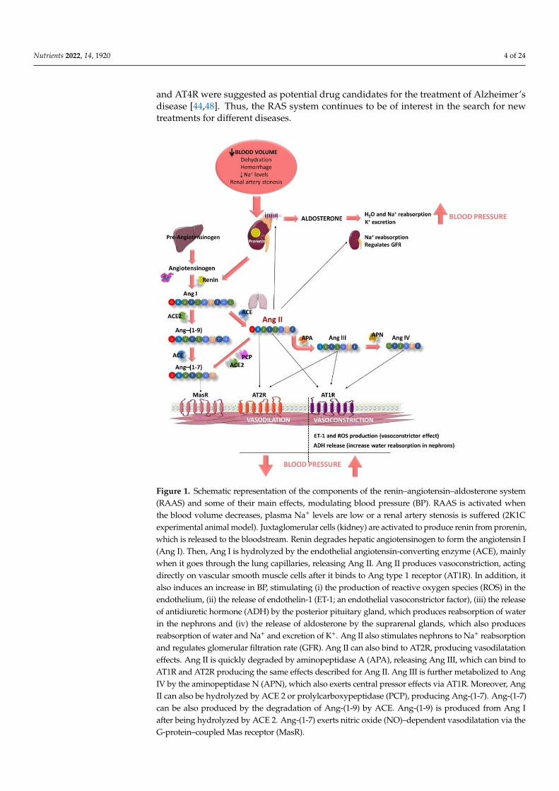
This peptide/hormone system is activated by different causes, such as a decrease inblood volume due to dehydration, or hemorrhage or/and a decrease in Na+ levels (Figure 1).This fact produces the activation of the juxtaglomerular cells, located in the kidney afferentarterioles, producing the hydrolysis of prorenin, the precursor of renin [28]. Renin entersthe bloodstream and reaches the liver, where this aspartyl protease triggers the cleavageof the angiotensinogen to form the decapeptide Ang I (Ang-(1-10)) [29]. This peptide ishydrolyzed by the ACE, mainly when it goes through the lung capillaries [30]. ACE issynthetized by the endothelial cells and its extracellular location in these cells helps theenzyme to interact easily with its substrate. As a result of its activity, the octapeptide AngII (a potent vasoconstrictor) is formed [30]. In addition, ACE is also known to hydrolyzebradykinin, a vasodilator peptide, producing its inactivation and contributing to a reductionin the vasodilator factors [31]. It is worth noting that this enzyme is considered key in the BPregulation. Ang II, also known as Ang-(1-8), can bind to two different receptors: Ang type1 receptor (AT1R) and Ang type 2 receptor (AT2R). These two receptors exert antagonisteffects. The main actions of Ang II are associated with its binding to AT1R and are relatedto the development of CVD. This pathway is known as the ACE–Ang II–AT1R axis [32].Ang II triggers intracrine, autocrine and paracrine responses with different physiologicaleffects [28], as AT1R is located in most of organs and is coupled to different G proteins [33].Ang II effects include producing vasoconstriction, acting directly on vascular smoothmuscle cells [33]. It also increases total peripheral resistance through its vasoconstrictoreffects on systemic arterioles [34]. This vasoconstrictor effect seems to be modulated bythe endothelium, as it has been reported that Ang II can stimulate the release of differentendothelial factors including the vasoconstrictor endothelin-1 (ET-1) [33] or reactive oxygenspecies (ROS) [35]. Moreover, Ang II stimulates the zona glomerulosa of the adrenal cortexto secrete aldosterone [28] and its release increases water and sodium reabsorption andpotassium excretion in the distal tubule and collecting duct of the nephron [36]. It alsoactivates the early proximal tubule (Na+–H+ antiporter) to Na+ reabsorption and regulatesthe glomerular filtration rate by the contraction of the efferent and afferent glomerulararterioles [37]. Furthermore, Ang II also acts in the hypothalamic level, firstly stimulatingthe sensation of thirst and consequently, promoting the intake of water. In addition, Ang IIstimulates the release of the antidiuretic hormone (ADH, vasopressin) in response to thethirst in the posterior pituitary gland. This ADH acts on the collecting ducts of the nephron,increasing water reabsorption in this area, thus reducing urinary loss [28]. Altogether, theseprocesses contribute to an increase in BP (Figure 1).
The half-life of Ang II in plasma is short (1–2 min) and it is degraded in its N-terminalposition by aminopeptidase A, releasing another active peptide called Ang III (Ang-(2-8)) [33]. This peptide exerts agonistic effects to those shown by Ang II, including releaseof aldosterone, pressor and dipsogenic effects or stimulation of Na+ intake. It also bindsto AT1R and AT2R to exert its effects [38–40]. Ang III is further metabolized to Ang IV(Ang-(3-8)) by the aminopeptidase N, which also exerts central pressor effects via AT1R,although Ang III can also bind to AT4R or insulin-regulated aminopeptidase (IRAP) [41].Moreover, Ang II can also be hydrolyzed by other enzymes, including ACE 2 or pro-lylcarboxypeptidase, producing the peptide Ang-(1-7). Ang-(1-7) can be also producedby the degradation of Ang-(1-9) by ACE, previously obtained by the action of ACE 2on Ang I [40]. This pathway is called the ACE2–Ang-(1-7)–Mas receptor (MasR) axis.It has been reported that Ang-(1-7) exerts nitric oxide (NO)–dependent vasodilatation,and antihypertensive, anti-inflammatory, antifibrotic and antiangiogenic effects via theG-protein–coupled MasR [42].
In addition to CVD, the role of RAS components in other diseases was recently revealed.For example, the role of ACE 2 in COVID-19 as SARS-CoV2 uses this enzyme to enter themucosa and also modulates its gene expression [43]. ACE and Ang II also play a role inAlzheimer’s disease [44]: brain ACE expression was related to Alzheimer’s disease severityand amyloid-beta (Aβ) load and Ang II is responsible for the development of neurovasculardamage and dysfunction via the AT1R pathway [45–47]. Moreover, agonists of brain AT2R
Nutrients 2022, 14, 1920 4 of 24
and AT4R were suggested as potential drug candidates for the treatment of Alzheimer’sdisease [44,48]. Thus, the RAS system continues to be of interest in the search for newtreatments for different diseases.
Nutrients 2022, 14, x FOR PEER REVIEW 4 of 25
In addition to CVD, the role of RAS components in other diseases was recently
revealed. For example, the role of ACE 2 in COVID-19 as SARS-CoV2 uses this enzyme to
enter the mucosa and also modulates its gene expression [43]. ACE and Ang II also play a
role in Alzheimer’s disease [44]: brain ACE expression was related to Alzheimer’s disease
severity and amyloid-beta (Aβ) load and Ang II is responsible for the development of
neurovascular damage and dysfunction via the AT1R pathway [45–47]. Moreover,
agonists of brain AT2R and AT4R were suggested as potential drug candidates for the
treatment of Alzheimer’s disease [44,48]. Thus, the RAS system continues to be of interest
in the search for new treatments for different diseases.
Figure 1. Schematic representation of the components of the renin–angiotensin–aldosterone system
(RAAS) and some of their main effects, modulating blood pressure (BP). RAAS is activated when
the blood volume decreases, plasma Na+ levels are low or a renal artery stenosis is suffered (2K1C
experimental animal model). Juxtaglomerular cells (kidney) are activated to produce renin from
prorenin, which is released to the bloodstream. Renin degrades hepatic angiotensinogen to form the
angiotensin I (Ang I). Then, Ang I is hydrolyzed by the endothelial angiotensin-converting enzyme
(ACE), mainly when it goes through the lung capillaries, releasing Ang II. Ang II produces
vasoconstriction, acting directly on vascular smooth muscle cells after it binds to Ang type 1 receptor
(AT1R). In addition, it also induces an increase in BP, stimulating (i) the production of reactive
oxygen species (ROS) in the endothelium, (ii) the release of endothelin-1 (ET-1; an endothelial
vasoconstrictor factor), (iii) the release of antidiuretic hormone (ADH) by the posterior pituitary
gland, which produces reabsorption of water in the nephrons and (iv) the release of aldosterone by
the suprarenal glands, which also produces reabsorption of water and Na+ and excretion of K+. Ang
II also stimulates nephrons to Na+ reabsorption and regulates glomerular filtration rate (GFR). Ang
II can also bind to AT2R, producing vasodilatation effects. Ang II is quickly degraded by
aminopeptidase A (APA), releasing Ang III, which can bind to AT1R and AT2R producing the same
Figure 1. Schematic representation of the components of the renin–angiotensin–aldosterone system(RAAS) and some of their main effects, modulating blood pressure (BP). RAAS is activated whenthe blood volume decreases, plasma Na+ levels are low or a renal artery stenosis is suffered (2K1Cexperimental animal model). Juxtaglomerular cells (kidney) are activated to produce renin from prorenin,which is released to the bloodstream. Renin degrades hepatic angiotensinogen to form the angiotensin I(Ang I). Then, Ang I is hydrolyzed by the endothelial angiotensin-converting enzyme (ACE), mainlywhen it goes through the lung capillaries, releasing Ang II. Ang II produces vasoconstriction, actingdirectly on vascular smooth muscle cells after it binds to Ang type 1 receptor (AT1R). In addition, italso induces an increase in BP, stimulating (i) the production of reactive oxygen species (ROS) in theendothelium, (ii) the release of endothelin-1 (ET-1; an endothelial vasoconstrictor factor), (iii) the releaseof antidiuretic hormone (ADH) by the posterior pituitary gland, which produces reabsorption of waterin the nephrons and (iv) the release of aldosterone by the suprarenal glands, which also producesreabsorption of water and Na+ and excretion of K+. Ang II also stimulates nephrons to Na+ reabsorptionand regulates glomerular filtration rate (GFR). Ang II can also bind to AT2R, producing vasodilatationeffects. Ang II is quickly degraded by aminopeptidase A (APA), releasing Ang III, which can bind toAT1R and AT2R producing the same effects described for Ang II. Ang III is further metabolized to AngIV by the aminopeptidase N (APN), which also exerts central pressor effects via AT1R. Moreover, AngII can also be hydrolyzed by ACE 2 or prolylcarboxypeptidase (PCP), producing Ang-(1-7). Ang-(1-7)can be also produced by the degradation of Ang-(1-9) by ACE. Ang-(1-9) is produced from Ang Iafter being hydrolyzed by ACE 2. Ang-(1-7) exerts nitric oxide (NO)–dependent vasodilatation via theG-protein–coupled Mas receptor (MasR).
Nutrients 2022, 14, 1920 5 of 24
2.2. Endothelial Function
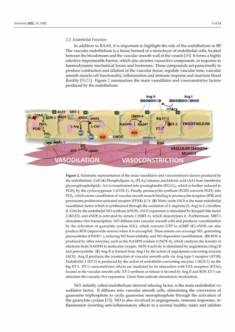
In addition to RAAS, it is important to highlight the role of the endothelium in BP.The vascular endothelium is a tissue formed of a monolayer of endothelial cells, locatedbetween the bloodstream and the vascular smooth wall of the vessels [49]. It forms a highlyselective impermeable barrier, which also secretes vasoactive compounds, in response tohaemodynamic mechanical forces and hormones. These compounds act paracrinally toproduce contraction and dilation of the vascular tissue, regulate vascular tone, vascularsmooth muscle cell functionality, inflammation and immune response and maintain bloodfluidity [50,51]. Figure 2 summarizes the main vasodilator and vasoconstrictor factorsproduced by the endothelium.
Nutrients 2022, 14, x FOR PEER REVIEW 7 of 25
Figure 2. Schematic representation of the main vasodilator and vasoconstrictor factors produced by
the endothelium. Cell (A) Phospholipase A2 (PLA2) releases arachidonic acid (AA) from membrane
glycerophospholipids. AA is transformed into prostaglandin (PG) G2, which is further reduced to
PGH2 by the cyclooxygenase 1 (COX-1). Finally, prostacyclin synthase (PGIS) converts PGH2 into
PGI2, which exerts vasodilation of vascular smooth muscle binding to prostacyclin receptors (IPR)
and peroxisome proliferator-activated receptor (PPAR) β/δ. (B) Nitric oxide (NO) is the main
endothelial vasodilator factor which is synthesized through the oxidation of L-arginine (L-Arg) to
L-citrulline (L-Citr) by the endothelial NO synthase (eNOS). eNOS expression is stimulated by
Kruppel-like-factor 2 (KLF2), and eNOS is activated by sirtuin 1 (SIRT-1), which deacetylates it.
Furthermore, SIRT-1 stimulates eNos transcription. NO diffuses into vascular smooth cells and
produces vasodilatation by the activation of guanylate cyclase (GC), which converts GTP to cGMP.
(C) eNOS can also produce ROS (superoxide anions) when it is uncoupled. These anions can
scavenge NO, generating peroxynitrites (ONOO−), reducing NO bioavailability and NO-dependent
vasodilatation. (D) ROS is produced by other enzymes, such as the NADPH oxidase 4 (NOX-4),
which catalyzes the transfer of electrons from NADPH to molecular oxygen. NOX-4 activity is
stimulated by angiotensin (Ang) II and peroxynitrite. (E) Ang II is formed from Ang I by the action
of angiotensin-converting enzyme (ACE). Ang II produces the constriction of vascular smooth cells
via Ang type 1 receptor (AT1R). Endothelin 1 (ET-1) is produced by the action of endothelin-
converting enzyme 1 (ECE-1) on the big ET-1. ET-1 vasoconstrictor effects are mediated by its
interaction with ETA receptors (ETAr), located in the vascular smooth cells. ET-1 synthesis or release
is favored by Ang II and ROS. ET-1 can stimulate the vascular Nox expression. Green lines indicate
stimulation/modulation.
3. Biological Rhythms and Blood Pressure
Many physiological processes including BP and heart rate follow a biological rhythm.
These rhythms are organized in cycles that allow the organisms to adapt to constant
changes in their environment, such as light and dark periods or even seasonal changes,
thus optimizing their metabolic functions and energy expenditure [83]. In mammals, these
rhythms are controlled by synchronized endogenous clocks, which are located both in the
central nervous system and in peripheral areas throughout the body. Because of this
synchronization and their connection to the environment [83,84], these clocks are able to
modulate many biological processes, such as neuronal, endocrine, metabolic and
behavioral functions [85]. The main factor that regulates and controls the endogenous
clocks is the 24-h light/dark cycle of Earth, also called the photoperiod. Nevertheless, other
environmental or behavioral factors, such as meal timing and exercise are also essential in
the modulation of these clocks [86].
3.1. Molecular Machinery behind Circadian Rhtyhms
In mammals, the central clock of the circadian rhythms, which synchronizes all
existing peripheral clocks, is located in the suprachiasmatic nucleus (SCN), specifically in
Figure 2. Schematic representation of the main vasodilator and vasoconstrictor factors produced bythe endothelium. Cell (A) Phospholipase A2 (PLA2) releases arachidonic acid (AA) from membraneglycerophospholipids. AA is transformed into prostaglandin (PG) G2, which is further reduced toPGH2 by the cyclooxygenase 1 (COX-1). Finally, prostacyclin synthase (PGIS) converts PGH2 intoPGI2, which exerts vasodilation of vascular smooth muscle binding to prostacyclin receptors (IPR) andperoxisome proliferator-activated receptor (PPAR) β/δ. (B) Nitric oxide (NO) is the main endothelialvasodilator factor which is synthesized through the oxidation of L-arginine (L-Arg) to L-citrulline(L-Citr) by the endothelial NO synthase (eNOS). eNOS expression is stimulated by Kruppel-like-factor2 (KLF2), and eNOS is activated by sirtuin 1 (SIRT-1), which deacetylates it. Furthermore, SIRT-1stimulates eNos transcription. NO diffuses into vascular smooth cells and produces vasodilatationby the activation of guanylate cyclase (GC), which converts GTP to cGMP. (C) eNOS can alsoproduce ROS (superoxide anions) when it is uncoupled. These anions can scavenge NO, generatingperoxynitrites (ONOO−), reducing NO bioavailability and NO-dependent vasodilatation. (D) ROS isproduced by other enzymes, such as the NADPH oxidase 4 (NOX-4), which catalyzes the transfer ofelectrons from NADPH to molecular oxygen. NOX-4 activity is stimulated by angiotensin (Ang) IIand peroxynitrite. (E) Ang II is formed from Ang I by the action of angiotensin-converting enzyme(ACE). Ang II produces the constriction of vascular smooth cells via Ang type 1 receptor (AT1R).Endothelin 1 (ET-1) is produced by the action of endothelin-converting enzyme 1 (ECE-1) on thebig ET-1. ET-1 vasoconstrictor effects are mediated by its interaction with ETA receptors (ETAr),located in the vascular smooth cells. ET-1 synthesis or release is favored by Ang II and ROS. ET-1 canstimulate the vascular Nox expression. Green lines indicate stimulation/modulation.
NO, initially called endothelium-derived relaxing factor, is the main endothelial va-sodilator factor. It diffuses into vascular smooth cells, stimulating the conversion ofguanosine triphosphate to cyclic guanosine monophosphate through the activation ofthe guanylate cyclase [52]. NO is also involved in angiogenesis, immune responses, in-flammation (exerting anti-inflammatory effects in a normal healthy state) and inhibits

Nutrients 2022, 14, 1920 6 of 24
white cell activation and platelet aggregation, among other effects [50,53,54]. In the en-dothelium, NO is synthesized through the oxidation of L-arginine to L-citrulline, in areaction catalyzed by the constitutive isoform of the enzyme NO synthase (eNOS or NOSIII), using as co-substrates nicotinamide-adenine-dinucleotide phosphate and oxygen [55].This monomeric enzyme contains two domains (the reductase and oxygenase domains)that form dimers, which are considered the active form of the enzyme [54]. In the plasmacell membrane, this enzyme is found attached to caveolin-1, which acts by inhibiting theenzyme [56]. eNOS activation is produced in response to shear stress, vascular endothelialgrowth factor, HDL and intracellular Ca2+ levels [57]. It is a Ca2+-dependent activation,although eNOS can be also activated in its absence [58]. Moreover, eNOS activity dependson different cofactors (flavin adenine dinucleotide, flavin mononucleotide and (6R-)5,6,7,8-tetrahydrobiopterin (BH4)), the phosphorylation of different amino acids, post-translationallipid modifications [54,55] and the SIRT-1 activity, which deacetylates it. Furthermore,SIRT-1 stimulates eNOS transcription [59,60] and Kruppel-like-factor 2 (KLF2) stimulateseNOS expression [61].
Instead of NO, eNOS can also produce superoxide anions. This process is called“eNOS uncoupling”. It can happen when L-arginine or BH4 levels are low (BH4 stabilizesthe eNOS dimer), or asymmetric dimethylarginine (an endogenous eNOS inhibitor) levelsincrease [62]. For example, reduction in BH4 levels can be produced by a decrease inBH4 production or by an increase in its oxidation due to excessive ROS levels, namelyperoxynitrite [63,64]. Consequently, it generates a reduction in NO bioavailability andan increase in ROS levels, altering the endothelial function. This is associated with HTNand other CVD. Moreover, NO availability can also be reduced by superoxide anions,which can scavenge NO, generating peroxynitrites and avoiding NO-dependent vasodi-latation [65]. Moreover, peroxynitrite can oxidize low-density lipoproteins which increasearginase activity, producing a reduction in L-arginine levels and also stimulating NADPHoxidases (NOX) and xanthine oxidase to produce ROS [62]; consequently, peroxynitriteand its ROS-induced production contribute to eNOS uncoupling. In addition to eNOSin its uncoupled state, endothelial cells produce ROS in the mitochondrial respirationand by means of xanthine oxidoreductase and NOX (mainly NOX-4 in these cells) [66,67].Moreover, endothelial ROS production can be increased by different factors, such as AngII action, as it can stimulate NOX-4 activity [68,69]. In the homeostatic state, the gener-ated free radical is counter-balanced by endogenous antioxidant mechanisms, which canbe enzymes, such as superoxide dismutase (SOD) or catalase (CAT), or non-enzymaticcompounds, such as reduced glutathione (GSH) or ascorbate. An unbalance between ROSproduction and degradation results in oxidative stress, representing the main cause ofendothelial dysfunction [51].
Prostaglandin (PG) or prostacyclin I2 (PGI2) is another important vasodilator factorproduced by the endothelium, mainly in response to shear stress [49]. However, it isconsidered that it plays a secondary role in vasodilation, exerting its effect mainly whenthe levels of NO are not high enough [70]. This factor is synthesized by a multi-stepenzyme-catalyzed reaction [71]. Firstly, phospholipase A2 releases arachidonic acid frommembrane glycerophospholipids, whose activation depends on Ca2+ levels [72]. Secondly,the free arachidonic acid is transformed in PGG2, which is further reduced to PGH2 bythe action of the cyclooxygenase (COX). This enzyme shows oxygenase and peroxidaseactivities [71] and the predominant isoform in endothelial cells is COX-1 [73]. Finally,prostacyclin synthase (PGIS) converts PGH2 into PGI2. The effects of PGI2 are mediatedby its binding to cell surface prostacyclin receptors (IPR) and intracellular peroxisomeproliferator-activated receptor (PPAR) β/δ. Activation of both pathways produces a multi-step reaction that results in a reduction in intracellular Ca2+ levels of vascular smoothcells and a further vasodilation of the vessel [71]. Moreover, it is known that NO inducesthe release of PGI2 and vice versa [49]. PGI2 can also act on juxtaglomerular apparatus,inducing the release of renin by kidney [74].

Nutrients 2022, 14, 1920 7 of 24
On the other hand, the endothelium also synthesizes and releases vasoconstrictorcompounds, such as ET-1 which is also involved in vascular and myocardial hypertrophyand promotes inflammation as it stimulates the release of interleukins (IL-6, IL-1 andIL-8) [75]. ET-1 is produced in different steps, comprising the hydrolysis of prepro-ET-1into big ET-1 by proteases and the further hydrolysis in Trp-21 of big ET-1 in its activeform ET-1, catalyzed by the endothelin-converting enzyme 1 (ECE-1) [76]. This process istightly regulated by different factors. In this regard, ET-1 synthesis or release is favoredby Ang II, ADH, ROS, cytokines (tumor necrosis factor-alpha and IL-1), norepinephrine,thrombin or shear stress, while it is reduced by NO, atrial natriuretic peptide, cyclicnucleotides and KLF2 [77–79]. The vasoconstrictor effects of ET-1 are mediated by itsinteraction with ETA and ETB receptors (mainly ETA receptors), located in the vascularsmooth cells [80]. However, ET-1 can also bind to ETB receptors in the endothelial cells,presenting an opposite effect to that showed by ETA activation. Specifically, ET-1 via ETBfavors the release of endothelial prostacyclin and NO, ET-1 clearance, and inhibits ECE-1expression [79]. In addition, it has been observed that ET-1 can stimulate the vascular Noxexpression [81]. Another vasoconstrictor produced by the endothelium is Ang II, as ACE isexpressed in endothelial cells. This local Ang II helps to maintain normal BP, although it isnot essential [31,82]
The balanced release of vasoconstrictor and vasodilator factors by the endotheliumleads to a controlled homeostasis of vascular tone and BP [49]. The imbalance betweenvasodilator and vasoconstrictor factors may trigger the development of some CVD, suchas HTN.
3. Biological Rhythms and Blood Pressure
Many physiological processes including BP and heart rate follow a biological rhythm.These rhythms are organized in cycles that allow the organisms to adapt to constantchanges in their environment, such as light and dark periods or even seasonal changes,thus optimizing their metabolic functions and energy expenditure [83]. In mammals, theserhythms are controlled by synchronized endogenous clocks, which are located both inthe central nervous system and in peripheral areas throughout the body. Because of thissynchronization and their connection to the environment [83,84], these clocks are able tomodulate many biological processes, such as neuronal, endocrine, metabolic and behavioralfunctions [85]. The main factor that regulates and controls the endogenous clocks is the 24-hlight/dark cycle of Earth, also called the photoperiod. Nevertheless, other environmentalor behavioral factors, such as meal timing and exercise are also essential in the modulationof these clocks [86].
3.1. Molecular Machinery behind Circadian Rhtyhms
In mammals, the central clock of the circadian rhythms, which synchronizes all existingperipheral clocks, is located in the suprachiasmatic nucleus (SCN), specifically in the ventralperiventricular zone of the anterior hypothalamus. The SCN receives information aboutexternal light through its connection with the retina and sends it to other organs, therebygenerating behavioral and biological rhythms [87].
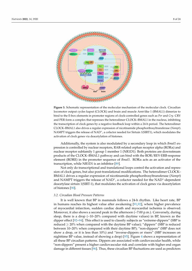
At the molecular level, the circadian clock is controlled by a set of genes calledclock genes, which codify many transcription factors that undergo an autoregulatorytranscription–translation feedback loop (Figure 3). The most important clock proteinsare circadian locomotor output cycles kaput (CLOCK) and brain and muscle Arnt-like1 (BMAL1). They dimerize to bind to the E-box elements in promoter regions of clock-controlled genes, such as Per1, Per2 and Per3 (period 1, 2 and 3) and Cry1 and Cry2(cryptochrome 1 and 2). CRY and PER form a complex that represses the heterodimerCLOCK–BMAL1 in the nucleus, thus inhibiting the transcription of clock genes by anegative feedback loop within a 24-h period [88].
Nutrients 2022, 14, 1920 8 of 24
Nutrients 2022, 14, x FOR PEER REVIEW 8 of 25
the ventral periventricular zone of the anterior hypothalamus. The SCN receives
information about external light through its connection with the retina and sends it to
other organs, thereby generating behavioral and biological rhythms [87].
At the molecular level, the circadian clock is controlled by a set of genes called clock
genes, which codify many transcription factors that undergo an autoregulatory
transcription–translation feedback loop (Figure 3). The most important clock proteins are
circadian locomotor output cycles kaput (CLOCK) and brain and muscle Arnt-like 1
(BMAL1). They dimerize to bind to the E-box elements in promoter regions of clock-
controlled genes, such as Per1, Per2 and Per3 (period 1, 2 and 3) and Cry1 and Cry2
(cryptochrome 1 and 2). CRY and PER form a complex that represses the heterodimer
CLOCK–BMAL1 in the nucleus, thus inhibiting the transcription of clock genes by a
negative feedback loop within a 24-h period [88].
Additionally, the system is also modulated by a secondary loop in which Bmal1
expression is controlled by nuclear receptors, RAR-related orphan receptor alpha (RORα)
and nuclear receptor subfamily 1 group 1 member 1 (NR1D1). Both proteins are
downstream products of the CLOCK–BMAL1 pathway and can bind with the ROR/REV-
ERB-response element (RORE) in the promoter sequence of Bmal1. RORα acts as an
activator of the transcription, while NR1D1 is an inhibitor [89].
Not only do transcriptional and translational loops control the activation and
repression of clock genes, but also post-translational modifications. The heterodimer
CLOCK–BMAL1 drives a regular expression of nicotinamide phosphoribosyltransferase
(Nampt) and NAMPT triggers the release of NAD+, a cofactor needed for the NAD+-
dependent deacetylase sirtuin 1(SIRT-1), that modulates the activation of clock genes via
deacetylation of histones [90].
Figure 3. Schematic representation of the molecular mechanism of the molecular clock. Circadian
locomotor output cycles kaput (CLOCK) and brain and muscle Arnt-like 1 (BMAL1) dimerize to
bind to the E-box elements in promoter regions of clock-controlled genes such as Per and Cry. CRY
and PER form a complex that represses the heterodimer CLOCK–BMAL1 in the nucleus, inhibiting
the transcription of clock genes by a negative feedback loop within a 24-h period. The heterodimer
CLOCK–BMAL1 also drives a regular expression of nicotinamide phosphoribosyltransferase
(Nampt). NAMPT triggers the release of NAD+, a cofactor needed for Sirtuin 1(SIRT1), which
modulates the activation of clock genes via deacetylation of histones.
3.2. Circadian Blood Pressure Patterns
It is well known that BP in mammals follows a 24-h rhythm. Like heart rate, BP in
humans reaches its highest value after awakening [91,92], where higher prevalence of
Figure 3. Schematic representation of the molecular mechanism of the molecular clock. Circadianlocomotor output cycles kaput (CLOCK) and brain and muscle Arnt-like 1 (BMAL1) dimerize tobind to the E-box elements in promoter regions of clock-controlled genes such as Per and Cry. CRYand PER form a complex that represses the heterodimer CLOCK–BMAL1 in the nucleus, inhibitingthe transcription of clock genes by a negative feedback loop within a 24-h period. The heterodimerCLOCK–BMAL1 also drives a regular expression of nicotinamide phosphoribosyltransferase (Nampt).NAMPT triggers the release of NAD+, a cofactor needed for Sirtuin 1(SIRT1), which modulates theactivation of clock genes via deacetylation of histones.
Additionally, the system is also modulated by a secondary loop in which Bmal1 ex-pression is controlled by nuclear receptors, RAR-related orphan receptor alpha (RORα) andnuclear receptor subfamily 1 group 1 member 1 (NR1D1). Both proteins are downstreamproducts of the CLOCK–BMAL1 pathway and can bind with the ROR/REV-ERB-responseelement (RORE) in the promoter sequence of Bmal1. RORα acts as an activator of thetranscription, while NR1D1 is an inhibitor [89].
Not only do transcriptional and translational loops control the activation and repres-sion of clock genes, but also post-translational modifications. The heterodimer CLOCK–BMAL1 drives a regular expression of nicotinamide phosphoribosyltransferase (Nampt)and NAMPT triggers the release of NAD+, a cofactor needed for the NAD+-dependentdeacetylase sirtuin 1(SIRT-1), that modulates the activation of clock genes via deacetylationof histones [90].
3.2. Circadian Blood Pressure Patterns
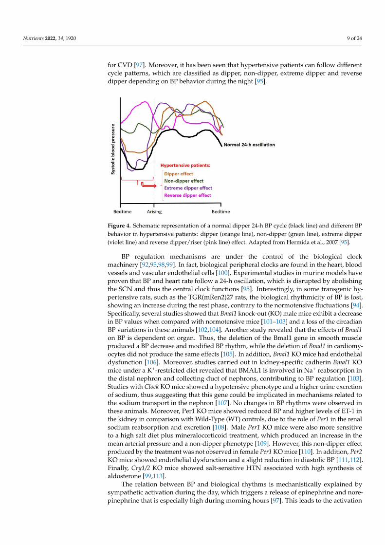
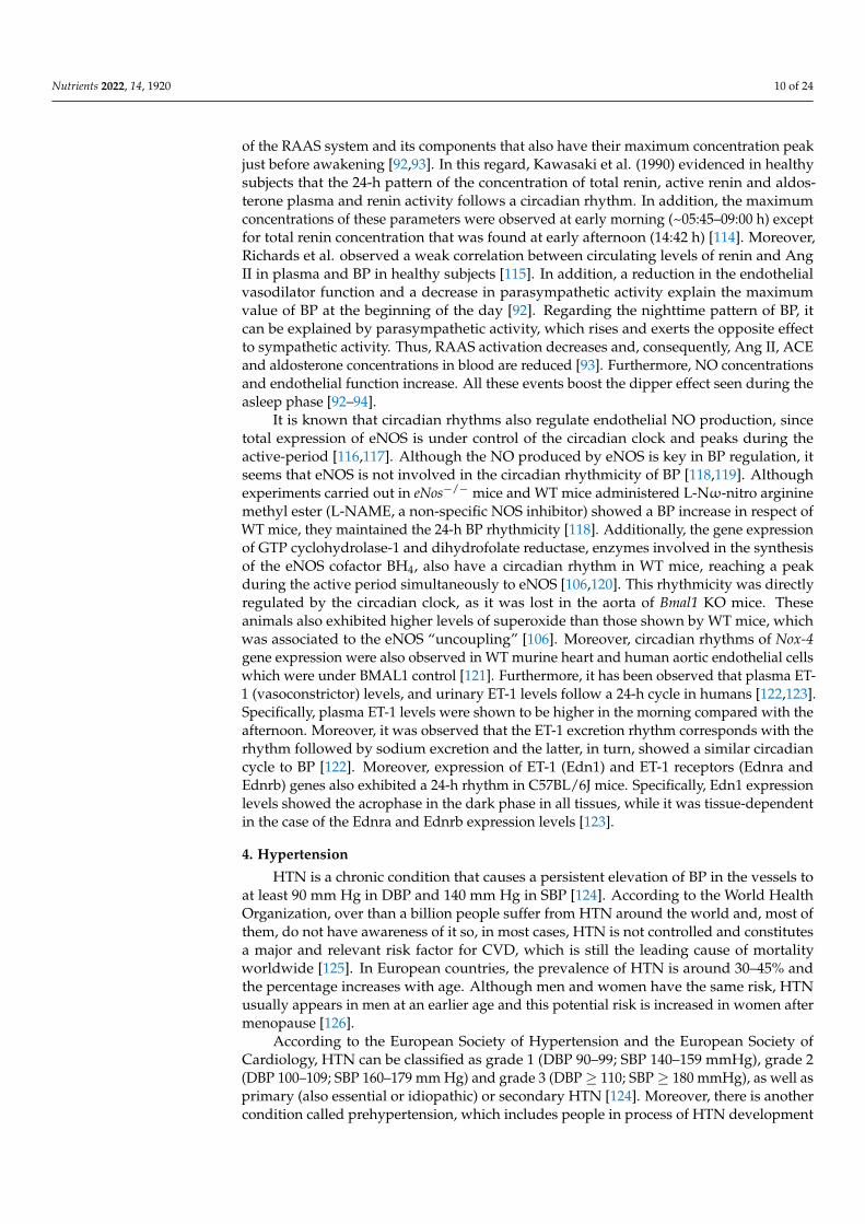
It is well known that BP in mammals follows a 24-h rhythm. Like heart rate, BPin humans reaches its highest value after awakening [91,92], where higher prevalenceof myocardial infarction, sudden cardiac death and myocardial ischemia is observed.Moreover, it also shows a second peak in the afternoon (~7:00 p.m.). Conversely, duringsleep, there is a drop (~10–20% compared with daytime values) in BP, known as thedipper effect [92–94]. This effect is used to classify subjects as “extreme-dippers” (SBP isreduced ≥ 20% when compared with the daytime BP value); “dippers” (SBP is reducedbetween 10–20% when compared with their daytime BP); “non-dippers” (SBP does notshow a drop, or it is less than 10%) and “inverse-dippers or risers” (SBP increases onnighttime BP value, instead of showing a drop) [95]. Figure 4 shows a representation ofall these BP circadian patterns. Dippers are associated with cardiovascular health, while“non-dippers” present a higher cardiovascular risk and correlate with higher end organdamage in different tissues [96]. Thus, these circadian BP fluctuations are used as predictors
Nutrients 2022, 14, 1920 9 of 24
for CVD [97]. Moreover, it has been seen that hypertensive patients can follow differentcycle patterns, which are classified as dipper, non-dipper, extreme dipper and reversedipper depending on BP behavior during the night [95].
Nutrients 2022, 14, x FOR PEER REVIEW 9 of 25
myocardial infarction, sudden cardiac death and myocardial ischemia is observed.
Moreover, it also shows a second peak in the afternoon (~7:00 p.m.). Conversely, during
sleep, there is a drop (~10–20% compared with daytime values) in BP, known as the dipper
effect [92–94]. This effect is used to classify subjects as “extreme-dippers” (SBP is reduced
≥ 20% when compared with the daytime BP value); “dippers” (SBP is reduced between
10–20% when compared with their daytime BP); “non-dippers” (SBP does not show a
drop, or it is less than 10%) and “inverse-dippers or risers” (SBP increases on nighttime
BP value, instead of showing a drop) [95]. Figure 4 shows a representation of all these BP
circadian patterns. Dippers are associated with cardiovascular health, while “non-
dippers” present a higher cardiovascular risk and correlate with higher end organ damage
in different tissues [96]. Thus, these circadian BP fluctuations are used as predictors for
CVD [97]. Moreover, it has been seen that hypertensive patients can follow different cycle
patterns, which are classified as dipper, non-dipper, extreme dipper and reverse dipper
depending on BP behavior during the night [95].
Figure 4. Schematic representation of a normal dipper 24-h BP cycle (black line) and different BP
behavior in hypertensive patients: dipper (orange line), non-dipper (green line), extreme dipper
(violet line) and reverse dipper/riser (pink line) effect. Adapted from Hermida et al. 2007 [95].
BP regulation mechanisms are under the control of the biological clock machinery
[92,95,98,99]. In fact, biological peripheral clocks are found in the heart, blood vessels and
vascular endothelial cells [100]. Experimental studies in murine models have proven that
BP and heart rate follow a 24-h oscillation, which is disrupted by abolishing the SCN and
thus the central clock functions [95]. Interestingly, in some transgenic hypertensive rats,
such as the TGR(mRen2)27 rats, the biological rhythmicity of BP is lost, showing an
increase during the rest phase, contrary to the normotensive fluctuations [94]. Specifically,
several studies showed that Bmal1 knock-out (KO) male mice exhibit a decrease in BP
values when compared with normotensive mice [101–103] and a loss of the circadian BP
variations in these animals [102,104]. Another study revealed that the effects of Bmal1 on
BP is dependent on organ. Thus, the deletion of the Bmal1 gene in smooth muscle
produced a BP decrease and modified BP rhythm, while the deletion of Bmal1 in
cardiomyocytes did not produce the same effects [105]. In addition, Bmal1 KO mice had
endothelial dysfunction [106]. Moreover, studies carried out in kidney-specific cadherin
Bmal1 KO mice under a K+-restricted diet revealed that BMAL1 is involved in Na+
reabsorption in the distal nephron and collecting duct of nephrons, contributing to BP
regulation [103]. Studies with Clock KO mice showed a hypotensive phenotype and a
higher urine excretion of sodium, thus suggesting that this gene could be implicated in
mechanisms related to the sodium transport in the nephron [107]. No changes in BP
rhythms were observed in these animals. Moreover, Per1 KO mice showed reduced BP
Figure 4. Schematic representation of a normal dipper 24-h BP cycle (black line) and different BPbehavior in hypertensive patients: dipper (orange line), non-dipper (green line), extreme dipper(violet line) and reverse dipper/riser (pink line) effect. Adapted from Hermida et al., 2007 [95].
BP regulation mechanisms are under the control of the biological clockmachinery [92,95,98,99]. In fact, biological peripheral clocks are found in the heart, bloodvessels and vascular endothelial cells [100]. Experimental studies in murine models haveproven that BP and heart rate follow a 24-h oscillation, which is disrupted by abolishingthe SCN and thus the central clock functions [95]. Interestingly, in some transgenic hy-pertensive rats, such as the TGR(mRen2)27 rats, the biological rhythmicity of BP is lost,showing an increase during the rest phase, contrary to the normotensive fluctuations [94].Specifically, several studies showed that Bmal1 knock-out (KO) male mice exhibit a decreasein BP values when compared with normotensive mice [101–103] and a loss of the circadianBP variations in these animals [102,104]. Another study revealed that the effects of Bmal1on BP is dependent on organ. Thus, the deletion of the Bmal1 gene in smooth muscleproduced a BP decrease and modified BP rhythm, while the deletion of Bmal1 in cardiomy-ocytes did not produce the same effects [105]. In addition, Bmal1 KO mice had endothelialdysfunction [106]. Moreover, studies carried out in kidney-specific cadherin Bmal1 KOmice under a K+-restricted diet revealed that BMAL1 is involved in Na+ reabsorption inthe distal nephron and collecting duct of nephrons, contributing to BP regulation [103].Studies with Clock KO mice showed a hypotensive phenotype and a higher urine excretionof sodium, thus suggesting that this gene could be implicated in mechanisms related tothe sodium transport in the nephron [107]. No changes in BP rhythms were observed inthese animals. Moreover, Per1 KO mice showed reduced BP and higher levels of ET-1 inthe kidney in comparison with Wild-Type (WT) controls, due to the role of Per1 in the renalsodium reabsorption and excretion [108]. Male Per1 KO mice were also more sensitiveto a high salt diet plus mineralocorticoid treatment, which produced an increase in themean arterial pressure and a non-dipper phenotype [109]. However, this non-dipper effectproduced by the treatment was not observed in female Per1 KO mice [110]. In addition, Per2KO mice showed endothelial dysfunction and a slight reduction in diastolic BP [111,112].Finally, Cry1/2 KO mice showed salt-sensitive HTN associated with high synthesis ofaldosterone [99,113].
The relation between BP and biological rhythms is mechanistically explained bysympathetic activation during the day, which triggers a release of epinephrine and nore-pinephrine that is especially high during morning hours [97]. This leads to the activation
Nutrients 2022, 14, 1920 10 of 24
of the RAAS system and its components that also have their maximum concentration peakjust before awakening [92,93]. In this regard, Kawasaki et al. (1990) evidenced in healthysubjects that the 24-h pattern of the concentration of total renin, active renin and aldos-terone plasma and renin activity follows a circadian rhythm. In addition, the maximumconcentrations of these parameters were observed at early morning (~05:45–09:00 h) exceptfor total renin concentration that was found at early afternoon (14:42 h) [114]. Moreover,Richards et al. observed a weak correlation between circulating levels of renin and AngII in plasma and BP in healthy subjects [115]. In addition, a reduction in the endothelialvasodilator function and a decrease in parasympathetic activity explain the maximumvalue of BP at the beginning of the day [92]. Regarding the nighttime pattern of BP, itcan be explained by parasympathetic activity, which rises and exerts the opposite effectto sympathetic activity. Thus, RAAS activation decreases and, consequently, Ang II, ACEand aldosterone concentrations in blood are reduced [93]. Furthermore, NO concentrationsand endothelial function increase. All these events boost the dipper effect seen during theasleep phase [92–94].
It is known that circadian rhythms also regulate endothelial NO production, sincetotal expression of eNOS is under control of the circadian clock and peaks during theactive-period [116,117]. Although the NO produced by eNOS is key in BP regulation, itseems that eNOS is not involved in the circadian rhythmicity of BP [118,119]. Althoughexperiments carried out in eNos−/− mice and WT mice administered L-Nω-nitro argininemethyl ester (L-NAME, a non-specific NOS inhibitor) showed a BP increase in respect ofWT mice, they maintained the 24-h BP rhythmicity [118]. Additionally, the gene expressionof GTP cyclohydrolase-1 and dihydrofolate reductase, enzymes involved in the synthesisof the eNOS cofactor BH4, also have a circadian rhythm in WT mice, reaching a peakduring the active period simultaneously to eNOS [106,120]. This rhythmicity was directlyregulated by the circadian clock, as it was lost in the aorta of Bmal1 KO mice. Theseanimals also exhibited higher levels of superoxide than those shown by WT mice, whichwas associated to the eNOS “uncoupling” [106]. Moreover, circadian rhythms of Nox-4gene expression were also observed in WT murine heart and human aortic endothelial cellswhich were under BMAL1 control [121]. Furthermore, it has been observed that plasma ET-1 (vasoconstrictor) levels, and urinary ET-1 levels follow a 24-h cycle in humans [122,123].Specifically, plasma ET-1 levels were shown to be higher in the morning compared with theafternoon. Moreover, it was observed that the ET-1 excretion rhythm corresponds with therhythm followed by sodium excretion and the latter, in turn, showed a similar circadiancycle to BP [122]. Moreover, expression of ET-1 (Edn1) and ET-1 receptors (Ednra andEdnrb) genes also exhibited a 24-h rhythm in C57BL/6J mice. Specifically, Edn1 expressionlevels showed the acrophase in the dark phase in all tissues, while it was tissue-dependentin the case of the Ednra and Ednrb expression levels [123].
4. Hypertension
HTN is a chronic condition that causes a persistent elevation of BP in the vessels toat least 90 mm Hg in DBP and 140 mm Hg in SBP [124]. According to the World HealthOrganization, over than a billion people suffer from HTN around the world and, most ofthem, do not have awareness of it so, in most cases, HTN is not controlled and constitutesa major and relevant risk factor for CVD, which is still the leading cause of mortalityworldwide [125]. In European countries, the prevalence of HTN is around 30–45% andthe percentage increases with age. Although men and women have the same risk, HTNusually appears in men at an earlier age and this potential risk is increased in women aftermenopause [126].
According to the European Society of Hypertension and the European Society ofCardiology, HTN can be classified as grade 1 (DBP 90–99; SBP 140–159 mmHg), grade 2(DBP 100–109; SBP 160–179 mm Hg) and grade 3 (DBP ≥ 110; SBP ≥ 180 mmHg), as well asprimary (also essential or idiopathic) or secondary HTN [124]. Moreover, there is anothercondition called prehypertension, which includes people in process of HTN development
Nutrients 2022, 14, 1920 11 of 24
(DBP 80–99; SBP 120–139 mmHg). Primary HTN, the most extended (≈95% of HTN)emerges due to unknown causes but some factors are strongly correlated such as age, dietor pharmacology treatments [127,128]. Moreover, genetics and environmental factors suchas obesity, sedentary lifestyle, alcohol, high salt/Na+ diet, K+/vitamin D deficiency, etc. canalso be involved and lead to an earlier appearance of HTN and CVD [128,129]. When HTNis caused by other pathologies (renal, thyroid, hormonal, vascular or metabolic disorders),it is classified as secondary HTN [127].
The high BP levels recorded in hypertensive patients originate in the disruptionand functional alteration of regulation systems, such as RAAS and other disorders, forexample, endothelial dysfunction which starts with an imbalanced secretion of vasodilatorand vasoconstrictor molecules. When the endothelium is damaged, pro-inflammatoryand pro-thrombotic factors (chemokines, cytokines, and adhesion molecules) are releasedand interact with leucocytes and platelets, thereby provoking the loss of integrity ofendothelial cells that can be detached from the vascular wall. In addition, the increase inpro-inflammatory chemokines contributes to T cells and macrophage infiltration, generatingtissue injury [130].
Another consequence caused by HTN is ROS overproduction. In this situation, ROScontent might reach levels that cannot be buffered by endogenous antioxidant mechanisms,leading to oxidative stress. As was previously mentioned in Section 2, “uncoupled” eNOSand NOX are particularly relevant in endothelial ROS generation [49,62]. Since the RAASis overactivated in HTN, Ang II is overproduced, affecting BP via increasing ET-1 andROS production (stimulating NOX-4 activity and eNOS uncoupling) (Figure 2) [68,79].Moreover, ROS overproduction triggers adipose inflammation, glucose intolerance andinsulin resistance. In addition, the generated superoxide anions can react with NO, increas-ing peroxynitrite production and decreasing NO bioavailability. In turn, peroxynitrite islinked to alterations of redox-sensitive genes and transcription factors [49,131]. The resultsof all these processes negatively affect the vascular physiology, contributing to damageprogression related to CVD [131].
Moreover, biological rhythm disruptions can lead to HTN or CVD clinical pictures.For instance, populations such as shift workers (particularly nighttime shift workers) thatsuffer a misalignment in the natural light–dark schedule showed alterations in their BPlevels in comparison with daily workers. In addition, alterations in sleep duration weredemonstrated to be associated with cardiometabolic disorders development [132–134]. Fur-thermore, as was mentioned in Section 3.2, the deletion of Per1 in male mice in combinationwith the consumption of a high salt diet plus mineralocorticoid treatment produced anincrease in the mean arterial pressure and a non-dipper phenotype, which is associated toHTN [109].
4.1. Treatments for Hypertension
Since HTN is assumed as a relevant health problem worldwide, a plethora of potentialpreventive and corrective solutions have been investigated. Among them, well-establishedand novel pharmacological approaches have been defined and are efficient to manageHTN in most of the cases; however, they can exert side effects in some patients and theyare not suitable for prehypertensive patients. Thus, new bioactive compounds obtainedfrom natural sources or food have emerged as an adequate alternative. Moreover, lifestylemeasures such as salt/alcohol intake restrictions, exercise, diet, weight loss, etc. must notbe forgotten [135–137].
4.1.1. Pharmacological Treatments
The therapy against HTN includes a wide range of drugs that interact and inhibitRAAS components and other HTN agonists. Thus, one of the most investigated andprescribed treatments are those that involve ACE inhibitors, such as captopril or lisinopril,as well as benazepril, ramipril and imidapril. While ACE inhibitors prevent the productionof Ang II, different Ang II receptor blockers (ARB), such as valsartan and olmesartan, reduce
Nutrients 2022, 14, 1920 12 of 24
its action, thereby avoiding blood vessel constriction [138,139]. Furthermore, inhibitors ofβ-adrenergic receptor blockers are utilized to attenuate heart β-adrenoreceptor activation.These compounds, such as propranolol, are commonly used combined with other drugs andtheir proposed mechanism of action includes heart rate and cardiac output reduction, reninrelease inhibition, venous return and plasma volume reduction and vascular complianceimprovement, among others [140]. Furthermore, during recent decades, calcium channelblockers (amlodipine, felodipine, isradipine) have been widely used (monotherapy orcombined therapy) because of their good tolerability by hypertensive patients and theireffectiveness in reducing BP via blocking calcium entry into cardiovascular cells and thustriggering a vasodilator effect [141]. Moreover, different diuretic drugs are used in severalcases of HTN, acting in several areas of nephrons. For instance, thiazide-type diureticsblock Na+-Cl- cotransporters in the distal convoluted tubule and, consequently, reduce Na+
reabsorption. Also, loop-active agents, such as torasemide, block Na+-K+-Cl- cotransportersin the thick ascending limb of the loop of Henle and potassium-sparing diuretics act onNa+-K+ pumps, decreasing K+ excretion in the late distal tubule and collecting duct [142].
Despite the high diversity of pharmacological treatments than can be prescribed forthe different HTN grades, a substantial percentage of hypertensive patients show uncon-trolled BP because of intolerance or nonadherence to the abovementioned antihypertensiveagents [135,143]. Thus, novel drugs, devices and procedures are being investigated inpreclinical and clinical studies. In this sense, several drugs are in preclinical or phase-I ofdevelopment, such as those based on the inhibition of dopamine β-hydroxylase, aminopep-tidase A and Na+/H+ exchanger 3, as well as vaccines targeting Ang II and AT1R andantioxidants such as vitamin D. Other agents are at a more advanced stage (phase-II/III),such as newer mineralocorticoid receptor antagonists, inhibitors of aldosterone synthase,vasopeptidases and soluble epoxyde hydrolase, and agonists of natriuretic peptide A andvasoactive intestinal peptide receptor 2 [135,143].
4.1.2. Natural Bioactive Compounds
Although antihypertensive drugs have shown effectiveness against HTN, other alterna-tives are considered when the disorder is in development. For instance, diet modificationscombined with healthy lifestyles were shown to prevent and alleviate the disruptionscaused by the disease [137,144]. Besides recommended eating plans, such as DASH (Di-etary Approaches to Stop HTN), that advises a reduced salt/sodium and saturated fatintake and includes fruit and vegetables, specific natural matrices have been subjected toinvestigation to obtain bioactive compound-enriched products [144,145]. Thus, bioactivecompounds extracted from natural sources and food have been targeted to develop novelapproaches that might have a place in a patient’s diet. Among these compounds, severalmolecules including peptides, phenolic compounds, vitamins, carotenoids, alkaloids andorganosulfur compounds have shown antihypertensive potential, although not all themechanisms of action are fully elucidated [137,146].
These antihypertensive compounds are searched and selected, based on their ACEinhibitory (ACEi) properties. This is due to the fact that pharmacological ACE inhibitorsare the first-line treatments for HTN and an inhibition of ACE results in an effective BPreduction [147]. In this sense, protein hydrolysates and peptides obtained from variedfood matrices exert significant ability to inhibit or reduce ACE activity. For instance, thoseprepared from vegetal products and by-products, such as from fruits, legumes, cereals,wine lees, pseudocereals, herbs and spices, etc. showed potent in vitro ACEi activity and,in some cases, in vivo studies were conducted to validate the BP-lowering effects [148–151].Moreover, animal sources, such as dairy products, meat, fish or eggs and also specificby-products from these industries, such as chicken feet, led to potent ACE inhibitors, aswell as other alternative matrices, such as algae or mushrooms [149,152–158]. In addition, ithas been reported that some food matrices including different dairy products, such as kefiror cheese and unfiltered olive oil, can also contain ACE inhibitory and /or antihypertensivepeptides [8,159–161]. Peptide length and amino acid position in the chain is very important
Nutrients 2022, 14, 1920 13 of 24
to inhibit ACE. Thus, peptides of small size (3–12 amino acids), the presence of hydrophobicamino acids or those containing hydrophobic-branched side chains at the C-terminal endand the presence of the branched aliphatic amino acids at N-terminal position were relatedto a higher ACEi activity than peptides not presenting these characteristics [157].
Other important compounds with potent ACEi activities are the phenolic compounds.Their activity has been linked to the number of the hydroxyl group on the benzene ring [162].Moreover, the ACEi activity of the flavonoid family has been associated with the presenceof the catechol group in the B-ring and the ketone group at the C4 and the double bondbetween the C2 and C3 positions in the C-ring of the structure [163]. In addition toindividual phenolic compounds, ACEi activities were reported for phenolic-rich extracts orbeverages obtained from wine, tea, legumes, barley, winery by-products and algaes amongothers [3,164–167].
In addition to ACEi activity, protein hydrolysates were found in macroalga Palmariapalmate [168], lima bean [169], soy bean [170] and flaxseed [171], phenolic-rich extractsfrom tree peony petals [172] and from leaves of Cuphea ignea A. DC. [173] with in vitrorenin-inhibitory activity, some of them with a BP-lowering effect in vivo.
Some of these ACE or renin-inhibitory compounds are able to modulate the RAAScomponents after being consumed by hypertensive animals. For instance, such is thecase of Hibiscus sabdariffa L. extracts, which produced a reduction in plasma Ang II, ACEand aldosterone levels in L-NAME–induced hypertensive rats [174]. However, althoughnatural antihypertensive compounds are mainly searched and selected based on theiractivity on RAAS, it is very common that they can decrease BP by acting on other BPregulation pathways after ingestion by hypertensive animals. For instance, they can acton the endothelium, restoring the endothelial dysfunction and oxidative stress associatedwith HTN. Thus, they can increase the NO bioavailability in animals treated with bioactivecompounds by favoring the NO release (acting on the activity and expression of eNOS andSIRT-1), as well as decreasing ROS levels (downregulating Nox expression and increasingthe activity or upregulating the expression of different endogenous antioxidant enzymesincluding SOD, CAT, gluthatione peroxidase, heme oxygenase, gamma-glutamylcysteinesynthetase and glutathione S-transferase enzymes) [3]. A decrease in plasma levels in thevasoconstrictor ET-1 and a downregulation of its gene expression (Edn1) [3] have also beenreported. For instance, a chicken foot hydrolysate, selected according its good ACEi activity,showed a potent antihypertensive effect after its acute administration to spontaneouslyhypertensive rats (SHR) and after its long-term administration to diet-induced hypertensiverats (CHR) [156,175]. It was found that this bioactive hydrolysate upregulated aortic Sirt1expression, downregulated Edn expression and increased hepatic GSH levels (the mainendogenous antioxidant) after its long-term administration to CHR [175]. In addition,the peptide sequence AVFQHNCQE was found in the hydrolysate, showing good in vitroACEi activity (IC50 = 44.8 µM) and potent antihypertensive effects in SHR [157]. Moreover,administration of an acute dose (10 mg/kg body weight) of this chicken foot-derivedpeptide to SHR produced an improvement of endothelial function and oxidative stress inthese animals, as a downregulation of aorta Edn1 and Nox-4 gene expression was found andan increase in hepatic GSH levels in respect of control SHR [176]. Moreover, the peptideAVPYPQ, identified in a kefir beverage, showed in vitro ACEi, antioxidant and free radicalscavenger activities [177–179]. Administration of this peptide (10 mg/kg/day) for 7 daysto 2K1C mice, an animal model of secondary HTN, reduced both BP and heart rate. Itproduced a ROS-level reduction in vascular smooth muscle cells (acting on ROS productionpathways: NOX and mitochondria), attenuated aortic thickening and reduced structuraldamage in the aortic endothelium in comparison with control 2K1C mice [179].
Similarly to these abovementioned peptide examples, the mechanisms involved inthe BP-lowering effect of a liquid fraction of wine lees (rich in phenolic compounds)in SHR, selected also by its ACEi effect, was an improvement of endothelial function(downregulating of Nox-4 and Edn gene expression and upregulating of eNos and Sirt1gene expression) and of oxidative stress (increasing hepatic GSH levels) [147,180,181].
Nutrients 2022, 14, 1920 14 of 24
Another example is grape seed proanthocyanidin extract (GSPE), that evidenced ACEiand antihypertensive effects in several hypertensive animal models (SHR and CHR) andproduced an upregulation of Sirt1 expression and a downregulation of Edn expression inthe aorta of CHR administered with the extract (25 mg/kg) for 3 weeks [182]. Additionally,it decreased plasma ET-1 levels, upregulated aorta eNos expression and downregulatedaorta Nox expression in CHR consuming a single dose (375 mg/kg) of the extract [183].Moreover, NOX-activity modulation has been reported for individual phenolic compounds,such as catechins, hesperidin and curcumin that can be obtained from tea, citrus fruits andturmeric, respectively [184].
It is worth noting the in vitro antioxidant effects shown by some of these natural com-pounds, such as phenolic compounds. Thus, these compounds can scavenge or attenuatethe generation of ROS within the oxidative stress status linked to HTN and, therefore,hinder the disease progression. A great number of works have been carried out referringto the antioxidant activity of phenolic compounds. This antioxidant and antihypertensivepotential was observed for phenol-rich products from fruit and vegetables, such as grapes,cherries, berries, tea leaves, etc. [185–187].
In addition to dietary bioactive compounds, the importance of probiotics in the BPregulation was reported. For example, the administration of the probiotic kefir bever-age (0.3 mL/100 g bw) to SHR for 60 days produced a significant reduction in BP andheart rate in these animals [188,189]. Its antihypertensive effect was associated with animprovement of endothelial dysfunction. Specifically, it improved the responsiveness ofvessels (aorta) to acetylcholine-induced endothelium-dependent vasorelaxation in responseto reduced levels of aortic ROS (•O2
−, H2O2, and ONOO−/•OH−), increased levels ofNO in aorta and increased the circulating endothelial progenitor cells levels, in respectof control SHR [189]. The correlation between probiotics administration and endothelialdysfunction amelioration was also noticed after Lactobacillus fermentum or Bifidobacteriumbreve supplementation (1 billion colony forming units per day) to SHR for 18 weeks [190].Furthermore, administration of the probiotic VSL#3 (50 billion bacteria/kg/bw/daily) tochronic bile-duct-ligated (CBDL) rats prevented endothelial dysfunction, oxidative stress,inflammation and overactivation of aorta RAS associated with this animal model [191].Another example is the administration of Lactobacillus coryniformis CECT5711 to high-fat-induced obese mice for 12 weeks, which improved the endothelial dysfunction and restoredROS levels (reducing NOX activity and increasing antioxidant enzymes) of these animalsin respect of control mice [192].
5. Antihypertensive Compounds and their Role in Biological Rhythms5.1. Pharmacological Treatments and Circadian Rhythms
Traditionally, the clinical approaches for the management of HTN is based on themodulation of the treatment dose or the combination of different drugs, ignoring how therhythmicity of the pathology impacts on the efficacy, kinetics, dynamics and toxicity ofthe therapeutic products. In this regard, the study of chronopharmacology in antihyper-tensive treatment aims to synchronize drug concentrations with the circadian rhythmsof the disease, in order to increase the effectiveness and to reduce the side effects of thetreatment [193].
Patients with HTN generally take BP-lowering medication in the morning [194],despite several clinical trials having demonstrated that the efficacy of the available an-tihypertensive drugs varies depending on whether they are ingested in the morning orevening [195]. For example, ACE inhibitors, i.e., drugs such as captopril, ramipril or lisino-pril, generally improved tolerance and antihypertensive efficacy when they were taken atbedtime [196]. By contrast, treatments with other drugs, such as imidapril, did not exertdifferent effects between morning and evening dosage or, in the case of benazepril, evenresulted in a better antihypertensive activity after morning administration [197].
In a similar manner to ACE inhibitors, treatments with ARB in monotherapy enhancedthe reduction in BP values when dosed at a rest phase [196]. Evening treatments with

Nutrients 2022, 14, 1920 15 of 24
valsartan, both in dipper and non-dipper volunteers, showed a reduction in asleep BPvalues and normalized the 24-h BP pattern [93]. Similarly, olmesartan consumed at bedtimewas more effective in reducing SBP/DBP, as well as increasing the BP decline producedduring sleep-time in essential hypertensive patients than the same drug consumed in amorning dosage [198].
Concerning other therapies, calcium channel blockers showed a reduction in BPindependently of the treatment-time regimen [199]. However, the diuretic torasemide wasmuch more effective when administered at bedtime compared with morning time [200].Little information is available regarding the relationship between dosing time and efficacyof β-adrenergic receptor blockers. Only nebivolol, dosed at bedtime, maintained its efficacythroughout the daytime and slightly attenuated the nocturnal reduction in antihypertensiveeffect generally observed in β-adrenergic receptor blockers [201].
5.2. Bioactive Compounds and Circadian Rhythms
As mentioned above, alternative therapeutic approaches are considered for preventingthe development of HTN. Considering that drugs used to treat diseases such as HTN exertdifferential effects depending on administration time, as well as bioactive compoundstargeting the same molecular pathways than drugs, it is logical to think that the effective-ness of bioactive compounds could also vary depending on administration time. Thus,chrononutrition, a discipline that studies the relationship between temporal eating patterns,biological rhythms and metabolic health, has emerged as a potential therapeutic option [85].Specifically, an appropriate composition of the diet and the timing of food intake canpreserve the circadian rhythmicity and promote healthy metabolic and cardiovascular sys-tems [202]. In this regard, phenolic compounds have been described as compounds able toregulate BP and endothelial function, and which can also interact with the circadian rhythmby affecting the expression clock genes (see [88,203] for more details). Consequently, aswell as for conventional drugs, the dosing schedule of phenolic compounds could modifytheir ability to restore metabolic disorders [203].
One experimental study in rats revealed that resveratrol, a well-known antioxidant,generated different oxidative effects depending on the administration schedule [204]. Specif-ically, the study analyzed the levels in heart, liver and kidney of thiobarbituric acid reactivespecies (TBARS), lipid peroxidation-derived products that have been correlated with car-diovascular risk factors such as HTN [205]. Measurements showed a high decrease inTBARS values after light span administration and, conversely, a dramatic increase inTBARS concentrations when resveratrol was administered during the dark phase. Based onthis evidence, the authors concluded that time of consumption of resveratrol may impactits function in the cardiovascular system, thus recommending morning intake in humansto obtain better therapeutic results.
Similarly, GSPE has widely shown potential beneficial effects on different metabolicsyndrome components, including antihypertensive properties in an obese rat model [182].In another study with rats on standard diet, GSPE also modulated the circadian rhythmby targeting BMAL1 acetylation in the liver, only after administration at the beginningof the dark (active) phase [206]. Consequently, this phenolic extract modulated the levelsof NAMPT and NAD+ in the liver depending on the time of GSPE administration, thusintegrating the impact of GSPE on both biological rhythms and metabolic pathways [206].
There are several studies in rodents, as well in humans, highlighting the antihyperten-sive effects of epigallocatechin-3-gallate (EGCG), a phenolic compound found mostly ingreen tea [207,208]. Interestingly, in a murine model of diet-induced obesity, only duringthe dark phase, did this compound restore the alteration of the biological rhythms due tothe diet by regulating the expression of Bmal1, Clock and Cry1 through the modulation ofthe SIRT1–PGC-1α loop, in liver and adipose tissue [209].
Moreover, it has been suggested that cacao liquor procyanidin (CLPr), an antihyper-tensive extract rich in phenolic compounds (epicatechins catechins and procyanidins) [88]could regulate clock gene expression in the liver by modulating the secretion of glucagon-
Nutrients 2022, 14, 1920 16 of 24
like peptide-1 in mice [210]. Additionally, CLPr administration, only at the light period (restphase), suppressed postprandial hyperglycemia by activation of the AMP-activated proteinkinase (AMPK) and the resulting translocation of glucose transporter type 4 (GLUT4), thusdemonstrating that the beneficial effects of this compound on metabolic syndrome wasrelated to its administration timing [211].
It is undeniable that circadian oscillations should be considered in the traditionaland alternative therapies for HTN. Unfortunately, despite the increasing evidence of theimportance of chrononutrition in the management of metabolic disorders, and the evi-dence describing the cardioprotective properties and the modulatory effect of phenoliccompounds over the regulation of clock genes and restoration of circadian rhythms, thereis a lack of studies assessing the optimal timing of bioactive compound dosing to enhancetheir ability to regulate HTN.
6. Conclusions and Future Perspectives
The relevance of controlling BP is confirmed by the impact of HTN and CVD as a majorproblem within public health. Thus, novel approaches using pharmacological drugs arebeing investigated and, when HTN is still in development, natural bioactive compoundsand nutritional interventions emerge as promising alternatives.
The study of the mechanisms of action of these bioactive molecules (e.g., phenolic com-pounds or bioactive peptides) is crucial but the consumption timing must be highlighted.In this sense, chrononutrition is a novel concept that gathers together biological rhythmsalong with diet and bioactive compounds. Thus, the effect of biological oscillations on BPregulation mechanisms, as well as on compound digestion and metabolization, should beconsidered in the development of new antihypertensive ingredients, since these factorscould alter the effectiveness of these compounds against HTN. In addition, it is known thatgut microbiota is also sensitive to biological rhythms [212] and, therefore, it is plausible tobelieve that these changes may also alter BP. However, little is known about these changesand their consequences and further research is needed.
Although several studies have been conducted following strategies based on chrononu-trition, further studies must be carried out to fully understand the relation between bioactivecompounds, rhythms and HTN.
Author Contributions: Conceptualization, B.M., F.I.B. and M.S.; Funding acquisition, B.M., F.I.B.and M.S.; Writing—Original Draft, N.I.-B., D.M., E.C., L.R.-M., F.I.B. and M.S.; Writing—Reviewand Editing, F.I.B., B.M. and M.S. All authors have read and agreed to the published version ofthe manuscript.
Funding: This work has been supported by Grant number: RETOS COLABORACIÓN: RTC-2017-6044-2 from the Ministerio de Economía y Competitividad and the European Regional DevelopmentFund (FEDER). N.I.-B. thanks the Ministerio de Ciencia, Innovación y Universidades and EuropeanSocial Fund for grant PEJ2018-003154-A and D.M thanks the Ministerio de Ciencia e Innovación forgrant FJC2020-044585-I (Juan de la Cierva-Formación call).
Institutional Review Board Statement: Not applicable.
Informed Consent Statement: Not applicable.
Data Availability Statement: Not applicable.
Acknowledgments: F.I.B. is a Serra Húnter Fellow.
Conflicts of Interest: The authors declare no conflict of interest.
References1. Unger, T.; Borghi, C.; Charchar, F.; Khan, N.A.; Poulter, N.R.; Prabhakaran, D.; Ramirez, A.; Schlaich, M.; Stergiou, G.S.;
Tomaszewski, M.; et al. 2020 International Society of Hypertension Global Hypertension Practice Guidelines. Hypertension 2020,75, 1334–1357. [CrossRef] [PubMed]
2. Thomopoulos, C.; Parati, G.; Zanchetti, A. Effects of blood pressure lowering on outcome incidence in hypertension. 1. Overview,meta-analyses, and meta-regression analyses of randomized trials. J. Hypertens. 2014, 32, 2285–2295. [CrossRef] [PubMed]
Nutrients 2022, 14, 1920 17 of 24
3. López-Fernández-Sobrino, R.L.; Torres-Fuentes, C.; Bravo, F.I.; Muguerza, B. Winery by-products as a valuable source for naturalantihypertensive agents. Crit. Rev. Food Sci. Nutr. 2022, 1–14. [CrossRef] [PubMed]
4. Maria, A.G.; Graziano, R.; Nicolantonio, D. Carotenoids: Potential allies of cardiovascular health? Food Nutr. Res. 2015, 59, 26762.[CrossRef]
5. Majumder, K.; Wu, J. Molecular Targets of Antihypertensive Peptides: Understanding the Mechanisms of Action Based on thePathophysiology of Hypertension. Int. J. Mol. Sci. 2014, 16, 256–283. [CrossRef]
6. Asif, M.A.; Lisa, S.R.; Qais, N. Exploring the Anti-Hypertensive Properties of Medicinal Plants and Their Bioactive Metabolites:An Extensive Review. Am. J. Plant Sci. 2021, 12, 1705–1740. [CrossRef]
7. Mähler, A.; Wilck, N.; Rauch, G.; Dechend, R.; Müller, D.N. Effect of a probiotic on blood pressure in grade 1 hypertension(HYPRO): Protocol of a randomized controlled study. Trials 2020, 21, 1032. [CrossRef]
8. Pimenta, F.S.; Luaces-Regueira, M.; Ton, A.M.; Campagnaro, B.P.; Campos-Toimil, M.; MC Pereira, T.; Vasquez, E.C. Mechanismsof Action of Kefir in Chronic Cardiovascular and Metabolic Diseases. Cell. Physiol. Biochem. 2018, 48, 1901–1914. [CrossRef]
9. Silveira-Nunes, G.; Durso, D.F.; De Oliviera, L.R.A., Jr.; Cunha, E.H.M.; Maioli, T.U.; Vieira, A.T.; Speziali, E.; Corrêa-Oliveira,R.; Martins-Filho, O.A.; Teixeira-Carvalho, A.; et al. Hypertension Is Associated with Intestinal Microbiota Dysbiosis andInflammation in a Brazilian Population. Front. Pharmacol. 2020, 11, 258. [CrossRef]
10. Barona, J.; Aristizabal, J.C.; Blesso, C.N.; Volek, J.S.; Fernandez, M.L. Grape Polyphenols Reduce Blood Pressure and IncreaseFlow-Mediated Vasodilation in Men with Metabolic Syndrome. J. Nutr. 2012, 142, 1626–1632. [CrossRef]
11. Li, S.-H.; Zhao, P.; Tian, H.-B.; Chen, L.-H.; Cui, L.-Q. Effect of Grape Polyphenols on Blood Pressure: A Meta-Analysis ofRandomized Controlled Trials. PLoS ONE 2015, 10, e0137665. [CrossRef] [PubMed]
12. Queipo-Ortuño, M.I.; Boto-Ordóñez, M.; Murri, M.; Gomez-Zumaquero, J.M.; Clemente-Postigo, M.; Estruch, R.; Cardona Diaz, F.;Andrés-Lacueva, C.; Tinahones, F.J. Influence of red wine polyphenols and ethanol on the gut microbiota ecology and biochemicalbiomarkers. Am. J. Clin. Nutr. 2012, 95, 1323–1334. [CrossRef] [PubMed]
13. Sano, A.; Uchida, R.; Saito, M.; Shioya, N.; Komori, Y.; Tho, Y.; Hashizume, N. Beneficial Effects of Grape Seed Extract onMalondialdehyde-Modified LDL. J. Nutr. Sci. Vitaminol. 2007, 53, 174–182. [CrossRef] [PubMed]
14. Mellen, P.B.; Daniel, K.R.; Brosnihan, K.B.; Hansen, K.J.; Herrington, D.M. Effect of Muscadine Grape Seed Supplementation onVascular Function in Subjects with or at Risk for Cardiovascular Disease: A Randomized Crossover Trial. J. Am. Coll. Nutr. 2010,29, 469–475. [CrossRef] [PubMed]
15. Ward, N.C.; Hodgson, J.M.; Croft, K.; Burke, V.; Beilin, L.J.; Puddey, I.B. The combination of vitamin C and grape-seed polyphenolsincreases blood pressure: A randomized, double-blind, placebo-controlled trial. J. Hypertens. 2005, 23, 427–434. [CrossRef]
16. Miralles, B.; Amigo, L.; Recio, I. Critical Review and Perspectives on Food-Derived Antihypertensive Peptides. J. Agric. FoodChem. 2018, 66, 9384–9390. [CrossRef]
17. Gallego, M.; Mora, L.; Toldrá, F. Health relevance of antihypertensive peptides in foods. Curr. Opin. Food Sci. 2018, 19, 8–14.[CrossRef]
18. Magder, S. The meaning of blood pressure. Crit. Care 2018, 22, 1–10. [CrossRef]19. Brzezinski, W.A. Blood pressure. In Clinical Methods: The History, Physical, and Laboratory Examinations; Walker, H., Hall, W., Hurst,
J., Eds.; Butterworths: Boston, MA, USA, 1990; ISBN 040990077X.20. Bartels, K.; Esper, S.A.; Thiele, R. Blood Pressure Monitoring for the Anesthesiologist. Anesth. Analg. 2016, 122, 1866–1879.
[CrossRef]21. Margalef, M.; Bravo, F.I.; Arola-Arnal, A.; Muguerza, B. Natural Angiotensin Converting Enzyme (ACE) inhibitors with
antihypetensive properties. In Natural Products Targeting Clinically Relevant Enzymes; Andrade, P., Valentao, P., Pereira, D.M., Eds.;Wiley-VCH GmbH & Co.: Weinheim, Germany, 2017; pp. 45–67.
22. Valensi, P. Autonomic nervous system activity changes in patients with hypertension and overweight: Role and therapeuticimplications. Cardiovasc. Diabetol. 2021, 20, 1–12. [CrossRef]
23. Hart, E.; Charkoudian, N. Sympathetic Neural Regulation of Blood Pressure: Influences of Sex and Aging. Physiology 2014, 29,8–15. [CrossRef] [PubMed]
24. Tigerstedt, R.; Bergman, P.Q. Niere und Kreislauf1. Skand. Arch. Physiol. 1898, 8, 223–271. [CrossRef]25. Goldblatt, H.; Lynch, J.; Hanzal, R.F.; Summerville, W.W. Studies on Experimental Hypertension. J. Exp. Med. 1934, 59, 347–379.
[CrossRef] [PubMed]26. Milei, J. A cornerstone in the history of hypertension: The seventieth anniversary of the discovery of angiotensin. J. Cardiovasc.
Med. 2010, 11, 260–264. [CrossRef]27. Basso, N.; Terragno, N.A. History About the Discovery of the Renin-Angiotensin System. Hypertension 2001, 38, 1246–1249.
[CrossRef]28. Patel, S.; Rauf, A.; Khan, H.; Abu-Izneid, T. Renin-angiotensin-aldosterone (RAAS): The ubiquitous system for homeostasis and
pathologies. Biomed. Pharmacother. 2017, 94, 317–325. [CrossRef]29. Soubrier, F.; Cambien, F. Renin-angiotensin system genes as candidate genes in cardiovascular diseases. Trends Cardiovasc. Med.
1993, 3, 250–258. [CrossRef]30. Riordan, J.F. Angiotensin-I-converting enzyme and its relatives. Genome Biol. 2003, 4, 225. [CrossRef]31. Watanabe, T.; Barker, T.A.; Berk, B.C. Angiotensin II and the endothelium: Diverse signals and effects. Hypertension 2005, 45,
163–169. [CrossRef]
Nutrients 2022, 14, 1920 18 of 24
32. Iwai, M.; Horiuchi, M. Devil and angel in the renin–angiotensin system: ACE–angiotensin II–AT1 receptor axis vs. ACE2–angiotensin-(1–7)–Mas receptor axis. Hypertens. Res. 2009, 32, 533–536. [CrossRef]
33. Pueyo, M.E.; Michel, J.-B. Angiotensin II receptors in endothelial cells. Gen. Pharmacol. Vasc. Syst. 1997, 29, 691–696. [CrossRef]34. Fountain, J.H.; Lappin, S.L. Physiology, Renin Angiotensin System; StatPearls Publishing: Treasure Island, FL, USA, 2022.35. Touyz, R.M.; Chen, X.; Tabet, F.; Yao, G.; He, G.; Quinn, M.; Pagano, P.J.; Schiffrin, E.L. Expression of a Functionally Active
gp91phox-Containing Neutrophil-Type NAD(P)H Oxidase in Smooth Muscle Cells from Human Resistance Arteries. Circ. Res.2002, 90, 1205–1213. [CrossRef] [PubMed]
36. Osborn, J.W.; Foss, J.D. Renal Nerves and Long-Term Control of Arterial Pressure. Compr. Physiol. 2017, 7, 263–320. [CrossRef]37. Gupta, M. Vasoactive substances as mediators of renal injury. In Comprehensive Toxicology; McQueen, C.A., Ed.; Elsevier:
Amsterdam, The Netherlands, 2010; pp. 305–327.38. Blair-West, J.R.; Carey, K.D.; Denton, D.A.; Madden, L.J.; Weisinger, R.S.; Shade, R.E. Possible contribution of brain angiotensin III
to ingestive behaviors in baboons. Am. J. Physiol. Integr. Comp. Physiol. 2001, 281, R1633–R1636. [CrossRef] [PubMed]39. Tseng, C.J.; Chou, L.L.; Ger, L.P.; Tung, C.S. Cardiovascular effects of angiotensin III in brainstem nuclei of normotensive and
hypertensive rats. J. Pharmacol. Exp. Ther. 1994, 268, 558–564. [PubMed]40. Yugandhar, V.G.; Clark, M.A. Angiotensin III: A physiological relevant peptide of the renin angiotensin system. Peptides 2013, 46,
26–32. [CrossRef] [PubMed]41. De Bundel, D.; Smolders, I.; Vanderheyden, P.; Michotte, Y. Ang II and Ang IV: Unraveling the Mechanism of Action on Synaptic
Plasticity, Memory, and Epilepsy. CNS Neurosci. Ther. 2008, 14, 315–339. [CrossRef]42. Touyz, R.M.; Montezano, A.C. Angiotensin-(1–7) and Vascular Function. Hypertension 2018, 71, 68–69. [CrossRef]43. Shang, J.; Ye, G.; Shi, K.; Wan, Y.; Luo, C.; Aihara, H.; Geng, Q.; Auerbach, A.; Li, F. Structural basis of receptor recognition by
SARS-CoV-2. Nature 2020, 581, 221–224. [CrossRef]44. Loera-Valencia, R.; Eroli, F.; Garcia-Ptacek, S.; Maioli, S. Brain Renin–Angiotensin System as Novel and Potential Therapeutic
Target for Alzheimer’s Disease. Int. J. Mol. Sci. 2021, 22, 10139. [CrossRef]45. Wright, J.W.; Harding, J.W. Contributions by the Brain Renin-Angiotensin System to Memory, Cognition, and Alzheimer’s
Disease. J. Alzheimer’s Dis. 2019, 67, 469–480. [CrossRef] [PubMed]46. Kauwe, J.S.K.; Bailey, M.H.; Ridge, P.G.; Perry, R.; Wadsworth, M.E.; Hoyt, K.L.; Staley, L.A.; Karch, C.; Harari, O.; Cruchaga, C.;
et al. Genome-Wide Association Study of CSF Levels of 59 Alzheimer’s Disease Candidate Proteins: Significant Associations withProteins Involved in Amyloid Processing and Inflammation. PLoS Genet. 2014, 10, e1004758. [CrossRef] [PubMed]
47. Jochemsen, H.M.; Teunissen, C.E.; Ashby, E.L.; Van Der Flier, W.M.; Jones, R.E.; Geerlings, M.I.; Scheltens, P.; Kehoe, P.G.; Muller,M. The association of angiotensin-converting enzyme with biomarkers for Alzheimer’s disease. Alzheimer’s Res. Ther. 2014, 6, 27.[CrossRef] [PubMed]
48. Ahmed, H.A.; Ishrat, T. The Brain AT2R—A Potential Target for Therapy in Alzheimer’s Disease and Vascular CognitiveImpairment: A Comprehensive Review of Clinical and Experimental Therapeutics. Mol. Neurobiol. 2020, 57, 1–27. [CrossRef][PubMed]
49. Flammer, A.J.; Lüscher, T.F. Three decades of endothelium research: From the detection of nitric oxide to the everyday implemen-tation of endothelial function measurements in cardiovascular diseases. Swiss Med. Wkly. 2010, 140, 1–9. [CrossRef]
50. Desideri, G.; Ferri, C. Cardiovascular risk and endothelial dysfunction: The preferential route for atherosclerosis. Curr. Pharm.Biotechnol. 2011, 12, 1343–1353. [CrossRef]
51. Incalza, M.A.; D’Oria, R.; Natalicchio, A.; Perrini, S.; Laviola, L.; Giorgino, F. Oxidative stress and reactive oxygen species inendothelial dysfunction associated with cardiovascular and metabolic diseases. Vasc. Pharmacol. 2018, 100, 459–469. [CrossRef]
52. Ghalayini, I.F. Nitric oxide–cyclic GMP pathway with some emphasis on cavernosal contractility. Int. J. Impot. Res. 2004, 16,459–469. [CrossRef]
53. Sharma, J.N.; Al-Omran, A.; Parvathy, S.S. Role of nitric oxide in inflammatory diseases. Inflammopharmacology 2007, 15, 252–259.[CrossRef]
54. Chen, W.; Xiao, H.; Rizzo, A.; Zhang, W.; Mai, Y.; Ye, M. Endothelial Nitric Oxide Synthase Dimerization Is Regulated by HeatShock Protein 90 Rather than by Phosphorylation. PLoS ONE 2014, 9, e105479. [CrossRef]
55. Förstermann, U.; Sessa, W.C. Nitric oxide synthases: Regulation and function. Eur. Heart J. 2012, 33, 829–837. [CrossRef] [PubMed]56. Sowa, G.; Pypaert, M.; Sessa, W.C. Distinction between signaling mechanisms in lipid rafts vs. caveolae. Proc. Natl. Acad. Sci.
USA 2001, 98, 14072–14077. [CrossRef] [PubMed]57. Hecker, M.; Mülsch, A.; Bassenge, E.; Förstermann, U.; Busse, R. Subcellular localization and characterization of nitric oxide
synthase(s) in endothelial cells: Physiological implications. Biochem. J. 1994, 299, 247–252. [CrossRef] [PubMed]58. Andrew, P.J. Enzymatic function of nitric oxide synthases. Cardiovasc. Res. 1999, 43, 521–531. [CrossRef]59. Mattagajasingh, I.; Kim, C.-S.; Naqvi, A.; Yamamori, T.; Hoffman, T.A.; Jung, S.-B.; DeRicco, J.; Kasuno, K.; Irani, K. SIRT1
promotes endothelium-dependent vascular relaxation by activating endothelial nitric oxide synthase. Proc. Natl. Acad. Sci. USA2007, 104, 14855–14860. [CrossRef]
60. Zhang, Q.-J.; Wang, Z.; Chen, H.-Z.; Zhou, S.; Zheng, W.; Liu, G.; Wei, Y.-S.; Cai, H.; Liu, D.-P.; Liang, C.-C. Endothelium-specificoverexpression of class III deacetylase SIRT1 decreases atherosclerosis in apolipoprotein E-deficient mice. Cardiovasc. Res. 2008,80, 191–199. [CrossRef] [PubMed]
Nutrients 2022, 14, 1920 19 of 24
61. Fujiyoshi, M.; de Meijer, V.E.; Porte, R.J. Machine perfusion for donor organ repair: From vision to everyday clinical practice. InOrgan Repair and Regeneration; Orlando, G., Shaf Keshavjee, Eds.; Elsevier: Amsterdam, The Netherlands, 2021; pp. 43–73.
62. Łuczak, A.; Madej, M.; Kasprzyk, A.; Doroszko, A. Role of the eNOS Uncoupling and the Nitric Oxide Metabolic Pathway in thePathogenesis of Autoimmune Rheumatic Diseases. Oxidative Med. Cell. Longev. 2020, 2020, 1417981. [CrossRef]
63. Rosenkranz-Weiss, P.; Sessa, W.C.; Milstien, S.; Kaufman, S.; A Watson, C.; Pober, J.S. Regulation of nitric oxide synthesisby proinflammatory cytokines in human umbilical vein endothelial cells. Elevations in tetrahydrobiopterin levels enhanceendothelial nitric oxide synthase specific activity. J. Clin. Investig. 1994, 93, 2236–2243. [CrossRef]
64. Li, H.; Förstermann, U. Uncoupling of ENOS in cardiovascular disease. In Nitric Oxide; Ignarro, L., Freeman, B., Eds.; AcademicPress: Cambridge, MA, USA, 2017; pp. 117–124.
65. Rubanyi, G.M.; Vanhoutte, P.M. Superoxide anions and hyperoxia inactivate endothelium-derived relaxing factor. Am. J. Physiol.Circ. Physiol. 1986, 250, H822–H827. [CrossRef]
66. Panth, N.; Paudel, K.R.; Parajuli, K. Reactive Oxygen Species: A Key Hallmark of Cardiovascular Disease. Adv. Med. 2016, 2016,9152732. [CrossRef]
67. Drummond, G.R.; Sobey, C.G. Endothelial NADPH Oxidases: Which NOX to Target in Vascular Disease? Trends Endocrinol. Metab.2014, 25, 452–463. [CrossRef] [PubMed]
68. Garrido, A.M.; Griendling, K.K. NADPH oxidases and angiotensin II receptor signaling. Mol. Cell. Endocrinol. 2009, 302, 148–158.[CrossRef] [PubMed]
69. Griendling, K.K.; Minieri, C.A.; Ollerenshaw, J.D.; Alexander, R.W. Angiotensin II stimulates NADH and NADPH oxidase activityin cultured vascular smooth muscle cells. Circ. Res. 1994, 74, 1141–1148. [CrossRef] [PubMed]
70. Sandoo, A.; Veldhuijzen Van Zanten, J.J.C.S.; Metsios, G.S.; Carroll, D.; Kitas, G.D. The Endothelium and Its Role in RegulatingVascular Tone. Open Cardiovasc. Med. J. 2010, 4, 302–312. [CrossRef]
71. Mitchell, J.A.; Ali, F.; Bailey, L.; Moreno, L.; Harrington, L.S. Role of Nitric Oxide and Prostacyclin as Vasoactive HormonesReleased by the Endothelium. Exp. Physiol. 2008, 93, 141–147. [CrossRef] [PubMed]
72. Murakami, M.; Kudo, I. Phospholipase A2. J. Biochem. 2002, 131, 285–292. [CrossRef] [PubMed]73. Mitchell, J.A.; Warner, T. COX isoforms in the cardiovascular system: Understanding the activities of non-steroidal anti-
inflammatory drugs. Nat. Rev. Drug Discov. 2006, 5, 75–86. [CrossRef]74. Frölich, J.C. Prostacyclin in Hypertension. Z. Kardiol. 1992, 81, 303–309.75. Spieker, L.E.; Flammer, A.J.; Lüscher, T.F. The vascular endothelium in hypertension. Handb. Exp. Pharmacol. 2006, 176, 249–283.76. Xu, D.; Emoto, N.; Giaid, A.; Slaughter, C.; Kaw, S.; Dewit, D.; Yanagisawa, M. ECE-1: A membrane-bound metalloprotease that
catalyzes the proteolytic activation of big endothelin-1. Cell 1994, 78, 473–485. [CrossRef]77. Groenendijk, B.C.W.; Van Der Heiden, K.; Hierck, B.P.; Poelmann, R.E. The Role of Shear Stress on ET-1, KLF2, and NOS-3
Expression in the Developing Cardiovascular System of Chicken Embryos in a Venous Ligation Model. Physiology 2007, 22,380–389. [CrossRef] [PubMed]
78. Corder, R.; Carrier, M.; Khan, N.; Klemm, P.; Vane, J.R. Cytokine Regulation of Endothelin-1 Release from Bovine AorticEndothelial Cells. J. Cardiovasc. Pharmacol. 1995, 26 (Suppl. 3), S56-8. [CrossRef] [PubMed]
79. Marasciulo, M.M.A.M.A.P.F.L.; Montagnani, M.; Potenza, M.A. Endothelin-1: The Yin and Yang on Vascular Function. Curr. Med.Chem. 2006, 13, 1655–1665. [CrossRef]
80. Opgenorth, T.J. Endothelin receptor antagonism. In Advances in Pharmacology; August, J.T., Anders, M.W., Murad, F., Coyle, J.T.,Eds.; Academic Press: Cambridge, MA, USA, 1995; pp. 1–65.
81. Duerrschmidt, N.; Wippich, N.; Goettsch, W.; Broemme, H.-J.; Morawietz, H. Endothelin-1 Induces NAD(P)H Oxidase in HumanEndothelial Cells. Biochem. Biophys. Res. Commun. 2000, 269, 713–717. [CrossRef] [PubMed]
82. Cole, J.M.; Khokhlova, N.; Sutliff, R.L.; Adams, J.W.; Disher, K.M.; Zhao, H.; Capecchi, M.R.; Corvol, P.; Bernstein, K.E. MiceLacking Endothelial ACE. Hypertension 2003, 41, 313–321. [CrossRef]
83. Heyde, I.; Oster, H. Differentiating external zeitgeber impact on peripheral circadian clock resetting. Sci. Rep. 2019, 9, 1–13.[CrossRef]
84. Gerhart-Hines, Z.; Lazar, M.A. Circadian Metabolism in the Light of Evolution. Endocr. Rev. 2015, 36, 289–304. [CrossRef]85. Raphael, T.E.; Au, K.H.; Goldman, S.R. Is Is. Change 2009, 198–229. [CrossRef]86. Lewis, P.; Korf, H.; Kuffer, L.; Groß, J.V.; Erren, T.C. Exercise time cues (zeitgebers) for human circadian systems can foster health
and improve performance: A systematic review. BMJ Open Sport Exerc. Med. 2018, 4, e000443. [CrossRef]87. Hannibal, J. Neurotransmitters of the retino-hypothalamic tract. Cell Tissue Res. 2002, 309, 73–88. [CrossRef]88. Arola-Arnal, A.; Cruz-Carrión, Á.; Torres-Fuentes, C.; Ávila-Román, J.; Aragonès, G.; Mulero, M.; Bravo, F.I.; Muguerza, B.; Arola,
L.; Suárez, M. Chrononutrition and Polyphenols: Roles and Diseases. Nutrients 2019, 11, 2602. [CrossRef] [PubMed]89. Golombek, D.A.; Bussi, I.L.; Agostino, P.V. Minutes, days and years: Molecular interactions among different scales of biological
timing. Philos. Trans. R. Soc. B Biol. Sci. 2014, 369, 20120465. [CrossRef] [PubMed]90. Miller, B.H.; McDearmon, E.L.; Panda, S.; Hayes, K.R.; Zhang, J.; Andrews, J.L.; Antoch, M.P.; Walker, J.R.; Esser, K.A.; Hogenesch,
J.B.; et al. Circadian and CLOCK-controlled regulation of the mouse transcriptome and cell proliferation. Proc. Natl. Acad. Sci.USA 2007, 104, 3342–3347. [CrossRef]
91. Latha, K.; Uhumwangho, M.; Sunil, S.; Srikanth, M.; Murthy, K.R. Chronobiology and Chronotherapy of Hypertension–A Review.Int. J. Health Res. 2010, 3, 121–131. [CrossRef]
Nutrients 2022, 14, 1920 20 of 24
92. Buurma, M.; Van Diemen, J.J.K.; Thijs, A.; Numans, M.E.; Bonten, T.N. Circadian Rhythm of Cardiovascular Disease: The Potentialof Chronotherapy with Aspirin. Front. Cardiovasc. Med. 2019, 6, 84. [CrossRef]
93. Hermida, R.C.; Ayala, D.E.; Fernández, J.R.; Portaluppi, F.; Fabbian, F.; Smolensky, M.H. Circadian Rhythms in Blood PressureRegulation and Optimization of Hypertension Treatment with ACE Inhibitor and ARB Medications. Am. J. Hypertens. 2011, 24,383–391. [CrossRef]
94. Lemmer, B. The importance of circadian rhythms on drug response in hypertension and coronary heart disease—From mice andman. Pharmacol. Ther. 2006, 111, 629–651. [CrossRef]
95. Hermida, R.C.; Ayala, D.E.; Portaluppi, F. Circadian variation of blood pressure: The basis for the chronotherapy of hypertension.Adv. Drug Deliv. Rev. 2007, 59, 904–922. [CrossRef]
96. Elcik, D.; Kelesoglu, S.; Bing; Inan; Kalay, N.; Topsakal, R. Clinical effect of non-dipper and dipper hypertension with acutecoronary syndrome patients. Erciyes Med. J. 2020, 43, 282. [CrossRef]
97. Ohashi, N.; Isobe, S.; Ishigaki, S.; Yasuda, H. Circadian rhythm of blood pressure and the renin–angiotensin system in the kidney.Hypertens. Res. 2017, 40, 413–422. [CrossRef]
98. Costello, H.M.; Gumz, M.L. Circadian Rhythm, Clock Genes, and Hypertension: Recent Advances in Hypertension. Hypertension2021, 78, 1185–1196. [CrossRef] [PubMed]
99. Douma, L.G.; Gumz, M.L. Circadian clock-mediated regulation of blood pressure. Free Radic. Biol. Med. 2018, 119, 108–114.[CrossRef] [PubMed]
100. Manfredini, R.; Fabbian, F.; Cappadona, R.; Modesti, P.A. Daylight saving time, circadian rhythms, and cardiovascular health.Intern. Emerg. Med. 2018, 13, 641–646. [CrossRef] [PubMed]
101. Zhang, D.; Pollock, D.M. Diurnal Regulation of Renal Electrolyte Excretion: The Role of Paracrine Factors. Annu. Rev. Physiol.2020, 82, 343–363. [CrossRef]
102. Curtis, A.M.; Cheng, Y.; Kapoor, S.; Reilly, D.; Price, T.S.; FitzGerald, G.A. Circadian variation of blood pressure and the vascularresponse to asynchronous stress. Proc. Natl. Acad. Sci. USA 2007, 104, 3450–3455. [CrossRef]
103. Crislip, G.R.; Douma, L.G.; Masten, S.H.; Cheng, K.-Y.; Lynch, I.J.; Johnston, J.G.; Barral, D.; Glasford, K.B.; Holzworth, M.R.;Verlander, J.W.; et al. Differences in renal BMAL1 contribution to Na+ homeostasis and blood pressure control in male and femalemice. Am. J. Physiol. Physiol. 2020, 318, F1463–F1477. [CrossRef]
104. Witte, K.; Schnecko, A.; Buijs, R.M.; van der Vliet, J.; Scalbert, E.; Delagrange, P.; Guardiola-Lemaitre, B.; Lemmer, B. Effects of ScnLesions on Orcadian Blood Pressure Rhythm in Normotensive and Transgenic Hypertensive Rats. Chrono. Int. 1998, 15, 135–145.[CrossRef]
105. Xie, Z.; Su, W.; Liu, S.; Zhao, G.; Esser, K.; Schroder, E.A.; Lefta, M.; Stauss, H.; Guo, Z.; Gong, M.C. Smooth-muscle BMAL1participates in blood pressure circadian rhythm regulation. J. Clin. Investig. 2015, 125, 324–336. [CrossRef]
106. Anea, C.B.; Cheng, B.; Sharma, S.; Kumar, S.; Caldwell, R.W.; Yao, L.; Ali, M.I.; Merloiu, A.M.; Stepp, D.W.; Black, S.M.; et al.Increased Superoxide and Endothelial NO Synthase Uncoupling in Blood Vessels of Bmal1-Knockout Mice. Circ. Res. 2012, 111,1157–1165. [CrossRef]
107. Zuber, A.M.; Centeno, G.; Pradervand, S.; Nikolaeva, S.; Maquelin, L.; Cardinaux, L.; Bonny, O.; Firsov, D. Molecular clock isinvolved in predictive circadian adjustment of renal function. Proc. Natl. Acad. Sci. USA 2009, 106, 16523–16528. [CrossRef]
108. Stow, L.R.; Richards, J.; Cheng, K.-Y.; Lynch, I.J.; Jeffers, L.; Greenlee, M.M.; Cain, B.D.; Wingo, C.S.; Gumz, M.L. The CircadianProtein Period 1 Contributes to Blood Pressure Control and Coordinately Regulates Renal Sodium Transport Genes. Hypertension2012, 59, 1151–1156. [CrossRef]
109. Solocinski, K.; Holzworth, M.; Wen, X.; Cheng, K.-Y.; Lynch, I.J.; Cain, B.D.; Wingo, C.S.; Gumz, M.L. Desoxycorticosteronepivalate-salt treatment leads to non-dipping hypertension in Per1 knockout mice. Acta Physiol. 2017, 220, 72–82. [CrossRef][PubMed]
110. Douma, L.G.; Solocinski, K.; Holzworth, M.R.; Crislip, G.R.; Masten, S.H.; Miller, A.H.; Cheng, K.-Y.; Lynch, I.J.; Cain, B.D.;Wingo, C.S.; et al. Female C57BL/6J mice lacking the circadian clock protein PER1 are protected from nondipping hypertension.Am. J. Physiol. Integr. Comp. Physiol. 2019, 316, R50–R58. [CrossRef]
111. Viswambharan, H.; Carvas, J.M.; Antic, V.; Marecic, A.; Jud, C.; Zaugg, C.E.; Ming, X.-F.; Montani, J.-P.; Albrecht, U.; Yang, Z.Mutation of the Circadian Clock Gene Per2 Alters Vascular Endothelial Function. Circulation 2007, 115, 2188–2195. [CrossRef]
112. Vukolic, A.; Antic, V.; Van Vliet, B.N.; Yang, Z.; Albrecht, U.; Montani, J.-P. Role of mutation of the circadian clock gene Per2 incardiovascular circadian rhythms. Am. J. Physiol. Integr. Comp. Physiol. 2010, 298, R627–R634. [CrossRef]
113. Doi, M.; Takahashi, Y.; Komatsu, R.; Yamazaki, F.; Yamada, H.; Haraguchi, S.; Emoto, N.; Okuno, Y.; Tsujimoto, G.; Kanematsu, A.;et al. Salt-sensitive hypertension in circadian clock–deficient Cry-null mice involves dysregulated adrenal Hsd3b6. Nat. Med.2010, 16, 67–74. [CrossRef]
114. Kawasaki, T.; Cugini, P.; Uezono, K.; Sasaki, H.; Itoh, K.; Nishiura, M.; Shinkawa, K. Circadian Variations of Total Renin, ActiveRenin, Plasma Renin Activity and Plasma Aldosterone in Clinically Healthy Young Subjects. Horm. Metab. Res. 1990, 22, 636–639.[CrossRef] [PubMed]
115. Richards, A.M.; Nicholls, M.G.; Espiner, E.A.; Ikram, H.; Cullens, M.; Hinton, D. Diurnal Patterns of Blood Pressure, Heart Rateand Vasoactive Hormones in Normal Man. Clin. Exp. Hypertens. Part A Theory Pr. 1986, 8, 153–166. [CrossRef] [PubMed]
116. Denniff, M.; Turrell, H.E.; Vanezis, A.; Rodrigo, G.C. The time-of-day variation in vascular smooth muscle contractility dependson a nitric oxide signalling pathway. J. Mol. Cell. Cardiol. 2014, 66, 133–140. [CrossRef]
Nutrients 2022, 14, 1920 21 of 24
117. Shang, X.; Pati, P.; Anea, C.B.; Fulton, D.J.; Rudic, R.D. Differential Regulation of BMAL1, CLOCK, and Endothelial Signaling inthe Aortic Arch and Ligated Common Carotid Artery. J. Vasc. Res. 2016, 53, 269–278. [CrossRef] [PubMed]
118. Lemmer, B.; Arraj, M. Effect of NO Synthase Inhibition on Cardiovascular Circadian Rhythms in Wild-Type and eNOS-Knock-OutMice. Chrono. Int. 2008, 25, 501–510. [CrossRef]
119. Arraj, M.; Lemmer, B. Endothelial Nitric Oxide Is Not Involved in Circadian Rhythm Generation of Blood Pressure: Experimentsin Wild-Type C57 and eNOS Knock-Out Mice under Light-Dark and Free-Run Conditions. Chrono. Int. 2007, 24, 1231–1240.[CrossRef] [PubMed]
120. Rodrigo, G.C.; Herbert, K.E. Regulation of vascular function and blood pressure by circadian variation in redox signalling. FreeRadic. Biol. Med. 2018, 119, 115–120. [CrossRef]
121. Anea, C.B.; Zhang, M.; Chen, F.; Ali, M.I.; Hart, C.M.M.; Stepp, D.W.; Kovalenkov, Y.; Merloiu, A.-M.; Pati, P.; Fulton, D.; et al.Circadian Clock Control of Nox4 and Reactive Oxygen Species in the Vasculature. PLoS ONE 2013, 8, e78626. [CrossRef]
122. Hwang, Y.-S.; Hsieh, T.-J.; Lee, Y.-J.; Tsai, J.-H. Circadian rhythm of urinary endothelin-1 excretion in mild hypertensive patients.Am. J. Hypertens. 1998, 11, 1344–1351. [CrossRef]
123. Douma, L.G.; Barral, D.; Gumz, M.L. Interplay of the Circadian Clock and Endothelin System. Physiology 2021, 36, 35–43.[CrossRef]
124. Mancia, G.; Zanchetti, A. Choice of antihypertensive drugs in the European Society of Hypertension–European Society ofCardiology guidelines: Specific indications rather than ranking for general usage. J. Hypertens. 2008, 26, 164–168. [CrossRef][PubMed]
125. Mc Namara, K.; Alzubaidi, H.; Jackson, J.K. Cardiovascular disease as a leading cause of death: How are pharmacists gettinginvolved? Integr. Pharm. Res. Pr. 2019, 8, 1–11. [CrossRef]
126. Blacher, J.; Kretz, S.; Sorbets, E.; Lelong, H.; Vallée, A.; Lopez-Sublet, M. Épidémiologie de l’HTA: Différences femme/homme.Press. Med. 2019, 48, 1240–1243. [CrossRef]
127. Liu, C.; Huang, Y. Chinese Herbal Medicine on Cardiovascular Diseases and the Mechanisms of Action. Front. Pharmacol. 2016, 7,469. [CrossRef]
128. Tabassum, N.; Ahmad, F. Role of natural herbs in the treatment of hypertension. Pharmacogn. Rev. 2011, 5, 30–40. [CrossRef][PubMed]
129. Elliott, W.J. Systemic Hypertension. Curr. Probl. Cardiol. 2007, 32, 201–259. [CrossRef] [PubMed]130. Guzik, T.J.; Touyz, R.M. Oxidative Stress, Inflammation, and Vascular Aging in Hypertension. Hypertension 2017, 70, 660–667.
[CrossRef] [PubMed]131. Masi, S.; Uliana, M.; Virdis, A. Angiotensin II and vascular damage in hypertension: Role of oxidative stress and sympathetic
activation. Vasc. Pharmacol. 2019, 115, 13–17. [CrossRef]132. Cannizzaro, E.; Cirrincione, L.; Mazzucco, W.; Scorciapino, A.; Catalano, C.; Ramaci, T.; Ledda, C.; Plescia, F. Night-Time Shift
Work and Related Stress Responses: A Study on Security Guards. Int. J. Environ. Res. Public Health 2020, 17, 562. [CrossRef]133. Depner, C.M.; Stothard, E.R.; Wright, K.P., Jr. Metabolic Consequences of Sleep and Circadian Disorders. Curr. Diabetes Rep. 2014,
14, 507. [CrossRef]134. Torres-Fuentes, C.; Suárez, M.; Aragonès, G.; Mulero, M.; Ávila-Román, J.; Arola-Arnal, A.; Salvadó, M.J.; Arola, L.; Bravo, F.I.;
Muguerza, B. Cardioprotective properties of phenolic compounds: A role for biological rhythms. Mol. Nutr. Food Res. 2022,2100990. [CrossRef]
135. Ghatage, T.; Goyal, S.G.; Dhar, A.; Bhat, A. Novel therapeutics for the treatment of hypertension and its associated complications:Peptide- and nonpeptide-based strategies. Hypertens. Res. 2021, 44, 740–755. [CrossRef]
136. Sayarlioglu, H. Rational approaches to the treatment of hypertension: Modification of lifestyle measures. Kidney Int. Suppl. 2013,3, 346–348. [CrossRef]
137. Huang, W.-Y.; Davidge, S.T.; Wu, J. Bioactive Natural Constituents from Food Sources—Potential Use in Hypertension Preventionand Treatment. Crit. Rev. Food Sci. Nutr. 2013, 53, 615–630. [CrossRef]
138. Bhatti, M.S.; Alseud, K.M.; Uddin, J.; El-Seedi, H.R.; Musharraf, S.G. Repurposing of Pharmaceutical Drugs by High-throughputApproach for Antihypertensive Activity as Inhibitors of Angiotensin-Converting Enzyme (ACE) Using HPLC-ESI-MS/MSMethod. Arab. J. Chem. 2021, 14, 103279. [CrossRef]
139. Hermida, R.C.; Ayala, D.E.; Mojón-Ojea, A.; Fernández, J.R. Influence of circadian time of hypertension treatment on cardiovascu-lar risk: Results of the MAPEC study. Chrono. Int. 2010, 27, 1629–1651. [CrossRef] [PubMed]
140. Frishman, W.H. Beta-Adrenergic Receptor Blockers in Hypertension: Alive and Well. Prog. Cardiovasc. Dis. 2016, 59, 247–252.[CrossRef] [PubMed]
141. Tocci, G.; Battistoni, A.; Passerini, J.; Musumeci, M.B.; Francia, P.; Ferrucci, A.; Volpe, M. Calcium Channel Blockers andHypertension. J. Cardiovasc. Pharmacol. Ther. 2015, 20, 121–130. [CrossRef]
142. Vazir, A.; Sundaram, V.; Harper, A.R. Diuretic therapy. In Encyclopedia of Cardiovascular Research and Medicine; Vasan, R.S., Sawyer,D.B., Eds.; Elsevier: Amsterdam, The Netherlands, 2018; pp. 107–116.
143. Oparil, S.; Schmieder, R.E. New Approaches in the Treatment of Hypertension. Circ. Res. 2015, 116, 1074–1095. [CrossRef][PubMed]
144. Feyh, A.; Bracero, L.; Lakhani, H.V.; Santhanam, P.; I Shapiro, J.; Khitan, Z.; Sodhi, K. Role of Dietary Components in ModulatingHypertension. J. Clin. Exp. Cardiol. 2016, 7, 1–7. [CrossRef]
Nutrients 2022, 14, 1920 22 of 24
145. Siervo, M.; Lara, J.; Chowdhury, S.; Ashor, A.; Oggioni, C.; Mathers, J.C. Effects of the Dietary Approach to Stop Hypertension(DASH) diet on cardiovascular risk factors: A systematic review and meta-analysis. Br. J. Nutr. 2015, 113, 1–15. [CrossRef]
146. Kaur, S.; Das, M. Functional foods: An overview. Food Sci. Biotechnol. 2011, 20, 861–875. [CrossRef]147. López-Fernández-Sobrino, R.; Soliz-Rueda, J.; Margalef, M.; Arola-Arnal, A.; Suárez, M.; Bravo, F.; Muguerza, B. ACE Inhibitory
and Antihypertensive Activities of Wine Lees and Relationship among Bioactivity and Phenolic Profile. Nutrients 2021, 13, 679.[CrossRef]
148. Morales, D.; Miguel, M.; Garcés-Rimón, M. Pseudocereals: A novel source of biologically active peptides. Crit. Rev. Food Sci. Nutr.2021, 61, 1537–1544. [CrossRef]
149. Shobako, N. Hypotensive peptides derived from plant proteins. Peptides 2021, 142, 170573. [CrossRef] [PubMed]150. Sosalagere, C.; Kehinde, B.A.; Sharma, P. Isolation and functionalities of bioactive peptides from fruits and vege16s: A reviews.
Food Chem. 2021, 366, 130494. [CrossRef] [PubMed]151. Bravo, F.I.; Mas-Capdevila, A.; López-Fernández-Sobrino, R.; Torres-Fuentes, C.; Mulero, M.; Alcaide-Hidalgo, J.M.; Muguerza, B.
Identification of novel antihypertensive peptides from wine lees hydrolysate. Food Chem. 2021, 366, 130690. [CrossRef] [PubMed]152. Mora, L.; Gallego, M.; Toldrá, F. ACEI-Inhibitory Peptides Naturally Generated in Meat and Meat Products and Their Health
Relevance. Nutrients 2018, 10, 1259. [CrossRef] [PubMed]153. Morales, D.; Piris, A.J.; Ruiz-Rodriguez, A.; Prodanov, M.; Soler-Rivas, C. Extraction of bioactive compounds against cardiovascu-
lar diseases from Lentinula edodes using a sequential extraction method. Biotechnol. Prog. 2018, 34, 746–755. [CrossRef]154. Sultan, S.; Huma, N.; Butt, M.S.; Aleem, M.; Abbas, M. Therapeutic potential of dairy bioactive peptides: A contemporary
perspective. Crit. Rev. Food Sci. Nutr. 2018, 58, 105–115. [CrossRef]155. Rathnapala, E.C.N.; Ahn, D.U.; Abeyrathne, S. Functional properties of ovotransferrin from chicken egg white and its derived
peptides: A review. Food Sci. Biotechnol. 2021, 30, 619–630. [CrossRef]156. Mas-Capdevila, A.; Pons, Z.; Aleixandre, A.; Bravo, F.I.; Muguerza, B. Dose-Related Antihypertensive Properties and the
Corresponding Mechanisms of a Chicken Foot Hydrolysate in Hypertensive Rats. Nutrients 2018, 10, 1295. [CrossRef]157. Bravo, F.I.; Mas-Capdevila, A.; Margalef, M.; Arola-Arnal, A.; Muguerza, B. Novel Antihypertensive Peptides Derived from
Chicken Foot Proteins. Mol. Nutr. Food Res. 2019, 63, e1801176. [CrossRef]158. Paiva, L.; Lima, E.; Neto, A.; Baptista, J. Angiotensin I-Converting Enzyme (ACE) Inhibitory Activity, Antioxidant Properties,
Phenolic Content and Amino Acid Profiles of Fucus spiralis L. Protein Hydrolysate Fractions. Mar. Drugs 2017, 15, 311. [CrossRef]159. Alcaide-Hidalgo, J.M.; Margalef, M.; Bravo, F.I.; Muguerza, B.; López-Huertas, E. Virgin olive oil (unfiltered) extract contains
peptides and possesses ACE inhibitory and antihypertensive activity. Clin. Nutr. 2019, 39, 1242–1249. [CrossRef] [PubMed]160. Durak, M.Z. Antihypertensive Peptides in Dairy Products. Am. J. Biomed. Sci. Res. 2020, 7, 191–195. [CrossRef]161. Alcaide-Hidalgo, J.M.; Romero, M.; Duarte, J.; López-Huertas, E. Antihypertensive Effects of Virgin Olive Oil (Unfiltered) Low
Molecular Weight Peptides with ACE Inhibitory Activity in Spontaneously Hypertensive Rats. Nutrients 2020, 12, 271. [CrossRef][PubMed]
162. Al Shukor, N.; Van Camp, J.; Gonzales, G.B.; Staljanssens, D.; Struijs, K.; Zotti, M.J.; Raes, K.; Smagghe, G. Angiotensin-ConvertingEnzyme Inhibitory Effects by Plant Phenolic Compounds: A Study of Structure Activity Relationships. J. Agric. Food Chem. 2013,61, 11832–11839. [CrossRef]
163. Guerrero, L.; Castillo, J.; Quiñones, M.; Garcia-Vallvé, S.; Arola, L.; Pujadas, G.; Muguerza, B. Inhibition of Angiotensin-ConvertingEnzyme Activity by Flavonoids: Structure-Activity Relationship Studies. PLoS ONE 2012, 7, e49493. [CrossRef] [PubMed]
164. Zhang, Y.; Pechan, T.; Chang, S.K. Antioxidant and angiotensin-I converting enzyme inhibitory activities of phenolic extracts andfractions derived from three phenolic-rich legume varieties. J. Funct. Foods 2018, 42, 289–297. [CrossRef] [PubMed]
165. Ra, J.-E.; Woo, S.-Y.; Jin, H.; Lee, M.J.; Kim, H.Y.; Ham, H.; Chung, I.-M.; Seo, W.D. Evaluation of antihypertensive polyphenols ofbarley (Hordeum vulgare L.) seedlings via their effects on angiotensin-converting enzyme (ACE) inhibition. Appl. Biol. Chem.2020, 63, 38. [CrossRef]
166. Paiva, L.; Lima, E.; Neto, A.; Baptista, J. Angiotensin I-converting enzyme (ACE) inhibitory activity of Fucus spiralis macroalgaeand influence of the extracts storage temperature—A short report. J. Pharm. Biomed. Anal. 2016, 131, 503–507. [CrossRef]
167. Margalef, M.; Bravo, F.I.; Muguerza, B.; Arola-Arnal, A. Natural Angiotensin Converting Enzyme (ACE) inhibitors withantihypertensive properties. In Natural Products Targeting Clinically Relevant Enzymes; Wiley: Hoboken, NJ, USA, 2017; pp. 45–67.
168. Fitzgerald, C.; Mora-Soler, L.; Gallagher, E.; O’Connor, P.; Prieto, J.; Soler-Vila, A.; Hayes, M. Isolation and Characterization ofBioactive Pro-Peptides with in Vitro Renin Inhibitory Activities from the Macroalga Palmaria palmata. J. Agric. Food Chem. 2012, 60,7421–7427. [CrossRef] [PubMed]
169. Ciau-Solís, N.A.; Acevedo-Fernández, J.J.; Betancur-Ancona, D. In Vitro renin–angiotensin system inhibition and In Vivoantihypertensive activity of peptide fractions from lima bean (Phaseolus lunatus L.). J. Sci. Food Agric. 2018, 98, 781–786. [CrossRef][PubMed]
170. Takahashi, S.; Hori, K.; Shinbo, M.; Hiwatashi, K.; Gotoh, T.; Yamada, S. Isolation of Human Renin Inhibitor from Soybean:Soyasaponin I Is the Novel Human Renin Inhibitor in Soybean. Biosci. Biotechnol. Biochem. 2008, 72, 3232–3236. [CrossRef][PubMed]
171. Udenigwe, C.C.; Adebiyi, A.P.; Doyen, A.; Li, H.; Bazinet, L.; Aluko, R.E. Low molecular weight flaxseed protein-derivedarginine-containing peptides reduced blood pressure of spontaneously hypertensive rats faster than amino acid form of arginineand native flaxseed protein. Food Chem. 2012, 132, 468–475. [CrossRef] [PubMed]
Nutrients 2022, 14, 1920 23 of 24
172. Gao, Y.; Li, X.; Liu, X.; Yang, W.; Li, M.; Li, J.; Li, F. Aqueous extracts of tree peony petals: Renin and angiotensin I-convertingenzyme inhibitory activities in different colours and flowering stages. RSC Adv. 2022, 12, 7735–7741. [CrossRef] [PubMed]
173. Ismail, W.M.; Ezzat, S.M.; E Michel, H.; El Deeb, K.S.; El-Fishawy, A.M. Angiotensin-converting enzyme and renin inhibitionactivities, antioxidant properties, phenolic and flavonoid contents of Cuphea ignea A. DC. J. Rep. Pharm. Sci. 2020, 9, 92.[CrossRef]
174. Salem, M.A.; Ezzat, S.M.; Ahmed, K.A.; Alseekh, S.; Fernie, A.R.; Essam, R.M. A Comparative Study of the Antihypertensive andCardioprotective Potentials of Hot and Cold Aqueous Extracts of Hibiscus sabdariffa L. in Relation to Their Metabolic Profiles.Front. Pharmacol. 2022, 13. [CrossRef]
175. Mas-Capdevila, A.; Iglesias-Carres, L.; Arola-Arnal, A.; Suarez, M.; Muguerza, B.; Bravo, F.I. Long-term administration of proteinhydrolysate from chicken feet induces antihypertensive effect and confers vasoprotective pattern in diet-induced hypertensiverats. J. Funct. Foods 2019, 55, 28–35. [CrossRef]
176. Mas-Capdevila, A.; Iglesias-Carres, L.; Arola-Arnal, A.; Aragonès, G.; Aleixandre, A.; Bravo, F.I.; Muguerza, B. Evidence thatNitric Oxide is Involved in the Blood Pressure Lowering Effect of the Peptide AVFQHNCQE in Spontaneously Hypertensive Rats.Nutrients 2019, 11, 225. [CrossRef]
177. Tonolo, F.; Folda, A.; Cesaro, L.; Scalcon, V.; Marin, O.; Ferro, S.; Bindoli, A.; Rigobello, M.P. Milk-derived bioactive peptidesexhibit antioxidant activity through the Keap1-Nrf2 signaling pathway. J. Funct. Foods 2020, 64, 103696. [CrossRef]
178. Tonolo, F.; Sandre, M.; Ferro, S.; Folda, A.; Scalcon, V.; Scutari, G.; Feller, E.; Marin, O.; Bindoli, A.; Rigobello, M.P. Milk-derivedbioactive peptides protect against oxidative stress in a Caco-2 cell model. Food Funct. 2018, 9, 1245–1253. [CrossRef]
179. Aires, R.; Amorim, F.G.; Côco, L.Z.; da Conceição, A.P.; Zanardo, T.C.; Taufner, G.H.; Nogueira, B.V.; Vasquez, E.C.; Pereira,T.M.C.; Campagnaro, B.P.P.; et al. Use of kefir peptide (Kef-1) as an emerging approach for the treatment of oxidative stress andinflammation in 2K1C mice. Food Funct. 2022, 13, 1965–1974. [CrossRef]
180. López-Fernández-Sobrino, R.; Soliz-Rueda, J.; Suárez, M.; Mulero, M.; Arola, L.; Bravo, F.; Muguerza, B. Blood Pressure-LoweringEffect of Wine Lees: Dose-Response Study, Effect of Dealcoholization and Possible Mechanisms of Action. Nutrients 2021, 13,1142. [CrossRef]
181. López-Fernández-Sobrino, R.; Soliz-Rueda, J.; Ávila-Román, J.; Arola-Arnal, A.; Suárez, M.; Muguerza, B.; Bravo, F. BloodPressure-Lowering Effect of Wine Lees Phenolic Compounds Is Mediated by Endothelial-Derived Factors: Role of Sirtuin 1.Antioxidants 2021, 10, 1073. [CrossRef] [PubMed]
182. Mas-Capdevila, A.; Iglesias-Carres, L.; Arola-Arnal, A.; Suárez, M.; Bravo, F.I.; Muguerza, B. Changes in arterial blood pressurecaused by long-term administration of grape seed proanthocyanidins in rats with established hypertension. Food Funct. 2020, 11,8735–8742. [CrossRef] [PubMed]
183. Pons, Z.; Margalef, M.; Bravo, F.; Arola-Arnal, A.; Muguerza, B. Grape seed flavanols decrease blood pressure via Sirt-1 andconfer a vasoprotective pattern in rats. J. Funct. Foods 2016, 24, 164–172. [CrossRef]
184. Yousefian, M.; Shakour, N.; Hosseinzadeh, H.; Hayes, A.W.; Hadizadeh, F.; Karimi, G. The natural phenolic compounds asmodulators of NADPH oxidases in hypertension. Phytomedicine 2019, 55, 200–213. [CrossRef]
185. Pap, N.; Fidelis, M.; Azevedo, L.; do Carmo, M.A.V.; Wang, D.; Mocan, A.; Pereira, E.P.R.; Xavier-Santos, D.; Sant’Ana, A.S.;Yang, B.; et al. Berry polyphenols and human health: Evidence of antioxidant, anti-inflammatory, microbiota modulation, andcell-protecting effects. Curr. Opin. Food Sci. 2021, 42, 167–186. [CrossRef]
186. Figueira, J.; Porto-Figueira, P.; Berenguer, C.; Pereira, J.; Câmara, J. Evaluation of the Health-Promoting Properties of SelectedFruits. Molecules 2021, 26, 4202. [CrossRef]
187. Yan, Z.; Zhong, Y.; Duan, Y.; Chen, Q.; Li, F. Antioxidant mechanism of tea polyphenols and its impact on health benefits. Anim.Nutr. 2020, 6, 115–123. [CrossRef]
188. Klippel, B.F.; Duemke, L.B.; Leal, M.A.; Friques, A.G.F.; Dantas, E.M.; Dalvi, R.F.; Gava, A.L.; Pereira, T.M.C.; Andrade, T.U.;Meyrelles, S.S.; et al. Effects of Kefir on the Cardiac Autonomic Tones and Baroreflex Sensitivity in Spontaneously HypertensiveRats. Front. Physiol. 2016, 7, 211. [CrossRef]
189. Friques, A.G.F.; Arpini, C.; Kalil, I.C.; Gava, A.L.; Leal, M.A.; Porto, M.L.; Nogueira, B.V.; Dias, A.T.; Andrade, T.U.; Pereira,T.M.C.; et al. Chronic administration of the probiotic kefir improves the endothelial function in spontaneously hypertensive rats.J. Transl. Med. 2015, 13, 1–16. [CrossRef]
190. Robles-Vera, I.; Toral, M.; De La Visitación, N.; Sánchez, M.; Gómez-Guzmán, M.; Romero, M.; Yang, T.; Izquierdo-Garcia, J.L.;Jiménez, R.; Ruiz-Cabello, J.; et al. Probiotics Prevent Dysbiosis and the Rise in Blood Pressure in Genetic Hypertension: Role ofShort-Chain Fatty Acids. Mol. Nutr. Food Res. 2020, 64, e1900616. [CrossRef] [PubMed]
191. Rashid, S.K.; Khodja, N.I.; Auger, C.; Alhosin, M.; Boehm, N.; Oswald-Mammosser, M.; Schini-Kerth, V.B. Probiotics (VSL#3)Prevent Endothelial Dysfunction in Rats with Portal Hypertension: Role of the Angiotensin System. PLoS ONE 2014, 9, e97458.[CrossRef]
192. Toral, M.; Gómez-Guzmán, M.; Jiménez, R.; Romero, M.; Sánchez, M.; Utrilla, M.P.; Garrido-Mesa, N.; Rodríguez-Cabezas,M.E.; Olivares, M.; Gálvez, J.; et al. The probiotic Lactobacillus coryniformis CECT5711 reduces the vascular pro-oxidant andpro-inflammatory status in obese mice. Clin. Sci. 2014, 127, 33–45. [CrossRef] [PubMed]
193. Lemmer, B. Chronopharmacology and its impact on antihypertensive treatment. Acta Physiol. Pharmacol. Bulg. 1999, 24, 71–80.
Nutrients 2022, 14, 1920 24 of 24
194. de la Sierra, A.; Redon, J.; Banegas, J.R.; Segura, J.; Parati, G.; Gorostidi, M.; de la Cruz, J.J.; Sobrino, J.; Llisterri, J.L.; Alonso, J.;et al. Prevalence and Factors Associated with Circadian Blood Pressure Patterns in Hypertensive Patients. Hypertension 2009, 53,466–472. [CrossRef] [PubMed]
195. Potucek, P.; Klimas, J. Chronopharmacology of high blood pressure—A critical review of clinical evidence. Eur. Pharm. J. 2019, 66,32–36. [CrossRef]
196. Hermida, R.C.; Ayala, D.E.; Smolensky, M.H.; Fernandez, J.R.; Mojón, A.; Portaluppi, F. Chronotherapy with conventional bloodpressure medications improves management of hypertension and reduces cardiovascular and stroke risks. Hypertens. Res. 2016,39, 277–292. [CrossRef]
197. Kohno, I.; Ijiri, H.; Takusagawa, M.; Yin, D.; Sano, S.; Ishihara, T.; Sawanobori, T.; Komori, S.; Tamura, K. Effect of Imidapril inDipper and Nondipper Hypertensive Patients: Comparison Between Morning and Evening Administration. Chrono. Int. 2000, 17,209–219. [CrossRef]
198. Hermida, R.C.; Ayala, D.E.; Chayán, L.; Mojón-Ojea, A.; Fernández, J.R. Administration-Time-Dependent Effects of Olmesartanon the Ambulatory Blood Pressure of Essential Hypertension Patients. Chrono. Int. 2009, 26, 61–79. [CrossRef]
199. Smolensky, M.H.; Hermida, R.C.; Ayala, D.E.; Tiseo, R.; Portaluppi, F. Administration–time-dependent effects of blood pressure-lowering medications: Basis for the chronotherapy of hypertension. Blood Press. Monit. 2010, 15, 173–180. [CrossRef]
200. Hermida, R.C.; Ayala, D.E.; Mojón, A.; Chayán, L.; Dominguez, M.J.; Fontao, M.J.; Soler, R.; Alonso, I.; Fernandez, J.R.; Mojón-Ojea,A. Comparison of the Effects on Ambulatory Blood Pressure of Awakening versus Bedtime Administration of Torasemide inEssential Hypertension. Chrono. Int. 2008, 25, 950–970. [CrossRef] [PubMed]
201. Hermida, R.C.; Calvo, C.; Ayala, D.E.; Lopez, J.E.; Covelo, M. Administration time-dependent effects of nebivolol on ambulatoryblood pressure in patients with essential hypertension. Am. J. Hypertens. 2006, 18, A63. [CrossRef]
202. Potter, G.D.M.; Cade, J.; Grant, P.J.; Hardie, L.J. Nutrition and the circadian system. Br. J. Nutr. 2016, 116, 434–442. [CrossRef][PubMed]
203. Ávila-Román, J.; Soliz-Rueda, J.R.; Bravo, F.I.; Aragonès, G.; Suárez, M.; Arola-Arnal, A.; Mulero, M.; Salvadó, M.-J.; Arola, L.;Torres-Fuentes, C.; et al. Phenolic compounds and biological rhythms: Who takes the lead? Trends Food Sci. Technol. 2021, 113,77–85. [CrossRef]
204. Gadacha, W.; Ben-Attia, M.; Bonnefont-Rousselot, D.; Aouani, E.; Ghanem-Boughanmi, N.; Touitou, Y. Resveratrol opposite effectson rat tissue lipoperoxidation: Pro-oxidant during day-time and antioxidant at night. Redox Rep. 2009, 14, 154–158. [CrossRef][PubMed]
205. Ghiadoni, L.; Magagna, A.; Versari, D.; Kardasz, I.; Huang, Y.; Taddei, S.; Salvetti, A. Different Effect of Antihypertensive Drugson Conduit Artery Endothelial Function. Hypertension 2003, 41, 1281–1286. [CrossRef]
206. Ribas-Latre, A.; Baselga-Escudero, L.; Casanova, E.; Arola-Arnal, A.; Salvado, J.; Bladé, C.; Arola, L. Dietary proanthocyanidinsmodulate BMAL1 acetylation, Nampt expression and NAD levels in rat liver. Sci. Rep. 2015, 5, 10954. [CrossRef]
207. Potenza, M.A.; Marasciulo, F.L.; Tarquinio, M.; Tiravanti, E.; Colantuono, G.; Federici, A.; Kim, J.-A.; Quon, M.J.; Montagnani,M. EGCG, a green tea polyphenol, improves endothelial function and insulin sensitivity, reduces blood pressure, and protectsagainst myocardial I/R injury in SHR. Am. J. Physiol. Endocrinol. Metab. 2007, 292, E1378–E1387. [CrossRef]
208. Xu, R.; Yang, K.; Ding, J.; Chen, G. Effect of green tea supplementation on blood pressure. Medicine 2020, 99, e19047. [CrossRef]209. Mi, Y.; Qi, G.; Fan, R.; Ji, X.; Liu, Z.; Liu, X. EGCG ameliorates diet-induced metabolic syndrome associating with the circadian
clock. Biochim. Biophys. Acta (BBA)-Mol. Basis Dis. 2017, 1863, 1575–1589. [CrossRef]210. Hironao, K.-Y.; Mitsuhashi, Y.; Huang, S.; Oike, H.; Ashida, H.; Yamashita, Y. Cacao polyphenols regulate the circadian clock gene
expression and through glucagon-like peptide-1 secretion. J. Clin. Biochem. Nutr. 2020, 67, 53–60. [CrossRef] [PubMed]211. Hironao, K.-Y.; Ashida, H.; Yamashita, Y. The cacao procyanidin extract-caused anti-hyperglycemic effect was changed by the
administration timings. J. Clin. Biochem. Nutr. 2020, 67, 61–66. [CrossRef] [PubMed]212. Arreaza-Gil, V.; Escobar-Martínez, I.; Suárez, M.; Bravo, F.I.; Muguerza, B.; Arola-Arnal, A.; Torres-Fuentes, C. Gut Seasons:
Photoperiod Effects on Fecal Microbiota in Healthy and Cafeteria-Induced Obese Fisher 344 Rats. Nutrients 2022, 14, 722.[CrossRef] [PubMed]


