Embed Size (px)
Citation preview
RESEARCH ARTICLE
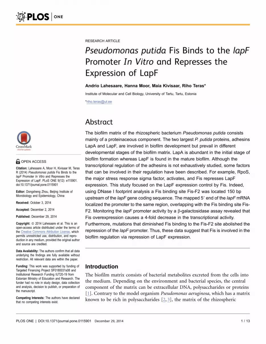
Pseudomonas putida Fis Binds to the lapFPromoter In Vitro and Represses theExpression of LapFAndrio Lahesaare, Hanna Moor, Maia Kivisaar, Riho Teras*
Institute of Molecular and Cell Biology, University of Tartu, Tartu, Estonia
Abstract
The biofilm matrix of the rhizospheric bacterium Pseudomonas putida consists
mainly of a proteinaceous component. The two largest P. putida proteins, adhesins
LapA and LapF, are involved in biofilm development but prevail in different
developmental stages of the biofilm matrix. LapA is abundant in the initial stage of
biofilm formation whereas LapF is found in the mature biofilm. Although the
transcriptional regulation of the adhesins is not exhaustively studied, some factors
that can be involved in their regulation have been described. For example, RpoS,
the major stress response sigma factor, activates, and Fis represses LapF
expression. This study focused on the LapF expression control by Fis. Indeed,
using DNase I footprint analysis a Fis binding site Fis-F2 was located 150 bp
upstream of the lapF gene coding sequence. The mapped 59 end of the lapFmRNA
localized the promoter to the same region, overlapping with the Fis binding site Fis-
F2. Monitoring the lapF promoter activity by a b-galactosidase assay revealed that
Fis overexpression causes a 4-fold decrease in the transcriptional activity.
Furthermore, mutations that diminished Fis binding to the Fis-F2 site abolished the
repression of the lapF promoter. Thus, these data suggest that Fis is involved in the
biofilm regulation via repression of LapF expression.
Introduction
The biofilm matrix consists of bacterial metabolites excreted from the cells into
the medium. Depending on the environment and bacterial species, the central
component of the matrix can be extracellular DNA, polysaccharides or proteins
[1]. Contrary to the model organism Pseudomonas aeruginosa, which has a matrix
known to be rich in polysaccharides [2, 3], the matrix of the rhizospheric
OPEN ACCESS
Citation: Lahesaare A, Moor H, Kivisaar M, TerasR (2014) Pseudomonas putida Fis Binds to thelapF Promoter In Vitro and Represses theExpression of LapF. PLoS ONE 9(12): e115901.doi:10.1371/journal.pone.0115901
Editor: Dongsheng Zhou, Beijing Institute ofMicrobiology and Epidemiology, China
Received: October 3, 2014
Accepted: December 2, 2014
Published: December 29, 2014
Copyright: � 2014 Lahesaare et al. This is anopen-access article distributed under the terms ofthe Creative Commons Attribution License, whichpermits unrestricted use, distribution, and repro-duction in any medium, provided the original authorand source are credited.
Data Availability: The authors confirm that all dataunderlying the findings are fully available withoutrestriction. All relevant data are within the paper.
Funding: This work was supported by funding ofTargeted Financing Project SF0180031s08 andInstitutional Research Funding IUT20-19 fromEstonian Ministry of Education and Research. Thefunder had no role in study design, data collectionand analysis, decision to publish, or preparation ofthe manuscript.
Competing Interests: The authors have declaredthat no competing interests exist.
PLOS ONE | DOI:10.1371/journal.pone.0115901 December 29, 2014 1 / 13

bacterium P. putida consists mainly of proteins [4]. The two largest proteins in P.
putida, the adhesins LapA and LapF, are known to be crucial for P. putida’s
biofilm formation [5–8]. These adhesins are involved in biofilm development and
prevail in different developmental stages of the biofilm matrix – LapA is necessary
at the beginning of biofilm formation and LapF in mature biofilm [5, 7–10]. LapA
is required for cell-surface interactions and therefore is needed for adhesion to
plant roots [11] or seeds leading to biofilm initiation [5]. P. putida’s LapF
provides cell-cell interactions, participating in the development of mature biofilm
[10, 12]. P. putida’s LapF (PP0806) consists of 6310 amino acids and it has only
one large repeat domain [5, 7, 12]. LapF is encoded by the first gene of the lapFHIJ
operon. The other genes encode for an ABC transporter which could be involved
in the secretion of LapF to the cell surface [12]. However, there is still no direct
evidence that LapHIJ is involved in the transport of LapF [12].
LapF is extensively expressed in the stationary phase but not in logarithmically
growing cells [8–10]. One reason for the lapF transcription activation in the
stationary phase cells is a positive effect of RpoS, the sigma factor that is required
for starvation and stress responses [10]. Additionally, several other indirect
influences to the expression of LapF have been described. Martinez-Gil et al.
(2014) monitored the expression of LapA and LapF by b-galactosidase assay and
RT-PCR in several P. putida strains obtained by random mini-Tn5 mutagenesis
[9]. They found that the GacA/S two-component system is indirectly involved in
the regulation of the LapF expression by modulating the expression of RpoS
(sigmaS), and that c-di-GMP overproduction reduces LapF expression [9].
Furthemore, although the overexpression of the global regulator Fis enhances P.
putida biofilm formation [13], it reduces the quantity of LapF about 4 times in the
P. putida stationary-phase cells [8].
Fis is a small DNA-binding and -bending homodimeric protein, which is
known as a trigger of fast growth in Enterobacteriaceae [14, 15]. It is speculated
that Fis is substitutable by other nucleoid-associated proteins such as IHF, HU, H-
NS or Dps in Escherichia coli, though none of the other proteins have noticeable
amino acid sequence similarity to Fis [14]. However, although E. coli fis knock-out
mutants are viable, it seems that the deletion of the fis gene is lethal for
Pseudomonas species [13, 16, 17]. Fis participates in several important processes
such as regulation of transcription and recombination [14, 15], but it is not
commonly associated with biofilm development. Although the involvement of Fis
in biofilm formation has been described in some studies [18–22], no one has
shown the direct transcription regulation of adhesin genes in P. putida by Fis.
In this study, the 59 end of the lapF mRNA is mapped, and the location of the
lapF promoter is determined. A Fis binding site Fis-F2, which overlaps the lapF
promoter, is identified by DNase I footprint and gel-shift assays. The adverse
effect of Fis overexpression to the transcription from the lapF promoter is shown
by a b-galactosidase assay. However, when testing a mutated Fis-binding site Fis-
F2-mut, Fis does not protect the DNA against DNase I cleavage, nor does it
repress transcription from lapF promoter, confirming that Fis directly represses
the lapF promoter.
Fis Binds to the lapF Promoter
PLOS ONE | DOI:10.1371/journal.pone.0115901 December 29, 2014 2 / 13
Materials and Methods
Bacterial strains, plasmids, oligonucleotides and media
The bacterial strains and plasmids used in this study are described in Table 1.
Bacteria were grown in LB medium [23]. Solid media contained 1.5% Difco agar.
Antibiotics were added at the following concentrations: ampicillin, 100 mg ml21;
gentamicin, 10 mg ml21; kanamycin, 50 mg ml21; penicillin, 1500 mg ml21;
streptomycin, 200 mg ml21. E. coli was incubated at 37 C̊ and P. putida at 30 C̊.
Bacteria were electrotransformed as described by Sharma & Schimke [24]. E. coli
strain DH5a (Invitrogen), was used for DNA cloning.
Prediction of Fis-binding sites on the promoter region of the lapFgene
Possible Fis-binding sequences on the promoter regions of the lap genes were
predicted using the E. coli Fis-binding sites matrix [25] and the matrix-scan
program available at the Regulatory Sequence Analysis Tools homepage (http://
rsat.ulb.ac.be/). The 2500 bp to +100 bp DNA region of the lapF gene was used
for the prediction of potential Fis-binding sites. The Markov model of zero order
(Bernoulli model), organism-specific probability of nucleotides in the upstream
region of genes in P. putida KT2440 and a P-value upper threshold of 0.001 were
selected for the conditions of the background model. The rest of the parameters
were left at the program’s default values.
DNA manipulations
The promoter probe vector pBLKT (Table 1) was constructed to measure b-
galactosidase activities. The 3320-bp DNA fragment containing the promoterless
lacZ reporter gene from pHLU102 [26] was cloned into pBBR1-MCS-5 [27] using
the PaeI and Acc65I restrictases, resulting in pMCS5-lacZ (Table 1). The KmR
gene was amplified by PCR from the plasmid pUTmini-Tn5 Km2 [28] by KmSac
primers (59-CAGGAGCTCGTTCGATTTATTCAACAAAGCC-39). The 1033-bp
PCR fragment containing the KmR gene was cleaved with Ecl136II and inserted
into pMCS5-lacZ, which was opened with PaeI and BglII and thereafter blunted
by Klenow I fragment, resulting in pBLK (Table 1). The pKST1T2 [29] was
cleaved with BcuI and EcoRV and a 452-bp DNA fragment containing the T1T2
transcription terminator was obtained. The T1T2 fragment was inserted into
pBLK, which was opened with PdiI and BcuI restrictases, resulting in the
promoter probe vector pBLKT (Table 1).
Two pBLKT derivatives containing the lapF promoter region were constructed.
The lapF promoter regions were amplified by PCR, and both fragments were
cloned into the pBLKT BamHI site. A 235-bp PCR product was amplified by
LapF-fw (59-TAGATCTTTCGCTGAGGCTTTTCTAC-3) and PP0806-rev (59-
TGGATCCACTTCGGATTGCTTATCGG-39) oligonucleotides. The PCR product
was cut with BglII and BamHI and resulted in two fragments. The fragment
Fis Binds to the lapF Promoter
PLOS ONE | DOI:10.1371/journal.pone.0115901 December 29, 2014 3 / 13

containing the 2198 bp to 222 bp region of the lapF upstream DNA was used to
obtain pBLKT-F-Fis.
For the construction of pBLKT-F-Fis-mut, site-directed mutagenesis of wild-
type Fis-F2 was performed using two sequential PCRs and the P. putida PSm
chromosome as a template. These amplifications resulted in a fragment with five
substituted nucleotides in the Fis-F2 binding site but otherwise identical to the
one that was used for obtaining pBLKT-F-Fis. In the first PCR, the
oligonucleotides LapFII-mut carrying five substitutions (59-
CATCTGGTTGCTGCCTTCCGGGCTGCTATATC-39, substitutions in bold) and
PP0806-rev were used for DNA amplification (see sequence in Fig. 1A). In the
second PCR, LapF-fw and the product of the first PCR were used as primers for
DNA amplification from the P. putida PSm chromosome. Thereafter the PCR
fragment was restricted with BamHI and BglII. The fragment containing the
2198 bp to 222 bp region of the mutated lapF upstream DNA was inserted to
pBLKT, resulting in pBLKT-F-Fis-mut. All designed plasmids were sequenced in
order to exclude PCR-generated errors in the cloned DNA fragments.
DNase I footprinting
DNase I footprint assays were performed for the identification of P. putida Fis-
binding sequences on the lapF promoter region. PCR-amplified fragments were
used for DNase I footprint assay and were generated as follows. The 149 bp-long
Table 1. Bacterial strains and plasmids used in this study.
Strain and plasmid Genotype or description Source/reference
E. coli
DH5a supE44 DlacU169(f80 lacZDM15) recA1 endA1 hsdR17 thi-1 gyrA96 relA1 Invitrogen
P. putida
PSm PaW85; chromosomal mini-Tn7-VSm1 (Smr) [13]
F15 PaW85; chromosomal mini-Tn7-VGm-term-lacIq-Ptac-fis-T1T2 (Gmr) [13]
Plasmids
pBBR1-MCS-5 Cloning vector (Gmr) [27]
pBLK 1033 bp DNA fragment containing Kmr gene cloned into pMC5-lacZ opened with BglII and PaeI (Kmr) This study
pBLKT Promoter probe vector, a 452 bp DNA fragment containing the T1T2 transcription terminator cloned intopBLK opened with BcuI and PdiI (Kmr)
This study
pKST1T2 pBluescript KS(+) containing the transcription terminator sequence T1T2 (Ampr) [29]
pHLU102 Promoterless lacZ gene [26]
pMCS5-lacZ 3320 bp fragment containing the lacZ gene cloned into pBBR1-MCS-5 by PaeI and Acc65I restrictases(Gmr)
This study
pUTmini-Tn5Km2 Suicide vector, source of Km resistance gene (Ampr, Kmr) [41]
pBLKT-F-Fis 177-bp-long promoter region of the lapF gene cloned into pBLKT BamHI site (Kmr) This study
pBLKT-F-Fis-mut 177-bp-long promoter region of the lapF gene with the mutated Fis-F2 site cloned into pBLKT BamHIsite (Kmr)
This study
pLA1-12 Carrying LF2 site in left end DNA of Tn4652; (Ampr) [30]
pRA1-12 Carrying RF1 site in right end DNA of Tn4652; (Ampr) [30]
doi:10.1371/journal.pone.0115901.t001
Fis Binds to the lapF Promoter
PLOS ONE | DOI:10.1371/journal.pone.0115901 December 29, 2014 4 / 13

DNA fragment upstream of the lapF gene was amplified using the LapF-fw and
LapF-down2 (59-CGTAACAGAGCCCTTTCAG-39) oligonucleotides to identify
Fis binding sites upstream of the lapF gene. Depending on the template (pBLKT-
F-Fis or pBLKT-F-Fis-mut), the PCR-amplified fragments contained either the
wild-type Fis-F2 or the mutated Fis-F2-mut Fis-binding site. The following
procedures: labelling PCR products with [c-32P]-ATP, preparing reaction
Fig. 1. Mapping of Fis binding sites, promoter, and 59 end of mRNA at the lapF promoter region. (A) Sequence of the lapF promoter region anddownstream DNA. The start codon of the lapF gene is shown in bold. The first nucleotide of the mRNA 59 end is written in bold and is designated as F-1. Thepotential –10 and –35 elements of the lapF promoter are shown in grey boxes. The Fis binding site Fis-F2 is shown in black brackets and the in silicopredicted Fis binding site Fis-F1 is shown in dashed brackets. The E. coli Fis binding consensus according to Finkel and Johnson (1992) and Shao et al.(2008) is shown above the lapF promoter sequence [36, 37]. The most important nucleotides for the E. coli Fis binding are shown in bold. The pointmutations in the Fis-F2 antisense strand are indicated by arrows (B) Agarose-gel electrophoresis of cDNA amplified by the RACE method for theidentification of the lapF mRNA 59 end. The arrow points to the PCR product used to determine the mRNA 59 end. (C) Protection of the lapF upstream DNAagainst DNase I cleavage by binding of Fis on the sense and the antisense strands. Lines at the right side of the panels indicate the regions protected by Fisfrom DNase I cleavage at the positions 2165 to 2132 on the sense strand and –158 to 2133 on the antisense strand corresponding to Fis-F2.
doi:10.1371/journal.pone.0115901.g001
Fis Binds to the lapF Promoter
PLOS ONE | DOI:10.1371/journal.pone.0115901 December 29, 2014 5 / 13

mixtures and gel electrophoresis were carried on as described in Teras et. al (2009)
[16].
Gel mobility shift assay
The same labelled PCR-amplified fragments that were used for DNase I footprint
assays, were used for the gel mobility assay. Additionally, the non-labelled PCR
product containing the Fis binding site LF2 [30] and a PCR product without Fis-
binding site RF1 [30] were used in out-competition experiments. The unlabelled
DNA fragment LF2 was amplified using the oligonucleotides TnLsisse (59-
GCAAAGACTGCTTCGCGCCC-39) and SIDD-2 (59-
AGAGCTCCTGTACGTGCGCTT-39). Oligonucleotides PRH8 (59-
GCTGAGCTCAGACGGTGGATGACCAGC-39) and Tnots (59-
GGGGTTATGCCGAGATAAGGC-39) were used for the amplification of
unlabelled DNA RF1. Plasmids pLA1-12 and pRA1-12 [30] were used for
amplifying LF2 and RF1, respectively. Amounts of competing DNA in the reaction
mixes were calculated in molecules.
Binding reactions with purified P. putida His-tagged Fis were carried out with
261010 molecules (750–1000 c.p.m.) of labelled DNA fragment in a reaction
buffer (24 mM Tris/HCl pH 7.5, 50 mM KCl, 10 mM MgCl2, 1 mM CaCl2,
0.1 mM EDTA, 5% glycerol, 0.05 mg BSA ml21 and 0.05 mg salmon sperm DNA
ml21) in a final volume of 20 ml. The mixtures were preincubated for 20 min at
room temperature. After incubation, reaction mixtures were applied to a 5% non-
denaturing polyacrylamide gel buffered with TBE (50 mM Tris, 60 mM boric
acid, 5 mM EDTA; pH 7.5). Electrophoresis was carried out at 4 C̊ at 10 V cm21
for 3 h. Gels were vacuum dried and exposed to a Typhoon Trio screen (GE
Healthcare).
Identification of 59 ends of mRNA by RACE
The mRNA 59 end of the lapF gene was identified by RACE (rapid amplification
of cDNA ends) as described by Sambrook and Russell (2001) [31]. 1.5 mg of
purified total RNA and the LapF-RACE1 primer (59-
GCCGACGAAGACCATATC-39) were used for the amplification of the first
strand of cDNA. The second strands of cDNA were amplified using primers
Adapt-pikkC (59-GACTCGAGTCGACATCGA(C)17-39) or Adapt-pikkT (59-
GACTCGAGTCGACATCGA(T)17-39), with 59 ends binding accordingly to poly-
G or poly-A, synthesised by terminal deoxynucleotidyltransferase (TdT) to the 39
ends of the first strand of cDNA. For the second PCR, the Adapt-lyh (59-
GACTCGAGTCGACATCG-39) and PP0806-rev primers were used. Zymo
Research DNA Clean & Concentrator-5 kit was used for DNA purification
between the RACE stages.
Fis Binds to the lapF Promoter
PLOS ONE | DOI:10.1371/journal.pone.0115901 December 29, 2014 6 / 13

Measurement of b-galactosidase activity
To measure b-galactosidase activities, P. putida cells were grown in LB medium
with or without 1 mM IPTG supplementation for 18 hours. As a source of b-
galactosidase, the pBLKT-F-Fis and pBLKT-F-Fis-mut constructs containing lapF
promoter region in front of the lacZ gene were used (Table 1). The measurement
of b-galactosidase from cell suspension was performed according to the protocol
of Miller (1992) [23]. At least eight independent measurements were performed.
Statistical analysis
The factorial analysis of variance (ANOVA) method and post-hoc Bonferroni test
at a significance level 0.05 were used to assess the variability of experimental data.
The calculations were performed using Statistica 10 software.
Results and Discussion
Mapping of the Fis-binding sites in the lapF promoter region
We have previously shown that Fis overexpression decreases the quantity of LapF
about four times in P. putida cells [8]. Therefore, we were interested in the
possible interaction of Fis and the lapF promoter region. We used in silico
prediction of Fis-binding sites on the upstream region of the lapF gene to obtain
initial information for later transcriptional studies. Surprisingly, no Fis-binding
sites were predicted in silico for the upstream sequence of lapF under the selected
conditions (detailed information in the Material and Methods section). However,
there was one predicted Fis-binding sequence (Fis-F1) approximately 65 bp
downstream the lapF start codon (Fig. 1A). The weight-score and p-value for Fis-
F1 were respectively 6.5 and 0.0002 for the sense strand and 5.0 and 0.00091 for
the antisense strand. The applied matrix’s maximum weight score was 12.5 and
minimum weight score was 228. Despite the in silico prediction, we could not
verify Fis binding to the predicted Fis-F1 sequence by DNase I footprint analysis
(data not shown). Instead, DNase I footprint analysis revealed another Fis-
binding site, Fis-F2, located approximately 2150 bp upstream of the lapF gene
(Fig. 1A and C).
To identify the promoter sequences of the lapF gene, we mapped the 59 end of
the lapF mRNA by RACE method. We identified one 59 end of the mRNA at the
position 2120 upstream of the lapF coding sequence (Fig. 1A and B). Since the
lapF transcription has been previously shown to be activated by RpoS in
stationary phase cells [10], we expected to find a recognisable –10 element of the
sigmaS-dependent promoter. It is known that sigmaS-dependent promoters, like
the Pm promoter from the P. putida TOL plasmid and the PalkS promoter from
Pseudomonas oleovorans OCT plasmid, depend on RpoS both in E. coli and in P.
putida [32–34]. Therefore we used the consensuses of E. coli sigmaS-dependent
promoters to predict lapF promoter elements. A putative –10 promoter element
sequence GCTATATC was located six nucleotides upstream of the mapped 59
Fis Binds to the lapF Promoter
PLOS ONE | DOI:10.1371/journal.pone.0115901 December 29, 2014 7 / 13
mRNA end F-I (Fig. 1A). This is similar to the –10 consensus sequence of the E.
coli sigmaS-dependent promoters KCTAYACT, where ‘‘K’’ is G or T and ‘‘Y’’ is
pyrimidine [35]. The sequence of the –35 promoter element is not well-conserved
among E. coli sigmaS-dependent promoters and can differ from the consensus of
the sigma70-dependent –35 element TTGACA [35]. Even more, the preferred
spacer length between the –10 and –35 elements is more flexible compared to
sigma70-dependent promoters, varying from 15 to 19 nucleotides [35]. However,
although the sequence of the –35 element is not conserved, modifications in this
region can change the transcription initiation from sigmaS-dependent promoters
[35]. Indeed, no recognisable sequence was found (Fig. 1A) that would be similar
with the –35 element of sigma70-dependent promoters’ consensus TTGACA [35].
Considering the overlapping position of the –10 hexamer and the Fis binding site
Fis-F2 it was plausible that Fis could repress transcription from the lapF promoter
by impeding RNA polymerase binding.
To confirm direct binding of Fis to the promoter of the lapF gene, we mutated
the five nucleotides of Fis-F2 that could be the most important for Fis binding
(Fig. 1A). According to Finkel and Johnson (1992) and Shao et al. (2008) the
most important nucleotides for E. coli Fis binding are the 1st, 4th, 5th, 11th, 12th
and 15th nucleotides in the GnTYAWWWWWTRAnC consensus. In the
consensus, ‘‘Y’’ is pyrimidine, ‘‘W’’ is A or T, ‘‘R’’ is purine and ‘‘n’’ is any
nucleotide [36, 37]. Considering the fact that the potential –35 hexamer of the
lapF promoter is located within the Fis-F2 binding site, we could only mutate the
nucleotides that did not belong to the potential –35 and –10 elements of the
promoter (Fig. 1A and C). The C nucleotide substitution with A at the position
2159 bp from the lapF gene happened by chance during PCR. However, this
mutation was inside the Fis-F2 site without overlapping the potential –35 element.
Therefore, we decided to use this construct for following experiments.
The DNase I footprint analysis carried out with the Fis-F2-mut DNA and the
purified Fis revealed that unlike the wild-type Fis-F2, Fis did not prevent DNase I
cleavage of the Fis-F2-mut sequence (Fig. 1C). Thus, Fis did not bind the mutated
Fis-F2 sequence or bound it with a weak affinity, which was undetectable by the
DNase I footprint analysis.
Additionally, Fis binding to the DNA containing Fis-F2 was assessed by gel
mobility shift analysis. To assess Fis specific binding to the Fis-F2 sequence,
unlabelled DNA containing the Fis-binding site LF2 from the left end of Tn4652
[30] was used to outcompete Fis from the lapF promoter DNA-Fis complex
(Fig. 2). Although Fis bound relatively similarly to the Fis-F2 and Fis-F2-mut
DNA fragments, LF2 outcompeted Fis from Fis-F2-mut complex more easily than
from Fis-F2 complex (Fig. 2). Thus, the introduced mutations in the Fis-F2
sequence had an adverse influence on Fis binding in vitro.
The impact of Fis on the transcription of the lapF gene
To elucidate the possible effect of Fis on the transcription of the lapF gene, the
promoter region was cloned to the front of the reporter gene lacZ in a promoter
Fis Binds to the lapF Promoter
PLOS ONE | DOI:10.1371/journal.pone.0115901 December 29, 2014 8 / 13

probe vector pBLKT (Table 1). The b-galactosidase activity was measured in
stationary-phase cells of the P. putida strains PSm (wild-type) and F15 (IPTG-
inducible Fis overexpression strain). Indeed, we could solely examine Fis’s impact
to lapF expression using the fis overexpression strain F15, since all our attempts to
reduce Fis’ amount by fis gene deletion or conditional gene expression resulted in
a lethal genotype or unexpected recombination of the native fis to somewhere in
the P. putida genome [13, 16].
The influence of IPTG to the lacZ gene expression was assessed in LB medium.
Comparing the LacZ activities of the PSm cells carrying the pBLKT derivates, we
did not observe any statistically significant differences in the results when the cells
were grown in LB medium with and without 1 mM IPTG (Fig. 3). This control
experiment demonstrated that IPTG itself has no influence on the expression of
the lacZ gene in P. putida. Also, we measured the expression of lacZ from the
promoterless pBLKT in the cells of PSm and F15. In all cases, the expression of the
lacZ gene resulted in b-galactosidase activities lower than 0.9 Miller Units and no
effect of the presence of IPTG in the LB medium or the use of the host strain was
observed (data not shown).
The LacZ activity was measured in P. putida harbouring the pBLKT constructs
containing the lapF promoter region in front of the reporter gene lacZ. As
expected, Fis overexpression drastically reduced the b-galactosidase activity in
cells carrying the lapF promoter-lacZ transcriptional fusion. We measured LacZ
activity in P. putida harbouring pBLKT-F-Fis, the plasmid that contained the
DNA region encompassing 2198 to 222 bp from the lapF start codon inserted in
front of the reporter gene lacZ. The LacZ activity was comparable in the wild-type
cells (PSm) and F15 grown without IPTG and did not have statistically significant
differences. However, Fis overexpression in F15 reduced the LacZ activity 4.2
times (p,0.001) compared to F15 grown without IPTG (Fig. 3A). Thus, the b-
galactosidase results confirmed our previous finding that Fis overexpression
decreases the amount of LapF in P. putida stationary-phase cells when LapF is
efficiently expressed [8].
Fig. 2. Gel shift assay of the Fis binding to the lapF promoter DNA containing the wild-type Fis binding site Fis-F2 and the mutated site Fis-F2-mut.261010 molecules of radioactively labelled PCR products containing Fis-F2 (lanes 1–10) and Fis-F2-mut site (lanes 11–20) were used for Fis binding. Fiswas outcompeted from Fis-DNA complex with unlabelled PCR product containing the Fis binding site (LF2) and PCR product without Fis-binding site (RF1).Added unlabelled DNA was calculated in molecules. 0.46 mM Fis was used in each reaction mixture except mixtures without Fis in lanes 2 and 12.
doi:10.1371/journal.pone.0115901.g002
Fis Binds to the lapF Promoter
PLOS ONE | DOI:10.1371/journal.pone.0115901 December 29, 2014 9 / 13

We employed a pBLKT-F-Fis-mut construct to elucidate Fis’s effect on the lapF
gene’s transcription by b-galactosidase assay. The pBLKT-F-Fis-mut construct was
identical to pBLKT-F-Fis, except for point mutations in the Fis-F2 binding site
that reduced Fis binding in in vitro assays. Fis overexpression did not decrease the
activity of LacZ in P. putida F15 harbouring pBLKT-F-Fis-mut (Fig. 3B),
indicating direct regulation of LapF expression by Fis. However, if compared to
the wild-type strain PSm, the LacZ activity in F15 remained 1.8 times (p,0.001)
or 1.7 times lower (p50.001), depending on the absence or presence of 1 mM
IPTG in the growth medium. In addition, the LacZ activity measured in the wild-
type strain PSm cells carrying the pBLKT-F-Fis-mut construct was approximately
three times higher than that in cells with pBLKT-F-Fis. Hence, we suppose that
the introduced mutations in the Fis-F2 site increased the general expression of
LacZ, but at the same time had an adverse effect on Fis binding.
Thus, the Fis-binding site Fis-F2 is needed for Fis to repress the lapF promoter.
Both the results of the b-galactosidase assay (Fig. 3) and the DNase I footprint
analysis (Fig. 1) indicate that the transcription of lapF is repressed by Fis.
Moreover, our data confirm our previous finding that Fis overexpression can
diminish the quantity of LapF in P. putida stationary phase cells, where LapF is
strongly expressed [8].
Considering the dependence of RpoS and Fis amounts on P. putida growth
phases, the expression of LapF seems to be tightly controlled by the physiological
state of P. putida. In fast growing bacteria, the expression of Fis is favoured, and
the expression of RpoS is down-regulated [38, 39]. When the growth rate of
bacteria decelerates and they enter stationary phase, the expressions of RpoS and
Fig. 3. Effect of overexpression of Fis on the level of transcription from the lapF promoter. b-Galactosidase (b-Gal) activity expressed from the lapFpromoter-lacZ reporter constructs were measured in the wild-type strain PSm and the Fis-overexpressing strain F15 of P. putida grown for 18 hours in LBmedium supplemented with 1 mM IPTG and without IPTG supplementation. Vertical bars denote 95% confidence intervals of means. Data of at least 8independent measurements is shown. The length of the lapF promoter region inserted upstream of the lacZ reporter gene in different pBLKT constructs isshown above diagrams. The Fis-binding site Fis-F2 (gray box) and 59 end of lapF mRNA F-I (arrow) are shown in drawings.
doi:10.1371/journal.pone.0115901.g003
Fis Binds to the lapF Promoter
PLOS ONE | DOI:10.1371/journal.pone.0115901 December 29, 2014 10 / 13

Fis will be reversed [38, 39]. Waite et al. (2006) compared gene expression in
planktonic and sessile Pseudomonas aeruginosa PAO1 by microarray analysis. As
expected, they found that RpoS and Fis are oppositely regulated during biofilm
development [40]. The level of fis mRNA was highest in the developing biofilm
and slightly decreased in mature biofilm. Contrary to fis, the level of rpoS mRNA
is highest in mature biofilm [40]. Considering the overlapping position of the lapF
–10 element and Fis binding site Fis-F2, it is likely that Fis could repress
transcription from the lapF promoter by impeding RNA polymerase binding.
Therefore, it is more plausible that LapF expression is regulated by two competing
regulators: Fis and RpoS. In logarithmically growing P. putida or at the beginning
of biofilm development, LapF is not expressed or weakly expressed due to
repression of Fis and the deficiency of activator RpoS. During growth speed
deceleration or biofilm development, RpoS level increases and Fis level decreases,
allowing LapF to be expressed. Thereby, the overexpression of Fis in stationary
phase can unbalance the ratio of regulators leading to the Fis-induced
transcriptional repression of lapF (Fig. 3) [8].
Summarizing the data presented in this report, we can conclude that Fis
represses transcription from lapF promoter via the Fis-binding site Fis-F2. We
identified a Fis binding site Fis-F2 in vitro by DNase I footprint and by using a b-
galactosidase assay were able to show that Fis overexpression represses
transcription from the lapF promoter region. Fis binding is weaker to the mutated
Fis-F2-mut site and therefore Fis overexpression does not repress transcription
from the lapF promoter. This confirms that Fis directly represses the lapF
promoter.
Acknowledgments
We thank Signe Saumaa and Andres Ainelo for their comments on the
manuscript.
Author ContributionsConceived and designed the experiments: AL HM RT. Performed the
experiments: AL HM RT. Analyzed the data: AL HM RT. Contributed reagents/
materials/analysis tools: MK. Contributed to the writing of the manuscript: AL
HM MK RT.
References
1. Sutherland IW (2001) The biofilm matrix–an immobilized but dynamic microbial environment. TrendsMicrobiol 9: 222–227.
2. Friedman L, Kolter R (2004) Genes involved in matrix formation in Pseudomonas aeruginosa PA14biofilms. Mol Microbiol 51: 675–690.
3. Ryder C, Byrd M, Wozniak DJ (2007) Role of polysaccharides in Pseudomonas aeruginosa biofilmdevelopment. Curr Opin Microbiol 10: 644–648.
Fis Binds to the lapF Promoter
PLOS ONE | DOI:10.1371/journal.pone.0115901 December 29, 2014 11 / 13
4. Jahn A, Griebe T., Nielsen PH (1999) Composition of Pseudomonas putida Biofilms: Accumulation ofPreteins in the Biofilm Matrix. Biofouling 14: 49–57.
5. Espinosa-Urgel M, Salido A, Ramos JL (2000) Genetic analysis of functions involved in adhesion ofPseudomonas putida to seeds. J Bacteriol 182: 2363–2369.
6. Gjermansen M, Nilsson M, Yang L, Tolker-Nielsen T (2010) Characterization of starvation-induceddispersion in Pseudomonas putida biofilms: genetic elements and molecular mechanisms. Mol Microbiol75: 815–826.
7. Hinsa SM, Espinosa-Urgel M, Ramos JL, O’Toole GA (2003) Transition from reversible to irreversibleattachment during biofilm formation by Pseudomonas fluorescens WCS365 requires an ABC transporterand a large secreted protein. Mol Microbiol 49: 905–918.
8. Moor H, Teppo A, Lahesaare A, Kivisaar M, Teras R (2014) Fis overexpression enhancesPseudomonas putida biofilm formation by regulating the ratio of LapA and LapF. Microbiology.
9. Martinez-Gil M, Ramos-Gonzalez MI, Espinosa-Urgel M (2014) Roles of cyclic Di-GMP and the Gacsystem in transcriptional control of the genes coding for the Pseudomonas putida adhesins LapA andLapF. J Bacteriol 196: 1484–1495.
10. Martinez-Gil M, Yousef-Coronado F, Espinosa-Urgel M (2010) LapF, the second largestPseudomonas putida protein, contributes to plant root colonization and determines biofilmarchitecture. Mol Microbiol 77: 549–561.
11. Yousef-Coronado F, Travieso ML, Espinosa-Urgel M (2008) Different, overlapping mechanisms forcolonization of abiotic and plant surfaces by Pseudomonas putida. FEMS Microbiol Lett 288: 118–124.
12. Fuqua C (2010) Passing the baton between laps: adhesion and cohesion in Pseudomonas putidabiofilms. Mol Microbiol 77: 533–536.
13. Jakovleva J, Teppo A, Velts A, Saumaa S, Moor H, et al. (2012) Fis regulates the competitiveness ofPseudomonas putida on barley roots by inducing biofilm formation. Microbiology 158: 708–720.
14. Dorman CJ (2009) Nucleoid-Assiociated Proteins and Bacterial Physiology. In: Laskin AI, Sariaslani, S,Gadd, G M., editor. Advances in Applied Microbiology: Elsevier.
15. Browning DF, Grainger DC, Busby SJ (2010) Effects of nucleoid-associated proteins on bacterialchromosome structure and gene expression. Curr Opin Microbiol 13: 773–780.
16. Teras R, Jakovleva J, Kivisaar M (2009) Fis negatively affects binding of Tn4652 transposase by out-competing IHF from the left end of Tn4652. Microbiology 155: 1203–1214.
17. Liberati NT, Urbach JM, Miyata S, Lee DG, Drenkard E, et al. (2006) An ordered, nonredundant libraryof Pseudomonas aeruginosa strain PA14 transposon insertion mutants. Proc Natl Acad Sci U S A 103:2833–2838.
18. Chaudhuri RR, Sebaihia M, Hobman JL, Webber MA, Leyton DL, et al. (2010) Complete genomesequence and comparative metabolic profiling of the prototypical enteroaggregative Escherichia colistrain 042. PLoS One 5: e8801.
19. Czeczulin JR, Balepur S, Hicks S, Phillips A, Hall R, et al. (1997) Aggregative adherence fimbria II, asecond fimbrial antigen mediating aggregative adherence in enteroaggregative Escherichia coli. InfectImmun 65: 4135–4145.
20. Sheikh J, Hicks S, Dall’Agnol M, Phillips AD, Nataro JP (2001) Roles for Fis and YafK in biofilmformation by enteroaggregative Escherichia coli. Mol Microbiol 41: 983–997.
21. Saldana Z, Xicohtencatl-Cortes J, Avelino F, Phillips AD, Kaper JB, et al. (2009) Synergistic role ofcurli and cellulose in cell adherence and biofilm formation of attaching and effacing Escherichia coli andidentification of Fis as a negative regulator of curli. Environ Microbiol 11: 992–1006.
22. Prigent-Combaret C, Zghidi-Abouzid O, Effantin G, Lejeune P, Reverchon S, et al. (2012) Thenucleoid-associated protein Fis directly modulates the synthesis of cellulose, an essential component ofpellicle-biofilms in the phytopathogenic bacterium Dickeya dadantii. Mol Microbiol 86: 172–186.
23. Miller JH (1992) A short course in bacterial genetics: a laboratory manual and handbook for Escherichiacoli and related bacteria. Cold Spring Harbor, NY: Cold Spring Harbor Laboratory Press.
24. Sharma RC, Schimke RT (1996) Preparation of electrocompetent E. coli using salt-free growth medium.Biotechniques 20: 42–44.
Fis Binds to the lapF Promoter
PLOS ONE | DOI:10.1371/journal.pone.0115901 December 29, 2014 12 / 13

25. Gama-Castro S, Salgado H, Peralta-Gil M, Santos-Zavaleta A, Muniz-Rascado L, et al. (2011)RegulonDB version 7.0: transcriptional regulation of Escherichia coli K-12 integrated within geneticsensory response units (Gensor Units). Nucleic Acids Res 39: D98–105.
26. Lang H, Palva ET (1993) The ompS gene of Vibrio cholerae encodes a growth-phase-dependentmaltoporin. Mol Microbiol 10: 891–901.
27. Kovach ME, Elzer PH, Hill DS, Robertson GT, Farris MA, et al. (1995) Four new derivatives of thebroad-host-range cloning vector pBBR1MCS, carrying different antibiotic-resistance cassettes. Gene166: 175–176.
28. de Lorenzo V, Herrero M, Jakubzik U, Timmis KN (1990) Mini-Tn5 transposon derivatives for insertionmutagenesis, promoter probing, and chromosomal insertion of cloned DNA in gram-negative eubacteria.J Bacteriol 172: 6568–6572.
29. Tavita K, Mikkel K, Tark-Dame M, Jerabek H, Teras R, et al. (2012) Homologous recombination isfacilitated in starving populations of Pseudomonas putida by phenol stress and affected by chromosomallocation of the recombination target. Mutat Res 737: 12–24.
30. Teras R, Horak R, Kivisaar M (2000) Transcription from fusion promoters generated duringtransposition of transposon Tn4652 is positively affected by integration host factor in Pseudomonasputida. J Bacteriol 182: 589–598.
31. Sambrook J, Russel DW (2001) Molecular Cloning: A Laboratory Manual. New York: Cold SpringHarbor Laboratory Press.
32. Canosa I, Yuste L, Rojo F (1999) Role of the alternative sigma factor sigmaS in expression of the AlkSregulator of the Pseudomonas oleovorans alkane degradation pathway. J Bacteriol 181: 1748–1754.
33. Marques S, Gallegos MT, Ramos JL (1995) Role of sigma S in transcription from the positivelycontrolled Pm promoter of the TOL plasmid of Pseudomonas putida. Mol Microbiol 18: 851–857.
34. Miura K, Inouye S, Nakazawa A (1998) The rpoS gene regulates OP2, an operon for the lower pathwayof xylene catabolism on the TOL plasmid, and the stress response in Pseudomonas putida mt-2. MolGen Genet 259: 72–78.
35. Typas A, Becker G, Hengge R (2007) The molecular basis of selective promoter activation by thesigmaS subunit of RNA polymerase. Mol Microbiol 63: 1296–1306.
36. Finkel SE, Johnson RC (1992) The Fis protein: it’s not just for DNA inversion anymore. Mol Microbiol 6:3257–3265.
37. Shao Y, Feldman-Cohen LS, Osuna R (2008) Functional Characterization of the Escherichia coli Fis-DNA Binding Sequence. Journal of Molecular Biology 376: 771–785.
38. Yuste L, Hervas AB, Canosa I, Tobes R, Jimenez JI, et al. (2006) Growth phase-dependentexpression of the Pseudomonas putida KT2440 transcriptional machinery analysed with a genome-wideDNA microarray. Environ Microbiol 8: 165–177.
39. Bertani I, Sevo M, Kojic M, Venturi V (2003) Role of GacA, LasI, RhlI, Ppk, PsrA, Vfr and ClpXP in theregulation of the stationary-phase sigma factor rpoS/RpoS in Pseudomonas. Arch Microbiol 180: 264–271.
40. Waite RD, Paccanaro A, Papakonstantinopoulou A, Hurst JM, Saqi M, et al. (2006) Clustering ofPseudomonas aeruginosa transcriptomes from planktonic cultures, developing and mature biofilmsreveals distinct expression profiles. BMC Genomics 7: 162.
41. de Lorenzo V, Cases I, Herrero M, Timmis KN (1993) Early and late responses of TOL promoters topathway inducers: identification of postexponential promoters in Pseudomonas putida with lacZ-tetbicistronic reporters. J Bacteriol 175: 6902–6907.
Fis Binds to the lapF Promoter
PLOS ONE | DOI:10.1371/journal.pone.0115901 December 29, 2014 13 / 13