Embed Size (px)
Citation preview
137137137137137Mem Inst Oswaldo Cruz, Rio de Janeiro, Vol. 101(Suppl. I): 137-143, 2006
Protein tyrosine kinases in Schistosoma mansoniDiana Bahia/+, Luiza Freire Andrade*, Fernanda Ludolf*, Renato Arruda Mortara**,
Guilherme Oliveira/*
Centro de Pesquisas René Rachou-Fiocruz, Av. Augusto de Lima 1715, 30190-002 Belo Horizonte, MG, Brasil*Programa de Pós-Graduação e Pesquisa, Santa Casa de Misericórdia de Belo Horizonte, Belo Horizonte, MG, Brasil
**Escola Paulista de Medicina, Unifesp, São Paulo, SP, Brasil
The identification and description of signal transduction molecules and mechanisms are essential to elucidateSchistosoma mansoni host-parasite interactions and parasite biology. This mini review focuses on recent advance-ments in the study of signalling molecules and transduction mechanisms in S. mansoni, drawing special attention tothe recently identified and characterised protein tyrosine kinases of S. mansoni.
Key words: signal transduction - protein kinase - schistosoma - tyrosine kinase
The identification and characterisation of signal trans-duction molecules and mechanisms are essential to eluci-date Schistosoma mansoni host-parasite interactions andparasite biology. Protein tyrosine kinases (PTKs) are im-portant molecules for intra- and inter-cellular communica-tion as well as for survival in eukaryotes, playing a majorrole in signal transduction processes (Hanks et al. 1988).PTKs also participate in cellular mechanisms that controlseveral biological processes such as adhesion, cytoskel-eton reorganisation, and migration. These proteins arealso known to be involved in developmental and differen-tiation processes of cells. Therefore, the study of PTKsmay unveil strategies that can be used for identifying newcandidate drug targets.
Phosphorylation of protein substrates catalysed bykinases is an essential mechanism by which importantintracellular and extracellular signals are transmittedthroughout the cell and to the nucleus (Cheetham 2004).During kinase catalysis, the γ-phosphate group from ATPis transferred to the protein substrate, thereby changingthe substrate properties (e.g. its structure, location or itsactivity as enzyme) (Manning et al. 2002a).
The recent success in cancer treatment that includesspecific tyrosine kinase inhibitors strongly validates theclinical relevance of basic research on tyrosine phospho-rylation. Functional profiling of the tyrosine phospho-proteome is likely to lead to the identification of noveltargets for drug discovery, providing a basis for novelmolecular target approaches (Machida et al. 2003). Manyof the 500 or so identified human protein kinases are at-tractive drug targets to treat cancer, inflammation pro-cesses and autoimmune diseases.
PTKsPTKs comprise proteins found in multicellular organ-
isms (Neet & Hunter 1996). PTKs may be found (i) an-chored in the cell membrane, acting as receptors, (ii) freein the cytoplasm, participating in signalling cascades, and(iii) in the nucleus, directly associated with gene activa-tion processes. PTK catalytic domain activation resultsfrom the interaction with other signalling proteins, whichallows specifically signalling propagation. There is noevidence of PTK in yeast. However, 49 from 239 Droso-phila melanogaster PKs and 105 out of 454 Caeno-rhabditis elegans PKs have been classified as PTKs (Man-ning et al. 2002a). There are two major classes of PTKs,receptor tyrosine kinases (RTKs) and non-receptor ty-rosine kinases, also named cytoplasmic or cellular tyrosinekinases (NRTKs) (Neet & Hunter 1996).
Receptor tyrosine kinases (RTKs) - RTKs contain threedistinct regions: an extracellular binding domain, a trans-membrane helix, and a cytoplasmatic domain that con-tains the kinase activity (Hubbard & Till 2000). RTK acti-vation is generally triggered by the interaction of a ligandwith a specific biding site on the receptor extracellulardomain. Following RTK stimulation at the extracellular site,the catalytic domain on the cytoplasmic side of membraneis activated by the dimerisation of the receptor, leading toautophosphorylation (Alberts et al. 1994) and then to theactivation of kinase activity, providing a new biding sitefor intracellular adapter molecules.
RTK families show diversified extracellular domains(Heldin 1996). The first protein receptor described as be-ing protein kinase tyrosine-specific was the epidermalgrowth factor receptor (EGFr) (Yarden & Ullrich 1988).However, there are growth and differentiation factor re-ceptors that also belong to RTKs.
Non-RTKs (NRTK) - NRTKs are a set of intracellularsignalling proteins that has been identified as being ableto interact with PTK phosphotyrosine (Neet & Hunter1996). Although intracellular signalling proteins that bindto activated PTK phosphotyrosine residues have variousfunctions and structures, they generally share highly con-served non-catalytic domains, known as SH2 and SH3.One of the main features of NRTK is the presence of the
Financial support: Fapemig (DB CBB-174/02)+Corresponding author and CNPq fellow. Present address:Departamento de Genética, ESALQ, USP, Av. Pádua Dias 11,13400-970 Piracicaba, SP, Brasil.E-mail: [email protected] 25 May 2006Accepted 26 June 2006
138138138138138 PTK in S. mansoni • Diana Bahia et al.
SH2 domain. SH2 domains are small protein modules thatbind specifically to tyrosine-phosphorylated peptides.There are more than 100 SH2 domains in the human ge-nome, and different SH2 domains bind to different classesof tyrosine-phosphorylated ligands. These domains playa critical role in the propagation of signals in the cell,mediating the relocation and complex formation of pro-teins in response to changes in tyrosine phosphorylation(Machida et al. 2003).Protein kinases and schistosome biology
Animal cells normally divide when stimulated bygrowth factors, which are generally produced by othercells and act via RTKs. Given that mutations leading toamino acid substitutions on protein kinases are commonin cancer, diabetes and other diseases, a better under-standing on how such enzymes regulate a wide range offunctions may enable further therapeutic interventions(Plowman et al. 1999). Further knowledge on PTKs mayprovide new strategies for drug development, an approachintensively pursued in cancer research (Traxler 2003, Harari2004). As sensing and responding to the environment areessential in the complex life cycle of schistosomes, theknowledge gained by studying signal transduction pro-teins and their mechanisms will be important for under-standing the biology of the organism.S. mansoni proteins involved in signal transduction
It is now clear that the ability of S. mansoni to survivefor decades in the blood-stream of its host and the sexualmaturation of the female that depends on a close contactwith the male are processes that require molecular com-munication (Schussler et al. 1997, Kunz 2001, Kapp et al.2004).
Recently, a number of signalling molecules have beenidentified and cloned in schistosomes, including trans-membrane and cellular receptors (Table I).
Some of the signalling proteins seem to be involvedwith the SmRK (S. mansoni receptor kinase) signallingpathway. SmRK is a divergent member of the serin/threonin kinase TGF-β receptor (transforming growth fac-tor beta receptor family), possibly participating in the hostresponse to growth factors such as: cell migration, differ-entiation, adhesion and apoptosis. SmRK1 is a surfacemembrane receptor serine/threonine (Davies et al. 1998)belonging to the S. mansoni TGF-β superfamily that maybe important in mediating host-parasite interactions as-sociated with parasite development. Sm14-3-3ε is acytoplasmatic protein associated with TGF-β (McGonigleet al. 2001a). The overexpression of Sm14-3-3a leads toincreased TGF-β signalling, whereas eIF2α (eukaryoticinitiation factor 2 alpha subunit) leads to TGF-β inhibition(McGonigle et al. 2002). Smads are able to interact withreceptor molecules carrying the message to the nucleus.Both Smads (SmSmad1 and SmSmad2) and SmRK1 arefound in the same developmental stages (lung stage andadult parasites). SmSmad2 interacts with SmRK1, whileSmSmad4 interacts with SmSmad1 and SmSmad2, besidesphosphorylating Erk1/2 (kinase regulated by extracellularsignal) (Beall et al. 2000, Beall & Pearce 2001, Osman et al.2001, 2004). FKBP12 influences a variety of signal trans-
duction pathways that regulate cell division, differentia-tion, and ion homeostasis. Among these, TGF-β signal-ling and calcineurin (CN) phosphatase activity are modu-lated by FKBP12 via binding to TGF-β family type I recep-tors (TGFbR-I) or to the CN subunit A, respectively (Chenet al. 1997). The S. mansoni FKBP12 homologue (Sm-FKBP12) is a direct partner of SmRK1 and both are presentand interact in the female gonads (Knobloch et al. 2004).
Other signalling proteins found in S. mansoni partici-pate in several different pathways and functions. SmPKC1has been recently described by our group as the first β1-type protein kinase C identified in S. mansoni. Immuno-localisation studies indicated that SmPKC1 was stronglyassociated with the ridge cyton and excretory vesicles insporocysts, while in skin-stage schistosomula, SmPKC1was clearly expressed in the acetabular gland, tegument,and duct (Bahia et al. 2006a). SmMAK16 contains a nuclearsignalling portion and a site for CK2 (casein kinase 2)phosphorylation, being related to the biogenesis of theribosome 60S subunit as well as to the cell cycle withhigher expression levels in female worms (Milhon et al.2000). SmRXR are nuclear receptors and gene transcrip-tion activators. The SmRXR gene is constitutively ex-pressed and thus must play multiple roles throughout theschistosome life cycle. SmRXR is located in vitellinic cells,and may play a role in the activation of the eggshell p14gene precursor. (Freebern et al. 1999a,b, Fantappié et al.2001). SmFTZF1 is another nuclear receptor with a highlyconserved DNA binding domain, related with develop-mental and sexual differentiation (de Mendonça et al. 2002).
TABLE ISignalling molecules identified in Schistosoma mansoni
Protein References
CaBPs Siddiqui et al. 1991MAP kinase Schussler et al. 1997GAP Schussler et al. 1997HSF Lantner et al. 1998SmRK1/SmTbRI Davies et al. 1998SmRXR1 Freebern et al. 1999aSmRXR2 Freebern et al. 1999bSmRas1 Kampkotter et al. 1999,
Osman et al. 1999Sh-TOR Inal 1999SmSmad1 Beall et al. 2000SmMAK16 Milhon et al. 2000SmSmad2 Beall et al. 2000,
Osman et al. 2001Sm14-3-3e McGonigle et al. 2001aSMA3 Da’dara et al. 2001SIP McGonigle et al. 2001bSmFTZ-F1 De Mendonça et al. 2002eIF2a McGonigle et al. 2002SmRhoI Santos et al. 2002,
Vermeire et al. 2003SmSmad4 Osman et al. 2004SmRK2/SmTbRII Forrester et al. 2004SmFKBP12 Knobloch et al. 2004SchP2X Agboh et al. 2004SmPKC1 Bahia et al. 2006a
139139139139139Mem Inst Oswaldo Cruz, Rio de Janeiro, Vol. 101(Suppl. I), 2006
SchP2X is related to the ATP ionic channel opening(Agboh et al. 2004). SmRhoI (Santos et al. 2002, Vermeireet al. 2003) is a GTPase that possibly participates in thecytoskeleton organisation, gene transcription, cell cycleand membrane transport and is expressed at higher levelsin female worms (Vermeire et al. 2003). SMA3 is a Ca-AT-Pase homologue found in the adult tegument, suggestingthat SMA3 functions to help control Ca homeostasis withinthe tegument and may play a role in signal transduction atthe host-parasite interface (Da’dara et al. 2001).PTKs identified in S. mansoni
Very few PTKs have been identified and characterisedin S. mansoni: three of them are RTKs (SmRTK-1, SmIR-1,SER), and four are NRTKs (TK5, TK4, TK3, SmFes), (TableII).
SmRTK1 is a membrane protein with an extracellularbinding domain similar to several protein domains thatshare the Venus Flytap-VFT structure and the cytoplas-matic TK domain, which is similar to the insulin receptor(IR) catalytic domain. The SmRTK1 gene is expressedthroughout all developmental stages. In males, it is pref-erably found in parenchyma cells. In females, an intenselabelling was associated with ovocytes present in theovary and in the ovary duct. SmRTK1 is believed to con-stitute an original GABA-activated RTK, which is involvedin pheromone recognition, necessary for the developmentof the female ovaries (Vicogne et al. 2003). SmRTK1 waslocalised in sporocysts. The preferential localisation ofSmRTK1 in sporocysts germinal cells and ovocytes couldpoint to a role in schistosome growth and differentiation.
SmIR-1 is a tyrosine kinase similar to the family mem-bers of IR. It has all the features of IR with a conservedligand-binding domain. Immunohistochemical studieshave shown that SmIR-1 is mainly expressed at the basalmembrane level of the tegument in adult worms (Dissouset al. 2006, Khayath et al. unpublished results). It mightplay a role in glucose uptake regulation.
SER is an epithelial growth factor receptor that con-tains a TK domain homologous to the TK domain of theerbB family. The gene is translated into a 170 kDa proteinthat contains a signal peptide, a cystein-enriched extra-cellular domain, a transmembrane hydrophobic sequence,and an intracellular TK domain. The SER protein is presentin cercariae and, more strongly, in adult worm muscles(Ramachandran et al. 1996), suggesting that it could par-ticipate in muscle development and function. The genesproduce three variant transcripts, resulting from SER al-ternative splicing (Shoemaker et al. 1992). SER seems tobe activated by vertebrate EGF ligands besides activat-ing ERK signalling, suggesting a conservation of the EGFRfunction in Schistosoma (Vicogne et al. 2004).
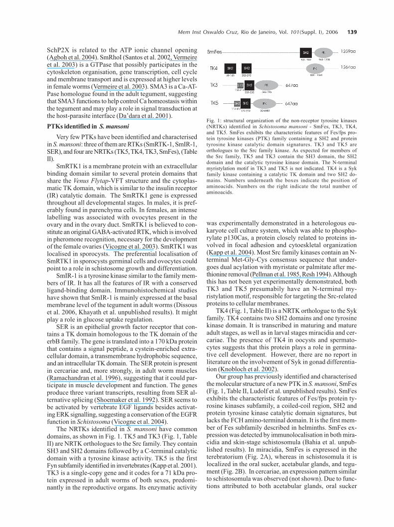
The NRTKs identified in S. mansoni have commondomains, as shown in Fig. 1. TK5 and TK3 (Fig. 1, TableII) are NRTK orthologues to the Src family. They containSH3 and SH2 domains followed by a C-terminal catalyticdomain with a tyrosine kinase activity. TK5 is the firstFyn subfamily identified in invertebrates (Kapp et al. 2001).TK3 is a single-copy gene and it codes for a 71 kDa pro-tein expressed in adult worms of both sexes, predomi-nantly in the reproductive organs. Its enzymatic activity
was experimentally demonstrated in a heterologous eu-karyote cell culture system, which was able to phospho-rylate p130Cas, a protein closely related to proteins in-volved in focal adhesion and cytoeskletal organization(Kapp et al. 2004). Most Src family kinases contain an N-terminal Met-Gly-Cys consensus sequence that under-goes dual acylation with myristate or palmitate after me-thionine removal (Pellman et al. 1985, Resh 1994). Althoughthis has not been yet experimentally demonstrated, bothTK3 and TK5 presumabily have an N-terminal my-ristylation motif, responsible for targeting the Src-relatedproteins to cellular membranes.
TK4 (Fig. 1, Table II) is a NRTK orthologue to the Sykfamily. TK4 contains two SH2 domains and one tyrosinekinase domain. It is transcribed in maturing and matureadult stages, as well as in larval stages miracidia and cer-cariae. The presence of TK4 in oocysts and spermato-cytes suggests that this protein plays a role in germina-tive cell development. However, there are no report inliterature on the involvement of Syk in gonad differentia-tion (Knobloch et al. 2002).
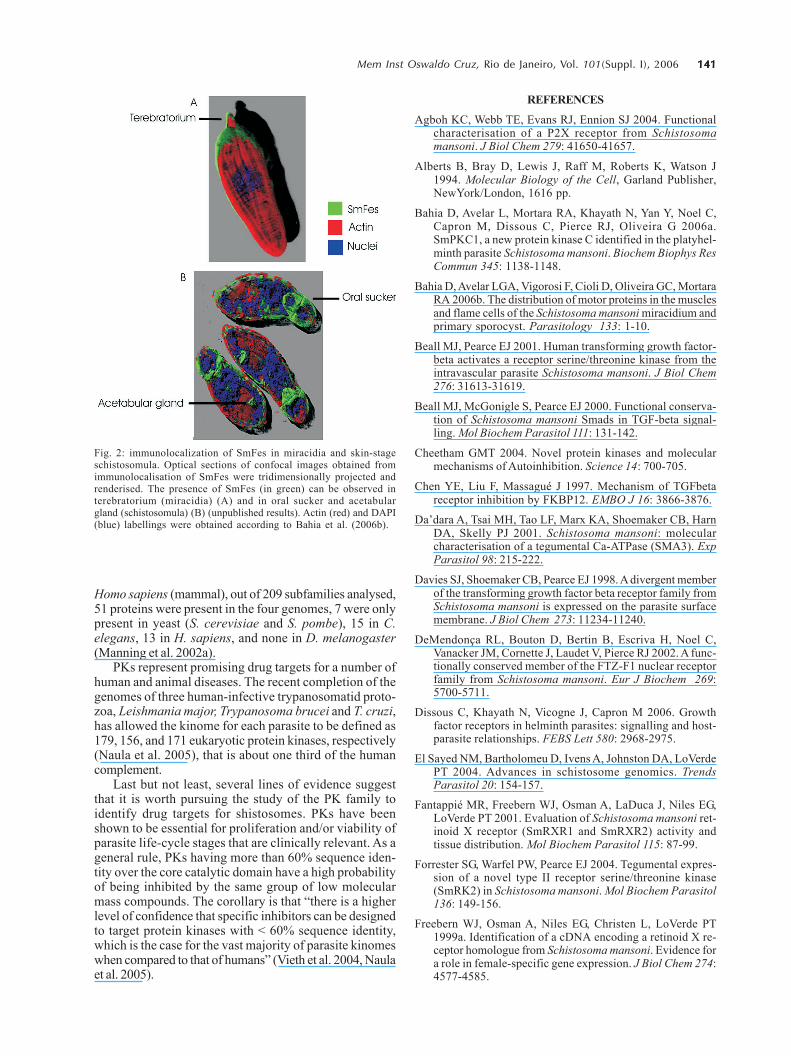
Our group has previously identified and characterisedthe molecular structure of a new PTK in S. mansoni, SmFes(Fig. 1, Table II, Ludolf et al. unpublished results). SmFesexhibits the characteristic features of Fes/fps protein ty-rosine kinases subfamily, a coiled-coil region, SH2 andprotein tyrosine kinase catalytic domain signatures, butlacks the FCH amino-terminal domain. It is the first mem-ber of Fes subfamily described in helminths. SmFes ex-pression was detected by immunolocalisation in both mira-cidia and skin-stage schistosomula (Bahia et al. unpub-lished results). In miracidia, SmFes is expressed in theterebratorium (Fig. 2A), whereas in schistosomula it islocalized in the oral sucker, acetabular glands, and tegu-ment (Fig. 2B). In cercariae, an expression pattern similarto schistosomula was observed (not shown). Due to func-tions attributed to both acetabular glands, oral sucker
Fig. 1: structural organization of the non-receptor tyrosine kinases(NRTKs) identified in Schistosoma mansoni - SmFes, TK3, TK4,and TK5. SmFes exhibits the characteristic features of Fes/fps pro-tein tyrosine kinases (PTK) family containing a SH2 and proteintyrosine kinase catalytic domain signatures. TK3 and TK5 areorthologues to the Src family kinase. As expected for members ofthe Src family, TK5 and TK3 contain the SH3 domain, the SH2domain and the catalytic tyrosine kinase domain. The N-terminalmyristylation motif in TK3 and TK5 is not indicated. TK4 is a Sykfamily kinase containing a catalytic TK domain and two SH2 do-mains. Numbers underneath the boxes indicate the position ofaminoacids. Numbers on the right indicate the total number ofaminoacids.
140140140140140 PTK in S. mansoni • Diana Bahia et al.
and terebratorium, these findings seem to suggest thatSmFes plays a pivotal role in the signal transduction path-way involved in the larvae transformation after penetra-tion into intermediate and definitive hosts.Presence of PKs in sequenced genomes
PKs have been characterised not only through tradi-tional biochemical techniques but also by catalytic do-main analyses from aminoacid sequences of their primarystructure (Hanks et al. 1988). PKs comprise one of thelargest families of proteins, which correspond to 1.5 to2.5% of eukaryote genes (Manning et al. 2002a). The de-velopment of genomic studies has led to the identifica-tion of an increasing number of PK in various animal spe-cies.
The genome of S. mansoni is currently being as-sembled and annotated and two large scale transcriptomeprojects described the majority of the genes of this spe-
cies (El Sayed et al. 2004, Oliveira et al. 2004, Oliveira &Bahia 2004, Verjovski et al. 2004). It is expected that therelease of a full analysis of these projects will reveal alarge number of proteins related to parasite-environmentand parasite-host interactions, among them PTks.
By using C. elegans as model for studies on signaltransduction, as it was the first fully sequenced multicel-lular organism, Pks were grouped into the second biggestfamily of the protein domains in theses worms, compris-ing 411 fully sequenced PKs (Plowman et al. 1999). Se-quencing of the human genome revealed 518 PK codinggenes, 1.7% of the entire human gene content. Amongthe 258 PKs analysed, 83 domain types were identified,most of which are found to be closely related to signallingprotein interaction domains, e.g., SH2, which recognisesand binds to phosphorylated tyrosine residues (Manninget al. 2002b). By comparing kinomes from S. cerevisiae(yeast), C. elegans (worm), D. melanogaster (insect) and
TABLE II Protein tyrosine kinases (receptor tyrosine kinases-RTKs and non-receptor tyrosine kinases- NRTKs) identified in
Schistosoma mansoni
Family Function Localisation References
SER EGFR Participates in schistosome Predominantly in the muscle Ramachandran et al. 1996(170kDa) signal transduction, perhaps of adult male and femaleGenBank related to muscle development wormsM86396 or function
SmRTK1 Insulin Probably with a role in male- In male: in parenchymal cells. Vicogne et al. 2003(172kDa) receptors female communication. In female: in ovocytes and in theGenBank Involved in the recognition ovary duct. In miracidia andAF101194 of a male pheromone signal newly-transformed sporocysts
necessary for the development in cells surrounding the neuralof the female ovaries mass and probably representing
the parasite germinal cells
SmIR-1 Insulin Possible role in the regulation Basal membrane of the Unpublished results(170kDa) receptors of the uptake of glucose via the tegument in the adult wormsGenBank activity of SGTP1 and SGTP4AF314754
TK3 Src Seems to play a role in signal Predominantly expressed in the Kapp et al. 2004(71kDa) transduction pathways reproductive organs such asGenBank organising the cytoskeleton in testes (male) and ovary asAJ585205 the gonads of schistosomes well as the vitellarium (female)
TK5 Src-like Seems to play a role during Expressed in the adult worms Kapp et al. 2001(73kDa) embryogenesis as well as gut and, furthermore, occurs in theGenBank formation and/or function free-living larval stagesAF232691
TK4 Syk May play a role in germ cell In larval stages and adult Knobloch et al. 2002(140kDa) development schistosomes. Significant signalsGenBank were detected in ovocytes (female)AJ421472 and in spermatocytes (male)
SmFes Fes/Fps May play a role in the signal SmFes expression was detected Unpublished results(143kDa) transduction pathway involved by immunolocalisation in bothGenBank in the larvae transformation miracidia and schistosomulaAF515706 after penetration into skin-stage. In miracidia, SmFes
intermediate and definitive is expressed in the terebratorium,hosts whereas in schistosomula it is
localised in the oral sucker,acetabular glands and tegument
141141141141141Mem Inst Oswaldo Cruz, Rio de Janeiro, Vol. 101(Suppl. I), 2006
Homo sapiens (mammal), out of 209 subfamilies analysed,51 proteins were present in the four genomes, 7 were onlypresent in yeast (S. cerevisiae and S. pombe), 15 in C.elegans, 13 in H. sapiens, and none in D. melanogaster(Manning et al. 2002a).
PKs represent promising drug targets for a number ofhuman and animal diseases. The recent completion of thegenomes of three human-infective trypanosomatid proto-zoa, Leishmania major, Trypanosoma brucei and T. cruzi,has allowed the kinome for each parasite to be defined as179, 156, and 171 eukaryotic protein kinases, respectively(Naula et al. 2005), that is about one third of the humancomplement.
Last but not least, several lines of evidence suggestthat it is worth pursuing the study of the PK family toidentify drug targets for shistosomes. PKs have beenshown to be essential for proliferation and/or viability ofparasite life-cycle stages that are clinically relevant. As ageneral rule, PKs having more than 60% sequence iden-tity over the core catalytic domain have a high probabilityof being inhibited by the same group of low molecularmass compounds. The corollary is that “there is a higherlevel of confidence that specific inhibitors can be designedto target protein kinases with < 60% sequence identity,which is the case for the vast majority of parasite kinomeswhen compared to that of humans” (Vieth et al. 2004, Naulaet al. 2005).
REFERENCES
Agboh KC, Webb TE, Evans RJ, Ennion SJ 2004. Functionalcharacterisation of a P2X receptor from Schistosomamansoni. J Biol Chem 279: 41650-41657.
Alberts B, Bray D, Lewis J, Raff M, Roberts K, Watson J1994. Molecular Biology of the Cell, Garland Publisher,NewYork/London, 1616 pp.
Bahia D, Avelar L, Mortara RA, Khayath N, Yan Y, Noel C,Capron M, Dissous C, Pierce RJ, Oliveira G 2006a.SmPKC1, a new protein kinase C identified in the platyhel-minth parasite Schistosoma mansoni. Biochem Biophys ResCommun 345: 1138-1148.
Bahia D, Avelar LGA, Vigorosi F, Cioli D, Oliveira GC, MortaraRA 2006b. The distribution of motor proteins in the musclesand flame cells of the Schistosoma mansoni miracidium andprimary sporocyst. Parasitology 133: 1-10.
Beall MJ, Pearce EJ 2001. Human transforming growth factor-beta activates a receptor serine/threonine kinase from theintravascular parasite Schistosoma mansoni. J Biol Chem276: 31613-31619.
Beall MJ, McGonigle S, Pearce EJ 2000. Functional conserva-tion of Schistosoma mansoni Smads in TGF-beta signal-ling. Mol Biochem Parasitol 111: 131-142.
Cheetham GMT 2004. Novel protein kinases and molecularmechanisms of Autoinhibition. Science 14: 700-705.
Chen YE, Liu F, Massagué J 1997. Mechanism of TGFbetareceptor inhibition by FKBP12. EMBO J 16: 3866-3876.
Da’dara A, Tsai MH, Tao LF, Marx KA, Shoemaker CB, HarnDA, Skelly PJ 2001. Schistosoma mansoni: molecularcharacterisation of a tegumental Ca-ATPase (SMA3). ExpParasitol 98: 215-222.
Davies SJ, Shoemaker CB, Pearce EJ 1998. A divergent memberof the transforming growth factor beta receptor family fromSchistosoma mansoni is expressed on the parasite surfacemembrane. J Biol Chem 273: 11234-11240.
DeMendonça RL, Bouton D, Bertin B, Escriva H, Noel C,Vanacker JM, Cornette J, Laudet V, Pierce RJ 2002. A func-tionally conserved member of the FTZ-F1 nuclear receptorfamily from Schistosoma mansoni. Eur J Biochem 269:5700-5711.
Dissous C, Khayath N, Vicogne J, Capron M 2006. Growthfactor receptors in helminth parasites: signalling and host-parasite relationships. FEBS Lett 580: 2968-2975.
El Sayed NM, Bartholomeu D, Ivens A, Johnston DA, LoVerdePT 2004. Advances in schistosome genomics. TrendsParasitol 20: 154-157.
Fantappié MR, Freebern WJ, Osman A, LaDuca J, Niles EG,LoVerde PT 2001. Evaluation of Schistosoma mansoni ret-inoid X receptor (SmRXR1 and SmRXR2) activity andtissue distribution. Mol Biochem Parasitol 115: 87-99.
Forrester SG, Warfel PW, Pearce EJ 2004. Tegumental expres-sion of a novel type II receptor serine/threonine kinase(SmRK2) in Schistosoma mansoni. Mol Biochem Parasitol136: 149-156.
Freebern WJ, Osman A, Niles EG, Christen L, LoVerde PT1999a. Identification of a cDNA encoding a retinoid X re-ceptor homologue from Schistosoma mansoni. Evidence fora role in female-specific gene expression. J Biol Chem 274:4577-4585.
Fig. 2: immunolocalization of SmFes in miracidia and skin-stageschistosomula. Optical sections of confocal images obtained fromimmunolocalisation of SmFes were tridimensionally projected andrenderised. The presence of SmFes (in green) can be observed interebratorium (miracidia) (A) and in oral sucker and acetabulargland (schistosomula) (B) (unpublished results). Actin (red) and DAPI(blue) labellings were obtained according to Bahia et al. (2006b).
142142142142142 PTK in S. mansoni • Diana Bahia et al.
Freebern WJ, Niles EG, LoVerde PT 1999b. RXR-2, a memberof the retinoid x receptor family in Schistosoma mansoni.Gene 233: 33-38.
Hanks SK, Quinn AM, Hunter T 1988. The protein kinasefamily: conserved features and deduced phylogeny of thecatalytic domains. Science 241: 42-52.
Harari PM 2004. Epidermal growth factor receptor inhibitionstrategies in oncology. Endocr Relat Cancer 11: 689-708.
Heldin CH 1996. Protein tyrosine kinase receptors. CancerSurv 27: 7-24.
Hubbard SR, Till JH 2000. Protein tyrosine kinase structureand function. Annu Rev Biochem 69: 373-398.
Inal JM 1999. Schistosoma TOR (trispanning orphan recep-tor), a novel, antigenic surface receptor of the blood-dwell-ing, Schistosoma parasite. Biochim Biophys Acta 1445: 283-298.
Kampkotter A, Ridgers I, Johnston DA, Rollinson D, Kunz W,Grevelding CG 1999. Schistosoma mansoni: cloning andcharacterisation of the Ras homologue. Exp Parasitol 91:280-283.
Kapp K, Knobloch J, Schussler P, Sroka S, Lammers R, KunzW, Grevelding CG 2004. The Schistosoma mansoni Srckinase TK3 is expressed in the gonads and likely involvedin cytoskeletal organization. Mol Biochem Parasitol 138:171-182.
Kapp K, Schussler P, Kunz W, Grevelding CG 2001. Identifi-cation, isolation and characterisation of a Fyn-like tyrosinekinase from Schistosoma mansoni. Parasitology 122: 317-327.
Knobloch J, Rossi A, Osman A, LoVerde PT, Klinkert MQ,Grevelding CG 2004. Cytological and biochemical evidencefor a gonad-preferential interplay of SmFKBP12 andSmTbetaR-I in Schistosoma mansoni. Mol BiochemParasitol 138: 227-236.
Knobloch J, Winnen R, Quack M, Kunz W, Grevelding CG2002. A novel Syk-family tyrosine kinase from Schisto-soma mansoni which is preferentially transcribed in repro-ductive organs. Gene 294: 87-97.
Kunz W 2001. Schistosome male-female interaction: inductionof germ-cell differentiation. Trends Parasitol 17: 227-231.
Lantner F, Ziv E, Ram D, Schechter I 1998. Different forms ofthe mRNA encoding the heat-shock transcription factor areexpressed during the life cycle of the parasitic helminthSchistosoma mansoni. Eur J Biochem 253: 390-398.
Machida K, Mayer BJ, Nollau P 2003. Profiling the globaltyrosine phosphorylation state. Mol Cell Proteomics 2:215-233.
Manning G, Plowman GD, Hunter T, Sudarsanam S 2002a.Evolution of protein kinase signalling from yeast to man.Trends Biochem Sci 27: 514-520.
Manning G, Whyte DB, Martinez R, Hunter T, Sudarsanam S2002b. The protein kinase complement of the human ge-nome. Science 298: 1912-1934.
McGonigle S, Beall MJ, Feeney EL, Pearce EJ 2001a. Con-served role for 14-3-3e downstream of type I TGFL recep-tors. FEBS Letters 490: 65-69.
McGonigle S, Beall MJ, Pearce EJ 2002. Eukaryotic initiation
factor 2 alpha subunit associates with TGF beta receptorsand 14-3-3 epsilon and acts as a modulator of the TGF betaresponse. Biochemistry 41: 579-587.
McGonigle S, Feeney EL, Beall MJ, Pearce EJ 2001b. SIP, anovel SH3 domain-containing protein, interacts with Schis-tosoma mansoni receptor kinase 1. Mol Biochem Parasitol114: 119-123.
Milhon JL, Albert TJ, Vande Waa EA, O’Leary KA, JacksonRN, Kessler MA, Schuler LA, Tracy JW 2000. SmMAK16,the Schistosoma mansoni homologue of MAK16 from yeast,targets protein transport to the nucleolus. Mol BiochemParasitol 108: 225-236.
Naula C, Parsons M, Mottram JC 2005. Protein kinases asdrug targets in trypanosomes and Leishmania. BiochimBiophys Acta 30: 151-159.
Neet K, Hunter T 1996. Vertebrate non-receptor protein-ty-rosine kinase families. Genes Cells 1: 147-169.
Oliveira G, Bahia D 2004. The genome of Schistosoma mansoni.Proc of the Third Braz Symp Math Comput Biol 1: 101-115.
Oliveira G, Rodrigues NB, Romanha AJ, Bahia D 2004. Ge-nome and genomics of shistosomes. Can J Zool 82: 375-390.
Osman A, Niles EG, LoVerde PT 1999. Characterization of theRas homologue of Schistosoma mansoni. Mol BiochemParasitol 100: 27-41.
Osman A, Niles EG, LoVerde PT 2001. Identification and char-acterization of a Smad2 homologue from Schistosomamansoni, a transforming growth factor-beta signal trans-ducer. J Biol Chem 276: 10072-10082.
Osman A, Niles EG, LoVerde PT 2004. Expression of func-tional Schistosoma mansoni Smad4: role in Erk-mediatedtransforming growth factor beta (TGF-beta) down-regula-tion. J Biol Chem 279: 6474-6486.
Pellman D, Garber EA, Cross FR, Hanafusa H 1985. An N-terminal peptide from p60src can direct myristylation andplasma membrane localization when fused to heterologousproteins. Nature 314: 374-377.
Plowman GD, Sudarsanam S, Bingham J, Whyte D, Hunter T1999. The protein kinases of Caenorhabditis elegans: amodel for signal transduction in multicellular organisms.Proc Natl Acad Sci USA 96: 13603-13610.
Ramachandran H, Skelly PJ, Shoemaker CB 1996. The Schisto-soma mansoni epidermal growth factor receptor homologue,SER, has tyrosine kinase activity and is localized in adultmuscle. Mol Biochem Parasitol 83: 1-10.
Resh MD 1994. Myristylation and palmitylation of Src familymembers: the fats of the matter. Cell 76: 411-413.
Santos TM, Machado CR, Franco GR, Pena SD 2002. Charac-terization and comparative functional analysis in yeast of aSchistosoma mansoni Rho1 GTPase gene. Mol BiochemParasitol 125: 103-112.
Schussler P, Grevelding CG, Kunz W 1997. Identification ofRas, MAP kinases, and a GAP protein in Schistosomamansoni by immunoblotting and their putative involve-ment in male-female interaction. Parasitology 115: 629-634.
Shoemaker CB, Ramachandran H, Landa A, dos Reis MG, SteinLD 1992. Alternative splicing of the Schistosoma mansonigene encoding a homologue of epidermal growth factor re-ceptor. Mol Biochem Parasitol 53: 17-32.
143143143143143Mem Inst Oswaldo Cruz, Rio de Janeiro, Vol. 101(Suppl. I), 2006
Siddiqui AA, Podesta RB, Clarke MW 1991. Schistosomamansoni: characterization and identification of calcium-bind-ing proteins associated with the apical plasma membraneand envelope. Exp Parasitol 72: 63-68.
Traxler P 2003. Tyrosine kinases as targets in cancer therapy -successes and failures. Expert Opin Ther Targets 7: 215-234.
Verjovski AS, Leite LC, Dias NE, Menck CF, Wilson RA 2004.Schistosome transcriptome: insights and perspectives forfunctional genomics. Trends Parasitol 20: 304-308.
Vermeire JJ, Osman A, LoVerde PT, Williams DL 2003. Char-acterization of a Rho homologue of Schistosoma mansoni.Int J Parasitol 33: 721-731.
Vicogne J, Cailliau K, Tulasne D, Browaeys E, Yan YT, Fafeur
V, Vilain JP, Legrand D, Trolet J, Dissous C 2004. Conser-vation of epidermal growth factor receptor function in thehuman parasitic helminth Schistosoma mansoni. J Biol Chem279: 37407-37414.
Vicogne J, Pin JP, Lardans V, Capron M, Noel C, Dissous C2003. An unusual receptor tyrosine kinase of Schistosomamansoni contains a Venus Flytrap module. Mol BiochemParasitol 126: 51-62.
Vieth M, Higgs RE, Robertson DH, Shapiro M, Gragg EA,Hemmerle H 2004. Kinomics-structural biology andchemogenomics of kinase inhibitors and targets. BiochimBiophys Acta 1697 (1-2): 243-257.
Yarden Y, Ullrich A 1988. Growth factor receptor tyrosine ki-nases. Annu Rev Biochem 57: 443-478.


